masters thesis combined - murdoch research...
TRANSCRIPT

THEDEGRADATIVEEFFECTSOFTEMPERATURE,
ULTRAVIOLETRADIATIONANDSODIUM
HYPOCHLORITEONTHEDETECTIONOFBLOODAT
CRIMESCENESUSINGTHEABACARD®
HEMATRACE® KIT
SarahEVANS
Athesissubmittedinfulfillmentoftherequirementsforthedegreeof
MasterofForensicScience(ProfessionalPractice)in
TheSchoolofVeterinaryandLifeSciences
MurdochUniversity
DrMarkReynolds
JamesSpeers
November,2016

ii
Declaration
Ideclarethatthisthesisdoesnotcontainanymaterialsubmittedpreviouslyforthe
awardofanyotherdegreeordiplomaatanyuniversityorothertertiaryinstitution.
Furthermore, to the best of my knowledge, it does not contain any material
previously published orwritten by another individual, exceptwhere due reference
has been made in the text. Finally, I declare that all reported experimentations
performedinthisresearchwerecarriedoutbymyself,exceptthatanycontribution
byothers,withwhomIhaveworkedisexplicitlyacknowledged.
Signed:SarahEvans

iii
Acknowledgements
TheauthorwouldliketoexpresstheirsinceregratitudetothefollowingpeoplefortheirsupportthroughoutthecompletionoftheMastersdegree:’DrMarkReynolds:Idon’tthinkanywordswoulddojusticetoexpresshowthankfulIamforallyourhelp,supportandguidancethroughoutthisprocess.Thankyouforbeing there to listen to allmy concerns and doubts that thiswould come togethersuccessfully. I am forever grateful for everything you have done to further myeducationand career andevenmoregrateful that I havegaineda fantastic lifelongfriend.Associate Professor James Speers: The dedication you have to your studentssupersedesanythingIhaveeverseenfromasupervisor.Thankyouforputtingintheextrahourstomakesurethisdegreewasattainable.Ithasbeenroughatsomepoints,forusall,butyourenthusiasmandsarcasmhasmadeitallworthit...alongwithtavTuesdays. Thank you for all your guidance andpatience, particularly in the editingprocesses.Thisdegreewouldnotbethesamewithoutyourwealthofknowledgeandhardworkyouput in toensure thestudentsare taughtbyonly thebest inWA!Sothankyoufromthebottomofmyheart!Sushil Madhogarhia, Abacus Diagnostics Inc: Thank you kindly for donating theHemaTrace®kits,includinghavingtopostthemtwice,thankstoAustraliancustoms.Thisprojectwouldnotbethesamewithoutyoursupport.DrDaveBerrymanandDrPeterSpencer,MurdochUniversity:Averybigthankstothebothofyouforallowingmetouseyourfacilities(theUVchamberandtheDNAlaboratory),includingatveryshortnotice.Yoursupportisgreatlyappreciated.

iv
TableofContents
TitlePage..........................................................................................................................................................i
Declaration......................................................................................................................................................ii
Acknowledgements....................................................................................................................................iii
PartOneLiteratureReview.....................................................................................................................1
PartTwoManuscript.................................................................................................................................48

v

1
- PartOne -
LITERATUREREVIEW
THEEFFECTOFTEMPERATURE,UVRADIATIONAND
SODIUMHYPOCHLORITEONTHEDETECTIONOF
BLOODATCRIMESCENESUSINGTHEABACARD®
HEMATRACE® KIT:ALITERARYREVIEW
2
TABLEOFCONTENTS
LISTOFFIGURES........................................................................................................................................3
LISTOFTABLES..........................................................................................................................................4
ABSTRACT....................................................................................................................................................51.0 INTRODUCTION:CRIMESCENESANDPRESUMPTIVETESTINGFORBLOOD..............6
2.0 DISCUSSION.......................................................................................................................................72.1 BIOLOGICALANDPHYSICALPROPERTIESOFBLOOD..................................................................82.2 HUMANHAEMOGLOBIN..........................................................................................................................92.2.1HAEMOGLOBINHUMANSPECIFIC....................................................................................................................132.2.2HAEMOGLOBINDEGRADATION.........................................................................................................................14
2.3 ABACARD®HEMATRACE®..................................................................................................................162.3.1ABACARD®HEMATRACE®:APRESUMPTIVEORCONFIRMATORYTEST?.....................................19
2.4 DRIEDBLOODSTAINS.............................................................................................................................192.5 DEGRADATIVEAGENTS.........................................................................................................................232.5.1 TEMPERATURE,HAEMOGLOBINANDABACARD®HEMATRACE®...........................................232.5.2 ULTRAVIOLETLIGHT,HAEMOGLOBINANDABACARD®HEMATRACE®..............................262.5.3 SODIUMHYPOCHLORITE,HAEMOGLOBINANDABACARD®HEMATRACE®.......................29
3.0 EXPERIMENTALDESIGNELEMENTS.......................................................................................313.1 AUSTRALIANENVIRONMENTALCONDITIONS..............................................................................313.1.1 TEMPERATURECONDITIONSINPERTHANDNORTHERNAUSTRALIA.......................................313.1.2 ULTRAVIOLETEXPOSURELEVELSINPERTHANDNORTHERNAUSTRALIA............................33
3.2 SODIUMHYPOCHLORITE......................................................................................................................353.3 SUBSTRATEEFFECTSANDEFFECTONSAMPLINGPROCEDURE............................................363.4 SOLUBILITYOFDRIEDBLOODSTAINS............................................................................................363.5 EXPOSURETIME......................................................................................................................................373.6 QUANTIFICATIONOFHAEMOGLOBINDEGRADATIONPRODUCTION..................................38
4.0 EXPERIMENTALAIMSANDHYPOTHESIS..............................................................................38
5.0 CONCLUSION...................................................................................................................................406.0 REFERENCELIST............................................................................................................................42

3
LISTOFFIGURES
Figure1: Crosssectionofaredbloodcellmembranedisplayingthelipidbylayer,
membraneproteinglycophorinandcytoskeletalproteins..................................................................8
Figure2: Fourhaeme-globinunitsformingasinglemoleculecomplexofhaemoglobin
(Lehmann&Huntsman,1974).........................................................................................................................9
Figure3: Themolecularstructureofhaemoglobinwithintheredbloodcelldisplayingthe
foursubunitseachwiththehaemegroup,ironatomandglobinchains....................................11
Figure4: TheOxyhaemoglobinDissociationCurvedisplayingtherelationshipbetween
haemoglobinsaturationandpartialpressure.Ashifttotheleft(greenline)isthe
resultoflowoxygendemandinthetissuesmeaningthehaemoglobinretains
affinityforoxygen.Ashifttotheright(purpleline)representswhenoxygenisin
highdemandfromthetissueandhencethehaemoglobin’saffinityforoxygenis
decreased................................................................................................................................................................12
Figure5 Schematicrepresentationoftheoxidativeprocessanddegradativeprocessof
haemoglobin(Hb)tooxyhaemoglobin(Oxy-Hb)andmethaemoglobin(Met-Hb)
invitroandinvivo(adaptedfromMarrone&Ballantyne,2009;Bremmeretal.,
2012).........................................................................................................................................................................15
Figure6 SchematicrepresentationoftheABACardHemaTraceprocess.....................................................17
Figure7 PossibleresultsfromtheHemaTraceKitdisplayinganegativeresult,positive
resultandinvalidresults(AbacusDiagnostics,2001).......................................................................18
Figure8 Adriedbloodstaindisplayingthecentralportion,thecoronaandperiphery
(Brutin,etal.,2011).............................................................................................................................................20
Figure9 Thefivedryingstagesofablooddropdepositedfromahealthyindividualat22°C
(Brutin,etal.,2011).............................................................................................................................................22
4
LISTOFTABLESTable1: Humanhaemoglobin(Hb)structuresthroughouthealthyhumandevelopmentand
theirsubunits..........................................................................................................................................................13
Table2: Thefivedryingphasesexhibitedfromdepositedbloodstains(Brutin,etal.,2011)..............21
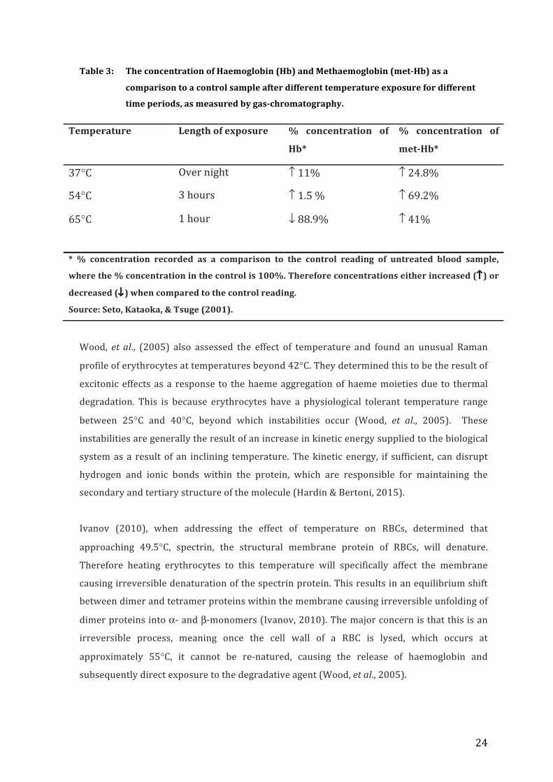
Table3: TheconcentrationofHaemoglobin(Hb)andMethaemoglobin(met-Hb)asa
comparisontoacontrolsampleafterdifferenttemperatureexposurefordifferent
timeperiods,asmeasuredbygas-chromatography.............................................................................24
Table4: MonthlytemperaturesrecordedforPerth(InternationalAirportStation)and
Broome(AirportStation)during2015displayedasmonthlyaveragemaximum
andminimumtemperatureandthemaximumtemperaturerecordedwithinthe
month................................................................................................................................................................................ 32
Table5: TheUVindexscaledisplayingthelevelofUVirradiationexposureatgroundlevel
(BureauofMeterology,2016)..........................................................................................................................33
Table6: LevelofUVexposureinPerth(southern)andBroome(northern)regionsof
WesternAustraliaasconductedbyNASAandtheTOMSmissionin2008(Bureau
ofMeterology,2016)............................................................................................................................................34
Table7: ConcentrationoftheactiveingredientscontainedinWhiteKingUltraBleach
product(Pental,2016)........................................................................................................................................35

5
ABSTRACT
Bloodisoneofthemostcommontypesofbiologicalevidencefoundatthesceneof
violent crimes.Whilst the first step in processing this evidence is observation and
documentation, this is closely followedbypresumptive testing.Due to the fact that
manysubstanceshaveanappearancesimilartoblood,thesamplemustbeanalysed
at the crime scene firstly to determine if the material is likely to be blood, and
secondly if it is likely to be of human origin. Depending on the case context, this
ensurestimeandresourcesarenotwastedtestingasubstanceoflittleornoforensic
value.However, this canbecomplicated if the selected testingkithas theability to
producefalse-negativeresults.
Therearemanydegradativesubstancesandenvironmentalconditionswithinacrime
sceneinwhichabloodstaincanbeexposedto.Substantialdegradationmayresultin
aninabilityforthepresumptivetesttorecognisethesampleasblood.TheABACard®
HemaTrace® from Abacus Diagnostics Inc. tests for the presence human
haemoglobin by antibody-antigen immunohematological chromatography, and is
routinely used by forensic Police forces and biological laboratories worldwide.
However,itiscurrentlyunknowninthescientificliterature,howcertaindegradative
agents, such as high temperature, high intensity ultra violet (UV) radiation and
sodium hypochlorite (household bleach) affect the haemoglobin within a blood
sampleintermsofsubsequentpresumptivetesting.Ifthehaemoglobinisstructurally
degraded beyond recognition, itmay not be able to bind to the antibodies present
withintheHemaTrace®kit,producingafalse-negativeresult.Thisliteraturereview
aims to address the affect these three degradative agents (high temperature, UV
radiationandbleach)haveonhumanhaemoglobinandthesubsequenttestingusing
the ABACard® HemaTrace® kit. The purpose of this literature review is to dictate
parameters for potential research that may aid in answering the investigative
question.

6
1.0 INTRODUCTION: CRIME SCENES AND PRESUMPTIVE
TESTINGFORBLOOD
Blood isoneof themostcommontypesofbiologicalevidence foundat thesceneofviolent
crimes. Not only can it be used for event sequencing and pattern reconstruction for
BloodstainPatternAnalysis,butalsothebiologicalpropertiesallowfortheanalysisofDNA
for human identification. The correct identification of human blood can therefore aid in
determininga suspect, exoneratingan innocent individualor linkingbloodlettingevents to
particularwounds or injuries (Virkler & Lednev, 2009). It is therefore critical to establish
whatbloodstainsbelongtowhom.However,beforethiscanbedone,thestainsmustfirstbe
identifiedasblood.Thisisbecauseothersubstancescanhaveasimilarappearancetoblood
ormaybeofanimaloriginand therefore irrelevant to thecriminal investigation(Virkler&
Lednev,2009).Thisisusuallydonethroughtheuseofpresumptivetestingatthecrimescene
and there are numerous commercially available kits for this purpose, such as ABACard®
HemaTrace, Seratec® HemDirect Hemoglobin, Galantos® Rapid Stain Identification of
Human Blood (RSID™-Blood) or HemaStick testing (Horjan, Barbaric, & Mrsic, 2016).
However, most presumptive tests have a trade-off between specificity and sensitivity.
Therefore, as the sensitivity of the test increases, meaning smaller concentrations of the
targetsubstancearerequired fordetection, there isan increasedchanceofcrossreactivity
withothersubstancesthatcanproduceerroneousresults(Horjan,Barbaric,&Mrsic,2016).
Consequently, it ispossibletoobtainfalsenegativeorfalsepositiveresultsfromsuchtests.
Whilstfalsepositiveresultscanmeanawasteofinvestigativetimeandresourcesanalysinga
substanceoflittleornoforensicvalue,afalsenegativeresultmaycausethedismissalofvital
forensicevidence.
ABACard® HemaTrace® (Abacus Diagnostics Inc.) is a highly sensitive commercially
availablekitusedforthepresumptivetestingofhumanbloodatcrimesceneswithminimal
cross reactivity from other species (Abacus Diagnostics , 2001). As a result, it is routinely
employed inmajor crime casesbyForensicPolice forcesworld-wide (AbacusDiagnostics ,
2001). It operates on the principle of protein chromatography and immunohematological
reactions, with the target substance being haemoglobin present within red blood cells
(Reynolds,2004).However,itisunknownwhatstatethehaemoglobinmustbepresentinto
allowsuccessfulbindingtotheantibodieswithintheHemaTrace®kit.Thisisbecausestudies
have shown that degraded blood samples have the capability of producing a negative
HemaTrace®result,despitebeingabletoobtainacompleteorpartialDNAprofile(Coy,etal.,

7
2005). This phenomenon may have been encountered in the extreme climate of Western
Australian,where forensic investigatorshaveobtainedanegativeresult for thepresenceof
bloodusingtheABACard®HemaTrace®kit,despitethesamplebeingofhumanorigin.With
little literature content addressing common degradative agents in isolation, rather than a
combinationofvariables,itisdifficulttoconcludewhatmaybeproducingthefalse-negative
results.Thisisascientificareathatthisliteraturereviewaimstoaddress,withthepurposeof
experimentalvalidation.
Bloodsamplesdepositedatcrimescenesarerarely incontrolledenvironments,butrather
exposed to degradative agents. The basis for the literature review stems from the
presumptionthatifthehaemoglobininabloodsourceisseverelydegraded,itmayaffectthe
ability for the antibodies to bind, resulting in a false negative test result. Therefore, the
purposeof this literary review is todetermine if threecommonlyencountereddegradative
agents (high temperature,ultraviolet radiationandsodiumhypochlorite) couldpotentially
degradeaknownbloodsamplebeyondthedetectableabilityof thepresumptivetestingkit
ABACard® HemaTrace®. This literary review will aid in the determination of an
experimental design to allow for the testing of the investigative question underAustralian
environmentalconditions.Resultsofthisstudywillaidforensicinvestigatorswhenselecting
samplesforpresumptivetestingthathavebeenexposedtothedegradativeagentsandmay
aid in interpreting and explaining false negative test results both in an investigative sense
andinacourtoflaw.
2.0 DISCUSSION
This section aims to address the literature that is currently available in regards to the
biologicalandphysicalpropertiesofbloodstainfoundatcrimescenes.This incudeswhat is
currentlyunderstoodaboutthedegradationprocesshumanhaemoglobinundergoesoutside
thebodyandthesubsequenttestingusingtheABACard®HemaTrace®bloodtestingkitfrom
Abacus Diagnostics Inc. This section will finish by discussing the known effects the three
degradative agents (high temperature, UV radiation and Sodium Hypochlorite) have on
bloodstainsandhumanhaemoglobin.
8
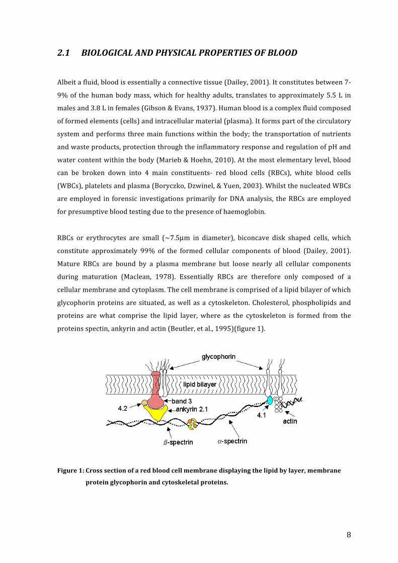
2.1 BIOLOGICALANDPHYSICALPROPERTIESOFBLOOD
Albeitafluid,bloodisessentiallyaconnectivetissue(Dailey,2001).Itconstitutesbetween7-
9%of thehumanbodymass,which forhealthyadults, translates toapproximately5.5L in
malesand3.8Linfemales(Gibson&Evans,1937).Humanbloodisacomplexfluidcomposed
offormedelements(cells)andintracellularmaterial(plasma).Itformspartofthecirculatory
systemandperforms threemain functionswithin thebody; the transportationofnutrients
andwasteproducts,protectionthroughtheinflammatoryresponseandregulationofpHand
watercontentwithinthebody(Marieb&Hoehn,2010).Atthemostelementarylevel,blood
can be broken down into 4 main constituents- red blood cells (RBCs), white blood cells
(WBCs),plateletsandplasma(Boryczko,Dzwinel,&Yuen,2003).WhilstthenucleatedWBCs
areemployed in forensic investigationsprimarily forDNAanalysis, theRBCsareemployed
forpresumptivebloodtestingduetothepresenceofhaemoglobin.
RBCs or erythrocytes are small (~7.5µm in diameter), biconcave disk shaped cells, which
constitute approximately 99% of the formed cellular components of blood (Dailey, 2001).
Mature RBCs are bound by a plasma membrane but loose nearly all cellular components
during maturation (Maclean, 1978). Essentially RBCs are therefore only composed of a
cellularmembraneandcytoplasm.Thecellmembraneiscomprisedofalipidbilayerofwhich
glycophorinproteinsaresituated,aswellasa cytoskeleton.Cholesterol,phospholipidsand
proteins are what comprise the lipid layer, where as the cytoskeleton is formed from the
proteinsspectin,ankyrinandactin(Beutler,etal.,1995)(figure1).
Figure1:Crosssectionofaredbloodcellmembranedisplayingthelipidbylayer,membrane
proteinglycophorinandcytoskeletalproteins.

9
DuetothefactthatRBCslooseallcellularcomponentsaftermaturation,theydonotcontaina
nucleus or organelles meaning they are incapable of aerobic respiration. This therefore
makes them ideal carrier cells for oxygen transportation (Maclean, 1978). This
transportationofoxygenthroughoutthebodytotargetcellsisachievedthroughtheprotein
haemoglobin,foundonlywithinRBCs.
2.2 HUMANHAEMOGLOBIN
Haemoglobinisaproteinsynthesisedforthetransportationofoxygenfromthecapillarygas-
exchange interfacewithin the lungs to cells around thebody (Marieb&Hoehn,2010).The
moleculehasacompositestructureformedbythejoiningofthehaemeandglobinunits.The
haemoglobin molecule is naturally comprised of aggregates of the single haemoglobin
monomer(Maclean,1978).Thisisintheformoffourmonomersboundtogethertoformthe
functionalcomplex(figure2).
Figure2: Four haeme-globin units forming a single molecule complex of haemoglobin
(Lehmann&Huntsman,1974).
Thehaemecomponentofthehaemoglobincomplexisaverystablecompoundofferrousiron
(Fe2+)andprotoporphyrin IX (Maclean,1978).Protoporphyrin IX is formedwhenSuccinyl-
CoAbindswithglycinetoformapyrrolemolecule(Maclean,1978).Fourpyrrolemolecules

10
thencombinetocreatethefinalprotoporphyrinIXmolecule.Theironatomiscoupledtothe
porphyrin ring by four nitrogen atoms. However, the iron atom makes an additional two
links; one to the globin polypeptide chain at the histidine F8 residue, and the other to the
oxygen atom being transported (Maclean, 1978). The bound oxygen molecule serves as
ligand,orcomplexingagent,andhastwoimportantcharacteristics.Firstly,theligandsiteis
only made available when the haeme is complexed to the globin chain. Therefore, haeme
alone cannot act as a transport molecule for oxygen (Beutler, et al., 1995). Secondly, the
bindingoftheoxygenmoleculeasaligandaffectsthespinstateoftheelectronssurrounding
the iron atom.This affects themanner inwhich the iron atom fits into theporphyrin ring,
whichinturnaffectsthetertiarystructureoftheprotein.Thisisimportantasitisthetertiary
structurethatgivestheproteinitsfunctionality(Maclean,1978).
Thebindingofthehaemeandglobincomponentsplaysacrucialroleinthestateoftheiron
atom.When complexed, the oxygen atom bound for transportation does not result in the
oxidationof the ironatomitself.Whenthe ferrous iron(Fe2+) isoxidised to the ferricstate
(Fe3+),itbecomesfunctionallyuselessasanoxygencarrier.Iftheglobinproteinisdenatured,
thispropertyislostandthehaemecannottransporttheoxygen(Maclean,1978).
Theglobincomponentofhaemoglobinisessentiallytheproteincomponentofthemolecule
and is comprised of a primary, secondary, tertiary and quaternary structure. The primary
structureisthenumberandarrangementofaminoacidsinthepolypeptidechain(Neuwirt&
Ponka,1977).Thenumberitselfdiffersbetweendifferentglobinsandbetweenspecies.The
secondary structure dictates the configuration the polypeptide adopts and is almost
invariably always a coil structure, referred to as theα-helix (Neuwirt&Ponka, 1977).The
tertiarystructureisathirddimensionaddedbythefoldingofthecoilstructureuponitself.
This occurs when the individual amino acids are added one at a time during synthesis in
ordertoprovideastableconfigurationduringthenaturalcoilingprocess(Neuwirt&Ponka,
1977).Thedisfigurationofthetertiarystructureisknownasdenaturationoftheproteinand
cannot be re-natured once lost, only resynthesis one amino acid at a time can restore the
conformation(Maclean,1978).
Haemoglobin exists as tetramer of four monomers constituted of α and β chains. Each
monomer consists of a single globin in which one haeme group is embedded (Neuwirt &
Ponka,1977)(Figure3).Thefourmonomersareheldtogetherbyhydrophobiclinksbetween
the adjacent polypeptide chains (Maclean, 1978). These links play an essential role in the
physiologicalallosterydisplayed.

11
Figure3: Themolecularstructureofhaemoglobinwithintheredbloodcelldisplayingthefour
subunitseachwiththehaemegroup,ironatomandglobinchains.
The allosteric binding properties exhibited by the haemoglobin at the oxygen-binding site,
arises from the interactionbetween the ironatomwithin thehaemegroupand theoxygen
molecule itself.Thishas resultant affectson thequaternary structureof theprotein.When
the oxygen binds to the haemoglobin, it triggers a biochemical cascade. As the iron atom
moves into the porphyrin plane of the haeme, the histidine F8 residue of the globin
polypeptidechainisalsopulledtowardsthisplaneasaconsequenceofbeingboundtheiron
atom(Wood,etal.,2005).Theconformationalchangeistransmittedthroughoutthepeptide
backboneresulting inachange to the tertiarystructureof thesubunit (Wood,etal.,2005).
This conformational change results in newbinding interactions between adjacent subunits
dictating the quaternary structure (Maclean, 1978). The interaction between the adjacent
subunitallowsforatransformationmeaningtheaccess foroxygentothebindingpocketof
the second haeme unit is made easier. This therefore increases the affinity of the
haemoglobinmolecule forasecondoxygenatominsolutionsofhighoxygenconcentration,
suchasinthelungs.

12
Theaffinityforoxygenexhibitedbyhaemoglobinisdictatedbytheconcentrationofoxygen
within in thesurroundingtissues.Thehaemoglobinproteinwillabsorbandreleaseoxygen
moleculeswhenthereisanimbalanceincomparativepressureoroxygenconcentrationina
solution.When transported to tissue cellswhere the oxygen tension is low, the binding is
decreased,resultinginaweakeningofthebondbetweentheoxygenandhaemeunit.When
thisbond isbroken, theoxygen is released intosolution.TheOxyhaemoglobinDissociation
Curvedescribes this relationship, relating thepressure (PaO2) andoxygenavailabilitywith
the saturationofhaemoglobin (SaO2) (Hooley, 2015).As thepressure increases, suchas in
the lungs during breathing, the affinity for oxygen in increased, resulting in complete
saturationofthehaemoglobin(Figure4).
Figure4: The Oxyhaemoglobin Dissociation Curve displaying the relationship between
haemoglobin saturationandpartialpressure. A shift to the left (green line) is the
resultoflowoxygendemandinthetissuesmeaningthehaemoglobinretainsaffinity
for oxygen. A shift to the right (purple line) represents when oxygen is in high
demand from the tissue and hence the haemoglobin’s affinity for oxygen is
decreased.
Therefore,whenbloodisinitiallydeposited,theamountofboundoxygenwilldependonthe
bloodsourcewithinthebody,whichwilleitherbehighlyoxygenatedorde-oxygenated.

13
2.2.1HAEMOGLOBINHUMANSPECIFIC
The confirmatory identification of human haemoglobin relies on the differentiation of the
proteinbetweenspecies.Thisisachievedthroughdifferentaminoacidsequenceswithinthe
protein. The phylogenic relationship between humans and higher primates suggests why
most haemoglobin detection kits, such as the ABACard® HemaTrace®, are only higher
primate specific, not human specific. However, a common cross-species interference is
experiencedwithferretsamples.Thisisduetothecommonα-chainwithinthehaemoglobin
betweenferretspeciesandhigherprimates.Inparticular,thisisexhibitedintheaminoacid
sequenceTNAVAH,whichspanstheresidues67–73(Johnston,Newman,&Frappier,2003).
Thissectionhasoptimaluseforhaemoglobinrecognitionfrommonoclonalantibodiesasthis
isthesectionthatexhibitsmaximumvariationbetweenhumanandanimalspecies(Johnston,
Newman,&Frappier,2003).Whilstithasnotbeenmadepublicallyavailabletheexactamino
acidsequencethattheABACard®HemaTrace®kitemploysastheantibody-bindingsite,itis
presumed to be a highly conservative sequence, such as the one mentioned by Johnston,
NewmanandFrappier(2003).
Even within humans, there is variation in the structure of haemoglobin throughout the
embryonic,foetalandadultstagesoflife(Maclean,1978)(table1).
Table1: Human haemoglobin (Hb) structures throughout healthy human development and
theirsubunits.
Developmentalstage Symbol Globinunits
Embryonic HbE1
HbE2
HbE3
α2ε2
e2ζ2
ζ2γ2
Foetal HbF α2γ2 (Note: the γ chain is duplicated and not absolutely
identical).
Adult HbA α2β2(constitutes97.5%oftotalhaemoglobin)
α2δ2
AdaptedfromMaclean(1978).

14
2.2.2HAEMOGLOBINDEGRADATION
RBCs have a natural lifespan of 100-120 days until they loose their flexibility and become
rigid and fragile. At this point they are usually trapped in smaller channels, fragment and
becomeengulfedanddestroyedbymacrophages, typically in the liveror spleen (Marieb&
Hoehn,2010).ThehaemolysisorrupturingoftheRBCs,resultinthereleaseofhaemoglobin
thatundergofurtherenzymaticdegradationprocessesbeforebeingrecycledorexcreted.The
degradationprocessofhumanhaemoglobin inside thebody fromsenescentRBCshasbeen
well documented through the literature (Lehmann & Huntsamn, 1974; Neuwirt & Ponka,
1977; Maclean, 1978; Marieb & Hoehn, 2010). However, the blood encountered at crime
sceneshasbeenexposedtotheexternalenvironment,andhencenotcontrolledbythebodies
regulation systems. The degradation process of haemoglobin outside the body from dried
bloodstains,intermsofchangestothemolecularstructureduringthedenaturationprocess,
isnot fullyunderstoodwithinthescientific literature.However, themolecularspecieshave
beenidentifiedduringthedifferentstagesofdegradation.
Bremmer,etal. (2012)determinedthatonceoutsidethebody, thehaemoglobin inbloodis
saturated by oxygen from the external environment. This results in all haemoglobin
molecules becoming oxyhaemoglobin, which is present in the ferrous state (Fe2+). The
oxyhaemoglobinisthenauto-oxidizedtoformmethaemoglobin,whichispresentintheferric
state(Fe3+),meaning itcanno longerbindoxygen. Ifwithinthebodiessystem,cytochrome
b5wouldreduce themethaemoglobinallowing its reversalback tohaemoglobin thatcould
re-oxygenate(figure5).However,duetothelimitedavailabilityofcytochromeb5outsidethe
body, the auto-oxidation of methaemoglobin is essentially irreversible (Bremmer, et al.,
2012).ThisprocesswassupportedbyWood,etal.(2005)whofoundnodifferenceinRaman
Spectra of haemoglobin that had been deoxygenated, left to rest in ambient temperatures,
and subsequently re-oxygenated, suggesting the methaemoglobin exposed to the
environment could not uptake the oxygen atoms, even when in abundance. Once the
methaemoglobin is formed, it is then denatured to form hemichrome,which is a low spin
formofmethaemoglobin formedbyan internal conformational change to thehaemegroup
(Sugawara,etal.,2003;Hanson&Ballantyne,2010)(figure5).
This process was supported by the findings of Marrone and Ballantyne (2009), who also
assessed the degradation process of haemoglobin from dried bloodstain. The authors
reinforcedthedegradationprocessofoxyhaemoglobintomethaemoglobinandsubsequently
hemichrome (Marrone&Ballantyne, 2009). However, the authors also detected free ferric

15
andferrousironatomswithinthebloodstains,whichtheyhypothesiseweredetachedfrom
the oxyhaemoglobin and methaemoglobin during each stage of denaturation (Marrone &
Ballantyne,2009)(figure5).
Figure5 Schematicrepresentationoftheoxidativeprocessanddegradativeprocessof
haemoglobin (Hb) to oxyhaemoglobin (Oxy-Hb) and methaemoglobin (Met-
Hb)invitroandinvivo(adaptedfromMarrone&Ballantyne,2009;Bremmer
etal.,2012).
Inadditiontothespeciesmentionedabove,theauthorsalsofoundafourthspeciespresent,
butwerenot able to identify themolecule using theUV Spectroscopy technique employed
(Marrone & Ballantyne, 2009). It was hypothesised that themolecule could potentially be
ferrylhaemoglobin or choleglobin. Ferrylhaemoglobin is formed when oxyhaemoglobin is
combined with hydrogen peroxide (H2O2), which can potentially form through Fenton
Chemistry reactions (Halliwell,Gutteridge,&Aruoma,1987).Alternatively, choleglobin is a
denatured form of haemoglobinwhen the porphyrin ring is hydroxylated or broken open,
howevertheirstudiescouldnotconfirmtheunknownspeciestobeeitherofthese(Marrone
& Ballantyne, 2009). It was concluded however, that dried blood samples undergo rapid
oxidation reactions (Marrone & Ballantyne, 2009). The authors hypothesised this to be
acceleratedby the formationof thehydroxyl radical (OH•) formed from the releaseof free
iron during the degradation of haemoglobin (Marrone & Ballantyne, 2009). Molchanova
(1981)alsodetected thepresenceof anoxidative specieswith thedenaturationprocessof

16
haemoglobin. The author found the production of a super oxide [O2-] during the auto-
oxidationofoxyhaemoglobinthatcouldpotentiallyassistinfurtheralterationstoremaining
RBCmembranes,acceleratingfurtherdegradationwiththedepositedbloodstain.
2.3 ABACARD®HEMATRACE®Knowledge of the degradation process of human haemoglobin is crucial for interpreting
presumptive testingkits for thedetectionof humanbloodat crime scenes.This is because
haemoglobin is the primary detection molecule for many kits, including the ABACard®
HemaTrace® test.TheHemaTrace®kit isanimmunohematologicaltestthatisusedforthe
detectionofhumanbloodbyidentificationofthehumanhaemoglobinpresentinthesample.
Thetestworksonthepremisesofanantigen/antibodyreactionandproteinchromatography
(Reynolds, 2004). Contained within the stationary phase of the test is an absorbent
membranematerial.Thebottomlayerofthemembraneispresentatthesamplewell,where
the test solution is inserted. The stationary phase containsmobile dye-taggedmonoclonal
antihuman antibodies located near the sample well, which will complex with the
haemoglobinifpresentinthesolution(Reynolds,2004)(figure6).Thehaemoglobinismade
available to bindwith the antibodies after haemolysis within the HemaTrace® buffer (pH
7.5)(Johnston,Newman,&Frappier,2003).Thiscomplexmigratestowardsthetestpanel‘T’
which contains immobilized antibodies (figure 6). These antibodies are polyclonal anti-
human haemoglobin antibodies, which capture the complexed haemoglobin so that an
antibody-antigen-antibody compound is formed (Reynolds, 2004). If the concentration of
haemoglobinisgreaterthantheminimumdetectionlevel,thedyewillprecipitateforminga
visible pink band in the ‘T’ panel representing a positive result for the presence of human
blood(Reynolds,2004).Anyexcessmobilemonoclonalantibodiesthatdonotbindinthe‘T’
panel continue tomigratealong themembrane towards to thecontrol ‘C’panel.Here, they
bindwithimmobilizedpolyclonalanti-immunoglobulinantibodiesandprecipitatetoforma
pinkbandinthe‘C’panel(Reynolds,2004)(figure6).Thisactsasaformofinternalcontrol
andindicatesthetesthasworkedasintended.

17
Figure6 SchematicrepresentationoftheABACardHemaTraceprocess.
If twopinkbands (one in the ‘T’panelandanother in the ‘C’panel)arepresent in the test
afteramaximumtimeof tenminutes (asrecommendedby themanufacturer), theresult is
positiveforhumanblood(figure7).Ifonlyasinglebandpresentinthe‘C’panel,theresultis
negative for human blood, provided therewas not a high dose hook effect. The high dose
hook effect results in false negative test results due to excessively high concentrations of
haemoglobin present in the sample. The haemoglobin inhibits the binding of the mobile
human haemoglobin-antibody complexes to the stationary antibodies. This is due to the
excessiveconcentrationofhaemoglobin,whichbecomesacompetitiveinhibitorfortheanti-
humanhaemoglobin-antibody complex, preventing binding in the ‘T’ panel. False-negative
resultscanalsobeobtainedifthetestsolutionistooviscous,resultinginaninabilityforthe
solution to migrate through the test membrane (Johnston, Newman, & Frappier, 2003).Alternatively,ifnobandappearsinthe‘C’panel,thetestresultisinvalid,meaningeitherthe
test did not operate as intended due to a defect or proper analysis procedure were not
adheredto(figure7).
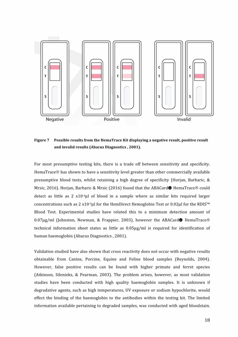
18
Figure7 PossibleresultsfromtheHemaTraceKitdisplayinganegativeresult,positiveresult
andinvalidresults(AbacusDiagnostics,2001).
Formost presumptive testing kits, there is a trade off between sensitivity and specificity.
HemaTrace®hasshowntohaveasensitivitylevelgreaterthanothercommerciallyavailable
presumptive blood tests, whilst retaining a high degree of specificity (Horjan, Barbaric, &
Mrsic,2016).Horjan,Barbaric&Mrsic(2016)foundthattheABACard® HemaTrace®could
detect as little as 2 x10⁻⁶µl of blood in a sample where as similar kits required larger
concentrationssuchas2x10-5µlfortheHemDirectHemoglobinTestor0.02µlfortheRDIS™
Blood Test. Experimental studies have related this to a minimum detection amount of
0.07µg/ml (Johnston, Newman, & Frappier, 2003), however the ABACard® HemaTrace®
technical information sheet states as little as 0.05µg/ml is required for identification of
humanhaemoglobin(AbacusDiagnostics,2001).
Validationstudiedhavealsoshownthatcrossreactivitydoesnotoccurwithnegativeresults
obtainable from Canine, Porcine, Equine and Feline blood samples (Reynolds, 2004).
However, false positive results can be found with higher primate and ferret species
(Atkinson, Silenieks, & Pearman, 2003). The problem arises, however, as most validation
studies have been conducted with high quality haemoglobin samples. It is unknown if
degradativeagents,suchashightemperatures,UVexposureorsodiumhypochlorite,would
effect the binding of the haemoglobin to the antibodieswithin the testing kit. The limited
informationavailablepertainingtodegradedsamples,wasconductedwithagedbloodstain.

19
Whilst Johnston, Newman and Frappier (2003) achieved positive HemaTrace® results for
bloodstainagedbetween25to30years,Horjan,BarbaricandMrsic(2016)achievedmixed
results. These authors found that of the five bloodstains tested, aged between 19 and 28
yearsinacontrolledenvironment,onlytwoproducedapositivereading(19and21yearold
samples).However, itwas not determined, or evenhypothesised,why three tests failed to
produceapositiveresult.Itshouldalsobenoted,thatnotonlywasaverysmallsamplesize
employedintheexperiment,replicatesampleswerenotperformed.
2.3.1 ABACARD® HEMATRACE®: A PRESUMPTIVE OR CONFIRMATORY
TEST?
There appears to be an inconsistency within the literature, as to whether the ABACard®
HemaTrace® Kit can be classified as a confirmatory crime scene testing kit, orwhether it
should remain as a presumptive test for the detection of human blood. The distinction of
whichtermisemployed,appearstobedictatedbythecontextinwhichtheauthorrefersto
theapplicabilityofthetest.Theauthorsthatrefertothetestas ‘confirmatory’dosoonthe
basisthatthetesthastheabilitytodistinguishbetweenhumanbloodandotherspecies(with
theacknowledgedexceptionof ferretandhigherprimates)andthereforecanprovidemore
specificinformationtoaninvestigatoronsiteabovewhatmostotherpresumptivetestsare
capableof(Reynolds,2004;Coy,etal.,2005).Conversely,otherauthorswhoclassifythetest
as presumptive, do so on the firm basis that the test has the potential to return a false
positiveresultforhumanbloodandconsequentlyrequiresfurthertestingbeforemakingany
conclusions(Horjan,Barbaric,&Mrsic,2016).
2.4 DRIEDBLOODSTAINSThedryingprocess a depositedbloodstain undergoes is a function of the surface area and
volumeof thebloodstain (Ramsthaler,etal.,2012).These factorsare further influencedby
the temperature and humidity of the external environment, the air circulation, vapour
pressure,thesurfacecharacteristicsonwhichthebloodwasdepositedandthecomposition
of theblood includingtheviscosity,allofwhichmay impactthedegradationprocessof the
RBC(Brutin,etal.,2011;Ramsthaler,etal.,2012).Whenabloodstainisdeposited,theRBCs
interactwitheachotherandwiththeperipheralwallsof thebloodstain.These interactions
aregovernedbybiology,chemistryand fluidmechanics (Brutin,etal.,2011).However, the

20
colloidal particles, predominantly the RBCs, are carried by the flow of motion within the
bloodstain as precipitation occurs. This iswhat causes the formation of a coronawithin a
dried bloodstain, described as the dark red ring below the periphery of the bloodstain
(Brutin,etal.,2011)(figure8).
Figure8 Adriedbloodstaindisplayingthecentralportion,thecoronaandperiphery(Brutin,
etal.,2011).
Thecrackformationsthatappearareformedbydehydrationofthecolloidalparticles,when
onafixed/stationarysurface.Itishypothesisedthatthisisduetothesalinityofthesolution
andtheinstabilityofcellularcomponentsduringdesiccationresultinginbucklingofthecells,
particularly in larger bloodstains (Brutin, et al., 2011). This occurs after the RBCs have
ruptured releasing the liquid cytoplasm to complete the drying stage (Brutin, etal., 2011).
Brutin,etal.(2011)proposedthedryingprocessofabloodstainoccursinfivephases(table
2):

21
Table2: Thefivedryingphasesexhibitedfromdepositedbloodstains(Brutin,etal.,2011)
These five stages of drying, as depicted in figure 9, are dramatically accelerated with
increases in temperature. Ramsthaler, etal. (2012) found that a blood drop deposited and
maintainedat20°Ctook60minutestodrytothepointthatasmearcouldnotbeachieved,
Phase %Dry Description
1
0-20% Directly after deposition, the cellular components, predominately the RBCs,
migrate to the periphery of the bloodstain. This is due to Marangoni
convection,whichisthetransferofsubstancesalongaliquidinterfaceduetoa
tension gradient (Brutin, et al., 2011). The RBCs then recede from the
periphery,leavingalightreddepositlineattheedgeofthebloodstain.
2 20-50% During this stage, crystallisation occurs at the edge of the drop, which
proceedsinwardstowardthecentre.AdarkredtorusringofRBCsisobserved
just below the periphery of the stain, displaying separation of the fluid
components.
3 50-70% Atthisdryingstage,thetorusringbeingstodesiccateandthecentralpartof
the bloodstain lightens in colour. It is at this stage the first cracks begin to
appearbetweentheperipheryandwhatwilleventuallybethecorona.Minor
cracksalsobegintoformbetweenfuturecoronaandthecentralportionofthe
stain.
4 70-85% At this stage, the drying process at the centre of the bloodstain is nearly
complete. The RBCs accumulate by convection to form a solid deposit just
belowtheperiphery,referredtoasthecorona.Circulardryingspotsbeginto
appear around the corona. It is at this point, theRBCs rupture releasing the
liquid cytoplasm portion, including the haemoglobin protein, for further
desiccation.
5 85-100% During this stage, large plaques of the corona will move slightly as the
cytoplasm of the RBCs dehydrates. Beyond this, no further physical changes
areobserved.

22
whereas itonlyrequired30minutesat24°Ctoachieve thesamestate.An increaseofonly
4°Cresultedinhalftherequireddryingtime(Ramsthaler,etal.,2012).
Figure9 Thefivedryingstagesofablooddropdepositedfromahealthyindividualat22°C
(Brutin,etal.,2011).
Thedegreeofdehydrationcanpotentiallyaffecttheabilitytosolubilisethebloodstainintoan
aqueous solution, such as a buffer for further laboratory testing. Blood, as a substance, is
readily soluble in water. For this reason, blood from a dried bloodstain can be simply
rehydrated, with the best capturing material being cotton, or a similar fabric (Hillman &
Schaler, 1981). This is supported by studies that have successfully captured, bymeans of
rehydration, bloodstains that are multiple years old (30 years) and exposed to various
temperatures (including fire conditions) (Johnston, Newman, & Frappier, 2003). However,
for hardened bloodstains, either by means of age or temperature, some authors suggest
extending the extraction/rehydration time, either by prolonging contact time between the
moistenedswabandthebloodsourceorprolongingtheimmersionofthestainedmaterialin
thesolutionbeyondregularprotocol(Johnston,Newman,&Frappier,2003;Horjan,Barbaric,
&Mrsic,2016).

23
2.5 DEGRADATIVEAGENTS
There are numerous degradative agents present in the environment that are capable of
comingintocontactwithblooddepositedatcrimescenes.Themostcommonlyencountered
agents are extreme temperatures, ultra violet (UV) radiation from the sun and sodium
hypochlorite (house-hold bleach). Whilst the effects of these agents have been well
documented for the purpose of DNA degradation and subsequent forensic analysis, their
specific effects on RBCs, in particular haemoglobin for the purpose of presumptive blood
testingremainlimitedinthescientificliterature.
2.5.1 TEMPERATURE,HAEMOGLOBINANDABACARD®HEMATRACE®
The temperature a blood sample is exposed to at a crime scene is essentially an
uncontrollablevariableandmaynotbeaccuratelydeterminableeither.Thisposesaproblem
for many forensic investigators, particularly those assessing information pertaining to the
drying or degradation process of the bloods components. This is because the drying and
denaturationprocessofblooddepositedoutsidethehumanbodyisdramaticallyaccelerated
whenexposedtoincreasingtemperatures(Brutin,etal.,2011).
The affect of heat on a blood sample causes considerable alterations to the haemoglobin
forms detected (Seto, Kataoka, & Tsuge, 2001).What can be considered as ‘mild’ heating,
between 50°C and 54°C is sufficient to significantly accelerate the denaturation of
haemoglobin tomethaemoglobin (Seto, Kataoka, & Tsuge, 2001). Seto, Katakok and Tsuge
(2001) measured the concentration of haemoglobin and its degradative product
methaemoglobin, by headspace-gas-chromatography in heat-treated samples. They found
that little change occurred overnight when stored at body temperature (37°C), however,
when exposed to 54°C for three hours, the concentration ofmethaemoglobin dramatically
increasedby69.2%(Seto,Kataoka,&Tsuge,2001).When theblood samplewasheated to
65°Cforanhour,theamountofmethaemoglobinincreasedby41%,however,theamountof
haemoglobin left in the solution, was decreased by 88.9% (Seto, Kataoka, & Tsuge,
2001)(table 3). This result revealed that nearly all of the haemoglobin had degraded to
methaemoglobin,however,furtherdegradation,potentiallytohemichrome,wasoccurringat
afasterratewithincreasedtemperatures.
24
Table3: TheconcentrationofHaemoglobin(Hb)andMethaemoglobin(met-Hb)asa
comparisontoacontrolsampleafterdifferenttemperatureexposurefordifferent
timeperiods,asmeasuredbygas-chromatography.
Temperature Lengthofexposure % concentration of
Hb*
% concentration of
met-Hb*
37°C Overnight ↑11% ↑24.8%
54°C 3hours ↑1.5% ↑69.2%
65°C 1hour ↓88.9% ↑41%
* % concentration recorded as a comparison to the control reading of untreated blood sample,
wherethe%concentrationinthecontrolis100%.Thereforeconcentrationseitherincreased(↑)or
decreased(↓)whencomparedtothecontrolreading.
Source:Seto,Kataoka,&Tsuge(2001).
Wood, et al., (2005) also assessed the effect of temperature and found an unusual Raman
profileoferythrocytesattemperaturesbeyond42°C.Theydeterminedthistobetheresultof
excitoniceffectsasa response to thehaemeaggregationofhaememoietiesdue to thermal
degradation. This is because erythrocytes have a physiological tolerant temperature range
between 25°C and 40°C, beyond which instabilities occur (Wood, et al., 2005). These
instabilitiesaregenerallytheresultofanincreaseinkineticenergysuppliedtothebiological
systemas a result of an inclining temperature.Thekinetic energy, if sufficient, candisrupt
hydrogen and ionic bonds within the protein, which are responsible for maintaining the
secondaryandtertiarystructureofthemolecule(Hardin&Bertoni,2015).
Ivanov (2010), when addressing the effect of temperature on RBCs, determined that
approaching 49.5°C, spectrin, the structural membrane protein of RBCs, will denature.
Therefore heating erythrocytes to this temperature will specifically affect the membrane
causingirreversibledenaturationofthespectrinprotein.Thisresultsinanequilibriumshift
betweendimerandtetramerproteinswithinthemembranecausingirreversibleunfoldingof
dimerproteinsintoα-andβ-monomers(Ivanov,2010).Themajorconcernisthatthisisan
irreversible process, meaning once the cell wall of a RBC is lysed, which occurs at
approximately 55°C, it cannot be re-natured, causing the release of haemoglobin and
subsequentlydirectexposuretothedegradativeagent(Wood,etal.,2005).

25
Cho and Choy (1980) suggest the stability of haemoglobin, when exposed to thermal
degradation, is dependent on both the spin state of the iron atom, which determines the
tertiaryandquaternarystructure,aswellasstericinteractionsbetweentheproteins.Steric
interactionsoccurduetotheamountofspaceeachatomwithinamoleculeoccupies.When
atomsarebroughttooclosetogether,thereisanassociatedcostinenergy,whichcanaffect
the molecules conformation and reactivity (Sapir & Harries, 2015). Furthermore, steric
interactionshavebeenshowntobetemperaturedepended(Sapir&Harries,2015).Wood,et
al. (2005)alsoacknowledgedthataggregationandhencethe lackofspace formoleculesto
occupyduringdesiccation,playsanimportantroleinhaemoglobindenaturation.Theyfound
thatathightemperatures,beyond42°C,aggregationofhaememoietiesispromoted(Wood,
et al., 2005). Upon aggregation, the distance between haeme units is diminished and
therefore themigration of energy, in the form of kinetic excitation through the porphyrin
structural network is facilitated (Wood, et al., 2005). This results in exitonic interactions
betweeninducetransitiondipolemomentsenablingthemovementofelectronsthroughout
theaggregate(Akins,etal.,1997).
Asestablished,athighertemperatures,thedenaturationofhaemoglobinoccursmorereadily.
This starts with the unfolding of the physical structure of the haemoglobin molecule
(Mechnik, et al., 2005). Drzazga, et al. (2001) determined via differential scanning
calorimetry, that the denaturation of haemoglobin from40°Cup to 80°C is followedby an
exothermicreaction.Thisisbecausetheaggregationofproteinsisclassifiedasanexothermic
response.ThereforeitwasestablishedthattheprimaryaggregationprocessofRBCsoccurs
duringor after the thermaldenaturationof themolecule.The authorsdetermined that the
thermaldenaturation(referredtohereasunfolding)occurs ina four-stageprocess.Firstly,
the tetramer structure (four joined haeme-globin subunits) is degraded to form a dimer,
whichdegradesfurthertoamonomer(Drzazga,etal.,2001).Onceintheformofamonomer,
unfoldingoftheindividualchainsoccurs(Drzazga,etal.,2001).Furthermore,theβ-subunit
denaturedbeforetheα-subunit.Theauthorsfoundthatthisprocesswasnotonlyaccelerated
athighertemperatures,buttheywerenotabletodetermineameltingprofileofhaemoglobin
withinthe40°Cto90°Crangetested(Drzazga,etal.,2001).ThiswassupportedbyMechnik,
etal.(2005),whodeterminedthattheunfoldingprocessofthehaemoglobintetramerbegins
between63°Cand67°C,butcouldnotdetermineameltingpointforisolatedhaemoglobin.
After haemoglobin is denatured to methaemoglobin, the subsequent process is to form
hemichrome(Bremmer,etal.,2012).Studieshaveshownthatmuchlowertemperaturesare
required for the denaturation of methaemoglobin into hemichrome, with values as low at
26
20°C to 36°C being recorded (Tsuruga, et al., 1998). Therefore, in circumstances of high
temperatures,thisprocesswouldbeaccelerated.
Although some sources have suggested the thermal denaturation of haemoglobin is partly
reversible,providingexposuredoesnotextendbeyond42°C for full recovery(Wood,etal.,
2005) or 68°C for partial renaturation (Mechnik, et al., 2005), most sources say the
denaturationprocessisirreversible(Cho&Choy,1980;Drzazge,eta.,2001;Seto,Kataoka,&
Tsuge, 2001). The subsequent affect this has on the ability to employ the ABACard®
HemaTrace® Kits for thermally denatured bloodstains is concerning. The kit relies on the
ability tobindhaemoglobinwith theantibodiespresent in thechromatographymembrane,
howeverthestructuralintegrityofthehaemoglobinrequiredtoachievethisisnotknown.If
thermal degradation of the haemoglobin is such that the destruction to the structural
integrityor the formationof different epitopesdoesnot allowbindingor recognition from
theantibodies,theHemaTrace®Kitwillnotbeabletoprovideanaccuratedepictionofthe
constituteswithinthebloodsample.Thismaythereforeresultinafalsenegativereadingfor
humanblood.
2.5.2 ULTRAVIOLETLIGHT,HAEMOGLOBINANDABACARD®HEMATRACE®UltraViolet(UV)lightformspartoftheelectromagneticspectrumandfallsbetween100nm
and400nm.Thisrangeisfurtherclassifiedintothreecategories:UV-Aorlongwave(400nm
–313nm),UV-Bormid-wave (315nm–280nm)andUV-Corshortwave (280nm–100
nm), all of which are emitted by sunlight (Alados, et al., 2004). The level of exposure to
surface irradiance isacombinationof thesolarzenithangle,surfaceelevation,cloudcover,
aerosol loading, optical properties, surface albedo and the vertical profile of the ozone
(Alados,etal.,2004).ProlongedorintenseexposuretoUVradiationisknowntocauselethal
damagetocells(Laroussi,2005),however,itseffectonthedegradationofblood,specifically
haemoglobin,islimitedinthescientificliterature,withtheexceptionbeingtheimpactonthe
challengeof‘aging’bloodstains.Majorityoftheresearchfocusisontheexposureofforensic
samplesforthepurposeofsubsequentDNAanalysis.
In terms of cell degradation, Laroussi (2005) established that UV exposure below 285 nm
producedaninsufficientpowerdensity,equivalentto50µW/cm-2,whichwasnotadequate

27
tocausesufficientcelldestruction.Thisthresholdrequiredtocausecelldamagecouldbethe
reasonwhydifferentauthorshavereportedmixedresultsintermsofthedegradativeeffect
of UV on haemoglobin. However, the literature lacks substantial and crucial information
pertinenttotheexperimentscarriedout.Forexample,whenaddressingtheUVexposureofa
bloodstain, few authors reference the intensity, wavelength or total UV dose of the light
sourceemployed,makingcomparisonsandconclusionsdifficulttoachieve(Bremmer,etal.,
2012).
FurthercompoundingtheproblemofaddressingthedegradativeeffectofUVradiation,isthe
fact that eachauthoremploysadifferentmethodof exposure, includingdifferent exposure
lengths, intensities,conditionsandmeasurementmethods.Forexample,Inoue,etal.(1992)
foundaslowerrateofbloodstainagingwhenexposedtofluorescentlight,whereasFujita,et
al. (2005) found the aging rate to be increased when sunlight is used as the UV agent.
However, contradictory, Bauer, Polzin and Patzelt (2003) found no difference in the RNA
degradationratebetweenbloodstainsthatwereexposedorshelteredfromsunlight.Without
specificknowledgeoftheexperimentalmethodsemployedbytheseauthors,itisdifficultto
drawanycomparativeconclusions.
Drzazga,etal.(2001)specificallyaddressedtheeffectofUVexposureonhumanhaemoglobin
andfoundthatthehaemoglobinwasdestabilisedafterafifteenminuteexposureperiodata
wavelengthof246nm.Thisresultwaslinkedtodenaturationtemperature,withtheauthors
reportingUV exposure reduced the transition temperatureby2°C, thereby suggesting that
UV exposure can assist the denaturation process of haemoglobin by creating an initial
destabilisation themolecule (Drzazga, et al., 2001). The advanced degradation process of
haemoglobinwhenUVandtemperaturearecoupled,wassupportedbyFujita,etal. (2005)
whoreportedthesameagingratebetweensunlightexposedbloodstainsmaintainedat20°C
andbloodstainsmaintainedat40°Cinadarkroom.
In terms of the specific effect UV has on RBCs and haemoglobin, different authors have
hypothesisedalternativemechanisms.Bauer,PolzinandPatzelt(2003)suggestedthattheUV
radiation from exposure to direct sunlight destroys the RNA nucleic acids within the
bloodstains, which accelerates the destabilisation and ultimately the degradation process.
This destruction was reported after an exposure period of two months. Others however,
suggesttheUVradiationattackscarbonbondstoformfreeradicals,whichfurtherreactwith
atmospheric oxygen destabilising essential bonds that results in a loss of the structural
integrity of the haemoglobin molecule (Drzazga, et al., 2001). Specifically Drzazga, et al.

28
(2001)statethatundertheexposureofUV,itisthehaemepockets(wheretheoxygenbeing
transportedisbound)thatdisorderfirst,beforeanyunfoldingoftheglobinchainsoccurs.
Muchlikethespecificmolecularthermal-degradationprocessofhaemoglobin,theprocessof
UV denaturation on the molecular structure is an area that requires additional research.
Inoue, et al. (1992) addressed the exposure of fluorescent light (300 – 400 Lux) to
bloodstainsand foundanaccelerated rateof transformation fromhaemoglobin intohaeme
andtheα-andβ-globinsub-unitsandfurtherintosmallerconstituents.Howevertheauthors
also found an additional species thatwas present in the fluorescent degraded bloodstains
thatwasnotpresentinfreshbloodstains(Inoue,etal.,1992).Usinghigh-performanceliquid
chromatography(HPLC),theauthorsdeterminedthespeciestohavearetentiontimeoffive
minutesandincreasedinconcentrationduringageingofthebloodstains.Theconcentration
of the unknown specieswas further increased in samples exposed to the fluorescent light
source(Inoue,etal.,1992).
Withoutacleardelineation in thescientific literaturepertaining to thespecificdegradative
effects UV has on the structural stability of haemoglobin or the required exposure times
necessarytoachievecompletedenaturation,itisdifficulttopredicttheimpactaUVexposed
blood sample will have on the HemaTrace® kit. Although some literature suggests it is
possibletoachieveapositiveHemaTrace®resultfromUVexposedbloodsamples,thelackof
controlledexperimentalconditionslimitstheirapplicability(Johnston,Newman,&Frappier,
2003).Forexample,thebloodsampleemployedinthestudy,wasleftoutsideforaperiodof
onemonth,wheretheaveragetemperaturewas21.4°C,UVindexwasmoderatetohighand
total precipitation was 40 mm (Johnston, Newman, & Frappier, 2003). However, the
combination of variables complicates the ability to directly assess the effect on the blood
samplefromeachagent’sexposure.Likewise,withoutinformationpertainingtotheintensity
of the UV radiation, in terms of the total UV dose the blood sample was subjected to,
conclusionstothedegradativepowerofUVisproblematic.
If it is a conformational alteration in the structure of the molecule that occurs, as
hypothesisedbyDrzazgaetal., (2001), then it ispossible themoleculewillbe incapableof
successfully binding to the antibodieswithin the test kit, producing a false-negative result.
This conceptwill therefore form thebasisofanexperimentalaimrequiringanalysis in the
proposedstudy.

29
2.5.3 SODIUMHYPOCHLORITE,HAEMOGLOBINANDABACARD®HEMATRACE®
Sodium hypochlorite is the primary chemical found in household bleach and is commonly
encounteredatcrimesceneswhenanindividualattemptstocleanorconcealabloodletting
event. Attempts to obscure evidence with bleach primarily arise from the established
degradativeeffects ithasonDNAand the subsequent forensicanalysisprocess (Coy,etal.,
2005).Bleach is apowerful oxidative reagent,meaning it has the capabilityof transferring
electrons during oxidation-reduction reactions (PubChem, 2016). This makes it a highly
degradative agent, particularly to organic compounds (PubChem, 2016). However, the
specificdegradationprocess inrelation the to theconformationalchanges to themolecular
structureofhaemoglobin in an environmentof oxidative stress is relativelyunknown. It is
howeverunderstoodthatbleachhasthepotentialtocausesignificantcellulardamage.Thisis
primarilyduetotheproductionoffreeradicalsthatcausetheremovalofoxygenatomsfrom
molecules,affectingcellularbondsthatarecrucial tomaintainingthestructural integrityof
themolecule(Dunne,etal.,2006).
Dunne,etal. (2006)assessedthedegradativeeffectofpowerfuloxidativeagentsonhuman
haemoglobin. The authors found that oxidative agents, such as sodium hypochlorite or
peroxides, causeshaemoglobin toundergoa stoichiometric conversion from the ferric iron
(Fe3+)statetoaferrylredoxstate(Fe4+),whichdonatestwoelectronstotheoxidativeagent
(Dunne,etal.,2006).Thisprocesscausestheproductionofacationicradicalspecies,which
hasa complexnaturewithin theerythrocyte.Ultimately, the cationic radical formed is less
stable and resides on the tyrosine and tryptophan amino acids in the globin polypeptide
chain, causing the haemoglobin molecule to become unstable (Dunne, et al., 2006). The
presence of oxidative agents, even in low concentrations, is sufficient to cause oxidative
damage,seenintheunfoldingofproteinsandirreversibleproteinaggregation(Winter,etal.,
2008).
Most studies that have addressed the effect of bleach on the degradation and subsequent
detection of haemoglobin have done so in the context of laundering/machine washing of
clothing.This isproblematic in the sense that extensivedilutionof thehaemoglobin in the
sample is occurring, which could be the cause a false negative detection results, not as a
direct result of bleach degradation. Nevertheless, this presents an applicable forensic
relevant scenario. However, much like experimentation with UV, extreme variations in
experimentaldesignmakethecomparisonofresultscomplicated.

30
Horjan,Barbaric&Mrsic (2016)depositedwholebloodontocotton fabric,whichwas then
lefttodryovernightbeforebeingsoakedin50mLofwater,subsequentlyrinsedandagain
left to dry before analysis. Three treatment typeswere assessed in the experiment:warm
water (40°C) with stain remover containing active oxygen (2% v/v), cold water with the
samestainremoverandwarmwater (40°C)withnostainremover.All samplesreturneda
positive result forhumanbloodusing theABACard®HemaTrace® kit. However, a similar
experiment conducted by Coy, et al. (2005)produced a very different result. The authors
againdepositedbloodstainsontocottonfabric inthefollowingamounts:wholeblood,1:20,
1:100,1:250and1:500dilutions.Theexhibitwasdriedbeforebeingplacedintoastandard
washingmachineofwhichastandardcoldcyclewasemployed.Thetestsamplesweremixed
with125mLofhouseholdbleach(%sodiumhypochloriteunknown),whichwasaddedafter
thewater levelreachedmaximuminthemachine(Coy,etal,2005).Allsamplesexposedto
bleach returned a negative result using the ABACard® HemaTrace® kit (Coy, etal, 2005).
Whilst the controlwhole blood sample and 1:20 dilution (no bleach exposure) returned a
positive result, all other control samples produced a negative result using the ABACard®
HemaTrace®kit(Coy,etal,2005).Thissuggestedthatthedilutionofthesample,bothprior
todepositionandtheadditionalmachinewater,haddilutedthehaemoglobininthesample
beyondthedetectablelimitsofthetest.
Theoxidativeeffectsofbleachthattargetsthebreakdownofmolecularchemicalbonds,also
target the chromophore or colour-containing component of a molecule. This results in a
whiteningeffectofthesubstrate.DuetothefactthattheABACard®HemaTrace®kitworks
onthepremisesofacolorimetricchangethroughthepresenceofdye-taggedantibodies,the
directeffectofbleachwithinthetestsolutiononthefunctioningofthepresumptivetestare
uncertain. However, the presence of bleachwithin the test solution adds another concern.
BleachisastrongalkalinesolutionwithapHof11-12foraSodiumHypochloritebaseorpH
13 for a chlorine base. The ABACard® HemaTrace® Technical information sheet
recommendedthepHofatestsolutiontoremainbelowpH9,beyondwhichtheresultwillbe
affected,whichiswhytheextractionbuffer isapHof7.5(AbacusDiagnostics ,2001). The
additionofconcentratedbleachtothetestsolutionmaypotentiallyincreasethepHbeyond
the functioning capability of the HemaTrace® kit, producing either false-negative or
inconclusiveresults.However,thiscanonlybehypothesises,duetothelackofliteraturethat
addressesthedirectaffectontheabilityfortheABACard®HemaTrace®kittodetecthuman
bloodafterexposuretoundilutedbleach.Thisisanareathatthecurrentresearchattemptsto
address.

31
3.0 EXPERIMENTALDESIGNELEMENTS
3.1 AUSTRALIANENVIRONMENTALCONDITIONS
TheAustraliansummermonthscanexperienceextremeweatherconditions,governedbythe
hot, sinking air of a subtropical high-pressure belt (Bureau of Meterology, 2016). The
Australian summer months fall between December and February and experience extreme
temperaturelevelsandUVexposure.
3.1.1 TEMPERATURECONDITIONSINPERTHANDNORTHERNAUSTRALIA
ThehottestmonthsforPerth,WesternAustralia,fallbetweenDecemberandFebruary,where
theaveragemaximumtemperaturesexceed30°C(BureauofMeterology,2016)(Table4).

32
Table4: Monthly temperatures recorded for Perth (International Airport Station) and
Broome(AirportStation)during2015displayedasmonthlyaveragemaximumand
minimumtemperatureandthemaximumtemperaturerecordedwithinthemonth.
Month Average
monthly
minimum
temperature
(°C)
Average
monthly
maximum
temperature
(°C)
Maximum
recorded
temperature
Perth
(°C)
Maximum
recorded
temperature
Broome
(°C)
January 17.7 33.8 44.2 44.1
February 18.6 33.1 39.6 42.7
March 16.2 29.9 38.6 42.2
April 13.4 25.7 30.5 41.0
May 8.8 21.4 26.1 38.7
June 10.0 21.0 24.9 36.2
July 9.1 18.7 21.8 36.0
August 9.2 19.4 27.4 37.8
September 9.3 22.7 31.6 41.3
October 12.2 27.0 34.7 42.8
November 15.6 29.0 39.2 44.3
December 16.0 30.1 41.7 44.8
During an Australian summer, it is not uncommon to encounter temperatures above 40°C
(Bureau of Meterology, 2016). However, certain circumstances can result in dramatically
highertemperaturesrecorded,suchasaparkedvehicleindirectsunlight.Inanexperiment
conductedinPerth,WA,thetemperaturerecordedinthetrunkofaparkedvehicleona45°C
day,reachedamaximumof70°C(Dadour,etal.,2011).Ingeneraltheauthorsconcludedthat
the temperature inside the cabin of a vehicles could reach 20°C - 30°C above the outside
ambienttemperature(Dadour,etal.,2011).Thesetemperaturesaresufficienttorupturethe
RBCspresent inabloodsample,whichoccursbetween40°Cand55°C(Wood,etal.,2005).
Furthermore, the literature suggests these temperatures would be sufficient to cause the
unfolding of the molecular structure of haemoglobin, which is reported to occur at
approximately65°C(Michnik,etal.,2005).

33
InorderforthecurrentresearchtoapplytothemajorityofWestAustralianforensiccases,
theexperimentaldesignneedstoincludeatemperaturerangecommonlyencountered.With
the average temperature during summer months being between 30.1°C – 33.8°C, but the
potential to reach between 39.6°C – 44.2°C, and further accelerated by 20°C - 30°C in
situationssuchasaparkedcar,theexperimentaltemperatureshouldrepresenttheaverage
ofthesevalues.Thisvaluehasbeenapproximatedat45°C.
3.1.2 ULTRA VIOLET EXPOSURE LEVELS IN PERTH AND NORTHERN
AUSTRALIA
The Australian Bureau ofMeteorology records themonthly averagemaximum level of UV
exposureatgroundlevelandreportsthis figureasaUVindexlevel.This isachievedbyUV
readingsspanningwavelengthsof290-400nm,whichareweightedbytheErythemalAction
Spectrum.TheUVindexrangesfromextremetolowexposure(table5)whereoneUVindex
isequalto25mW/m2.
Table5: TheUVindexscaledisplayingthelevelofUVirradiationexposureatgroundlevel
(BureauofMeterology,2016).
Description UVLevel
Extreme 11–14
VeryHigh 8–10
High 6–7
Moderate 3–5
Low 1–2
NB:Onelevelisequivalentto25mW/m2ofUVirradiation.
Perth, WA, reports levels of UV exposure all throughout the UV spectrum, with summer
months showing higher intensity. These intensities increase in the northern portion of
WesternAustralia,incomparisontoPerthandsouthernregions.Table6displaystheaverage
monthlyexposure in thePerth (southern)andBroome(northern) regionsasan integrated
analysisbyNASAusingtheTotalOzoneMappingSpectrometer(TOMS)missionin2008.

34
Table6: LevelofUVexposureinPerth(southern)andBroome(northern)regionsofWestern
AustraliaasconductedbyNASAandtheTOMSmissionin2008(Bureauof
Meterology,2016).
Month Averagemonthly
UVlevelinPerth
Averagemonthly
UVlevelinBroome
January 12 14
February 11 14
March 9 12
April 6 10
May 3 7
June 2 6
July 3 6
August 4 8
September 6 9
October 8 12
November 10 13
December 12 13
In addition to the Total Ozone Mapping Spectrometer (TOMS) mission in 2008, The
Australian Radiation Protection and Nuclear Safety Agency (ARPANSA)measures the total
doseofUVexposurecumulativeinatwenty-fourhourperiodforaspecificlocation.Thetotal
doseexperiencedattheearthssurfacelevel,ismeasuredasunitsofStandardErythemalDose
(SEDs)whereoneSED isequivalent to10mJ/cm2.OnanextremeUVexposureday (i.e.UV
Index12)theaveragedailydoseofUVapproximates55SEDs(ARPANSA,2016).
Of theUV radiation emitted (short,mid and longwaves)most irradiation that reaches the
groundlevelisintheformofUV-Aorlongwaveradiationwhichfallsinthespectralrangeof
315–400nm.Thestratosphericozoneabsorbsmostofthemid-rangeradiationbeforeitcan
reach the ground, where as short wave radiation is completely absorbed by the earth’s
atmosphere. Whilst the UV-A radiation is less intense than the UV-B rays that reach the
earth’s surface, the rays are 30 – 50 times more prevalent (Alados, et al., 2004). For this
reason,anyUVexposureemployedintheexperimentaldesignshouldbebetween315–400
nm. This level of exposure surpasses the UV levels recorded in the scientific literature for
denaturation.Drzazga,etal. (2001)reportedthatexposureat246nmissufficient tocause

35
destabilization of the haemoglobinmolecule,which Laroussi (2005) suggested aminimum
exposurelevelof285nmisrequiredtobegindenaturation.Exposurebetween315–400nm
will therefore be sufficient to determine if haemoglobin denaturation by means of UV
exposure is capable of producing a false-negative result using theABACard®HemaTrace®
Kit.
3.2 SODIUMHYPOCHLORITE
During the attempted clean-up of a bloodletting eventwith a bleaching agent, an offender
maynotdilutethebleach,particularlyiftheprimaryaimwastodestroyevidenceintheform
of nucleic acids employed for forensic DNA analysis. The active agent in most household
bleach issodiumhypochlorite,whichaccounts for less than10%of theconcentration,with
most common concentrations between 5.25% and 8.25%. White King Ultra Bleach is a
commonbleachingagent found inall supermarketsacrossAustralia.Theactive ingredients
aredetailedintable7(Pental,2016).
Table7: ConcentrationoftheactiveingredientscontainedinWhiteKingUltraBleachproduct
(Pental,2016).
Chemical Reported%Concentration
Sodiumhypochlorite <10
Sodiumhydroxide 1–5
Cocodimethylamineoxide 1
Sodiumlaurethsulphate 1
Foranyexperimentationconducted,undilutedhouseholdbleachshouldbeusedtodetermine
ifthesodiumhypochloriteishavingaspecificdegradationeffectonthehaemoglobinbeyond
thedetectablecapabilitiesoftheABACard®HemaTrace®kit.Theconcentrationofbleachto
bloodisnotavariablethatcanbestandardisedforreal-worldapplications.Thisisbecauseit
is highlydependentonhow the individualwould choose to clean the scene, limitedby the
amountofbleachavailabletotheindividualaswellastheamountonblooddepositedduring
the violent event.With little research in the literature relating to direct exposure between
bloodandbleach,abaseleveltobeginforapilotstudywouldbea1:1ratio.

36
3.3 SUBSTRATEEFFECTSANDEFFECTONSAMPLINGPROCEDURE
Thesurfaceinwhichabloodstainisdepositedcangreatlyaffectthesubsequentanalysesthat
can be performed. Whilst substrate is a variable that is always stated when addressing
bloodstainpatternanalysisandpatternrecognition,otherstudiesusingbloodasamedium,
suchasbloodstainagingordegradation,oftenfailtoacknowledgetheeffectofthesubstrate
(Bremmer, et al., 2012). When addressing washing/laundering, most experiments employ
cotton fabricasasubstrate(Horjan,etal.,2016),whereasotherstudiesaddressingdrying
propertiesemployaglassorsimilarsubstrate(Brutin,etal.,2011).Thecharacteristicsofthe
substrate, inparticular if theporosity,mayaffect the interactionbetween thehaemoglobin
present within the blood sample and the degradative agent. For example a highly porous
substrate may act as a protective barrier to any UV radiation exposure. Alternatively, a
surface may enhance the degradative process, such as a substrate that may amplify the
surfacetemperateaboveexperimentalconditions,suchasblacktarroads.
Fromapracticalsense,differentsubstratesdefinetheabilityormechanismofhowsamples
are collect. Some substrates allow total immersion of the blood sample in the extraction
buffer whilst still on the substrate, whilst others require a collection process such as
swabbing. Those that can be directly immersed are preferred by scientistwhen prolonged
extractionissuggested,suchasforseverelyagedbloodstains,howeverswabbinghasshown
to suffice (Johnston, Newman, & Frappier, 2003). If swabbing is employed, Johnston,
Newman, and Frappier (2003) recommend snipping the swab for immersion in the buffer,
ratherthanpullingfibres.
For the pilot study being conducted, the substrate should remain a constant variable
throughout the experimental design.Due to the colorimetric bleaching property of sodium
hypochlorite,bleachmaynotbeapreferredcleaningsolutionforaporoussurface.Therefore,
a non-pours surface that is unlikely to amplify temperature conditions, such as a light
colouredbathroom/kitchentileshouldbeemployed.
3.4 SOLUBILITYOFDRIEDBLOODSTAINS
Freshbloodandhumanhaemoglobinisreadilysolubleinaqueoussolutions(Weister,etal.,
2002).However,thispropertybecomeslesscapablewithhardenedbloodstains(Bremmer,et

37
al., 2012). The exposure of bloodstains to high temperatures or extreme UV levels for an
extendedperiodof timemay cause the severeaggregationofRBCsanddegradedproducts
suchthatcollectionandsubsequentimmiscibilityinabuffersolutionmaybeproblematic.The
blood sample needs to be able to form a homogenous solution to allow for successful
chromatographythroughthetestingmembrane.Toensurethis, thebloodstainmayrequire
significant rehydration via a moistened cotton swab during collection of the sample. As
suggested by the literature, this should be coupled with prolonged exposure time in the
buffer solution (Johnston,Newman,&Frappier, 2003).Whilst theABACard®HemaTrace®
TechnicalInformationsheetsuggeststheextractionperiodinthebuffertobefiveminutesat
roomtemperature,somesourcessuggestforagedbloodstainstheextractiontimeshouldbe
between thirty minutes (Johnston, Newman, & Frappier, 2003) and one hour (Horjan,
Barbaric, & Mrsic, 2016) depending on the age and concentration of the collected blood
sample (Atkinson, Silenieks, & Pearman, 2003). Therefore, regarding experimental design,
bloodstainsthathavebeensubjectedtoprolongedexposuretotemperatureandUV,should
remainintheextractionbufferforanextendedperiodoftime,uptothirtyminutes.Inorder
toassessthesolubilityofthesebloodstains,thesameextractionsolutionshouldbeprocessed
through a HemaTrace Kit at five minutes as per the technical protocol and after thirty
minutesaspertheliteraturetoassessthedifference.
3.5 EXPOSURETIME
Thedegradativeimpactofmanyagentsisdependentonthelevelandtimeofexposure.Most
degradative agents studied as a function of time display an increase in denaturation with
extended exposure periods (Seto, et al., 2001; Fujita, et al., 2005; Wood, et al., 2005;
Bremmer, et al., 2012; Horjan, et al. 2016). However, each agent (temperature, UV and
bleach) report variable exposure periods required to initiate the denaturation process of
haemoglobin,partlyduetothedifferentintensitiesatwhichsampleswereexposed.Forreal
worldapplicability,samplesshouldbeexposedatthechosenlevelofintensity/concentration
for time frames that forensic experts commonly encounterwith violent offences.However,
thistimeframecanbeanywherebetweenhourstomonthsandpotentiallyyears.Therefore,
theexperimentalexposuretimesselectedforeachagentmayrequiredictationbytheresults
collected.Whilstnegativeresultsmaybeachievedinshorttimeframesforsomeagents(such
as temperature which reports a fast process of thermal denaturation), other agents may
require timeperiods longer than those achievable in the allowedexperimental parameters
(uptotwoyearsindirectsunlight(Laroussi,2005).

38
3.6 QUANTIFICATIONOFHAEMOGLOBINDEGRADATION
PRODUCTION
Theexperimentaldesignwilldetermineifexposuretodegradativeagent:hightemperatures,
extreme UV exposure and household bleach, have the capabilities of returning a false-
negativeresultforthepresumptionofhumanbloodusingtheABACard®HemaTrace®kits.If
suchresultsareobtained,itwouldbeofvaluetobeabletoquantifythedegradationproducts
ofhaemoglobininordertogaugethelevelofdegradationachievedatthepointofobtainable
false-negative results. This quantification can be achieved through methods such as high
performance liquid chromatography to determine the physical state of the degraded
haemoglobin(HPLC)(Bremmer,etal.,2012).Thiswillprovideamorein-depthindicationas
tothequalityofthedegradedsample,beyondtheinformationofafalseorpositiveresult.
4.0 EXPERIMENTALAIMSANDHYPOTHESIS
Inlightoftheresearchpresentedintheliteraryreview,itisevidentthathightemperatures,
exposuretoultravioletlightandsodiumhypochlorite(intheformofhouseholdbleach),may
haveadegradativeeffectonthehaemoglobinwithinabloodsamplebeyondthecapabilities
of detection using the ABACard® HemaTrace® Kit. This may be due to extensive
denaturation and changes in the structureof themolecule resulting in the inability for the
molecule to bind to the antibodies present within the kit, producing a false negative test
result. If this is the case, then forensic investigators may dismiss vital forensic evidence,
thought tobe irrelevantat the timeofpresumptive testing.Theexperiment, asdictatedby
the literary review, therefore aims to determine if the commonly encountered degradative
agents:hightemperature,extremeUVlevelsandbleach,cancauseafalse-negativeresultfor
humanbloodusingtheABACard®HemaTrace®kit.Subsequentlytherearethreehypothesis
tobetested:
39
ExperimentalHypothesis1:
H0: Exposureofawholebloodsampleto45°Coveraone-weekperiodisnotsufficientto
causeafalse-negativetestresultforhumanbloodusingtheABACard®HemaTrace®
kit.
H1: Exposure of awhole blood sample to 45°C over a one-week period is sufficient to
causeafalse-negativetestresultforhumanbloodusingtheABACard®HemaTrace®
kit.
ExperimentalHypothesis2:
H0: Exposureofawholebloodsampletoawavelengthof315nmorequivalent55SEDs
dose/dayoveraone-weekperiodisnotsufficienttocauseafalse-negativetestresult
forhumanbloodusingtheABACard®HemaTrace®kit.
H1: Exposureofawholebloodsampletoawavelengthof315nmorequivalent55SEDs
dose/dayoveraone-weekperiodissufficienttocauseafalse-negativetestresultfor
humanbloodusingtheABACard®HemaTrace®kit.
ExperimentalHypothesis3:
H0: Exposure of a whole blood sample to a single drop (25µ) of 5.25% sodium
hypochlorite(householdbleach)overaone-weekperiodisnotsufficienttocausea
false-negativetestresultforhumanbloodusingtheABACard®HemaTrace®kit.
H1: Exposure of a whole blood sample to a single drop (25µ) of 5.25% sodium
hypochlorite(householdbleach)overaone-weekperiodissufficienttocauseafalse-
negativetestresultforhumanbloodusingtheABACard®HemaTrace®kit.

40
5.0 CONCLUSIONBlood isoneof themostcommontypesofbiologicalevidence foundat thesceneofviolent
crimes.NotonlycanitbeusedforreconstructivepurposesbyBloodstainPatternAnalysts,it
can also be employed for human identification throughDNA analysis. For this reason, it is
highlyimportantthatcrimesceneinvestigatorsareabletoidentifythesubstanceashuman
blood before any further processing is conducted. This is achieved through the use of
presumptiveblood testingkits at the crime scene, suchas theABACard®HemaTrace®kit
fromAbacusDiagnosticInc.However,aswithmostpresumptivetestingkits,thereisatrade-
off between sensitivity and specificity. The HemaTrace® kit reports a high degree of
specificity with very little cross-reactivity, whilst maintaining a sensitivity level of
0.05µg/mL(Abacus Diagnostics, 2001), which is lower than other commercially available
presumptivebloodtestkits.However,theliteraturehasshownthatthesetestingkitsarenot
immunetoproducingfalse-negativetestresults.Thisispredominantlyduetotheexposureof
degradativeagentscommonlyencounteredwithincrimescenes.
The ABACard® HemaTrace® Kit works on the basis of protein chromatography and
antibody-antigenbindingwhenhumanhaemoglobinispresentwithinasample.However,if
thehaemoglobin is structurallydegradedbeyondrecognition, itmaynotbeable tobind to
theantibodies,producinga false-negative testresult for thepresenceofhumanblood.This
could result in the dismissal of potentially vital forensic evidence. Therefore, crime scene
investigatorsneedtobeawareofdegradativeagentsthatthesamplemayhavebeenexposed
toandthesubsequentinterpretationofmiscellaneoustestresults.
Whilst there isextensive literatureavailablepertaining to thedegradationofDNAatcrime
scenes, there is very little that address the degradation of humanhaemoglobin.Whilst the
degradativeprocessisknownintermsofspeciesformation,theprecisemolecularchangesto
the molecule still remain relatively unknown. This complicates any interpretation of how
eachdegradativeagentspecificallyaffectsthehaemoglobin,beyondanunderstandingofthe
accelerationrateoftransformationintothedegradativespecies.Nevertheless,thisliterature
reviewaimedtoaddresstheimpactofthreecommondegradativeagents:hightemperature,
ultraviolet (UV) radiation and sodiumhypochlorite (bleach), on human blood samples and
thesubsequentimpactonpresumptivetestingusingtheABACard®HemaTrace®kit.
Hightemperatures(beyond42ᴼC)cansignificantlyincreasetherateoftransformationfrom
haemoglobintomethaemoglobinandsubsequentdegradativeproducts,suchashemichrome.

41
This is thought to be due to numerous factors including an increase in kinetic energy
resulting in molecular instability (Wood, et al., 2005), particularly in the structural
membrane proteins (Ivanov, 2010) as well as disruption of hydrogen and ionic bonds
(Hardin & Bertoni, 2015) and the spin state of the central iron atom(Cho & Choy, 1980).
Theseincombinationresultinincreaseddegradationofthehaemoglobinmolecule.
Ultravioletradiationformspartof theelectromagneticspectrumandisclassified intothree
categories:UV-Aorlongwave(400nm–313nm),UV-Bormid-wave(315nm–280nm)and
UV-Corshortwave(280nm–100nm).Whilstallthreewavelengthsareemittedbythesun,
most of the radiation that reaches the earth surface is in the form of longwave radiation.
However, the intensity of this light is dependent on numerous factors including but not
limited to surface elevation, the ozone shield and cloud cover. Prolonged exposure to high
intensity UV radiation is known to cause lethal cell damage, including damage to the
haemoglobin protein. Whilst most authors who addressed this topic agree that UV
denaturationiscausedbydestabilizationtothemolecularstructure,theauthors’hypothesis
thistobefordifferentreasons.Bauer,PolzinandPatzelt(2003)suggestthedenaturationis
duetothedestructionofRNAacidswithintheredbloodcells,whilstDrzazga,etal. (2001)
suggest it is the carbonbonds that aredestabilized causing theproductionof free radicals
thatfurtherattackstructuralbondswithinthehaemepocket.Althoughallauthorsagreethat
UVcanhavedegradativeeffectsonbiologicalsamples,comparisonofresultsisdifficultwith
a lack of specific information recorded in the literature, including the exposure intensities
and/or wavelength, exposure time or experimental parameters such as surface texture or
concentrationofhaemoglobin.
Sodiumhypochlorite is the active ingredient found in commonhouseholdbleach.Bleach is
commonlyencounteredincrimesceneswhenanindividualattemptstocleanorconcealthe
bloodevidence.Asapowerfuloxidativereagent, itcancausesignificantcellulardamageby
theproductionoffreeradicalsthatremoveoxygenatomsfromamoleculeanddestroybonds
that are crucial to maintaining the structural integrity. When bleach is exposed to
haemoglobin,itresultsinthesystematicunfoldingoftheproteinandirreversibleaggregation
(Winter, et al., 2008). However, majority of the studies that have addressed the effect of
bleach on subsequent presumptive blood testing, have done so in the context of machine
washing/launderingofclothing.Thedirecteffectofbleachonliquidbloodappearstobeless
establishedinthescientificliterature.

42
Whilst the threedegradative agents discussedhave thepotential to cause cellular damage,
their direct effect on human haemoglobin and the subsequent effect on the reliability of
presumptivebloodtestingis lessrecognised.Thisliteraturereviewhasestablishedthatthe
structural integrity of the haemoglobinmay be diminished after exposure to these agents,
suchthattheantibodieswithinapresumptivebloodtestingkitmaynotbeabletorecognise
the binding site of the molecule. Consequently, a false-negative result may be obtained.
However,furtherresearchisrequiredtodirectlyanswerthisquestion.Thefindingsofsucha
study will assist in explaining the causes of false-negative results and aid crime scene
investigators in critical decision-making when selecting bloodstains to test, if there is
suspicionofseveredegradation.
6.0 REFERENCELISTAbacusDiagnostics.(2001).ABAcard®HemaTrace®testkit–TechnicalInformation
Sheet.(R.1,Ed.)USA.Akins,D.L.,Ozcelik,S.,Zhu,H.,&Guo,C.(1997).Aggregation-EnhancesRaman
ScatteringofaCyanideDyeinHomogenousSolution.JournalofPhysicalChemistry,101,3251-3259.
Alados,I.,Mellado,J.A.,Ramos,F.,&Alados-Arboledas,L.(2004).EstimatingUV
ErythemalIrradiationbyMeansofNeuralNetworks.PhotochemistryandPhotobiology,80(2),351-358.
ARPANSA.(2016).UVDoseReportsforPerthandDarwin.Retrieved0910,2016from
UVDoseReports:www.ARPANSE.gov.auAtkinson,C.,Silenieks,T.,&Pearman,C.(2003).ValidationoftheABACardHematrace
KitsforDeterminingthePresenceofHigherPrimateHemoglobininBloodstains.ForensicScienceSA,EvidenceRecoveryandBiologyAnalyticalGroups,Adelaide.
Attinger,D.,Moore,C.,Donaldson,A.,Jafari,A.,&Stone,H.A.(2013).FluidDynamics
TopicsinBloodstainPatternAnalysis:ComparativeReviewandResearchOpportunities.ForensicScienceInternational,231(1),375-396.
Bauer,M.,Polzin,S.,&Patzelt,D.(2003).QuantificationofRNADegradationbySemi-
quantitativeDuplexandCompetitiveRT-PCR:APossibleIndicatoroftheAgeofBloodstains.ForensicScienceInternational,138(1-3),94-103.
Beutler,E.,Lichtman,M.A.,Coller,B.S.,&Kipps,T.J.(1995).WilliamsHematology
(5thEded.).NY,USA:McGraw-HillInc.

43
Boryczko,K.,Dzwinel,W.,&Yuen,D.A.(2003).DynamicClusteringofRedBloodCellsinCapillaryVessels.JournalofMolecularModelling,9(1),16-23.
Bremmer,R.H.,deBruin,K.G.,vanGemert,M.J.,vanLeeuwen,T.G.,&Aalders,M.C.
(2012).ForensicQuestforAgeDeterminationofBloodstains.ForensicScienceInternational,216(1-3),1-11.
Brutin,D.,Sobac,B.,Loquet,B.,&Sampol,J.(2011).PatternFormationinDrying
DropsofBlood.JournalofFluidMechanics,667(1),85-65.BureauofMeterology.(2016).AustralianClimateSiteData.Retrieved0906,2016
fromAustralianGovernmentBureauofMeterology:www.bom.gov.auBureauofMeterology.(2016).GriddedAverageUVIndexMetaData.Retrieved0906,
2016fromAustralianGovernmentBureauofMeterology:www.bom.gov.auCho,K.C.,&Choy,C.L.(1980).ThermalStabilityofHemoglobinandMyoglobinEffect
ofSpinStates.BiochimicaetBiophysicaActa,622,320-330.Coy,K.L.,Lewis,K.E.,Fulmer,A.,Hudson,A.,&Cruz,T.(2005).ObtainingTypable
DNAfromBloodstainsthatSerologicallyTestNegative.JournalofForensicIdentification,55(5),633-643.
Dadour,I.R.,Almanjahie,I.,Fowkes,N.D.,Keady,G.,&Vijayan,K.(2011).
TemperatureVariationsinaParkedVehicle.ForensicScienceInternational,207(1),205-211.
Dailey,J.F.(2001).Blood(2ndEded.).Ipswich,MN:MecialConsultingGroup.Drzazga,Z.,Michnik,A.,Bartoszek,M.,&Beck,E.(2001).ThermalStabilityof
HaemoglobinSolutionsUnderDCandACMagneticFieldandUVandIRRadiation.JournalofThermalAnalysisandCalorimetry,65,575-583.
Dunne,J.,Caron,A.,Menu,P.,Alayash,A.,Buehler,P.,Wilson,M.,etal.(2006).
AscorbateRemovesKeyPrecursorstoOxidativeDamagebyCall-FreeHaemoglobininVitroandinVivo.BiochemicalJournal,399(3),513-534.
Fujita,Y.,Tsuchiya,K.,Abe,S.,Takiguchi,Y.,Kubo,S.,&Sakurai,H.(2005).Estimation
oftheAgeofHumanBloodstainsbyElectronParamagneticResonanceSpectroscopy:Long-TermControlledExperimentontheEffectsofEnvironmentalFactors.ForensicScienceInternational,152(1),39-43.
Gibson,J.G.,&Evans,W.A.(1937).ClinicalStudiesoftheBloodVolumeII;The
RelationofPlasmaandTotalBloodVolumetoVenousPressure,BloodVelocityRate,PhysicalMeasurments,AgeandSexinNinetyNormalHumans.TheJournalofClinicalInvestigation,16(3),317-328.

44
Halliwell,B.,Gutteridge,J.M.,&Aruoma,O.I.(1987).TheDeoxyriboeMethod:ASimple'Test-Tube'AssayforDeterminationofRateConstantsforReactionsofHydroxylRadicals.AnalyticalBiochemistry,165(1),216-219.
Hanson,E.K.,&Ballantyne,J.(2010).ABlueSpecturalShiftoftheHemoglobinSoret
BandCorrelateswithAge(TimeSinceDeposition)ofDriedBloodstains.PolsOne,5(9),1-11.
Hardin,J.,&Bertoni,G.(2015).Becker'sWorldoftheCell(9thEded.).Fl,USA:
Pearsons.Hillman,K.,&Schaler,R.C.(1981).TheRehydrationandIsolationofLeukocytes
FromDriedBloodstains.JournalofForensicSciences,26(3),543-551.Hooley,J.(2015).DecodingtheOxyhaemoglobinDissociationCurve.AmericanNurse
Today,10(1),18-23.Horjan,I.,Barbaric,L.,&Mrsic,G.(2016).ApplicabilityofThreeComerciallyAvailable
KitsforForensicIdentificationofBloodStains.JournalofForensicandLegalMedecine,38(1),101-105.
Inoue,H.,Takabe,F.,Iwasa,M.,Maeno,Y.,&Seko,Y.(1992).ANewMarkerfor
EstimationofBloodstainAgebyHighPerformanceLiquidChromatography.ForensicScienceInternational,57(1),17-27.
Ivanov,I.T.(2010).ImpendanceSpectroscopyofHumanErythrocyteMembrane:
EffectofFrequencyattheSpectrinenaturationTransitionTemperature.Bioelectrochemistry,78(2),181-185.
Johnston,S.,Newman,J.,&Frappier,R.(2003).ValidationStudyoftheAbacus
DiagnosticsABACardHemaTraceMembraneTestfortheForensicIdentificationofHumanBlood.CanadianSocietyofForensicScienceJournal,36(3),173-183.
Laroussi,M.(2005).LowTemperaturePlasma-BasedSteralization:Overviewand
State-of-the-Art.PlasmaProcessesandPolymers,2(5),291-400.Lehmann,H.,&Huntsman,R.(1974).Man'sHaemoglobins(2ndEded.).North
Holland,Amsterdam:NorthHollandPublishingCompany.Maclean,N.(1978).Haemoglobin.London,UK:EdwardArdonldLimited.Marieb,E.N.,&Hoehn,K.(2010).HumanAnatomyandPhysiology(8thEded.).San
Francisco,CA:PearsonBenjaminCummings.Marrone,A.,&Ballantyne,J.(2009).ChangesinDryStateHemoglobinOverTimeDo
NotIncreasethePotentialforOxidativeDNADamageinDriedBlood.PloSOne,4(4),1-8.

45
Michnik,A.,Drzazga,Z.,Kluczewska,A.,&Muchalik,K.(2005).DifferentialScanningMicrocalorimetryStudyoftheThermalDenaturationofHaemoglobin.BiophysicalChemistry,118(1),93-101.
Molchanova,T.(1981).AlphaandBetaHumanHemoglobins.Hemichromesandtheir
StabilityAccordingtotheProteolysisinVitro.DoctoralThesis,EngelgardtInstituteofMolecularBiology,RussianAcademyofForensicSciencesandHematologicalScientificCentre,Moscow,RU.
Neuwirt,J.,&Ponka,P.(1977).RegulationofHaemoglobinSynthesis.Prague,CZ:
MartinusNijhoff.Pental.(2016).WhiteKingUltraBleachMaterialsSafetyDataSheet.PubChem.(2016).CompoundSummary:SodiumHypochlorite.Retrieved0901,2016
fromPubChemOpenChemistryDatabase:https://pubchem.ncbi.nlm.nih.gov/compound/sodium_hypochlorite#section=Top
Ramsthaler,F.,Schmidt,P.,Bux,R.,Potente,S.,Kaiser,C.,&Kettner,M.(2012).Drying
PropertiesofBloodstainonCommonIndoorSurfaces.InternationalJournalofLegalMedicine,126(1),739-746.
Reynolds,M.(2004).TheABAcard®HemaTrace®.AConfirmatoryIdentificationof
HumanBloodLocatedatCrimeScenes.InternationalAssociationofBloodstainPatternAnalysis(IABPA)News,4-10.
Sapir,L.,&Harries,D.(2015).IstheDepletionForceEntropic?MolecularCrowding
BeyondStericInteractions.CurrentOpinioninColloid&InterfaceScience,20(1),3-10.
Seto,Y.,Kataoka,M.,&Tsuge,K.(2001).StabilityofBloodCarbonMonoxideand
HemoglobinsDuringHeating.ForensicScienceInternational,121(1),144-150.Sugawara,Y.,Kadono,E.,Suzuki,A.,Yukata,Y.,Shinasaki,Y.,Nichimura,N.,etal.
(2003).HemichromeFormationObservedinHumanHaemoglobinUnderVariousBufferConditions.AcraPhysiologicaScandinavica,179(1),49-59.
Virkler,K.,&Lednev,I.K.(2009).AnalysisofBodyFluidsforForensicPurposes:
FromLaboratoryTestingtoNon-DestructiveRapidConfirmatoryIdentificationataCrimeScene.ForensicScienceInternational,188(1),1-17.
Weister,M.J.,Winsett,D.W.,Richards,J.H.,Doefler,D.L.,&Costa,D.L.(2002).
PartitioningofBenzeneinBlood:InfuienceofHemoglobinTypesinHumansandAnimals.EnvironmentalHealthPerspectives,110(3),255-261.
Winter,J.,Ilbert,M.,Graf,P.,Ozcelik,D.,&Jakob,U.(2008).BleachActivatesaRedox-
RegulatedChaperonebyOxidativeProteinUnfolding.Cell,135(4),691-701.
46
Wood,B.R.,Hammer,L.,Davis,L.,&McNaughton,D.(2005).RamanMicrospectroscopyandImagingProvidesInsightIntoHemeAggregationandDenaturationWithinHumanErythrocytes.JournalofBiomedicalOptocs,10(1).

47

48
- PartTwo -
MANUSCRIPT
THEEFFECTOFTEMPERATURE,ULTRAVIOLETRADIATION
ANDSODIUMHYPOCHLORITEONTHEDETECTIONOF
BLOODATCRIMESCENESUSINGTHEABACARD®
HEMATRACE® KIT

49
THE EFFECT OF TEMPERATURE, ULTRAVIOLET RADIATION AND SODIUM
HYPOCHLORITEONTHEDETECTIONOFBLOODATCRIMESCENESUSINGTHE
ABACARD® HEMATRACE® KIT
SarahEvans1,MarkReynolds2,JamesSpeers1
1MurdochUniversity,SchoolofVeterinaryandLifeSciences,Perth,WA.2WestAustralianPolice,ForensicDivision.
ABSTRACT
The ABACard® HemaTrace® (Abacus Diagnostics, Inc.) is an immunoassay test
employed for the qualitative detection of human haemoglobin. However, if the
proteinisstructurallydegradedbeyondrecognitionfromthetestantibodies,thekit
mayhavethepotentialtoproduceafalse-negativeresult.Thiscouldpotentiallyresult
in the dismissal of vital forensic evidence. This study evaluated the effect of
temperature (55°C), ultraviolet (UV) radiation (500mJ/cm2/24 hours), and sodium
hypochlorite(42g/Lintheformofhouseholdbleach)onhumanhaemoglobinforthe
purpose of subsequent analysis using the ABACard® HemaTrace® kit. After total
exposureperiodsoftwoweeks(temperature),95days(UV)andoneweek(sodium
hypochlorite), theABACard®HemaTrace®kitwasable topositivelydetecthuman
haemoglobin. Immunoassay kits that test for the presence of human haemoglobin,
such as theABACard®HemaTrace®kit, are therefore capable of detecting human
bloodevenafterevenlevelofexposuretodegradativeagentsemployedinthisstudy.
__________________________________________________________________________________________________
KeyWords:ForensicScience,ABACard®HemaTrace®,Haemoglobindegradation,
Temperature,UltravioletRadiation,SodiumHypochlorite.

50
INTRODUCTION
Bloodisoneofthemostcommontypesofbiologicalevidencefoundatthesceneof
violent crimes. Not only can it be employed for event sequencing and pattern
reconstruction for Bloodstain Pattern Analysis, but also the biological properties
allowfortheanalysisofDNA.Thecorrectidentificationofhumanbloodcantherefore
aid in determining a suspect, exonerating an innocent individual or linking
bloodlettingeventstoparticularwoundsorinjuries[1].Duetothefactthatbloodcan
have a similar appearance to other substances, it is of upmost importance that the
material be identified as blood before further analysis is conducted. However, the
blood found at crime scenes is often exposed to many elements that have the
potential to degrade biological proteins. This is an important issue for forensic
scientists as the structural integrity of blood proteins, such as haemoglobin, may
affect the ability to employ immunochromatographic test kits for the forensic
identificationof humanblood at the crime sceneor in the laboratory.Without this
knowledge, it isdifficulttoexplainhoworwhycrimesceneandlaboratorytestkits
canproducefalse-negativeresultsforhumanblood,particularlywhenthesubstance
isstillcapableofproducingtypableDNAresults[2].
Immunochromatographic test kits, such as the ABACard® HemaTrace® (Abacus
Diagnostics, Inc.) detect the presence of human haemoglobin (with cross-reactivity
fromhigherprimateandferrethaemoglobin[3])byrecognitionoftheuniqueamino
acidsequenceT-N-A-V-A-H-V [4]. Thissequence, foundontheα-chainof theglobin
unit, is recognised by themonoclonal antihuman haemoglobin antibodieswith the
testmembrane [4].However, ifexposuretodegradativeagentsat thecrimescene is
sufficienttocausethestructuraldenaturationofthehaemoglobinprotein,suchthat

51
the amino acid sequence cannot be recognised by the antibodies, a false-positive
resultmaybeattainable.
Thermaldenaturationof haemoglobin is achievedwhenexcessive kinetic energy is
applied to the cellularmatrix causing instability. Erythrocytes have a physiological
tolerancebetween25°Cand40°C [5],beyondwhichtheexcitoniceffectscancause
disruption to thehydrogenand ionicbondswithin thehaemoglobinprotein,which
areresponsibletomaintainingthesecondaryandtertiarystructure[6].Furthermore,
the primary structural membrane protein of erythrocytes, spectrin, will begin
denature at 49.5° C [7], causing the eventual rupture of the cell wall, which is
irreversible beyond 55° C [5]. Whilst the unfolding process of the haemoglobin
tetramerisreportedtobeginoncethemoleculereachesbetween63°Cand67°C[8],
othersourcessuggestheatingbetween50°Cand54°Cmaybesufficienttobeginthe
degradationprocessofhaemoglobinintomethaemoglobin[9].
UVradiation formspartof theelectromagneticspectrumandfallsbetween100nm
and400nm.However,theamountofUVradiationthatreachestheearth’ssurfaceis
dependentonanumberoffactorsincludingthesolarzenithangle,surfaceelevation,
cloudcover,aerosolloading,opticalproperties,surfacealbedoandtheverticalprofile
oftheozone[10].UVexposurecanhavelethalconsequencesforcellularsurvivalasthe
radiationattacksthecarbonbondswithinthemoleculecausingtheproductionoffree
radicals, which further react with atmospheric oxygen to destabilise structural
bonds[11].Morespecifically,underUVexposureit isthehaemepocketthatcontains
the bound oxygen being transported, that disorders before any unfolding of the
globin chain occurs [11]. It is somewhat unclear what intensity, wavelength and

52
duration of UV exposure is required for complete structural denaturation of the
haemoglobinproteinasmanyauthorswhohaveaddressedthis fail tospecify these
parameters[12].
Sodium hypochlorite is the primary oxidative agent found in household bleach.
Bleachisasignificantchemicalinforensicinvestigations,duetoitsoxidativepower
thatcancause thedestructionofDNAmoleculesandhence issometimesemployed
whenthereisanattempttocleanorconcealabloodlettingevent.Whenhaemoglobin
is combined with an oxidative agent, such as sodium hypochlorite, the molecule
undergoes a stoichiometric conversion from the ferric iron (Fe3+) state to a ferryl
redox (Fe4+) state, which donates two electrons to the oxidative agent [13]. This
process causes the production of an unstable cationic radical species that resides
particularly on the tyrosine and tryptophan amino acids within the globin chain,
causing the haemoglobin molecule to become unstable [13]. Furthermore, as an
oxidativeagent,sodiumhypochloritehas thepotential toproduce freeradicals that
removeoxygenatomsfromhaemoglobinmolecules,assistingincreatinginstabilityin
structuralbonds[13].
Thisobjectiveofthepresentstudywastoaddresstheeffectthesethreedegradative
agents:temperature,ultraviolet(UV)radiationandsodiumhypochlorite(intheform
of household bleach) on a human blood sample for the purpose of subsequent
analysisusingtheABACard®HemaTrace®kit.

53
MATERIALSANDMETHODS
Thebloodstainsthatweresubjectedtothedegradativeagents:temperature,UVand
sodium hypochlorite, were deposited onto three white-gloss ceramic kitchen tiles
(30 cm x 30 cm) thatwere sterilised using 70° Cwater and 70% ethanol solution
prior to deposition. The tileswere gridded to isolate the bloodstains into different
analysistimeperiodswithfivereplicatesateachtimepoint.4mLofhumanvenous
bloodwasextracted froma25yearold,healthy femaleand immediatelydeposited
ontothetilesurfacesinapproximately25µldroplets(Figure1).
Figure1: Experimentalset-upofbloodstainsgriddedontothetilewithpositiveandnegativesamplingareasandfivereplicatetestsamplesforeachtimeperiod.
Beforethetilesweretransferredtotheexposurecabinets,thebloodstainswereleft
to partially dry at room temperature (19° C – 20° C) until a fixed periphery was

54
establishedinordertoavoidrunning.Priortoanyexposure,apositive(freshblood
fromthesamedonor)andnegativecontrol(tilesurface)samplewasanalysedusing
theABACard®HemaTrace®kit.
TemperatureExposure
ThebloodstainedtilewasplacedintoaConthermDigitalSeries5Incubatorpre-setat
55°C. Samples were analysed in replicates of five after an exposure period of six
hours,oneweekandtwoweeks.Inadditiontothefiveminutebufferextractiontime,
athirtyminuteextractiontimewasalsoemployedforsamplesexposedafteroneand
two weeks. Furthermore, samples at these time frames required rehydration with
onedropofextractionbufferimmediatelypriortoswabbing.
UltravioletRadiation
ThebloodstainedtilethatwasemployedforUVexposurewasplacedintoaBioRAD
GSGeneLinkerUVChamber.UsingtheprogramcodeC4(250mJ/cm2percycle)the
bloodstainswereexposedtoUVlevelsupto52,500mJ/cm2,withintervalsampling
occurring at four exposure periods (500mJ/cm2, 750mJ/cm2, 17,500mJ/cm2 and
35,000mJ/cm2)(Table 1). These periodswere calculated to represent an exposure
periodequivalenttoadailydoseofUVonanextremeday(level11-14).Thesamples
wereexposedtoanequivalenttimeperiodupto95dayswhereoneday’sexposure
corresponds to 55 standard erthermal doses (SEDs) of UV (Table 1). A buffer
extractionperiodoffiveminuteswasemployedforallsamples.

55
Table1: TheamountofUVexposuretothebloodstainandtheequivalenttimein
daysthiswouldrepresent.
UVexposure(mJ/cm2) NumberofStandard
ErthermalDoses(SEDs)
Equivalentnumberofexposuredays
500 50 1750 75 1317,500 1750 3135,000 3500 6352,500 5250 95
NB:Thenumberofexposuredayswasroundedtothenearestwholeday.
SodiumHypochloriteExposure
Approximately25µlofColes™BrandBleach(42g/LSodiumHypochlorite,4%w/v
available chlorine, 9 g/L sodium hydroxide) was deposited directly on top of the
partiallydriedbloodstains.Thetilewaslefttositatambienttemperature(19°C-20°
C)awayfromdirectsunlight.Thebloodsampleswereanalysedinreplicatesoffiveat
time periods of 6 hours, 30 hours and 1 week. Rehydration of the blood samples
exposed for a one week period was achieved using 25µl of extraction buffer
immediatelypriortoanalysis.Inadditiontoafiveminutebufferextractiontimethat
wasemployed forall samples,a30minuteextractionperiodwasemployed for the
oneweeksamples.
Toassesstheaffectneatbleachhasonthefunctioningof thedye-taggedantibodies
within theABACard®HemaTrace®kit,200µlofundilutedbleachwasrun through
thetestingkit.
56
SamplingandTestingProcedure
Thebloodsampleswerecollectedandanalysedasper theABACard®HemaTrace®
technicalinformationsheetusingsterilecottonswabstocollectthebloodsamples[3].
Apositiveresultwasrecordediftwopinklineswerepresentinthetestandcontrol
panels. Inordertoremoveanysubjectiveinterpretationofthepresence/absenceof
veryfaintlines,ifthecolourpigmentwastoofainttoconfidentlyscoreandcouldnot
bevisibleonphotographicdocumentation,itwasrecordedasa‘partialnegative’for
thepurposeofthisexperimentation.Anegativeresultwasrecordedifapinklinewas
visibleinthecontrolpanel,butfailedtoproducealineinthetestpanel.Anytestthat
did not produce two lines (control and test) would be classed as inconclusive. All
results were recorded within ten minutes of analysis from the ABACard®
HemaTrace®asperthetechnicalinformationsheet[3].
RESULTS
After thebloodstainswereexposedat55°C fora sixhourperiod,all five replicates
returnedapositiveHemaTrace®result.Afteraone-weekcontinualexposureperiod,
onepositive result, two faintpositive results and twopartial negative resultswere
obtainedfollowinga5minuteextractiontime(Table2).Duetothefaintandpartial
negative results, a 30 minute extraction time was also employed. This returned a
result of one positive, three faint positive and one negative result for the five
replicates(Table2).
Foranalysisafter twoweeksofexposureat55°C, theABACard®HemaTrace®kits
produced three positive results, one faint positive and one negative. After further

57
extraction time in thebuffer, theseresultschanged to twopositivesand three faint
positiveresults(Table2).
After a UV dosage equivalent to one, 13 and 31 days, all five replicates returned
positiveresults(Table2).However,aftera63dayexposuredosage, theABACard®
HemaTrace®kitsproducedthreepositiveresults,onefaintpositiveandonepartial
negativeresult(Table2).Furthermore,whenthedosageperiodwasextendedto95
days, the HemaTrace® results returned four positive and one faint positive result
(Table2).
After six hours exposure to the bleach solution, the ABACard® HemaTrace® kits
produced three positive results and two faint positive results (Table 2).When this
exposure timewas extended to30hours, the testsproduced fourpositive andone
partiallynegativeresult(Table2).However,afteroneweekofexposuretothebleach
solution,theHemaTrace®kitsproducedfourpositiveresultsandonenegativeafter
5 minutes in the extraction buffer. Due to the presence of the negative result, an
additionalextractionphaseinthebuffer(30minutes)wasemployed,whichreturned
fivepositiveresults(Table2).

58
Table2: ABACard® HemaTrace® resultsafteratotaltwoweeksat55°C,95days
ofUVexposureandoneweekexposureto25µ lsodiumhypochlorite,
withintervaltestingresults.
Temperatureexposureat55°CReplicate
ExposureTime6hours 1week 2weeks5min
extraction5min
extraction30min
extraction5min
extraction30min
extraction1 + -** + + +*2 + +* +* + +3 + + +* +* +*4 + +* +* - +*5 + -** - + +
Ultravioletradiationat550mJ/cm2/day
Replicate
Exposuretimeastheequivalentdailydosage1 13 31 63 95
5minextraction
5minextraction
5minextraction
5minextraction
5minextraction
1 + + + +* +2 + + + + +3 + + + + +4 + + + -** +*5 + + + + +
Sodiumhypochlorite(bleach)exposure
ReplicateExposureTime
6Hours 30Hours 1Week5minextraction 5minextraction 5minextraction 30minextraction
1 +* + - +2 + + + +3 + + + +4 + + + +5 +* -** + +
Scoringsystem+PositiveHemaTrace®result-NegativeHemaTrace®result+*VeryfaintpositiveHemaTrace®result-**PartialNegative:potentialpositivetestresult,buttoofainttoconfidentiallyclassaspositive~Inconclusiveduetonocontrollineproduced.
Inordertoassesstheeffectstraightbleachhasonthefunctioningofthedye-tagged
antibodieswithintheABACard®HemaTrace®kit,approximately200µlofundiluted
bleachwasrunthroughthetestingkit.Thisproducedtwofaintgreylineswithinthe
controlpanelandonefaintgreylineinthetestpanelwithintenminutesofanalysis.

59
DISCUSSION
Bloodsamplesdepositedatcrimescenesmaybeexposedtoanumberofdegradative
agents. This can include exposure to high temperatures in summer climates,
ultravioletradiationfromthesunandoxidativeagents,suchassodiumhypochlorite
presentinhouseholdcleaningproducts.Thespecificeffecttheseagentshaveonthe
integrity of the blood and the molecular structure of the biological components,
includinghaemoglobinisrelativelyunknown[5].Furthermore,theeffectofdegraded
samples on the subsequent testing at the crime scene and in forensic laboratories
using immunochromatographic tests, such as the ABACard® HemaTrace® kit,
remainunknown.Thisknowledgewouldhelptoexplainifandwhyimmunoassaytest
kits have the ability to produce false-negative results, in order to assist with the
analysisprocedureandinterpretationofresults.
Temperature
After a twoweek exposureperiod at 55°C andwith a30minutebuffer extraction
time, the ABACard® HemaTrace® kit was able to correctly identify the human
haemoglobin, with five positive results obtained. These results are somewhat
contradictory to the literature surrounding thermal degradation of haemoglobin,
whichstatesthatredbloodcells(RBCs)haveaphysicaltolerancebetween25°Cand
40°C[5].Beyondthistemperaturerange,thecellexperiencesanexcessiveamountof
kineticenergy,whichultimatelycausesthecelltobecomeunstable[5].Furthermore,
spectrin,whichisthestructuralproteinwithinthemembraneofaRBCdenaturesat
49.5°Cafteronlya tenminuteperiod[7].Therefore, at anexposure temperatureof
55° C over a two week period, it is expected that the RBC would have ruptured,

60
releasingthehaemoglobintodirectexposurefromtheheat.Therefore,byobtaining
positiveresultsaftertheexperimentalperiod,itissuggestivethatalthoughheathas
anaffectonthestabilityoftheRBCs,itdoesnotimpactthehaemoglobinaminoacid
bindingsequencethattheABACard®HemaTrace®recognises.
Once the haemoglobin has been exposed to the environmental temperature, the
unfolding of the tetramer will begin. Michnik, et al., suggest this unfolding of the
haemoglobin proteinwill occur between 63° C and 67° C [8], whichwas above the
experimental range. However, others have suggested that the melting point of
haemoglobincouldbeaslargeas90°C[11].Theseparameterssurpasstheachievable
environmental temperatures experienced in Western Australia (WA). Even in
situationswherethetemperaturecanbesubstantiallyincreased,suchasthetrunkof
aparkedvehicle,temperaturesapproximating90°Carenotachievable[14].Thiswas
testedinanexperimentconductedinPerth,WA,wherethetemperaturerecordedin
thetrunkofaparkedvehicleona45°Cdayreachedamaximumtemperatureof70°C
[14]. In general, the authors concluded that the temperature inside the cabin of a
vehiclecouldreach 20°C–30°Cabovetheoutside temperature[14].Therefore, in
ordertoreachthereportedhaemoglobinmeltingpointat90°C,a60°Cdaywouldbe
required,whichhasneverbeenrecordedinWA,withthecurrentrecordstandingat
49.4°CinDecember2011,inthePilbararegion[15].
Thereforeitcanbeconcludedthatexposureofabloodstainto55°Coveratwoweek
period is not sufficient to cause denaturation of the haemoglobinmolecule beyond
thecapabilitiesoftheABACard®HemaTrace®kittoidentifythesubstanceashuman
blood.Therefore, if thermaldenaturationofabloodsample is suspectedata crime

61
sceneandthesuspectedtemperatureexposureiswithintheparametersemployedin
this study, then the ABACard® HemaTrace® still has the capabilities of correctly
identifyinghumanhaemoglobin.
UltravioletRadiation
TheUVradiationemployedinthestudywasonthebasisofa ‘dailydosage’,which
exposedthebloodsamplestotheequivalentamountofUVthatwouldbeexperienced
on theearth’s surface ina24hourperiod foran ‘extreme’UV levelday.WhilstUV
lampsorcabinetsareoftenemployedforexposureexperiments, thesemethodsare
difficulttocontroltheintensityoftheradiationandtheamountofexposurethatthe
lightemits,withvariablessuchaswavelength,distancebetweenthesampleandlight,
andangleoflightneedingconsideration.Forthisreason,itisdifficulttoensurethat
theintensity(powerperunit)isequivalenttothedamagethatwouldbeexperienced
attheearth’ssurfacefromtheexposedwavelength.Likewise,UVlampsandcabinets
makeitdifficulttocontrolthequantityofUVthatwouldrealisticallyreachtheearth’s
surface. For this reason, a ‘daily dosage’ method was employed, that would best
representtheUVexposurethatisexperiencedattheearth’ssurface.Thisismeasured
by theAustralianRadiationProtection andNuclear SafetyAgency (ARPNSA) in the
form of a Standard Erthermal Dosage (SED), which can then be converted into
mJ/cm2.
TheamountofUVexposureutilisedinthisstudywasonthebasisofan‘extreme’UV
day,asdefinedbyalevel11-14dayontheWorldHealthOrganisation'sGlobalSolar
UV Index[16]. This level of exposure is not uncommon inWestern Australia,where

62
‘extreme’ level days are experienced during summermonths, particularly between
NovemberandMarch[17].Onadayof‘extreme’exposure,theaverageSEDreadingis
55per24-hourperiod,whichequatestoa550mJ/cm2dosageofUV.
Positive results were obtained for all five replicates after one, 13 and 31 days of
‘extreme’UVexposure.At63daysexposure,onepartialnegativeresultwasobtained,
however at 95 days, all replicates returned positive results. Therefore it can be
concluded, that exposure of a bloodstain to a UV level equivalent to 95 days at an
‘extreme’ level, is not sufficient to degrade the amino acid recognition sequence
withinthehaemoglobinbeyondthedetectablelimitsoftheABACard®HemaTrace®
kit.
These results are difficult to compare to other literary sources, due to varied
experimentalparametersincludingthechoiceofdirectsunlightorUVlampexposure,
differentwavelengths,exposuretimesandradiationintensities[12].Theseparameters
need to be defined if attempting to compare experimental findings. As a result of
highly varied experimental parameters, the literature is conflicted about the
degradative power of UV when exposed to bloodstains [12]. For example, when
addressedinthecontextofthebloodstainagingprocess,authorsreportcontradicting
conclusions ranging fromnegative toneutral topositivedamaging effects ofUVon
the RBC [18-20]. Whilst this study addressed the degradative effects of UV on
haemoglobin, not the aging process, it supports the notion that the damaging
potentialofUVradiationonabloodsampleappearstobemethod-specific.

63
SodiumHypochlorite
Sodium hypochlorite is a powerful oxidation agent found in common household
bleach. The bleach employed in this studywas not diluted in order to test the full
denaturation power of the solution when exposed to a bloodstain. After six hours
exposure, all bloodstain returned a positive result, however after 30 hours, this
changed to four positive and one partial negative. This single negative result was
replicatedatoneweeksexposureafteranextraction timeof fiveminutes,however
after30minutesintheextractionbuffer,allfivereplicatedreturnedapositiveresult.
The ability to achieve full positive results after one weeks exposure, is surprising
consideringthatthepresenceofoxidativeagents,suchassodiumhypochlorite,even
in low concentrations, is sufficient to cause oxidative damage and the subsequent
unfolding of proteins and cellular aggregation [21]. Whilst severe aggregation was
observedinthebloodstainsafter30hourswiththebloodstainsformingaconsistency
of amouldable soft-elastic plastic, this combination of exposure concentration and
time was not sufficient to degrade the haemoglobin beyond recognition of the
ABACard®HemaTrace®kit.
Dunne,etal., suggest thismaybedue to themanner inwhichsodiumhypochlorite
attacksorganiccompounds[13].Sodiumhypochlorite,whenexposedtohaemoglobin,
changestheironstatefromferric(Fe3+)toaferrylredoxstate(Fe4+),whichinturn
producesacationicradicalspecieswithintheRBC[13].Theseradicalspeciesresideon
thetyrosineandtryptophanaminoacidswithintheglobinchainandaretheprimary
reason for causing instability within the haemoglobin protein [13]. The amino acid
bindingsequencethat theABACard®HemaTrace®kitrecognises,doesnotcontain

64
tyrosineortryptophan[4].Thereforeiftheinstabilityinthepolypeptidechainoccurs
at these points, it is possible that the sodium hypochlorite is denaturing the
haemoglobin unit, but not in the amino acid binding sequence. Therefore, the
ABACard®HemaTrace®kit is still capable of recognising the human haemoglobin
returningpositiveresultsafteroneweekofexposure.
In addition to testing the effect of sodium hypochlorite on blood for subsequent
immunoassaytesting,thisstudyalsoaddressedtheeffectundilutedbleachhadonthe
directmechanismoftheABACard®HemaTrace®kit.Thebleachhadadirectaffect
onthepinkdyeparticleswithinthetest,asthebandsproducedweregrey.However,
unexpectedly, in addition to the lines produced in the control panel, the solution
produced a faint grey line in the test panel. The significance of the line being grey
rather than pink is unknown.Oxidative stress in antibodiesmay negatively impact
theiraffinityandfinespecificity[22],whichmayexplainwhythepinkdyewasunable
tobeexpressed,oralternatively,itmaybethedyethatwasdirectlythatwasaffected.
Ifthepresenceofalineregardlessofcolourissignificant,thenbleachmaypotentially
be a solution capable of producing false-positive results using the ABACard®
HemaTrace®kit,howeverthisisanareaofstudythatrequiresfurtherresearch.
The sampling of all bloodstains was completed in accordance with the ABACard®
HemaTrace® technical information sheet [3], however rehydration of some blood
samples was required prior to swabbing, which is not specified in the sampling
protocol. Thiswas completedonbloodstains thatdisplayed severe cell aggregation
(bleach samples after 30 hours and oneweek) or extensive cracking (temperature
samplesafteroneandtwoweeks).Asaresult,thesebloodstainsnolongeradheredto

65
thetilesurfaceanddirectswabbingwouldhaveresultedintheentirebloodvolume
being collected. Had this been done, it risked the chances of obtaining a negative
result due to the ‘high dose hook’ effect,which occurswhen there is saturation of
haemoglobin on the testmembrane[3]. For this reason, a single drop of extraction
buffer was used for rehydration, followed immediately by agitation and collection
usingamoistenedswab.
Thetechnicalinformationsheetspecifiesafiveminuteextractiontimeinthesupplied
buffer [3]. However, if the bloodstains are aged (up to five years), a 30 minutes
extractiontimeisrecommended[3].Thesetwoextractiontimeswereanalysedwithin
the experiment conducted. In three instances (temperature exposure after one and
twoweeksandbleachexposureafteroneweek),thelongerextractiontimechanged
the outcome of the result where a negative or partial negative result produced a
positiveresultaftera30minuteextraction.Atnopointdidapositiveresultchangeto
a negative result with additional extraction time, however a partial negative was
confirmedtobenegative(temperatureexposureafteroneweek).Therefore,foraged
bloodstainsorstainsthatshowsignsofenvironmentalstressbymeansofcrackingor
extremecellaggregation,itisrecommendedthata30minutebufferextractionperiod
beemployediftheresultreturnsaninitialnegativeorpartialnegativeresultaftera
five minute extraction time. The test can be reproduced/repeated with the same
sample in the givenvolumeof buffer,which is sufficient tobe replicated4-8 times
dependingontheinitialvolumeemployed[3].
TheABACard®HemaTrace®technicalsheetstatesthattheintensityofthetestband
cannotbeusedasaquantitativeorqualitativemeasureofthehaemoglobinwithinthe

66
test solution[3].Although therewasan increase in faint test resultsobtainedas the
exposureperiodincreased,thiscannotbeusedasadirectindicationthatthequality
of thehaemoglobin inthesamplewasdegrading.Thiswouldrequiresubstantiating
by more comprehensive testing, such as high performance liquid chromatography
(HPLC)inordertoquantifytheconcentrationofhaemoglobindegradationproducts
within the bloodstain [12]. However, it can be concluded, that the experimental
parameterswerenotsufficienttocausethedegradationofthehaemoglobinbeyond
thedetectablecapabilitiesoftheABACard®HemaTrace®kits.However,thismaybe
due to the nature of the amino acid binding site on the haemoglobin that the kits
recognise.
It was hypothesised that false-negative results could potentially be achieved if the
haemoglobin molecule was structurally degraded beyond recognition from the
antibodieswithin the test.However, thisdoesnot implycompletedegradation.The
testonlyrequiresrecognitionoftheaminoacidsequenceT-N-A-V-A-H-Vonthealpha
chainoftheglobinunitthatisspecifictohuman/higherprimatehaemoglobin[4].Itis
knownthatthehaemepocketdenaturesbeforetheglobinunit[11,23-24].Therefore, if
the binding sequence remains intact, despite degradation of the surrounding
structure, then the test would still be able to provide a positive result. This may
explain why positive result were still obtained from the experimental parameters,
when the literature suggests that thedegreeof exposure to thedegradative agents
wouldhavebeensufficienttocausehaemoglobindenaturation.Ifthedegradationof
theaminoacidbindingsiteoccurs later inthedegradativecascade, thenthiswould
explainwhytheABACard®HemaTrace®kitwouldbeabletostillrecogniseseverely
degradedhaemoglobin.
67
Whilsttheexperimentalparametersemployedinthepilotstudyattemptedtomimic
conditions thatmay be encountered in forensic cases, the experimental conditions
requirefurtheranalysis.Thisincludesemployingcyclingconditionsfortemperature
and UV exposure to mimic day and night exposure levels, assessing the effect of
combined variables and employing a range of exposure levels including different
temperatures, UV intensities and bleach concentrations or bleach-to-blood ratios.
Furthermore,inordertounderstandwhysomeforensicsamplesmayproducefalse-
negative results, additional variables need to be analysed including humidity,
moisture and bacterial growth. Only one substrate was employed in the current
study, which is a limiting factor as samples may behave differently to alternative
substrates.Thesefactorsshouldbeconsideredinfurtherresearchconductedinthis
area.
CONCLUSION
The parameters that were employed in the study for the thermal denaturation,
ultraviolet radiation and sodium hypochlorite exposure to haemoglobin were not
sufficienttocauseseveredegradationtothemoleculesuchthatfalse-negativeresults
were obtained by the ABACard® HemaTrace® testing kit. This was despite the
surrounding literature suggesting that the level of exposure should have been
sufficient to cause degradation of the haemoglobin molecule. Therefore, it is
concludedthatalthoughthehaemoglobinmoleculemayhavebeguntodegrade,the
amino acid binding sequence recognised by the antibodies within the ABACard®
HemaTrace®kit, remained intact toprovidepositive results. However, inorder to
obtainfullpositiveresultsforallfivereplicatesamples,anextendedextractiontime

68
in thebufferwasrequired.Therefore, it isrecommendedthat ifanegativeresult is
obtainedandtheinvestigatorsuspectsthematerialtobehumanblood,thesolution
shouldbere-testedafteranextendedbufferextractionperiod.Althoughthecurrent
study has provided an insight into how some environmental agents affect blood
samples,furtherresearchisrequiredwithinthefieldtoexplainwhypotentialfalse-
negative results for human blood can be achieved when employing
immunochromatographictestkits,suchastheABACard®HemaTrace®kit.
ACKNOWLEDGEMENTS
TheauthorwouldliketothankAbacusDiagnostics,Inc.fortheirsupportinsupplying
materialscentraltothestudyconducted.Inaddition,Iwouldliketoextendthanksto
the co-authors for their valuable comments.This researchwas fundedbyMurdoch
University,Perth,WA.
DISCLAIMER
Theauthorsdonotendorseanyproductsforthepurposeofbloodidentification,nor
isthereadeclaredconflictofinterestwithinthestudyconducted.
REFERENCES
[1] Virkler,K.,&Lednev,I.K.(2009).AnalysisofBodyFluidsforForensicPurposes:FromLaboratoryTesting toNon-DestructiveRapidConfirmatory IdentificationataCrimeScene.ForensicScienceInternational,188(1),1-17.
[2] Coy,K. L., Lewis,K. E., Fulmer,A.,Hudson,A.,&Cruz,T. (2005).ObtainingTypable
DNA from Bloodstains that Serologically Test Negative. Journal of ForensicIdentification,55(5),633-643.
[3] AbacusDiagnostics, Inc. (2013).TheABACard®HemaTrace®Technical Information
SheetfortheForensicIdentificationofHumanBlood.Rev7.[4] Johnston, S., Newman, J., & Frappier, R. (2003). Validation Study of the Abacus
Diagnostics ABACard HemaTrace Membrane Test for the Forensic Identification ofHumanBlood.CanadianSocietyofForensicScienceJournal,36(3),173-183.

69
[5] Wood, B. R., Hammer, L., Davis, L., & McNaughton, D. (2005). Raman
Microspectroscopy and Imaging Provides Insight Into Heme Aggregation andDenaturationWithinHumanErythrocytes.JournalofBiomedicalOptocs,10(1).
[6] Hardin, J., & Bertoni, G. (2015). Becker's World of the Cell (9th Ed ed.). Fl, USA:
Pearsons.[7] Ivanov, I. T. (2010). Impendance Spectroscopy of Human Erythrocyte Membrane:
Effect of Frequency at the Spectrin Denaturation Transition Temperature.Bioelectrochemistry,78(2),181-185.
[8] Michnik,A.,Drzazga,Z.,Kluczewska,A.,&Muchalik,K. (2005).DifferentialScanning
Microcalorimetry Study of the Thermal Denaturation of Haemoglobin. BiophysicalChemistry,118(1),93-101.
[9] Seto, Y., Kataoka, M., & Tsuge, K. (2001). Stability of Blood Carbon Monoxide and
HemoglobinsDuringHeating.ForensicScienceInternational,121(1),144-150.[10] Alados, I., Mellado, J. A., Ramos, F., & Alados-Arboledas, L. (2004). Estimating UV
Erythemal Irradiation by Means of Neural Networks. Photochemistry andPhotobiology,80(2),351-358.
[11] Drzazga, Z., Michnik, A., Bartoszek, M., & Beck, E. (2001). Thermal Stability of
Haemoglobin Solutions Under DC and ACMagnetic Field and UV and IR Radiation.JournalofThermalAnalysisandCalorimetry,65,575-583.
[12] Bremmer,R.H.,deBruin,K.G.,vanGemert,M.J.,vanLeeuwen,T.G.,&Aalders,M.C.
(2012). Forensic Quest for Age Determination of Bloodstains. Forensic ScienceInternational,216(1-3),1-11.
[13] Dunne, J., Caron, A., Menu, P., Alayash, A., Buehler, P., Wilson, M., et al. (2006).
AscorbateRemovesKeyPrecursorstoOxidativeDamagebyCall-FreeHaemoglobininVitroandinVivo.BiochemicalJournal,399(3),513-534.
[14] Dadour, I. R., Almanjahie, I., Fowkes, N. D., Keady, G., & Vijayan, K. (2011).
TemperatureVariations inaParkedVehicle.ForensicScienceInternational ,207 (1),205-211.
[15] Bureau of Meteorology. (2016). Special Climate Statements. Retrieved 04 11, 2016,
fromAustralianGovernmentBureauofMeterology:www.bom.gov.au.[16] WorldHealthOrganization. (2002).GlobalSolarUVIndex.Geneva,CH:WHOLibrary
Cataloguing-in-PublicationData.ISBN9241590076.[17] BureauofMeterology.(2016).GriddedAverageUVIndexMetaData.Retrieved0906,
2016,fromAustralianGovernmentBureauofMeterology:www.bom.gov.au.[18] Inoue, H., Takabe, F., Iwasa, M., Maeno, Y., & Seko, Y. (1992). A New Marker for
EstimationofBloodstainAgebyHighPerformanceLiquidChromatography.ForensicScienceInternational,57(1),17-27.

70
[19] Bauer,M.,Polzin,S.,&Patzelt,D.(2003).QuantificationofRNADegradationbySemi-quantitative Duplex and Competitive RT-PCR: A Possible Indicator of the Age ofBloodstains.ForensicScienceInternational,138(1-3),94-103.
[20] Fujita,Y.,Tsuchiya,K.,Abe,S.,Takiguchi,Y.,Kubo,S.,&Sakurai,H.(2005).Estimation
oftheAgeofHumanBloodstainsbyElectronParamagneticResonanceSpectroscopy:Long-TermControlledExperimenton theEffectsofEnvironmentalFactors.ForensicScienceInternational,152(1),39-43.
[21] Winter,J.,Ilbert,M.,Graf,P.,Ozcelik,D.,&Jakob,U.(2008).BleachActivatesaRedox-
RegulatedChaperonebyOxidativeProteinUnfolding.Cell,135(4),691-701.[22] Delgado Alves, J., Mason, L. J., Ames, P. R., Chen, P. P., Rauch, J., Levine, J. S., et al.
(2005). Antiphospholipid antibodies are associatedwith enhanced oxidative stress,decreased plasma nitric oxide and paraoxonase activity in an experimental mousemodel.Rheumatology,44(10),1238-1244.
[23] Sugawara, Y., Kadono, E., Suzuki, A., Yukata, Y., Shinasaki, Y., Nichimura, N., et al.
(2003). Hemichrome Formation Observed in Human Haemoglobin Under VariousBufferConditions.AcraPhysiologicaScandinavica,179(1),49-59.
[24] Hanson,E.K.,&Ballantyne,J.(2010).ABlueSpecturalShiftoftheHemoglobinSoret
BandCorrelateswithAge (TimeSinceDeposition) ofDriedBloodstains.PolsOne ,5(9),1-11.