mechanisms underlying disorders of consciousness: bridging
TRANSCRIPT
Neurocrit Care (2021) 35:S37–S54https://doi.org/10.1007/s12028-021-01281-6
THE CURING COMA CAMPAIGN
Mechanisms Underlying Disorders of Consciousness: Bridging Gaps to Move Toward an Integrated Translational ScienceAndrea I. Luppi1,2* , Joshua Cain3*, Lennart R. B. Spindler1,2*, Urszula J. Górska4*, Daniel Toker3, Andrew E. Hudson5, Emery N. Brown6,7, Michael N. Diringer8, Robert D. Stevens9, Marcello Massimini10,11, Martin M. Monti3,12, Emmanuel A. Stamatakis1,2, Melanie Boly4 and the Curing Coma Campaign and Its Contributing Collaborators
© 2021 The Author(s)
Abstract
Aim: In order to successfully detect, classify, prognosticate, and develop targeted therapies for patients with disor-ders of consciousness (DOC), it is crucial to improve our mechanistic understanding of how severe brain injuries result in these disorders.
Methods: To address this need, the Curing Coma Campaign convened a Mechanisms Sub-Group of the Coma Sci-ence Work Group (CSWG), aiming to identify the most pressing knowledge gaps and the most promising approaches to bridge them.
Results: We identified a key conceptual gap in the need to differentiate the neural mechanisms of consciousness per se, from those underpinning connectedness to the environment and behavioral responsiveness. Further, we characterised three fundamental gaps in DOC research: (1) a lack of mechanistic integration between structural brain damage and abnormal brain function in DOC; (2) a lack of translational bridges between micro- and macro-scale neural phenomena; and (3) an incomplete exploration of possible synergies between data-driven and theory-driven approaches.
Conclusion: In this white paper, we discuss research priorities that would enable us to begin to close these knowl-edge gaps. We propose that a fundamental step towards this goal will be to combine translational, multi-scale, and multimodal data, with new biomarkers, theory-driven approaches, and computational models, to produce an inte-grated account of neural mechanisms in DOC. Importantly, we envision that reciprocal interaction between domains will establish a “virtuous cycle,” leading towards a critical vantage point of integrated knowledge that will enable the advancement of the scientific understanding of DOC and consequently, an improvement of clinical practice.
Keywords: Brain injury, Coma, Consciousness, Electroencephalography, Magnetic resonance imaging, Neuroimaging, Mechanism
*Correspondence: [email protected]; [email protected]; [email protected]; [email protected] 1 Division of Anaesthesia, School of Clinical Medicine, University of Cambridge, Cambridge, UK3 Department of Psychology, University of California, Los Angeles, Los Angeles, CA, USA4 Department of Psychiatry, University of Wisconsin–Madison, Madison, WI, USA
Full list of author information is available at the end of the article
Andrea I. Luppi, Joshua Cain, Lennart R. B. Spindler, Urszula J. Górska are Co-first authors.
Emmanuel A. Stamatakis, Melanie Boly are Co-senior authors.
S38
Introduction and Current State of ScienceThe last two decades have seen growing interest in the neuroscience of disorders of consciousness (DOC). Sig-nificant progress has led to the differentiation of clinical phenotypes, including the discovery of new syndromic entities such as cognitive-motor dissociation (CMD) syndrome, a condition characterized by behavioral unre-sponsiveness paired with evidence of covert conscious-ness (voluntary brain activity). At the same time, this progress has also highlighted the critical limitations in our practical ability to diagnose covert consciousness at the bedside, predict long-term trajectories and outcomes, and enhance neurological recovery with therapies that target specific biological mechanisms.
The discovery through functional magnetic resonance imaging (fMRI) studies [1–3] and scalp electroencepha-lography (EEG) [4] that ~15–20% of patients with DOC who lack overt behavioral responsiveness may neverthe-less be covertly conscious highlights the critical need for diagnostic tools that rely on brain activity. Although some theory-driven approaches already seem promising [5, 6], further elucidation of the mechanisms underlying uncon-sciousness in DOC, as distinct from responsiveness, is a fundamental requirement to guide the development of new accurate bedside diagnostic tools. Additionally, almost 90% of patients with chronic DOC do not recover 1 year post injury [7]. Accordingly, there is a need to draw on large-scale and, ideally, longitudinal clinical stud-ies to properly model prognostic trajectories. Although some new pharmacological (e.g., amantadine, zolpidem) and interventional therapies (e.g., deep brain stimulation [8], low-intensity-focused ultrasound [9]) have recently emerged for patients with DOC, both case reports and randomized controlled trials demonstrate only moderate success. Thus, a better mechanistic understanding of the different pathways leading to altered consciousness after brain injury could help develop more effective therapies as well as tailor more personalized treatment options to specific patients.
In the present white paper, we aim to identify the most pressing current gaps in our understanding of DOC and
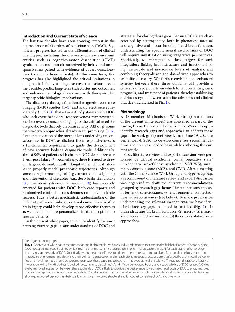
strategies for closing those gaps. Because DOCs are char-acterized by heterogeneity, both in phenotype (arousal and cognitive and motor functions) and brain function, understanding the specific neural mechanisms of DOC will require investigation using integrative perspectives. Specifically, we conceptualize three targets for such integration: linking brain structure and function, link-ing microscale and macroscale levels of analysis, and combining theory-driven and data-driven approaches to scientific discovery. We further envision that enhanced synergy between these three domains will provide a critical vantage point from which to empower diagnosis, prognosis, and treatment of patients, thereby establishing a virtuous cycle between scientific advances and clinical practice (highlighted in Fig. 1).
MethodologyA 13-member Mechanisms Work Group (co-authors of the present white paper) was convened as part of the Curing Coma Campaign, Coma Science Work Group to identify research gaps and approaches to address these gaps. The work group met weekly from June 19, 2020, to September 4, 2020, to develop consensus recommenda-tions and on an as-needed basis while authoring the cur-rent article.
First, literature review and expert discussion were per-formed by clinical syndrome: coma, vegetative state/unresponsive wakefulness syndrome (VS/UWS), mini-mally conscious state (MCS), and CMD. After a meeting with the Coma Science Work Group endotype subgroup, a second round of literature review and expert discussion was organized to draft the current recommendations, grouped by research gap theme. The mechanisms are cast in terms of consciousness vs. environmental connected-ness vs. responsiveness (see below). To make progress on understanding the relevant mechanisms, we have iden-tified three key gaps that need to be filled (Fig. 1): (1) brain structure vs. brain function, (2) micro- vs macro-scale neural mechanisms, and (3) theories vs. data-driven approaches.
Fig. 1 Overview of white paper recommendations. In this article, we have subdivided the gaps that exist in the field of disorders of consciousness (DOC) research into subdisciplines while stressing their mutual interdependence. The term “subdiscipline” is used for each branch of knowledge that makes up the study of DOC. Specifically, we suggest that efforts should be made to integrate structural and functional correlates, micro- and macroscale phenomena, and data- and theory-driven perspectives. Within each discipline (e.g., structural correlates), specific gaps should be identi-fied and novel methods should be selected to answer these gaps and to reach an improved state of the science. Throughout this process, iterative integration with other disciplines is desired (bottom; note disciplines “A” and “B” can be replaced by any given subdiscipline of DOC research). Collec-tively, improved integration between these subfields of DOC is likely to provide the best avenue toward the clinical goals of DOC science: improved diagnosis, prognosis, and treatment (center circle). Circular arrows represent iterative processes, whereas two-headed arrows represent bidirection-ality, e.g., improved diagnosis is likely to allow for more fine-tuned structural and functional correlates of DOC and vice versa
(See figure on next page.)
S39
S40
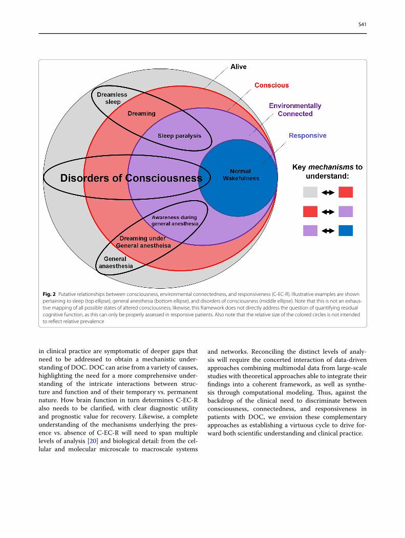
Results of Gap AnalysisMechanisms of Interest and Current Gaps: Consciousness, Environmental Connectedness, and ResponsivenessDisorders of consciousness are characterized by a wide array of symptoms and etiologies. Consequently, a num-ber of classification schemes across different dimensions have been proposed that are based on aspects such as cognitive functions [10], awareness [11] and sensory, motor, and arousal behavioral functions [12]. With the acknowledgment that any one framework cannot fully capture all the constructs relevant to DOC, we propose to contextualize our recommendations within the frame-work provided by Sanders et al. [13], which distinguishes between consciousness, environmental connectedness, and responsiveness (C-EC-R), as outlined below.
Lack of responsiveness remains the clinical criterion for DOC. However, there is a clear conceptual distinc-tion between responsiveness (specifically, nonreflexive behavioral responses) and consciousness (presence of subjective experience, regardless of what the experience is about [13]; note that here we will use this term to also encompass awareness). The possibility for a dissociation between these two dimensions is sharply illustrated by the (rare) case of patients experiencing intraoperative awareness during anesthesia, whose unresponsiveness due to paralysis is mistaken for evidence of unconscious-ness [14–16]. Likewise, independent studies during sleep and anesthesia (in which retrospective reports can be obtained on awakening) demonstrate that subjective experience frequently occurs even in the absence of any behavioral responsiveness [17, 18]. Within DOC, routine assessment based on overt responsiveness may be miss-ing covert consciousness in about ~15–20% of patients [3, 4], a fact that has practical and ethical implications for the management of these patients.
In addition to consciousness and responsiveness, a third relevant dimension is environmental connected-ness (as originally proposed in the context of anesthe-sia [13]). Environmental connectedness corresponds to consciousness of the external environment so that what happens in the environment influences the con-tents of one’s consciousness (unlike, e.g., during some dream states). On the basis of this framework of C-EC-R, patients with CMD can be characterized as being conscious and environmentally connected but not (overtly) responsive. An intriguingly similar example of dissociation between consciousness and responsiveness is found in patients studied under anesthesia by using the isolated forearm technique, whereby an inflated cuff on the arm prevents it from being paralyzed dur-ing general anesthesia [13]. Despite the absence of spontaneous behaviors, such patients can perform sim-ple commands, such as squeezing the physician’s hand
(although the potential confound of arousing nocicep-tion due to the inflated cuff should also be considered). Examples of conscious but environmentally discon-nected states include dreaming and dissociative states induced by ketamine; examples of states that are con-scious and environmentally connected but unrespon-sive are awareness during general anesthesia and sleep paralysis, whereby the paralysis that naturally occurs during rapid eye movement (REM) sleep persists for a short time after awakening [19]. Of course, it is also important to consider the notion of behavioral arousal; although likely neither sufficient for consciousness (given the presence of sleep–wake cycles in patients with VS/UWS) nor necessary for it (given the possibil-ity of dreams during deep non-REM sleep), arousal may nevertheless be a background prerequisite for respon-siveness and possibly also for full-fledged environmen-tal connectedness (Fig. 2).
Current Gaps in Coma ScienceCorrectly characterizing each patient in terms of the three dimensions described in the C-EC-R framework may be valuable for improving diagnosis and determin-ing the prognosis and appropriate standards of care; for instance, it is especially pressing to establish communi-cation with unresponsive but connected patients and not only identify reliable biomarkers for conscious contents, such as emotional distress or pain (i.e., the conscious processing of nociceptive information), but also identify whether some environmental stimuli trigger them (envi-ronmental connectedness). Additionally, by capitalizing on the availability of subjective reports after awakening from sleep or anesthesia in the laboratory environment, it will be crucial to identify ways to detect environmental connectedness based on brain function alone.
By identifying the neural mechanisms that deter-mine transitions between the dimensions of the C-EC-R framework, it may be feasible to devise targeted treat-ment strategies aimed at restoring each in their own right and to identify which personalized avenues may be the most advantageous for a given patient. Specifically, efforts should be made not only to investigate whether specific mechanisms identified from studies of anesthe-sia and sleep [13] could also apply to patients with DOC (e.g., to what extent could the isolated forearm scenario constitute a good model for CMD?) but also to investi-gate how these different states differ from one another (anesthesia is reversible on a short time scale, but DOC may not always be). Additionally, investigations should seek to identify possible dissociations between each of these aspects in subgroups of patients with DOC.
Crucially, the limitations of our present ability to dis-criminate between elements of the C-EC-R framework
S41
in clinical practice are symptomatic of deeper gaps that need to be addressed to obtain a mechanistic under-standing of DOC. DOC can arise from a variety of causes, highlighting the need for a more comprehensive under-standing of the intricate interactions between struc-ture and function and of their temporary vs. permanent nature. How brain function in turn determines C-EC-R also needs to be clarified, with clear diagnostic utility and prognostic value for recovery. Likewise, a complete understanding of the mechanisms underlying the pres-ence vs. absence of C-EC-R will need to span multiple levels of analysis [20] and biological detail: from the cel-lular and molecular microscale to macroscale systems
Fig. 2 Putative relationships between consciousness, environmental connectedness, and responsiveness (C-EC-R). Illustrative examples are shown pertaining to sleep (top ellipse), general anesthesia (bottom ellipse), and disorders of consciousness (middle ellipse). Note that this is not an exhaus-tive mapping of all possible states of altered consciousness; likewise, this framework does not directly address the question of quantifying residual cognitive function, as this can only be properly assessed in responsive patients. Also note that the relative size of the colored circles is not intended to reflect relative prevalence
and networks. Reconciling the distinct levels of analy-sis will require the concerted interaction of data-driven approaches combining multimodal data from large-scale studies with theoretical approaches able to integrate their findings into a coherent framework, as well as synthe-sis through computational modeling. Thus, against the backdrop of the clinical need to discriminate between consciousness, connectedness, and responsiveness in patients with DOC, we envision these complementary approaches as establishing a virtuous cycle to drive for-ward both scientific understanding and clinical practice.
S42
Box 1 Brain connectivity and networks
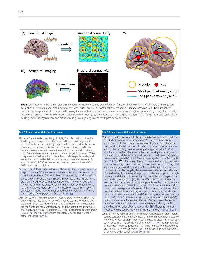
Measures of effective connectivity have also been introduced to identify directed information flow (from region A to region B and not vice versa). Some effective connectivity approaches rely on probabilistic accounts to infer the direction of interactions from statistical relation-ships in the data (e.g., transfer entropy, Granger causality [31–33]). Another approach to characterize the directionality and strength of interactions, albeit limited to a small number of brain areas, is dynamic causal modeling (DCM), which has also been applied to patients with DOC [34]. The DCM framework is used to infer the direction of connec-tivity between regions by comparing possible models of how regional signals were generated. First, alternative models are constructed on the basis of possible coupling between regions, viewed as nodes in a directed network. In a second step, the models are compared through Bayesian model selection to identify the model that best explains the empirically observed data [35]. Finally, effective connectivity can be assessed by a perturb-and-measure approach, in which causal interac-tions are measured by directly stimulating a subset of neurons and by measuring the responses of the rest of the system. In addition to func-tional and effective connectivity, structural connectivity can also be measured in vivo in humans from diffusion magnetic resonance imag-ing data (Fig. 3b), for instance, through diffusion tensor imaging (DTI), which can measure the relative diffusion of water molecules along white matter fibers connecting different regions (although without providing information about directionality) [36]. Thus, structural con-nectivity and FC can be related in the same individual [24, 25, 37, 38].
Whether functional or structural, the interactions between brain regions can be conceived as a network (Fig. 3c), and the mathematical study of networks, known as graph theory, can be used to obtain insights about such networks at multiple levels of resolution [39, 40]: from properties of individual nodes (e.g., degree, measuring how well connected they are [41, 42]) to network modules [26] to macroscale properties such as small-world organization [24, 25, 30, 43–45].
Fig. 3 Connectivity in the human brain. a Functional connectivity can be quantified from functional neuroimaging, for example, as the Pearson correlation between regional blood-oxygen-level-dependent time series from functional magnetic resonance imaging (MRI). b Structural con-nectivity can be quantified from structural imaging, for example, as the number of streamlines between regions, estimated by using diffusion MRI. c Network analysis can provide information about individual nodes (e.g., identification of high-degree nodes, or “hubs”) as well as mesoscopic proper-ties (e.g., modular organization) and macroscale (e.g., average length of shortest path between nodes)
Box 1 Brain connectivity and networks
The term “functional connectivity” (FC) (Fig. 3a) reflects the notion that similarity between patterns of activity of different brain regions (in terms of statistical dependency) may arise from interactions between those regions. At the spatial and temporal resolutions afforded by noninvasive neuroimaging techniques in humans, neural activity is most frequently estimated in terms of electrophysiology (using EEG or magnetoencephalography) or from the blood-oxygen-level-depend-ent signal measured by fMRI. Activity is simultaneously measured for each sensor (for EEG/magnetoencephalography) or each voxel (for fMRI) over a period of time.
On the basis of these measurements of brain activity, the most common ways to quantify FC are measures of linear association between pairs of regional time series (primarily, Pearson correlation, but also methods based on phase coherence or spectral properties of the signals), which are therefore agnostic to interactions between more than two ele-ments and ignore the direction of information flow between the two regions. However, more sophisticated measures also exist, capable of addressing various shortcomings of traditional FC (although often at the expense of computational feasibility) [21–26].
Distinct sets of brain regions, termed “resting-state networks,” spontane-ously organize into consistently cofluctuating assemblies during both tasks and also at rest. Prominent among these resting-state networks are the frontoparietal control network and the default mode network: these networks typically exhibit inversely correlated time courses at rest [27, 28], but their interactions are consistently perturbed in uncon-scious individuals [29, 30].
S43
Brain Structure vs. Brain FunctionAdvancing the science of DOC requires a precise map-ping of the heterogenous structural and functional brain alterations observed in DOC to clinically relevant dimen-sions (e.g., C-EC-R) across a variety of temporal and spa-tial scales. Although it is sometimes possible to precisely map neurological dysfunction to a specific location of damaged tissue, it is important to emphasize that brain regions are intricately interconnected, such that local structural or functional changes may well have far-reach-ing or even global repercussions on other components of the network (diaschisis [46]). Classical lesion-based methods have found success focusing on specific regions of interest (ROI); however, future work should attempt an integration of these approaches with the notion that there exists a many-to-many mapping between brain structure and functional brain states [47]. A full mapping between structural and functional correlates of DOC will require the leveraging of multimodal neuroimaging and neurophysiological techniques, combined with novel analytical methods for integrating them, including the emerging approach of whole-brain computational mod-eling (Box 2).
Historically, circumscribed lesions and changes in activity within damaged brains have been used to identify ROI and model simplified circuits that may be relevant to DOC symptoms. For instance, the influential mesocircuit model proposes that because of pathological changes fol-lowing severe brain injuries, a reduction of thalamocor-tical and thalamostriatal outflow withdraws drive to the frontal cortex and striatum, thus implicating basal gan-glia–thalamocortical circuits in the symptoms of DOC [10]. Compared to network or computational perspec-tives, these focal lesion-based models have the benefit of providing clear targets for treatment (e.g., via deep brain stimulation, low-intensity-focused ultrasound, pharmacological). For instance, emphasis on the role of the thalamus in DOC has compelled the development of techniques for its stimulation, which have been associ-ated with improved behavioral responsiveness in a subset of patients with DOC [9, 48]. Yet the precise functional roles of key ROI, such as the thalamus, remain far from fully characterized; indeed, it remains unclear why nearly full ablation of the thalamus does not appear to result in a loss of consciousness in rodent models [49] despite its consistent association with DOC. Thus, focal characteri-zation of structure–function relationships retains unique value but must continue to be refined.
For instance, although the original mesocircuit model emphasizes pallido-thalamo-cortical communication, this model has been updated by the recent discovery of
direct structural connections between the globus pal-lidus and the cortex, first in rodents [50] and then in humans [51] by using DTI. In parallel, DCM (see Box 1) was applied to the mesocircuit and specifically implicated pallidocortical communication in the transition between states of consciousness under anesthesia [52]. This exam-ple illustrates the kind of multidisciplinary workflow that should inform the placement of ROI within increas-ingly complex frameworks (network, computational) for understanding the structural and functional relationships underlying DOC.
Compared to ROI approaches, the structural correlates of DOC may be further detailed by employing mass-uni-variate, voxel-wise analysis, which can produce mappings of behavioral symptoms to structural alterations at the millimeter scale (e.g., voxel-level symptom mapping [53, 54]; also see voxel-level shape analysis [55, 56]). Similar approaches have been applied to the structural connec-tome derived from DTI (known as connectome-based lesion symptoms mapping [57, 58]). Although these methods allow for a more spatially precise connection between large-scale functions (e.g., behavioral arousal) and structural damage, they do not capture the perhaps more elusive reorganizations in functional networks that often follow structural insult and that ultimately produce changes in behavior (e.g., diaschisis) as well as inadequate or maladaptive compensatory mechanisms, which may also produce DOC symptoms. Indeed, regions that are only secondarily affected may be particularly promising treatment targets because of their relative retained struc-tural integrity, which may hold greater potential to reach preinjury levels of functioning. To capture the relation-ship between gray matter atrophy, white matter discon-nection, and functional interactions, there is a need to better integrate structural correlates with the full range of functional modalities available to us instead of behavioral symptoms alone.
A jumping-off point for multimodal integration in DOC may be to overlay the structural correlates of DOC with the healthy human connectome (both structural and functional) to derive likely locations for a diaschisis effect, a method that avoids the often challenging process of multimodal data collection within patients themselves [59, 60]. Such an approach could be used to build whole-brain computational models, including the known struc-tural correlates of DOC and known large-scale functional correlates (Box 2).
S44
Box 2 Whole-brain computational models
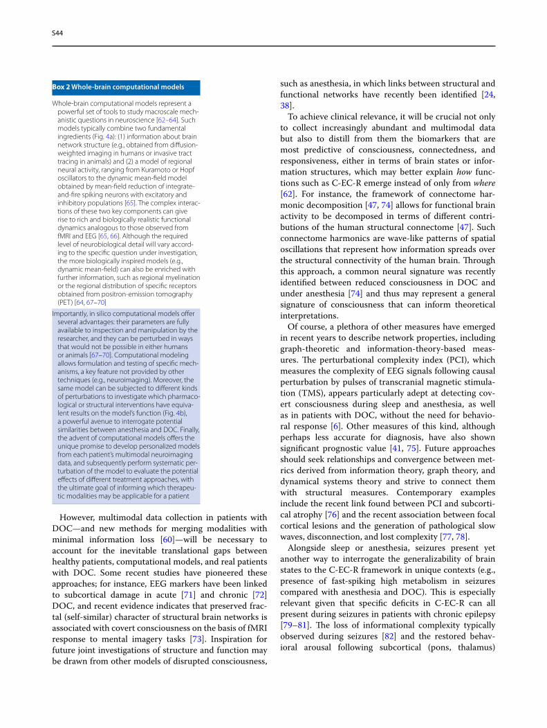
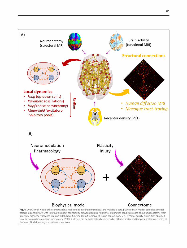
Whole-brain computational models represent a powerful set of tools to study macroscale mech-anistic questions in neuroscience [62–64]. Such models typically combine two fundamental ingredients (Fig. 4a): (1) information about brain network structure (e.g., obtained from diffusion-weighted imaging in humans or invasive tract tracing in animals) and (2) a model of regional neural activity, ranging from Kuramoto or Hopf oscillators to the dynamic mean-field model obtained by mean-field reduction of integrate-and-fire spiking neurons with excitatory and inhibitory populations [65]. The complex interac-tions of these two key components can give rise to rich and biologically realistic functional dynamics analogous to those observed from fMRI and EEG [65, 66]. Although the required level of neurobiological detail will vary accord-ing to the specific question under investigation, the more biologically inspired models (e.g., dynamic mean-field) can also be enriched with further information, such as regional myelination or the regional distribution of specific receptors obtained from positron-emission tomography (PET) [64, 67–70]
Importantly, in silico computational models offer several advantages: their parameters are fully available to inspection and manipulation by the researcher, and they can be perturbed in ways that would not be possible in either humans or animals [67–70]. Computational modeling allows formulation and testing of specific mech-anisms, a key feature not provided by other techniques (e.g., neuroimaging). Moreover, the same model can be subjected to different kinds of perturbations to investigate which pharmaco-logical or structural interventions have equiva-lent results on the model’s function (Fig. 4b), a powerful avenue to interrogate potential similarities between anesthesia and DOC. Finally, the advent of computational models offers the unique promise to develop personalized models from each patient’s multimodal neuroimaging data, and subsequently perform systematic per-turbation of the model to evaluate the potential effects of different treatment approaches, with the ultimate goal of informing which therapeu-tic modalities may be applicable for a patient
However, multimodal data collection in patients with DOC—and new methods for merging modalities with minimal information loss [60]—will be necessary to account for the inevitable translational gaps between healthy patients, computational models, and real patients with DOC. Some recent studies have pioneered these approaches; for instance, EEG markers have been linked to subcortical damage in acute [71] and chronic [72] DOC, and recent evidence indicates that preserved frac-tal (self-similar) character of structural brain networks is associated with covert consciousness on the basis of fMRI response to mental imagery tasks [73]. Inspiration for future joint investigations of structure and function may be drawn from other models of disrupted consciousness,
such as anesthesia, in which links between structural and functional networks have recently been identified [24, 38].
To achieve clinical relevance, it will be crucial not only to collect increasingly abundant and multimodal data but also to distill from them the biomarkers that are most predictive of consciousness, connectedness, and responsiveness, either in terms of brain states or infor-mation structures, which may better explain how func-tions such as C-EC-R emerge instead of only from where [62]. For instance, the framework of connectome har-monic decomposition [47, 74] allows for functional brain activity to be decomposed in terms of different contri-butions of the human structural connectome [47]. Such connectome harmonics are wave-like patterns of spatial oscillations that represent how information spreads over the structural connectivity of the human brain. Through this approach, a common neural signature was recently identified between reduced consciousness in DOC and under anesthesia [74] and thus may represent a general signature of consciousness that can inform theoretical interpretations.
Of course, a plethora of other measures have emerged in recent years to describe network properties, including graph-theoretic and information-theory-based meas-ures. The perturbational complexity index (PCI), which measures the complexity of EEG signals following causal perturbation by pulses of transcranial magnetic stimula-tion (TMS), appears particularly adept at detecting cov-ert consciousness during sleep and anesthesia, as well as in patients with DOC, without the need for behavio-ral response [6]. Other measures of this kind, although perhaps less accurate for diagnosis, have also shown significant prognostic value [41, 75]. Future approaches should seek relationships and convergence between met-rics derived from information theory, graph theory, and dynamical systems theory and strive to connect them with structural measures. Contemporary examples include the recent link found between PCI and subcorti-cal atrophy [76] and the recent association between focal cortical lesions and the generation of pathological slow waves, disconnection, and lost complexity [77, 78].
Alongside sleep or anesthesia, seizures present yet another way to interrogate the generalizability of brain states to the C-EC-R framework in unique contexts (e.g., presence of fast-spiking high metabolism in seizures compared with anesthesia and DOC). This is especially relevant given that specific deficits in C-EC-R can all present during seizures in patients with chronic epilepsy [79–81]. The loss of informational complexity typically observed during seizures [82] and the restored behav-ioral arousal following subcortical (pons, thalamus)
S45
Fig. 4 Overview of whole-brain computational modeling to integrate multimodal and multiscale data. a Whole-brain models combine a model of local regional activity with information about connectivity between regions. Additional information can be provided about neuroanatomy (from structural magnetic resonance imaging [MRI]), brain function (from functional MRI), and neurobiology (e.g., receptor density distribution obtained from in vivo positron-emission tomography [PET]). b Models can be systematically perturbed at different spatial and temporal scales, intervening at the level of individual regions or their connections
S46
stimulation during focal seizures in rats [83] suggests that generalizability is likely to be found; however, this area is ripe for more investigation.
Importantly, the first steps toward the integration of structure and function in DOC are largely taking place by experimental, analytic, and computational integration of the various neuroimaging methods that probe macro-scale phenomena. However, a full mapping of structure–function relationships must inevitably dive deeper into the microscale neural substrate on which macroscale net-works arise. Indeed, modeling the biological correlates of DOC across all spatial scales is likely to improve the location of individual patients within the heterogeneous space of DOC manifestations (e.g., as defined by C-EC-R). Thus, in the next section, we detail the gaps that exist in our understanding of microscale neural underpinnings of DOC and coma and how macro and micro levels of understanding may be bridged.
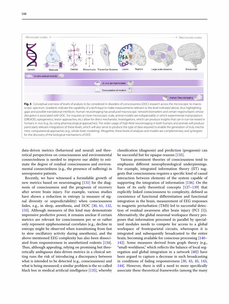
Linking Micro‑ and MacroscalesA comprehensive understanding of DOC requires that insights from macroscopic and mesoscopic levels of inquiry sampled by neuroimaging are integrated with insights from more microscopic scales: the relevant con-tributions at the systems, cellular, genetic, and molecular levels. Because these can largely only be studied directly in animal models, bridging this gap necessitates a closer association of preclinical and clinical research and the generation of hypotheses that can be bidirectionally tested in both animal and human research approaches.
Animal models that allow relevant direct measure-ments of such microscopic facets remain limited for coma [84] and fully absent for VS/UWS and MCS. The development of functionally relevant animal models will need to distinguish between the various neurologi-cal events (e.g., traumatic brain injury, anoxia, hypoxia among others) associated with DOC and the subsequent neurological syndromes of DOC (e.g., MCS, UWS/VS) [85, 86] to characterize both etiology-specific and generalizable DOC mechanisms. This could be further enhanced through comparison with results from sleep and anesthesia research, in which microscopic mecha-nisms have been more comprehensively probed in pre-clinical work [87], producing mechanistic frameworks that span from whole-brain phenomena, such as indi-vidual susceptibility, to anesthetic-state transitions (neu-ral inertia), all the way to genetic susceptibility factors for anesthesia [38, 88, 89]. Indeed, work in rodents and corti-cal slices alike has recently demonstrated that neuronal “off” periods determine a dramatic collapse of large-scale interactions and complexity during non-REM sleep and anesthesia [90–92], which can also be assessed by using
EEG coupled with TMS in humans with brain damage [77, 78].
In terms of experimental approaches, novel noninva-sive in vivo optogenetic techniques allow for the modula-tion of very specific and deeply seated targets in animal models [93, 94]. Leveraging these technological develop-ments will enable systematic hypothesis-driven investi-gations of mechanisms that have been suggested both in previous anesthetic, sleep, and lesion studies in both ani-mals and humans. Importantly, combining optogenetic stimulation with simultaneous high-resolution neuro-imaging [95] could provide biomarkers to serve as direct translational interfaces between animal and human research.
Firstly, a systems-level microscopic perspective requires a comprehensive investigation of subcorti-cal structures in neuroimaging given the implications of these structures in animal and translational research across trauma, anesthesia, and sleep. Specifically, the historical dichotomy between the cortex as the sub-strate of contents of consciousness and the thalamus and brainstem as substrates of arousal has oversimpli-fied the various and diverse roles of subcortical systems [96]. As a first step, efforts should be directed toward a detailed mapping of how key subcortical structures (e.g., thalamus, brainstem nuclei, and basal ganglia) interact with specific cortical layers by using high-field structural and functional neuroimaging and complementary ani-mal models [97, 98]. A relevant example of how current oversimplified views of the subcortical–cortical interplay can be refined into region- and lamina-specific accounts is provided by Redinbaugh et al. [97], who used tha-lamic stimulation in the anesthetized macaque to reveal that consciousness-relevant thalamic influence differs between deep and superficial cortical layers.
Similarly, critical insights at the microscopic systems level are to be gained from the brainstem neuromodula-tory nuclei, which have been extensively studied in ani-mals by using anesthetic and lesion approaches [99–101]. Their associated transmitter systems and brain-wide neu-romodulatory projections have been variously implicated as causing coma [60, 102, 103]. In healthy patients, they have been found to possibly drive both tonic and phasic large-scale in vivo brain activity [104–106]. Established and novel single-photon emission computed tomogra-phy approaches [107] and innovative magnetic resonance sequences (e.g., magnetization transfer images [108]), combined with increased magnetic resonance field strengths, will allow these nuclei and their brain-wide projections to be more directly probed. These approaches should delineate whether dysfunction of these nuclei alters their modulation of the whole-brain connec-tome, which in turn may cause the striking macroscopic
S47
network disruptions commonly observed in DOC. This should use complementary approaches in both animal and patient research as well as computational modeling. Specifically, recent identification of the dopaminergic system’s relevance for wakefulness from anesthesia mod-els has begun to provide a mechanistic foundation of why dopamine may have emerged as a key pharmacological treatment target in DOC [99, 109–112]. To maximize therapeutic potential, comprehensive assessments of associated brainstem nuclei should also aim to delineate whether their dysfunctions in patients with DOC are pre- or postsynaptic using approaches, such as those dem-onstrated by Fridman et al. [113], combining PET with bolus administrations of pre- or postsynaptically acting pharmacological agents to characterize a presynaptic dopaminergic release deficit in patients with DOC.
Further progress along the microscopic perspective requires that differential involvements of particular neu-ronal types and subtypes in consciousness alterations be delineated. These studies will largely have to be delivered by using single-unit recordings and conditional expres-sion approaches in novel animal models to delineate the relevance of neuronal types, e.g., pyramidal neu-rons [114]. For instance, anesthetic-induced decoupling between apical and basal compartments of layer 5 pyram-idal neurons in rodents impairs large-scale functional integration in the brain [115]. These animal experiments can, however, also be complemented by work in human neuroimaging by using whole-brain transcriptomic maps from microarray data, as demonstrated by Craig et al. [116], who used this approach to delineate that GABAe-rgic cortical interneuron subtypes are differently affected in anesthesia.
Indeed, it is key to not only characterize the involve-ment of different neuronal subtypes but equally charac-terize the role of glia and glial subtypes in DOC [117], especially given that widespread white matter damage is commonly associated with these conditions [118, 119]. Both animal models of glial modulation and alteration in response to trauma/anoxia [120] and new approaches in future neuroimaging studies [121] will have to construc-tively integrate white matter structure and function [121, 122]. An area of particular promise is the study of how microglia mediate systemic and regional inflammation following both traumatic brain injury and anoxia [123], leading to DOC. PET ligands for activated microglia and concomitant longitudinal collection of inflammatory and cell death markers in blood and/or cerebrospinal fluid should also distinguish inflammation in acute and chronic phases of DOC [124]. Animal models should aim to distinguish neuroprotective and pathogenic inflamma-tory effects in vivo [125] and distinguish whether phar-macological intervention can induce neuroprotective
states, leading to more favorable outcomes. Similar mul-timodal techniques can also be used to begin to probe the roles of other glial cell types, such as astrocytes, whose role in overall homeostasis of the central nervous system has been demonstrated in traumatic brain injury and thus may play a similar role on the DOC spectrum [126, 127].
Finally, the subcellular and thus most microscopic level holds great promise for future explorations. In particular, it is necessary to identify genetic and molecular media-tors of clinical progression and outcome in DOC. Beyond relying on analogies from anesthesia, it is required to delineate specific receptors and particular pathways that may be associated with the development or persis-tence of DOC. Although no specific genetic factors have yet been associated with DOC, genetic risk factors for adverse outcomes in traumatic brain injury/anoxia serve as useful and possibly intertwined starting points [128]. However, because human genome-wide association stud-ies require prohibitively large population sizes, insights across all other levels of microscopic analysis (see Fig. 5) should be combined into biologically and physiologically relevant collections of gene candidates to be assessed with approaches such as transcriptome-wide association studies [129]. These could identify viable genetic candi-dates that in turn could be incorporated into both animal (assessing, e.g., loss-of-function and gain-of-function alterations) and human experiments (e.g., using micro-array data [116]), thereby feeding back relevant, testable knowledge across all levels of analysis at which we need to consider DOC mechanisms as well as the theoretical frameworks that build on them.
Theory vs. Data‑Driven ApproachesRecent research continues to enable a better understand-ing of the neuronal causes and clinical manifestations of DOC. Among the approaches that have recently been developed, we may distinguish purely empirical ones and those driven by assumptions of certain theories of con-sciousness. However, in their current state, it seems that both theories and data-driven approaches are unable to make predictions precise enough to enable a clear dis-tinction between the neural mechanisms of the three dimensions of the C-EC-R framework. Yet this mecha-nistic understanding would carry immense clinical and ethical significance. For example, one of the key issues for clinicians is the ability to better predict the presence vs. absence of pain in unresponsive patients [130] and what may trigger it. Obtaining such diagnostic biomark-ers would require a mechanistic understanding of the foundations of presence vs. absence of both conscious-ness and environmental connectedness [13] without reli-ance on behavior alone. Thus, a convergence between
S48
data-driven metrics (behavioral and neural) and theo-retical perspectives on consciousness and environmental connectedness is needed to improve our ability to esti-mate the degree of residual consciousness and environ-mental connectedness (e.g., the presence of suffering) in unresponsive patients.
Recently, we have witnessed a formidable growth of new metrics based on neuroimaging [131] for the diag-nosis of consciousness and the prognosis of recovery after severe brain injury. For example, various studies have shown a reduction in entropy (a measure of sig-nal diversity or unpredictability) when consciousness fades, e.g., in sleep, anesthesia, and DOC [30, 61, 132, 133]. Although measures of this kind may demonstrate impressive predictive power, it remains unclear if certain metrics are relevant for consciousness per se or rather only represent epiphenomenal correlates (e.g., decline in entropy might be observed when transitioning from fast to slow oscillatory activity during anesthesia), and the above-mentioned EEG complexity has also been dissoci-ated from responsiveness in anesthetized rodents [134]. Thus, although appealing, relying on promising but theo-retically ambiguous data-driven metrics in a clinical set-ting runs the risk of introducing a discrepancy between what is intended to be detected (e.g., consciousness) and what is being measured; a similar problem is the so-called black box in medical artificial intelligence [135], whereby
classification (diagnosis) and prediction (prognosis) can be successful but for opaque reasons [135].
Various prominent theories of consciousness tend to emphasize different neurophysiological underpinnings. For example, integrated information theory (IIT) sug-gests that consciousness requires a specific kind of causal interaction between elements of the system capable of supporting the integration of information [136]. On the basis of its early theoretical concepts [137–139] that explicitly linked consciousness to complexity, defined as coexistence of functional differentiation and functional integration in the brain, measurement of EEG responses to magnetic perturbation (TMS) led to successful detec-tion of residual awareness after brain injury (PCI [5]). Alternatively, the global neuronal workspace theory pro-poses that information processed in parallel by special-ized modules needs to compete for access to a global workspace of frontoparietal circuits, whereupon it is integrated and subsequently broadcasted to the entire brain, becoming available for conscious processing [140–142]. Some measures derived from graph theory (e.g., “small-worldness,” which reflects the balance of local seg-regation and global integration in a network [40]) have been argued to capture a decrease in such broadcasting in conditions of fading responsiveness [30, 43, 45, 143, 144]. However, there is still a need to more specifically associate these theoretical frameworks (among the many
Fig. 5 Conceptual overview of levels of analysis to be considered in disorders of consciousness (DOC) research across the microscopic-to-macro-scopic spectrum. Gradients indicate the capability of a technique to make measurements relevant to the level indicated above, thus highlighting gaps and possible translational interfaces. Human neuroimaging has produced macroscopic network biomarkers and certain regions/layers whose disruption is associated with DOC. For inquiries at more microscopic scale, animal models are indispensable, in which experimental manipulations (DREADD, optogenetics, lesion approaches, etc.) allow for direct mechanistic investigations, which can produce insights that can in turn be tested in humans in vivo (e.g., by using pharmacological approaches). The wider usage of high-field neuroimaging in both humans and animals will produce particularly relevant integrations of these levels, which will also serve to produce the type of data required to enable the generation of truly mecha-nistic computational approaches (e.g., whole-brain modeling). Altogether, these levels of analyses and models are complementary and synergistic for the discovery of the biological mechanisms of DOC
S49
others) with concrete biomarkers so that empirical data may more clearly inform on the validity of particular theoretical interpretations over others. To do so, there is a pressing need to explicitly clarify, for example, the constructs of integration and information, as featured in different theoretical accounts, as well as their underlying assumptions.
In response to the current gaps and inconsisten-cies identified above, both data- and theory-driven approaches should advance toward unified terminol-ogy and contents that can be readily compared. First, we should aim for precise theoretical predictions for environmental disconnection and unconsciousness that are distinct from behavioral responsiveness, both at the information level and in terms of possible bio-logical mechanisms. Indeed, there can be distinct bio-logical mechanisms leading to the same functional or informational end point (e.g., multiple possible causes leading to brain states of low complexity). Both data- and theory-driven biomarkers for DOC should first be veri-fied in conditions in which subjective reports are avail-able (sleep, anesthesia, seizures) and then be precisely described in relation to their target: the distinction in C-EC-R (see Fig. 2).
To ameliorate the inconsistencies outlined here, theo-ries should help to identify markers that may be most discriminative, and in the case of existing data-driven markers, their proponents should be able to explain their relevance to particular theoretical perspectives. Ideally, data-driven approaches should focus on a comprehensive set of markers indexing different biological levels, e.g., from metabolism and resting-state EEG/fMRI measures to effective connectivity or task-based paradigms. Simi-larly, comprehensive theoretical frameworks should span different descriptive levels (e.g., circuit level, information level, and topological level) and explicitly connect their predictions to different types of empirical markers.
We acknowledge that comparisons between existing theories are challenging because they sometimes oper-ate on different definitions of consciousness. Further-more, biological mechanistic frameworks, such as the mesocircuit hypothesis [10], may be compatible with a mathematical framework, such as IIT, when viewed as providing the required background conditions (e.g., neu-romodulators maintaining adequate excitability) that enable the physical substrate of consciousness itself to function [76, 145]. Although some recent efforts have attempted to reconcile concepts derived from different theories of consciousness [146, 152], a direct involve-ment of the proponents of each theory in an adversarial context is most helpful to identify commonalities and differences in theoretical predictions and come up with specific experiments through which their predictions can
be explicitly compared. For example, some incompat-ible predictions of global neuronal workspace and IIT are now being explicitly tested [147]. Ultimately, such adversarial collaboration may provide different degrees of data-driven support for different theoretical frameworks to make inferences about the presence of consciousness and/or environmental connectedness in patients with DOC.
Conclusion and Future DirectionsIn this position paper, we suggest that bringing together and integrating different levels of analyses and modali-ties, different scales, and different approaches, while at the same time attending to discrete relevant dimensions (C-EC-R), will prove a challenging but vital approach to push forward our mechanistic understanding of DOC (Table 1).
To achieve these ambitious goals, the field will need to leverage multimodal data in the same patients over time but also in diverse patients and across different states of altered consciousness (e.g., sleep, anesthesia, seizures), as well as in animal models, to enable the generalization of results. Several categories of techniques stand out for their ability to provide both improvements within each area and further integration between them, namely, (1) increasingly multilevel animal models, (2) analytic tech-niques for aggregating the various neural correlates of DOC (e.g., machine learning), and (3) increasingly multi-level whole-brain computational models.
Although direct animal models of DOC remain absent and our ability to disentangle consciousness from respon-siveness in animals remains limited because of subjective reports being unattainable, animal models continue to be an important avenue for expanding our understand-ing of the biological mechanisms of DOC. Specifically, the expansion of multimodal methods for experimen-tal manipulation in animals provides opportunities to selectively probe mechanisms and neuronal populations in the brain. Such work in animals could provide trans-lational bridges from foundational microscale alterations and effectors to the neuroimaging findings commonly observed in patients with DOC [148] and thus catalyze the identification of translatable treatment targets and strategies.
To make sense of the wealth of data that such increas-ingly multimodal and multivariate approaches will pro-duce, interrogation through new analytic tools will also be called for. Machine learning based on neuroimaging data has already been used to predict prognosis in patients with DOC [41, 42] as well as to differentiate states of con-sciousness (wakefulness from DOC or anesthesia) [75]. Nevertheless, predictive models derived from machine learning can help to both reduce redundancy between

S50
metrics and identify synergy between many identifiable neural correlates of DOC to distill clinically relevant pre-dictions (e.g., probability of recovery, likelihood of covert consciousness).
In this vein, the increasing richness of empirical data can be leveraged to develop increasingly realistic com-putational models of DOC. In silico whole-brain models offer an especially promising avenue, with their potential to combine macroscale information about brain structure and function with considerations of microscale neurobi-ology [62–64] (Box 2) as well as related empirical data and theoretical perspectives at multiple levels (e.g., how systems-level accounts of DOC interact with information perspectives). Indeed, successful models of anesthetic states have already been put forth [149–151]. Therefore, computational models could provide means to address each of the three gaps we have identified, with the poten-tial to aid personalized medicine.
Throughout this article, we have emphasized that closing the gaps we have identified must be understood as a synergistic endeavor. Addressing gaps at one level (e.g., the relationship between micro- and macroscale) will also inform our understanding of the interactions between brain structure and function. At the same time, closer translational interfaces between these areas of study will thereby inevitably emerge. Likewise, in addi-tion to addressing the specific questions and challenges outlined here, large-scale multimodal data sets across humans and animals will provide the basis on which data-driven approaches and subsequent modeling can be developed further and refined. Finally, bidirectional
interaction between scientific investigation and clinical practice will continue to play a fundamental role, with models and biomarkers informing prognosis and treat-ment while being informed by clinical insight, engender-ing a virtuous cycle. Nevertheless, we do not consider bridging these gaps as the end point of coma science but only as a new vantage point—a vantage point of inte-grated knowledge across levels, imaging modalities, and theoretical approaches—from which to pursue our goal: curing coma.
Author details1 Division of Anaesthesia, School of Clinical Medicine, University of Cambridge, Cambridge, UK. 2 Department of Clinical Neurosciences, University of Cam-bridge, Cambridge, UK. 3 Department of Psychology, University of California, Los Angeles, Los Angeles, CA, USA. 4 Department of Psychiatry, University of Wisconsin–Madison, Madison, WI, USA. 5 Department of Anesthesia and Perioperative Medicine, David Geffen School of Medicine, University of California, Los Angeles, Los Angeles, CA, USA. 6 Department of Anesthesia, Critical Care, and Pain Medicine, Massachusetts General Hospital and Harvard Medical School, Harvard University, Boston, MA, USA. 7 Department of Brain and Cognitive Sciences, Massachusetts Institute of Technology, Cambridge, MA, USA. 8 Department of Neurology and Neurosurgery, Washington Univer-sity in St. Louis, St. Louis, MO, USA. 9 Departments of Anesthesiology and Criti-cal Care Medicine, Neurology and Neurosurgery, and Radiology, School of Medicine, Johns Hopkins University, Baltimore, MD, USA. 10 Dipartimento di Scienze Biomediche e Cliniche “L. Sacco”, Università Degli Studi Di Milano, Milan, Italy. 11 Istituto Di Ricovero e Cura a Carattere Scientifico, Fondazione Don Carlo Gnocchi, Milan, Italy. 12 Brain Injury Research Center, Department of Neurosurgery, David Geffen School of Medicine, University of California, Los Angeles, Los Angeles, CA, USA.
AcknowledgementsThe authors wish to acknowledge the Curing Coma Campaign collabora-tors participating in the overall program and Angela Comanducci for helpful discussion. Figure 4 was created with biorender.com.
Table 1 Future research needs for investigating mechanisms of consciousness toward improved diagnosis, prognosis, and treatment of DOC
C-EC-R consciousness, environmental connectedness, and responsiveness, DOC disorders of consciousness
Research need: establish a framework for differentiating clinical subtypes of DOC with concepts of C-EC-R
Research need: identify links between structural brain damage and abnormal brain function in DOCForm a complete mapping of DOC structural damage to functions of interest across multiple temporal and spatial scales
Identify structural correlates of relevant biomarkers of brain function in DOC patients derived from different approaches, e.g., information theory, graph theory, and dynamical systems theory
Research need: provide an integrated understanding of DOC across biological micro‑ and macroscalesDevelop clinically relevant animal models of DOC for translational research approaches
Identify the role of subcortical structures and their interplay with the cortex in heterogeneous DOC
Associate microscale (neuronal and nonneuronal), and subcellular (molecular and genetic) mediators, with in vivo manifestations in patients with DOC
Research need: induce integration between theory‑driven and data‑driven approachesDevelop precise theoretical predictions and further biomarkers to address each dimension of the C-EC-R framework
Build a comprehensive set of data-driven and theory-driven biomarkers addressing different levels of analysis
Compare and develop existing theories in adversarial collaboration between theory leaders
Research need: integrate levels of description, imaging modalities, and theoretical approachesAnalyze multilevel and multimodal data from large-scale data sets to construct more realistic computational models of DOC
Develop personalized medicine models to guide treatment based on individual patients’ multimodal data
S51
Author ContributionsAll authors contributed to the development and design of the concepts in the article through attending the online meetings, performing the literature review, and drafting or critically revising the article for important intellectual content. All authors approved the final version. Authors AIL, JC, LRBS, and UJG contributed equally as first authors. EAS and MB contributed equally as last authors.
Conflicts of interestThe authors declare no conflicts of interest.
Source of SupportThis work was supported by the Neurocritical Care Society; the Tiny Blue Dot Foundation; the Templeton World Charity Foundation, Inc.; grant NInDS K23NS112473; National Institutes of Health, National Institute of General Medical Sciences grant R01GM135420; National Institute of General Medical Sciences grant 1K08GM121961; the Johns Hopkins University Discovery Award and Stimulating and Advancing ACCM Research Award; the European Union’s Horizon 2020 Framework Program for Research and Innovation under the Spe-cific Grant Agreement No. 945539 (Human Brain Project SGA3); the Canadian Institute for Advanced Research (RCZB/072 RG93193); the Stephen Erskine Fellowship (Queens’ College, Cambridge, UK); the Cambridge European Trust; and the Gates Cambridge Trust.
Open AccessThis article is licensed under a Creative Commons Attribution 4.0 International License, which permits use, sharing, adaptation, distribution and reproduction in any medium or format, as long as you give appropriate credit to the original author(s) and the source, provide a link to the Creative Commons licence, and indicate if changes were made. The images or other third party material in this article are included in the article’s Creative Commons licence, unless indicated otherwise in a credit line to the material. If material is not included in the article’s Creative Commons licence and your intended use is not permitted by statutory regulation or exceeds the permitted use, you will need to obtain permission directly from the copyright holder. To view a copy of this licence, visit http:// creat iveco mmons. org/ licen ses/ by/4. 0/.
Publisher’s NoteSpringer Nature remains neutral with regard to jurisdictional claims in pub-lished maps and institutional affiliations.
Received: 24 March 2021 Accepted: 17 May 2021
References 1. Owen AM, Coleman MR, Boly M, Davis MH, Laureys S, Pickard JD.
Detecting awareness in the vegetative state. Science. 2006;313:1402. 2. Monti MM, Vanhaudenhuyse A, Coleman MR, Boly M, Pickard JD,
Tshibanda L, et al. Willful modulation of brain activity in disorders of consciousness. N Engl J Med. 2010;362:579–89.
3. Naci L, Sinai L, Owen AM. Detecting and interpreting conscious experiences in behaviorally non-responsive patients. Neuroimage. 2017;145:304–13.
4. Claassen J, Doyle K, Matory A, Couch C, Burger KM, Velazquez A, et al. Detection of brain activation in unresponsive patients with acute brain injury. N Engl J Med. 2019;380:2497–505.
5. Casali AG, Gosseries O, Rosanova M, Boly M, Sarasso S, Casali KR, et al. A theoretically based index of consciousness independent of sensory processing and behavior. Sci Transl Med. 2013;5:198ra105.
6. Casarotto S, Comanducci A, Rosanova M, Sarasso S, Fecchio M, Napoli-tani M, et al. Stratification of unresponsive patients by an indepen-dently validated index of brain complexity. Ann Neurol. 2016;80:718–29.
7. Estraneo A, Moretta P, Loreto V, Santoro L, Trojano L. Clinical and neu-ropsychological long-term outcomes after late recovery of responsive-ness: a case series. Arch Phys Med Rehabil. 2014;95:711–6.
8. Schnakers C, Monti MM. Disorders of consciousness after severe brain injury: therapeutic options. Curr Opin Neurol. 2017;30:573–9.
9. Cain JA, Spivak NM, Coetzee JP, Crone JS, Johnson MA, Lutkenhoff ES, et al. Ultrasonic thalamic stimulation in chronic disorders of conscious-ness. Brain Stimul. 2021;14:301–3.
10. Schiff ND. Recovery of consciousness after brain injury: a mesocircuit hypothesis. Trends Neurosci. 2010;33:1–9.
11. Laureys S. The neural correlate of (un)awareness: lessons from the vegetative state. Trends Cogn Sci. 2005;9:556–9.
12. Giacino JT. The vegetative and minimally conscious states: consensus-based criteria for establishing diagnosis and prognosis. NeuroRehabili-tation. 2004;19:293–8.
13. Sanders RD, Tononi G, Laureys S, Sleigh JW. Unresponsiveness ≠ uncon-sciousness. Anesthesiology. 2012;116:946–59.
14. Moerman N, Bonke B, Oosting J. Awareness and recall during general anesthesia. Facts and feelings Anesthesiology. 1993;79:454–64.
15. Ghoneim MM, Block RI, Haffarnan M, Mathews MJ. Awareness during anesthesia: risk factors, causes and sequelae: a review of reported cases in the literature. Anesth Analg. 2009;108:527–35.
16. Sebel PS, Bowdle TA, Ghoneim MM, Rampil IJ, Padilla RE, Gan TJ, et al. The incidence of awareness during anesthesia: a multicenter United States study. Anesth Analg. 2004;99:833–9.
17. Nir Y, Tononi G. Dreaming and the brain: from phenomenology to neurophysiology. Trends Cogn Sci. 2010;14:88–100.
18. Leslie K, Sleigh J, Paech MJ, Voss L, Lim CW, Sleigh C. Dreaming and electroencephalographic changes during anesthesia maintained with propofol or desflurane. Anesthesiology. 2009;111:547–55.
19. Cheyne JA, Newby-Clark IR, Rueffer SD. Relations among hypnagogic and hypnopompic experiences associated with sleep paralysis. J Sleep Res. 1999;8:313–7.
20. Marr D. Vision: a computational investigation into the human represen-tation and processing of visual information. Cambridge (MA): The MIT Press; 2010.
21. Crone JS, Schurz M, Höller Y, Bergmann J, Monti M, Schmid E, et al. Impaired consciousness is linked to changes in effective connectivity of the posterior cingulate cortex within the default mode network. Neuroimage. 2015;110:101–9.
22. Deco G, Vidaurre D, Kringelbach ML. Revisiting the global workspace orchestrating the hierarchical organization of the human brain. Nat Hum Behav. 2021;5:497–511.
23. Mediano PAM, Rosas F, Carhart-Harris RL, Seth AK, Barrett AB. Beyond integrated information: A taxonomy of information dynamics phenom-ena. arXiv. 2019; arXiv: 1909. 02297.
24. Barttfeld P, Uhrig L, Sitt JD, Sigman M, Jarraya B, Dehaene S. Signature of consciousness in the dynamics of resting-state brain activity. Proc Natl Acad Sci USA. 2015;112:887–92.
25. Zamani Esfahlani F, Jo Y, Faskowitz J, Byrge L, Kennedy DP, Sporns O, et al. High-amplitude cofluctuations in cortical activity drive functional connectivity. Proc Natl Acad Sci USA. 2020;117:28393–401.
26. Shine JM, Bissett PG, Bell PT, Koyejo O, Balsters JH, Gorgolewski KJ, et al. The dynamics of functional brain networks: integrated network states during cognitive task performance. Neuron. 2016;92:544–54.
27. Raichle ME, MacLeod AM, Snyder AZ, Powers WJ, Gusnard DA, Shul-man GL. A default mode of brain function. Proc Natl Acad Sci USA. 2001;98:676–82.
28. Fox MD, Snyder AZ, Vincent JL, Corbetta M, Van Essen DC, Raichle ME. The human brain is intrinsically organized into dynamic, anticorrelated functional networks. Proc Natl Acad Sci USA. 2005;102:9673–8.
29. Di Perri C, Bahri MA, Amico E, Thibaut A, Heine L, Antonopoulos G, et al. Neural correlates of consciousness in patients who have emerged from a minimally conscious state: a cross-sectional multimodal imaging study. Lancet Neurol. 2016;15:830–42.
30. Luppi AI, Craig MM, Pappas I, Finoia P, Williams GB, Allanson J, et al. Consciousness-specific dynamic interactions of brain integration and functional diversity. Nat Commun. 2019;10:4616.
31. Barrett AB, Murphy M, Bruno M-A, Noirhomme Q, Boly M, Laureys S, et al. Granger causality analysis of steady-state electroencepha-lographic signals during propofol-induced anaesthesia. PLoS ONE. 2012;7:e29072.
32. Schreiber T. Measuring information transfer. Phys Rev Lett. 2000;85:461–4.
S52
33. Massey J. Causality, feedback and directed information. Proceedings of the 1990 International Symposium on Information Theory and its Applications. 1990.
34. Boly M, Garrido MI, Gosseries O, Bruno M-A, Boveroux P, Schnakers C, et al. Preserved feedforward but impaired top-down processes in the vegetative state. Science. 2011;332:858–62.
35. Stephan KE, Harrison LM, Kiebel SJ, David O, Penny WD, Friston KJ. Dynamic causal models of neural system dynamics: current state and future extensions. J Biosci. 2007;32:129–44.
36. Hagmann P, Cammoun L, Gigandet X, Meuli R, Honey CJ, Wedeen VJ, et al. Mapping the structural core of human cerebral cortex. PLoS Biol. 2008;6:e159.
37. Amico E, Marinazzo D, Di Perri C, Heine L, Annen J, Martial C, et al. Mapping the functional connectome traits of levels of consciousness. Neuroimage. 2017;148:201–11.
38. Uhrig L, Sitt JD, Jacob A, Tasserie J, Barttfeld P, Dupont M, et al. Resting-state dynamics as a cortical signature of anesthesia in monkeys. Anesthesiology. 2018;129:942–58.
39. Sporns O. From simple graphs to the connectome: networks in neuro-imaging. Neuroimage. 2012;62:881–6.
40. Bullmore E, Sporns O. The economy of brain network organization. Nat Rev Neurosci. 2012;13:336–49.
41. Chennu S, Annen J, Wannez S, Thibaut A, Chatelle C, Cassol H, et al. Brain networks predict metabolism, diagnosis and prognosis at the bedside in disorders of consciousness. Brain. 2017;140:2120–32.
42. Pappas I, Adapa RM, Menon DK, Stamatakis EA. Brain network disin-tegration during sedation is mediated by the complexity of sparsely connected regions. Neuroimage. 2019;186:221–33.
43. Achard S, Delon-Martin C, Vértes PE, Renard F, Schenck M, Schneider F, et al. Hubs of brain functional networks are radically reorganized in comatose patients. Proc Natl Acad Sci USA. 2012;109:20608–13.
44. Crone JS, Soddu A, Höller Y, Vanhaudenhuyse A, Schurz M, Bergmann J, et al. Altered network properties of the fronto-parietal network and the thalamus in impaired consciousness. Neuroimage Clin. 2013;4:240–8.
45. Monti MM, Lutkenhoff ES, Rubinov M, Boveroux P, Vanhaudenhuyse A, Gosseries O, et al. Dynamic change of global and local information processing in propofol-induced loss and recovery of consciousness. PLoS Comput Biol. 2013;9:e1003271.
46. Carrera E, Tononi G. Diaschisis: past, present, future. Brain. 2014;137:2408–22.
47. Atasoy S, Donnelly I, Pearson J. Human brain networks function in connectome-specific harmonic waves. Nat Commun. 2016;7:10340.
48. Schiff ND, Giacino JT, Kalmar K, Victor JD, Baker K, Gerber M, et al. Behav-ioural improvements with thalamic stimulation after severe traumatic brain injury. Nature. 2007;448:600–3.
49. Fuller PM, Sherman D, Pedersen NP, Saper CB, Lu J. Reassessment of the structural basis of the ascending arousal system. J Comp Neurol. 2011;519:933–56.
50. Chen MC, Ferrari L, Sacchet MD, Foland-Ross LC, Qiu M-H, Gotlib IH, et al. Identification of a direct GABAergic pallidocortical pathway in rodents. Eur J Neurosci. 2015;41:748–59.
51. Zheng ZS, Monti MM. Thalamic and extra-thalamic connections of the Globus Pallidus in the human brain: the ultradirect pathway. BioRxiv. 2019; https:// doi. org/ 10. 1101/ 688283
52. Crone JS, Lutkenhoff ES, Bio BJ, Laureys S, Monti MM. Testing proposed neuronal models of effective connectivity within the cortico-basal ganglia-thalamo-cortical loop during loss of consciousness. Cereb Cortex. 2017;27:2727–38.
53. Bates E, Wilson SM, Saygin AP, Dick F, Sereno MI, Knight RT, et al. Voxel-based lesion-symptom mapping. Nat Neurosci. 2003;6:448–50.
54. Tyler LK, Marslen-Wilson W, Stamatakis EA. Dissociating neuro-cognitive component processes: voxel-based correlational methodology. Neu-ropsychologia. 2005;43:771–8.
55. Lutkenhoff ES, McArthur DL, Hua X, Thompson PM, Vespa PM, Monti MM. Thalamic atrophy in antero-medial and dorsal nuclei correlates with six-month outcome after severe brain injury. Neuroimage Clin. 2013;3:396–404.
56. Lutkenhoff ES, Chiang J, Tshibanda L, Kamau E, Kirsch M, Pickard JD, et al. Thalamic and extrathalamic mechanisms of consciousness after severe brain injury. Ann Neurol. 2015;78:68–76.
57. Gleichgerrcht E, Fridriksson J, Rorden C, Bonilha L. Connectome-based lesion-symptom mapping (CLSM): a novel approach to map neurologi-cal function. Neuroimage Clin. 2017;16:461–7.
58. Zheng ZS, Reggente N, Lutkenhoff E, Owen AM, Monti MM. Disentan-gling disorders of consciousness: insights from diffusion tensor imaging and machine learning. Hum Brain Mapp. 2017;38:431–43.
59. Boes AD, Prasad S, Liu H, Liu Q, Pascual-Leone A, Caviness VS, et al. Network localization of neurological symptoms from focal brain lesions. Brain. 2015;138:3061–75.
60. Fischer DB, Boes AD, Demertzi A, Evrard HC, Laureys S, Edlow BL, et al. A human brain network derived from coma-causing brainstem lesions. Neurology. 2016;87:2427–34.
61. Demertzi A, Tagliazucchi E, Dehaene S, Deco G, Barttfeld P, Raimondo F, et al. Human consciousness is supported by dynamic complex patterns of brain signal coordination. Sci Adv. 2019;5:eaat7603.
62. Cofré R, Herzog R, Mediano PAM, Piccinini J, Rosas FE, Sanz Perl Y, et al. Whole-brain models to explore altered states of consciousness from the bottom up. Brain Sci. 2020;10:626.
63. Kringelbach ML, Deco G. Brain states and transitions: insights from computational neuroscience. Cell Rep. 2020;32:108128.
64. Luppi AI, Mediano PAM, Rosas FE, Allanson J, Pickard JD, Williams GB, et al. Paths to oblivion: common neural mechanisms of anaesthesia and disorders of consciousness. BioRxiv. 2021; https:// doi. org/ 10. 1101/ 2021. 02. 14. 431140
65. Cabral J, Kringelbach ML, Deco G. Functional connectivity dynamically evolves on multiple time-scales over a static structural connectome: models and mechanisms. Neuroimage. 2017;160:84–96.
66. Bensaid S, Modolo J, Merlet I, Wendling F, Benquet P. COALIA: a com-putational model of human EEG for consciousness research. Front Syst Neurosci. 2019;13:59.
67. Demirtaş M, Burt JB, Helmer M, Ji JL, Adkinson BD, Glasser MF, et al. Hier-archical heterogeneity across human cortex shapes large-scale neural dynamics. Neuron. 2019;101:1181-94.e13.
68. Wang P, Kong R, Kong X, Liégeois R, Orban C, Deco G, et al. Inversion of a large-scale circuit model reveals a cortical hierarchy in the dynamic resting human brain. Sci Adv. 2019;5:eaat7854.
69. Deco G, Cruzat J, Cabral J, Knudsen GM, Carhart-Harris RL, Whybrow PC, et al. Whole-brain multimodal neuroimaging model using serotonin receptor maps explains non-linear functional effects of LSD. Curr Biol. 2018;28:3065-74.e6.
70. Kringelbach ML, Cruzat J, Cabral J, Knudsen GM, Carhart-Harris R, Whybrow PC, et al. Dynamic coupling of whole-brain neuronal and neurotransmitter systems. Proc Natl Acad Sci USA. 2020;117:9566–76.
71. Schnakers C, Lutkenhoff ES, Bio BJ, McArthur DL, Vespa PM, Monti MM. Acute EEG spectra characteristics predict thalamic atrophy after severe TBI. J Neurol Neurosurg Psychiatry. 2019;90:617–9.
72. Lutkenhoff ES, Nigri A, Rossi Sebastiano D, Sattin D, Visani E, Rosazza C, et al. EEG power spectra and subcortical pathology in chronic disorders of consciousness. Psychol Med. 2020. https:// doi. org/ 10. 1017/ S0033 29172 00033 0X.
73. Luppi AI, Craig MM, Coppola P, Peattie ARD, Finoia P, Williams GB, et al. Preserved fractal character of structural brain networks is associated with covert consciousness after severe brain injury. Neuroimage Clin. 2021;30:102682.
74. Luppi AI, Vohryzek J, Kringelbach ML, Mediano PAM, Craig MM, Adapa R, et al. Connectome harmonic decomposition of human brain dynam-ics reveals a landscape of consciousness. bioRxiv. 2020; https:// doi. org/ 10. 1101/ 2020. 08. 10. 244459.
75. Demertzi A, Antonopoulos G, Heine L, Voss HU, Crone JS, de Los AC, et al. Intrinsic functional connectivity differentiates minimally conscious from unresponsive patients. Brain. 2015;138:2619–31.
76. Lutkenhoff ES, Johnson MA, Casarotto S, Massimini M, Monti MM. Sub-cortical atrophy correlates with the perturbational complexity index in patients with disorders of consciousness. Brain Stimul. 2020;13:1426–35.
77. Sarasso S, D’Ambrosio S, Fecchio M, Casarotto S, Viganò A, Landi C, et al. Local sleep-like cortical reactivity in the awake brain after focal injury. Brain. 2020;143:3672–84.
78. Rosanova M, Fecchio M, Casarotto S, Sarasso S, Casali AG, Pigorini A, et al. Sleep-like cortical OFF-periods disrupt causality and complexity in the brain of unresponsive wakefulness syndrome patients. Nat Com-mun. 2018;9:4427.
S53
79. Johanson M, Revonsuo A, Chaplin J, Wedlund J-E. Level and contents of consciousness in connection with partial epileptic seizures. Epilepsy Behav. 2003;4:279–85.
80. Johanson M, Valli K, Revonsuo A, Chaplin JE, Wedlund J-E. Alterations in the contents of consciousness in partial epileptic seizures. Epilepsy Behav. 2008;13:366–71.
81. Laccheo I, Sonmezturk H, Bhatt AB, Tomycz L, Shi Y, Ringel M, et al. Non-convulsive status epilepticus and non-convulsive seizures in neurologi-cal ICU patients. Neurocrit Care. 2015;22:202–11.
82. Mateos DM, Guevara Erra R, Wennberg R, Perez Velazquez JL. Measures of entropy and complexity in altered states of consciousness. Cogn Neurodyn. 2018;12:73–84.
83. Kundishora AJ, Gummadavelli A, Ma C, Liu M, McCafferty C, Schiff ND, et al. Restoring conscious arousal during focal limbic seizures with deep brain stimulation. Cereb Cortex. 2017;27:1964–75.
84. Pais-Roldán P, Edlow BL, Jiang Y, Stelzer J, Zou M, Yu X. Multimodal assessment of recovery from coma in a rat model of diffuse brainstem tegmentum injury. Neuroimage. 2019;189:615–30.
85. O’Donnell JC, Browne KD, Kilbaugh TJ, Chen HI, Whyte J, Cullen DK. Challenges and demand for modeling disorders of consciousness fol-lowing traumatic brain injury. Neurosci Biobehav Rev. 2019;98:336–46.
86. Wojnarowicz MW, Fisher AM, Minaeva O, Goldstein LE. Considerations for experimental animal models of concussion, traumatic brain injury, and chronic traumatic encephalopathy-these matters matter. Front Neurol. 2017;8:240.
87. Brown EN, Purdon PL, Van Dort CJ. General anesthesia and altered states of arousal: a systems neuroscience analysis. Annu Rev Neurosci. 2011;34:601–28.
88. Proekt A, Hudson AE. A stochastic basis for neural inertia in emergence from general anaesthesia. Br J Anaesth. 2018;121:86–94.
89. Friedman EB, Sun Y, Moore JT, Hung H-T, Meng QC, Perera P, et al. A conserved behavioral state barrier impedes transitions between anesthetic-induced unconsciousness and wakefulness: evidence for neural inertia. PLoS ONE. 2010;5:e11903.
90. Pigorini A, Sarasso S, Proserpio P, Szymanski C, Arnulfo G, Casarotto S, et al. Bistability breaks-off deterministic responses to intracortical stimu-lation during non-REM sleep. Neuroimage. 2015;112:105–13.
91. D’Andola M, Rebollo B, Casali AG, Weinert JF, Pigorini A, Villa R, et al. Bistability, causality, and complexity in cortical networks: an in vitro perturbational study. Cereb Cortex. 2018;28:2233–42.
92. Dasilva M, Camassa A, Navarro-Guzman A, Pazienti A, Perez-Mendez L, Zamora-López G, et al. Modulation of cortical slow oscillations and complexity across anesthesia levels. Neuroimage. 2021;224:117415.
93. Hong G. Seeing the sound. Science. 2020;369:638. 94. Taylor NE, Van Dort CJ, Kenny JD, Pei J, Guidera JA, Vlasov KY, et al.
Optogenetic activation of dopamine neurons in the ventral tegmental area induces reanimation from general anesthesia. Proc Natl Acad Sci USA. 2016;113:12826–31.
95. Reynaud O, da Silva AR, Gruetter R, Jelescu IO. Multi-slice passband bSSFP for human and rodent fMRI at ultra-high field. J Magn Reson. 2019;305:31–40.
96. Satpute AB, Kragel PA, Barrett LF, Wager TD, Bianciardi M. Deconstruct-ing arousal into wakeful, autonomic and affective varieties. Neurosci Lett. 2019;693:19–28.
97. Redinbaugh MJ, Phillips JM, Kambi NA, Mohanta S, Andryk S, Dooley GL, et al. Thalamus modulates consciousness via layer-specific control of cortex. Neuron. 2020;106:66-75.e12.
98. Bastos AM, Donoghue JA, Brincat SL, Mahnke M, Yanar J, Correa J, et al. Neural effects of propofol-induced unconsciousness and its reversal using thalamic stimulation. Elife. 2021;10:e60824.
99. Palmiter RD. Dopamine signaling as a neural correlate of consciousness. Neuroscience. 2011;198:213–20.
100. Brown EN, Pavone KJ, Naranjo M. Multimodal general anesthesia: theory and practice. Anesth Analg. 2018;127:1246–58.
101. Brown EN, Lydic R, Schiff ND. General anesthesia, sleep, and coma. N Engl J Med. 2010;363:2638–50.
102. Edlow BL, Takahashi E, Wu O, Benner T, Dai G, Bu L, et al. Neuroanatomic connectivity of the human ascending arousal system critical to con-sciousness and its disorders. J Neuropathol Exp Neurol. 2012;71:531–46.
103. Edlow BL, Haynes RL, Takahashi E, Klein JP, Cummings P, Benner T, et al. Disconnection of the ascending arousal system in traumatic coma. J Neuropathol Exp Neurol. 2013;72:505–23.
104. van den Brink RL, Pfeffer T, Warren CM, Murphy PR, Tona K-D, van der Wee NJA, et al. Catecholaminergic neuromodulation shapes intrinsic MRI functional connectivity in the human brain. J Neurosci. 2016;36:7865–76.
105. Shine JM, Breakspear M, Bell PT, Ehgoetz Martens KA, Shine R, Koyejo O, et al. Human cognition involves the dynamic integration of neural activity and neuromodulatory systems. Nat Neurosci. 2019;22:289–96.
106. Avery MC, Krichmar JL. Neuromodulatory systems and their interac-tions: a review of models, theories, and experiments. Front Neural Circuits. 2017;11:108.
107. Palermo G, Ceravolo R. Molecular imaging of the dopamine trans-porter. Cells. 2019;8:872.
108. Ye R, Rua C, O’Callaghan C, Jones PS, Hezemans FH, Kaalund SS, et al. An in vivo probabilistic atlas of the human locus coeruleus at ultra-high field. Neuroimage. 2021;225:117487.
109. Solt K, Van Dort CJ, Chemali JJ, Taylor NE, Kenny JD, Brown EN. Electri-cal stimulation of the ventral tegmental area induces reanimation from general anesthesia. Anesthesiology. 2014;121:311–9.
110. Taylor NE, Chemali JJ, Brown EN, Solt K. Activation of D1 dopamine receptors induces emergence from isoflurane general anesthesia. Anesthesiology. 2013;118:30–9.
111. Edlow BL, Claassen J, Schiff ND, Greer DM. Recovery from disorders of consciousness: mechanisms, prognosis and emerging therapies. Nat Rev Neurol. 2021;17:135–56.
112. Solt K, Cotten JF, Cimenser A, Wong KFK, Chemali JJ, Brown EN. Meth-ylphenidate actively induces emergence from general anesthesia. Anesthesiology. 2011;115:791–803.
113. Fridman EA, Osborne JR, Mozley PD, Victor JD, Schiff ND. Presynaptic dopamine deficit in minimally conscious state patients following traumatic brain injury. Brain. 2019;142:1887–93.
114. Aru J, Suzuki M, Larkum ME. Cellular mechanisms of conscious pro-cessing. Trends Cogn Sci. 2020;24:814–25.
115. Suzuki M, Larkum ME. General anesthesia decouples cortical pyrami-dal neurons. Cell. 2020;180:666-76.e13.
116. Craig MM, Misic B, Pappas I, Adapa RM, Menon DK, Stamatakis EA. Propofol sedation-induced alterations in brain connectivity reflect parvalbumin interneurone distribution in human cerebral cortex. Br J Anaesth. 2021;126:835–44.
117. Robertson JM. The gliocentric brain. Int J Mol Sci. 2018;19:3033. 118. Wu X, Zhang J, Cui Z, Tang W, Shao C, Hu J, et al. White matter deficits
underlying the impaired consciousness level in patients with disor-ders of consciousness. Neurosci Bull. 2018;34:668–78.
119. Stafford CA, Owen AM, Fernández-Espejo D. The neural basis of external responsiveness in prolonged disorders of consciousness. Neuroimage Clin. 2019;22:101791.
120. Karve IP, Taylor JM, Crack PJ. The contribution of astrocytes and micro-glia to traumatic brain injury. Br J Pharmacol. 2016;173:692–702.
121. Grajauskas LA, Frizzell T, Song X, D’Arcy RCN. White matter fMRI activation cannot be treated as a nuisance regressor: overcoming a historical blind spot. Front Neurosci. 2019;13:1024.
122. Takata N, Sugiura Y, Yoshida K, Koizumi M, Hiroshi N, Honda K, et al. Optogenetic astrocyte activation evokes BOLD fMRI response with oxygen consumption without neuronal activity modulation. Glia. 2018;66:2013–23.
123. Simon DW, McGeachy MJ, Bayır H, Clark RSB, Loane DJ, Kochanek PM. The far-reaching scope of neuroinflammation after traumatic brain injury. Nat Rev Neurol. 2017;13:171–91.
124. Scott G, Hellyer PJ, Ramlackhansingh AF, Brooks DJ, Matthews PM, Sharp DJ. Thalamic inflammation after brain trauma is associated with thalamo-cortical white matter damage. J Neuroinflammation. 2015;12:224.
125. Corps KN, Roth TL, McGavern DB. Inflammation and neuroprotection in traumatic brain injury. JAMA Neurol. 2015;72:355–62.
126. Burda JE, Sofroniew MV. Seducing astrocytes to the dark side. Cell Res. 2017;27:726–7.
127. Theparambil SM, Hosford PS, Ruminot I, Kopach O, Reynolds JR, Sand-oval PY, et al. Astrocytes regulate brain extracellular pH via a neuronal activity-dependent bicarbonate shuttle. Nat Commun. 2020;11:5073.
S54
128. Zeiler FA, McFadyen C, Newcombe VFJ, Synnot A, Donoghue EL, Ripatti S, et al. Genetic influences on patient-oriented outcomes in traumatic brain injury: a living systematic review of non-apolipoprotein e single-nucleotide polymorphisms. J Neurotrauma. 2021;38:1107–23.
129. Wainberg M, Sinnott-Armstrong N, Mancuso N, Barbeira AN, Knowles DA, Golan D, et al. Opportunities and challenges for transcriptome-wide association studies. Nat Genet. 2019;51:592–9.
130. Chatelle C, Thibaut A, Whyte J, De Val MD, Laureys S, Schnakers C. Pain issues in disorders of consciousness. Brain Inj. 2014;28:1202–8.
131. Varley TF, Luppi AI, Pappas I, Naci L, Adapa R, Owen AM, et al. Con-sciousness and brain functional complexity in propofol anaesthesia. Sci Rep. 2020;10:1018.
132. Liu X, Lauer KK, Ward BD, Roberts CJ, Liu S, Gollapudy S, et al. Regional entropy of functional imaging signals varies differently in sensory and cognitive systems during propofol-modulated loss and return of behavioral responsiveness. Brain Imaging Behav. 2019;13:514–25.
133. Schartner M, Seth A, Noirhomme Q, Boly M, Bruno M-A, Laureys S, et al. Complexity of multi-dimensional spontaneous EEG decreases during propofol induced general anaesthesia. PLoS ONE. 2015;10:e0133532.
134. Pal D, Li D, Dean JG, Brito MA, Liu T, Fryzel AM, et al. Level of con-sciousness is dissociable from electroencephalographic measures of cortical connectivity, slow oscillations, and complexity. J Neurosci. 2020;40:605–18.
135. Castelvecchi D. Can we open the black box of AI? Nature. 2016;538:20–3.
136. Oizumi M, Albantakis L, Tononi G. From the phenomenology to the mechanisms of consciousness: Integrated Information Theory 3.0. PLoS Comput Biol. 2014;10:e1003588.
137. Tononi G, Edelman GM. Consciousness and complexity. Science. 1998;282:1846–51.
138. Tononi G. An information integration theory of consciousness. BMC Neurosci. 2004;5:42.
139. Tononi G, Boly M, Massimini M, Koch C. Integrated information theory: from consciousness to its physical substrate. Nat Rev Neurosci. 2016;17:450–61.
140. Baars BJ. Global workspace theory of consciousness: toward a cognitive neuroscience of human experience. Prog Brain Res. 2005;150:45–53.
141. Dehaene S, Changeux J-P. Experimental and theoretical approaches to conscious processing. Neuron. 2011;70:200–27.
142. Mashour GA, Roelfsema P, Changeux J-P, Dehaene S. Conscious processing and the global neuronal workspace hypothesis. Neuron. 2020;105:776–98.
143. Barttfeld P, Bekinschtein TA, Salles A, Stamatakis EA, Adapa R, Menon DK, et al. Factoring the brain signatures of anesthesia concentration and level of arousal across individuals. Neuroimage Clin. 2015;9:385–91.
144. Vatansever D, Schröter M, Adapa RM, Bullmore ET, Menon DK, Stamata-kis EA. Reorganisation of brain hubs across altered states of conscious-ness. Sci Rep. 2020;10:3402.
145. Boly M, Massimini M, Tsuchiya N, Postle BR, Koch C, Tononi G. Are the neural correlates of consciousness in the front or in the back of the cerebral cortex? Clinical and neuroimaging evidence. J Neurosci. 2017;37:9603–13.
146. Safron A. An integrated world modeling theory (IWMT) of con-sciousness: combining integrated information and global neuronal workspace theories with the free energy principle and active inference framework; toward solving the hard problem and characterizing agen-tic causation. Front Artif Intell. 2020;3:30.
147. Melloni L. Accelerating research on consciousness: an adversarial collaboration to test contradictory predictions of global neuronal workspace and integrated information theory. Templeton World Charity Foundation, Inc. https:// www. templ etonw orldc harity. org/ proje cts- datab ase/ accel erati ng- resea rch- consc iousn ess- adver sarial- colla borat ion- test- contr adict ory.
148. Berlingeri M, Magnani FG, Salvato G, Rosanova M, Bottini G. Neuroimag-ing studies on disorders of consciousness: a meta-analytic evaluation. J Clin Med. 2019;8:516.
149. Ching S, Purdon PL, Vijayan S, Kopell NJ, Brown EN. A neurophysiolog-ical-metabolic model for burst suppression. Proc Natl Acad Sci USA. 2012;109:3095–100.
150. Ching S, Cimenser A, Purdon PL, Brown EN, Kopell NJ. Thalamocortical model for a propofol-induced alpha-rhythm associated with loss of consciousness. Proc Natl Acad Sci USA. 2010;107:22665–70.
151. Vijayan S, Ching S, Purdon PL, Brown EN, Kopell NJ. Thalamocortical mechanisms for the anteriorization of α rhythms during propofol-induced unconsciousness. J Neurosci. 2013;33:11070–5.
152. Luppi AI, Mediano PA, Rosas FE, Allanson J, Carhart-Harris RL, Williams GB, et al. A Synergistic Workspace for Human Consciousness Revealed by Integrated Information Decomposition. bioRxiv. 2020; https:// doi. org/ 10. 1101/ 2020. 11. 25. 398081