membranes for the guided bone regeneration · sang-woon lee: membranes for the guided bone...
TRANSCRIPT

239
Maxillofac Plast Reconstr Surg 2014;36(6):239-246http://dx.doi.org/10.14402/jkamprs.2014.36.6.239ISSN 2288-8101(Print) ISSN 2288-8586(Online)
Review Article
RECEIVED October 1, 2014, REVISED October 7, 2014, ACCEPTED October 17, 2014
Correspondence to Seong-Gon KimDepartment of Oral and Maxillofacial Surgery, College of Dentistry, Gangneung-Wonju National University7 Jukheon-gil, Gangneung 210-702, KoreaTel: 82-33-640-2468, Fax: 82-33-641-2477, E-mail: [email protected]
Copyright © 2014 by The Korean Association of Maxillofacial Plastic and Reconstructive Surgeons. All rights reserved.CC This is an open access article distributed under the terms of the Creative Commons Attribution Non-Commercial License (http://creativecommons.org/licenses/ by-nc/3.0) which permits unrestricted non-commercial use, distribution, and reproduction in any medium, provided the original work is properly cited.
Membranes for the Guided Bone Regeneration
Sang-Woon Lee, Seong-Gon Kim1
Department of Oral and Maxillofacial Surgery, Gangneung Asan Hospital, 1Department of Oral and Maxillofacial Surgery, College of Dentistry, Gangneung-Wonju National University
Abstract
Many kinds of membrane have been used for the guided bone regeneration (GBR) technique. However, most membranes
do not fulfill all requirements for the ideal membrane for the GBR technique. Among them, collagen membrane has been
most widely used. However, its high price and weak tensile strength in wet condition are limitations for wide clinical application.
Synthetic polymers have also been used for the GBR technique. Recently, silk based membrane has been considered as
a membrane for the GBR technique. Despite many promising preclinical data for use of a silk membrane, clinical data regarding
the silk membrane has been limited. However, silk based material has been used clinically as vessel-tie material and an
electrospun silk membrane was applied successfully to patients. No adverse effect related to the silk suture has been reported.
Considering that silk membrane can be provided to patients at a cheap price, its clinical application should be encouraged.
Key words: Membrane, Bone, Silk, Collagen, Polymer
Introduction
In recent decades, guided bone regeneration (GBR) pro-
cedures have been commonly performed to repair bone
defect due to pathologic lesions or to augment alveolar
bone for dental implant treatment[1]. In the GBR proce-
dure, the role of barrier membrane is crucial for proper
bone regeneration. It can prevent in-growth of soft tissue
to the bone defect, and maintain the defect space during
bone tissue regeneration. To achieve maximum bone re-
generation, GBR membrane should have several character-
istics, including (1) biocompatibility; (2) proper stiffness
for space maintenance; (3) prevent epithelial cell migration;
and (4) appropriate resorption time after proper bone re-
generation[2].
Many tissue engineering studies have been conducted
for development of an ideal GBR membrane from various
natural and synthetic sources. Clinically, collagen mem-
brane and expanded polytetrafluoroethylene (ePTFE)
membrane have been widely used for the GBR procedure.
Numerous clinical studies with these membranes have
demonstrated their clinical usefulness. However, these
membranes still have limitations in terms of ideal character-
istics of GBR membrane.
In the clinical aspect, the indications for GBR membrane
have increased. GBR membrane has mainly been used for
bone augmentation surgery[3]. Recently, GBR membrane
has been used for mandibular third molar extraction[4] or

240 Sang-Woon Lee: Membranes for the Guided Bone Regeneration
Maxillofac Plast Reconstr Surg
Table 1. Summary of commercially available membrane for guided bone regeneration
Product Manufacturer Biodegradation Crosslinking Raw materials
AlloDermBio-Arm Bio-GideBiomendCytoblast RTM collagenGuidossOSSiX plusOsseoGuard FlexEZCureLyoplantRapidermRapigideSuredermCytoflex (open membrane
TEF guard)Cytoplast (Ti-250 or Ti-150
Titanium-Reinforced)Cytoplast TXT200Gore-TEXOpen-tex
BioHorizonsACE Surgical Supply
CompanyGeistlichZimmer DentalOsteogenics BiomedicalNibecOraPharmaBIOMET 3iBiomatlanteB. Braun Melsungen AGDalim medicalDalim medicalHans GBRUnicare biomedical Osteogenics biomedical Osteogenics biomedical W. L. Gore and AssociatesPurgo
YesYes
YesYesYesYesYesYesNoYesYesYesYesNo
No
NoNoNo
Not presentedYes (formaldehyde crosslinking) NoYes (glutaldehyde crosslinking)Not presentedYesYes (sugar based crosslinking)YesYesNoNot presentedNot presentedNot presentedNo No NoNoNo
Acellular dermal matrix human skinPorcine type I collagen Porcine type I, III collagenBovine type I collagenBovine type I collagenPorcine type I collagenPorcine-based collagenBovine type I, III collagenPorcine-based collagenBovine collagenPorcine type I collagenPorcine type I collagenHuman skin tissueMicro-porous, PTFE membrane High-density PTFE membrane High-density PTFE membraneePTFE membraneHigh-density PTFE (100%) membrane
PTFE, polytetrafluoroethylene; ePTFE, expanded polytetrafluoroethylene.
Fig. 1. Scanning electron microscopic view of collagen membrane.
periodontal flap surgery[5]. GBR membrane is also used
for treatment of peri-implant bone loss[6]. Although the
indications for GBR membrane have increased, its clinical
application has not shown a rapid increase. The main ob-
stacle for its wide clinical application may be its high price.
In this article, commercially available GBR membranes are
selectively reviewed. In addition, silk materials are re-
viewed as GBR membrane. The limitations of each material
and the future perspective are also discussed.
Collagen
Collagen membrane is a representative absorbable GBR
membrane. Commercially available membranes are shown
in Table 1. Collagen, the major constituent of connective
tissue, is a structural component. It showed excellent bio-
compatibility when applied in tissue engineering[7]. Type
I and III collagens derived from porcine, bovine, and hu-
man were mainly used in production of GBR membrane[8].
Thus, its antigenicity should be eliminated through specific
chemical processes.
Rapid degradation is another disadvantage of collagen
materials. To overcome rapid degradation, cross-linking
treatments using glutaraldehyde, formaldehyde, or enzyme
were performed depending on commercial products[9,10],
which can control the absorption times of the collagen
membrane during the bone regeneration period. However,
some fixatives, such as glutaraldehyde, can be cyto-
toxic[11]. In general, the surface of collagen membrane
is modified for acceleration of tissue integration (Fig. 1).
In clinical use, collagen membrane generally has less
stiffness compared with non-absorbable membrane such
as ePTFE or titanium mesh[12]. Thus, the space maintaining
ability was lower than that of ePTFE or titanium mesh.
The collagen membrane can be used for labial or buccal
bone augmentation procedure combined with autogenous
block bone graft[13]. Therefore, the bone graft has fre-
quently accompanied the collagen membrane application
during the GBR procedure[14]. The complication ratio of

Sang-Woon Lee: Membranes for the Guided Bone Regeneration 241
Vol. 36 No. 6, November 2014
Fig. 3. Foreign body giant cells were attached to the silk implants(H&E, ×200).
Fig. 2. Scanning electron microscopic view of expanded polytetrafluoroethylene membrane.
the collagen membrane has been lower in the GBR
procedure. Premature exposure of the collagen membrane
shows severely compromised amounts of bone re-
generation[15].
Synthetic Polymers
Aliphatic polyesters such as polylactic acid (PLA), poly-
glycolic acid (PGA), poly(ε-caprolactone), and poly-
dioxanone have been used for production of synthetic pol-
ymers[16]. Synthetic polymers have traditionally been used
for the plate and screw systems in orthopedic surgery[17].
In dentistry, the PLA membrane was first used for perio-
dontal tissue regeneration[18]. After that, various GBR
membranes, for example, Guidor (Sunstar Americas Inc.
Chicago, IL, USA), Resolut (W.L. Gore & Associates Inc.,
Newark, NJ, USA), Atrisorb (Atrix Laboratories Inc., Fort
Collins, CO, USA), Epi-Guide (Kensey Nash Corp.,
Research Triangle Park, NC, USA), and Biomesh (Samyang
Corp., Seoul, Korea) have been commercially available.
The PLA polymer showed a slower hydrolysis rate com-
pared with the PGA polymer in the human body[19]. For
proper degradation of polymer, PLA polymer has mainly
been combined with the PGA polymer as a copolymer;
these polymers degrade by enzymatic hydrolysis[20]. Thus,
Poly(lactic-co-glycolic) acid (PLGA) has mainly been used
in dentistry for synthesis of GBR membrane[21]. The com-
positional change of PLGA affects the hydrolysis rate and
mechanical strength of the GBR membrane[22]. Synthetic
polymer membranes showed less inflammation when ap-
plied in the GBR procedures[23]. In addition, it can also
be used as a carrier for drug delivery[24]. Compared to
collagen membrane, when using the synthetic polymer
membrane, there is no possibility of cross infection and
less limitation of its production. As most synthetic polymer
is poorly bio-degradable, it should be removed after bone
regeneration. Synthetic polymer is usually encapsulated by
the fibrotic capsule[25]. Without incorporating bio-active
molecules, synthetic polymer membrane itself does not
have osteoinduction ability[26]. Therefore, compared to
collagen membrane, new bone formation in the bony de-
fect was lower[12].
Among the synthetic polymers, ePTFE has been widely
used as a GBR membrane (Fig. 2). The ePTFE membrane
is used with autogenous bone grafting for GBR[27]. In cases
of autogenous bone grafting, premature exposure of ePTFE
membrane does not influence the clinical outcome[27].
Immediate implant installations after tooth extraction and
augmentation with ePTFE membranes have predictable re-
sults[28]. However, contamination of ePTFE membrane has
shown unfavorable results. Infection is a serious risk factor
for arterio-venous PTFE grafts[29]. The extent of bacterial
contamination of the ePTFE membrane is an indicator of
the long-term success of the GBR procedure[30].
Silk
Silk, a macromolecule produced by Bombyx mori, has
242 Sang-Woon Lee: Membranes for the Guided Bone Regeneration
Maxillofac Plast Reconstr Surg
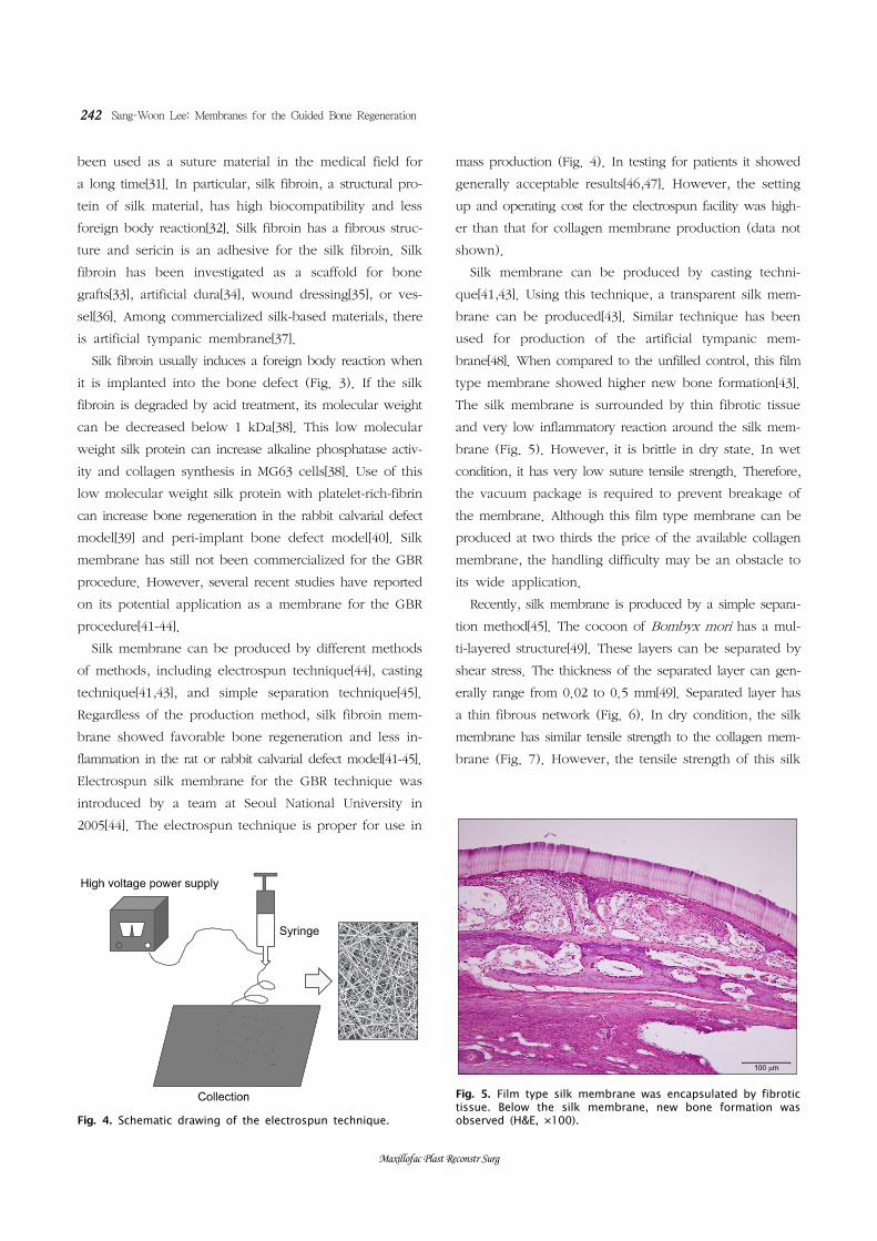
Fig. 4. Schematic drawing of the electrospun technique.
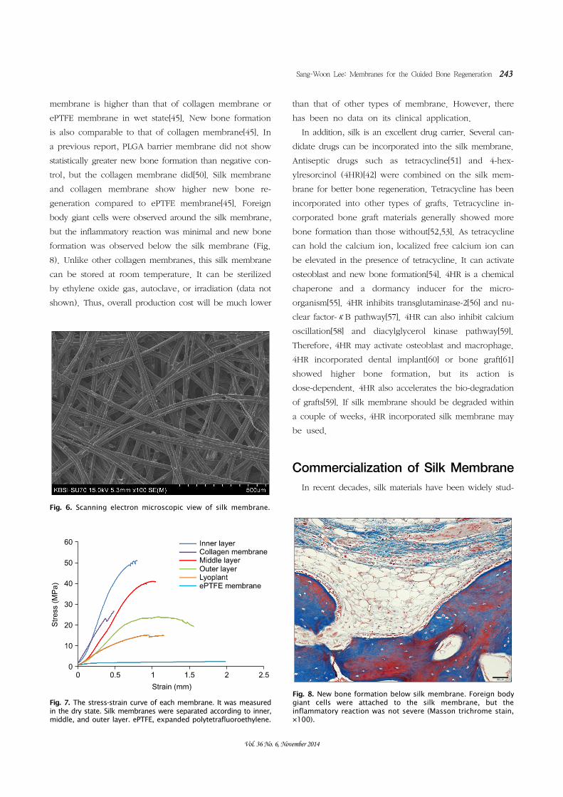
Fig. 5. Film type silk membrane was encapsulated by fibrotic tissue. Below the silk membrane, new bone formation was observed (H&E, ×100).
been used as a suture material in the medical field for
a long time[31]. In particular, silk fibroin, a structural pro-
tein of silk material, has high biocompatibility and less
foreign body reaction[32]. Silk fibroin has a fibrous struc-
ture and sericin is an adhesive for the silk fibroin. Silk
fibroin has been investigated as a scaffold for bone
grafts[33], artificial dura[34], wound dressing[35], or ves-
sel[36]. Among commercialized silk-based materials, there
is artificial tympanic membrane[37].
Silk fibroin usually induces a foreign body reaction when
it is implanted into the bone defect (Fig. 3). If the silk
fibroin is degraded by acid treatment, its molecular weight
can be decreased below 1 kDa[38]. This low molecular
weight silk protein can increase alkaline phosphatase activ-
ity and collagen synthesis in MG63 cells[38]. Use of this
low molecular weight silk protein with platelet-rich-fibrin
can increase bone regeneration in the rabbit calvarial defect
model[39] and peri-implant bone defect model[40]. Silk
membrane has still not been commercialized for the GBR
procedure. However, several recent studies have reported
on its potential application as a membrane for the GBR
procedure[41-44].
Silk membrane can be produced by different methods
of methods, including electrospun technique[44], casting
technique[41,43], and simple separation technique[45].
Regardless of the production method, silk fibroin mem-
brane showed favorable bone regeneration and less in-
flammation in the rat or rabbit calvarial defect model[41-45].
Electrospun silk membrane for the GBR technique was
introduced by a team at Seoul National University in
2005[44]. The electrospun technique is proper for use in
mass production (Fig. 4). In testing for patients it showed
generally acceptable results[46,47]. However, the setting
up and operating cost for the electrospun facility was high-
er than that for collagen membrane production (data not
shown).
Silk membrane can be produced by casting techni-
que[41,43]. Using this technique, a transparent silk mem-
brane can be produced[43]. Similar technique has been
used for production of the artificial tympanic mem-
brane[48]. When compared to the unfilled control, this film
type membrane showed higher new bone formation[43].
The silk membrane is surrounded by thin fibrotic tissue
and very low inflammatory reaction around the silk mem-
brane (Fig. 5). However, it is brittle in dry state. In wet
condition, it has very low suture tensile strength. Therefore,
the vacuum package is required to prevent breakage of
the membrane. Although this film type membrane can be
produced at two thirds the price of the available collagen
membrane, the handling difficulty may be an obstacle to
its wide application.
Recently, silk membrane is produced by a simple separa-
tion method[45]. The cocoon of Bombyx mori has a mul-
ti-layered structure[49]. These layers can be separated by
shear stress. The thickness of the separated layer can gen-
erally range from 0.02 to 0.5 mm[49]. Separated layer has
a thin fibrous network (Fig. 6). In dry condition, the silk
membrane has similar tensile strength to the collagen mem-
brane (Fig. 7). However, the tensile strength of this silk
Sang-Woon Lee: Membranes for the Guided Bone Regeneration 243
Vol. 36 No. 6, November 2014
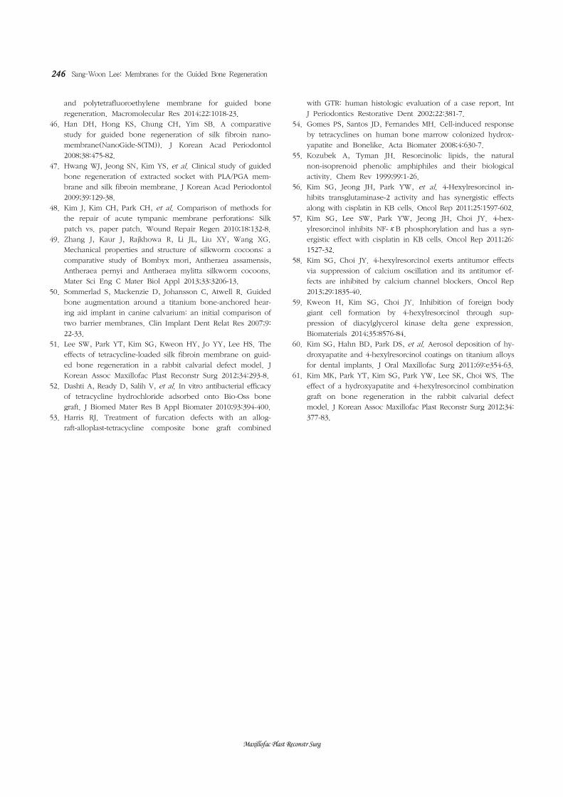
Fig. 8. New bone formation below silk membrane. Foreign bodygiant cells were attached to the silk membrane, but the inflammatory reaction was not severe (Masson trichrome stain, ×100).
Fig. 7. The stress-strain curve of each membrane. It was measuredin the dry state. Silk membranes were separated according to inner,middle, and outer layer. ePTFE, expanded polytetrafluoroethylene.
Fig. 6. Scanning electron microscopic view of silk membrane.
membrane is higher than that of collagen membrane or
ePTFE membrane in wet state[45]. New bone formation
is also comparable to that of collagen membrane[45]. In
a previous report, PLGA barrier membrane did not show
statistically greater new bone formation than negative con-
trol, but the collagen membrane did[50]. Silk membrane
and collagen membrane show higher new bone re-
generation compared to ePTFE membrane[45]. Foreign
body giant cells were observed around the silk membrane,
but the inflammatory reaction was minimal and new bone
formation was observed below the silk membrane (Fig.
8). Unlike other collagen membranes, this silk membrane
can be stored at room temperature. It can be sterilized
by ethylene oxide gas, autoclave, or irradiation (data not
shown). Thus, overall production cost will be much lower
than that of other types of membrane. However, there
has been no data on its clinical application.
In addition, silk is an excellent drug carrier. Several can-
didate drugs can be incorporated into the silk membrane.
Antiseptic drugs such as tetracycline[51] and 4-hex-
ylresorcinol (4HR)[42] were combined on the silk mem-
brane for better bone regeneration. Tetracycline has been
incorporated into other types of grafts. Tetracycline in-
corporated bone graft materials generally showed more
bone formation than those without[52,53]. As tetracycline
can hold the calcium ion, localized free calcium ion can
be elevated in the presence of tetracycline. It can activate
osteoblast and new bone formation[54]. 4HR is a chemical
chaperone and a dormancy inducer for the micro-
organism[55]. 4HR inhibits transglutaminase-2[56] and nu-
clear factor-κB pathway[57]. 4HR can also inhibit calcium
oscillation[58] and diacylglycerol kinase pathway[59].
Therefore, 4HR may activate osteoblast and macrophage.
4HR incorporated dental implant[60] or bone graft[61]
showed higher bone formation, but its action is
dose-dependent. 4HR also accelerates the bio-degradation
of grafts[59]. If silk membrane should be degraded within
a couple of weeks, 4HR incorporated silk membrane may
be used.
Commercialization of Silk Membrane
In recent decades, silk materials have been widely stud-

244 Sang-Woon Lee: Membranes for the Guided Bone Regeneration
Maxillofac Plast Reconstr Surg
ied for dental and medical application. However, only film
type silk membrane has been approved as a substitute
for the tympanic membrane by the Korean Food and Drug
Administration. In addition, the silk tympanic membrane
is not widely used the imbalance between the cost for
production and the price suggested by the health
insurance. In the case of the tympanic membrane, most
patients are healed naturally without artificial membrane.
Only severely injured patients may need the artificial tym-
panic membrane. Therefore, its clinical application may
be limited.
Unlike the silk tympanic membrane, silk membrane pro-
duced by simple separation method does not require the
degumming process[46]. Therefore, there was no risk of
residual bio-hazard salts that were added during the de-
gumming process. However, separation itself should be
done manually; it was very labor intensive work. The size
of the silk membrane produced by simple separation[45]
is dependent on the cocoon size. Therefore, a large sized
membrane cannot be produced by use of this technique.
Thus, this silk membrane cannot be used for covering max-
illary sinus wall defect or cystic cavity wall defect. Despite
these limitations, this new silk membrane can be widely
used for covering small sized intra-oral defect such as ex-
traction socket, periodontal defect, and peri-implant defect.
As the silk material is classified as a non-biodegradable
material[32], the clinical method for the silk membrane is
generally in accordance with that of small sized ePTFE
membrane. Compared to vessel tie silk material, the silk
membrane for GBR, located mainly in the submucosal lay-
er, can be easily removed. Whether it can be used for
an open-membrane technique like collagen membrane is
not clear. It should be tested in the clinical application.
Conclusion
There have been numerous patients who potentially
need the GBR membrane. However, the cost for using
the membrane is a main obstacle for its wide applications.
When the silk membrane produced by simple separation
method is commercialized, its price will be much lower
than that of any other currently available types of
membrane. Development of better material is a vital com-
ponent of public health care.
Acknowledgements
This work was supported by a grant from the
Next-Generation BioGreen21 Program (No. PJ009013),
Rural Development Administration, Republic of Korea.
References1. Nguyen TT, Mui B, Mehrabzadeh M, et al. Regeneration of
tissues of the oral complex: current clinical trends and re-
search advances. J Can Dent Assoc 2013;79:d1.
2. Rakhmatia YD, Ayukawa Y, Furuhashi A, Koyano K.
Current barrier membranes: titanium mesh and other mem-
branes for guided bone regeneration in dental applications.
J Prosthodont Res 2013;57:3-14.
3. Khojasteh A, Morad G, Behnia H. Clinical importance of re-
cipient site characteristics for vertical ridge augmentation: a
systematic review of literature and proposal of a classification.
J Oral Implantol 2013;39:386-98.
4. Corinaldesi G, Lizio G, Badiali G, Morselli-Labate AM,
Marchetti C. Treatment of intrabony defects after impacted
mandibular third molar removal with bioabsorbable and
non-resorbable membranes. J Periodontol 2011;82:1404-13.
5. Cortellini P, Tonetti MS. Clinical performance of a re-
generative strategy for intrabony defects: scientific evidence
and clinical experience. J Periodontol 2005;76:341-50.
6. Schwarz F, Hegewald A, Sahm N, Becker J. Long-term fol-
low-up of simultaneous guided bone regeneration using na-
tive and cross-linked collagen membranes over 6 years. Clin
Oral Implants Res 2014;25:1010-5.
7. Chattopadhyay S, Raines RT. Review collagen-based bio-
materials for wound healing. Biopolymers 2014;101:821-33.
8. Parrish LC, Miyamoto T, Fong N, Mattson JS, Cerutis DR.
Non-bioabsorbable vs. bioabsorbable membrane: assessment
of their clinical efficacy in guided tissue regeneration
technique. A systematic review. J Oral Sci 2009;51:383-400.
9. Veríssimo DM, Leitão RF, Ribeiro RA, et al. Polyanionic col-
lagen membranes for guided tissue regeneration: effect of
progressive glutaraldehyde cross-linking on biocompatibility
and degradation. Acta Biomater 2010;6:4011-8.
10. Rothamel D, Schwarz F, Sager M, Herten M, Sculean A,
Becker J. Biodegradation of differently cross-linked collagen
membranes: an experimental study in the rat. Clin Oral
Implants Res 2005;16:369-78.
11. Speer DP, Chvapil M, Eskelson CD, Ulreich J. Biological ef-
fects of residual glutaraldehyde in glutaraldehyde-tanned col-
lagen biomaterials. J Biomed Mater Res 1980;14:753-64.
12. Caffesse RG, Nasjleti CE, Morrison EC, Sanchez R. Guided
tissue regeneration: comparison of bioabsorbable and
non-bioabsorbable membranes. Histologic and histometric
study in dogs. J Periodontol 1994;65:583-91.
13. Proussaefs P, Lozada J. The use of resorbable collagen
membrane in conjunction with autogenous bone graft and
inorganic bovine mineral for buccal/labial alveolar ridge
augmentation: a pilot study. J Prosthet Dent 2003;90:530-8.
14. Urban IA, Jovanovic SA, Lozada JL. Vertical ridge augmenta-
tion using guided bone regeneration (GBR) in three clinical

Sang-Woon Lee: Membranes for the Guided Bone Regeneration 245
Vol. 36 No. 6, November 2014
scenarios prior to implant placement: a retrospective study
of 35 patients 12 to 72 months after loading. Int J Oral
Maxillofac Implants 2009;24:502-10.
15. Bornstein MM, Bosshardt D, Buser D. Effect of two different
bioabsorbable collagen membranes on guided bone re-
generation: a comparative histomorphometric study in the
dog mandible. J Periodontol 2007;78:1943-53.
16. Zhao L, Li N, Wang K, Shi C, Zhang L, Luan Y. A review
of polypeptide-based polymersomes. Biomaterials 2014;35:
1284-301.
17. Rokkanen PU. Absorbable materials in orthopaedic surgery.
Ann Med 1991;23:109-15.
18. Galgut P, Pitrola R, Waite I, Doyle C, Smith R. Histological
evaluation of biodegradable and non-degradable membranes
placed transcutaneously in rats. J Clin Periodontol 1991;18:
581-6.
19. Daniels AU, Andriano KP, Smutz WP, Chang MK, Heller J.
Evaluation of absorbable poly(ortho esters) for use in surgi-
cal implants. J Appl Biomater 1994;5:51-64.
20. Athanasiou KA, Agrawal CM, Barber FA, Burkhart SS.
Orthopaedic applications for PLA-PGA biodegradable polymers.
Arthroscopy 1998;14:726-37.
21. Vuddhakanok S, Solt CW, Mitchell JC, Foreman DW, Alger
FA. Histologic evaluation of periodontal attachment appara-
tus following the insertion of a biodegradable copolymer
barrier in humans. J Periodontol 1993;64:202-10.
22. Urakami K, Higashi A, Umemoto K, Godo M, Watanabe C,
Hashimoto K. Compositional analysis of copoly (DL-lactic/gly-
colic acid) (PLGA) by pyrolysis-gas chromatography/mass
spectrometry combined with one-step thermally assisted hy-
drolysis and methylation in the presence of tetramethylammo-
nium hydroxide. Chem Pharm Bull (Tokyo) 2001;49:203-5.
23. De Stefano D, De Rosa G, Maiuri MC, et al. Oligonucleotide
decoy to NF-kappaB slowly released from PLGA micro-
spheres reduces chronic inflammation in rat. Pharmacol Res
2009;60:33-40.
24. Tseng YY, Liao JY, Chen WA, Kao YC, Liu SJ. Sustainable
release of carmustine from biodegradable poly[((D,L))-lac-
tide-co-glycolide] nanofibrous membranes in the cerebral
cavity: in vitro and in vivo studies. Expert Opin Drug Deliv
2013;10:879-88.
25. Orenstein SB, Saberski ER, Kreutzer DL, Novitsky YW.
Comparative analysis of histopathologic effects of synthetic
meshes based on material, weight, and pore size in mice. J
Surg Res 2012;176:423-9.
26. Jones AA, Buser D, Schenk R, Wozney J, Cochran DL. The
effect of rhBMP-2 around endosseous implants with and with-
out membranes in the canine model. J Periodontol 2006;77:
1184-93.
27. Lindfors LT, Tervonen EA, Sándor GK, Ylikontiola LP.
Guided bone regeneration using a titanium-reinforced ePTFE
membrane and particulate autogenous bone: the effect of
smoking and membrane exposure. Oral Surg Oral Med Oral
Pathol Oral Radiol Endod 2010;109:825-30.
28. Becker W, Dahlin C, Lekholm U, et al. Five-year evaluation
of implants placed at extraction and with dehiscences and fen-
estration defects augmented with ePTFE membranes: results
from a prospective multicenter study. Clin Implant Dent Relat
Res 1999;1:27-32.
29. Bachleda P, Utikal P, Kalinova L, et al. Infectious complica-
tions of arteriovenous ePTFE grafts for hemodialysis. Biomed
Pap Med Fac Univ Palacky Olomouc Czech Repub 2010;154:
13-9.
30. Selvig KA, Kersten BG, Chamberlain AD, Wikesjö UM,
Nilvéus RE. Regenerative surgery of intrabony periodontal
defects using ePTFE barrier membranes: scanning electron
microscopic evaluation of retrieved membranes versus clin-
ical healing. J Periodontol 1992;63:974-8.
31. Cao Y, Wang B. Biodegradation of silk biomaterials. Int J
Mol Sci 2009;10:1514-24.
32. Kundu B, Rajkhowa R, Kundu SC, Wang X. Silk fibroin bio-
materials for tissue regenerations. Adv Drug Deliv Rev 2013;
65:457-70.
33. Seok H, Park YT, Kim SG, Jin HJ. The effect of silk fibroin
particles coated with hydroxyapatites on bone regeneration
in the rat calvarial defect model. J Korean Assoc Maxillofac
Plast Reconstr Surg 2013;35:13-7.
34. Kim DW, Eum WS, Jang SH, et al. A transparent artificial dura
mater made of silk fibroin as an inhibitor of inflammation in
craniotomized rats. J Neurosurg 2011;114:485-90.
35. Kanokpanont S, Damrongsakkul S, Ratanavaraporn J, Aramwit
P. Physico-chemical properties and efficacy of silk fibroin fab-
ric coated with different waxes as wound dressing. Int J Biol
Macromol 2013;55:88-97.
36. Liu S, Dong C, Lu G, et al. Bilayered vascular grafts based
on silk proteins. Acta Biomater 2013;9:8991-9003.
37. Shen Y, Redmond SL, Teh BM, et al. Scaffolds for tympanic
membrane regeneration in rats. Tissue Eng Part A 2013;19:
657-68.
38. Kim JY, Choi JY, Jeong JH, et al. Low molecular weight silk
fibroin increases alkaline phosphatase and type I collagen
expression in MG63 cells. BMB Rep 2010;43:52-6.
39. Lee EH, Kim JY, Kweon HY, et al. A combination graft of
low-molecular-weight silk fibroin with Choukroun plate-
let-rich fibrin for rabbit calvarial defect. Oral Surg Oral Med
Oral Pathol Oral Radiol Endod 2010;109:e33-8.
40. Jang ES, Park JW, Kweon H, et al. Restoration of peri-im-
plant defects in immediate implant installations by
Choukroun platelet-rich fibrin and silk fibroin powder com-
bination graft. Oral Surg Oral Med Oral Pathol Oral Radiol
Endod 2010;109:831-6.
41. Lee SW, Kim SG, Song JY, et al. Silk fibroin and 4-hex-
ylresorcinol incorporation membrane for guided bone
regeneration. J Craniofac Surg 2013;24:1927-30.
42. Song JM, Shin SH, Kim YD, et al. Comparative study of chi-
tosan/fibroin-hydroxyapatite and collagen membranes for
guided bone regeneration in rat calvarial defects: micro-com-
puted tomography analysis. Int J Oral Sci 2014;6:87-93.
43. Song JY, Kim SG, Lee JW, et al. Accelerated healing with
the use of a silk fibroin membrane for the guided bone re-
generation technique. Oral Surg Oral Med Oral Pathol Oral
Radiol Endod 2011;112:e26-33.
44. Kim KH, Jeong L, Park HN, et al. Biological efficacy of silk
fibroin nanofiber membranes for guided bone regeneration.
J Biotechnol 2005;120:327-39.
45. Ha YY, Park YW, Kweon HY, Jo YY, Kim SG. Comparison
of the physical properties and In vivo bioactivities of silk-
worm-cocoon-derived silk membrane, collagen membrane,
246 Sang-Woon Lee: Membranes for the Guided Bone Regeneration
Maxillofac Plast Reconstr Surg
and polytetrafluoroethylene membrane for guided bone
regeneration. Macromolecular Res 2014;22:1018-23.
46. Han DH, Hong KS, Chung CH, Yim SB. A comparative
study for guided bone regeneration of silk fibroin nano-
membrane(NanoGide-S(TM)). J Korean Acad Periodontol
2008;38:475-82.
47. Hwang WJ, Jeong SN, Kim YS, et al. Clinical study of guided
bone regeneration of extracted socket with PLA/PGA mem-
brane and silk fibroin membrane. J Korean Acad Periodontol
2009;39:129-38.
48. Kim J, Kim CH, Park CH, et al. Comparison of methods for
the repair of acute tympanic membrane perforations: Silk
patch vs. paper patch. Wound Repair Regen 2010;18:132-8.
49. Zhang J, Kaur J, Rajkhowa R, Li JL, Liu XY, Wang XG.
Mechanical properties and structure of silkworm cocoons: a
comparative study of Bombyx mori, Antheraea assamensis,
Antheraea pernyi and Antheraea mylitta silkworm cocoons.
Mater Sci Eng C Mater Biol Appl 2013;33:3206-13.
50. Sommerlad S, Mackenzie D, Johansson C, Atwell R. Guided
bone augmentation around a titanium bone-anchored hear-
ing aid implant in canine calvarium: an initial comparison of
two barrier membranes. Clin Implant Dent Relat Res 2007;9:
22-33.
51. Lee SW, Park YT, Kim SG, Kweon HY, Jo YY, Lee HS. The
effects of tetracycline-loaded silk fibroin membrane on guid-
ed bone regeneration in a rabbit calvarial defect model. J
Korean Assoc Maxillofac Plast Reconstr Surg 2012;34:293-8.
52. Dashti A, Ready D, Salih V, et al. In vitro antibacterial efficacy
of tetracycline hydrochloride adsorbed onto Bio-Oss bone
graft. J Biomed Mater Res B Appl Biomater 2010;93:394-400.
53. Harris RJ. Treatment of furcation defects with an allog-
raft-alloplast-tetracycline composite bone graft combined
with GTR: human histologic evaluation of a case report. Int
J Periodontics Restorative Dent 2002;22:381-7.
54. Gomes PS, Santos JD, Fernandes MH. Cell-induced response
by tetracyclines on human bone marrow colonized hydrox-
yapatite and Bonelike. Acta Biomater 2008;4:630-7.
55. Kozubek A, Tyman JH. Resorcinolic lipids, the natural
non-isoprenoid phenolic amphiphiles and their biological
activity. Chem Rev 1999;99:1-26.
56. Kim SG, Jeong JH, Park YW, et al. 4-Hexylresorcinol in-
hibits transglutaminase-2 activity and has synergistic effects
along with cisplatin in KB cells. Oncol Rep 2011;25:1597-602.
57. Kim SG, Lee SW, Park YW, Jeong JH, Choi JY. 4-hex-
ylresorcinol inhibits NF-κB phosphorylation and has a syn-
ergistic effect with cisplatin in KB cells. Oncol Rep 2011;26:
1527-32.
58. Kim SG, Choi JY. 4-hexylresorcinol exerts antitumor effects
via suppression of calcium oscillation and its antitumor ef-
fects are inhibited by calcium channel blockers. Oncol Rep
2013;29:1835-40.
59. Kweon H, Kim SG, Choi JY. Inhibition of foreign body
giant cell formation by 4-hexylresorcinol through sup-
pression of diacylglycerol kinase delta gene expression.
Biomaterials 2014;35:8576-84.
60. Kim SG, Hahn BD, Park DS, et al. Aerosol deposition of hy-
droxyapatite and 4-hexylresorcinol coatings on titanium alloys
for dental implants. J Oral Maxillofac Surg 2011;69:e354-63.
61. Kim MK, Park YT, Kim SG, Park YW, Lee SK, Choi WS. The
effect of a hydroxyapatite and 4-hexylresorcinol combination
graft on bone regeneration in the rabbit calvarial defect
model. J Korean Assoc Maxillofac Plast Reconstr Surg 2012;34:
377-83.