meniere's disease in finland kotimÄki - university of...
TRANSCRIPT

MENIERE'S DISEASE IN FINLANDAn epidemiological and clinical study on occurrence, clinical picture and policy
JOUKOKOTIMÄKI
Department of Otorhinolaryngology,University of Oulu
OULU 2003


JOUKO KOTIMÄKI
MENIERE'S DISEASE IN FINLANDAn epidemiological and clinical study on occurrence, clinical picture and policy
Academic Dissertation to be presented with the assent ofthe Faculty of Medicine, University of Oulu, for publicdiscussion in the Auditorium 7 of the University Hospitalof Oulu, on October 17th, 2003, at 12 noon.
OULUN YLIOPISTO, OULU 2003

Copyright © 2003University of Oulu, 2003
Supervised byProfessor Martti SorriDocent Eero AantaaProfessor Kalevi Jokinen
Reviewed byProfessor Iain W. S. MairDocent Hannu Valtonen
ISBN 951-42-7113-0 (URL: http://herkules.oulu.fi/isbn9514271130/)
ALSO AVAILABLE IN PRINTED FORMATActa Univ. Oul. D 747, 2003ISBN 951-42-7112-2ISSN 0355-3221 (URL: http://herkules.oulu.fi/issn03553221/)
OULU UNIVERSITY PRESSOULU 2003

Kotimäki, Jouko, Meniere's disease in Finland. An epidemiological and clinical studyon occurrence, clinical picture and policyDepartment of Otorhinolaryngology, University of Oulu, P.O.Box 5000, FIN-90014 University ofOulu, Finland Oulu, Finland2003
Abstract
The symptom complex originating from the inner ear, known as Meniere's disease, was studiedespecially from the epidemiologic point of view. A total of 442 patients' charts were retrospectivelyanalysed in several hospital districts of Finland. The period of 1992-1996 was covered. The mainfocus was on the epidemiological assessment of the disease in Finland. To clarify the epidemiologicalfigures, the validity of the diagnostic assessment was examined using the latest guidelines (1995) ofthe Committee on Hearing and Equilibrium of the American Academy of Otolaryngology - Head andNeck Surgery (AAO-HNS) as a gold standard.
The diagnostic tools used in the different hospitals were documented and evaluated, anddiagnostic accuracy at the different levels of the health care system was evaluated. The clinical pictureof Meniere's disease was characterised, and the therapeutic modalities used were evaluated. Theaudiometric configurations were classified according to two principles. The prognosis of hearingimpairment was specified by creating a multivariable model.
Half of the patients (N = 221) fulfilled the AAO-HNS criteria for definite disease. The prevalenceand incidence of definite cases of Meniere's disease appeared to be lower in Finland than could beexpected based on previous international studies. A prevalence of at least 43 per 100,000 and anaverage annual incidence of 4.3 per 100,000 were obtained. The prevalence rates in the catchmentareas of the university and central hospitals did not differ statistically, but a significant (p < 0.001)difference was found between the average prevalences in the northern and southern Finnish hospitaldistricts.
Fluctuation of hearing in repeated audiometric measurements appeared to be a highly sensitive(94%) diagnostic test to detect definite Meniere's disease. According to the multivariable modelcreated in this study, the hearing impairment in Meniere's disease affects equally males and females,and the deterioration is about 1 dB per year due to the duration of the disease and 0.5 dB per year dueto aging. The disease was controlled conservatively in 69% of the cases. A gently sloping high-frequency audiometric pattern was most prevalent according to the EU Work Group classification anda flat pattern according to the mid-frequency-based classification.
The variability of diagnostic criteria, diagnostic tools and therapeutic modalities shows an evidentneed for up-to-date therapy recommendations for Meniere's disease in Finland.
Keywords: audiometry, diagnostic battery, diagnostic criteria, endolymphatic hydrops,hearing impairment, hearing loss, prevalence


Acknowledgements
This research was conducted in the Department of Otorhinolaryngology, Oulu University Hospital, during the years 1997-2003.
I wish to express my sincere gratitude to Professor Juhani Nuutinen, M.D., and Professor Kalevi Jokinen, M.D., who have been the Heads of the Department during this period, for supervision and encouragement in my scientific career. I am also grateful to Professor Emeritus Antti Palva, M.D., who contributed to my decision to specialize in ear, nose and throat diseases and, as chairman of Antti Palva Foundation, also financially supported my research.
I owe my deepest gratitude to my main supervisor, Professor Martti Sorri, M.D., and Docent Eero Aantaa, M.D., who was also my supervisor. These two enthusiastic scientists and clinicians were the initiators of this research and deserve my deep admiration. Their broad knowledge about Meniere’s disease and their connections with the Finnish Meniere Federation made this work possible.
I express my appreciation to the official referees of this thesis, Professor Iain W.S. Mair, M.D., and Docent Hannu Valtonen, M.D., for their careful review of this manuscript. Professor Mair is especially acknowledged by his altruistic correction of the language during the review process.
My special thanks are due to my statistical and epidemiological co-author, Arto Muhli, chief system analyst in the Computer Services Centre, University of Oulu. I would also like to thank Professor Esa Läärä, Lic.Sci, from the Department of Mathematical Sciences, University of Oulu, and Pasi Ohtonen, M.Sc., for their valuable help with the statistical and epidemiological analyses in the first work.
I wish to express my gratitude to my chief during the final stages of my study, Arja Vuorialho, M.D., who was actively encouraged me to complete my dissertation. I also thank my colleagues in the Department of Otorhinolaryngology in Oulu University Hospital, Kuhmo Health Care Centre and Kainuu Central Hospital.
I am grateful to Mr. Malcolm Hicks, M.A., for revising the English language of original paper I, to Mr. Keith Kosola, licensed translator, for revising the English language of the original papers II-III and to Mrs. Sirkka-Liisa Leinonen, Lic.Phil., for revising the English language of the original papers IV and V and the final manuscript. Special thanks go to Mrs. Raili Puhakka for her friendly assistance.

I owe my sincere gratitude to the Finnish Meniere Federation and the co-ordinators of this nationwide association for their co-operation and encouragement during this work. Through them I have learned much about the daily life of patients with Meniere’s disease.
I am deeply thankful to my parents Hilja and Teuvo, who encouraged me to choose an academic career and gave various kinds of help to our family, especially by arranging periods of rest for me and my wife.
Finally, I extend my loving thanks to the dearest people in my life, my wife Sanna, for her tireless love and support, which have been necessary for continuing this research. Her flexibility in bearing most of the responsibility for parenting our children during the most intensive periods of my research is incredible. Our dear children, Lotta-Maria, Eero, Lauri, Anna-Inkeri, Riikka and Louna-Kaisa, who have had to adapt to different environments during our stays in Kuhmo, Oulu, Kuhmo again and, finally, Kajaani, deserve my loving thanks.
This research was supported financially by RAY Finland via the Federation of the Hard of Hearing, Antti Palva Foundation, Finnish Audiological Association and Korvatautien Tutkimussäätiö Foundation, for which I am grateful.
Kajaani 31.07.2003 Jouko Kotimäki

Abbreviations
AAO-HNS American Academy of Otolaryngology - Head and Neck Surgery ABR auditory brain stem responses AP action potential CI confidence interval CT computerized tomography dB decibel DPOAE distortion-product otoacoustic emissions ECoG electrocochleography ELH endolymphatic hydrops ELS endolymphatic sac ENG electronystagmography ENT ear-nose-throat FLS functional level scale GABA gamma-amino butyric acid HL hearing level ICD International Statistical Classification of Diseases and Related Health Problems JSER Japanese Society for Equilibrium Research kHz kilohertz ML maximum likelihood MRI magnetic resonance imaging PTA pure-tone average SD standard deviation SP summation potential


List of original publications
This thesis is based on the following articles, which are referred to in the text by their Roman numerals. I Kotimäki J, Sorri M, Aantaa E, Nuutinen J. (1999) Prevalence of Meniere’s disease in
Finland. Laryngoscope 109: 748–53. II Kotimäki J, Sorri M, Aantaa E, Nuutinen J. (2000) Regional differences in the
prevalence of Meniere’s disease in Finland. In: Sterkers O, Ferrary E, Dauman R, Sauvage JP, Tran Ba Huy (eds) Meniere’s disease ─Update 1999, p.393−397. The Hague, The Netherlands, Kugler Publications.
III Kotimäki J, Sorri M, Muhli A. (2001) Prognosis of Hearing impairment in Meniere’s disease. Acta Otolaryngol (Stockh) Suppl 545: 14–18.
IV Kotimäki J, Sorri M, Muhli A. (2003) Diagnostic policy to confirm a suspicion of Meniere’s disease in Finland. A retrospective analysis. Audiological Medicine 2:115–122.
V Kotimäki J, Sorri M, Muhli A. (2003) Clinical picture and audiometric configurations in Meniere’s disease. Audiological Medicine 2: 123–131.
The original papers in this thesis have been reproduced with the permission of the original publishers.

Contents
Abstract Acknowledgements Abbreviations List of original publications Contents 1 Introduction ...................................................................................................................12 2 Review of the literature .................................................................................................13
2.1 Definition................................................................................................................13 2.2 Epidemiology .........................................................................................................15
2.2.1 Occurrence.......................................................................................................15 2.2.2 Age at the onset of the disease.........................................................................16 2.2.3 Sex distribution................................................................................................17 2.2.4 Occupational distribution ................................................................................18 2.2.5 Sidedness .........................................................................................................18 2.2.6 Race .................................................................................................................18 2.2.7 Individual and environmental factors ..............................................................19 2.2.8 Concomitant diseases ......................................................................................19
2.3 Pathological findings and pathophysiology............................................................21 2.4 Aetiopathogenesis...................................................................................................22 2.5 Clinical manifestations ...........................................................................................24
2.5.1 Vestibular symptoms........................................................................................25 2.5.2 Cochlear symptoms .........................................................................................26
2.6 Natural course.........................................................................................................27 2.7 Diagnosis ................................................................................................................28
2.7.1 History .............................................................................................................28 2.7.2 Examination.....................................................................................................29
2.7.2.1 Audiological tests ................................................................................29 2.7.2.2 Vestibular tests.....................................................................................31 2.7.2.3 Radiographic examinations .................................................................32 2.7.2.4 Blood tests...........................................................................................32 2.7.2.5 Differential diagnosis ..........................................................................33

2.8 Therapy...................................................................................................................34 2.8.1 General aspects of therapy...............................................................................34 2.8.2 Medication.......................................................................................................35 2.8.3 Surgical therapy...............................................................................................41
2.8.3.1 Conservative surgery...........................................................................41 2.8.3.2 Ablative surgery ..................................................................................42
2.8.4 Pressure treatment ...........................................................................................43 2.8.5 Other treatment modalities ..............................................................................44 2.8.6 Rehabilitation ..................................................................................................45
3 Aims of the present research..........................................................................................47 4 Subjects and methods ....................................................................................................48
4.1 Enrolment of patients..............................................................................................48 4.2 Database and statistical methods ............................................................................49 4.3 Parameters registered in the database .....................................................................49 4.4 Epidemiologic concepts..........................................................................................50 4.5 Ethical considerations.............................................................................................50
5 Results and comments ...................................................................................................51 5.1 Prevalence and incidence of Meniere’s disease in Finland (Study I)......................51 5.2 Regional differences in the prevalence of Meniere’s disease in Finland (Study II) ................................................................................................................53 5.3 Prognosis of hearing impairment in Meniere’s disease (Study III) ........................55 5.4 Diagnostic policy used in Finland to confirm a suspicion of Meniere’s disease (Study IV) ..................................................................................................58 5.5 Clinical picture and treatment of Meniere’s disease (Study V) ..............................61 5.6 Audiometric configurations of Meniere’s disease (Study V) .................................64 5.7 Regional differences in the diagnostics and treatment of Meniere’s disease..........65 5.8 Patient’s perspective ...............................................................................................67
6 General discussion.........................................................................................................70 6.1 Epidemiologic and diagnostic problems.................................................................70 6.2 Problems with the therapy of Meniere’s disease ....................................................71 6.3 Suggestions for the diagnosis and therapy of Meniere’s disease............................72
7 Summary and conclusions .............................................................................................77 References Appendices

1 Introduction
Meniere’s disease is a complex of symptoms originating from the inner ear, which was first described by Prosper Ménière over 140 years ago. The exact aetiology has remained obscure up till today, although information has been accumulating concerning this topic. The aetiology is assumed to be multifactorial, but endolymphatic hydrops has been recognized as a pathophysiologic condition behind the symptoms of the disease. This knowledge has resulted in a multitude of therapeutic modalities aimed at relieving the symptoms of the disease. Unfortunately, none of these has proven to alter the natural progression of the disease. Of the main symptoms of the disease, which consist of vertigo, hearing impairment and tinnitus or aural fullness, the first is best controlled by modern therapeutic modalities.
The diagnostics of this disease has not been uniform regardless of the several guidelines published by both American and Japanese groups of experts. The diagnostic inaccuracy has naturally led to marked variation of the evaluations concerning the epidemiology of the disease. The reports concerning the effects of various therapeutic modalities also suffer from the ambiguous diagnostic criteria. Diagnostic tools have also changed during the last few decades, reflecting especially the rapid development of technical equipment. The latest criteria for diagnosing and reporting the results of therapy were published in 1995 by the Committee on Hearing and Equilibrium of the American Academy of Otolaryngology - Head and Neck surgery, and these criteria were used as a gold standard to identify the patients with Meniere’s disease in this study.
The need for more information about Meniere’s disease, especially its epidemiology, in Finland and the possibilities of the Finnish health care system to serve these patients was considered as very important by the voluntary patient association, Finnish Meniere Federation, during the last decade. The federation decided to start a project to explore these topics and to provide information of the disease both to patients and to health care workers. Financial support for this nationwide Meniere project was provided by RAY Finland via the Federation of Hard of Hearing in Finland. The author of this dissertation was invited to serve as the main investigator for this project, which began in 1997.

2 Review of the literature
2.1 Definition
Meniere’s disease is a clinical disorder that was described for the first time by Prosper Ménière (1861). It is defined as an idiopathic syndrome of endolymphatic hydrops by the Committee on Hearing and Equilibrium of the American Academy of Otolaryngology - Head and Neck Surgery (AAO-HNS 1995). It is a nosologic entity characterised by the following main symptoms: tinnitus, fluctuating hearing loss and repeated attacks of vertigo (Pfaltz & Matefi 1981). The term ‘Meniere’s disease’ refers to idiopathic disease, while the term ‘Meniere’s syndrome’ should be used when there is an underlying primary disease causing similar symptoms (Pfaltz & Matefi 1981). The diagnostic criteria have been considered by the AAO-HNS three times (1972, 1985, and 1995). The latest criteria are shown in Table 1.

14
Table 1. Diagnosis of Meniere’s disease according to AAO-HNS (1995)
Diagnosis of Meniere’s disease Definitive Meniere's disease plus histopathologic confirmation Two or more definitive spontaneous episodes of vertigo for 20 minutes or longer Audiometrically documented hearing loss (frequencies 0.5,1,2 and 3 kHz) on at least one occasion Tinnitus or aural fullness in the treated ear
Certain Definite
Other causes excluded One definitive episode of vertigo Probable Other criteria as for definitive Meniere's disease Episodic vertigo of the Meniere type without documented hearing loss, or Possible Sensorineural hearing loss, fluctuating or fixed, with disequilibrium but without definitive episodes
Other causes excluded In Japan, too, evaluation criteria for reporting the results of the treatment of Meniere’s disease were first published in the 1970’s (Watanabe 1976), and they have since been revised twice (Komazusaki et al. 1988, Mizukoshi et al. 1995) by the Japanese Society for Equilibrium Research (JSER). The latest JSER criteria are shown in Table 2.
Table 2. Diagnosis of Meniere’s disease according to JSER (Mizukoshi et al.1995)
Diagnosis of Meniere’s disease Vertiginous spell lasting for 20 min to 24 hours Nystagmus always observed during the spell Adjunctive spells between definite spells may occur Tinnitus, which is evaluated objectively and subjectively (standard tinnitus evaluation tests) Hearing impairment in four-frequency PTA at 0.25, 0.5, 1 and 2 kHz Test for detecting endolymphatic hydrops (glycerol test, ECOG, furosemide test)
Definite disease
Exclusion of central nervous system and other cochleovestibular disorders Suspected disease Criteria for definite disease are not fulfilled
AAO-HNS (1995) introduced a staging system to be applied only to cases of certain and definite Meniere’s disease. Staging is based solely on hearing, which is the most readily measurable variable and the variable most closely related to the natural history of Meniere’s disease. The staging system is shown in Table 3.

15
Table 3. Staging of definite and certain Meniere’s disease according to AAO-HNS (1995)
Stage Four-tone average (dB) (0.5, 1,2 and 3 kHz) 1 <25 2 26-40 3 41-70 4 > 70
2.2 Epidemiology
2.2.1 Occurrence
The occurrence of Meniere’s disease has been evaluated since the Second World War. The inconsistency in establishing the diagnosis and the different criteria used for the diagnosis have confused the knowledge about epidemiological figures (Stahle et al. 1978, Arenberg et al. 1980).
Naito (1962) reported that the frequency of Meniere patients had increased from 100 to 3400 per 100,000 ENT clinic outpatients in Japan since the end of the Second World War, concluding that the increase was due to the “westernization” of the Japanese way of life. Two British estimates gave the occurrence as 157 cases per 100,000 (Cawthorne and Hewlett 1954) and 100 per 100,000 (Harrison & Naftalin 1968). A more recent Japanese evaluation is available from Toyoama Prefecture, where the prevalence remained almost unchanged at 16−17 per 100,000 during the period 1974−1990 (Watanabe et al. 1995). The marked variation of these figures may be explained by the different criteria for the disease; in the study of Cawthorne and Hewlett (1954), for example, as many as 61 % of the patients referred because of vertigo were diagnosed to have Meniere’s disease, while in the recent study of Ballester et al. (2002), only 5.1 % out of the 8423 neurotological patients met the AAO-HNS (1995) criteria for definite Meniere’s disease.
In Sweden, Stahle et al. (1978) estimated the annual incidence for 1973 to be 46 per 100,000, and by extrapolating these figures, Arenberg et al. (1980) concluded that there was a total of at least 2,425,000 patients in the United States in 1973. The incidence in Northern Ireland was found to be 10-20 per 100,000 by Wilmot (1979). In Italy, the average annual incidence was 8.2 per 100,000 in south-eastern Latium in 1973−1985 (Celestino & Ralli 1991) and 27.5 per 100,000 in the Siennese area over the period 1981−1990 (Biagini et al. 1991). Wladislavosky-Waserman et al. (1984) reported the prevalence rate at the beginning of year 1980 in Rochester, United States, to be 218 per 100,000, while in the Italian study by Celestino and Ralli (1991), the prevalence was 205 per 100,000.
Differences in the geographical distribution of the disease have also been reported to occur. The disease was shown to be more common in the southern and central regions compared to the northern region of Japan according to Watanabe et al. (1981). The differences in health care facilities may, however, affect geographical figures.

16
The figures of occurrence are shown summarized in Table 4. It seems likely that there are true differences in the prevalence and incidence of Meniere’s disease between different countries. However, the variable diagnostic criteria and the different epidemiological concepts and methods make the results difficult to compare.
Table 4. Studies of the incidence and prevalence of Meniere’s disease Investigator Published
(year) Country Occurrence Base Real quality of
data Cawthorne and Hewlett
1954 Great Britain 157/100,000 population combination of incidence and prevalence?
Harrison and Naftalin
1968 Great Britain 100/100,000 ENT patients clinical estimate
Stahle et al. 1978 Sweden 46/100,000 population combination of incidence and prevalence
Wilmot 1979 Northern Ireland 10-20 /100,000 ENT patients clinical estimate Okafor 1984 Nigeria 400/100,000 ENT patients relative frequency
15/100,000 population incidence Wladislavosky-Waserman et al.
1984 Rochester MN, United States
218/100,000 population prevalence
Celestino and Ralli 1991 Italy 8.2/100,000 population incidence 205/100,000 population prevalence Biagini et al. 1991 Italy 27/100,000 population incidence Watanabe et al. 1995 Japan 17/100,000 population relative frequency Clinical estimate: an occurrence figure for an unspecified population and period Relative frequency: a ratio of the number of clinical cases to the total number of admitted patients.
2.2.2 Age at the onset of the disease
Meniere’s disease is relatively rare in persons aged under 18 years. Meyerhoff et al. (1978) suggested that 3 % of patients with Meniere’s disease were children, while Filipo and Barbara estimated the proportion of children to be 7 % (1985). In the series of Oosterweld (1980), 3.5 % of the patients were younger than 20 years at the time of the first attack. By combining the experience of four European centers, Häusler et al. (1987) estimated that children represented 1 % of all patients with Meniere’s disease at those centers. In a recent study from Japan, Akagi et al. (2001) reported the incidence of Meniere’s disease in pediatric patients with vertigo to be 2.9 %. Pediatric cases hence appear to represent only a small percentage of all patients with Meniere’s disease.
Several studies have been published showing that symptoms of the disease usually appear between the ages of 20 and 60 years, and the average age at onset is in the fourth decade of life (Thomas & Harrison 1971, Okafor 1984, Wladislavosky-Waserman 1984,

17
Celestino & Ralli 1991, Parker 1995, Watanabe et al. 1995). Because the onset of the disease may be monosymptomatic, the delay between the first symptoms and the definitive diagnosis may be several years. In the study of Friberg et al. (1984), this delay was about three years.
Onset is not uncommon even during the sixth and seventh decades (Oosterweld 1980). According to Ballester et al. (2002), 9 % of all Meniere patients begin to develop their disease at the age of 65 or more. They also found that one of the most striking features of Meniere’s disease in the elderly is the high incidence of drop attacks, which often leads to a misdiagnosis of stroke or brain stem ischemia.
2.2.3 Sex distribution
According to several investigators (Cawthorne & Hewlett 1954, Thomas & Harris 1971, Oosterweld 1980, Celestino & Ralli 1991), there is no major difference between the sexes in Meniere’s disease. Some earlier reports (Harrison & Naftalin 1968, Hedgecock 1968) showed a slight male preponderance. In his series of 356 Swedish patients, Stahle (1976a) reported 57 % males and 43 % females, but in another survey conducted by the same author a contrary situation was seen (Stahle et al. 1978). Female preponderance was also reported by Watanabe (1995), Parker (1995) and Tokomasu et al. (1996). In Japan, the male-female ratio was reported to change towards female predominance during the period 1975−1990 (Watanabe et al. 1995), while Wladislavosky-Waserman et al. (1984) reported a progressive decline for women during 1951−1980. Table 5 shows the sex distribution of Meniere’s disease in some studies with larger numbers of subjects. Actually, no true difference between the sexes has been proven.
Table 5. Sex distribution of patients with Meniere’s disease in previous studies
Investigator Number of cases Male (%) Female (%) Male/female ratio Cawthorne & Hewlett 1954 900 54 46 1.17 Stahle 1976a 356 57 43 1.33 Stahle et al.1978 257 40 60 0.67 Pfaltz & Matefi 1981 100 52 48 1.08 Wladislavosky-Waserman et al. 1984
180 39 61 0.64
Celestino & Ralli 1991 111 47 53 0.88 Watanabe et al. 1995 953 28 72 0.40 Tokomasu et al. 1996 151 44 56 0.80

18
2.2.4 Occupational distribution
Wladislavosky-Waserman et al. (1984) found no statistical predominance for any occupation among Meniere patients in Rochester, United States. In their Italian study, Celestino & Ralli (1991) found a 3.4-fold incidence among hospital staff compared to age-matched individuals in the remaining study population, which might be indicative of easier access to medical examinations. A high incidence of Meniere’s disease among professional and managerial occupations was reported in Japan (Watanabe et al. 1995). Occupational factors obviously play no marked role in the epidemiology of Meniere’s disease.
2.2.5 Sidedness
There seems to be no side predominance, as both ears are equally affected in most studies (Stahle 1976a, Meyerhoff et al. 1981, Wladislavosky-Waserman 1984). Bilaterality of the disease has been shown to increase in relation to its duration (Stahle et al. 1991), with bilateral involvement ranging from 5 to 47 % in the recent literature (Table 6). According to the most recent studies (Conlon & Gibson 1999, Friedrichs & Thornton 2001), 10−27 % of the contralateral asymptomatic ears of Meniere patients show evidence of endolymphatic hydrops in electrocochleography or in a travelling wave velocity test.
Table 6. Proportion of bilaterality in the recent literature
Source Patients N
Bilaterality %
Oosterweld 1980 408 10 Pfaltz & Matéfi 1981 100 5 Friberg et al. 1984 161 47 Wladislavosky-Waserman et al. 1984 180 34.4 Kitahara et al. 1990 480 41 Kodama et al. 1995 480 28 Watanabe et al. 1995 520 9.2 Tokomasu et al. 1996 151 12.6 Perez et al. 1997 65 11
2.2.6 Race
Wiet (1979) reported the disease to be extremely rare in American Indians of the Phoenix area in Arizona. In his report of 1500 cases from Pittsburgh, Pennsylvania, Caparosa (1963) concluded that the disease occurs only occasionally in the black race. Some other

19
investigators (Kitahara et al. 1971, Nsamba 1972) have described the same phenomenon. However, Okafor (1984) reported a relative frequency of 400 per 100,000 for Meniere’s disease in the black race among patients of an ENT clinic of a Nigerian hospital. According to Morrison (1995), the frequency of Meniere’s disease among Caucasians was 50−100 per 100,000, and the Swedish series gave an incidence of 46 per 100,000 (Stahle et al. 1978). No significant differences in occurrence emerged between Japan and Sweden (Imoto et al. 1984). The possible disparity between races may be explained by differences in the analysis and also by social factors and the availability of health care services.
2.2.7 Individual and environmental factors
Watanabe et al. (1995) reported that, in Japan, Meniere’s disease is more common among married persons, non-obese persons and ones with a nervous and precise character. Paparella (1991) postulated that attacks may be triggered by nervous tension, anxiety and abuse of salt. Celestino (2000) emphasized the role of psychological problems in the recurrence of attacks. When clarifying the psychological profile of patients with Meniere’s disease, Coker et al. (1989) demonstrated depression in 80 % of the patients with active vestibular symptoms compared to 32 % of those classified into the inactive group. In an earlier study (Crary & Wexler 1977), the investigators postulated that psychological stress results from the disease symptoms and may play a role in their precipitation. Friberg and Stahle presented (1999) two potential alternatives of psychological influence on Meniere’s disease: the psychological disturbances are secondary to the vestibular disorders (somatopsychic effect), or the psychological disturbances are primary to the vestibular disorders (psychosomatic effect).
Psychological factors undoubtedly have some effect on the severity of the subjective symptoms of Meniere’s disease. Psychological stress seems to precipitate vertiginous spells and aggravate tinnitus. It should be noted, however, that neither every person with stress will develop symptoms of Meniere’s disease, nor every patient with the disease will present with anxiety or depression. Thus, the role of psychological factors behind the symptoms should not be overemphasized.
Watanabe et al. (1995) proposed that weather may also influence the disease: the attacks of vertigo occurred most often when a cold front had just passed. Some seasonal variation in the attacks of vertigo has also been described in Italy (Celestino et al. 1987) and, to a less extent, in the United States (Wladislavosky-Waserman et al. 1984). The mechanism of the possible effect of weather remains unclear.
2.2.8 Concomitant diseases
Migraine has been reported to occur more often in Meniere patients than in the general population by various investigators (Morrison 1981, Rassekh & Harker 1992, Parker

20
1995). Episodic headache was reported by 83 % of Meniere patients according to Dolowitz (1979), and 64 % of Meniere patients had headache in the study of Kentala (1996). In the series of patients with severe or moderately severe Meniere’s disease, Eklund (1999) showed the occurrence of headache to be 70 % and that of severe headache 58 %. There thus seems to be some comorbidity between headache or migraine and Meniere’s disease, although the explanation for this remains still unknown.
A much higher prevalence of signs and symptoms of craniomandibular disorders was found in patients with Meniere’s disease than in the general population (Björne & Agerberg 1996). Temporomandibular joint syndrome was diagnosed in 43.5 % of 138 patients with inner ear dysfunctions, 22 of whom had Meniere’s disease (Kempf et al. 1993).
Haid et al. (2000) found the paranasal sinuses to be affected in 42 % of the patients with Meniere’s disease who underwent CT or MRI examination. In a previous study of Haid et al. (1995), the affection of paranasal sinuses was 24 %, and a high prevalence of internal diseases, such as hypotonia (42 %), hypertonia (20 %), hyperlipidemia (53 %), hyperuricemia (25 %) and diabetes mellitus (26 %), was found. On the other hand, Jones and Davis (2000) did not find any difference in lipid levels between Meniere patients and those with normal hearing.
The fluctuating sensorineural hearing loss following or coninciding with chronic otitis media led Paparella et al. (1979) to hypothesise that endolymphatic hydrops may result from chronic otitis media. In the animal models of Meyerhoff et al. (1980), 45 % of animals with artificially induced otitis media showed endolymphatic hydrops. Shojaku et al. (1995) found a higher ratio of otitis media in the past history of severe cases of Meniere’s disease compared to non-severe patients, suggesting that otitis media in the past may contribute to the severity of Meniere’s disease. Actually, when an underlying disease can be shown, Meniere’s syndrome rather than Meniere’s disease is concerned.
The connections between Meniere’s disease and acute idiopathic sensorineural hearing loss, also known as sudden deafness, have been discussed by several authors (Yoshida & Okamoto 1978, Ino et al. 1979, Filipo 1993). Filipo et al. (1997a) found high SP/AP ratios in electrocochleography in patients with sudden deafness, supporting a possible hydropic aetiology. In the study of Abe et al. (1992), 6 patients out of 80 (7.5. %), and in the study of Schaaf et al. (2001), 3 patients out of 81 (3.7 %) with low-tone sudden deafness subsequently progressed to Meniere’s disease. The average follow-up periods in these studies were 33 and 64 months, respectively. Kallinen et al. (2001) recently reported on the 8-year follow-up of patients with sudden deafness: only one of 116 patients was later diagnosed to have Meniere’s disease.
Otosclerosis has been found to occur concomitantly with Meniere’s disease (Paparella & Chasin 1966, Liston et al. 1984, Franklin et al. 1990, Shea et al. 1994). Menieres’s disease was shown to be far more common than otosclerosis in Sweden by Levin et al. (1988).
Allergy, especially to foods, was found in 14 % of Meniere patients in the 28-year follow-up by Pulec (2000). However, the results of inventories concerning allergy in Meniere’s disease are variable (Stahle 1976b, Derebery & Berliner 2000, Boulassel et al. 2001) and do not support a significant etiologic role of allergy in this disease.

21
As a summary of concomitant diseases, it may be stated that several types of comorbidity with Meniere’s disease have been reported, but the comorbidity may be mostly random without any true causal relationship with Meniere’s disease.
2.3 Pathological findings and pathophysiology
The lumen of the membranous labyrinth of the inner ear is filled with endolymph, which, in contrast to the perilymph that fills the surrounding spaces, contains a low level of sodium and a high level of potassium and resembles intracellular fluid in both humans and animals (Fernandez 1967). The superior part of the membranous labyrinth consists of the utricle and the semicircular canals, whilst the inferior part includes the saccule and the cochlea. These two parts are connected to the endolymphatic sac (ELS) by the endolymphatic duct. Endolymph flows longitudinally, as first proposed by Guild (1927), towards the endolymphatic sac by an osmotic gradient maintained by the stria vascularis (Sterkers et al. 1984). A narrow bony channel, the cochlear aqueduct, connects the perilymph and the cerebrospinal fluid.
The role of the endolymphatic duct and sac in the regulation of endolymphatic volume was shown experimentally by Kimura and Schuknecht (1965). The epithelium of the ELS is considered to absorb the endolymphatic fluid generated from the stria vascularis in the cochlea and the dark cells of the vestibular organ (Kimura & Schuknecht 1965, Lundquist 1976, Bagger-Sjöbäck & Rask-Andersen 1986). In recent studies, ELS has been shown to be able to secrete highly hydrophilic glycoproteins and to secrete a naturopeptide and possibly a hormone called ‘saccin’, which increases the volume of endolymph (Qvortrup et al. 1996, Gibson & Arenberg 2000). ELS is thus not a passive space but an active organ with specific properties.
In Great Britain, Hallpike and Cairns (1938) found signs of endolymphatic hydrops (ELH) in a histopathological examination of the temporal bones of two Meniere patients who had died of complications of vestibular neurectomy. Independently of this, Yamakawa (1938) in Japan made a similar observation. The main changes in ELH are dilatation of the utricle, saccule and scala media (Schuknecht 1976). Further investigations have confirmed the correlation between ELH and Meniere’s disease (Antoli-Candela 1976, Johnstone & Patuzzi 1991, Sperling et al. 1993). In a series of 19 temporal bones from 14 patients with Meniere’s disease, Antoli-Candela (1976) showed that cochleosaccular hydrops was present in all cases, and ruptured membranes of the labyrinth were seen in 13 of the 19 cases. The findings of Okuno and Sando (1987) were compatible. Vosteen and Morgenstern (1986) considered the disturbance in the balance of ions between the production and resorption of endolymph to be primary in generating hydrops and its symptoms.
There are variable hypotheses to explain the mechanisms underlying the symptoms of Meniere’s disease. Schuknecht (1976) suggested that endolymph became contaminated with perilymph through ruptures of the inner ear membranes, which would explain the attacks of vertigo and the temporary hearing impairment in the early stages of the disease. In advanced disease, the hearing impairment and vestibular dysfunction may be due to

22
multiple obstructions and ruptures of the membranous labyrinth (Shea 1993). There is electron microscopic evidence of ciliary fusion and retraction of the outer hair cells from the cuticular plate in the temporal bones of patients with advanced disease (Kimura et al. 1976, Nadol & Thornton 1987). The recent drainage theory by Gibson & Arenberg (2000) postulates that debris from the inflammatory reaction within the cochlea becomes lodged within the endolymphatic duct. The duct tries to reconstitute an orderly longitudinal flow by secreting flow-increasing glycoproteins, and the ELS secretes ‘saccine’, which increases endolymphatic volume. This enhanced longitudinal flow is, according to this theory, the cause of vertigo.
The development of laboratory methods has enabled closer histopathological examinations, in which fibrosis of the ELS and vestibular epithelium (Wackym 1995) and selective loss of type II hair cells and Scarpa’s ganglion cells (Tsuji et al. 2000) in patients with Meniere’s disease have been shown. These findings may explain the irreversible disturbances of hearing and equilibrium seen in the final stages of the disease.
From the literature, it is evident that well-controlled homeostasis of inner ear fluids is necessary for normal cochlear and vestibular function. This homeostasis is preserved by biochemical and hormonal mechanisms, in which the ELS plays an active role. The pathological processes in Meniere’s disease lead to endolymphatic hydrops, which, however, is reversible in the early stages of the disease. In advanced disease, irreversible morphologic changes occur, leading to permanent impairment of inner ear functions.
2.4 Aetiopathogenesis
Allergy was first proposed to be a possible etiologic factor by Duke (1923). In his recent report, Pulec (2000) considers allergy an etiologic factor in 14 % of patients. Allergic or immunological aetiologies have also been proposed by several other investigators (Clemis 1967, Powers & House 1969, Derebery 1996, Derebery & Berliner 2000). On the other hand, involvement of type I allergy in Meniere’s disease seems unlikely (Stahle et al. 1974, Stahle 1976b, Yoshino et al. 1996, Boulassel et al. 2001).
Other diseases may lie behind the symptoms of Meniere’s disease. In addition to allergy, Pulec (2000) found multiple aetiologies in his 28-year progress report: adrenal pituitary insufficiency (7 %), congenital or acquired syphilis 6 (%), hypothyroidism (2 %), vascular causes (3 %), estrogen insufficiency (2 %) and a combination of the above coniditions (12 %). Trauma (physical or acoustic) was found in 5 %, internal auditory canal stenosis in 3 % and a viral etiology in 1 % of the patients, and the remaining 45 % were interpreted as idiopathic, although he speculated a viral aetiology in these cases.
Histopathological findings suggestive of chronic inflammation of the inner ear in patients with Meniere’s disease have also been found (Arnold & Altermatt 1995). A viral aetiopathogenesis was previously proposed by Williams et al. (1987), and recent investigations have shown some evidence of the role of cytomegalovirus (Arenberg et al. 1997) and, especially, herpes simplex virus (Bergström et al. 1992, Calenoff et al. 1995, Arnold & Niedermeyer 1997). Gacek & Gacek (2001) studied temporal bones from Meniere patients and found morphologic changes to support the concept that symptoms
23
of Meniere’s disease may be due to reactivation of latent viral vestibular ganglionitis. On the other hand, Welling et al. (1997) found no viral DNA in vestibular ganglion tissue from 11 patients with Meniere’s disease, concluding that infection of vestibular ganglia with herpes simplex virus, cytomegalovirus or varicella zoster virus does not appear to play a major role in the pathoaetiology of the disease. It can thus be concluded that viral infections probably have some role in the aetiopathogenesis of Meniere’s disease, but their role should not be overemphasized, because evidence of these infections often is either lacking or indirect. Autoimmune mechanisms, especially the humoral immunological response of ELS, have been studied by several investigators in recent years (Soliman 1996, Yoshino et al. 1996, Alleman et al. 1997). Multiple antigens are presumed to be involved in the immunopathology of Meniere’s disease (Yoo et al. 2001). Ikeda et al. (2000) reported a higher incidence of reactions with sialyl-i ganglioside in the sera of patients with Meniere’s disease compared to normal subjects. A subgroup of patients with Meniere’s disease was shown to have reactivity to the recombinant purified glutathione-S-transferase-Raf-1 protein (Cheng et al. 2000). Elevated levels of antibodies to type II collagen were found in the serum and endolymphatic and perilymphatic fluids of Meniere patients by Yoshino et al. (1996). According to Harris and Ryan (1995), an autoimmune process may be involved in patients with a more progressive Meniere’s disease with a tendency towards worse hearing and bilateral involvement. Atlas et al. (1998) found evidence of antibodies reactive to inner ear proteins from patients with classical Meniere’s disease, and the incidence of these antibodies correlated significantly with disease activity. In Japan, a high incidence (6 %) of Meniere cases was found among patients with a suspicion of autoimmune aetiology (Tomoda et al. 1993). With the accumulating data on autoimmune background, it may be possible to outline a separate autoimmune-based subgroup of Meniere’s disease in the future.
Genetic factors have been suspected to be significant in the aetiology of Meniere’s disease. However, the data from various studies indicate only 5−15 % heredity. In Great Britain, Morrison (1981) reported a family history in 5 % of Meniere patients. Japanese investigators (Mizukoshi et al. 1979) found 5.8 % of 520 patients with Meniere’s disease to have a close relative with the same disease. Birgerson et al. (1987) reported a higher occurrence, up to 14 % of 91 Meniere patients, whose disease could be classified as being of familial origin in Uppsala county, Sweden. Bernstein et al. (1996) reported that a gene closely linked to major histocompatibility complex genes is responsible for the type II collagen disorders that may cause hearing impairment. Arweiler et al. (1995) proved in their study that human leukocyte antigen A2 was represented in 90 % of patients with a positive family history of Meniere’s disease and in 75 % of patients with solitary Meniere’s disease in contrast to only 29 % in the average European population. A study of Morrison (1995) documented a familial variant of Meniere’s disease with an autosomal dominant inheritance pattern with 60 % penetrance. A genetic disorder may be manifested as small, underdeveloped and malfunctioning ELS, abnormally placed since birth (Shea 1993).
A statistical correlation between anterior and medial displacements of the lateral sinus in patients with Meniere’s disease has been shown by Paparella & Sajjadi (1998).
Altered glycoprotein metabolism was proposed as a basic pathologic mechanism by Gibson & Arenberg (1991) and Hebbar et al. (1991). In earlier studies, disturbances in

24
glucose, lipid or thyroid metabolism (Karjalainen et al. 1984, Kinney 1980) could not be shown to be aetiologic factors of Meniere’s disease.
The influence of mental stress upon the pathogenesis of Meniere’s disease has frequently been discussed. Cawthorne & Hewlett (1954) did not consider stress to have an aetiologic role, and later investigators (Pulec & House 1973, Andersson et al. 1997) have come to the same conclusion. On the contrary, Hincliffe (1967) and Stephens (1975) postulated that patients with Meniere’s disease are of a certain personality type. Intravenous infusion of adrenaline has been shown to cause hearing loss and osmolarity changes in the serum and perilymph of guinea pigs (Juhn et al. 1991). According to Yardley et al. (1994), the interaction between vertigo and anxiety in humans is evident. The release of stress hormones may thus contribute to the onset of the disease. However, Mateijsen et al. (2001b) did not find an association between elevated plasma aldosterone levels and Meniere’s disease.
Despite extensive research, the precise aetiology of Meniere’s disease still remains obscure. The concept of multifactorial aetiology seems to be widely accepted in the literature. It is probable that several factors may lead to endolymphatic hydrops, which manifests as a clinical entity of Meniere’s disease. The more accurate exploration of multiple aetiologies will open up opportunities for more specific treatment modalities and may, in the near future, create a need for a revision of the diagnostic and therapeutic strategies.
2.5 Clinical manifestations
The classical triad of tinnitus or aural fullness with episodic vertigo and hearing impairment is often not seen at the beginning of the disease. In earlier studies, the whole triad has been documented in 18−41 % of the patients at the onset of Meniere’s disease (Tokomasu et al. 1996, Haid et al. 1995, Pfaltz & Matefi 1981, Stahle & Klockhoff 1986). The disease often begins in a monosymptomatic form, and according to Shea (1993), only cochlear symptoms occur at the first stage. There are, however, very little data on how often each of the main symptoms is present at the beginning of the disease. After a period of variable duration, the complete triad of symptoms will appear (Friberg & Stahle 1999). In a recent study of 111 definite Meniere patients by Mateijsen (2001), almost half of the patients suffered from the complete triad of symptoms at the onset, and one year later, 66 % of the patients had all of the three symptoms. As none of the three symptoms were among the first occurring in all patients, he concluded that no single symptom can be used to define the onset of the disease. Because of this vague onset, the former diagnostic criteria (AAO-HNS 1972, 1985) included separate cochlear and vestibular varieties of the disease, but based on the latest criteria (AAO-HNS 1995), these forms are only recognized as ‘possible disease’.

25
2.5.1 Vestibular symptoms
AAO-HNS (1995) characterizes the episodic vertigo of the Meniere type. It is spontaneous rotational vertigo lasting for 20 minutes to several hours, and it is usually accompanied by disequilibrium, which may last for days. Nausea and vomiting are often present during the spell of Meniere’s disease, but consciousness is not lost. Of the cardinal symptoms of the disease, vertigo was shown to be the most disabling one (Cohen et al. 1995).
During the period between definitive spells, various adjunctive spells, such as positional vertigo, may occur (Mizukoshi et al. 1995). According to Paparella (1984), positional vertigo during and/or between attacks was experienced by 86 % of the patients.
Short drop attacks characterised by a sudden loss of balance with preserved consciousness are called Tumarkin attacks according to the author who first described this phenomenon (Tumarkin 1936). These attacks, also known as otolith crisis (Ödkvist & Bergenius 1988), are assumed to be triggered by changes in inner ear pressure affecting otolith function, and they may occur both in the early and in the late stage of the disease (Baloh et al. 1990). Ballester et al. (2002) reported the occurrence of drop attacks in 11−25 % of elderly patients with Meniere’s disease, concluding that the attacks are more frequent in this age group than in the general patient populations with Meniere’s disease.
To help to assess the effects on episodic vertigo on daily activities, AAO-HNS (1995) introduced a six-point functional level scale (Table 7), which is based on patients’ subjective experiences.
Table 7. Functional level scale according to AAO-HNS (1995) regarding the current state of overall function, not just functioning during attacks
FLS-scale Patient’s subjective experience 1 My dizziness has no effects on my activities at all 2 When I am dizzy, I have to stop what I am doing for a while, but it soon passes and I can resume
activities. I continue to work, drive and engage in any activity I choose without restriction. I have not changed any plans or activities to accommodate my dizziness.
3 When I am dizzy, I have to stop what I am doing for a while, but it does pass and I can resume activities. I continue to work, drive and engage in most activities I choose, but I have had to change some plans and make some allowance for my dizziness.
4 I am able to work, drive, travel, take care of a family, or engage in most essential activities, but I must exert a great deal of effort to do so. I must constantly make adjustments in my activities and budget my energies. I am barely making it.
5 I am unable to work, drive, or take care of my family. I am unable to do most of the active things that I used to do. Even essential activities must be limited. I am disabled.
6 I have been disabled for one year or longer and/or I receive compensation (money) because of my dizziness or balance problem.

26
2.5.2 Cochlear symptoms
Not very much objective information about tinnitus in Meniere’s disease is available. Paparella (1991) suggested that the pitch tends to be related to the region of the most severe hearing loss, and the magnitude of tinnitus is roughly proportional to the severity of hearing loss. According to Vernon et al. (1980), tinnitus in Meniere’s disease is most commonly of the low-frequency type. In a study of 564 patients with the six most common diseases involving vertigo, a questionnaire with four levels of tinnitus was used by Kentala (1996). The most intense tinnitus was experienced by the patients with Meniere’s disease. In the survey of Kentala and Pyykkö (2000), 62 % of the patients with Meniere’s disease reported their tinnitus to be moderate or severe, and its intensity even increased over time. The latest Japanese diagnostic criteria include the recommendation that both the subjective and the “objective” tinnitus sounds should be assessed according to the standard tinnitus tests of the Japanese Audiological Society (Mizukoshi et al. 1995).
A sensation of aural fullness may precede a definite vertiginous spell (Paparella 1991), and it is considered a symptom alternative to tinnitus in the criteria of AAO-HNS (1985, 1995). In an earlier study by Paparella (1984), aural pressure was experienced by 74.1 % of the patients. Magliulo et al. (2001), having found a high rate (58 %) of positive glycerol tests in selected patients with aural fullness as their only audiologic symptom, concluded that patients with aural fullness in the absence of other associated symptoms may potentially be in the initial stages of Meniere’s disease.
Hearing impairment in Meniere’s disease is of sensorineural type and may include decreased speech discrimination, diplacusis and loudness intolerance (Paparella 1991). Hearing impairment is most commonly seen at the low frequencies or as a flat audiometric pattern (Enander & Stahle 1967, Thomas & Harrison 1971, Meyerhoff et al. 1981, Stahle & Klockhoff 1986). Lee et al. (1995) found a prevalence of 50 % of peak audiograms (nearly normal hearing at around 2 kHz and decreased sensorineural hearing at lower and higher frequencies) and considered this type of audiogram to be diagnostic of Meniere’s disease. Rising and peak audiograms appeared more commonly in patients with disease of short duration (Meyerhoff et al. 1981). The duration of the disease has been shown to increase the number of flat hearing impairment curves (Thomas & Harrison 1971, Friberg et al. 1984). Ge et al. (2000) showed that a low-frequency hearing loss may occur in all stages of the disease, and Mateijsen et al. (2001a) reported that the shape of the hearing loss does not depend on the duration of the disease. Friberg & Stahle (1995) reported the hearing deterioration to end at the level of 50-60 dB and considered it to be the main reason for disability in the late phases of the disease.
Lermoyez (1919) described a variant of Meniere’s disease, where transient improvement of hearing occurs during and immediately after a vertigo attack. The incidence of this Lermoyez’ syndrome among Meniere patients has been reported to be almost 18 % (Schmidt & Schoonhoven 1989). According to Arenberg et al. (1991), there is insufficient temporal bone histopathological evidence to support ELH as the underlying pathophysiological mechanism of Lermoyez’ syndrome.

27
2.6 Natural course
The onset of Meniere’s disease is often monosymptomatic. As stated earlier, the whole triad of symptoms have been reported to appear initially in 18−41 % (Tokomasu et al. 1996, Haid et al. 1995, Pfaltz & Matefi 1981, Stahle & Klockhoff 1986). The period between the primary symptoms and the manifestation of other symptoms varies from months to several years (Enander & Stahle 1967, Thomas & Harrison 1971, Wladislavosky-Waserman et al. 1984, Tokomasu et al. 1996), with an estimated average of 6−18 months.
The natural course of Meniere’s disease is variable, but frequently progressive. To characterize the natural course of the disease, different staging systems have been proposed. Shea (1993) presented his classification of the five stages of the disease and correlated the possibilities of different treatment modalities to these stages. Filipo & Barbara published (1997) their staging with attention to clinical “signposts” that characterise the evolution of the disease, including the influence of therapeutic interventions. Gibson & Arenberg (2000) characterised three chronological main stages of Meniere’s disease. What these classifications by various authors have in common is that, in the initial stages, both the underlying pathology and the symptoms are reversible, while in the final stages, called ‘burnt-out’ Meniere’s disease, hearing is permanently affected with no or minimal fluctuation and no vertigo attacks.
Although hearing usually fluctuates in the early stages of Meniere’s disease, fluctuation is not universally present and has not been considered essential to the diagnosis (AAO-HNS, 1995). The fluctuation of hearing and the occurrence of dizzy spells tend to stabilize over time with the progression of the disease (Shea 1993). Based on their observations on long-term follow-up of patients with Meniere’s disease, Stahle et al. (1991) concluded that, after about 5−10 years of disease, the cochlear and vestibular functional losses stop at a hearing threshold of 50-60 dB, a speech discrimination capacity of 50−60 % and a caloric response around 50 % of normal. Silverstein et al. (1989) reported that vertiginous episodes seem to resolve almost completely, as resolution was seen in 57 % of the patients at two years and in 71 % at eight years of follow-up. In the advanced stages of the disease, unsteadiness, especially in the dark, is often present (Stahle & Klockhoff 1986, Shea 1993). This is explained by the deterioration of vestibular function, which increases the importance of visual control of equilibrium.
Long-term results of various studies have shown that the natural progression of Meniere’s disease is not significantly altered by any medical or surgical treatment (Filipo & Barbara 1997, Kinney et al. 1997, Wazen et al. 1998). In the study by Quaranta et al. (1998), no significant differences were seen at the end of a 7-year follow-up in the capacity to control vertigo between an operated and a non-operated group, but the findings suggested that vertigo attacks can be earlier controlled by surgery. Although the proven effect of various treatment modalities on Meniere’s disease is not ideal, a majority (79 %) of patients with the disease reported their quality of life in general to be very good or good in the study of Söderman et al. (2001).

28
2.7 Diagnosis
The diagnosis of Meniere’s disease should be based on a careful history and physical examination and on the exclusion of other diseases (AAO-HNS 1995). The diagnostic criteria used in the past and the more recent, redefined criteria are presented in Chapter 2.1. According to the latest criteria (AAO-HNS 1995), only the classical form, including the whole triad of the disease (tinnitus, hearing impairment and vertigo attacks), is considered as definite disease. The diagnosis must be established independently for each ear in order for a case to be considered bilateral Meniere’s disease. Otoneurological expert systems have been developed (Viikki et al. 1999) to help to collect data and to diagnose both central and peripheral diseases causing vertigo. When constructing a decision tree for neurotologic diseases, Kentala et al. (2000) reported Meniere’s disease to be the most difficult to classify correctly.
2.7.1 History
According to Paparella (1991), in 90 % of cases with Meniere’s disease, the diagnosis can be made from a good and complete history. Both the latest criteria of AAO-HNS (1995) and the Japanese Society for Equilibrium Research, JSER (Mizukoshi 1995), emphasize the diagnostic importance history. Especially in the differential diagnosis of vertiginous diseases, the importance of detailed questions cannot be overestimated.
The detailed questions related to both vestibular and cochlear symptoms are included in the history-taking. The questions should concern the nature, duration and frequency of vertigo attacks and the possible concomitant nausea or vomiting; the onset of hearing impairment and the possible fluctuation of hearing; the presence of tinnitus or aural fullness and the possible aggravation of these symptoms during or before vertigo attacks. Adjunctive positive signs in the patient’s history, such as diplacusis, loudness intolerance, motion intolerance during an acute attack and positional vertigo between acute attacks, should be elicited. The triggering factors for acute attacks, such as anxiety, allergies and so forth, should also be asked (Paparella 1991). The medications used by the patients and the effect of the medications are valuable information. Positive family history or former head or ear traumas may help in the diagnostic workup.

29
2.7.2 Examination
2.7.2.1 Audiological tests
Audiometry. Paparella (1991) suggests that a routine audiologic workup, including pure-tone with air and bone conduction thresholds, tympanometry and discrimination of speech, should be adopted when examining a patient with symptoms of Meniere’s disease. Audiometry is the basic examination for both diagnosis and follow-up. A four-tone average of 0.5, 1, 2 and 3 kHz has been adopted in the guidelines of AAO-HNS (1985, 1995), while in Japan, where hearing level at 3 kHz is not generally measured, a four-frequency PTA of 0.25, 0.5, 1 and 2 kHz is used (Mizukoshi et al. 1995). In these JSER criteria, fluctuation in repeated audiometric measurements is considered important, while the latest criteria of AAO-HNS (1995) do not consider fluctuation essential to the diagnosis, provided that hearing loss is documented at some time. Both AAO-HNS and JSER consider a change of 10 dB or more in PTA or a greater than 15 % change in word recognition score a clinically significant change during diagnostic tests and treatment.
Glycerol test. The glycerol test is based on the reduction of endolymphatic hydrops by osmotic dehydration and was introduced by Klockhoff and Lindblom (1966), who reported the test to be useful for Meniere patients with fluctuating hearing loss. Klockhoff (1976) reported the glycerol test to be positive in about 60 % of Meniere patients tested at random times. In the series of Snyder (1982), 66 % of the ears with Meniere’s disease yielded significant hearing improvement, i.e., a positive finding. The combination of glycerol test with electrocochleography was shown by Mori et al. (1985) to increase the detection rate of endolymphatic hydrops from 29 % (both tests positive) up to 84 % (either test positive). According to the Japanese diagnostic criteria (Mizukoshi et al. 1995), a test for detecting endolymphatic hydrops (glycerol test, electrocochleography, furosemide test) should be routinely used. In contrast to this, Paparella (1991) postulated that the glycerol test, although widely used in the past, has appeared inefficacious and has largely been abandoned. However, he suggests the test as useful when surgery is being considered. The glycerol test is not included in the latest AAO-HNS (1995) criteria.
Auditory brain stem evoked responses. Registration of auditory brain stem evoked responses (ABR) is a non-invasive method used, in this context, primarily to rule out retrocochlear pathology in unilateral sensorineural hearing impairment. In a study of 306 patients (566 ears) with unilateral sensorineural hearing loss (Watson 1999), 15 % failed the test, and follow-up confirmed 3 % to have some form of retrocochlear abnormality. Haapaniemi and co-workers (2000a) analysed 41 patients with unilateral acoustic neuroma, and 40 (98 %) showed an abnormal ABR finding. The sensitivity of ABR correlated well with the size of the tumour in the study of Robinette et al. (2000): ABR identified 100 % of large tumours (> 2.0 cm). The weakness of ABR is its poor specificity in hearing loss above 60 dB HL (Watson 1999).
Attempts have also been made to use ABR as an objective method to indicate endolymphatic hydrops (Thornton et al. 1991). The travelling wave velocity test, which

30
uses derived auditory brainstem responses, has been shown to be altered, reflecting endolymphatic hydrops in 27 % of the asymptomatic ears of unilateral Meniere patients (Friedrichs & Thornton 2001).
Electrocochleography. Transtympanic electrocochleography (ECOG) was first described by Portmann et al. (1967), and it provides, among other things, a way to examine the electrical changes that occur in Meniere’s disease. In ECOG, an increased summation potential/action potential ratio is attributable to depression of the basilar membrane by endolymphatic hydrops (Van Deelen et al. 1988, Sass 1998). ECOG has been shown to be a specific and sensitive method indicative of the presence of endolymphatic hydrops (Morrison et al. 1980, Gibson 1991, Johansson et al. 1997, Sass 1998). In the study of Sass (1998), specificity was 95 % and sensitivity 62 %, and upon the inclusion of 1 kHz burst-evoked summation potential amplitudes, sensitivity increased to 82 % without any change in specificity. On the other hand, only 28 % of the patients with all the four cardinal symptoms of Meniere’s disease had abnormal ECOGs in the study of Levine et al. (1998). Mori et al. (1985) compared ECOG to the glycerol test in 51 ears affected by Meniere’s disease. The sensitivities of these two tests were 63 % and 51 %, respectively.
ECOG recordings can also be made by the ear canal or tympanic membrane technique. An invasive transtympanic electrode provides much larger amplitudes than non-invasive electrodes, due to its closer proximity to the cochlea (Haapaniemi et al. 2000b). The invasiveness of transtympanic records may have restricted the spreading of this diagnostic modality. On the other hand, when comparing non-invasive ear canal and tympanic membrane ECOG recordings with the invasive transtympanic technique, Haapaniemi et al. (2000b) reported that the transtympanic technique was preferred by patients because it was painless, unlike the tympanic membrane technique. In addition, the transtympanic technique also allows a good signal-to-noise ratio, thus making diagnostic interpretations easier and more reliable compared to the tympanic membrane technique (Haapaniemi et al. 2000b).
Otoacoustic emissions. Otoacoustic emissions are sounds emitted within the cochlea that can be detected in the external auditory meatus by using a sensitive microphone with the signal-averaging technique (Kemp 1978). Distortion-product otoacoustic emissions (DPOAE) have also been introduced into the diagnostics of Meniere’s disease. Perez et al. (1997) classified 65 patients into four stages, with reference mainly to the duration of the disease, and found a significant reduction in amplitude and an increment in the DPOAE threshold to correlate with the stage of the disease in the affected ears. This effect may not, however, be specific, but probably reflects the cochlear damage. In patients with aural fullness as their only audiologic symptom, DPOAE responses reached significantly higher levels compared to those before glycerol administration in the study of Magliulo and co-workers (2001). The authors concluded that DPOAE combined with the glycerol test may be helpful in detecting the initial stages of Meniere’s disease. The prevalence of spontaneous otoacoustic emissions has been found to be greater in patients with hydrops than in volunteers without an otologic history (Ceranic et al 2000, Haginomori et al. 2001), but the role of emission studies as additional information in the diagnosis of Meniere’s disease has not yet been confirmed.

31
Tympanic membrane displacement analysis. Tympanic membrane displacement analysis is a recent method used to clinically estimate labyrinthine and intracranial fluid pressures (Reid et al. 1990). Based on their survey of 25 Meniere patients, Bouccara et al. (1998) concluded that this rapid and non-invasive method with its relatively good intra-individual reproducibility is of value in the diagnostics of Meniere’s disease. The inner ear pressure changes induced by glycerol were recorded with a tympanic membrane displacement analyser by Albera et al. (2001). This method may prove to be a beneficial addition to the diagnostic battery.
2.7.2.2 Vestibular tests
Electronystagmography. Electronystagmography is the basic method to examine the function of the vestibular organ. Dix and Hallpike (1952) used this test to assess changes in Meniere’s disease. The major objectives of the test are to classify the origin of vertigo as central or peripheral and to try to find out the affected ear. During an acute spell of Meniere’s disease, horizontal or horizontal rotatory nystagmus is observed with naked eyes and with Frenzel’s spectacles (Mizukoshi et al. 1995). Nystagmus may be electronystagmographically recorded hours or days after a severe spell, when the brain is still compensating for the sudden vestibular insult (Paparella 1991).
Bilateral caloric irrigation with cool and warm water or air is often combined with electronystagmography. According to Friberg & Stahle (1999), the caloric reaction in Meniere’s disease has four variants: normal, reduced, directional preponderance and a combination of the latter two. The most common finding in caloric stimulation is hypoactivity of the involved vestibular labyrinth (Paparella 1991). The caloric response decreases with the duration of the disease, and the number of patients with a reduced response increases proportionally, accounting in most studies for about half of the patients (Oosterveld 1981, Pfaltz & Matefi 1981, Meyerhoff et al. 1981, Dobie et al. 1982).
Posturography. Posturography is a method by which the influence of a peripheral vestibular disorder on balance can be evaluated (Aalto et al. 1988, Pyykkö et al. 1993, Ödkvist & Ledin 1993). Patients with Meniere’s disease and acoustic neuroma had higher sway velocity values than healthy controls in all testing conditions in a linearly oscillating platform study by Pyykkö et al. (1995a). In baseline measurements in non-visual conditions, patients with Meniere’s disease swayed more than patients with acoustic neuroma. When evaluating the effects of gentamicin treatment on postural compensation with a custom-made force platform, Pyykkö et al. (1999) observed a tendency of postural stability to improve slightly during follow-up of 2 years. Evans & Krebs (1999), however, reported posturography to be expensive and to correlate poorly with vestibular function in patients with chronic vestibular hypofunction.

32
2.7.2.3 Radiographic examinations
Conventional tomography and, during the past two decades, computed tomography (CT) scans or magnetic resonance imaging (MRI) have been used to exclude retrocochlear pathologic processes causing symptoms similar to those seen in Meniere’s disease. Dawes & Jeannon (1998) analysed retrospectively 334 patients with asymmetric hearing loss of at least 20 dB, referred for MRI screening for acoustic neuroma: a tumour was detected in 12 (3.7 %) of the cases.
Radiological examinations of the vestibular aqueduct in Meniere patients have been performed using conventional tomography or CT, and the main findings include reduction in the visualisation and shortening of the vestibular aqueduct compared to normal subjects (Willbrand et al. 1978, Lorenzi et al. 2000).
Casselman et al. (1993) introduced a new imaging technique called three-dimensional Fourier transformation constructive interference in steady-state MRI, which has enabled visualisation and identification of the membranous labyrinth, including the endolymphatic sac and duct. Detection of endolymphatic hydrops by visualising the position of Reissner’s membrane with three-dimensional magnetic resonance microscopy has been reported in guinea pigs (Henson et al. 1994, Salt et al. 1995) and also in human temporal bone (Koizuka et al. 2000).
High-resolution MRI can be used to image the endolymphatic duct and sac, as shown by Tanioka et al. (1997). In their study, visible abnormalities and the lack of a visible endolymphatic duct and sac correlated with the clinical course of Meniere’s disease. However, the diagnosis of Meniere’s disease still cannot be made by this method (Lorenzi et al. 2000).
2.7.2.4 Blood tests
Metabolic screening, including carbohydrate and lipid metabolism and thyroid function tests, has not proven useful in the diagnostics of Meniere's disease (Quaranta et al. 1982, Karjalainen et al. 1984, Paparella 1991). Paparella (1991) suggested laboratory tests only for special cases: treponemal antigen test for syphilis and suitable tests for patients in whom autoimmune disease is highly suspect. In their retrospective case-controlled study of patients with sensorineural hearing loss, Jones and Davis (2000) did not find any difference in lipid levels between Meniere’s disease patients and those with normal hearing (hearing thresholds < 25 dB HL, mean of 0.5, 1, 2, 4 kHz). No differences in plasma aldosterone levels were seen between patients with Meniere’s disease and controls by Matejsen et al. (2001b)
33
2.7.2.5 Differential diagnosis
There are various diseases that may mimic either one or more symptoms of Meniere’s disease. The vague onset of Meniere’s disease creates a challenge for differential diagnosis, and the ultimate diagnosis may be preceded by several visits to health care units.
Autoimmune sensorineural hearing loss was described for the first time by McCabe (1979). He proposed the diagnosis to be based on relatively distinct clinical manifestations (progressive sensorineural, usually bilateral but possibly also asymmetric hearing loss over weeks or months) positive immune laboratory tests and a positive treatment response. About 80 % of the patients with a high-risk clinical profile of idiopathic bilateral progressive sensorineural hearing loss have a positive response to immunosuppressive regimen (Hughes et al. 1994).
Cogan’s syndrome is an autoimmune disorder with tinnitus, sensorineural hearing loss, episodic vertigo and non-syphilitic interstitial keratitis (Cogan 1945). In addition, manifestations of widespread vasculitis may occur (Schuknecht & Gulya 1983). Otosyphilis has similar clinical manifestations as Cogan’s syndrome (Schuknecht & Gulya 1983), and although it is very rare in Finland, it is included in the AAO-HNS criteria (1995) as a disease to be excluded prior to a diagnosis of Meniere’s disease.
Acute idiopathic sensorineural hearing loss, known as sudden deafness, may simulate Meniere’s disease or be the first sign of Meniere’s disease. The connections between sudden deafness and Meniere’s disease are discussed in Chapter 2.2.8.
Mondini’s dysplasia, which was described over 200 years ago (Mondini 1791), is a developmental anomaly of the otic capsules characterised by malformations of the cochlea and semicircular canals, and it may mimic the symptoms of Meniere’s disease (Johnsson et al. 1984).
Jannetta (1975) was the first to describe the vascular loop syndrome, a neurovascular cross-compression, as a cause of eighth cranial nerve dysfunction. Patients may present with either cochlear or vestibular symptoms, which include tinnitus, diplacusis, vertigo and hearing loss (Jannetta 1975).
Viral labyrinthitis/neuritis may manifest as episodic hearing loss and vertigo (Stahle & Klockhoff 1986). Patients with delayed endolymphatic hydrops have a profound hearing loss in one ear, usually from infection or trauma, and much later develop either episodic vertigo or fluctuating hearing loss in the ipsilateral or contralateral ear (Schuknecht 1978). A rupture of the round and/or oval window, perilymphatic fistula, described for the first time by Goodhill (1971), may also manifest with symptoms of Meniere’s disease and can be excluded by tympanoscopy (Pyykkö et al. 1995b). Vertigo, hearing loss and tinnitus are also experienced in acoustic neuromas (Kentala 1996, Kentala & Pyykkö 2000). The hearing loss in acoustic neuroma may also be of reversible type (Berg et al. 1986).
Lyme borreliosis seems to be able to mimic Meniere’s disease (Peltomaa et al. 1998): in their series of 2055 consecutive vertigo patients in an area endemic for Lyme borreliosis, 8 patients were diagnosed as having Lyme borreliosis.
In addition, there are several other important and rather common differential diagnoses of Meniere’s disease, such as benign paroxysmal positional vertigo, migraine,

34
temporomandibular joint dysfunction, etc. (Paparella 1991, Kempf et al. 1993, AAO-HNS 1995, Kentala 1996). The most important differential diagnoses are shown summarized in Table 8.
Table 8. Diseases to be considered on the basis of the main symptom in the differential diagnosis of Meniere’s disease
Vertigo Tinnitus Hearing impairment Acoustic neuroma Acoustic neuroma Acoustic neuroma Benign paroxysmal positional vertigo
Autoimmune sensorineural hearing loss
Autoimmune sensorineural hearing loss
Central nervous causes Barotrauma Barotrauma Cogan’s syndrome Cogan’s syndrome Cogan’s syndrome Delayed endolymphatic hydrops
Migraine Delayed endolymphatic hydrops
Herpes Zoster Other tumours of the cerebellopontine angle
Labyrinthine fistula
Idiopathic postural vertigo Otosyphilis Labyrinthitis Intralabyrinthine haemorrhage Ototoxicity Mondini’s dysplasia Labyrinthine fistula Temporomandibular joint
syndrome Otosclerosis
Labyrinthitis Vascular loop syndrome Otosyphilis Lyme borreliosis Paget’s disease Migraine Postinfectious hydrops following a
mumps infection Multiple sclerosis Sudden deafness Otosyphilis Temporomandibular joint syndrome Postural imbalance of elderly Vascular loop syndrome Psychogenic vertigo Temporomandibular joint syndrome
Vascular loop syndrome Vestibular epilepsy Vestibular neuritis
2.8 Therapy
2.8.1 General aspects of therapy
Torok (1977) concluded that all medical treatments have one common feature: all claim success, but not in 100 % of cases. Recovery actually varies from 60% to 80 %. He also pessimistically stated that nothing more is offered today than half a century ago. The

35
management of Meniere’s disease has, however, generated a large amount of medical literature. In their review of the literature on drug therapy for Meniere’s disease, Claes and Van De Heyning (2000) found, by using Medline, a total of 152 publications between January 1978 and September 1998 dealing specifically with medical treatment. In this review, no treatment was found to have had a beneficial effect on hearing or on the long-term evolution of the disease, but some efficacy of betahistine and diuretics was shown in double-blind studies on long-term control of vertigo.
When reviewing critically treatment reports from 1989 to 1999, Thorp et al. (2000) complained of the small number of randomised controlled trials. They explained this shortage by the reluctance of investigators to conduct controlled trials involving a non-surgical treatment option in the control arm and by ethical considerations. The placebo effect of various drug trials has been evident (Klockhoff & Lindblom 1967, Thomsen et al. 1979). In a recent study that used questionnaires concerning the quality of life and symptom-specific instruments, no difference in overall quality of life was found between patients treated conservatively, surgically or with intratympanic gentamicin (Söderman et al. 2001).
A low-salt diet has been suggested to patients with Meniere’s disease since Furstenberg et al. (1934) noted the “salt sensitivity” of many patients with Meniere’s disease. A low-sodium diet and diuretics have been shown to reduce significantly the frequency and severity of vertigo attacks (Jackson et al. 1981, Santos et al. 1993). Claes and Van De Heyning (2000) suggested that all patients should observe a low-salt diet with an intake of no more than 1 g of NaCl-enhanced salt per day, although no evidence for the benefit of this regimen was reviewed.
Supportive psychological therapy is considered very important by several authors (Coker et al. 1989, Paparella 1991, Kinney et al. 1997). Elwood et al. (1982) pointed out the importance of supplementary psychological treatments of vertigo by showing that behavioural therapy aimed to reduce the anxiety about having an attack in public helped the patient to recapture his social life and to cope with stress. According to Blair (1984), psychological support with patient education may be the most important part of conservative management and should include explanation of the disease to the patient, describing it with emphasis on its non-life-threatening nature and its various expectations in natural history and/or therapy.
2.8.2 Medication
Diuretics and dehydrating agents. Diuretics have been used since the 1930’s, when Furstenberg et al. (1934) published their findings concerning the use of diuretics and dietary salt restriction. Diuretics (hydrochlorothiazide, acetazolamide or a combination of hydrochlorothiazide and triamterene) are still the first choice in the medical therapy of Meniere’s disease (Claes & Van De Heyning 2000). Only one double-blinded, cross-over, placebo-controlled study has been published (Van Deelen & Huizing 1986), where the effects of using diuretics were shown to control vertigo, but no long-term effect on hearing levels was found. Osmotic diuretics, such as oral urea, daily or twice a week,

36
may have beneficial effects on fluctuating symptoms (Angelborg et al. 1977, Stahle 1984). Intravenous infusions of osmotic diuretics glycerol and mannitol were given to patients with Meniere’s disease by Filipo et al. (1997b). Of the cochlear symptoms, aural pressure improved in about half of the patients in both the glycerol and the mannitol groups, whilst improvement of the hearing threshold was recorded in 33 % and 24 %, respectively, and no relief for tinnitus was found in either group. Diuretics combined with a low-salt diet yielded complete or substantial control of vertigo in 79 % of the patients after 24 months of therapy in the study of Santos et al. (1993). In addition, hearing improved in 35 % of the patients, but deteriorated in 22 %, which does not support the special effect of this treatment modality on hearing.
Vascular agents. The results from animal and human experiments have suggested a reduced ability of the hydropic cochlea to autoregulate blood flow, which, in turn, has raised a question of the role of vasoactive medication (Angelborg 1986). A histamine analogue, betahistine, has been shown to produce vasodilatation of the capillaries, arterioles and arterial venous arcades in the stria vascularis and the spiral ligament and to lower endolymphatic pressure (Martinez 1972). Using laser Doppler flowmetry, Laurikainen et al. (1993) found that the drug increases vascular conductivity in the rat cochlea in a dose-dependent fashion by a mechanism that primarily involves cholinergic receptors. The mechanism of action of betahistine may be due to inhibition of presynaptic H3 receptors, and it may also have a direct effect on postsynaptic H1/H2 receptors and/or an effect modulated by other autonomic receptors (Laurikainen et al. 1998). These authors reported that the diameter of the anterior inferior cerebellar artery of the guinea pig appeared to increase by 17-20 % after administration of intravenous betahistine.
Betahistine may also promote and facilitate central vestibular compensation by enhancing histamine synthesis within the central vestibular nuclei through antagonism of H3 autoreceptors (Lacour & Sterkers 2001).
Oosterweld (1984) found betahistine to be significantly more effective than placebo in reducing both the incidence and the severity of dizziness, and this observation was confirmed by Meyer (1985). Instead, when comparing a slow-release form of betahistine with placebo, Schmidt and Huizing (1992) did not find any difference in their ability to control imbalance. Betahistine did not improve statistically significantly tinnitus or hearing impairment in the study of Oosterweld (1984). Betahistine has only minor side effects, and it does not seem to inhibit central habituation or compensation (Aantaa 1991).
A cerebrally active calcium antagonist flunarizine was shown to have a positive therapeutic effect on Meniere’s disease (Haid 1988), although it was less effective than betahistine. Experiences of the use of another calcium channel blocker nimodipine on 12 patients with Meniere’s disease were reported by Lassen et al. (1996). The results, however, were not very encouraging, because one third of the patients had to undergo surgery.
There is some evidence of the beneficial effect of vasoactive medication in Meniere’s disease. Of these agents, betahistine has been shown to be effective and safe. The other vasodilating agents did not seem to be any better than betahistine.
Vestibular sedatives. Drugs of the phenothiazine group with antihistamine properties (H1-blocking agents) are potent vestibular depressants with antiemetic activity (Paparella

37
1991). Antihistamines dimenhydrinate, meclizine and diphenhydramine have been studied in double-blind trials and found to be more effective than placebo in controlling acute vertigo attacks (Scherer & Bschorr 1980, Babin et al. 1984, Pyykkö et al. 1988). Cinnarizine is an antihistamine that also suppresses post-rotatory dizziness and nystagmus (Cobb et al. 1976). In an earlier study by Philipszoon (1962), cinnarizine proved to be more effective than placebo and in another cross-over trial (Towse 1980), its efficacy was found to be equivalent to prochlorperazine.
An anticholinergic drug used traditionally to decrease gastric acid and salivary gland secretion, glycopyrrolate, was found to reduce significantly the perception of dizziness compared with placebo in Meniere patients (Storper et al. 1998).
Benzodiazepines and carbamates have proved useful because of their selective effect on vestibular nuclei (Bojrab 1994). Diazepam acts as a GABA receptor inhibitor and has been shown to decrease activity in the vestibular nuclei (McCabe 1973). It is widely used as a vestibular sedative because of its additional tranquillizing effects (Claes & Van De Heyning 2000). The addictive properties of benzodiazepines should, however, be taken into account.
Gejrot (1976) administered lidocaine intravenously during acute attacks of Meniere’s disease. He considered the outcome excellent, because tinnitus disappeared in 20 minutes and nausea in 1−2 hours. However, no later studies are available to confirm these findings.
Immunosuppressants. Brookes (1986) showed the presence of circulating immune complexes in 54 % of the patients with Meniere’s disease. According to a Japanese study (Tomoda et al. 1993), up to 6 % of patients with Meniere’s disease may have autoimmune etiology. In such cases, successful responses to steroid therapy have been reported (Tomoda et al. 1993, Hughes et al. 1994). However, no double-blind studies have been conducted. Histopathological changes have been shown to occur in the cochlea in animals after removal of adrenal steroids (Lohuis et al. 1990). Corticosteroid receptors have also been identified within the inner ear (Pitovski et al. 1994). The results of using both systemic and intratympanic dexamethasone for Meniere’s disease are still debatable: Shea et al. (2000) reported an improvement rate of 93 % in dizziness and 34 % in hearing, while other investigators (Silverstein et al. 1998, Hirvonen et al. 2000) found only minimal or no benefit of this treatment modality.
As adjunct to steroids, the use of immunosuppressants, such as cyclophosphamide, have been reported in cases of suspected autoimmune aetiology (McCabe 1989). Sismanis et al. (1997) used oral methotrexate for patients with bilateral Meniere’s disease and other types of progressive sensorineural hearing loss and reported hearing improvement in 70 %, relief of vertigo in 73 % and decrease of tinnitus in 50 % of the patients. Pyykkö et al. (1997) presented the results of immunosuppressive therapy in patients with one deafened ear due either to cochlear hydrops or Meniere's disease, and a progressive or fluctuant hearing loss in the only hearing ear. Azathioprine combined with prednisolone yielded a significant hearing improvement for 6 out of 10 patients. A low-dose oral methotrexate was shown to be effective and safe in the treatment of bilateral Meniere’s disease of immune-mediated origin (Kilpatrick et al. 2000). Immunosuppressants seem to have a distinct position in the treatment of Meniere’s disease with autoimmune characteristics.

38
Intratympanic treatment. The use of intratympanic dexamethasone injections combined with its systemic administration was discussed in the previous chapter. Dexamethasone has also been used transtympanically alone: Sennaroglu et al. (1999) reported control of vertigo in 72 % of 24 patients with intractable vertigo caused by Meniere’s disease. The duration of the daily treatment with dexamethasone drops via a ventilating tube was three months. Arriaga & Goldman (1998) did not find any dramatic short-term hearing impairment after a single application of dexamethasone directly to the round window. Based on rat experiments, Fukushima at al. (2002) suggested that intratympanic steroids may contribute to the homeostasis of the inner ear via the small transmembrane water transporters, aquaporins. The role of intratympanic dexamethasone therapy remains unclear, but it may be helpful in cases where immunological abnormalities are present.
Lidocaine administered intratympanically yielded an immediate improvement of vertigo in 87 % and an improvement of tinnitus in 68 % of the patients in the study of 28 patients with Meniere’s disease by Fradis et al. (1985). Middle ear infusion with lidocaine yielded better vertigo control than that of dexamethasone in a comparative experiment by Sakata et al. (1986). No effect on hearing impairment was found in this study. Lidocaine has been used intratympanically especially in acute attacks of Meniere’s disease. Laurikainen et al. (1996) showed that intratympanically administered lidocaine has a specific effect on the organ of Corti structures in human, without significantly affecting the auditory nerve or the central auditory pathways. As a therapeutic modality with low risks, intratympanic lidocaine may be worth using in acute attacks, but long-term effects are probably lacking.
Aminoglycosides streptomycin and gentamicin are vestibulotoxic and specifically destroy the dark cells in the ampullary crista which secrete endolymph (Black and Pesznecker 1993). Fowler (1948) first reported the use of systemic streptomycin in the treatment of Meniere’s disease, and Schuknecht (1957) was the first to report perfusion of the labyrinth through the round window with streptomycin. In bilateral Meniere’s disease, streptomycin has been administered intramuscularly, but the therapy, unfortunately, involves oscillopsia and gait disturbances as disabling side effects (Singleton & Schuknecht 1968, Silverstein 1984). The safer protocols of gentamicin therapy used nowadays do not leave a role for intramuscular streptomycin in the treatment of Meniere’s disease.
Results from intratympanic injections of gentamicin were first reported by Beck & Schmidt (1978). With intratympanically administered aminoglycosides, only the diseased ear was affected, but there was a considerable risk of hearing loss (Singleton & Schuknecht 1968). According to Graham & Goldsmith (1994), the indication for ototoxic labyrinthectomy by intratympanic gentamicin is similar to the indications for surgical labyrinthectomy. Experiences of the treatment of bilateral disease have also been reported (Pyykkö et al. 1994).
Over the last few years, intratympanic gentamicin therapy has become a popular method for the treatment of Meniere’s disease, and the majority of recent publications on the medical treatment of Meniere’s disease are reports on the experience of using intratympanic gentamicin or streptomycin (Claes & Van De Heyning 2000). Chemical labyrinthectomy performed with this method has been widely accepted as an alternative to surgical deafferentation, with a vertigo control rate of 85 % or better (Beck 1986, Laitakari 1990, Parnes & Riddell 1993, Rauch & Oas 1997, McFeely et al. 1998). A

39
similar control rate has been achieved by a variety methods of administration and doses, and the recent discussion has concentrated on the most effective and safe protocol (Driscoll et al. 1997, Hirsch & Kamerer 1997, Atlas & Parnes 1999, Longridge & Mallinson 2000). A single transtympanic injection of gentamicin was reported to yield complete or good control of vertigo (AAO-HNS classes A or B) in 82 % of patients in four-year follow-up (Harner et al. 2001). In their recent review, Assimakopoulos & Patrikakos (2003) propose transtympanic gentamicin as a quick, easy, well-tolerated, ambulatory and cost-effective method for the treatment of persistent vertigo in patients with Meniere’s disease. The reports on intratympanic therapy of Meniere’s disease are shown summarized in Table 9.
As a conclusion, it is rather difficult to show any single medication that would be effective in the treatment of Meniere’s disease. Above all, the fluctuating nature of the disease makes evaluations unreliable. The patient samples should be much larger than in most of the studies to reliably prove the efficacy or inefficacy of the treatment, and randomly controlled trials are needed. At any rate, the adoption of a low-salt diet can be recommended to all patients with Meniere’s disease, and diuretics and betahistine have their place as first-line medications. Vestibular sedatives should be restricted to periods of disabling attacks. Immunosuppressive therapy is not indicated in uncomplicated Meniere’s disease, but may be considered in cases with characteristics indicative of autoimmune disease and in the aggressive forms of inner ear disease, especially when the hearing in an only hearing ear should be restored. In cases of disabling vertigo that fails to respond to milder treatment modalities, intratympanic gentamicin is the drug of choice.

40
Table 9. Outcomes of intratympanic treatment reports on vertigo and hearing in patients with Meniere’s disease.
Intratympanic treatment of Meniere’s disease
Number of patients Vertigo control Hearing changes
Streptomycin Adair & Kerr 1999 17 94 % 55 % preserved,
45 % worsened Shigeno et al. 2000 9 89 % 11 % improved,
33 % worsened Shea et al. 2000 (Streptomycin+ Dexamethasone)
44 93 % 34 % improved, 2 % worsened
Gentamicin
Laitakari 1990 20 90 % 5 % improved, 45 % worsened
Hirsch & Kamerer 1997
28 91 % 27 % improved, 31 % worsened
Driscoll et al. 1997 23 84 % No change Harner et al. 1998 43 84 % No change (mean PTA
change –2.8 dB) Silverstein et al. 1999
32 75 % 90 % preserved, 10 % worsened
Atlas et Parnes 1999 68 90 % 26 % improved, 17 % worsened
Hoffer et al. 2001 27 93 % 96 % preserved, 4 % worsened
Dexamethasone
Arriaga & Goldman 1998
15 Not reported 33 % improved, 20 % worsened
Silverstein et al. 1998
20 Not reported No change
Sennaroglu et al. 1999
24 72 % 17 % improved, 37 % worsened
Hirvonen et al. 2000 17 No change No change Lidocaine
Fradis et al. 1985 28 86 % 54 % improved, 46 % no change
Sakata et al. 1986 47 89 % No change

41
2.8.3 Surgical therapy
2.8.3.1 Conservative surgery
The first operation to address the pathogenesis of Meniere’s disease was successfully carried out by Portmann (1927), who described the technique of ELS decompression surgery. Since then, various techniques for shunting the sac have been introduced. House (1962) described the creation of an endolymphatic sub-arachnoid shunt with a silicone tube. Sacculotomy, introduced by Fick (1964) and Cody et al. (1967), was an attempt to decompress the endolymphatic system via a puncture of the saccule through the oval window. Schuknecht (1982) developed an internal shunt between perilymph and endolymph, called cochleosacculotomy. Recently, an effort was made to solve the problem of long-term recurrence following saccus decompression, presumably due to fibrosis around the saccotomy, with topical peroperative application of an antiproliferative agent Mitomycin C (Yazawa et al. 1999).
Success rates of 70−90 % in controlling vertigo have been reported after decompression of ELS during follow-up for at least 2 years (Portmann 1965, Huang et al. 1991, Quaranta et al. 1998). Huang et al. (1991), however, found chronological peaks of recurrence of symptoms at about 2 years and again at a minimum of 6 years after ELS surgery. The study of Silverstein et al. (1988) showed that ELS surgery does not alter the long-term natural course of vertigo control in Meniere’s disease. Correspondingly, Quaranta et al. (1998) found no significant difference in vertigo control between the ELS-operated and control groups of natural history during a follow-up of 6 years, although after 4 years of follow-up, the ELS group still had better vertigo control. The enthusiasm about ELS surgery has decreased since Thomsen et al. (1981) reported their double-blind trial of the effect of opening the sac: no effect was found compared to a sham operation, cortical mastoidectomy. A controversial interpretation of these results has recently been proposed by Welling & Nagaraja (2000). After re-classification and re-analysis of the same material and categorisation of partial control of vertigo as a positive outcome, their conclusions differed considerably from those of Thomsen et al. in 5 key areas, including postoperative vertigo, nausea and vomiting, tinnitus and a sum score. Based on these indexes, they found a significant difference favouring sac surgery.
Thomsen et al. (1998) compared in a randomised controlled study patients with an endolymphatic sac shunt operation and ones with a ventilating tube inserted in the tympanic membrane: dizzy spells were equally reduced in both groups and the patients’ hearing and tinnitus were statistically unaffected, but in the shunt group, 2 patients out of 15 developed severe hearing loss. The limited number of patients (29), however, justifies only restricted conclusions. The insertion of ventilation tubes in the treatment of Meniere’s disease was first introduced by Tumarkin (1966). He suggested the hypothesis that phases of underpressure in the middle ear are associated with consecutive impairment of labyrinthine function by pressure transmission through the round window. Montandon et al. (1988) confirmed the positive effect of ventilation tubes on vertigo, suggesting that patients with endolymphatic hydrops are particularly sensitive to middle ear pressure. The insertion of transtympanic ventilation tubes was reported to be effective

42
for the suppression of vertigo in over 70 % of the elderly patients by Ballester et al. (2002). On the other hand, some other investigators (Hall & Brackmann 1977, Maier et al. 1997) could not find any beneficial effect and considered the earlier positive findings a placebo effect. Densert et al. (1997) found no improvement in any of the electrophysiological recordings after the insertion of a ventilation tube.
As a conclusion concerning the role of conservative surgery in the treatment of Meniere’s disease, ELS surgery seems to relieve vertigo, but the effect may diminish over time, presumably due to obstruction of the shunt. Proven long-term effects of this procedure are scarce. ELS decompression has, during the last few years, given place to gentamicin treatment, and this tendency will probably continue in the future. The insertion of ventilation tubes, with minimal danger to hearing, may be a feasible alternative when diet and medication fail and in the case of elderly patients. If the effect of the tube remains insufficient, gentamicin treatment can be easily administered via the tube.
2.8.3.2 Ablative surgery
At the beginning of 20th century, sectioning of the cochleovestibular nerve bundle or only its vestibular portion via the posterior fossa route was pioneered by Frazier (1912), Dandy (1933) and McKenzie (1936). The high morbidity and mortality associated with this surgical procedure led to the development of new routes and better techniques. Retrosigmoid vestibular neurotomy provides straightforward and safe access to the eighth cranial nerve in the cerebellopontine angle (Magnan et al. 1991). Based on a literature review on vestibular neurectomy, Van De Heyning et al. (1997) concluded that more than 90 % of vertiginous patients will benefit from this procedure. Endoscope-assisted vestibular neurotomy, introduced by Oppel & Handrock, (1984) takes advantage of recent optical imaging systems (Wackym et al. 1998). The incidence of postoperative cerebrospinal fluid leakage after vestibular neurectomy has been reported to be approximately 10 % (Silverstein et al. 1992).
Schuknecht (1956) and Cawthorne (1957) independently described transcanal oval window labyrinthectomy. To improve the results of the transcanal approach, Silverstein (1976) introduced the technique of transmeatal labyrinthectomy. Transmastoid labyrinthectomy results in more complete and thorough removal of vestibular function and can be combined with preganglionic sectioning of the vestibular nerve. Rates of success well over 90 % have been reported (Pulec 1974, Graham & Kemink 1984).
Ultrasonic destruction of the labyrinth was introduced by Arslan (1953). Kosoff et al. (1967) developed a probe that allowed ultrasonic irradiation of the round window via tympanotomy. Stahle (1976c) reported 356 patients treated by ultrasound irradiation, with an improvement rate of 71 %. Hearing deteriorated in about 40 % of the patients, which finding is partially explained by the long follow-up time. Labyrinthine cryosurgery has been used since 1965 to achieve total labyrinthectomy without adverse effects on hearing (Wolfson 1984). According to his report, vertigo was controlled in 73 % of the 225 procedures followed up for one year or more, and hearing seemed to remain unaffected.

43
Recently, Adamczyk & Antonelli (2001) reported the potassium titanyl phosphate laser-assisted ablation of semicircular canals. In this laser trial, no significant loss of outer or inner hair cells in hydropic ears of guinea pigs was found.
As an optional therapeutic modality in the future, cochlear implantation may allow the restoration of hearing. Morgan et al. (1999) presented a patient with severe debilitating vertigo due to Meniere’s disease. Her right ear had become deafened earlier as a complication of otosclerosis surgery, and the only hearing ear was affected by Meniere’s disease. She was treated by chemical labyrinthectomy and cochlear implantation. Relief of vertigo and restoration of useful hearing were achieved.
The surgical treatment modalities and the results of recent studies are shown summarized in Table 10.
Table 10. Surgical treatment modalities of Meniere’s disease
Conservative surgery Number of patients Vertigo control Follow-up time ELS surgery
Quaranta et al. 1998 38 85 % 6 years Gianoli et al. 1998 35 92 % 2 years
Insertion of tympanic ventilation tube
Montadon et al. 1988 28 82 % > 2 years Ballester et al. 2002 19 73 % not told
Ablative surgery Vestibular neurectomy
Wackym et al. 1998 10 100 % >6 months Kubo et al. 1995 14 100 % > 2 years Magnan et al. 1991 96 95 % > 2 years
Labyrinthectomy Pereira & Kerr 1996 18 89 % > 2 years
Cryosurgery of labyrinth Wolfson 1984 225 73 % > 1 year
2.8.4 Pressure treatment
The first attempts to control the symptoms of Meniere’s disease by ambient pressure changes were made when patients with acute symptoms were exposed to pressure changes in an overpressure chamber (Densert et al. 1975, Ingelstedt et al. 1976). The hypothetical basis of the effect of pressure changes is, according to Densert et al. (1981), that an inducement of a positive pressure change in the inner ear compartment could lead to increased exchange of the inner ear fluids via the inner ear pressure communication routes. Densert et al. (1986) studied this relationship between inner ear function and ambient pressure changes in a series of experimental and clinical investigations and

44
observed relief of vertigo and improvement of hearing function in patients with advanced Meniere’s disease. Kitahara et al. (1994) reported an improvement rate of 32 % among patients with Meniere’s disease after treatment in an underpressure chamber.
In a placebo-controlled, randomized clinical study of 39 definitive Meniere patients, Densert et al. (1997) showed that electrocochleographic parameters can be improved by the application of positive pressure pulses of low amplitude in the middle ear. In a recent study of pressure treatment (Densert & Sass 2001), 34 out of 37 patients experienced freedom from vertigo spells or a significant decrease in the frequency of vertigo spells during the 2-year follow-up. A study of Fattori et al. (1996) compared therapy with glycerol in the acute phase followed by betahistine with hyperbaric treatment or alternobaric treatment in an acute regimen followed by long-term treatment of five sessions per month for 2 years. Favourable results were obtained especially by alternobaric treatment.
Based on the good results on vertigo, Ödkvist (2001) proposed that pressure treatment should be used initially before gentamicin treatment. Despite the promising reports, Claes and Van De Heyning (2000) wondered whether pressure therapy will ever become a recognized treatment modality. The relatively high price of the commercial pressure pulse generator from the patient’s perspective may hinder the spreading of pressure treatment, which seems, however, to be a method worth trying with a rather low risk of side effects, especially before more aggressive treatment.
2.8.5 Other treatment modalities
The cardinal symptoms of Meniere’s disease, i.e., vertigo, hearing impairment and tinnitus, have a marked negative effect on the daily lives of Meniere patients (Hägnebo et al. 1997, Anderson & Harris 2001). Their emotional disability was found to be greater than their physical disability by Kinney et al. (1997). Psychological factors often markedly contribute to the symptoms of Meniere’s disease, particularly vertigo and tinnitus.
Therapeutic modalities aimed at relaxing tension in the neck and shoulder regions may be useful additional methods in the treatment of patients with Meniere’s disease. Steinberger & Pansini (1983) suggested acupuncture to be beneficial in the treatment of vertigo in patients with Meniere’s disease.
While the management of vertigo is successful in most cases, tinnitus still constitutes a challenge to clinical practitioners. Feenstra (1997) concluded that more than 95 % of tinnitus patients do not require anything but careful examination and explanation, i.e., directive counselling. Several treatment modalities have been proposed to relieve disabling tinnitus, e.g., homeopathy (Simpson et al. 1998), laser (Mirz et al. 1999) and alcohol (Stephens 1999). Unfortunately, no beneficial effect has been shown. According to Vernon (1988), the masking of tinnitus through the use of hearing aids may relieve tinnitus in advanced Meniere’s disease. In their report concerning the effects of widely recommended alternative therapies on idiopathic tinnitus, Laurikainen et al. (2000)

45
concluded that none of these therapies can be recommended on the basis of medical evidence.
Recent progress in tinnitus therapy has resulted in a new treatment approach, tinnitus retraining therapy. It is based on the neurophysiological model of tinnitus and was introduced by Jastreboff & Hazell (1993). It consists of two main parts: ‘directive counselling’ and ‘sound therapy’. The idea is to retrain the patient’s nervous system through the natural process of habituation. Despite promising reports, criticism has also been presented (Kroener-Herwig et al. 2000).
Support from those who have been ill earlier and hence have more experience of the disease is very important for patients with chronic disease, e.g. Meniere’s disease. According to Kinney et al. (1997) encouragement by family support systems, social support systems and Meniere’s disease support groups may relieve the emotional effects of Meniere’s disease. Such support can be provided especially by voluntary patient associations and by the short-term adaptive courses arranged in Finland.
2.8.6 Rehabilitation
Physical therapy is often of great value in the treatment of vertigo overall. In patients with vestibular loss, the physical therapy should be directed towards the enhancement of central compensation (Ödkvist & Ödkvist 1988). A physical therapy programme of graded exercises was shown to reduce functional disability in the study of Cohen (1992). In the review of vestibular therapy programs, Telian & Shephard (1996) distinguished between three types of approach: habituation exercises, postural control exercises and general conditioning activities. Vestibular rehabilitation exercises help the patient to adjust more quickly to the loss of vestibular receptors in the operated ear (Smith-Wheelock et al. 1991). Ödkvist et al. (1997) recommend that all patients receiving gentamicin therapy should do a training programme comprising slow and rapid eye and head movements, and balance training should be initiated to ensure prompt recovery from the vestibular loss. Achievement of postural control after gentamicin therapy may be enhanced by introductory habituation training by a physiotherapist before treatment (Pyykkö et al. 1999).
Rehabilitation of patients with severe disabling tinnitus by increasing knowledge and adding supportive elements (presentations of anatomy and physiology, psychological and social aspects, discussions, relaxation, etc.) was studied by Laurikainen et al. (2000). Tinnitus, as part of the Meniere triad, may be disabling, especially during silent periods in daily life. In the above-mentioned study of Laurikainen et al., group therapy was found to be extremely helpful for patients with idiopathic tinnitus, although the tinnitus sensation evaluated on a visual analogue scale or by psychometric measures did not reveal beneficial effects. The discrepancy between the subjective reports by patients and the results of the available tests is commonly recognized and was also acknowledged by these authors.
The fitting of hearing aids is an important part of rehabilitation. They should be considered in Meniere’s disease whenever the hearing impairment is severe and patient

46
compliance is good, and they may also be useful when there is severe tinnitus in the relevant ear (Hesse et al. 2000). A fluctuating hearing threshold level may cause problems with hearing aid fitting in Meniere patients (Hesse et al. 2000). Hearing aids are often of some help, but patients may find it difficult to adapt to them (Ballester et al. 2002). Patients with low-frequency sensorineural hearing loss benefit more from modern digital hearing aids, which largely eliminate the inherent low-frequency noise (Hesse et al. 2000).

3 Aims of the present research
This study was undertaken to clarify the epidemiology, diagnosis, treatment and clinical picture of Meniere’s disease in Finland.
The specific aims of the substudies were
1. To study the prevalence and incidence of Meniere’s disease in Finland and to compare the epidemiological figures with those presented in earlier studies.
2. To clarify the regional differences in the prevalence of Meniere’s disease in Finland.
3. To characterise the clinical picture of Meniere’s disease.
4. To evaluate the prognosis of hearing impairment and the audiometric configurations of Meniere’s disease.
5. To examine the methods used in Finland to confirm a suspicion of Meniere’s disease and to evaluate the diagnostic value of these methods.
6. To examine the methods used in Finland to treat Meniere’s disease and to outline recommendations for Finnish hospitals for diagnosing and treating Meniere’s disease.

4 Subjects and methods
4.1 Enrolment of patients
The patients treated for Meniere’s disease were picked from the computerized patient registers of outpatients and inpatients of seven Finnish hospitals during the period 1992-1996. The inclusion criterion was that the patients, when visiting the otorhinolaryngology clinics of the target hospitals, had received an appropriate diagnosis, based on the International Statistical Classification of Diseases and Related Health Problems (ICD-9 or ICD-10), implying Meniere's disease.
The hospitals were selected so that half of the ultimate series would be drawn from the registers of two university hospitals (one located in northern and the other in southern Finland) and the rest from central hospitals with about half of the total series from northern areas and about half from southern areas of Finland. This was done in order to gain information from both various districts and various levels of the Finnish health care system. The total population in the regions covered by the target hospitals comprised 1,535,950 people at the end of the year 1996, while the total population of Finland was 5.1 million people. The sample can thus be considered representative.
Both old and new cases were included to evaluate both the prevalence and the incidence of the disease. Only one visit to hospital was recorded for each patient within each year. A total of 1,550 patients were thus found, and every fifth patient on each hospital’s computer list was randomly picked, which procedure yielded 306 patients with the diagnostic code of Meniere’s disease during the period concerned.
Persons with a chronic disease with disabling symptoms need to have more information about the disease and to get support from those who have been ill earlier and thus have more experience about the disease. It is also very important to share feelings with fellow patients, especially at the beginning of the disease, when many open questions appear and the patient is worried and uncertain. An association for Meniere patients in Finland, called Finnish Meniere Federation, was founded in 1993 to serve the members of local Meniere patient associations in different parts of the country. These local associations had emerged a few years earlier, first in the neighbourhoods of the universities of Oulu, Turku and Helsinki. Up till now, 17 local associations have been

49
founded, comprising the whole country, with the number of members exceeding 1100. The Finnish Meniere Federation was another source of patients for this study. A questionnaire and a consent form were sent to members of the associations located in the catchment areas of the selected hospitals. Consent was asked for examining the patients’ charts in the hospitals where they had been treated. A total of 141 patients were thus found, of whom 122 also filled out and returned the questionnaire where they were asked about the symptoms, diagnosis and treatment of their disease. The results of these questionnaires are not included in the studies I−V, but are presented and commented on in the results and comments section and in the general discussion. The questionnaire is shown in the Appendix section (Appendix 1).
The two groups, one from the hospital registers and the other from the patient association, were statistically compared by age, sex, marital status and the duration of the disease until the time of examination, and no significant differences were found. These groups were then pooled to form a total sample of 442 patients (5 patients belonged to both groups and were included in each group in further processing), from which a total of 221 definite cases (131 and 90 from the Finnish hospitals and the Federation, respectively) were obtained.
4.2 Database and statistical methods
A specially designed Microsoft Access-based software (Appendix 2) was used to gather the information needed for the purposes of this study in a form that allowed statistical analysis. The data were collected personally by the author from the patients’ charts retrospectively as long backwards as it was possible, in many cases back to the late 1960’s.
The SSPS 7.5 software was used for statistical processing and analysis of the collected data. The prevalence of the disease in the studies I and II was obtained by calculating a Maximum Likelihood (ML) estimate (Fisher 1922) for an unknown population prevalence by presuming that the number of cases is distributed binomially (Appendix 3). The Delta method (Agresti 1990) was applied to construct the estimates of confidence intervals. A multivariate model was created in study III, where the hearing level of the affected ear was the outcome variable, and the other variables were age, gender and duration of the disease. SAS ® Proprietary Software Release 8.2 (TS2MO) was used to calculate the specificity and sensitivity of the diagnostic tools in study IV.
4.3 Parameters registered in the database
The parameters registered from the patient charts in addition to general information (age, gender, marital status, occupation) were the time of diagnosis (the date when the diagnosis was established in a hospital was considered the beginning of the disease, because this is the first unambiguous point of time for determining the onset of Meniere’s

50
disease), the sidedness of the disease, the first and last available pure-tone audiometry, concomitant chronic disorders, possible previous internal ear problems, such as sudden deafness or vestibular neuritis, and the duration and type of vertigo attacks and their influence on functional ability. Invasive treatment modalities (surgical procedures and intratympanic gentamicin and lidocaine therapy) and rehabilitation courses attended were also saved in the data file. The presence of tinnitus or aural fullness and the type and duration of vertigo and the frequency of dizzy spells were similarly documented
Each patient’s charts were retrospectively re-evaluated according to the guidelines published by the Committee on Hearing and Equilibrium of the American Academy of Otolaryngology - Head and Neck Surgery (AAO-HNS 1995), and the patients were classified as definite, probable, possible and improbable/other disease classes. The class ‘certain disease’ was not included, because this status can only be proved at autopsy.
When examining the value of the diagnostic modalities (study IV), the following laboratory tests were in the focus of interest: electronystagmography with bithermal caloric irrigation with cool (30 o C) and warm (44 o C) air or water (ENG), glycerol test, evoked auditory brainstem responses (ABR), conventional tomography of the internal acoustic meatus, computed tomography of the head (CT) and magnetic resonance imaging of the head (MRI). Possible fluctuation of hearing, i.e. a 10 dB or greater improvement in at least 2 frequency thresholds or a greater than 15 % change in the word recognition score compared to previous audiometric measurements was also documented (Mizukoshi et al. 1995, AAO-HNS 1995).
4.4 Epidemiologic concepts
In this study, the most important epidemiologic concepts were prevalence and incidence. According to MacMahon & Pugh (1975), the incidence of a disease is the number of newly diagnosed cases during a specific period, while prevalence is the frequency of the disease at a designated point in time. Since the exact time of the onset of an illness is often uncertain, incidence is measured, in practice, by the time of diagnosis, as was also done in this study.
4.5 Ethical considerations
The study protocol was approved by the Ethics Committee of the Medical Faculty of the University of Oulu.

5 Results and comments
5.1 Prevalence and incidence of Meniere’s disease in Finland (Study I)
Of the total of 306 patients treated for Meniere's disease in the target hospitals, 131 (43 %) could be identified in a careful re-evaluation as really having had this disease according to the latest AAO-HNS criteria (Table 11). The last class (Improbable/other disease) also includes 4 cases that turned out to be falsely coded, without any document referring to Meniere's disease, and 3 patients diagnosed for benign paroxysmal positional vertigo without tinnitus or hearing loss, falsely coded as cases of Meniere's disease. Correspondingly, of course, there may have been a few cases of Meniere's disease that had been falsely assigned some other diagnosis code.
Table 11. Classification of the patients with a diagnostic code for Meniere’s disease (N= 306) according to the AAO-HNS (1995) criteria
Classification N % Definite 131 43 Probable 15 5 Possible 120 39 Improbable/other disease 40 13 total 306 100
The prevalence of Meniere's disease obtained in this investigation was 43.2 per 100,000 (95 % CI 37.6−49.6) at the end of 1996. The calculated prevalence rates in the individual hospitals are presented in Table 12. The prevalence for the three southern hospitals varied from 31.7 to 54 per 100,000, with an average of 38 per 100,000, while that for the northern hospitals ranged from 29.9 to 56.2 per 100,000 with an average of 49.3 per 100,000, respectively. The averages for the university (tertiary level) and central hospitals (secondary level) were almost equal, 44.2 and 42.1 per 100,000, respectively.

52
Sixty-six new definite diagnoses were found during the investigation period, yielding an average annual incidence of 4.3 per 100,000 for the whole target population in question.
The names of the hospitals and the populations served by them are not presented here, as was agreed when planning the study, but it should be noted that the numbers 1 and 2 are university hospitals. The positive predictive value, i.e. the probability to recognise definite cases among the sample varied from 0.38 to 0.50 within the target hospitals, with the exception of the smallest hospital, where the sample size was, however, very small.
Table 12. Estimated prevalence of Meniere's disease at seven Finnish hospitals
Hospital Prevalence extrapolation
Number of patients with the
Meniere code
Sample Definite cases Positive predictive
value N:o 1 56.2/100,000 500 98 40 0.41
N:o 2 34.3/100,000 381 75 30 0.40
university hospitals 44.2/100,000
0.40
N:o 3 29.9/100,000 96 19 8 0.42
N:o 4 31.7/100,000 172 34 13 0.38
N:o 5 56.2/100,000 161 33 15 0.45
N:o 6 38.3/100,000 38 7 5 0.71
N:o 7 54.0/100,000 202 40 20 0.50
central hospitals 42.1/100,000 0.46
whole area 43.2/100,000 1,550 306 131 0.43
southern hospitals 38.0/100,000
northern hospitals 49.3/100,000
Positive predictive value: Definite cases per sample, i.e. the probability to recognise definite cases among the sample
Comment. This is the first published study concerning the prevalence of Meniere’s disease in Finland and, further, one of the few population-based studies on the occurrence of this disease in the international literature. As the validity of the definite diagnosis has been ensured using the latest criteria (AAO-HNS 1995) and the sample covers 30 % of the population of Finland, the results may be considered relatively reliable. One reason for the scarce epidemiologic literature with vague occurrence figures is that there has never been a long-standing consensus on the definition of Meniere’s disease (Stahle et al. 1978, Arenberg et al. 1980, Mateijsen 2001). On the other hand, the epidemiological surveys on a disease with gradual onset are not easy to carry out, and the principles of collecting study samples vary in different surveys. The latest, strict AAO-HNS (1995) criteria do not recognise the pure cochlear or vestibular forms of the disease. The

53
occurrence figures obtained according to earlier criteria or with no exact criteria are thus much higher. It is therefore not surprising that, in this study, which was made according to the latest criteria, Meniere’s disease seems to be rather rare in Finland, with lower prevalence and incidence rates than could be expected based on previous international surveys. If, however, probable and possible cases are taken into account, the disease is much more common.
Accuracy in the sense of definite cases per patient with the Meniere diagnostic code was almost equal in the different hospitals, but regional differences in the prevalence rates were found. One fact that may explain the wrong diagnoses (cases classified as improbable/other disease) is that there exists only one diagnostic code number for Meniere’s disease in ICD-9 and ICD-10, and no number is reserved for a suspicion of the disease. In this situation, the diagnostic code number for Meniere’s disease may be given during the first visit. Further visits may, however, show this suspicion to be wrong. A diagnostic code for unclear vertigo is included in the ICD –classifications, and this code may sometimes be used when the diagnostic assessment is not definite and further studies are recommended.
5.2 Regional differences in the prevalence of Meniere’s disease in Finland (Study II)
During the first study (Study I), it became obvious that the epidemiologic figures of Meniere’s disease according to the latest AAO-HNS (1995) criteria may not be equal in different parts of Finland. Previously, there were no reports on the regional differences of this disease in Finland, and the international literature only contained a few heterogeneous geographical surveys (Watanabe et al. 1981, Biagini et al. 1991, Celestino & Ralli 1991). Compared to study I, the hospital with the smallest population was excluded, because the service area and the population of this hospital were much smaller than those of the others. A few additional patient charts that were missing during the first study were obtained for this study, and some patients from the service area of another hospital were excluded. This slightly affected the occurrence figures compared to those of study I.
Four central (secondary level) hospitals and two university (tertiary level) hospitals were selected as the target catchments areas to clarify whether there are true differences in the prevalence of Meniere’s disease within our country and the possible effect of the health care level on epidemiologic figures. The target hospitals comprised a service area of 1,456,000 persons (28.7 % of the Finnish population). One university hospital and two of the central hospitals represented southern Finland, while the others were located in the north. The population served by these hospitals ranged from 130,000 to 443,200 persons, mean 244,200.
Only 121 patients out of the total of 293 patients fulfilled the criteria for definite Meniere’s disease. Their ages ranged from 33 to 89 years, mean 59.5 years. There was a slight female preponderance (58 % vs. 42 %). The left ear was affected in 46 %, the right ear in 39 %, and bilateral affliction was seen in 14 % of the patients (Table 13). The

54
prevalence varied from 30 to 56 / 100,000 of the population. The average prevalence of the university hospitals was equal to that of the central hospitals. Instead, a significant (p<0.001) difference was seen in the average prevalence between the northern and southern hospitals, 49.0/100,000 and 37.5/100,000, respectively.
Table 13. Demographic data of definite Meniere patients (N=121)
Variable Value Age (years)
range 33-89 mean 59.5 SD 11.3
Gender (%) male 42 female 58
Ear (%) left 46 right 39
Comment. This study shows that the prevalence of Meniere’s disease is higher in northern Finland. In Finland, the documentation of patient charts in hospitals is quite uniform, which provides a good opportunity for assessing the validity of the diagnosis of Meniere’s disease according to the AAO-HNS recommendation. Thus, the comparability of epidemiological figures between hospitals is also reliable. Nevertheless, the diagnostic policy cannot be expected to be absolutely exact. In fact, only 121/293 of our cases, or 41 %, met the criteria for definite disease. On the other hand, this reflects the strictness of the latest AAO-HNS criteria which, for example, exclude the purely cochlear or vestibular forms of the disease as well as the cases in which no hearing impairment could be verified.
Previously, some evidence of geographical differences within a country has been obtained in Italy (Celestino & Ralli 1991; Biagini et al. 1991) and in Japan (Watanabe et al. 1981). Socioeconomic and hereditary factors have been suggested to explain these differences (Naito, 1962; Fick 1968). As in the other Nordic countries, welfare is rather equally distributed in Finland, although variability in income levels exists. Public health services are available throughout the country free of charge or for a minimal charge, and private practitioners, including ENT specialists, offer their services in every city and town. It is therefore evident that the regional differences in prevalence cannot be attributed to differences in the standard of living or in the availability of health services. Nor do genetic factors provide a plausible explanation for the differences in prevalence, because Finland is a rather homogeneous genetic isolate with a foreign population of 2.2 % (Statistical Yearbook 1997). However, there may be speculation on the impact of environmental factors that possibly underlie the prevalence of Meniere’s disease. For example, during the late autumn and winter, northern Finland has less light and colder temperatures than the southern parts. No differences in the level of health care were shown in this study, but the possible emphasis on otoneurology in the education of doctors may have a minor influence on the regional differences in the prevalence figures.

55
Bilateral involvement has been shown to correlate with the duration of the disease, and the frequency of bilaterality has often been reported to be 10−15 %; for example, Haid et al. (1995) found 12 % of their cases to be bilateral in an investigation of 574 patients. Our finding of 14 % correlates well with these previous observations.
5.3 Prognosis of hearing impairment in Meniere’s disease (Study III)
In this study, the progression of hearing impairment in Meniere’s disease was retrospectively analysed by creating a multivariable model, which takes into account the influence of various factors on the deterioration of hearing. The factors of special interest, in addition to Meniere’s disease itself, were gender, the duration of the disease and aging. From the initial population of 221 patients with definite Meniere’s disease, 11 patients, who also had another disease causing hearing impairment (chronic otitis media, acoustic trauma, otosclerosis), were excluded. Additional five patients were excluded because of a missing date of the latest or earliest audiometry. Finally, a total of 205 patients with definite Meniere’s disease were included in the further analysis.
38 % of the patients were men and 62 % women, their ages ranging from 33 to 89 years (mean 58.8 and median 60 years). The right ear was affected in 40.0 % and the left ear in 45.9 %, and bilateral affliction was seen in 11.7 % of the patients.
The means of the differences between the hearing levels of the two ears (worse ear─better ear) in three age groups are shown in Fig. 1. The difference between the ears was greatest at low frequencies. According to this analysis, the duration of the disease does not seem to affect these differences.
Fig. 1. Differences between the two ears in three age groups.

56
The means of the hearing levels of the affected ear according to the duration of the disease are shown in Fig. 2. Flat hearing impairments in these curves can be interpreted as a high frequency-stressed decrease in the better ear.
Fig. 2. Mean hearing levels of the affected ear according to the duration of the disease.
To examine the effect of aging and the disease on hearing, a multivariable model was created. In this model, a 50-year-old patient was used as a reference. The hearing levels for each frequency were dependent variables. Gender was considered a potential confounding factor, but it was found neither to affect the estimates, nor to modify the effect of duration. Because of these findings, gender was left out of the model to avoid decreasing the degrees of freedom. The confounding factor in this model was age. The duration of the disease was found to be the most significant factor affecting hearing, with an average deterioration of the four-tone average of the pure-tone thresholds over 0.5, 1, 2 and 4 kHz (PTA 0.5−4 kHz) of 1 dB per year. In addition, a separate model was created based on frequencies and age groups, with the duration of the disease as the only explaining variable. The age groups were divided so that one third of the population belonged to each group. The decline of the hearing level/year (dB) due to the duration of the disease and the corresponding p-value are shown in Table 14. It seems that if the patient’s age at the onset of the disease is higher than 50 years, the duration of the disease

57
loses its hearing-impairing effect. Most probably, this is associated with presbyacusis in these older subjects.
Table 14. A model concerning the effect of the duration of the disease on hearing in different frequency categories and age-groups
Frequency <43 43 − 52 52−
Coeff. Int. Signif. Coeff. Int. Signif. Coeff. Int. Signif. 125 Hz 0.409 55.367 0.249 1.216 40.495 0.008 0.504 63.708 0.518 250 Hz 0.752 52.494 0.028 1.289 41.347 0.006 0.434 64.442 0.535 500 Hz 0.811 49.817 0.013 1.239 38.768 0.003 0.481 60.546 0.426 1 kHz 1.031 44.119 0.003 1.182 40.862 0.005 0.400 57.898 0.499 2 kHz 1.147 40.262 0.001 1.326 38.879 0.001 0.381 56.337 0.489 3 kHz 1.418 34.547 0.000 1.395 42.082 0.001 0.152 58.675 0.803 4 kHz 1.263 45.945 0.000 1.308 49.726 0.001 0.350 64.914 0.535 6 kHz 1.386 52.816 0.000 1.774 52.959 0.000 1.078 69.497 0.077 8 kHz 0.825 59.717 0.009 1.306 58.740 0.002 0.075 78.527 0.904 Coeff. = coefficient for the duration of the disease (years) in a linear model Int. = intercept, a constant for the model Signif. =significance
The effect of the duration of the disease seems to diminish at all frequencies over the years, especially after 50 years of age. The effect of aging was about 0.5 dB/year.
Comment. The date of diagnostic assessment was chosen as the onset of the disease, because no exact onset point for this disease has been defined in the literature and the whole triad of Meniere symptoms is seen in only 27 % of the patients at the beginning of the disease (Haid et al. 1995). In this study population, the impairing effect of the duration of the disease was constant, 1 dB/year, and twice as much as the effect of aging. This study result includes, however, marked variation between the study subjects. The natural course of Meniere’s disease is highly variable, but progresses, in most cases to moderate hearing impairment of the affected ear. In the study of Stahle et al. (1991), hearing was shown to deteriorate over time, with the natural progression of the disease often leading to hearing impairment of 50−60 dB and speech recognition worsening to a level of 50−60 %, within 5-10 years after the onset of the disease. The bilateral affection in this study was 12 % and it is known to increase over time (Friberg & Stahle 1999). The presence of endolymphatic hydrops in the contralateral, asymptomatic ear has been shown by electrocochleographic records in 10−35 % of Meniere’s disease patients (Moffat et al. 1992, Conlon & Gibson 1999).

58
5.4 Diagnostic policy used in Finland to confirm a suspicion of Meniere’s disease (Study IV)
The lack of long-standing concensus concerning the diagnostic criteria for Meniere’s disease has led to heterogeneous diagnostic batteries used to confirm the suspicion of Meniere’s disease both in Finland and elsewhere in the world (Matejsen 2001). The inconsistency in establishing the diagnosis by primary physicians has also caused variation in epidemiological estimates (Arenberg et al. 1980). In this study, we clarified the diagnostic policy concerning Meniere’s disease in Finnish hospitals and evaluated the value of both old and new diagnostic tests.
Of the total of 442 patients, 221 (50 %) fulfilled the diagnostic criteria for definite Meniere’s disease. Among the patients identified in the hospital registers, 131 (42.8%) were definite cases. The number of definite cases among the patients from the voluntary patient association was 95 (67.4 %), which included some patients who had already been enrolled through the hospital registers.
The numbers of patients tested and the findings of the glycerol test, ENG with caloric irrigation, ABR, CT and MRI scans and conventional tomography varied and are shown in Table 15 with reference to the diagnostic scale of AAO-HNS. The glycerol test was made on 92 (20.8 %) patients. An improvement of hearing levels or speech recognition indicative of endolymphatic hydrops was present in 16.3 and 21.1 % of the definite and probable Meniere’s disease cases, respectively.
ENG with a caloric irrigation test was performed on 326 (73.8 %) patients, and an abnormal finding was obtained in 43.4 and 52.6 % of the definite and probable Meniere’s disease cases, respectively.
Among the definite Meniere patients, fluctuation of hearing was found in the right ear in 83 patients (93 % of the affected right ears) and in the left ear in 96 patients (94 % of the affected left ears), while bilateral fluctuation of hearing was seen in 13 patients (43 % of the bilateral cases). Fluctuation of hearing shown in audiograms proved to be highly sensitive (94 %), but had low specificity (3.6 %). Both ENG and the glycerol test proved to be of rather low specificity and sensitivity, which obviously reflects the variable clinical course of the disease and the diagnostic criteria used in this study (Table 16).

59
Table 15. Findings in diagnostic tests, definite disease (N=221), probable disease (N=19), possible disease (N=149) and improbable disease (N=53)
Test done
Abnormal of tested
Diagnostic tests in each group
N % N % Definite Meniere’s disease (N=221)
Glycerol test 66 29.9 36 54.5 Electronystagmography with caloric irrigation 178 80.5 96 53.9 Evoked auditory brainstem responses 93 42.1 9 9.7 Computed tomography of head 97 43.9 3 3.1 Magnetic resonance imaging of head 33 14.9 5 15.2 Tomography of internal acoustic meatus 59 26.7 1 1.7
Probable Meniere’s disease (N=19)
Glycerol test 6 31.6 4 66.7 Electronystagmography with caloric irrigation 16 84.2 10 62.5 Evoked auditory brainstem responses 13 68.4 2 15.4 Computed tomography of head 9 47.4 - - Magnetic resonance imaging of head 4 21.1 1 25.0 Tomography of internal acoustic meatus 4 21.1 - -
Possible Meniere’s disease (N=149)
Glycerol test 18 12.1 5 27.8 Electronystagmography with caloric irrigation 112 75.2 59 52.7 Evoked auditory brainstem responses 55 36.9 7 12.7 Computed tomography of head 46 30.9 1 2.2 Magnetic resonance imaging of head 17 11.4 1 5.9 Tomography of internal acoustic meatus 21 14.1 -
Improbable disease (N=53)
Glycerol test 2 3.8 1 50.0 Electronystagmography with caloric irrigation 20 37.8 10 50.0 Evoked auditory brainstem responses 11 20.8 1 9.1 Computed tomography of head 17 32.1 2 11.8 Magnetic resonance imaging of head 3 5.7 - Tomography of internal acoustic meatus 1 1.9 -

60
Table 16. Specificity and sensitivity of glycerol test, electronystagmography with caloric irrigation and fluctuation found in repeated audiometric measurements in detecting definite Meniere cases (N=221).
Diagnostic test N Sensitivity % 95 % CI *) N Specificity % 95 % CI*)
Glycerol test 66 54.5 (42.5−66.6) 26 61.5 (42.8−80.2) Electronystagmography
with caloric irrication 178 53.9 (46.6−61.3) 148 46.6 (38.6−54.7)
Fluctuation of hearing 221 94.1 (91.0−97.2) 221 3.6 (1.2−6.1) *) CI = confidence interval
Imaging studies, done either as a primary test or because of an abnormal ABR finding, did not reveal any neoplasms. In the abnormal CT or MRI scans, signs of intracranial blood circulation disorders (ischaemic lesions in the white matrix, lacunar infarcts, one Arnold-Chiari malformation) were found in 13 cases. In the category of definite Meniere cases (N=221), ABR was registered in 93 (42.1 %) cases, while a CT scan was taken in 97 cases (43.9 %) and a MRI scan in 33 (15 %).
Comment. Because of both the lack of uniform diagnostic criteria and the variability of the diagnostic methods available in different hospitals, the diagnostic policy of detecting Meniere’s disease appeared to be highly variable in the Finnish hospitals. The diagnostic tools for Meniere's disease have changed markedly during the past few decades. Audiometric tests have retained their primary position in diagnostics, because a hearing impairment can be easily measured, while the other main symptoms of Meniere's disease, such as tinnitus or aural fullness, are more subjective and thus more difficult to measure. Vertigo is not an easily measurable symptom, either. Electronystagmography and posturography measure primarily vestibular function, but the sensation of balance is processed by higher brain functions, which continue to be beyond the reach of available equipment. Pure tone and speech audiometries (usually with words used for the latter) have retained their position during the development of the AAO-HNS criteria, 0.5, 1, 2 and 3 kHz being the frequencies in question (1972, 1985, 1995). In clinical work, however, frequencies between 0.25 and 8 kHz (including sometimes 3 kHz and usually 6 kHz) are routinely measured in Finland.
The diagnostic tests may be divided into those which try to show the presence of endolymphatic hydrops and those which try to exclude other diseases, especially retrocochlear disorders. The diagnostic battery for Meniere’s disease has undergone distinct evolution, but is still variable in various hospitals in Finland. There were also hospitals where ENG or ABR were not used. Glycerol test was also not in use in some hospitals. Most hospitals are nowadays equipped with MRI, and the easier availability results in more examinations. The cost-effectiveness of the modern technique should, however, be kept in mind, especially when less costly methods may also give the necessary information. No single superior diagnostic test is yet available for Meniere’s disease.

61
5.5 Clinical picture and treatment of Meniere’s disease (Study V)
A total of 221 definite Meniere cases fulfilling the latest AAO-HNS criteria (1995) were found. The patients’ ages at the time of data collection ranged from 33 to 89 years; the mean age was 58.9 years, the median age 60 years and SD 10.8 years. A total of 85 subjects (38.5 %) were male and 136 (61.5 %) female, the male-female ratio thus being 1:1.6. Altogether 134 (61 %) of the subjects were married, while 27 (12 %) were single, 12 (5 %) widowed and 20 (9 %) divorced, and data on marital status were missing for 28 subjects (13 %). The right ear was affected in 89 (40.3 %) and the left in 102 (46.2 %) of the patients. Bilateral involvement was documented in 30 (13.6 %) cases. Hearing aid users numbered 58 (26.2 %).
In AAO-HNS (1995) staging, the frequencies 0.5, 1, 2 and 3 kHz are considered. Because the frequency 3 kHz is not always measured in Finland, 4 kHz was used instead of 3 kHz when staging the patients with Meniere’s disease in this study. The worst stage (PTA0.5−4 kHz > 70 dB) was never seen in recent cases, but in the whole study population, moderate or severe hearing impairment (Stages 3 and 4) of the affected ear was seen in 166 (75.1 %) of the patients based on the latest audiogram. However, the first stage (up to 25 dB PTA) was still observed in 7 patients who had had the disease for 10 years or more (Table 17).
Table 17. Staging of definite Meniere patients (N=221) according to AAO-HNS (1995) in relation to the duration of the disease. Staging is based on the pure-tone average of the affected ear indicated in parenthesis
Stage (PTA0.5-4 kHz) Duration of the disease N % < 2 years 2 − 10 years 10 − years N % N % N % 1 (≤ 25) 6 23.1 13 50.0 7 26.9 26 11.8 2 (26-40) 9 31.0 14 48.3 6 20.7 29 13.1 3 (41-70) 19 17.0 54 48.2 39 34.8 112 50.7 4 (> 70) 0 0.0 30 55.6 24 44.4 54 24.4 221 100.0 PTA=four-tone average (dB) of the pure-tone thresholds over 0.5, 1, 2 and 4 kHz
The definite Meniere patients also had some concomitant chronic diseases, the most common of which are shown in Table 18. The overrepresentation of ischaemic heart disease among the patients with Meniere’s disease compared to the general population appeared to be statistically significant.

62
Table 18. Concomitant chronic diseases in definite Meniere patients (N=221) vs. the general population of Finland (The figures for the general population indicate the number of diagnoses conferring entitlement to special refund, obtained from Statistics Finland, in relation to population. Source: Statistical Yearbook of Finland 1997)
Meniere patients
Diagnosis
N % 95 % CI
General population
% Essential hypertension 22 10.0 (6.3 – 14.7) 8.6 Ischemic heart disease 19 8.6 (5.3 – 13.1) 2.9 Migraine 8 3.6 (1.6 – 7.0) 1) Bronchial asthma 7 3.2 (1.3 – 6.4) 2.9 Diabetes (both types) 6 2.7 (1.0 – 5.8) 2.2 Hypothyreosis 6 2.7 (1.0 – 5.8) 1.2 Other or no diagnosis 153 69.2 (63.1 – 75.3) 1)
Total 221 100.0
CI = Confidence interval 1) Data not available
Vertigo was the most predominant symptom of the patients. The majority, i.e. 153 (69 %), of the definite Meniere patients had been treated conservatively, i.e. by recommending a change of life style and habits, a low-salt diet and medication, usually diuretics and/or betahistine. Six patients had had both gentamicin therapy and surgery, four patients had received both gentamicin and lidocaine injections, and 14 patients had had both lidocaine therapy and surgery. 14 patients (6.3 %) diagnosed primarily for sudden deafness later turned out to be definite cases of Meniere’s disease
Based on patient charts, 45.2 % (100/221) of the patients had minor functional impairment (FLS 1−2) because of their Meniere symptoms, while 27.1 % (60/221) had severe problems (FLS 5−6) (Table 19). The current FLS grades of 10 patients (29.4 % of the 34 patients treated surgically) were 1–2, while 90 (48.1% of the non-operated) patients had grades indicating minimal disability (FLS 1−2). Of the patients with most functional problems (FLS 5−6) in their daily life (N= 60), 14 had had surgery (41.2 % of the operated ones) and 46 (25 % of the non-operated ones) patients had been treated conservatively.

63
Table 19. Functional levels (FLS; AAO-HNS 1995) of definite Meniere patients (N=221) according to treatment modality
Operation Gentamicin Lidocaine Total of FLS yes no yes no yes no N %
FLS 1−2 10 90 5 95 11 89 100 45.2 FLS 3−4 9 49 7 51 14 44 58 26.2 FLS 5−6 14 46 9 51 10 50 60 27.1 missing 1 2 0 3 1 2 3 1.4 Total 34 187 21 200 36 185 221 100.0
Half of the patients, i.e. 113 (51.1 %), were documented as being employed in the patient charts, while four patients (1.8 %) were unemployed and 103 (46.6 %) were retired. Meniere’s disease was the main or partial cause for retirement in 49 cases (47.6 % of the retired patients), while some other disease was the reason in 23 cases (22.3 % of the retired) and age in the remaining 31 cases (30.1 %). No single occupation was overpresented.
Comment. Neither marital nor occupational status seemed to play an important role in the occurrence of the disease. Watanabe et al. (1995) reported a higher frequency of Meniere’s disease among married than unmarried persons in Japan, but this finding has not been confirmed by other investigators. The occurrence of chronic diseases in this study population is in line with the prevalence of these diseases in Finland, with the exception of ischaemic heart disease, which occurred more often in patients with Meniere’s disease than in the general population. This may be explained, at least partly, by the higher mean age of the study population, 58.9 years, compared to the mean age of the Finnish population, 38.5 years (Statistical Yearbook 1997). No preponderance for any occupation was found among Meniere patients. This is in agreement with Wladislavosky-Waserman et al. (1984). In this study, 69 % of the patients were treated conservatively, which is in line with a recent report by Assimakopoulos & Patrikakos (2003): at least 70 per cent of the patients treated conservatively attained significant control of their symptoms.
Meniere’s disease is a clinical entity which, despite the strict criteria for definite disease, includes a wide variety of clinical manifestations. No standard Meniere patient can be determined. The therapeutic modalities for Meniere’s disease varied between the hospitals included in the inventory. The experiences and habits of doctors in each ENT department seem to have an effect on the treatment battery. This leads to a situation where the patient’s residence may affect the choice of therapy more than the disease or its severity. This fact may also be partially due to the chronic and non-curable nature of the disease. At any rate, a clear and uniform nationwide suggestion for therapy would be beneficial.

64
5.6 Audiometric configurations of Meniere’s disease (Study V)
A high-frequency gently sloping pattern was shown to be most prevalent in the EU Work Group classification (Stephens 1996) and a flat pattern in the mid-frequency-based classification (Sorri et al. 2000). The duration of the disease seemed to affect the audiometric configuration. A flat audiometric configuration was rarely seen in recent cases, and an ascending configuration was most typically seen in patients who had had the disease for 2 to 10 years. Regardless of the classification, the U-shaped configuration appeared to be very rare among the patients with Meniere’s disease. The EU classification failed to classify 37.6 % of the audiograms, whereas all audiograms could be classified with the mid-frequency-based classification (Tables 20 and 21).
Table 20. Audiometric configurations of definite Meniere patients (N=221) according to the EU Work Group classification (Stephens 1996) in relation to the duration of the disease
Configuration Duration of the disease Total < 2 years 2 − 10 years 10 − years N % N %
N %
N %
Mid-frequency U-shaped
0 0.0 0 0.0 1 1.3 1 0.5
Low-frequency ascending
4 11.8 20 18.0 7 9.2 31 14.0
Flat 0 38.2 51 45.9 32 42.1 96 43.4 High-frequency gently sloping
11 32.4 25 22.5 21 27.6 57 25.8
High-frequency steeply sloping
6 17.6 6 5.4 10 13.2 22 10.0
No fit 14 41.2 43 38.7 26 34.2 83 37.6 Total 34 100.0 111 100.0 76 100.0 221 100.0

65
Table 21. Audiometric configurations of definite Meniere patients (N=221) according to the mid-frequency-based classification (Sorri et al., 2000) in relation to the duration of the disease
Configuration Duration of the disease Total < 2 years 2 − 10 years 10 − years
N %
N % N % N % U-shaped 0 0.0 0 0.0 0 0.0 0 0.0 Ascending 4 11.8 20 18.0 8 10.5 32 14.5 Flat 13 38.2 51 45.9 32 42.1 96 43.4 Descending 11 32.4 34 30.6 26 34.2 71 32.1 A-shaped 6 17.6 6 5.4 10 13.2 22 10.0 Total 34 100.0 111 100.0 76 100.0 221 100.0
Comment. The audiometric configuration may vary in Meniere’s disease, and it is thus not specific for this disease. The configuration varies both interindividually and intraindividually, the latter because of the fluctuating nature of hearing impairment and because of the natural progression of the disease, which affect the hearing during the course of the disease (Filipo & Barbara 1997). These facts have an important influence on the audiometric pattern obtained from the patient with Meniere’s disease. According to earlier studies (Pfaltz & Matefi 1981; Paparella et al. 1982), ascending or low-frequency rising configurations are common in the early (duration of the disease < 2 years) stage of Meniere’s disease, whereas patients with advanced disease have nearly flat audiograms. The EU Work Group classification leaves more than one-third of the audiograms outside the classification and fits poorly to the classification of Meniere patients’ audiograms. Instead, the mid-frequency-based classification missed no audiograms, is easy to use and may thus be recommended for further studies. In the latest audiogram of the affected ear, moderate or severe hearing impairment was seen in 75 % of the patients.
5.7 Regional differences in the diagnostics and treatment of Meniere’s disease
There was great variety between the target hospitals in the practice of diagnosing Meniere’s disease. Auditory brain stem evoked responses (ABR) were measured in 42 % (93/221) of patients In one central hospita,l no ABRs had been done on the patients included in the study sample. In this same hospital, the relative number of CT scans was highest, as 80 % of the suspected Meniere patients had been scanned. The difference between the two university hospitals in the use of the diagnostic tests was surprisingly great (Table 22).
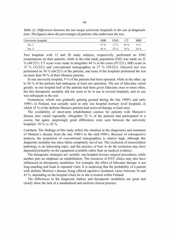
66
Table 22. Differences between the two target university hospitals in the use of diagnostic tests. The figures show the percentages of patients who underwent the test.
University hospital ABR ENG CT MRI No. 1 57 % 17 % 43 % 8 % No. 2 36 % 52 % 14 % 24 %
Two hospitals with 12 and 20 study subjects, respectively, performed no ENG examinations on their patients, while in the total study population ENG was made on 22 % (48/221). CT scans were made in altogether 44 % of the cases (97/221), MRI scans in 15 % (33/221) and conventional tomographies in 27 % (59/221). Glycerol test was performed on 30 % (66/221) of the patients, and none of the hospitals performed the test on more than 50 % of their Meniere patients.
In one university hospital, 9 % of the patients had been operated, while in the other, up to 28 % of the patients had undergone at least one operation. The use of lidocaine varied greatly: in one hospital half of the patients had been given lidocaine once or more often, but this therapeutic modality did not seem to be in use in several hospitals, and its use was infrequent in the rest.
Gentamicin, which was gradually gaining ground during the late 1980’s and early 1990’s in Finland, was actually used in only one hospital (tertiary level hospital), in which 18 % of the definite Meniere patients had received therapy at least once.
The availability of short-term rehabilitation courses for patients with Meniere’s disease also varied regionally. Altogether 32 % of the patients had participated in a course, but again, surprisingly great differences were seen between the university hospitals: 10 % vs. 43 %.
Comment. The findings of this study reflect the situation in the diagnostics and treatment of Meniere’s disease from the late 1960’s to the mid-1990’s. Because of retrospective analysis, the proportion of conventional tomographies is relative high, although this diagnostic modality has since fallen completely out of use. The exclusion of retrocochlear pathology is an interesting topic, and the practice of how to do the exclusion may have depended primarily on the equipment available rather than on medical evidence.
The therapeutic strategies are variable: one hospital favours surgical procedures, while another puts an emphasis on rehabilitation. The resources of ENT clinics may also have influenced on therapeutic modalities. For example, the effect of lidocaine therapy is not long-standing and leads to repeated visits. It is surprising that the probability of a patient with definite Meniere’s disease being offered operative treatment varies between 10 and 43 %, depending on the hospital where he or she is treated within Finland.
The differences in the diagnostic battery and therapeutic modalities are great and clearly show the lack of a standardised and uniform clinical practice.

67
5.8 Patient’s perspective
One important aim of this Meniere research project in Finland was to clarify the experiences and subjective observations of patients with Meniere’s disease concerning the diagnosis and treatment of their disease, i.e. the ability of the Finnish health care system to serve these patients. To obtain an adequate idea of this, a questionnaire was sent to 800 members of the Finnish Meniere Federation. A total of 630 patients returned the questionnaire. Because a voluntary patient association may also have members who do not have Meniere’s disease, these data as such could not be included in scientific reports, which were based on definite Meniere patients according to strict AAO-HNS (1995) criteria. A sample of patients who had fulfilled the questionnaire were asked a further permission to examine their patient charts in the hospital where they had been treated. 141 patients gave permission, and a group of 82 patients who had filled in the questionnaire and met the strict criteria for definite Meniere’s disease based on the patient charts were identified.
In this group, the delay from the beginning of the symptoms to the assessment of the diagnosis was < 6 months in 17 cases (20.7 %), 6 months−1 year in 21 (25.6 %), 1−10 years in 28 (34.1 %), 10−20 years in 11 (13.4 %) and more than 20 years in 3 (3.7 %). The diagnosis had been made in a tertiary level hospital in 48 cases (58.5 %), in a secondary level hospital in 25 (30.5 %) and in primary health care in 2 (2.4 %), and 4 (4.9 %) had been diagnosed by a private practitioner, while the remaining 3 (3.6 %) patients could not define the unit of diagnosis assessment. A total of 19 patients reported having used one alternative treatment modality, while 14 had two and 6 had used three or more other treatment modalities (Fig. 3).

68
Fig. 3. Other treatment modalities used by the definite Meniere patients (N=82) who returned a questionnaire.
Comment. According to the findings, the patients had not been treated equally in the different Finnish hospitals up till the end of 1996, and no radical changes in this respect are likely to have taken place thereafter. The delay from the beginning of the symptoms to the assessment of the diagnosis varies greatly, and so does the diagnostic battery used when Meniere’s disease is suspected. In the Swedish study by Friberg et al. (1984), the delay between onset and definitive diagnosis was about three years, and in a recent report from Norway, the delay was more than 10 years in 11 % of the patients (Rosenberger 2002). From the patients’ perspective, this delay is disabling, and the patient’s anxiety to find a definitive cause for his or her symptoms should be considered in the diagnostic protocol (Robinette et al. 2000).
The treatment modalities for Meniere’s disease may be considered as symptomatic treatments, because they do not alter the natural degenerative course of the condition (Claes & Van De Heyning 2000). This fact together with the proven good response of Meniere patients to placebo (Thomsen et al. 1979) makes these patients susceptible to a variety of treatment modalities outside conventional medicine. In the Norwegian study by Rosenberger (2002), 45 % of the patients used alternative medical treatments. The use of these treatment modalities may not always be revealed by the patient, for fear of impairing the patient-doctor relationship.
The differences in therapeutic modalities are explained mostly by the differences in clinical practice. The natural course of the disease and the placebo effect of all treatment

69
regimens make the evaluation of the treatment modalities difficult (Claes & Van De Heyning 2000). However, the delay from the onset of the disease to the diagnostic assessment and the multitude of treatment modalities available outside conventional medicine and used by Meniere patients reflect the need for more accurate diagnosis and more satisfying treatment modalities. Physicians should also show a more encouraging attitude.

6 General discussion
6.1 Epidemiologic and diagnostic problems
The symptom complex of Meniere’s disease has been known for more than 140 years, but the precise aetiology of this complex of symptoms still remains obscure. Curative treatment for this disease is also still lacking (Claes & Van De Heyning 2000). The diagnostic criteria have been repeatedly revised over the years, which may have caused confusion in clinical work. The diagnostic policy today differs markedly from that of the 1970’s, especially because of the rapid technical development but also because of the improved knowledge about this disease.
The lack of generally accepted diagnostic criteria has been weakening the epidemiologic evaluations of Meniere’s disease. The epidemiologic figures reported around the world are rather scarce and heterogeneous because of the different criteria used for diagnosing the disease. In addition, the confusion caused by incorrect epidemiologic concepts, such as incidence and prevalence, makes the evaluation of reports on the occurrence of the disease very difficult (Friberg & Stahle 1999). It is therefore quite understandable that it has not been possible to obtain any consistent idea of the epidemiology of Meniere’s disease in Finland earlier. However, the epidemiological knowledge of a chronic non-lethal disease, such as Meniere’s disease, is important both for the patients and their families and for the health care service system. This study is the first to clarify these topics in our country.
During the gathering of study data in various hospitals in Finland, the above problems were clearly visible. The diagnosis of Meniere’s disease in Finland is not based on unambiguous criteria, but is most often based on the personal clinical experience of the physician and the traditional practice of each ENT clinic. During the study period, the diagnostic criteria were being re-defined, which may have had an influence on diagnostic accuracy. The disease itself is not well known even among health care professionals. It is thus not surprising that only 50 % of this study population, who had diagnostic codes implying Meniere’s disease in the hospital registers, had a definite disease according to the standard used in this study, i.e. the AAO-HNS (1995) criteria. Another explanation for this poor diagnostic accuracy may be the ICD classification, which does not have a

71
code for a suspicion of Meniere’s disease. It is well known, and was also confirmed by this study, that the diagnosis of Meniere’s disease is not always made during the first visit to an ENT clinic, and the delay from the beginning of the symptoms to the assessment of a definite diagnosis may take several years (Friberg et al. 1984).
The diagnostic policy in Finnish hospitals appeared to be variable. The equipment available for the diagnostic assessment was heterogeneous. For example, some hospitals did not use the ENG technique, while in many others it was almost invariably used when examining patients with vertigo. The glycerol test belonged to the diagnostic battery in one hospital, but another never seemed to use this diagnostic test. The exclusion of retrocochlear pathology seemed to depend on the capacity of the hospital’s imaging unit, not on the real medical indications. ABR is still a reliable method to exclude acoustic neuroma (Haapaniemi et al. 2000a), but if MRI is easily available, many clinicians choose this more expensive method. At this time of critically increasing health care expenditure, judgement concerning the diagnostic procedures used when suspecting Meniere’s disease may not be directed by the technical equipment available but rather by the clinical experience and scientific literature.
Regional differences in the epidemiologic figures may thus be, at least partially, explained by environmental factors but also by the differences in the diagnostic policy and the level of interest in vertiginous inner ear diseases between hospitals. The services at various levels of the health care system are available for every citizen, and the standard of living in Finland, as in all Nordic countries, is equally distributed, which factors diminish the role of the socioeconomic factors in the regional variability of epidemiologic figures of Meniere’s disease.
6.2 Problems with the therapy of Meniere’s disease
Most of the above factors concerning the difficulties in the diagnostics of Meniere’s disease are also applicable to the therapy of this disease. The problems with the assessment of the definite diagnosis also reflect on the choice and introduction of the therapeutic modality. The therapeutic modalities used varied surprisingly much between the target hospitals. The rather small sample size may hide some important facts, but distinct differences in the clinical practice were seen in the treatment of Meniere’s disease. For example, short-term rehabilitation courses seemed to be arranged with different frequencies.
Furthermore, the fact that there is no curative treatment or even any treatment that could markedly alter the natural course of the disease constitutes a great challenge for a doctor treating patients with Meniere’s disease. There are, however, many therapeutic modalities which, especially in concomitance with a confidential doctor-patient relationship, can relieve the symptoms of Meniere’s disease to such an extent that the patient can cope with his or her daily life quite well.
In the historical perspective, no dramatic revolution has been experienced in the treatment of Meniere’s disease, although intratympanic gentamicin treatment may be seen as a marked improvement in controlling vertigo (Assimakopoulos & Patrikakos 2003).

72
The effects of this treatment modality on hearing impairment and tinnitus are not of special value, but gentamicin has almost completely eliminated the need for invasive surgery. The challenge for developing better treatment modalities still remains. The rather wide use of various methods outside conventional medicine by Meniere patients was shown in a Norwegian study (Rosenberger 2002) and also here (see Fig. 3, in Chapter 5.8, p. 70). This is indicative of the the need for better treatment modalities. Although Meniere’s disease will not lead to total deafness, a hearing aid is often needed. As many as 26.2 % of the 221 definite Meniere patients in this study were hearing aid users. Because of the fluctuating hearing thresholds, hearing aid fitting is not easy, and patients with Meniere’s disease may have problems to adapt to a hearing aid (Hesse et al. 2000, Ballester et al. 2002). Because of concomitant disorders or for iatrogenic reasons, moreover, some patients may totally lose their hearing. In these cases, cochlear implants may restore a social level of hearing, and at least one preliminary report on cochlear implant installation in a patient with Meniere’s disease has already been published (Morgan et al. 1999).
However, in developing both new techniques for diagnosing the disease and new therapeutic modalities to relieve the symptoms of the disease, the cost-benefit aspect should be kept in mind. A randomly controlled trial, which is considered the gold standard of methodology design, is difficult to carry out in Meniere’s disease because of two reasons according to Thorp et al. (2000): the reluctance of surgeons and other investigators to participate in controlled trials by offering a no-surgery option in the control arm and the ethical concerns that may be raised about randomization into a control arm in a clinical trial where the control group gets no therapy. In the field of complementary and alternative medicine, unfortunately, controlled trials are not required, and the anecdotal effects of a given treatment modality may raise gratuitous hope of full recovery from the disease. The proven good response of Meniere patients to placebo (Thomsen et al. 1979) makes these patients prone to a variety of treatment modalities outside conventional medicine. This was also shown in this study and is discussed in Chapter 2.8.6. Again, a good doctor-patient relationship may protect the patient from desperate and often costly trials.
This study shows that there is a great challenge for the Finnish health care system, as well as around the world, to develop adequate decision pathways for diagnosing Meniere’s disease and also a consistent treatment policy in order to offer equal chances to every patient suspected to have Meniere’s disease, regardless of where he or she lives. The cost-effectiveness of both diagnostic tests and treatment modalities should be further clarified.
6.3 Suggestions for the diagnosis and therapy of Meniere’s disease
The latest recommended AAO-HNS guidelines primarily aimed to enable the reporting of the treatment results in a way that makes computerized analysisg and comparison possible. The guidelines further offer uniform diagnostic criteria for epidemiological evaluation of the disease. The strictness of these criteria may, however, lead to

73
underestimation of the commonness of Meniere’s disease. On the other hand, the subdivision into four categories and the absence of a diagnostic protocol to establish the diagnosis and to exclude other causes for the symptomatology make the application of these criteria in clinical practice still unsatisfactory (Mateijsen 2001). Adjunctive findings suggestive of the diagnosis should also be emphasized more whenever Meniere’s disease is suspected. Especially the fluctuation of hearing seems to correlate well with the definite diagnosis according to the AAO-HNS criteria. For probable and possible cases, a suspicion of Meniere’s disease should have its own code in the ICD classification.
As there seems to be rather great variability in the diagnostic and therapeutic modalities within Finland, which also sets the patients in unequal positions, uniform diagnostic criteria and therapeutic scenarios, i.e., a clear decision pathway including history, current status and various laboratory tests, would be helpful. This ”current therapy” suggestion for Meniere’s disease in Finland should be assessed, and it would undoubtedly also serve to make the diagnosis of Meniere’s disease more uniform at the national level. Actually, the same phenomenon has been observed in various other filials of health care: the great variation in the diagnosis and treatment of recurrent tonsillitis, low back pain, breast cancer, etc. has led to current therapy suggestions in evidence-based medicine in Finland.
The suggestion for the diagnosis and treatment of Meniere’s disease should be compatible with the latest AAO-HNS recommendations, also taking into account the adjunctive findings suggesting the diagnosis, such as the fluctuation of hearing impairment, positive glycerol tests and positional vertigo between the dizzy spells of the disease. The exclusion of retrocohlear disease should be routinely used, and ABR is suggested as the primary screening method when there is no other special indication for intracranial imaging. The high costs and variable availability of these imaging modalities, especially at times of shortage of radiologists, and the high specificity of ABR make this test a more preferable front-line method.
In the treatment of patients with Meniere’s disease, a uniform practice should be adopted in every ENT clinic. Treatment should be based on a confident and consistent doctor-patient relationship, which relieves the anxiety caused by the disease. This relationship, including adequate information of the disease to the patient and a supportive and encouraging attitude of the doctor, is especially important in the case a chronic disabling disease, whose natural course cannot be markedly altered by any therapeutic procedure. The patient’s confidence in the doctor may protect him or her from desperate trials of ineffective treatment outside conventional medicine. Unnecessary surgical interventions may also often be avoided. Short-term rehabilitation courses should be adopted more often as part of the therapy and rehabilitation. The effects of these courses, which are an unknown and poorly reported entity, should be further clarified and reported. The recommendations for the diagnosis and treatment of Meniere’s disease in Finland are shown in pages 76 and 77.

74
Finnish recommendation for the diagnosis of Meniere’s disease:
Basic examinations:
ORL status, including audiometry and speech audiometry
Otoneurologic assessment: Romberg test,Unterberg test, spontaneous nystagmus with Frenzel spectacles, Dix-Hallpike test, Horizontal canal test, ENG with caloric irrigation, blood pressure
Laboratory tests (according to physician’s consideration): Blood cell count, glucose, THS, Borrelia, serum sodium and potassium levels. Glycerol test or ECOG if the diagnosis remains unclear after repeated visits
Positive findings to confirm the diagnosis of Meniere’s disease:
Main criteria:
At least 2 episodes of rotational vertigo with nausea/emesis lasting for 20 min−several hours
Tinnitus or aural pressure, which often increases before or during an acute spell
In the early disease: sensorineural hearing impairment documented in audiometry in the affected ear; the average (arithmetic mean) of hearing thresholds at 0.5, 1, 2 and 4 kHz being at least 15 dB poorer than on the opposite side. The hearing impairment may also be of the low-tone type: the average of threshold values at 0.25, 0.5 and 1 kHz are ≥ 15 dB higher than the average of 1, 2 and 4 kHz
Adjunctive criteria:
Adjunctive positive signs in patient’s history: diplacusis/loudness intolerance/ motion intolerance during an acute spell. Positional vertigo between spells
Fluctuation of hearing is not mandatory, but if reported by the patient or seen in repeated audiometry, strongly strengthens the suspicion of Meniere’s disease
Reliable exclusion of retrocochlear pathology primarily by ABR, if no other neurologic signs are present. If neurologic disorders are suspected or the ABR finding is not normal, head MRI with gadolinium should be done

75
Finnish recommendation for the therapy of Meniere’s disease:
Detailed information of the disease to the patient and a written information leaflet are given at the diagnostic assessment personally by the physician. A consistent and confidential doctor –patient relationship should be established.
Life style alterations. A low-salt diet should be adopted, and excessive consumption of coffee, alcohol and tobacco should be avoided, a regular sleepwaking state rhythm should be adopted, and factors causing stress should be abolished as far as possible.
Medication. If there are more than 2 attacks per month or the attacks are prolonged, oral medication should be started. The front-line medications are betahistine and/or diuretics, and the choice between them may be made according to the blood pressure level and possible relative or absolute contraindications. Every patient should also be supplied with antiemetic suppositories to be only used at acute attacks. Other medications (muscle relaxants, anxiolytes, hypnotics, antidepressants) should be prescribed only for special indications.
Follow-up. The effect of the above therapy modalities should be followed up for up to 3 months, and the medication should be adjusted or changed at the control visit according to the response. The minimum dose of medication sufficient to relieve symptoms should be the goal. If the response is excellent, the medication may be stopped, and gradual lowering of the dose is often recommended. The patient is also informed about the possibility to temporarily raise the dose in response to aggravation of the disease.
If conservative management fails to help the patient during 6 months, gentamicin application should be considered. An alternative for this is pressure treatment.
Surgical treatment (endolymphatic sac decompression, vestibular neurectomy) should be reserved only for cases resistant to all of the above-mentioned therapy modalities, especially when the hearing in the affected ear has been lost.
A hearing aid may be fitted into the ear with stable hearing. It may be of help for disabling tinnitus. A multi-channel hearing aid may also be fitted for a patient with fluctuant moderate or severe hearing impairment.
Other treatment modalities. Relaxation therapies (acupuncture, massage) may be of help to relieve neck and shoulder muscle tension, which may aggravate the symptoms. Short-term rehabilitation courses should be offered for every new patient with Meniere’s disease

76
The cost-effectiveness of both diagnostic tests and treatment modalities of Meniere’s disease should be further clarified. A neuro-otologist or a neuro-otologically oriented doctor should be available in every ENT clinic. Orientation and experience in diseases with vertigo, including Meniere’s disease, help the doctor to cope with the anxiety during the process of diagnostic assessment and the frustration shown by the patients if one treatment modality fails to be effective.
The patient with Meniere’s disease needs, over all, reassurance to cope with his or her daily life with the chronic and often disabling but non-lethal disease. It should be emphasized that Meniere’s disease and its surgical treatment did not prevent astronaut Alan Shephard from being appointed commander-in-chief of Apollo 14 (Guyot 1996).

7 Summary and conclusions
This study is a retrospective analysis of a study population with suspected Meniere’s disease treated as inpatients or outpatients in various Finnish hospitals during the years 1992-1996. A total of 442 patients’ charts were carefully analysed and the relevant data saved in a specially designed programme file. The latest AAO-HNS (1995) criteria for Meniere’s disease were used as a gold standard.
Study I. The average prevalence of definite Meniere's disease in Finland, according to the latest AAO-HNS criteria, was shown to be 43.2 per 100,000 of population. It is lower than could be expected based on previous international surveys, although most of them provide inadequate data for a reliable picture of the epidemiology of Meniere's disease. The average annual incidence was 4.3 per 100,000.
The prevalence of Meniere’s disease, as estimated on the basis of a retrospective population-based survey, including only the definite cases diagnosed according to the latest AAO-HNS recommendations, varies in Finland regionally. This difference could not be explained by the availability of health care services.
On the basis of these findings, it can be concluded that the epidemiological figures of Meniere’s disease in the literature are not unambiguous. The epidemiological concepts have been used vaguely, and the lack of uniform diagnostic criteria has made the topic unclear. For the patient, it is not unimportant whether or not he or she has Meniere’s disease. Accurate diagnostic criteria are thus necessary to improve specificity in recognizing definite cases of Meniere's disease, and such criteria would make the further research of epidemiological and clinical aspects of Meniere's disease more reliable.
Study II. The demographic data in this study were in line with the former literature: there is no major difference between the sexes or between the right and left ear. Bilaterality, 14 %, also remained within the limits of previous reports. Meniere’s disease appeared to be significantly more prevalent in northern Finland than further south.
Study III. A multivariable model was created to predict hearing impairment in Meniere’s disease. The model showed that the worsening effect on hearing was constant at ≈ 1 dB/year due to the duration of the disease. The effect of aging on hearing impairment (0.5 dB/year) was about half that of the effect of the duration of the disease. Gender did not modify the effect of the duration of the disease.

78
Study IV. A general gold standard for diagnosing Meniere’s disease is still lacking. The diagnostic battery for the disease has undergone distinct evolution, but pure-tone and speech audiometry still remain as basic elements of the diagnostic procedure. The more accurate diagnostic tools may serve as exclusion tests for other diseases, but some tests may also support the clinician’s suspicion of endolymphatic hydrops. No single method has proved to be superior in the diagnostics of Meniere’s disease. Fluctuation of hearing, however, is a highly specific observation correlating with definite cases of Meniere’s disease. Glycerol test may be used as an adjuvant test when the diagnosis is not easy to confirm, and ABR is an inexpensive and fairly reliable method to rule out retrocochlear pathology in unilateral hearing impairment. The need to explore the cost-benefit ratio of the modern diagnostic tools used to diagnose Meniere’s disease is evident. Despite the rapid technological evolution that is taking place in medicine, the diagnosis can mostly be made based on a good and complete history.
Study V. Meniere’s disease is an entity that, despite the strict criteria for definite disease, involves a wide variety of clinical manifestations. No standard Meniere patient can be determined. Vertigo was found to be the most disabling symptom of the disease. Patients with Meniere’s disease seemed to cope well with conservative treatment. Neither marital nor occupational status plays any major role in Meniere’s disease.
A high-frequency gently sloping audiometric configuration was most prevalent in the EU Work Group classification (Stephens 1996) and a flat pattern in the mid-frequency-based classification (Sorri et al. 2001). The duration of the disease seemed to affect the audiometric configuration. A flat audiometric configuration was rarely seen in recent cases, and an ascending configuration was most typically seen in patients who had had the disease for 2 to 10 years. The EU classification failed to classify 37.6 % of audiograms, whereas all audiograms could be classified with the mid-frequency-based classification. The mid-frequency-based classification may be recommended for the classification of hearing impairment in Meniere’s disease. No audiometric configuration is, however, specific for Meniere’s disease. In the latest audiogram of the affected ear, moderate or severe hearing impairment was seen in 75 % of the patients.
Final conclusion. The prevalence of Meniere’s disease in Finland was shown to be at least 43.2 and the annual incidence 4.3 per 100,000. The disease seemed to be more common in northern than southern Finland. Meniere’s disease is a clinical entity with a wide variety of clinical manifestations regardless the strict latest AAO-HNS (1995) criteria. The disease causes hearing deterioration, and the worsening effect due to the duration of the disease seemed to be constant at 1 dB/year. The audiometric pattern is also influenced by the duration of the disease. The most common audiometric pattern in Meniere’s disease was a high-frequency gently sloping pattern according to the EU Work Group classification (Stephens 1996) and a flat pattern in the mid-frequency-based classification (Sorri et al. 2001). Fluctuation of hearing was shown to be a specific observation in definite Meniere’s disease. Regional differences in the diagnostics and treatment of Meniere’s disease were found in Finland, and a recommendation for uniform clinical policy is proposed.
References
Aalto H, Pyykkö I, Starck J (1988) Computerized posturography, a development of the measuring system. Acta Otolaryngol (Stockh) Suppl 449: 71-75.
Aantaa E (1991) Treatment of acute vestibular vertigo. Acta Otolaryngol Suppl 479: 44-47. Abe T, Tsuiki T, Murai K, Kon Y, Ishikawa T, Asano Y (1992) Progressing cases from low tone
sudden deafness to Meniere’s disease - cochlear impairment in the so-called pre-Meniere’s disease. J Oto-Rhino-Laryngol Soc Japan 95 (9): 1352-1359.
Adamczyk M, Antonelli PJ (2001) Selective vestibular ablation by KTP laser in endolymphatic hydrops. Laryngoscope 111 (6): 1057-1062.
Agresti A (1990) Categorial data analysis, pp. 419-425. John Wiley & Sons Inc., New York. Akagi H, Yuen K, Maeda Y, Fukushima K, Kariya S, Orita Y, Kataoka Y, Ogawa T, Nishizaki K
(2001) Meniere’s disease in childhood. Int J Pediatr Otorhinolaryngol 61 (3): 259-264. Albera R, Cavalot A, Musto R, Fadda GL, Staffieri A, Di Girolamo S (2001) Tympanic membrane
displacement analyser tracing modifications induced by glycerol in Meniere’s disease. Audiology 40 (4): 185-190.
Alleman AM, Dornhoffer JL, Arenberg IK, Walker PD (1997) Demonstration of autoantibodies to the endolymphatic sac in Meniere’s disease. Laryngoscope 107: 211-215.
Anderson JP, Harris JP (2001) Impact of Meniere’s disease on quality of life. Otol Neurotol 22 (6): 888-894.
Andersson G, Hägnebo C, Yardley L (1997) Stress and symptoms of Meniere’s disease:a time-series analysis. J Psychosom Res 43: 595-603.
Angelborg C, Klockhoff I, Stahle J (1977) Urea and hearing in patients with Meniere’s disease. Scand Aud 6: 143-146.
Angelborg C (1986) Pharmacological treatment of Meniere’s disease. A review of common remedies with special emphasis on vasodilators, including hyperosmolar substances and hearing. Scand Aud Suppl 26: 33-36.
Antoli-Candela F (1976) The histopathology of Meniere’s disease. Acta Otolaryngol (Stockh) Suppl 340: 1-42.
Arenberg IK, Balkany TJ, Goldman G, Pillsbury HC (1980) The Incidence and prevalence of Meniere's disease - A statistical analysis of limits. Otolaryngol Clin North Am 13 (4): 597-601.
Arenberg IK, Wells JA, Shambaugh GE Jr (1991) Definitions and semantics: an overview of Meniere’s disease and endolymphatic hydrops. In: Arenberg IK (ed) Surgery of the inner ear, pp.3-7. Kugler Publications, Amsterdam.
Arenberg IK, Mihalco L (1993) Inner ear disease typing for Meniere's disease with audiometric and electrococleographic correlates. In: Hohmann D, ed. ECog, AOE and intraoperative monitoring, pp. 131-132. Kugler Publications, Amsterdam.

80Arenberg IK, Cabriac G, Marks S, Arenberg JG, Pfeiffer PR, Murray RS (1997) Cytomegalovirus
antibodies in endolymphatic sac biopsies of patients with endolymphatic hydrops and Meniere’s disease. Ann NY Acad Scienc 830: 314-318.
Arnold W, Altermatt HJ (1995) The signifigance of the human endolymphatic sac and its possible role in Meniere’s disease. Acta Otolaryngol (Stockh) Suppl 519: 36-42.
Arnold W, Niedelmeyer HP (1997) Herpes simplex virus antibodies in the perilymph of patients with Meniere’s disease. Arch Otolaryngol 123 (1): 53-56.
Arriaga MA, Goldman S (1998) Hearing results of intratympanic steroid treatment of endolymphatic hydrops. Laryngoscope 108: 1682-1685.
Arslan M (1953) Treatment of Meniere’s syndrome by direct application of ultrasound waves to the vestibular system. Proc Fifth Intl. Congress Otolaryngol, pp. 629-635. Amsterdam.
Arweiler DJ, Jahnke K, Grosse-Wilde H (1995) Meniere’s disease as an autosome hereditary disease. Laryngo- Rhino- Otologie 74 (8): 512-515.
Assimakopoulos D, Patrikakos G (2003) Treatment of Meniere’s disease by intratympanic gentamicin application. J Laryngol Otol 117: 10-16.
Atlas MD, Chai F, Boscato L (1998) Meniere’s disease: evidence of an immune process. Am J Otol 19: 628-631.
Atlas JT, Parnes LS (1999) Intratympanic gentamicin titration therapy for intractable Meniere’s disease. Am J Otol 20: 357-363.
Babin RW, Balkany TJ, Fee WE (1984) Transdermal scopolamine in the treatment of acute vertigo. Ann Otol Rhinol Laryngol 93: 25-27.
Bagger-Sjöbäck D, Rask-Andersen H (1986) The permeability barrier of the endolymphatic sac: a hypothesis of fluid and electrolyte exchange based on freeze fracturing. Am J Otol 7: 134-140.
Ballester M, Liard P, Vibert D, Häusler R (2002) Meniere’s disease in the elderly. Otol Neurotol 23 (1): 73-78.
Baloh RW, Jacobson K, Winder T (1990) Drop attacks with Meniere’s syndrome. Ann Neurol 28 (3): 384-387.
Beck C, Scmidt CL (1978) Ten years of experience with intratympanically applied streptomycin (gentamicin) in the therapy of morbus Meniere. Arch Otorhinolaryngol 221: 149-152.
Beck C (1986) Intratympanic application of gentamicin for treatment of Meniere’s disease. Keio J Med 35: 36-41.
Berg HM, Cohen NL, Hammerschlag PE, Waltzman SB (1986) Acoustic neuroma presenting as sudden hearing loss with recovery. Otolaryngol Head Neck Surg 94: 15-22.
Bergström T, Edström S, Tjellström A, Vahlne A (1992) Meniere’s disease and antibody reactivity to herpes simplex virus type 1 polypeptides. Am J Otol 13: 295-300.
Bernstein JM, Shanahan TC, Schaffer FM (1996) Further observations on the role of the MCH genes and certain hearing disorders. Acta Otolaryngol (Stockh) 116: 666-671.
Biagini C, Nuti D, Sensini I (1991) L'incidenza della malattia di Meniere nella USL 30 Area senese. Acta Otorhinolaryngol Ital 11 (4): 379-83.
Birgerson L, Gustavson KH, Stahle J (1987) Familial Meniere’s disease: a genetic investigation. Am J Otol 8: 323-326.
Björne A, Agerberg G (1996) Craniomandibular disorders in patients with Meniere’s disease: a controlled study. J Orofac Pain 10 (1): 28-37.
Black FO, Pesznecker SC (1993) Vestibular ototoxicity. Clinical considerations. Otolaryngol Clin North Am 26: 713-716.
Blair RL (1984) Medical management of vestibular dysfunction. Otolaryngol Clin North Am 17: 679-684.
Bojrab D, Bhansali S, Battista R (1994) Peripheral vestibular disorders. In: Jackler RK, Brackmann DE (eds). Neurotology, pp. 629-650. St.Louis, Missouri: Mosby.
Bouccara D, Ferrary E, El Garem H, Couloigner V, Coudert C, Sterkers O (1998) Inner ear pressure in Meniere’s disease and fluctuating hearing loss determined by tympanic membrane displacement analysis. Audiology 37: 255-261.
Boulassel MR, Alost M, Tomasi JP, Deggouj N, Gersdorff M (2001) No increased serum levels of antifood antibodies in patients with Meniere’s disease. J Oto-Rhino-Laryngol 63: 19-24.

81Brookes GB (1986) Circulating immune complexes in Meniere’s disease. Arch Otolaryngol Head
Neck Surg 112: 536-540. Calenoff E, Zhao JC, Derlacki EL, Harrison WH, Selmeczi K, Dutra JC, Olson IR, Hanson DG
(1995) Patients with Meniere’s disease possess IgE reacting with herpes family viruses. Arch Otolaryngol 121 (8): 861-864.
Caparosa RJ (1963) Medical treatment for Meniere’s disease. Laryngoscope 73: 666-672. Casselman JW, Kuhweide R, Deimling M, Ampe W, Dehaene I, Meeus L (1993) Constructive
interference in steady state (CISS-) 3DFT MR imaging of the inner ear and cerebellopontine angle. ANJR 14: 59-63.
Cawthorne T, Hewlett AB (1954) Meniere's disease. Proc Roy Soc Med 47: 633-670. Cawthorne T (1957) Meniere’s disease. Ann Otol Rhinol Laryngol 56:18-38. Celestino D, Ralli G, Merolla A, Gagliardi M, Magliulo G (1987) Seasonal recurrence of acute
episodes of Meniere’s disease. Acta Otorhinolaryngol Belg 41: 951-957. Celestino D, Ralli G (1991) Incidence of Meniere's disease in Italy. Am J Otol 12: 135-138. Celestino D (2000) Precipitating factors in Meniere’s disease. In: Sterkers O, Ferrary E, Dauman R,
Sauvage JP, Tran Ba Huy P (eds) Meniere’s disease1999 - Update, pp. 475-480. Kugler Publications, The Hague, The Netherlands.
Ceranic B, Prasher DK, Luxon LM (2000) Otoacoustic emissions as acoustic cochleography in diagnosis of Meniere’s disease. In: Sterkers O, Ferrary E, Dauman R, Sauvage JP, Tran Ba Huy P (eds) Meniere’s disease 1999 - Update, pp. 327-335. Kugler Publications, The Hague, The Netherlands.
Cheng KC, Matsuoka H, Lee KM, Kim N, Krug MS, Kwon SS, Mora M, Yoo TJ (2000) Proto-oncogene Raf-1 as an autoantigen in Meniere’s disease. Ann Otol Rhinol Laryngol 109 (12): 1093-1098.
Claes J, Van De Heyning PH (2000) A review of medical treatment for Meniere’s disease. Acta Otolaryngol Suppl 544: 34-39.
Clemis JD (1967) Allergy of the inner ear. Ann Allergy 25: 370-376. Cobb S, Coen LW, Gitomer SL, Iles R (1976) Effect on cinnarizine on nystagmus induced by
rotation deceleration. ENT Monthly 55: 24-30. Cody DTR, Simonton KM, Hallberg OE (1967) Automatic repetitive decompression of the saccule
in endolymphatic hydrops (tack operation). Preliminary report. Laryngoscope 77: 1480-1501. Cogan DG (1945) Syndrome of nonsyphilitic interstitial keratitis and vestibular-auditory
symptoms. Arch Ophtalmol 33: 144-149. Cohen H (1992) Vestibular rehabilitation reduces functional disability. Otolaryngol Head Neck
Surg 107 (5): 638-643. Cohen H, Ewell LR, Jenkins HA (1995) Disability in Meniere’s disease. Arch Otolaryngol Head
Neck Surg 121: 29-33. Coker NJ, Coker RR, Jenkins HA, Vincent KR (1989) Psychological profile of patients with
Meniere’s disease. Arch Otolaryngol Head Neck Surg 115: 1355-1357. Committee on Hearing and Equilibrium (1972) Ménière's disease: criteria for diagnosis and
evaluation of therapy for reporting. Trans Am Acad Ophtalmol Otolaryngol 76: 1462-1464. Committee on Hearing and Equilibrium (1985) Ménière's disease: criteria for diagnosis and
evaluation of therapy for reporting. AAO-HNS Bull 5 (July): 6-7. Committee on Hearing and Equilibrium (1995) Guidelines for the diagnosis and evaluation of
therapy in Meniere's disease. Otolaryngol Head Neck Surg 113: 181-185. Conlon BJ, Gibson WP (1999) Meniere’s disease: the incidence of hydrops in the contralateral
asymptomatic ear. Laryngoscope 109 (11): 1800-1802. Crary WG, Wexler M (1977) Meniere’s disease: a psychosomatic disorder. Psychol Rep 41: 603-
645. Dandy WE (1933) Treatment of Meniere’s disease by section of only vestibular portion of the
acoustic nerve. Bull Johns Hopkins Hosp 53: 52-55. Dawes PJD, Jeannon JP (1998) Audit of regional screening guidelines for vestibular schwannoma.
J Laryngol Otol 112: 860-864. Densert B, Densert O, Erlandsson B, Sheppard H (1986) Transmission of complex pressure waves
through the perilympatic fluid in cats. Acta Otolaryngol 102: 403-409.

82Densert B, Densert O, Arlinger S, Sass K, Ödkvist L (1997) Immediate effects of middle ear
pressure changes on the electrocochleographic recordings in patients with Meniere’s disease: a clinical placebo-controlled study. Am J Otol 6: 726-733.
Densert B, Sass K (2001) Control of symptoms in patients with Meniere’s disease using middle ear pressure applications: two years follow-up. Acta Otolaryngol 121 (5): 616-621.
Densert O, Ingelstedt S, Ivarsson A (1975) Immediate restoration of basal sensorineural hearing (Mb. Meniere) using a pressure chamber. Acta Otolaryngol 80: 93-100.
Densert O, Carlborg B, Stagg J (1981) Transmission of a low frequency pressure steps to the perilymphatic fluid. Acta Otolaryngol 91: 55-64.
Derebery MJ (1996) Allergic and immunologic aspects of Meniere’s disease. Otolaryngol Head Neck Surg 114: 360-365.
Derebery MJ, Berliner KI (2000) Prevalence of allergy in Meniere’s disease. Otolaryngol Head Neck Surg 123: 69-75.
Dix MR, Hallpike CS (1952) The pathology, symptomatology and diagnosis of certain common disorders of the vestibular system. Ann Otol Rhinol Laryngol 61: 987-1016.
Dobie RA, Snyder JM, Donaldson JA (1982) Electronystagmographic and audiologic findings in patients with Meniere’s disease. Acta Otolaryngol (Stockh) 94: 19-27.
Dolowitz D (1979) Meniere’s - an inner ear seizure. Laryngoscope 89: 67-77. Driscoll CL, Kaspenbauer JL, Facer GW, Harner SG, Beatty CW (1997) Low-dose intratympanic
gentamicin and the treatment of Meniere’s disease: preliminary results. Laryngoscope 107: 83-89.
Duke WW (1923) Meniere’s syndrome caused by allergy. JAMA 81: 2179. Eklund S (1999) Headache in Meniere’s disease. Auris Nasus Larynx 26 (4): 427-433. Elwood S, Carlton JH, Cliffe MJ (1982) A psychological contribution to the management of
Meniere’s disease. Practitioner 226: 1149-1152. Enander A, Stahle J (1967) Hearing in Meniere’s disease: a study of pure-tone audiograms in 334
patients. Acta Otolaryngol (Stockh) 64: 543-556. Evans MK, Krebs DE (1999) Posturography does not test vestibulospinal function. Otolaryngol
Head Neck Surg 120 (2): 164-173. Fattori B, De Laco G, Vannucci G, Casani A, Chilardi PL (1996) Alternobaric and hyperbaric
oxygen therapy in the immediate and long-term treatment of Meniere’s disease. Audiology 35: 322-324.
Feenstra L (1997) The management of tinnitus with or without Meniere’s disease. Acta Otolaryngol (Stockh) Suppl 526: 47-49.
Fernandez C (1967) Biochemistry of labyrinthine fluids: inorganic substances. Arch Otolaryngol 86: 226-233.
Fick IA (1964) Decompression of the labyrinth : A new surgical procedure for Meniere’s disease. Arch Otolaryngol 79: 447-458.
Fick I (1968) Sacculotomy. In: Pulec (ed) Symposium on Meniere’s disease, pp. 625-635. Saunders, Philadelphia, PA.
Filipo R, Barbara M (1985) Juvenile Meniere’s disease. J Laryngol Otol 99: 193-196. Filipo R (1993) The diagnostic role of electrocochleography in Meniere’s patients. In: Filipo R,
Barbara M (eds) Meniere’s disease: perspectives in the 90’s, pp. 245-250. Kugler Publications, Amsterdam.
Filipo R, Cordier A, Barbara M, Bertoli GA (1997) Electrocochleographic findings: Meniere’s disease versus sudden sensorineural hearing loss. Acta Otolaryngol (Stockh) Suppl 526: 21-23.
Filipo R, Barbara M, Cordier A, Mafera B, Romeo R, Attanasio G, Mancini P, Marzetti A (1997) Osmotic drugs in the treatment of cochlear disorders: a clinical and experimental study. Acta Otolaryngol 117: 229-231.
Filipo R, Barbara M (1997) Natural history of Meniere’s disease: staging patients or their symptoms? Acta Otolaryngol (Stockh) Suppl 526: 10-13.
Fisher RA (1922) On the interpretation of Chi-square from contingency tables, and the calculation of p. J Royal Stat Soc, 85: 87-94
Fowler EP (1948) Streptomycin treatment for vertigo. Trans Am Acad Ophtalmol Otolaryngol 52: 293-301.

83Fradis M, Podoshin L, Ben-David J, Reiner B (1985) Treatment of Meniere’s disease by
intratympanic injection with lidocaine. Arch Otolaryngol 111: 491-493. Franklin DJ, Pollack A, Fisch U (1990) Meniere’s symptoms resulting from bilateral otosclerotic
occlusion of the endolymphatic duct: an analysis of a causal relationship between otosclerosis and Meniere’s disease. Am J Otol 11: 135-140.
Frazier C (1912) Intracranial division of the auditory nerve for persistent aural vertigo. Surg Gynecol Obstet 15: 524-529.
Friberg U, Stahle J, Svedberg A (1984) The natural course of Meniere’s disease. Acta Otolaryngol (Stockh) Suppl 406: 72-77.
Friberg U, Stahle J (1999) The epidemiology of Meniere’s disease. In: Harris JP (ed) Meniere’s disease, pp. 17-28. Kugler Publications, The Hague, The Netherlands.
Friedrichs I, Thornton ARD (2001) Endolymphatic hydrops in asymptomatic ears in unilateral Meniere’s disease. Laryngoscope 111: 857-860.
Fukushima M, Kitahara T, Uno Y, Fuse Y, Doi K, Kubo T (2002) Effects of intratympanic injection of steroids on changes in rat inner ear aquaporin expression. Acta Otolaryngol 122: 600-606.
Furstenberg AC, Lashmet FH, Lathrop F (1934) Meniere’s symptom-complex: medical treatment. Ann Otol Rhinol Laryngol 43: 1035-1046.
Gacek RR, Gacek MR (2001) Meniere’s disease as a manifestation of vestibular ganglionitis. Am J Otol 22: 241-250.
Ge X, Shea JJ, Orchik DJ (2000) Low-frequency hearing loss in Meniere’s disease. In: Sterkers O, Ferrary E, Dauman R, Sauvage JP, Tran Ba Huy P (eds) Meniere’s disease1999 - Update, pp. 299-304. Kugler Publications, The Hague, The Netherlands.
Gejrot T (1976) Intravenous xylocaine in the treatment of attacks on Meniere’s disease. Acta Otolaryngol (Stockh) 82: 301-302.
Gianoli GJ, Larouere MJ, Kartush JM, Wayman J (1998) Sac-vein decompression for intractable Meniere’s disease: two-year treatment results. Otolaryngol Head Neck Surg 118: 22-29.
Gibson WPR (1991) The use of electrocochleography in the diagnosis of Meniere’s disease. Acta Otolaryngol (Stockh) Suppl 485: 46-52.
Gibson WPR, Arenberg IK (2000) The mechanism which cause the vertigo in Meniere’s disease. In: Sterkers O, Ferrary E, Dauman R, Sauvage JP, Tran Ba Huy P (eds) Meniere’s disease1999 - Update, pp. 451-454. Kugler Publications, The Hague, The Netherlands.
Graham MD, Kemink JL (1984) Transmastoid labyrinthectomy: surgical management of vertigo in nonserviceable hearing ear. A five-year experience. Am J Otol 5: 295-299.
Graham MD, Goldsmith MM. (1994) Labyrinthectomy. Indications and surgical technique. Otolaryngol Clin N Am 27 (2): 325-335.
Guild SR (1927) The circulation of the endolymph. Am J Anat 39: 57-81. Guyot J (1996) Should a pilot suffering from Meniere’s disease be grounded or lifted off to the
moon? ORL J Otorhinolaryngol Relat Spec 58: 304-305. Haapaniemi JJ, Laurikainen ET, Johansson R, Rinne T, Varpula M (2000) Audiovestibular findings
and location of an acoustic neuroma. Eur Arch Oto-Rhino-Laryngol 257 (5): 237-241. Haapaniemi J, Laurikainen E, Johansson R, Karjalainen S (2000) Transtympanic versus tympanic
membrane electrocochleography in examining cochleovestibular disorders. Acta Otolaryngol Suppl 543: 127-129.
Haginomori SI, Makimoto K, Tanaka H, Araki M, Takenaka H (2001) Spontaneous otoacoustic emissions in humans with endolymphatic hydrops. Laryngoscope 111: 96-101.
Haid CT, Watermeier D, Wolf SR, Berg M (1995) Clinical survey of Meniere’s disease: 574 cases. Acta Otolaryngol (Stockh) Suppl 520: 251-255.
Haid CT, Gerhardt O, Grossmann M, Nagel D (2000) Clinical hints in patients suffering from Meniere’s disease. In: Sterkers O, Ferrary E, Dauman R, Sauvage JP, Tran Ba Huy P (eds.) Meniere’s disease 1999 - Update, pp. 419-425. Kugler Publications, The Hague, The Netherlands.
Haid T (1988) Evaluation of flunarizine in patients with Meniere’s disease. Subjective and vestibular findings. Acta Otolaryngol Suppl 460: 149-153.

84Hall M, Brackmann DE (1977) Eustachian tube blockage and Meniere’s disease. Arch Otolaryngol
103: 355-357. Hallpike C, Cairns H (1938) Observations on the pathology of Meniere’s disease. J Laryngol Otol
53: 625-654. Harner SG, Driscoll CL, Facer GW, Beatty CW, McDonald TJ (2001) Long-term follow-up of
transtympanic gentamicin for Meniere’s syndrome. Otol Neurotol 22 (2): 210-214. Harris JP, Ryan A (1995) Fundamental immune mechanisms of the brain and inner ear.
Otolaryngol Head Neck Surg 112: 639-653. Harrison MS, Naftalin L (1968) Meniere's disease. Mechanism and Management. Springfield,
Illinois, Charles C Thomas. Hedgecock LD (1968) Audiometric findings in Meniere’s disease. Otolaryngol Clin N Am: 489-
497. Henson MM, Henson OW Jr, Gewalt SL, Wilson JL, Johnson GA (1994) Imaging the cochlea by
magnetic resonance microscopy. Hearing Res 75: 75-80. Hesse G, Schaaf H, Kolbe U, Brehmer D, Andres R (2000) The prescription of hearing Aids in
Meniere patients. In: Sterkers O, Ferrary E, Dauman R, Sauvage JP, Tran Ba Huy P (eds.) Meniere’s disease 1999 - Update, pp. 681-683. Kugler Publications, The Hague, The Netherlands.
Hincliffe R (1967) Personality profile in Meniere’s disease. J Laryngol Otol 81: 477-481. Hirsch BE, Kamerer DB (1997) Intratympanic gentamicin therapy for Meniere’s disease. Am J
Otol 18: 44-51. Hirvonen TP, Peltomaa M, Ylikoski J (2000) Intratympanic and systemic dexamethasone treatment
of Meniere’s disease. ORL 62: 117-120. Hoffer ME, Kopke RD, Weisskopf P, Gottshall K, Allen K, Wester D, Balaban C (2001) Use of the
round window microcatheter in the treatment of Meniere's disease. Laryngoscope 111: 2046-2049.
House WF (1962) Subarachnoidal shunt for drainage of endolymphatic hydrops. Laryngoscope 72: 713-729.
Huang TS, Lin CC, Chang YL (1991) Endolymphatic sac surgery for Meniere’s disease. A cumulative study of twelve years’ experience. Acta Otolaryngol Suppl 485: 145-154.
Hughes GB, Moscicki R, Barna BP, San-Martin JE (1994) Laboratory diagnosis of immune ear disease. Am J Otol 15: 198-202.
Hägnebo C, Melin L, Larsen HC, Lindberg P, Lyttkens L, Scott B (1997) The influence of vertigo, hearing impairment and tinnitus on the daily life of Meniere patients. Scand Aud 26: 69-76.
Häusler R, Toupet M, Guidetti G, Basseres F, Montandon P (1987) Meniere’s disease in children. Am J Otol 8: 187-193.
Ikeda A, Komatsuzaki A, Kasama T, Handa S, Taki T (2000) Detection of antibody to sialyl-i, a possible antigen in patients with Meniere’s disease. Biochimica et Biophysica Acta 1501 (2-3): 81-90.
Imoto T, Nakai Y, Stahle J (1984) Meniere’s disease in Uppsala and Osaka. Acta Otolaryngol (Stockh) 98: 519-523.
Ingelstedt S, Ivarsson A, Tjernström Ö (1976) Immediate relief of symptoms during acute attacks of Meniere’s disease using a pressure chamber. Acta Otolaryngol 82: 368-378.
Ino H, Mizukoshi K, Ishikawa K, Watanabe Y, Yamazaki H, Aoyagi M, Kato L (1979) Neurotological studies on Meniere’s disease and sudden deafness. Adv Oto-Rhino-Laryngol 25: 112-116.
Jannetta DJ (1975) Neurovascular cross-compression in patients with hyperactive dysfunction symptoms of the eighth cranial nerve. Surg Forum 26: 467-469.
Jastreboff PJ, Hazell JWP (1993) A neurophysiological approach to tinnitus: clinical implications. Br J Audiol 27:1.
Johansson RK, Haapaniemi JJ, Laurikainen EA (1997) Transtympanic electrocochleography in evaluation of cochleovestibular disorders. Acta Otolaryngol Suppl 529: 63-65, 1997.
Johnsson L-G, Hawkins JE, Rouse RC, Kingsley TC (1984) Four variations of the Mondini inner ear malformations seen in microdissections. Am J Otol 5: 242-257.

85Johnstone BM, Patuzzi RB (1991) Inner ear fluid dynamics and basilar membrane mechanics. In:
Arenberg KI (ed) Surgery of the inner ear, pp.85-89. Kugler Publications, Amsterdam / New York.
Jones NS, Davis A (2000) A retrospective case-controlled study of 1490 consecutive patients presenting to a Neuro-Otology Clinic to examine the relationship between blood lipid levels and sensorineural hearing loss. Clin Otolaryngol 25: 511-517.
Juhn SK, Ikeda K, Morizono T, Murphy M (1991). Pathophysiology of inner ear fluid imbalance. Acta Otolaryngol Suppl 485: 9-14.
Kallinen J, Laurikainen E, Bergroth L, Grenman R (2001) A follow-up study of patients suffering from sudden sensorineural hearing loss. Acta Otolaryngol 121 (7): 818-822.
Karjalainen K, Nuutinen J, Kärjä J, Vartiainen E (1984) Lack of utility of metabolic screening in Meniere's disease. Clin Otolaryngol 9 (1): 15-20.
Kemp DT (1978) Stimulated acoustic emissions from within the human auditory system. J Acoust Soc Am 64: 1386-1391.
Kempf HG, Roller R, Muhlbradt L (1993) Correlation between inner ear disorders and temporomandibular joint diseases. HNO 41 (1):7-10.
Kentala E (1996) Characteristics of six otologic diseases involving vertigo. Am J Otol 17: 883-892. Kentala E, Pyykkö I (2000) Tinnitus in Meniere’s disease. In: Sterkers O, Ferrary E, Dauman R,
Sauvage JP, Tran Ba Huy P (eds.) Meniere’s disease 1999 - Update, pp.499-503. Kugler Publications, The Hague, The Netherlands.
Kentala E, Viikki K, Pyykkö I, Juhola M (2000) Production of diagnostic rules from a neurotologic database with decision trees. Ann Otol Rhinol Laryngol 109 (2): 170-176.
Kilpatrick JK, Sismanis A, Spencer RF, Wise CM (2000) Low-dose oral metothrexate management of patients with bilateral Meniere’s disease. Ear Nose Throat J 79 (2): 82-92.
Kimura RS, Schuknecht HF (1965) Membranous hydrops in the inner ear of the guinea pig after obliteration of the endolymphatic sac. Pract Otol Rhinol Laryngol 27: 343-354.
Kimura RS, Ota CY, Schuknecht HF, Takahashi T (1976) Electron microscopic cochlear observations in bilateral Meniere’s disease. Ann Otol Rhinol Laryngol 85: 791-801.
Kinney SE (1980) The metabolic evaluation in Meniere’s disease. Otolaryngol Head Neck Surg 88: 594-598.
Kinney SE, Sandridge SA, Newman GW (1997) Long-term effects of Meniere’s disease on hearing and quality of life. Am J Otol 18: 67-73.
Kitahara M, Futaki T, Nakano K (1971) Ethnic aspect of Meniere’s disease. Int J Equilib Res (Suppl) 1: 104-110.
Kitahara M, Kitano N, Suzuki M (1990) Meniere’s disease with bilateral fluctuant hearing loss. In: Kitahara M (ed) Meniere’s Disease, p.13. Springer Verlag, Tokyo /Berlin /Heidelberg.
Kitahara M, Kodama A, Izukura H, Ozawa H (1994) Effect of atmospheric pressure on hearing in patients with Meniere’s disease. Acta Otolaryngol (Stockh) Suppl 510: 111-112.
Klockhoff I, Lindblom U (1966) Glycerol test in Meniere’s disease. Acta Otolaryngol (Stockh) Suppl 224: 449-457.
Klockhoff I, Lindblom U (1967) Meniere’s disease and hydrochlorthiazide: A critical analysis of symptoms and therapeutic effects. Acta Otolaryngol 63: 347-365.
Klockhoff I (1976) Diagnosis in Meniere’s disease. Arch Oto-Rhino-Laryng 212: 309-314. Kodama A, Kitahara M, Kitanishi T (1995) Clinical findings in Meniere’s disease with bilateral
fluctuant hearing loss. Acta Otolaryngol (Stockh) Suppl 519: 227-229. Koizuka I, Seo Y, Murakami M, Kato I (2000) Imaging of the fine structure of the inner ear using
magnetic resonance microscopy. In: Sterkers O, Ferrary E, Dauman R, Sauvage JP, Tran Ba Huy P (eds.) Meniere’s disease 1999 - Update, pp.567-570. Kugler Publications, The Hague, The Netherlands.
Komatsuzaki A, Futaki T, Harada Y et al. (1988) Meniere's disease. The guidelines for standardization of diagnostic criteria in vertiginous diseases, 1987. (The Committee for Standardization of Diagnostic Criteria in Vertiginous Diseases). Equilibrium Res 47: 247-249.
Kossoff G, Wadsworth JR, Dudley PF (1967) The round window ultrasonic technique for treatment of Meniere’s disease. Arch Otolaryngol 86: 535-542.

86Kroener-Herwig B, Biesinger E, Gerhards F, Goebel G, Verena Greimel K, Hiller W (2000)
Retraining therapy for chronic tinnitus. A critical analysis of its status. Scand Aud 29 (2): 67-78. Kubo I, Doi K, Koizuka I, Takeda N, Sugiyama N, Yamada K, Kohmura E, Hayakawa T
Assessment of auditory and vestibular functions after vestibular neurectomy for Meniere’s disease. Acta Otolaryngol 115: 149-153.
Lacour M, Sterkers O (2001) Histamine and betahistine in the treatment of vertigo. CNS Drugs 15: 853-870.
Laitakari K (1990) Intratympanic gentamycin in severe Meniere’s disease. Clin Otolaryngol 15 (6): 545-548.
Lassen LF, Hirsch BE, Kamerer DB (1996) Use of nimodipine in the medical treatment of Meniere’s disease: clinical experience. Am J Otol 17: 577-580.
Laurikainen EA, Miller JM, Quirk WS, Kallinen J, Ren T, Nuttall AL, Grenman R, Virolainen E (1993) Betahistine-induced vascular effects in the rat cochlea. Am J Otol 14: 24-30.
Laurikainen EA, Johansson RK, Kileny PR (1996) Effects of intratympanically delivered lidocaine on the auditory system in humans. Ear & Hearing 17 (1): 49-54.
Laurikainen E, Miller JM, Nuttall AL, Quirk WS (1998) The vascular mechanism of action of betahistine in the inner ear of the guinea pig. Eur Arch Oto-Rhino-Laryngol 255 (3): 119-123.
Laurikainen E, Johansson R, Akaan-Penttilä E, Haapaniemi J (2000) Treatment of severe tinnitus. Acta Otolaryngol Suppl 543: 77-78.
Lee CS, Paparella MM, Margolis RH, Le C (1995) Audiological profiles and Meniere’s disease. Ear Nose & Throat Journal 74 (8): 527-532.
Lermoyez M (1919) Le vertigo qui fait endendre. Presse Med 27: 1-3. Levin G, Fabian P, Stahle J (1988) Incidence of otosclerosis. Am J Otol 9: 299-301. Levine S, Margolis RH, Daly KA (1998) Use of electrocochleography in the diagnosis of
Meniere’s disease. Laryngoscope 108: 993-1000. Liston SL, Paparella MM, Mancini F, Anderson JH (1984) Otosclerosis and endolymphatic
hydrops. Laryngoscope 94: 1003-1007. Lohuis PJ, ten Cate WJ, Patterson KE, Rarey KE (1990) Modulation of rat stria vascularis in the
absence of circulating adrenocorticosteroids. Acta Otolaryngol (Stockh) 110: 348-356. Longridge NS, Mallinson AI (2000) Low-dose intratympanic gentamicin treatment for dizziness in
Meniere’s disease. J Otolaryngol 29 (1): 35-39. Lorenzi MC, Bento RF, Daniel MM, Leite CC (2000) Magnetic resonance imaging of the temporal
bone in patients with Meniere’s disease. Acta Otolaryngol 120 (5): 615-619. Lundquist PG (1976) The endolymphatic duct and sac in the guinea pig: an electron microscope
and experimental investigation. Acta Otolaryngol (Stockh) Suppl 212: 231-240. MacMahon B, Pugh TF (1975) Epidemiology: principles and methods, pp 60-62. Little, Brown and
Company, Boston. Magliuolo G, Cianfrone G, Triches L, Altissimi G, D’Amigo R (2001) Distortion-product
otoacoustic emissions and glycerol testing in endolymphatic hydrops. Laryngoscope 111 (1): 102-109.
Magnan J, Bremond G, Chays A, Gignac D, Florence A (1991) Vestibular neurotomy by retrosigmoid approach: Technique, indications and results. Am J Otol 12: 101-104.
Maier W, Fradis M, Ross U, Richter (1997) Middle ear pressure and dysfunction of the labyrinth: is there a relationship. Ann Otol Rhinol Laryngol 106: 478-482.
Martinez DM (1972) The effect of Serc (betahistine hydrochloride) on the circulation of the inner ear in experimental animals. Acta Otolaryngol Suppl 305: 29-47.
Mateijsen DJM (2001) Definition Meniere Groningen: A rational approach to Meniere’s disease.in Department for speech and communication through Hearing and Speech, Rijksuniversitet Groningen, Groningen.
Mateijsen DJM, Van Hengel PWJ, Van Huffelen WM, Wit HP, Albers WJ (2001) Pure-tone and speech audiometry in patients with Meniere’s disease. Clin Otolaryngol 26 (5): 379-387.
Mateijsen DJM, Kingma CM, de Jong PE, Wit HP, Albers FWJ (2001) Aldosterone assessment in patients with Meniere’s disease. ORL 63: 280-286.
McCabe BF (1979) Autoimmune sensorineural hearing loss. Ann Otol Rhinol Laryngol 88: 585-589.
87McCabe BF (1989) Autoimmune inner ear disease: therapy. Am J Otol 10: 196-197. McFeely EJ Jr, Singleton GT, Rodriquez FJ, Antonelli PJ (1998) Intratympanic gentamicin
treatment for Meniere’s disease. Otolaryngol Head Neck Surg 118: 589-596. McKenzie K (1936) Intracranial division of the vestibular portion of the auditory nerve for
Meniere’s disease. Can Med Assoc J 34: 369-391. Ménière P (1861) Pathologie auriculaire: Mémories sur des lésions de l’oreille interne donnant lieu
à des symptômes de congestion cérébrale apoplectiforme. Gazette Medicale de Paris 16: 597-601.
Meyer ED (1985) Zur Behandlung des Morbus Meniere mit Betahistindimesilat (Aequamen). Doppelblindstudie gegen Plazebo (crossover). Laryngol Rhinol Otol (Stuttg) 64: 269-272.
Meyerhoff WL, Paparella MM, Shea D (1978) Meniere’s disease in children. Laryngoscope 88: 1504-1511.
Meyerhoff WL, Shea DA, Giebink GS (1980) Experimental pneumococcal otitis media: a histopathologic study. Otolaryngol Head Neck Surg 88: 606-612.
Meyerhoff WL, Paparella MM, Gudbrandsson FK (1981) Clinical evaluation of Meniere’s disease. Laryngoscope 91: 1663-1668.
Mirz F, Zachariae R, Anderson SE, Neilsen AG, Johansen LV, Bjerring P, Pedersen CB (1999) The low-powered laser in the treatment of tinnitus. Clin Otolaryngol 24 (4): 346-354.
Mizukoshi K, Ino H, Ishikawa K, Watanabe Y, Yamazaki H, Kato I, Okubo J, Watanabe I (1979) Epidemiological survey of definite cases of Meniere’s disease collected by the seventeen members of the Meniere research committee of Japan in 1975-1976. Adv Otorhinolaryngol 25: 106-111.
Mizukoshi K, Watanabe Y, Shojaku H, Matsunaga T, Tokumasu K (1995) Preliminary Guidelines for Reporting Treatment Results in Meniere's Disease Conducted by the Committee of the Japanese Society for Equilibrium Research, 1993. Acta Otolaryngol (Stockh) Suppl 519: 211-15.
Moffat DA, Baguley DM, Harries MLL, Atlas M, Lynch CA. Bilateral electrocochleographic findings in unilateral Meniere’s disease. Otolaryngol Head Neck Surg 1992; 107:370-373
Mondini C (1791) Anatomia surdi nedi sectio. De Bononiensi Scientiarum et Artium Instituto Atque Academi Commentarii 7: 28-29, 419-431.
Montandon P, Guillemin P, Häusler R (1988) Prevention of vertigo in Meniere’s syndrome by means of transtympanic ventilation tubes. J Otorhinolaryngol Relat Spec 50: 377-381.
Morgan M, Flood L, Hawthorne M, Raje S (1999) Chemical labyrinthectomy and cochlear implantation for Meniere’s disease: an effective treatment or a last resort. J Laryngol Otol 113 (7): 666-669.
Mori N, Asai S, Suizu Y, Ohta K, Matsunaga T (1985) Comparison between electrocochleography and glycerol test in the diagnosis of Meniere’s disease. Scand Aud 14 (4): 209-213.
Morrison A, Moffat DA, O’Connor AF (1980) Clinical usefulness of electrocochleography in Meniere’s disease. Otolaryngol Clin N Am 13: 703-721.
Morrison AW (1981) Meniere’s disease. J R Soc Med 74: 183-189. Morrison AW (1995) Anticipation in Meniere’s disease. J Laryngol Otol 109: 499-502. Nadol JB, Thornton AR (1987) Ultrastructural findings in a case of Meniere’s disease. Ann Otol
Rhinol Laryngol 96: 449-454. Naito T (1962) Clinical studies on Meniere's disease. Rev Laryngol Otol Rhinol (Bordeaux) 83:
361-383. Nsamba C (1972) A comparative study of the aetiology of vertigo in the African. J Laryngol Otol
86: 917-925. Okafor BC (1984) Incidence of Meniere’s disease. J Larygol Otol 98: 775-779. Okuno T, Sando I (1987) Localization, frequency and severity of endolymphatic hydrops and the
pathology of the labyrinthine membrane in Meniere’s disease. Ann Otol Rhinol Laryngol 96: 438-445.
Oosterweld WJ (1980) Meniere’s disease, signs and symptoms. J Laryngol Otol 94: 885-894. Oosterweld WJ (1984) Betahistine dihydrochloride in the treatment of vertigo of peripheral
vestibular origin. A double-blind placebo-controlled study. J Laryngol Otol 98: 37-41.

88Oppel F, Handrock M (1984) Endoscopic section of the vestibular nerve by transpyramidal
retrolabyrinthine approach in Meniere’s disease. Adv Oto-Rhino-Laryngol 34: 234-241. Paparella MM, Chasin WD (1966) Otosclerosis and vertigo. J Laryngol Otol 80: 511-519. Paparella MM, deSousa LC, Mancini F (1983) Meniere’s syndrome and otitis media. Laryngoscope
93: 1408-1415. Paparella MM (1984) Pathogenesis of Meniere’s disease and Meniere’s syndrome. Acta
Otolaryngol Suppl 406: 10-25. Paparella MM, Sajjadi H (1988) Endolymphatic sac revision for recurrent Meniere’s disease. Am J
Otol 9: 441-447. Paparella M (1991) Methods of diagnosis and treatment of Meniere’s disease. Acta Otolaryngol
(Stockh) Suppl 485: 108-119. Parker W (1995) Meniere’s disease. Etiologic considerations. Arch Otolaryngol Head Neck Surg
121: 377-382. Parnes LS, Riddell D (1993) Irritative spontaneous nystagmus following intratympanic gentamicin
for Meniere’s disease. Laryngoscope 103: 745-749. Peltomaa M, Pyykkö I, Seppälä I, Viljanen M (1998) Lyme borreliosis - an unusual cause of
vertigo. Auris Nasus Larynx 25: 233-242. Pereira KD, Kerr AG (1996) Disability after labyrinthectomy. J Laryngol Otol 110: 216-218. Perez N, Espinosa JM, Fernandez S, Garcia-Tapia R (1997) Use of distortion-product otoacoustic
emissions for auditory evaluation in Meniere’s disease. Eur Arch Oto-Rhino-Laryngol 254 (7): 329-342.
Pfaltz CR, Matèfi L (1981) Meniere's disease or syndrome? A critical review of diagnose criteria. In: Vosteen KH, Schuknecht H, Pfaltz CR, Wersäll J, Kimura RS, Morgenstern C, Juhn K (eds.) Meniere's disease, pathogenesis, diagnosis and treatment, pp.1-10. Georg Thieme Verlag, Stuttgart, New York.
Philipszoon AJ (1962) Influence of cinnarizine on the labyrinth and on vertigo. Clin Pharmacol Ther 3: 184-190.
Pitovski DZ, Drescher MJ, Drescher DG (1994) Glucocorticoid receptors in the mammalian inner ear: RV 28362 binding sites. Hearing Res 77: 216-220.
Portmann G (1927) Vertigo: surgical treatment by opening the saccus endolymphaticus. Arch Otolaryngol 6: 309-315.
Portmann G (1965) Surgical treatment of vertigo by the opening of the endolymphatic sac. Laryngoscope 75: 1522-1532.
Portmann M, LeBert G, Aran JM (1967) Potentiels cochléares obtenus chez l’homme en dehors de toute intervention chirurgicale. Rev Lar Otol Rhinol 88: 157-164.
Powers WH, House WF (1969) The dizzy patients: allergic aspects. Laryngoscope 79: 1330-1338. Pulec J, House WF (1973) Meniere’s disease study: a three-year progress report. Int J Equilib Res
3: 156-161. Pulec J (1974) Labyrinthectomy: indications, techniques and results. Laryngoscope 84: 1552-1573. Pulec JL (2000) The natural history of Meniere’s disease. A 28-year progress report. In: Sterkers O,
Ferrary E, Dauman R, Sauvage JP, Tran Ba Huy P (eds) Meniere’s disease 1999 - Update, pp. 475-480. Kugler Publications, The Hague, The Netherlands.
Pyykkö I, Magnusson M, Schalen L, Enborn H (1988) Pharmacological treatment of vertigo. Acta Otolaryngol (Stockh) Suppl 455: 77-81.
Pyykkö I, Aalto H, Starck J, Ishizaki H (1993) Postural stabilization on a moving platform oscillating at high frequencies. Aviat Space Environ Med 64: 300-305.
Pyykkö I, Ishizaki H, Kaasinen S, Aalto H (1994) Intratympanic gentamicin in bilateral Meniere’s disease. Otolaryngol Head Neck Surg 110: 162-167.
Pyykkö I, Aalto H, Starck J, Ishizaki H (1995) Postural control in Meniere’s disease and acoustic neuroma when studied on a linearly oscillating platform. Acta Otolaryngol (Stockh) Suppl 520: 19-21.
Pyykkö I, Selmani Z, Ramsay H (1995) Middle ear imaging in neurotological work-up. Acta Otolaryngol (Stockh) Suppl 520: 273-276.
Pyykkö I, Ishizaki H, Peltomaa M (1997) Azathioprine with cortisone in treatment of hearing loss in only hearing ear. Acta Otolaryngol (Stockh) Suppl 529: 83-85.

89Pyykkö I, Eklund S, Ishizaki H, Aalto H (1999) Postural compensation after intratympanic
gentamicin treatment of Meniere’s disease. J Vest Res 9 (1): 19-26. Quaranta A, Amoroso C, Ettorre GC, Violante F (1982) Clinical and radiological findings in
subjects with unilateral Meniere's disorder. Clin Otolaryngol 7 (1): 29-34. Quaranta A, Marini F, Sallustio V (1998) Long-term outcome of Meniere’s disease: endolymphatic
mastoid shunt versus natural history. Audiol Neurootol 3: 54-60. Qvortrup K, Rostgaard J, Holstein-Rathlou N-H (1996) The inner ear produces a natriuretic
hormone. Am J Physiol 270: F1073-F1077. Rassekh CH, Harker LA (1992) The prevalence of migraine in Meniere’s disease. Laryngoscope
102: 135-138. Rauch SD, Oas JG (1997) Intratympanic gentamicin for treatment of intractable Meniere’s disease:
a preliminary report. Laryngoscope 107: 49-55. Reid A, Marchbanks R, Burge D, Martin A, Bateman D, Pickard D, Brightwell A (1990) The
relationship between intracranial pressure and tympanic membrane displacement. Br J Audiol 24: 123-129.
Robinette MS, Bauch CD, Olsen WO, Cevette MJ (2000) Auditory brainstem response and magnetic resonance imaging for acoustic neuromas; costs by prevalence. Arch Otolaryngol Head Neck Surg 126 (8): 963-966.
Rosenberger BJ (2002) The situation of the Meniere’s sufferer. A study with recommendations for action. The Norwegian Association for the Hard of Hearing People. Oslo.
Sakata E, Kitago Y, Murata Y, Teramoto K (1986) Treatment of Meniere’s disease. Middle ear infusion with lidocaine and steroid solution. Auris Nasus Larynx 13 (2): 79-89.
Salt AN, Henson MM, Gewalt SL, Keating AW, DeMott JE, Henson OW Jr (1995) Detection and quantification of endolymphatic hydrops in the guinea pig cochlea by magnetic resonance microscopy. Hearing Res 88: 79-86.
Santos PM, Hall RA, Snyder JM, Hughes LF, Dobie RA (1993) Diuretic and diet effect on Meniere’s disease evaluated by the 1985 Committee on Hearing and Equilibrium guidelines. Otolaryngol Head Neck Surg 109 (4): 680-689.
Sass K (1998) Sensitivity and specificity of transtympanic electrocochleography in Meniere’s disease. Acta Otolaryngol 118 (2): 150-156.
Schaaf H, Seling B, Rienhoff NK, Laubert A, Nelting M, Hesse G (2001) Is recurrent loss of low-frequency tone perception without vertigo a precursor of Meniere’s disease ? HNO 49: 543-547.
Scherer H, Bschorr J (1980) Bectrachtungen zur Wirksamkeitsmessung antivertiginoser Medikamente anhand zweier Standardpräparate und einer neu entwickelten Psychopharmakons. Laryngol Rhinol Otol (Stuttg) 59: 477-484.
Schmidt JT, Huizing EH (1992) The clinical drug trial in Meniere’s disease with emphasis on betahistine SR. Acta Otolaryngol (Stockh) Suppl 497: 1-189.
Schmidt PH, Schoonhoven R (1989) Lermoyez’s syndrome. A follow-up study in 12 patients. Acta Otolaryngol 107: 467-473.
Schuknecht HF (1956) Ablation therapy for the relief of Meniere’s disease. Laryngoscope 66: 859-870.
Schuknecht HF (1957) Ablation therapy in the management of Meniere’s disease. Acta Otolaryngol (Stockh) Suppl 132: 14-42.
Schuknecht HF (1976) Pathophysiology of endolymphatic hydrops Arch Oto-Rhino-Laryngol 212(4): 253-262.
Schuknecht HF (1978) Delayed endolymphatic hydrops. Ann Otol Rhinol Laryngol 87 (6): 743-748.
Schuknecht HF (1982) Cochleosacculotomy for Meniere’s disease: a theory, technique and results. Laryngoscope 92: 853-858.
Sennaroglu L, Dini FM, Sennaroglu G, Gursel B, Ozkan S (1999) Transtympanic dexamethasone application in Meniere’s disease: an alternative treatment for intractable vertigo. J Laryngol Otol 113 (3): 217-221.
Shea JJ (1993) Classification of Meniere’s disease. Am J Otol 3: 224-229. Shea JJ, Ge X, Orchik DJ (1984) Endolymphatic hydrops associated with otosclerosis. Am J Otol
15: 348-357.

90Shea JJ, Ge X, Orchik DJ (2000) Low-dose streptomycin/dexamethasone perfusion for Meniere’s
disease. In: Sterkers O, Ferrary E, Dauman R, Sauvage JP, Tran Ba Huy P (eds.) Meniere’s disease 1999 - Update, pp. 649-654. Kugler Publications, The Hague, The Netherlands.
Shigeno K, Kumagami H, Kobayashi T (2000) Intratympanic streptomycin therapy forMeniere’s disease. In: Sterkers O, Ferrary E, Dauman R, Sauvage JP, Tran Ba Huy P (eds.) Meniere’s disease 1999 - Update, pp. 669-676. Kugler Publications, The Hague, The Netherlands.
Shojaku H, Watanabe Y, Mizukoshi K, Kitahara M, Yazawa Y, Watanabe I, Ohkubo J (1995) Epidemiological study of severe cases of Meniere’s disease in Japan. Acta Otolaryngol (Stockh) Suppl 520: 415-418.
Silverstein H (1976) Transmeatal labyrinthectomy with and without cochleovestibular neurectomy. Laryngoscope 86: 1777-1791.
Silverstein H (1984) Streptomycin treatment for Meniere’s disease. Ann Otol Rhinol Laryngol 93: 44-48.
Silverstein H, Smouha E, Jones R (1989) Natural history versus surgery for Meniere’s disease. Otolaryngol Head Neck Surg 100: 6-16.
Silverstein H, Wanamaker H, Flanzer F, Rosenberg S (1992) Vestibular neurectomy in the United States 1990. Am J Otol 13: 23-30.
Silverstein H, Isaacson JE, Olds MJ, Rowan PT, Rosenberg S (1998) Dexamethasone inner ear perfusion for the treatment of Meniere’s disease: a prospective, randomized, double-blind, crossover trial. Am J Otol 19: 196-201.
Silverstein H, Arruda J, Rosenberg SI, Deems D, Hester TO (1999) Direct round window membrane application of gentamicin in the treatment of Meniere’s disease. Otolaryngol Head Neck Surg 120: 649-655.
Simpson JJ, Donaldson I, Davies WE (1998) Use of homeopathy in the treatment of tinnitus. Br J Aud 32: 227-233.
Singleton EF, Schuknecht HF (1968) Streptomycin sulfate in the management of Meniere’s disease. Otolaryngol Clin North Am Oct: 531-539.
Sismanis A, Wise CM, Johnson GD (1997) Metothrexate management of immune-mediated cochleovestibular disorders. Otolaryngol Head Neck Surg 116: 146-152.
Smith-Wheelock M, Shephard NT, Telian SA (1991) Physical therapy program for vestibular rehabilitation. Am J Otol 12: 218-225.
Snyder JM (1982) Predictability of the glycerin test in the diagnosis of Meniere’s disease.Clin Otolaryngol 7 (6): 389-397.
Soliman A (1996) A Subpopulation of Meniere’s patients produce antibodies that bind to endolymphatic sac antigens. Am J Otol 17: 76-80.
Sorri M, Muhli A, Mäki-Torkko E, Huttunen K (2000) Unambiguous system for describing audiogram configurations. Journal of Audiological Medicine; 9: 160-169.
Sperling NM, Paparella MM, Yoon TH, Zelterman D (1993) Symptomatic versus asymptomatic endolymphatic hydrops: a histopathological comparison. Laryngoscope 103: 277-285.
Stahle J, Deuschl H, Johansson SG (1974) Meniere’s disease and allergy: with special reference to immunoglobulin E and IgE antibody (reagin) in serum. Int J Equilib Res 4: 22-27.
Stahle J (1976) Advanced Meniere’s disease: a study of 356 severely disabled patients. Acta Otolaryngol (Stockh) 81: 113-119.
Stahle J (1976) Meniere’s disease: allergy, immunology, psychosomatic, hypo- and hypertonus. Arch Oto-Rhino-Laryngol 212: 287-292.
Stahle J (1976) Ultrasound treatment of Meniere’s disease. Long-term follow-up of 356 advanced cases. Acta Otolaryngol (Stockh) 81: 120-126.
Stahle J, Stahle C, Arenberg IK (1978) Incidence of Meniere's disease. Arch Otolaryngol 104: 99-102.
Stahle J (1984) Medical treatment of fluctuating hearing loss in Meniere’s disease. Am J Otol 5: 529-533.
Stahle J, Klockhoff I (1986) Diagnostic procedures, differential diagnosis and general conclusions. In: Pfaltz CR (ed) Controversial aspects of Meniere’s disease, pp. 71-86. Thieme Verlag, Stuttgart.

91Stahle J, Friberg U, Svedberg A (1991) Long-term progression of Meniere’s disease. Acta
Otolaryngol (Stockh) Suppl 485: 78-83. Statistical Yearbook of Finland 1997 (1997). Statistics Finland. Hämeenlinna: Karisto. Steinberger A, Pansini M (1983) The treatment of Meniere’s disease by acupuncture. Am J Chin
Med 11 (1-4): 102-105. Stephens SDG (1975) Personality tests in Meniere’s disorder. J Laryngol Otol 89: 479-490. Stephens SDG (1996) Audiological terms. EU Work Group in Genetics of Hearing Impairment
Infoletter; 2: 8-9. Stephens D (1999) Detrimental effect of alcohol in tinnitus. Clin Otolaryngol 24: 114-116. Sterkers O, Ferray E, Arniel C (1984) Inter and intracompartmental osmotic gradients within the rat
cochlea. Am J Physiol 247: 602-606. Storper IS, Spitzer JB, Scanlan M (1998) Use of glycopyrrolate in the treatment of Meniere’s
disease. Laryngoscope 108: 1442-1445. Söderman AC, Bergenius J, Bagger-Sjöbäck D, Tjell C, Langius A (2001) Patients’ subjective
evaluations of quality of life related to disease-specific symptoms, sense of coherence, and treatment in Meniere’s disease. Otol Neurotol 22 (4): 526-533.
Tanioka H, Kaga H, Zusho H, Araki T, Sasaki Y (1997) MRI of the endolymphatic duct and sac: findings in Meniere’s disease. Am J Neuroradiol 18 (1): 45-51.
Telian SA, Shephard NT (1996) Update on vestibular rehabilitation therapy. Otolaryngol Clin N Am 29 (2): 359-371.
Thomas K, Harrison MS (1971) Long-term follow up of 610 cases of Meniere’s disease. Proc R Soc Med 64: 853-857.
Thomsen J, Bech P, Prytz S, Vendsborg P, Zilstoff K (1979) Meniere’s disease: lithium treatment (demonstration of placebo effect in a double-blind cross-over trial). Clin Otolaryngol 4: 119-123.
Thomsen J, Bretlau P, Tos M (1981) Placebo effect in surgery for Meniere’s disease. Arch Otolaryngol 107: 271-277.
Thomsen J, Bonding P, Becker B, Stage J, Tos M (1998) The non-specific effect of endolymphatic sac surgery in treatment of Meniere’s disease: A prospective, randomized controlled study comparing ‘Classic’ endolymphatic sac surgery with the insertion of a ventilating tube in the tympanic membrane. Acta Otolaryngol 118 (6): 769-773.
Thornton AR, Farrell G (1991) Apparent travelling wave velocity changes in cases of endolymphatic hydrops. Scand Aud 20 (1): 13-18.
Thorp MA, Shehab ZP, Bance ML, Rutka JA (2000) Does evidence-based medicine exist in the treatment of Meniere’s disease. A critical review of the last decade of publications. Clin Otolaryngol 25: 456-460.
Tokomasu K, Fujino A, Naganuma H, Hoshino I, Arai M (1996) Initial symptoms and retrospective analysis of prognosis in Meniere’s disease. Acta Otolaryngol (Stockh) Suppl 524: 43-49.
Tomoda K, Suzuka Y, Iwai H, Yamashita T, Kumazawa T (1993) Meniere’s disease and autoimmunity: clinical study and survey. Acta Otolaryngol (Stockh) 500: 31-34.
Torok N (1977) Old and new in Meniere’s disease. Laryngoscope 103: 589-593. Towse G (1980) Cinnarizine - a labyrinthine sedative. J Laryngol Otol 94: 1009-1015. Tsuji K, Velazquez-Villasenor L, Rauch SD, Glynn RJ, Wall C 3rd, Merchant SN (2000)
Meniere’s disease. Ann Otol Rhinol Laryngol 109 (5): 26-31. Tumarkin A (1936) The otolith catastrophe: a new syndrome. Br Med J (Clin Res) 2: 175-177. Tumarkin A (1966) Thoughts on the treatment of labyrinthopathy. J Laryngol Otol 80: 1041-1053. Van Deelen GW, Huizing EH (1986) Use of a diuretic (Dyazide) in the treatment of Meniere’s
disease. A double-blind cross-over placebo-controlled study. ORL J Otorhinolaryngol Relat Spec 48: 287-292.
Van Deelen GW, Ruding PRJW, Smoorenburg GF, Veldman JE, Huizing EH (1988) Electrocochleographic changes in relation to cochlear histo-pathology in experimental endolymphatic hydrops. Acta Otolaryngol (Stockh) 105: 193-201.
Van De Heyning PH, Verlooy J, Schattelman I, Wuyts FL (1997) Selective vestibular neurectomy in Meniere’s disease: a review. Acta Otolaryngol (Stockh) Suppl 526: 58-66.

92Vernon J, Johnson R, Schleuning A (1980) The characteristics and natural history of tinnitus in
Meniere’s disease. Otolaryngol Clin Am 13 (4): 611-619. Vernon J (1988) Conservative treatment of tinnitus in Meniere’s disease. Am J Otol 9: 201-202. Viikki K, Kentala E, Juhola M, Pyykkö I (1999) Decision tree induction in the otoneurological
diseases. Med Inform & Intern 24 (4): 277-289. Vosteen KH, Morgenstern C (1986) Biochemical aspects of inner ear pathophysiology. In: Pfaltz
CR (ed) Controversial aspects of Meniere’s disease, pp. 16-28. Stuttgart: Thieme Verlag. Wackym PA (1995) Histopathological findings in Meniere’s disease. Otolaryngol Head Neck Surg
112: 90-100. Wackym P, King W, Barker F, Poe D (1998) Endoscope-assisted vestibular neurotomy.
Laryngoscope 108: 1787-1792. Watanabe I (1976) Diagnostic criteria of Meniere's disease drafted by the Meniere's Disease
Research Committee. Pract Otol (Kyoto) 69: 301-303. Watanabe I, Mizukoshi K, Nakae K, Okubo J (1981) Epidemiologic studies on Meniere’s disease
in Japan. In: Vosteen KH, Schuknecht H, Pfaltz R, Wersäll J, Kimura RS, Morgenstern C, Juhn K (eds) Meniere’s disease: Pathogenesis, Diagnosis and Treatment, pp. 194-199. Georg Thieme Verlag, Stuttgart/New York.
Watanabe Y, Mizukoshi K, Shojaku H, Watanabe I, Hinoki M, Kitahara M (1995) Epidemiological and clinical characteristics of Meniere's disease in Japan. Acta Otolaryngol (Stockh) Suppl 519: 206-210.
Watson DR (1999) A study of the effects of cochlear loss on the auditory brainstem response (ABR) specificity and false positive rate in retrocochlear assessment. Audiology 38: 155-164.
Wazen JJ, Spitzer J, Graig K, Anderson B (1998) Long-term hearing results following vestibular surgery in Meniere’s disease. Laryngoscope 108: 1470-1473.
Welling DB, Miles BA, Western L, Prior TW (1997) Detection of viral DNA in vestibular ganglia tissue from patients with Meniere’s disease. Am J Otol 18: 734-737.
Welling DB, Nagaraja HN (2000) Endolymphatic mastoid shunt: A re-evaluation of efficacy. Otolaryngol Head Neck Surg 122: 340-345.
Wiet RJ (1979) Patterns of ear disease in the south-western American Indian. Arch Otolaryngol 105: 381-385.
Willbrand HF, Stahle J, Rask-Andersen H (1978) Tomography in Meniere’s disease – why and how. Morphological clinical and radiographic aspects. Adv Oto-Rhino-Laryngol 24: 71-93.
Williams LL, Lowery HW, Shannon BT (1987) Evidence of persistent viral infection in Meniere’s disease. Arch Otolaryngol Head Neck Surg 113 (4): 397-400.
Wilmot TJ (1979) Meniere's disease. A review. Clin Otolaryngol 4: 131-143. Wladislavosky-Waserman P, Facer GW, Mokri B, Kurland LT (1984) Meniere's disease: a 30-year
epidemiologic and clinical study in Rochester, Mn. Laryngoscope 94 (8): 1098-1102. Wolfson RJ (1984) Labyrinthine cryosurgery for Meniere’s disease – present status.Otolaryngol
Head Neck Surg 92: 221-224. Yamakawa K (1938) Pathologic changes in a Meniere’s patient. Proc 42nd Meet Japanese
Otorhinolaryngol Soc 44: 2310-2312. Yardley L, Luxon LM, Haacke NP (1994) A longitudinal study of symptoms, anxiety andsubjective
well-being in patients with vertigo. Clin Otolaryngol 19: 109-116. Yoo TJ, Shea J Jr., Ge X, Kwon SS, Yazawa Y, Sener O, Mora F, Mora R, Mora M (2001)
Precence of autoantibodies in the sera of Meniere’s disease. Ann Otol Rhinol Laryngol 110 (5): 425-429.
Yoshida A, Okamoto K (1978) Follow up study of sudden deafness. Arch Oto-Rhino-Laryngol 221 (1): 77-80.
Yoshino K, Ohashi T, Urushibata T, Kenmochi M, Akagi M (1985) Antibodies to type II collagen and immune complexes in Meniere’s disease. Acta Otolaryngol (Stockh) Suppl 522: 79-85.
Ödkvist I, Ödkvist LM (1988) Physiotherapy in vertigo. Acta Otolaryngol (Stockh) Suppl 455: 74-76.
Ödkvist LM, Bergenius J (1988) Drop attacks in Meniere’s disease. Acta Otolaryngol (Stockh) Suppl 455: 82-85.

93Ödkvist LM, Ledin T (1993) Recovery of equilibrium after intratympanal gentamicin in Meniere’s
disease. Acta AWHO XII: 59-64. Ödkvist LM, Bergenius J, Möller C (1997) When and how to use gentamicin in the treatment of
Meniere’s disease. Acta Otolaryngol (Stockh) Suppl 526: 54-57. Ödkvist L (2001) Pressure treatment versus gentamicin for Meniere’s disease. Acta Otolaryngol
121 (2): 266-268.

Appendices

APPENDIX 1
MENIEREN TAUTIA KOSKEVA POTILASKYSELY
YLEISTIEDOT Sosiaaliturvatunnus___________-______ Mies____ Nainen____ Ikä____ Siviilisääty: Naimisissa____ Naimaton____ Eronnut____ Leski____ Ammatti (myös entinen)__________________________________ Asuinpaikkakunta_______________________________________ Muut pitkäaikaissairaudet:_____________________________ _______________________________ _______________________________ _______________________________ MENIEREN TAUTIA KOSKEVAT TIEDOT Sairastumisikä: alle 30 v____ 30–39 v____ 40–49 v____ 50–59 v____ 60 v tai yli____ Osaatko mainita laukaisevia tekijöitä: - tautiin sairastumiselle______________________________________________ - taudin kohtauksille________________________________________________ Oireileva korva: vasen____ oikea____ molemmat____ Oire(et), joka aiheutti hakeutumisen lääkärin vastaanotolle: huimaus____ kuulonalenema____ korvien soiminen____ muu, mikä______________

96
Oireita ollut kaiken kaikkiaan (taudin kestoaika): alle 1 v____ 1–10 v____ 10–20 v____ yli 20 v____ Kuinka kauan oireita oli ennen diagnoosin selviämistä: 0-6 kk____ 6 kk-1 v____ 1–10 v____. 10–20 v____ yli 20 v____ Missä diagnoosi asetettiin: Yliopistosairaala____ Keskussairaala____ Aluesairaala____ Terveyskeskus____ Yksityislääkäri____ muu, mikä_____________________________ Diagnoosi on vasta epäilyasteella____ Taudin kohtaukset: miten kauan on kulunut viimeisestä kohtauksesta: alle 1 kk____ 1 kk–1 v____ yli 1 v____ kuinka kauan kohtaus kestää: alle 5 min____ 5 min–1 tunti____ 1–3 tuntia____ yli 3 tuntia____ kohtauksia ei ole ollut lainkaan____ Kiusallisin oire tällä hetkellä: huimaus____ huimauksen pelko____ huono kuulo____ korvien soiminen____ muu, mikä__________________________ Taudin hoito: elämäntapaohjeet/dieetti____ lääkehoito: ei___ kyllä____: nesteenpoistolääke____ Betaserc____ pahoinvointilääke____ muu, mikä_________________________________________ Leikkaushoito: (paikka ja vuosi)____________________________________________ Korvan lääkeruiskutus: (paikka ja vuosi)____________________________________________ Muu hoito, myös ei-lääketieteellinen:_______________________________________________ Mikä hoito on auttanut parhaiten: ___________________________________________________________________ Sopeutumisvalmennuskurssi: olen käynyt (paikka ja vuosi): ___________________________________________________________________ Kurssin paras anti: ____________________________________________________________________

97
toivomuksia kurssien suhteen: ________________________________________________________________ en ole käynyt____ en ole kuullut asiasta_________________________________________ Muita terveisiä: _____________________________________________________________

APPENDIX 2
MENIEREN TAUTIA KOSKEVA TIEDONKERÄYSLOMAKE (SAIRAUSKERTOMUKSET) YLEISTÄ ID-NUMERO___ HENKILÖTUNNUS___________-_____ NAIMISISSA___NAIMATON___ERONNUT___LESKI___EI TIETOA___ AMMATTI_____________________ASUINPAIKKAKUNTA___________________ MUUT SAIRAUDET (PÄÄLUOKAT) ________________________ ________________________ ________________________ ________________________ MENIEREN TAUDIN KUVA TAUTIHISTORIA OIREIDEN ALKUPÄIVÄMÄÄRÄ_________EI TIETOA____ LAUKAISEVAT TEKIJÄT: SAIRASTUMINEN_________________________________EI KÄY ILMI____ KOHTAUKSET____________________________________EI KÄY ILMI____ OIREILEVA KORVA: VASEN___OIKEA___MOLEMMAT___ KOHTAUSTIHEYS: VIIMEISIN KOHTAUS (PÄIVÄMÄÄRÄ)___ EI TIETOA___
MONTAKO KOHTAUSTA_____ /SEURANTAPERIODI____ EI KÄY ILMI___
OIREKUVA: PÄÄAS. OIRE: HUIMAUS___ KUULONALENEMA___ TINNITUS___ MUUT OIREET___________________________________________________ AIEMPI ÄKILLINEN KUULONMENETYS: KYLLÄ___EI___EI TIETOA___

99
DIAGNOOSIN ASETUSAJANKOHTA______EI KÄY ILMI____
AIEMPI MAHDOLLINEN DIAGNOOSI___________PVM_________ ___________PVM_________ ___________PVM_________
KOHTAUKSET: KESTO: ALLE 5 min___ 5 min - 1h___1-3h___YLI 3 h___EI TIETOA___ ASTE: LIEVÄ (SEISOVILTAAN SAIRASTETTU)___ KESKIVAIKEA(KÄYTÄVÄ LEVOLLE)___ VAIKEA (PETI/SAIRAALAPOTILAS) ____ KOHTAUSOIREET: HUIMAUS____ TYYPPI: KIERTÄVÄ___ KAATAVA___ KEINUTTAVA___ VAIHTELEVA___ MUU___ KUULONALENEMA: KYLLÄ___EI___ TINNITUS: KYLLÄ___EI___ MUUT KOHTAUSOIREET:_________________________________________ OBJEKTIIVISET LÖYDÖKSET: AUDIOGRAMMI, KOPIO UUSIMMASTA JA VANHIMMASTA + PVM:T PUHEAUDIOGRAMMI(UUSIN): PK: DX___ SIN___ EK: DX___ SIN___ FLUKTUAATIO: OIKEA___VASEN___ ENG (TUOREIN): SPONTAANI NYSTAGMUS____ KALORISAATIO: NORMAALI___POIKKEAVA___EI TEHTY___ GLYSEROLITESTI: NORMAALI___POIKKEAVA___EI TEHTY___ BSER: NORMAALI___POIKKEAVA___EI TEHTY___ NYSTAGMUS TODETTU: PALJAIN SILMIN___ FRENZELIN LASEIN___EI TIETOA___ KUVANTAMISTUTKIMUKSET: CT (PVM)____NORMAALI___POIKKEAVA___,MITEN__________________________ MRI(PVM)____NORMAALI___POIKKEAVA___,MITEN__________________________ PORUSRTG/TOMO(PVM)___NORMAALI___POIKKEAVA____MITEN_________________________ HOITO SUOLARAJOITUS KIRJATTU SAIRASKERTOMUKSEEN___ KIRURGINEN TOIMENPIDE_______________________ VUOSI_____ AMINOGLYKOSIDI (VUOSI)____LIDOKAIINI(VUOSI)____ BETASERC____mg/PV DIUREETIT___tbl/PV PAHOINVOINTILÄÄKE___mg/PV MUU LÄÄKE, MIKÄ_______________________EI LÄÄKITYSTÄ___
100
SOPEUTUMISVALMENNUSKURSSI KÄYTY: EI___KYLLÄ(VUOSI)_____ JÄRJESTÄJÄ: SAIRAANHOITOPIIRI___ KUULONHUOLTOLIITTO___ MUU TAHO,MIKÄ______________________________ PSYKIATRIN KONSULTAATIO: KYLLÄ____EI____ MISTÄ KÄSIN_________LIITTYYKÖ MENIEREN TAUTIIN:KYLLÄ__EI__ NYKYTILA OIREET: (JOKSEENKIN) OIREETON____LIEVÄOIREINEN____VAIKEAOIREINEN____ TYÖKYKY:TYÖSSÄ____TYÖTÖN___ELÄKKEELLÄ___SAIRASELÄKKEELLÄ__ MENIEREN TAUDILLA OSUUTTA: KYLLÄ___ EI___ EI TIETOA___ MENIERE-DIAGNOOSIN TODENNÄKÖISYYS (AMERIKKALAISIN KRITEEREIN): VARMA___TODENNÄKÖINEN___MAHDOLLINEN___ EPÄTODENNÄKÖINEN MENIERE/MUU TAUTI ____ KUULOKOJE: EI___ KYLLÄ____
LUOVUTETTU KÄYTTÖÖN ENSIMMÄISEN KERRAN _____

APPENDIX 3
Path for enrolment in the study for the 131 definite Meniere patients (of whom 66 were new cases) representing the population of 1,535,950 people and calculation of the ML estimate for a binomially distributed unknown population prevalence. X1= 1,535,950 / \ P1 / \ / \ X2= 1,550 X1-X2= 1,534,400 / \ P2 / \ / \ X3= 306 X4= 1244 / \ / \ P3 / \ 1-P 3 P4 / \ / \ / \ X5= 131 X6=175 X7 X8 / \ / \ / \ X9=66 X10=65 X1= total population X2= patients treated with the Meniere diagnostic code in the target hospitals during the years 1992-96 X3= sample of patients taken for re-evaluation X4= patients not belonging to the sample X5= definite Meniere cases found in re-evaluation X6= patients of the sample not fulfilling the criteria for definite Meniere disease

102
X7= assumed number of definite Meniere cases among those not belonging to the sample X8= assumed number of patients not fulfilling the criteria for definite Meniere’s disease among those not belonging to the sample X9= new cases among the definite Meniere cases of the sample X10= old cases among the definite Meniere cases of the sample P1-4= prevalence estimates: P1= X2/X1
P2= X3/X2 P3= X5/X3 P4= X7/X4
Estimated number of definite Meniere cases among the 1244 suspected cases whose records were not examined: E(X7)= X4P3= 1244 x (131/306)= 532.6 Prevalence: (X5+E(X7) / 1,535,950)= 663.6 per 1,535,950 population = 43.2 per 100,000 population