mhc class i molecules with superenhanced cd8 … · recognition and nonspecifically activate ctls...
TRANSCRIPT

The Journal of Immunology
MHC Class I Molecules with Superenhanced CD8 BindingProperties Bypass the Requirement for Cognate TCRRecognition and Nonspecifically Activate CTLs
Linda Wooldridge,*,1 Mathew Clement,*,1 Anna Lissina,* Emily S. J. Edwards,*
Kristin Ladell,* Julia Ekeruche,* Rachel E. Hewitt,† Bruno Laugel,* Emma Gostick,*
David K. Cole,* Reno Debets,‡ Cor Berrevoets,‡ John J. Miles,*,x Scott R. Burrows,x
David A. Price,* and Andrew K. Sewell*
CD8+ CTLs are essential for effective immune defense against intracellular microbes and neoplasia. CTLs recognize short peptide
fragments presented in association with MHC class I (MHCI) molecules on the surface of infected or dysregulated cells. Ag
recognition involves the binding of both TCR and CD8 coreceptor to a single ligand (peptide MHCI [pMHCI]). The TCR/pMHCI
interaction confers Ag specificity, whereas the pMHCI/CD8 interaction mediates enhanced sensitivity to Ag. Striking biophysical
differences exist between the TCR/pMHCI and pMHCI/CD8 interactions; indeed, the pMHCI/CD8 interaction can be >100-fold
weaker than the cognate TCR/pMHCI interaction. In this study, we show that increasing the strength of the pMHCI/CD8
interaction by ∼15-fold results in nonspecific, cognate Ag-independent pMHCI tetramer binding at the cell surface. Furthermore,
pMHCI molecules with superenhanced affinity for CD8 activate CTLs in the absence of a specific TCR/pMHCI interaction to
elicit a full range of effector functions, including cytokine/chemokine release, degranulation and proliferation. Thus, the low
solution binding affinity of the pMHCI/CD8 interaction is essential for the maintenance of CTL Ag specificity. The Journal of
Immunology, 2010, 184: 3357–3366.
CD8+ CTLs recognize antigenic determinants in the form ofshort peptides derived from endogenous proteins bound toMHC class I (MHCI) molecules on the surface of target cells
and play a critical role in immune defense against intracellularpathogens and tumors. Ag specificity is conferred by the TCR, whichinteracts with the peptide-binding platform formed by the a1 and a2domains of MHCI (1, 2). In contrast, the surface gp CD8 binds to in-variant regions of MHCI and is capable of enhancing cellular sensi-tivity to Ag by up to six orders of magnitude (3, 4). CD8 mediates thisprofound enhancement of Ag sensitivity through a number of distinctmechanisms: 1) enhancement of the TCR/peptide MHCI (pMHCI)association rate (5–7); 2) stabilization of the TCR/pMHCI interaction(8, 9); 3) recruitment of essential kinases to the intracellular side of the
TCR/CD3/z complex (10, 11); and 4) localization of TCR/pMHCIcomplexes within specialized membrane microdomains that are en-riched for early intracellular signal transduction molecules and arethought to act as privileged sites for TCR-mediated cascade initiation(12, 13).TheMHCIbinding site forCD8 is separate from thepeptide-binding
domains that are recognizedby theTCR (2) and this spatial segregationallows both TCR and CD8 to bind a single MHCI molecule simulta-neously (14). Thus, CTL recognition ofAg involves the binding of tworeceptors (TCR and CD8) to a single ligand (pMHCI), a modus oper-andi that is unique toabTcell biology. The pMHCI/CD8 interaction ischaracterized by very low solution affinities (KD ∼150 mM) and rapidkinetics (Koff∼18 s21) (15, 16). Indeed, the affinity of the pMHCI/CD8interaction is even lower than the corresponding values measured forconventional molecular binding events involved in cell–cell recogni-tion, such as the CD2/CD48 interaction (KD = 60–90 mM) (15, 17). Instark contrast, the TCR/pMHCI interaction can be more than 100-foldstronger than the pMHCI/CD8 interaction (KD range for agonists from0.14 mM, the strongest natural TCR/pMHCI interaction measured todate) and exhibits considerably slower kinetics (Koff range for agonists0.01–1 s21) (1, 6, 18–20). It seems extremely unlikely that the strikingbiophysical characteristics of the pMHCI/CD8 interaction have oc-curred by accident. Indeed, this conclusion is strengthened by thefinding that the pMHCI/CD8 interaction is capable of exerting the vastmajority of its biological function when weakened even further (21),which suggests thatCD8has specifically evolved to operate at very lowsolution affinities.In this study,we probe the functional significance of the low solution
affinity pMHCI/CD8 interaction using pMHCI molecules with super-enhanced CD8 binding properties. Notably, we find that pMHCI mol-eculeswith affinities forCD8 that liewithin the typical range for agonistTCR/pMHCI interactions (KD∼10mM) are able to activate CTL in theabsence of a specific TCR/pMHCI interaction. Thus, the biophysical
*Department of Infection, Immunity, and Biochemisty, Cardiff University, Cardiff;†Micronutrient Status Research Section, Medical Research Council Human NutritionResearch, Cambridge, United Kingdom; ‡Laboratory of Experimental Tumor Immu-nology, Department of Medical Oncology, Erasmus Medical Center-Daniel den HoedCancer Center, Rotterdam, The Netherlands; and xDivision of Immunology, Queens-land Institute of Medical Research, Brisbane, Australia
1L.W. and M.C. contributed equally to this manuscript.
Received for publication July 23, 2009. Accepted for publication January 20, 2010.
L.W. is a Wellcome Trust Clinical Intermediate Fellow; M.C. is funded by the Well-come Trust. A.K.S. and A.L. are funded by the Cardiff University Link Chair scheme.D.A.P. is a Medical Research Council Senior Clinical Fellow; E.S.J.E., K. L., and E.G.are also funded by the Medical Research Council. D.K.C. is a Leverhulme EarlyCareer Fellow. J.J.M. is a National Health and Medical Research Council BiomedicalFellow.
Address correspondence and reprint requests to Dr. Linda Wooldridge, Henry Well-come Building, Heath Park, Cardiff University, Cardiff, CF144XN, U.K. E-mailaddress: [email protected]
Abbreviations used in this paper: 7-AAD, 7-amino-actinomycin D; C1R, Hmy.2 C1RB; hTERT, human telomerase reverse transcriptase; MHCI, MHC class I; pMHCI,peptide MHCI.
Copyright� 2010 by The American Association of Immunologists, Inc. 0022-1767/10/$16.00
www.jimmunol.org/cgi/doi/10.4049/jimmunol.0902398

characteristics of the pMHCI/CD8 interaction are essential for themaintenance of CTL Ag specificity.
Materials and MethodsCells
TheCTLclones 003 andNT1and theCTL line 868 are all specific for theHIV-1p17 Gag-derived epitope SLYNTVATL (residues 77–85) restricted by HLAAp0201 (A2 from this point forward) (22, 23). The followingA2-restrictedCTLclones were also used in this study: 1)Mel13, specific for theMelan-A–derivedepitope ELAGIGILTV (residues 26–35); and 2) ILA1, specific for the humantelomerase reverse transcriptase (hTERT)-derived epitope ILAKFLHWL(residues 540–548) (6, 24). In addition, the following non-A2–restricted CTLcloneswere used: 1) theHLAAp6801-restrictedCTLclone c23, specific for theHIV-1 Tat-derived epitope ITKGLGISYGR (residues 38–48) (25); 2) the HLABp0702-restricted CTL clone KD4, specific for the EBV EBNA3A-derivedepitope RPPIFIRRL (residues 379–387); 3) the HLA Bp0801-restricted CTLclone LC13, specific for the EBV EBNA3A-derived epitope FLRGRAYGL(residues 339–347) (26, 27); and 4) the HLA Bp3508-restricted CTL cloneSB27, specific for the EBV BZLF1-derived epitope LPEPLPQGQLTAY (res-idues 52–64) (28, 29). All CTLs were maintained in RPMI 1640 (Life Tech-nologies, Rockville, MD) containing 100 U/ml penicillin (Life Technologies),100 mg/ml streptomycin (Life Technologies), 2 mM L-glutamine (Life Tech-nologies), and 10% heat inactivated FCS (Life Technologies) (R10) supple-mented with 2.5% Cellkines (Helvetica Healthcare, Geneva, Switzerland), 200IU/ml IL-2 (PeproTech, Rocky Hill, NJ) and 25 ng/ml IL-15 (PeproTech).PBMCs were isolated by standard Ficoll-Hypaque density gradient centrifu-gation from healthy donor blood. The 293T-CD8a cells were manufactured byintroducing pBullet-human CD8a (30, 31) into 293T cells using vesicularstomatitis virus-pseudotyped Moloney murine leukemia virus particles. The293T-CD8a cells were cultured in DMEM (Life Technologies) supplementedwith 20% FCS (Life Technologies), 1 mM sodium pyruvate (Life Technolo-gies), 100 U/ml penicillin (Life Technologies), 100 mg/ml streptomycin (LifeTechnologies), and 2 mM L-glutamine (Life Technologies). Hmy.2 C1R B(C1R) cells expressing full-length A2 and variants thereof were generated asdescribed previously (21).
pMHCI tetramers
Tetrameric complexes of wild-type pMHCI molecules and mutants thereofwere produced, stored and used as described previously (9, 21). The followingA2-restricted peptide epitopes were used to refold the pMHCI molecules usedin this study: SLYNTVATL (HIV-1 p17 Gag, residues 77–85), LLFGYPVYV(HTLV-1 Tax, residues 11–19), GLCTLVAML (EBV BMLF1, residues 259–267),NLVPMVATV(CMVpp65, residues495–503),ELAGIGILTV(Melan-A,residues 26–35), and ILAKFLHWL (hTERT, residues 540–548). Tetrameric ormultimeric pMHCI reagents were constructed by the addition of streptavidinconjugated to PE, quantum dot 605 or quantum dot 800 (Life Technologies) atthe appropriate molar ratios.
Abs
The following mAbs were used in this study: purified anti-human CD8 (cloneDK-25;Dako,Carpinteria,CA),allophycocyanin-conjugatedanti-humanCD8(cloneRPA-T8;BDBiosciences, San Jose, CA), FITC-conjugated anti-humanCD8 (cloneSK1;BDBiosciences), PerCP-conjugated anti-humanCD8 (cloneSK1; BD Biosciences), PE-conjugated anti-human CD8b (clone 2ST8.5H7;Beckman Coulter, Fullerton, CA), PerCP-conjugated anti-human CD3 (cloneSK7; BD Biosciences), FITC-conjugated anti-human ab-TCR (clone BMA031; Serotec, Oxford, U.K.), FITC-conjugated or PE-conjugated anti-humangd-TCR (clone YB5.B8; BD Pharmingen, San Diego, CA), allophycocyanin-conjugated anti-human CD56 (clone AF12-7H3; Miltenyi Biotec, Auburn,CA), FITC-conjugated anti-human CD56 (clone MEM188; Caltag Labora-tories, Burlingame, CA ), FITC-conjugated anti-A2 (clone BB7.2; Serotec),and FITC-conjugated anti-human CD107a (clone H4A3; BD Biosciences).Unless specified, the anti-human CD8 mAbs used in this study target thea-chain of the coreceptor dimer. Dead cells were excluded from flow cyto-metric analyses with 7-amino-actinomycin D (7-AAD; BD Biosciences).
Flow cytometry
For pMHCI tetramer staining, 2.5 3 106 PBMC, 5 3 104 CTLs or 2 3 105
293T cells (untransfected or CD8a-transfected) were resuspended in PBS orFACS buffer (2% FCS/PBS) and stained with pMHCI tetramer at the con-centrations indicated for 20–30 min at 37˚C. Cells were subsequently stainedwith combinations of themAbsdescribed previously for 30min on ice. Prior tostaining, 293T cells were treated with Versene (Life Technologies) for 10 minat 37˚C. For anti-CD8 mAb blocking experiments: 2.5 3 106 PBMCs werepretreated with 10 mg/ml unconjugated anti-CD8 mAb (clone DK-25; Dako)
for 20minon ice prior to stainingwith 10mg/mlpMHCI tetramer for 45minonice. For A2 typing: 2.53 106 PBMCswere stainedwith 5ml FITC-conjugatedanti-A2 mAb (clone BB7.2; Serotec) for 30 min on ice. Samples were thenwashed twice and resuspended in PBS. Data were acquired using a FACSCa-libur or FACSAria II flow cytometer (BD Biosciences) and analyzed with ei-therCellQuest (BDBiosciences) or FlowJo (Tree Star, Ashland,OR) software.
TCR downregulation assay
The105 003CTLsperwellwere resuspended in a 96-well round-bottomedplatewith various concentrations of the indicated PE-conjugated tetramers (A2SLYNTVATL, A2/Kb SLYNTVATL, A2 LLFGYPVYV, or A2/Kb LLFGY-PVYV) diluted in 40 ml RPMI 1640 containing 2% FCS plus penicillin,streptomycin, and glutamine as described previously (R2) for 30 min at 37˚C.Cells were then washed, resuspended in ice-cold azide buffer (0.1% azide/2%FCS/PBS), and subsequently stained with FITC-conjugated anti–ab-TCR(clone BMA 031; Serotec), 7-AAD (BD Biosciences), and allophycocyanin-conjugated anti-CD8 (clone RPA-T8; BDBiosciences) for 30 min on ice. Aftertwo additional washes, cells were resuspended in ice-cold azide buffer. Datawere acquired using a FACSCalibur flow cytometer and analyzed with Cell-Quest software (BD Biosciences).
Cytokine/chemokine assays: ELISA, cytometric bead array,and ELISPOT
CTLswere incubated with C1RA2 cells, C1RA2/Kb cells, or medium alone atdifferent E:Tratios overnight at 37˚C. Subsequent to incubation, the supernatantwas harvested and assayed for MIP-1b, IFN-g, or RANTES by ELISA (R&DSystems, Minneapolis, MN). Remaining supernatant was assayed with thehumanTh1/Th2 cytokine kit (BDBiosciences) according to themanufacturer’sinstructions; data were acquired using a FACSCalibur flow cytometer and an-alyzed with CBA software (BD Biosciences). For tetramer-based ELISPOTassays, 23 103 CTL6 pMHCI tetramer at 1 mg/ml were applied to duplicatewells of PVDF-backed plates (Millipore, Bedford, MA) precoated with IFN-gcaptureAb1-DIK(Mabtech,Nacka,Sweden) in a total volumeof200mlR2andincubated for 4 h at 37˚C. To exclude activation by cognate peptide represen-tation or fluorochrome-mediated aggregation, cognate A2 D227K/T228A tet-ramers were included as controls; these tetramers do not bind CD8 and did notactivate 003 or 868 CTLs, despite efficient staining in both cases (data notshown). Plates were developed according to the manufacturer’s instructions(Mabtech) and spots were counted using an automated ELISpot Reader SystemELR02 (Autoimmun Diagnostika GmbH, Strassberg, Germany).
Degranulation assay
Surface CD107a mobilization was used to assess degranulation as describedpreviously (32).Briefly,CTLswere incubated for 4hat 37˚Cwith eitherC1RA2cells, C1R A2/Kb cells or medium alone at different E:T ratios; alternatively,CTLs were incubated with various pMHCI tetramers. Both FITC-conjugatedanti-CD107a (clone H4A3; BD Biosciences) and 0.7 ml/ml monensin (Golgi-Stop;BDBiosciences)wereaddedprior to incubation.Subsequent to incubation,the cells were washed twice and resuspended in PBS. Data were acquired usinga FACSCalibur flow cytometer and analyzed with FlowJo software (Tree Star).
CTL priming assay
TransfectedC1Rcellswerepulsedwith1mMELAGIGILTV(Melan-A26–35)peptide for 90 min, irradiated, and washed once in RPMI 1640 medium.Pulsed, irradiated C1R cells (2 3 105) were incubated with 106 fresh A2+
human PBMCs in R10; 200 IU/ml IL-2 was added on day 3. CD8+ cellsspecific for Melan-A26–35 were quantified on day 10 with wild-type A2ELAGIGILTV tetramer.
ResultsGeneration of MHCI molecules with superenhanced CD8binding affinity
Tetrameric fusion molecules comprising the a1/a2 peptide bindingplatform of A2 and the a3 domain of H2-Kb (A2/Kb from this pointforward) enable the monitoring of CD8+ T cell responses in A2transgenic mice (33). This reflects a requirement for the murineMHCI a3 domain to engage murine CD8 (11), thus enabling A2/Kb
reagents to stain murine CTL with lower affinity TCR/pMHCI in-teractions (so-called “low avidity” CTLs) (22). The A2/Kb H chainfoldedwith humanb2m interacts strongly with human CD8 (KD∼10mM, compared with A2 that binds to CD8 with a KD ∼150 mM) butexhibits unaltered A2-restricted TCR binding properties (9, 22).Thus, fusing themurinea3 domainwithA2a1/a2 domains increases
3358 NONSPECIFIC CTL ACTIVATION BY CD8 SUPERENHANCED MHCI
the strength of the pMHCI/CD8 interaction by ∼15-fold without af-fecting the TCR/pMHCI interaction.
Superenhanced CD8 binding results in nonspecific pMHCIligand interactions
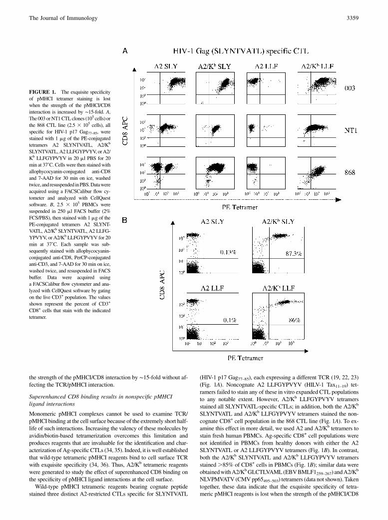
Monomeric pMHCI complexes cannot be used to examine TCR/pMHCI binding at the cell surface because of the extremely short half-life of such interactions. Increasing the valency of these molecules byavidin/biotin-based tetramerization overcomes this limitation andproduces reagents that are invaluable for the identification and char-acterization ofAg-specificCTLs (34, 35). Indeed, it is well establishedthat wild-type tetrameric pMHCI reagents bind to cell surface TCRwith exquisite specificity (34, 36). Thus, A2/Kb tetrameric reagentswere generated to study the effect of superenhanced CD8 binding onthe specificity of pMHCI ligand interactions at the cell surface.Wild-type pMHCI tetrameric reagents bearing cognate peptide
stained three distinct A2-restricted CTLs specific for SLYNTVATL
(HIV-1 p17 Gag77–85), each expressing a different TCR (19, 22, 23)(Fig. 1A). Noncognate A2 LLFGYPVYV (HILV-1 Tax11–19) tet-ramers failed to stain any of these in vitro expanded CTL populationsto any notable extent. However, A2/Kb LLFGYPVYV tetramersstained all SLYNTVATL-specific CTLs; in addition, both the A2/Kb
SLYNTVATL and A2/Kb LLFGYPVYV tetramers stained the non-cognate CD8+ cell population in the 868 CTL line (Fig. 1A). To ex-amine this effect in more detail, we used A2 and A2/Kb tetramers tostain fresh human PBMCs. Ag-specific CD8+ cell populations werenot identified in PBMCs from healthy donors with either the A2SLYNTVATL or A2 LLFGYPVYV tetramers (Fig. 1B). In contrast,both the A2/Kb SLYNTVATL and A2/Kb LLFGYPVYV tetramersstained.85% of CD8+ cells in PBMCs (Fig. 1B); similar data wereobtainedwithA2/KbGLCTLVAML(EBVBMLF1259–267) andA2/K
b
NLVPMVATV (CMVpp65495–503) tetramers (data not shown). Takentogether, these data indicate that the exquisite specificity of tetra-meric pMHCI reagents is lost when the strength of the pMHCI/CD8
FIGURE 1. The exquisite specificity
of pMHCI tetramer staining is lost
when the strength of the pMHCI/CD8
interaction is increased by ∼15-fold. A,The 003orNT1CTLclones (105 cells) or
the 868 CTL line (2.5 3 105 cells), all
specific for HIV-1 p17 Gag77–85, were
stained with 1 mg of the PE-conjugated
tetramers A2 SLYNTVATL, A2/Kb
SLYNTVATL,A2LLFGYPVYV, orA2/
Kb LLFGYPVYV in 20 ml PBS for 20
min at 37˚C. Cells were then stainedwith
allophycocyanin-conjugated anti-CD8
and 7-AAD for 30 min on ice, washed
twice, and resuspended inPBS.Datawere
acquired using a FACSCalibur flow cy-
tometer and analyzed with CellQuest
software. B, 2.5 3 105 PBMCs were
suspended in 250 ml FACS buffer (2%
FCS/PBS), then stained with 1 mg of the
PE-conjugated tetramers A2 SLYNT-
VATL, A2/Kb SLYNTVATL, A2 LLFG-
YPVYV, or A2/Kb LLFGYPVYV for 20
min at 37˚C. Each sample was sub-
sequently stained with allophycocyanin-
conjugated anti-CD8, PerCP-conjugated
anti-CD3, and 7-AAD for 30 min on ice,
washed twice, and resuspended in FACS
buffer. Data were acquired using
a FACSCalibur flow cytometer and ana-
lyzed with CellQuest software by gating
on the live CD3+ population. The values
shown represent the percent of CD3+
CD8+ cells that stain with the indicated
tetramer.
The Journal of Immunology 3359

interaction is increased by ∼15-fold. Thus, the low solution affinitiesof the wild-type pMHCI/CD8 interaction are required to maintainpMHCI binding specificity at the cell surface.
A2/Kb tetramers bind the majority of CTLs in peripheral blood
Noncognate A2/Kb tetramers were observed to bind ∼80% of theCD8a+ population in peripheral blood (Fig.1B). Although CD8a ispredominantly found on the surface of ab-TCR+ CTLs, it is alsofound on the surface of other lymphocytes, most notably some gd
T cells and NK cells.We therefore sought to determine the identity ofthe CD8a+ cells that stain with A2/Kb tetramers. Staining of fresh exvivo PBMCs isolated from healthy A2+ donors revealed that CD8awas expressed on ∼39%, 54%, and 32% of the ab-TCR+, NK cells,and gd-TCR+ populations, respectively, with some variation betweendonors (Fig. 2A). The majority of gd-TCR+ (∼93.6%) and NK cells(∼77%) failed to stainwith theA2/Kb ILAKFLHWL (hTERT540–548)tetramer and no significant binding was observed with the corre-sponding A2 tetramer (Fig. 2B). However, the vast majority of
ab-TCR+/CD8+ cells within the lymphocyte population stainednonspecifically with the A2/Kb ILAKFLHWL tetramer (Fig. 2C).Wehypothesized thatmostgd-TCR+ cells andNKcellsmight fail
to bind A2/Kb tetramers because they express the CD8aa homo-dimer rather than theCD8ab heterodimer, which is expressed on thesurface of CTLs. Thus, we generated a 293T cell line that expressedCD8aa (Fig. 3A) to examine the ability of A2/Kb tetramers to bindthis homodimeric form of the CD8 coreceptor on the cell surface. Incontrast to both A2 and A2 D227K/T228A tetramers, which exhibitnormal and abrogated interactions with CD8, respectively, A2/Kb
tetramers bound to most (74.3%) of the CD8aa+ 293 T cell trans-fectants (Fig. 3A, 3B); no binding was observed in the absence ofCD8aa surface expression (Fig. 3A). Thus, A2/Kb tetramers arecapable of binding to cell surface CD8aa.Why do A2/Kb tetramers bind predominantly to the CTL pop-
ulation in peripheral blood and not to other cells that express CD8?Fig. 3B shows that A2/Kb tetramer staining is directly proportional tothe level ofCD8aa expression, such that only cellswith a higher level
FIGURE 2. A2/Kb tetramers bind the
majority of CTLs in peripheral blood. A,
2.53 105 PBMCs from an A2+ donor were
stained with PerCP-conjugated anti-CD8,
7-AAD, and either FITC-conjugated anti–-
ab-TCR, allophycocyanin-conjugated anti-
CD56 or PE-conjugated anti–gd-TCR for
30 min on ice, washed twice, and re-
suspended in PBS.B, 2.53105A2+ PBMCs
were stained with 10 mg/ml of the PE-
conjugated tetramers A2 ILAKFLHWL or
A2/Kb ILAKFLHWL for 20 min at 37˚C.
After washing, cells were subsequently
stained with 7-AAD and either FITC-con-
jugated anti–gd-TCR or FITC-conjugated
anti-CD56 for 30 min on ice, washed twice,
and resuspended in PBS. C, 2.5 3 105 A2+
PBMCs were stained with 10 mg/ml of the
PE-conjugated tetramers A2 ILAKFLHWL
or A2/Kb ILAKFLHWL for 20 min at
37˚C. After washing, cells were stained
with allophycocyanin-conjugated anti-CD8,
FITC-conjugated anti–ab-TCR and 7-AAD
for 30 min on ice, washed twice, and re-
suspended in PBS. In A, B, and C, data were
acquired using a FACSCalibur flow cy-
tometer and analyzed with FlowJo software.
3360 NONSPECIFIC CTL ACTIVATION BY CD8 SUPERENHANCED MHCI

ofCD8aa expression stainwith this reagent. Examination of PBMCsfrom healthy donors revealed that CD8+ab-TCR+ cells express highlevels of CD8, whereas NK and gd-TCR+ cells express substantiallylower levels (Fig. 3C). Therefore, increasing the strength of thepMHCI/CD8 interaction allows pMHCI ligand binding at the cellsurface that can be mediated through the engagement of eitherCD8aa or CD8ab. However, our results suggest that binding is onlyobserved when cells express CD8 at levels above a certain threshold.Importantly, these data demonstrate that TCR expression is not re-quired for cell surface binding of A2/Kb tetramers.
A2/Kb tetramers activate CTLs irrespective of TCR specificity
It is well established that pMHCI tetramers can activate CTLs bearingcognate TCR [reviewed in (35)]. However, previous studies haveshown that pMHCI tetrameric binding at the cell surface does notnecessarily equate with activation (11, 37). Thus, we next examinedwhether nonspecific A2/Kb tetramer binding at the cell surface(Figs. 1–3) could activate human CTLs. Initially, we studied the A2-restricted SLYNTVATL-specific CTL clone 003 (23). Consistent withour findings previously stated, both A2 SLYNTVATL and A2/Kb
SLYNTVATL tetramers stained 003 CTLs efficiently, as did thenoncognate A2/Kb LLFGYPVYV tetramer; no staining was observedwith theA2LLFGYPVYV tetramer (Fig. 4A). On ligation, it is knownthat TCRs are downregulated from the cell surface (38). The cognate
A2 tetramer was able to induce significant TCR downregulation, evenat tetramer concentrations well below the limits of detection by flowcytometry; no TCR downregulation was observed with the non-cognate A2 LLFGYPVYV tetramer (Fig. 4B). In contrast, however,both the A2/Kb SLYNTVATL and A2/Kb LLFGYPVYV tetramersinducedTCRdownregulation, although this occurred to a lesser extentwith the noncognate form compared with either of the cognate tet-ramers (Fig. 4B). This TCR downregulation correlated with variousfunctional readouts typical of CTL effector activity, including theproduction of RANTES (Fig. 4C), IFN-g, and MIP-1b (data notshown). Similar results were observed with SLYNTVATL-specificCTLs bearing an alternative cognate TCR (Fig. 4D, 4E). Consistentwith the staining patterns (Fig. 4A), the activation of CTLs by non-cognate A2/Kb tetramers was less efficient than that induced by tet-ramers bearing the agonist peptide (Fig. 4C–E).To dissect this effect further at the single-cell level within a clonal
CTL population, we used a flow cytometric assay for degranulationbased on the detection of CD107a mobilized on to the cell surface(32). The noncognate A2/Kb tetramer, in this case folded around theGLCTLVAML peptide, induced degranulation in 15% of 003 CTLsat a concentration of 5 mg/ml (Fig. 4F); the cognate A2 SLYNT-VATL and A2/Kb SLYNTVATL tetramers induced almost 40%degranulation (data not shown). Notably, the cells that degranulatedin response to the A2/Kb GLCTLVAML tetramer were contained
FIGURE 3. Nonspecific A2/Kb tetramer binding is influenced byCD8 cell surface density. A and B, 23 105 293T cells were incubated6 10mg/ml of the PE-
conjugated tetramers A2 D227K/T228A ILAKFLHWL, A2 ILAKFLHWL, or A2/Kb ILAKFLHWL for 20 min at 37˚C, then stained with 7-AAD and either
FITC-conjugated anti-CD8or PE-conjugated anti-CD8b for 30min on ice,washed twice, and resuspended in PBS.C, 2.53105 PBMCswere stainedwith PerCP-
conjugated anti-CD8, 7-AAD, and either FITC-conjugated anti–ab-TCR, allophycocyanin-conjugated anti-CD56, or PE-conjugated anti–gd-TCR for 30min on
ice, washed twice, and resuspended in PBS. In A, B, and C, data were acquired using a FACSCalibur flow cytometer and analyzed with FlowJo software.
The Journal of Immunology 3361

almost exclusively within the tetramerhighCD8high population (Fig.4F). Thus, at least to some extent, the strong interaction betweenA2/Kb and CD8 can bypass the requirement for a specific TCR/pMHCI interaction and nonspecifically activate human CTLs.
Cell surface-expressed A2/Kb activates CTLs in the absence ofcognate Ag
To extend our investigation to the effects of cell surface pMHCI pre-sentation, C1R cells were transfected with either A2 or A2/Kb; stabletransfectants expressing similar cell surface MHCI densities were se-lected as targets for further experiments. Target cells expressing eitherA2 or A2/Kb were incubated overnight with three A2-restricted CTLclones with different peptide specificities (Mel13, 003, and ILA1).Targets that expressed A2 failed to activate any of the CTL clonessignificantly above background (Fig. 5A). Remarkably, however, theA2/Kb targets stimulated Mel13, 003, and ILA1 CTLs to produce
significant amounts of MIP-1b in the absence of specific peptide (Fig.5A).A2/Kb targets also elicited substantial levels ofTNFa and IFN-g attitratable E:T ratios (Fig. 5B), induced degranulation (Fig. 5C), andinduced significant levels of killing (data not shown) in the absence ofspecific TCR/pMHCI interactions.
Cell surface-expressedA2/Kb primes noncognateCTL expansions
Thymic output in healthyA2+ individuals is known to generate a highfrequency of naıve CD8+ T cells that can recognize the self-AgMelan-A26–35 (39); this system can be used to examine the priming ofCTLs directly ex vivo (40). We exploited these observations to in-vestigate the effect of superenhanced pMHCI/CD8 binding on CTLpriming. In priming experiments conductedwithC1R target cells, thepercentages of CTLs specific forMelan-A26–35 thatwere present after10 d in culture were related to the context of the pMHCI/CD8 in-teraction in which the cognate ELAGIGILTV peptidewas presented.
FIGURE 4. A2/Kb tetramers can activate CTLs in the absence of a specific TCR/pMHCI interaction.A, 105 003CTLswere suspended in 20ml PBS and stained
with the PE-conjugated tetramersA2 SLYNTVATL,A2/Kb SLYNTVATL,A2 LLFGYPVYV, or A2/Kb LLFGYPVYVat the indicated concentrations and 7-AAD
for 20min at 37˚C. Cells were then washed twice and resuspended in PBS. Data were acquired using a FACSCalibur flow cytometer and analyzed with CellQuest
software. B, 105 003 CTLs were suspended in 40 ml R2 with the PE-conjugated tetramers A2 SLYNTVATL, A2/Kb SLYNTVATL, A2 LLFGYPVYV, or A2/Kb
LLFGYPVYVat the indicated concentrations for 30 min at 37˚C. Cells were subsequently stained with FITC-conjugated anti–ab-TCR, 7-AAD, and allophy-
cocyanin-conjugated anti-CD8 for 30 min on ice in azide buffer (0.1% azide/2% FCS/PBS). After two washes, data were acquired using a FACSCalibur flow
cytometer and analyzedwith CellQuest software.C, 53 105 003CTLs were incubatedwith the PE-conjugated tetramers A2 SLYNTVATL,A2/Kb SLYNTVATL,
A2 LLFGYPVYV, or A2/Kb LLFGYPVYVat the indicated concentrations. After 4 h at 37˚C, supernatants were harvested and assayed for RANTES, IFN-g and
MIP-1b content by ELISA (only RANTES shown). D, 2 3 103 868 CTLs were incubated for 4 h at 37˚C with 1 mg/ml of the PE-conjugated tetramers A2
SLYNTVATL,A2/KbSLYNTVATL,A2LLFGYPVYV, orA2/KbLLFGYPVYVin an IFN-gELISpot assay.E, 1.253105 868CTLswere incubatedwith 1mg/ml
of the PE-conjugated tetramers A2 SLYNTVATL, A2/Kb SLYNTVATL, A2 LLFGYPVYV, or A2/Kb LLFGYPVYV for 4 h at 37˚C. The supernatant was sub-
sequently assayed for MIP-1b content by ELISA. C–E show the mean6 SD of two replicate assays. Results similar to A–E were also obtained with tetramers
conjugated to fluorochromes other than PE (data not shown). F, 003 CTLs were incubated with the PE-conjugated tetramers A2 SLYNTVATL, A2/Kb SLYNT-
VATL, A2 GLCTLVAML, or A2/Kb GLCTLVAML at the indicated concentrations for 4 h at 37˚C, then stainedwith allophycocyanin-conjugated anti-CD8 for 20
min on ice and assayed forCD107amobilization as described inMaterials andMethods. The inset plot shows staining for allophycocyanin-conjugated anti-CD8on
the x-axis andPE-conjugatedA2/KbGLCTLVAMLtetramer (5mg/ml) on they-axis.Backgated tetramer+CD107a+ cells are shown inblack and tetramer+CD107a2
cells are shown in gray. TetramerhighCD8high cells are preferentially activated by the A2/Kb tetramer.
3362 NONSPECIFIC CTL ACTIVATION BY CD8 SUPERENHANCED MHCI

Thus, in the absenceof a pMHCI/CD8 interaction (A2D227K/T228AC1R targets), only 1.5% of the CD8+ cell population was specific forMelan-A26–35; in contrast, 5.6% and 5.7% of the CD8+ populationbound the A2 ELAGIGILTV tetramer in the same experiment whenpriming was conducted with A2 and A2/Kb C1R targets, respectively(Fig. 6). Exposure to A2/Kb C1R targets also resulted in substantialexpansions of the total CD8+ population (Fig. 6). Similar resultswere obtained with multiple donors (data not shown). Thus, targetcells that expressMHCImolecules with superenhancedCD8-bindingproperties can induce nonspecific expansions of CD8+ cells in theabsence of cognate Ag.
Nonspecific A2/Kb-mediated CTL activation and tetramerstaining are not dependent on TCR expression
In earlier experiments, we observed that A2/Kb tetramers bound tothe majority of ab-TCR+CD8+ cells in PBMCs derived from A2+
donors (Fig. 2). To exclude the possibility that this phenomenon was
dependent on the presence of A2-restricted TCRs, we conductedstaining experiments with A22 PBMCs. As previously, the A2/Kb
ILAKFLHWL tetramer bound nonspecifically to the majority ofCD8+ cells (Fig. 7A). Furthermore, A2/Kb tetramer binding favoredCD8high cells and was abrogated by pretreatment with the anti-CD8mAbDK25 (Fig. 7A). Thus, consistent with the data shown in Fig. 3,nonspecific A2/Kb tetramer binding is a CD8-mediated effect that isnot dependent on the presence ofA2-restrictedTCRs. In addition, wedemonstrated in earlier experiments that A2/Kb, both in soluble andcell-associated form, nonspecifically activated A2-restricted CTL(Figs. 4, 5). To confirm that these functional correlates of nonspecificbinding were similarly independent of A2-restricted TCR expres-sion, we extended our studies to CTL clones restricted by non-A2MHCI molecules. In all cases, cell surface-expressed A2/Kb acti-vated CTL clones regardless of restriction element (Fig. 7B).
DiscussionCD8 has the potential to engage all pMHCI complexes, both selfand foreign, because it binds to largely nonpolymorphic regions ofthe MHCI molecule. Indeed, recent publications suggest that theability of CD8 to interact with nonstimulatory pMHCI complexeslowers T cell activation thresholds and enables CTLs to respond tolow copy numbers of specific pMHCI (41, 42). It therefore remainsunclear how the specificity of TCR recognition is maintained,despite the potential for multiple pMHCI/CD8 interactions at thecell surface. One possibility resides in the fact that the binding ofCD8 to MHCI is characterized by very low affinities and ex-tremely rapid kinetics. In this study, we have generated chimericA2/Kb MHCI molecules that increase the strength of the pMHCI/CD8 interaction by ∼15-fold to probe the biophysical and func-tional significance of the low solution binding affinities observedfor the pMHCI/CD8 interaction.Initially, we examined the effect of superenhanced CD8 binding on
pMHCItetramerbindingat thecell surface. Increasingthestrengthof thepMHCI/CD8interactionby∼15-fold resulted in the total lossofpMHCI
FIGURE 5. Cell surface-expressed A2/Kb activates
CTLs in the absence of cognate Ag. A, 2.5 3 104
Mel13, 003, or ILA1 CTLs were incubated for 12 h at
37˚C with 105 C1R cells stably transfected to express
equal levels of either A2 or A2/Kb at the cell surface.
Supernatant was subsequently assayed for MIP-1b
content by ELISA. The mean 6 SD of two replicate
assays is shown. B, 2.5 3 104 Mel13 CTLs were in-
cubated for 12 h at 37˚C with 105 C1R cells stably
transfected to express either A2 or A2/Kb at the cell
surface. Supernatant was assayed for IFN-g and TNFa
content by cytokine bead array. C, CD107a expression
by ILA1 and Mel13 CTLs after a 12-h incubation at
37˚CwithC1Rcells stably transfected to express either
A2 or A2/Kb on the cell surface. For A–C, C1R cells
were not previously pulsed with peptide.
FIGURE 6. Cell surface-expressed A2/Kb primes nonspecific expansion
of CD8+ cells. 106 A2+ PBMCs were incubated with 2 3 105 irradiated A2
D227K/T228A,A2, or A2/Kb C1R cells that had previously been pulsedwith
1 mM ELAGIGILTV (Melan-A26–35) peptide in R10. From day 3, IL-2 was
added in increments to reach a maximum concentration of 200 IU/ml by day
10. Lines were subsequently stained with PE-conjugated A2 ELAGIGILTV
tetramer, followed by allophycocyanin-conjugated anti-CD8 and 7-AAD.
Data were acquired using a FACSCalibur flow cytometer and analyzed with
FlowJo software.
The Journal of Immunology 3363

tetramer binding specificity. Thus, irrespective of restriction elementand the presented peptide, A2/Kb tetramers bound to the surface of allCTL clones examined in this study and to the majority of CTLs presentwithin PBMCs (Figs. 1, 2, 7A). In addition, A2/Kb tetramers bound tothe cell surface in the absence of TCR expression (Fig. 3) and non-specific binding was abrogated by pretreatment with an anti-CD8 Ab(Fig. 7A), thereby demonstrating that the observed loss of pMHCI tet-ramer binding specificity was CD8 mediated and TCR independent.These findings indicate that the low solution binding affinities observedfor the pMHCI/CD8 interaction are essential for the preservation ofpMHCI ligand binding specificity at the cell surface.IthaspreviouslybeendocumentedthatpMHCItetramersareefficient
activators of cognate CTLs [reviewed in (35)]. However, pMHCI tet-ramer staining does not necessarily equate with cellular activation.Therefore, we proceeded to examine the ability of A2/Kb tetramers toactivate CTL clones. Notably, we found that A2/Kb tetramers activatedCTL clones in a nonspecific manner (Fig. 4). Activation resulted ina full range of effector functions, including cytokine/chemokine re-lease, degranulation, and killing. Flow cytometric assessment of de-granulation by analysis of CD107a mobilization revealed that CTLswith higher surface expression of CD8 were the cells most likely toactivate in response toA2/Kbmolecules. This finding led us to examinethe effects of cell-surface presentedAg. Strikingly, exposure of PBMCsto C1R target cells bearing A2/Kb molecules caused a general non-specific expansion of CD8+ cells during the course of the experiment(Fig. 6). Furthermore, A2/Kb C1R cells, unlike their wild-type A2counterparts, were capable of stimulating effector function in all CTL
clones tested regardless of specificity and MHCI restriction (Figs. 5,7B). Although we cannot exclude the possibility that inclusion of themurinea3domain induces conformational changes at theTcell surfaceonbinding toCD8 that favor noncognate activation, this seemsunlikelygiven that: 1) theTCRbindingsite remainsunaltered (9, 22); 2) adegreeof noncognate activation can be observed in long-term assays withnonchimeric human MHCI molecules that exhibit incrementally en-hanced CD8 binding (data not shown); and 3) murine and humanpMHCI/CD8aa cocrystals exhibit similar binding orientations (14,43). Furthermore, these results are consistent with the observation thatthymus leukemia Ag, which interacts strongly (KD ∼12 mM) with cellsurface CD8aa expressed by intraepithelial lymphocytes, can modu-late T cell responses independently of the TCR (44–46).How does a superenhanced pMHCI/CD8 interaction result in
nonspecific CTL activation?We have previously demonstrated thatan incremental increase in the pMHCI/CD8 interaction (A2Q115E)results in enhanced immunogenicity of cognate Ags and that thiseffect is mediated by enhanced early intracellular signal trans-duction (9, 47). In contrast, the stimulatory properties of A2/Kb
molecules exhibited no peptide specificity requirements whatso-ever; indeed, cell surface-expressed A2/Kb was shown to activateeven non-A2–restricted CTL clones (Fig. 7B), thereby confirmingthat cognate TCR/pMHCI interactions are not required. Combinedwith the ability of A2/Kb to engage multiple CD8 molecules at thecell surface, these results suggest that A2/Kb cross-links CD8 andinduces activation in an “Ab-like” manner. Indeed, this is con-sistent with previous studies demonstrating that Ab-induced CD8
FIGURE 7. Noncognate A2/Kb-mediated CTL activation and tetramer binding is not influenced byMHCI restriction. A, 2.53 105 PBMCswere suspended
in 250ml FACSbuffer (2%FCS/PBS) and stainedwith FITC-conjugated anti-A2 and 7-AAD for 30min on ice, thenwashed twice, and resuspended in PBS. For
pMHCI tetramer staining experiments, 2.53 105 PBMCs were suspended in 50ml FACS buffer (2% FCS/PBS) and incubated6 10mg/ml unconjugated anti-
CD8 for 20min on ice, then stainedwith 10mg/ml of the PE-conjugated tetramers A2 ILAKFLHWLorA2/Kb ILAKFLHWL for 45min on ice. After washing,
cells were subsequently stained with allophycocyanin-conjugated anti-CD8 and 7-AAD, washed again, and resuspended in PBS. Data were acquired using
a FACSCalibur flow cytometer and analyzed with FlowJo software.B, 2.53 104 CTLs were incubated for 12 h at 37˚Cwith 105 unpulsed C1R cells expressing
either A2 or A2/Kb on the cell surface. The following CTL clones were used: 1) the HLAAp6801-restricted CTL clone c23, specific for the HIV-1 Tat-derived
epitope ITKGLGISYGR (residues 38–48); 2) the HLA Bp0702-restricted CTL clone KD4, specific for the EBV EBNA3A-derived epitope RPPIFIRRL
(residues 379–387); 3) theHLABp0801-restricted CTL cloneLC13, specific for the EBVEBNA3A-derived epitope FLRGRAYGL (residues 339–347); and 4)
the HLA Bp3508-restricted CTL clone SB27, specific for the EBV BZLF1-derived epitope LPEPLPQGQLTAY (residues 52–64). Supernatant was sub-
sequently assayed for MIP-1b content by ELISA. The mean6 SD of two replicate assays is shown.
3364 NONSPECIFIC CTL ACTIVATION BY CD8 SUPERENHANCED MHCI

cross-linking can induce T cell signaling (48, 49) and elicitdownstream effector functions, such as chemokine release (50);such effects are predictable given that the CD8a tail is coupled top56lck, an essential component of the early intracellular signalingpathway (10). It is interesting to note that the murine pMHCI/CD8interaction is significantly stronger (KD ∼30 mM) than the equiv-alent human interaction (KD ∼150 mM) (11), but does not result innoncognate CTL activation. It is therefore likely that a pMHCI/CD8 interaction affinity threshold exists for the maintenance ofCTL activation specificity. The strength of the murine pMHCI/CD8 interaction is 3-fold weaker than the strength of the in-teraction measured between A2/Kb and human CD8, thereby stilloperating at a level below this threshold.In summary, we used chimeric MHCI molecules that exhibit
a superenhanced interaction with CD8 to probe the physical andfunctionalsignificanceofthe lowsolutionbindingaffinitiespreviouslydescribed for the pMHCI/CD8 interaction. We found that increasingthe strengthof the pMHCI/CD8 interactionby∼15-fold resulted in: 1)total loss of pMHCI binding specificity at the cell surface; 2) non-cognate pMHCI tetramer-mediated activation; and 3) nonspecificactivation and proliferation triggered by cell surface-expressedpMHCI molecules. Thus, the low solution binding affinity of thepMHCI/CD8 interaction is essential for the preservation of pMHCIligand binding specificity at the cell surface and its attendant func-tional repercussions.
AcknowledgmentsWe thank E. Wang for provision of the allophycocyanin-conjugated mAb
specific for human CD56.
DisclosuresThe authors have no financial conflicts of interest.
References1. Davis, M. M., J. J. Boniface, Z. Reich, D. Lyons, J. Hampl, B. Arden, and Y. Chien.
1998. Ligand recognition by alpha beta T cell receptors. Annu. Rev. Immunol. 16:523–544.
2. Rudolph, M. G., and I. A. Wilson. 2002. The specificity of TCR/pMHC interaction.Curr. Opin. Immunol. 14: 52–65.
3. Zamoyska, R. 1998. CD4 and CD8: modulators of T-cell receptor recognition ofantigen and of immune responses? Curr. Opin. Immunol. 10: 82–87.
4. Holler, P. D., and D. M. Kranz. 2003. Quantitative analysis of the contribution ofTCR/pepMHC affinity and CD8 to T cell activation. Immunity 18: 255–264.
5. Gakamsky, D. M., I. F. Luescher, A. Pramanik, R. B. Kopito, F. Lemonnier,H. Vogel, R. Rigler, and I. Pecht. 2005. CD8 kinetically promotes ligand bindingto the T-cell antigen receptor. Biophys. J. 89: 2121–2133.
6. Laugel, B., H. A. van den Berg, E. Gostick, D. K. Cole, L. Wooldridge, J. Boulter,A. Milicic, D. A. Price, and A. K. Sewell. 2007. Different T cell receptor affinitythresholds and CD8 coreceptor dependence govern cytotoxic T lymphocyte acti-vation and tetramer binding properties. J. Biol. Chem. 282: 23799–23810.
7. van den Berg, H. A., L. Wooldridge, B. Laugel, and A. K. Sewell. 2007. CoreceptorCD8-driven modulation of T cell antigen receptor specificity. J Theor Biol 249:395-408.
8. Luescher, I. F., E. Vivier, A. Layer, J. Mahiou, F. Godeau, B. Malissen, and P. Romero.1995. CD8 modulation of T-cell antigen receptor-ligand interactions on living cytotoxicT lymphocytes. Nature 373: 353–356.
9. Wooldridge, L., H.A. van denBerg,M.Glick, E.Gostick, B. Laugel, S. L. Hutchinson,A. Milicic, J. M. Brenchley, D. C. Douek, D. A. Price, and A. K. Sewell. 2005. In-teraction between the CD8 coreceptor and major histocompatibility complex class Istabilizes T cell receptor-antigen complexes at the cell surface. J. Biol. Chem. 280:27491–27501.
10. Veillette, A., M. A. Bookman, E. M. Horak, and J. B. Bolen. 1988. The CD4 andCD8 T cell surface antigens are associated with the internal membrane tyrosine-protein kinase p56lck. Cell 55: 301–308.
11. Purbhoo, M. A., J. M. Boulter, D. A. Price, A. L. Vuidepot, C. S. Hourigan,P. R. Dunbar, K. Olson, S. J. Dawson, R. E. Phillips, B. K. Jakobsen, et al. 2001.The human CD8 coreceptor effects cytotoxic T cell activation and antigensensitivity primarily by mediating complete phosphorylation of the T cell re-ceptor zeta chain. J. Biol. Chem. 276: 32786–32792.
12. Arcaro, A., C. Gregoire, T. R. Bakker, L. Baldi, M. Jordan, L. Goffin, N. Boucheron,F. Wurm, P. A. van der Merwe, B. Malissen, and I. F. Luescher. 2001. CD8betaendows CD8 with efficient coreceptor function by coupling T cell receptor/CD3 toraft-associated CD8/p56(lck) complexes. J. Exp. Med. 194: 1485–1495.
13. Arcaro, A., C. Gregoire, N. Boucheron, S. Stotz, E. Palmer, B. Malissen, andI. F. Luescher. 2000. Essential role of CD8 palmitoylation in CD8 coreceptorfunction. J. Immunol. 165: 2068–2076.
14. Gao, G. F., J. Tormo, U. C. Gerth, J. R. Wyer, A. J. McMichael, D. I. Stuart, J. I. Bell,E. Y. Jones, and B. K. Jakobsen. 1997. Crystal structure of the complex betweenhuman CD8alpha(alpha) and HLA-A2. Nature 387: 630–634.
15. Wyer, J. R., B. E. Willcox, G. F. Gao, U. C. Gerth, S. J. Davis, J. I. Bell, P. A. vander Merwe, and B. K. Jakobsen. 1999. T cell receptor and coreceptor CD8 al-phaalpha bind peptide-MHC independently and with distinct kinetics. Immunity10: 219–225.
16. Gao, G. F., B. E. Willcox, J. R. Wyer, J. M. Boulter, C. A. O’Callaghan, K. Maenaka,D. I. Stuart, E. Y. Jones, P. A. Van Der Merwe, J. I. Bell, and B. K. Jakobsen. 2000.Classical and nonclassical class I major histocompatibility complex molecules exhibitsubtle conformational differences that affect binding to CD8alphaalpha. J. Biol.Chem. 275: 15232–15238.
17. Dustin, M. L., D. E. Golan, D. M. Zhu, J. M. Miller, W. Meier, E. A. Davies, andP. A. van der Merwe. 1997. Low affinity interaction of human or rat T cell adhesionmolecule CD2 with its ligand aligns adhering membranes to achieve high physi-ological affinity. J. Biol. Chem. 272: 30889–30898.
18. Cole, D. K., N. J. Pumphrey, J. M. Boulter, M. Sami, J. I. Bell, E. Gostick, D. A. Price,G. F. Gao, A. K. Sewell, and B. K. Jakobsen. 2007. Human TCR-binding affinity isgoverned by MHC class restriction. J. Immunol. 178: 5727–5734.
19. Varela-Rohena, A., P. E. Molloy, S. M. Dunn, Y. Li, M. M. Suhoski, R. G. Carroll,A. Milicic, T. Mahon, D. H. Sutton, B. Laugel, et al. 2008. Control of HIV-1immune escape by CD8 T cells expressing enhanced T-cell receptor. Nat. Med. 14:1390–1395.
20. Stone, J. D., A. S. Chervin, and D. M. Kranz. 2009. T-cell receptor binding af-finities and kinetics: impact on T-cell activity and specificity. Immunology 126:165–176.
21. Hutchinson, S. L., L. Wooldridge, S. Tafuro, B. Laugel, M. Glick, J. M. Boulter,B. K. Jakobsen, D. A. Price, and A. K. Sewell. 2003. The CD8 T cell coreceptorexhibits disproportionate biological activity at extremely low binding affinities.J. Biol. Chem. 278: 24285–24293.
22. Choi, E. M., J. L. Chen, L. Wooldridge, M. Salio, A. Lissina, N. Lissin, I. F. Hermans,J. D. Silk, F. Mirza, M. J. Palmowski, et al. 2003. High avidity antigen-specific CTLidentified by CD8-independent tetramer staining. J. Immunol. 171: 5116–5123.
23. Sewell, A. K., G. C. Harcourt, P. J. Goulder, D. A. Price, and R. E. Phillips. 1997.Antagonism of cytotoxic T lymphocyte-mediated lysis by natural HIV-1 alteredpeptide ligands requires simultaneous presentation of agonist and antagonistpeptides. Eur. J. Immunol. 27: 2323–2329.
24. Purbhoo, M. A., Y. Li, D. H. Sutton, J. E. Brewer, E. Gostick, G. Bossi,B. Laugel, R. Moysey, E. Baston, N. Liddy, et al. 2007. The HLA Ap0201-re-stricted hTERT(540-548) peptide is not detected on tumor cells by a CTL cloneor a high-affinity T-cell receptor. Mol. Cancer Ther. 6: 2081–2091.
25. Gostick, E., D. K. Cole, S. L. Hutchinson, L. Wooldridge, S. Tafuro, B. Laugel,A. Lissina, A. Oxenius, J. M. Boulter, D. A. Price, and A. K. Sewell. 2007.Functional and biophysical characterization of an HLA-Ap6801-restricted HIV-specific T cell receptor. Eur. J. Immunol. 37: 479–486.
26. Burrows, S. R., S. L. Silins, D. J. Moss, R. Khanna, I. S. Misko, and V. P. Argaet.1995. T cell receptor repertoire for a viral epitope in humans is diversified bytolerance to a background major histocompatibility complex antigen. J. Exp.Med. 182: 1703–1715.
27. Kjer-Nielsen, L., C. S. Clements, A. W. Purcell, A. G. Brooks, J. C. Whisstock,S. R. Burrows, J. McCluskey, and J. Rossjohn. 2003. A structural basis for theselection of dominant alphabeta T cell receptors in antiviral immunity. Immunity18: 53–64.
28. Tynan, F. E., S. R. Burrows, A. M. Buckle, C. S. Clements, N. A. Borg, J. J. Miles,T. Beddoe, J. C. Whisstock, M. C. Wilce, S. L. Silins, et al. 2005. T cell receptorrecognition of a ‘super-bulged’ major histocompatibility complex class I-boundpeptide. Nat. Immunol. 6: 1114–1122.
29. Tynan, F. E., N. A. Borg, J. J. Miles, T. Beddoe, D. El-Hassen, S. L. Silins,W. J. van Zuylen, A. W. Purcell, L. Kjer-Nielsen, J. McCluskey, et al. 2005. Highresolution structures of highly bulged viral epitopes bound to major histocom-patibility complex class I. Implications for T-cell receptor engagement and T-cellimmunodominance. J. Biol. Chem. 280: 23900–23909.
30. Willemsen, R., C. Ronteltap, M. Heuveling, R. Debets, and R. Bolhuis. 2005.Redirecting human CD4+ T lymphocytes to the MHC class I-restricted mela-noma antigen MAGE-A1 by TCR alphabeta gene transfer requires CD8alpha.Gene Ther. 12: 140–146.
31. Willemsen, R. A., Z. Sebestyen, C. Ronteltap, C. Berrevoets, J. Drexhage, andR. Debets. 2006. CD8 alpha coreceptor to improve TCR gene transfer to treat mel-anoma: down-regulation of tumor-specific production of IL-4, IL-5, and IL-10.J. Immunol. 177: 991–998.
32. Betts, M. R., J. M. Brenchley, D. A. Price, S. C. De Rosa, D. C. Douek,M. Roederer, and R. A. Koup. 2003. Sensitive and viable identification of antigen-specific CD8+ T cells by a flow cytometric assay for degranulation. J. Immunol.Methods 281: 65–78.
33. Choi, E. M., M. Palmowski, J. Chen, and V. Cerundolo. 2002. The use of chi-meric A2K(b) tetramers to monitor HLA A2 immune responses in HLA A2transgenic mice. J. Immunol. Methods 268: 35–41.
34. Altman, J. D., P. A. Moss, P. J. Goulder, D. H. Barouch, M. G. McHeyzer-Williams, J. I. Bell, A. J. McMichael, and M. M. Davis. 1996. Phenotypicanalysis of antigen-specific T lymphocytes. Science 274: 94–96.
35. Wooldridge, L., A. Lissina, D. K. Cole, H. A. van den Berg, D. A. Price, andA. K. Sewell. 2009. Tricks with tetramers: how to get the most from multimericpeptide-MHC. Immunology 126: 147–164.
The Journal of Immunology 3365

36. Burrows, S. R., N. Kienzle, A. Winterhalter, M. Bharadwaj, J. D. Altman, andA. Brooks. 2000. Peptide-MHC class I tetrameric complexes display exquisiteligand specificity. J. Immunol. 165: 6229–6234.
37. Schott, E., and H. L. Ploegh. 2002. Mouse MHC class I tetramers that are unableto bind to CD8 reveal the need for CD8 engagement in order to activate naiveCD8 T cells. Eur. J. Immunol. 32: 3425–3434.
38. Valitutti, S., S. Muller, M. Cella, E. Padovan, and A. Lanzavecchia. 1995. Serialtriggering of many T-cell receptors by a few peptide-MHC complexes. Nature375: 148–151.
39. Zippelius, A., M. J. Pittet, P. Batard, N. Rufer, M. de Smedt, P. Guillaume,K. Ellefsen, D. Valmori, D. Lienard, J. Plum, et al. 2002. Thymic selectiongenerates a large T cell pool recognizing a self-peptide in humans. J. Exp. Med.195: 485–494.
40. Salio, M., M. J. Palmowski, A. Atzberger, I. F. Hermans, and V. Cerundolo.2004. CpG-matured murine plasmacytoid dendritic cells are capable of in vivopriming of functional CD8 T cell responses to endogenous but not exogenousantigens. J. Exp. Med. 199: 567–579.
41. Yachi, P. P., J. Ampudia, N. R. Gascoigne, and T. Zal. 2005. Nonstimulatorypeptides contribute to antigen-induced CD8-T cell receptor interaction at theimmunological synapse. Nat. Immunol. 6: 785–792.
42. Anikeeva, N., T. Lebedeva, A. R. Clapp, E. R. Goldman, M. L. Dustin, H. Mattoussi,and Y. Sykulev. 2006. Quantum dot/peptide-MHC biosensors reveal strong CD8-dependent cooperation between self and viral antigens that augment the T cell re-sponse. Proc. Natl. Acad. Sci. USA 103: 16846–16851.
43. Kern, P. S., M. K. Teng, A. Smolyar, J. H. Liu, J. Liu, R. E. Hussey, R. Spoerl,H. C. Chang, E. L. Reinherz, and J. H. Wang. 1998. Structural basis of CD8coreceptor function revealed by crystallographic analysis of a murine CD8al-phaalpha ectodomain fragment in complex with H-2Kb. Immunity 9: 519–530.
44. Leishman, A. J., O. V. Naidenko, A. Attinger, F. Koning, C. J. Lena, Y. Xiong,
H. C. Chang, E. Reinherz, M. Kronenberg, and H. Cheroutre. 2001. T cell re-
sponses modulated through interaction between CD8alphaalpha and the non-
classical MHC class I molecule, TL. Science 294: 1936–1939.45. Tsujimura, K., Y. Obata, E. Kondo, K. Nishida, Y. Matsudaira, Y. Akatsuka,
K. Kuzushima, and T. Takahashi. 2003. Thymus leukemia antigen (TL)-specific
cytotoxic T lymphocytes recognize the alpha1/alpha2 domain of TL free from
antigenic peptides. Int. Immunol. 15: 1319–1326.46. Cole, D.K., andG. F. Gao. 2004. CD8: adhesionmolecule, co-receptor and immuno-
modulator. Cell. Mol. Immunol. 1: 81–88.47. Wooldridge, L., A. Lissina, J. Vernazza, E. Gostick, B. Laugel, S. L. Hutchinson,
F. Mirza, P. R. Dunbar, J. M. Boulter, M. Glick, et al. 2007. Enhanced immu-
nogenicity of CTL antigens through mutation of the CD8 binding MHC class I
invariant region. Eur. J. Immunol. 37: 1323–1333.48. Grebe, K. M., R. L. Clarke, and T. A. Potter. 2004. Ligation of CD8 leads to
apoptosis of thymocytes that have not undergone positive selection. Proc. Natl.
Acad. Sci. USA 101: 10410–10415.49. Clarke, R. L., S. Thiemann, Y. Refaeli, G. Werlen, and T. A. Potter. 2009. A new
function for LAT and CD8 during CD8-mediated apoptosis that is independent of
TCR signal transduction. Eur. J. Immunol. 39: 1619–1631.50. Wooldridge, L., S. L. Hutchinson, E. M. Choi, A. Lissina, E. Jones, F. Mirza,
P. R. Dunbar, D. A. Price, V. Cerundolo, and A. K. Sewell. 2003. Anti-CD8
antibodies can inhibit or enhance peptide-MHC class I (pMHCI) multimer
binding: this is paralleled by their effects on CTL activation and occurs in the
absence of an interaction between pMHCI and CD8 on the cell surface. J. Im-
munol. 171: 6650–6660.
3366 NONSPECIFIC CTL ACTIVATION BY CD8 SUPERENHANCED MHCI