minireview multifunctional micellar nanomedicine for cancer therapy
TRANSCRIPT
MINIREVIEW
Multifunctional Micellar Nanomedicine forCancer Therapy
ELVIN BLANCO,* CHASE W. KESSINGER,* BARAN D. SUMER,� AND JINMING GAO*,1
*Department of Pharmacology, Simmons Comprehensive Cancer Center, University of TexasSouthwestern Medical Center at Dallas, Dallas, Texas 75390; and �Department of Otolaryngology,
Southwestern Medical School, University of Texas Southwestern Medical Center at Dallas,Dallas, Texas 75235
Polymeric micelles are supramolecular, core-shell nanoparticles
that offer considerable advantages for cancer diagnosis and
therapy. Their relatively small size (10–100 nm), ability to
solubilize hydrophobic drugs as well as imaging agents, and
improved pharmacokinetics provide a useful bioengineering
platform for cancer applications. Several polymeric micelle
formulations are currently undergoing phase I/II clinical trials,
which have shown improved antitumor efficacy and reduced
systemic toxicity. This minireview will focus on recent advance-
ments in the multifunctional design of micellar nanomedicine
with tumor targeting, stimulated drug release, and cancer
imaging capabilities. Such functionalization strategies result in
enhanced micellar accumulation at tumor sites, higher drug
bioavailability, as well as improved tumor diagnosis and visual-
ization of therapy. Ultimately, integrated nanotherapeutic sys-
tems (e.g., theranostic nanomedicine) may prove essential to
address the challenges of tumor heterogeneity and adaptive
resistance to achieve efficacious treatment of cancer. Exp Biol
Med 234:123–131, 2009
Key words: polymeric micelles; cancer nanomedicine; drug delivery
systems; tumor imaging; cancer targeting; controlled release
Introduction
Cancer remains as one of the leading causes of
mortality worldwide, affecting over 10 million new patients
every year. Currently, the treatment options include surgical
resection, radiation, and chemotherapy. However, although
over 90 chemotherapeutic drugs have been approved by the
FDA for clinical use, their efficacy has been severely
hindered by dose-limiting toxicity and patient morbidity.
Recently, nanoscale (10–200 nm) therapeutic systems have
emerged as novel therapeutic modalities for cancer treat-
ment (1–3). These systems include polymeric micelles,
polymer-drug conjugates, dendrimers, liposomes, and
inorganic particulates. Compared to conventional small
molecule-based therapy, nanotherapeutic systems have
several potential advantages for cancer therapy, including
higher payload capacity, prolonged blood circulation times,
reduced toxicity to healthy tissues, and improved anti-tumor
efficacy. In this article, we will review key advances of one
of these emerging nanotherapeutic systems, polymeric
micelles (4–7), and discuss their potential for cancer
therapy.
Polymeric Micelles: Properties and Advantages forCancer Treatment
The use of polymeric micelles for cancer treatment was
first reported in the early 1980s by Ringsdorf and coworkers
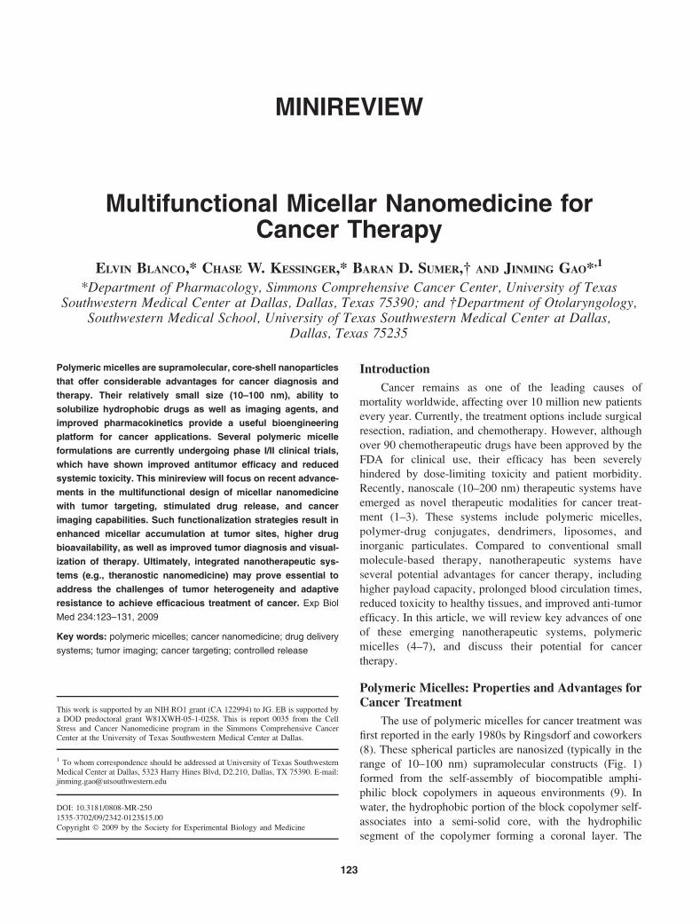
(8). These spherical particles are nanosized (typically in the
range of 10–100 nm) supramolecular constructs (Fig. 1)
formed from the self-assembly of biocompatible amphi-
philic block copolymers in aqueous environments (9). In
water, the hydrophobic portion of the block copolymer self-
associates into a semi-solid core, with the hydrophilic
segment of the copolymer forming a coronal layer. The
This work is supported by an NIH RO1 grant (CA 122994) to JG. EB is supported bya DOD predoctoral grant W81XWH-05-1-0258. This is report 0035 from the CellStress and Cancer Nanomedicine program in the Simmons Comprehensive CancerCenter at the University of Texas Southwestern Medical Center at Dallas.
1 To whom correspondence should be addressed at University of Texas SouthwesternMedical Center at Dallas, 5323 Harry Hines Blvd, D2.210, Dallas, TX 75390. E-mail:[email protected]
123
DOI: 10.3181/0808-MR-250
1535-3702/09/2342-0123$15.00
Copyright � 2009 by the Society for Experimental Biology and Medicine
resulting core-shell architecture is important for drug
delivery purposes, because the hydrophobic core can act
as a reservoir for water insoluble drugs, while the outer shell
protects the micelle from rapid clearance (10). Although
several functional aspects of the constituent blocks have
been explored (e.g., temperature or pH sensitive blocks), the
most important criteria are biocompatibility and/or biode-
gradability. Currently, the most commonly used corona-
forming polymer is polyethylene glycol (PEG), with a
molecular weight range from 2 to 15 kD. Core-forming
blocks typically consist of poly(propylene oxide) (PPO),
poly(D,L-lactic acid) (PDLLA), poly(e-caprolactone)
(PCL), and poly(L-aspartic acid), to name a few (6).
Given their lipophilic nature, most anticancer drugs are
inherently water insoluble. As an example, paclitaxel, a
highly effective anticancer agent that inhibits microtubule
growth by binding to the b subunit of tubulin, has a water
solubility of 0.0015 mg/mL. While this degree of hydro-
phobicity is favorable for drug permeation through cell
membranes, intravenous (i.v.) administration would result in
rapid drug aggregation and formation of capillary embo-
lisms (11). By encapsulation of the drug within the
hydrophobic core of the micelle, the apparent solubility of
the drug can be significantly increased. For example,
micelle encapsulation of paclitaxel increased the solubility
over three orders of magnitude from 0.0015 to 2 mg/mL
(12). Hence, polymer micelles allow for the in vivo use of
previously existing drugs otherwise deemed too hydro-
phobic or toxic, without having to manipulate the chemical
structure of the agent. Additionally, encapsulating the drug
within the polymer core affords drug stability by hindering
enzymatic degradation and inactivation.
The hydrophilic micellar corona also plays an important
role in in vivo applications by reducing particle recognition
by opsonin proteins (13). In the absence of this brush-like
coating, the micelle would undergo rapid phagocytic
clearance by the reticuloendothelial system (RES) (14).
Additionally, the critical micelle concentration (CMC, the
concentration threshold of polymers at which micelles are
formed) is very low for polymeric micelles, typically on the
order of 10�6–10�7 M, resulting in stable constructs that are
not easily dissociable in vivo (15). These characteristics
together contribute to longer blood circulation times, and
this longevity results in an increase in the bioavailability of
the drug. The long circulation times and small size of
polymer micelles also aid in the preferential accumulation of
micelles in tumor tissue through the enhanced permeability
and retention (EPR) effect, which allows for passive
targeting due to fenestrations between endothelial cells in
angiogenic tumor vessels (16, 17).
Comparison of Polymeric Micelles to OtherNanotherapeutic Systems
Polymer-drug conjugates (18), dendrimers (19) and
liposomes (20) represent other major polymer-based nano-
therapeutic systems, each with different chemical structures
and biological properties. Among these systems, polymer-
drug conjugates and liposomes have a longer history of
development and to this date have found the most success in
the clinics. For example, SMANCS, a conjugate of
neocarzinostatin (NCS) and poly(styrene-co-maleic acid)
(SMA), was developed by Maeda and coworkers in the
1980s, and has been clinically approved for liver cancer
treatment (21). The blood half-life of SMANCS is 10 times
higher than that of NCS, which leads to enhanced tumor
targeting via the EPR effect. Most importantly, the
improved stability and tumor selectivity resulted in
increased antitumor efficacy during hepatocellular carcino-
ma treatment (22). Currently, other types of polymer-drug
conjugates are also gaining prominence, with dextran-
doxorubicin, PEG-camptothecin, and polyglutamate-pacli-
taxel conjugates in phase I, II, and III clinical trials (1). With
regards to liposomal delivery systems, a doxorubicin-
containing, PEGylated formulation, Doxilt, has been
clinically approved to treat Kaposi’s sarcoma and several
types of solid tumors (23). While dendrimers have yet to
find their way into clinical use, preliminary research with
methotrexate-containing polyamidoamine dendrimers has
shown growth reduction of subcutaneous tumors in mice
(24). Several micellar systems are currently in Phase I/II
clinical trials for the delivery of doxorubicin (DOX) and
paclitaxel. Among these, Kataoka and coworkers prepared
DOX micelles from a poly(ethylene glycol)–poly(L-aspartic
acid) block copolymer, resulting in significantly improved
preclinical antitumor efficacy (25). The micelle formulation,
currently in clinical trials under the name NK911, nearly
tripled the half-life of free drug (from 48 mins to 2.3–2.8
hrs) and reduced the clearance of the drug (26).
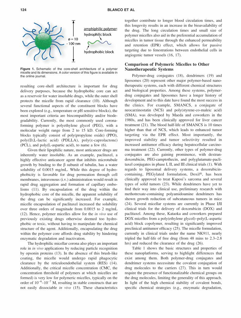
Table 1 shows the basic structures and properties of
these nanoplatforms, serving to highlight differences that
exist among them. Both polymer-drug conjugates and
dendrimer systems necessitate the covalent conjugation of
drug molecules to the carriers (27). This in turn would
require the presence of functionalizable chemical groups on
the drug molecules, limiting the generality of this approach.
In light of the high chemical stability of covalent bonds,
specific chemical strategies (e.g., enzymatic degradation,
Figure 1. Schematic of the core-shell architecture of a polymermicelle and its dimensions. A color version of this figure is available inthe online journal.
124 BLANCO ET AL
acid-catalyzed hydrolysis) are necessary to release the drug
molecules at tumor sites (28). Moreover, due to the small
size of these systems (typically , 10 nm), they can easily
cross basement membranes in the glomeruli of kidneys and
be quickly cleared, leading to much shortened blood half-
lives (3, 29).
Liposomes are vesicular nanostructures self-assembled
from the phospholipid and cholesterol molecules that
typically form cell membranes (20). As a result of their
inner hydrophilic compartment, liposomes are more suitable
for the delivery of water-soluble agents such as therapeutic
proteins or DNAs. Poorly soluble drugs can be entrapped
within the hydrophobic bilayer membrane, but the loading
capacity is limited due to membrane destabilization effects
(30). Stealth liposomes, where hydrophilic polymers such as
PEG have been conjugated on the liposomal surface, have
considerably prolonged blood circulation times, and effec-
tive passive targeting to solid tumors through the EPR effect
has been noted in numerous studies (31, 32). However, due
to intrinsic structural constraints, most liposomal particles
are over 90 nm in diameter, which may considerably limit
their transport in tumor tissues. For example, Yuan and
coworkers have shown limited liposomal penetration to only
30 lm (a few cell layers) following particle extravasation
(33). Later studies have shown that fibril collagen is the
main structural barrier for interstitial transport (34). More-
over, drug release from conventional liposomal formula-
tions is quite limited once these particles reach the tumor
target. To overcome this problem, thermo- and pH-sensitive
liposomes have been explored to provide responsive release
of drugs in hopes of improving bioavailability (35, 36).
Polymeric micelles provide a unique and complemen-
tary nanoplatform to the above nanosystems for drug
delivery applications. The hydrophobic cores of micelles
provide a natural carrier environment that allows easy
encapsulation of poorly soluble anticancer drugs. The non-
covalent encapsulation strategy makes it feasible to entrap
drugs without the requirement of reactive chemical groups.
Meanwhile, the unique chemistry of the polymer constitu-
ents does allow for the chemical conjugation of anticancer
drugs, such as doxorubicin (37, 38), to these chains,
effectively enhancing drug loading and hindering premature
drug release upon administration. Additionally, the size of
polymeric micelles, 10–100 nm, can be easily controlled by
varying the hydrophobic block of the amphiphilic copoly-
mer (39). This size range also permits for evasion of renal
filtration while allowing for increased tumor penetration
compared to liposomes (7).
Despite many advantages of polymer micelles for invivo applications, several challenges exist and represent an
active field of research. For example, the small micellar size
of 10–100 nm limits the amount of drug that can be
incorporated within the core, with higher drug loading
coming at the cost of increased micelle size and aggregation
(40, 41). This small size and limited drug loading in turn
results in faster release from the micelles (42), which may
cause premature release prior to the micelle reaching its
intended site of action. Therefore, chemical conjugation
Table 1. Summary of different nanotherapeutic technologies proposed for cancer therapy. A color version of thistable is available in the online journal.
MULTIFUNCTIONAL MICELLES FOR CANCER THERAPY 125
strategies, as mentioned above, as well as increasing the
compatibility of the micelle core with the intended drug
(43), are being investigated to address these concerns.
Additionally, several questions have been raised regarding
the long-term stability of polymer micelles (9). In response
to these issues, several groups are examining core-cross-
linked micelles in order to enhance in vivo stability (44, 45).
Finally, questions regarding the antitumor efficacy of
micelles in the clinical setting have been raised. In this
regard, many labs have explored methods to ensure
accumulation of the micelles at the tumor site and release
its contents in a controlled, predetermined fashion. In the
sections below, we will discuss strategies employed to
achieve micelle multifunctionalization, namely active tar-
geting, stimulated drug release, and imaging sensitivity for
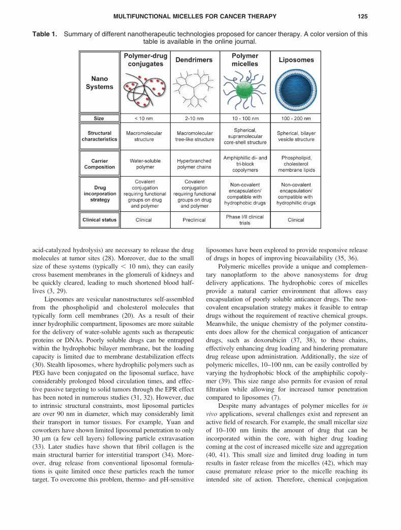
cancer applications (Fig. 2).
Active Targeting of Micelles
Active targeting strategies, which involve the function-
alization of the micelle surface with a ligand that recognizes
tumor-specific receptors, are an intense area of study with
several potential advantages. These include increased
accumulation at tumor sites as well as increased uptake
into cancer cells via receptor-mediated endocytosis (46).
Commonly used ligands are grouped into the following
classes: small organic molecules, peptides, carbohydrates,
monoclonal antibodies, and DNA/RNA aptamers.
An example of a small organic molecule for cancer
targeting applications is folic acid, whose receptor is over-
expressed (100–300 times) in a variety of tumors (47). Park
and coworkers functionalized DOX-containing PEG-PLGA
micelles with folic acid and were able to show significantly
increased uptake and cytotoxicity in KB cells (38). In vivostudies showed that folate-labeled micelles led to a 2-fold
decrease in tumor growth rate compared to non-targeted
micelles. Peptides are also actively explored as ligands for
tumor-targeted drug delivery. Recent work by our labo-
ratory has investigated the use of cyclic(Arg-Gly-Asp-D-
Phe-Lys) (cRGDfK) peptide, which targets the avb3 integrin
overexpressed on the surface of angiogenic tumor vessels
(48).
Carbohydrate molecules, such as galactose and lactose,
have also been used to functionalize micelles. These ligands
Figure 2. Multifunctional design of a micelle nanomedicine platform with cancer targeting, imaging, and controlled release properties. A colorversion of this figure is available in the online journal.
126 BLANCO ET AL
have high affinity for the asialoglycoprotein receptor
(ASGPR) overexpressed in hepatocellular carcinoma (49).
A galactose-labeled poly(ethylene glycol)-co-poly(c-benzyl
L-glutamate) block copolymer was used by Cho and
coworkers to produce micelles encapsulating paclitaxel,
and exhibited a 30% increased uptake in ASGPR cells (50).
Monoclonal antibodies represent another wide class of
active targeting ligands. Recently, Torchilin and coworkers
reported diacyllipid-PEG (PE-PEG) micelles conjugated
with an anti-cancer monoclonal antibody (mAb 2C5) or an
anti-myosin mAb 2G4 antibody to target lung cancer cells
(11). Micelles encoded with 2C5 were able to increase
paclitaxel accumulation (four-fold after 2 hrs) and cytotox-
icity in lung tumors over control micelles. Finally, tumor-
specific aptamers, DNA or RNA oligonucleotides identified
by library screening, are also gaining potential as targeting
ligands. Docetaxel-loaded PEG-PLGA micelles were re-
cently conjugated with an RNA aptamer specific for the
prostate specific membrane antigen (PSMA) to treat prostate
tumors (51). In vivo studies in LNCaP xenografts showed
overall increased anti-tumor efficacy and lesser systemic
toxicity than non-targeted micelles, and more importantly,
total tumor regression in five of seven mice was reported.
Stimulus Responsive Drug Release
Upon entering the tumor site, it is desirable that the
therapeutic agent be released from the micelles in a
controlled fashion in order to reach cytotoxic levels. To
achieve this, several strategies have been explored that
include pH-, temperature-, and ultrasound-stimulated re-
lease.
It is now well known that tumor tissues tend to have
lower pH values (as low as 5.7) than normal tissue
environments (pH 7.4), due to the glycolytic metabolism
of cancer cells. Additionally, the process of endocytosis, or
the sequestration of the nanocarriers into vesicles (e.g., late
endosomes, and heavily degradative lysosomes) is one
associated with low pH values of ;5.0–5.5. Hence, changes
in pH values encountered by micelles upon intravenous
injection provide a possible venue through which to achieve
stimulated release of drugs. Two different strategies have
been reported to induce pH-sensitive release of drugs from
micelles. These include the use of acid-labile bonds and
non-covalent strategies involving selective protonation of
pH-sensitive components inside the micelle. In the first
strategy, Kataoka and coworkers were able to formulate
micelles where doxorubicin was conjugated to the PEG-
pAsp copolymer via a hydrazone linkage (37). The resulting
micelles had high loading of DOX (42.5%) and pH sensitive
release; 3% of the drug was released after 48 hrs in pH 7.4
and 25% release of drug was achieved at the same time at
pH 5.5. In vivo studies showed increased tumor accumu-
lation, greater tolerance for the drug, and tumor regression
in 50% of mice. Non-covalent strategies for pH-sensitive
release were explored by several groups (52–54). For
example, Tang et al. devised a triblock polymer of PEG,
poly(2-(dimethylamino)ethyl methacrylate) (DMA), and
poly(2-diethylamino)ethyl acrylate (DEA) resulting in a
system that dissolves completely in acidic solution but
forms micelles at high pH (pH 8.0) (54). Acid sensitive
release of dipyridamole was observed with a 50% increase
of drug release at pH 3.0 over that at pH 7.4.
Technologies that permit for site-specific elevation of
temperature have led to the development of heat-sensitive
polymer micelles. The polymer of choice is poly(N-
isopropylacrylamide) or pNIPAM, which has a lower
critical solution temperature (LCST) of 328C (55). Okano
and coworkers reported micelles where poly(butyl meth-
acrylate) (PBMA) was used to form the hydrophobic core
while pNIPAM was used as the thermosensitive corona
(56). The resulting pNIPAM-b-PBMA micelles were loaded
with DOX and released 15% of the drug after 15 hrs at
308C, compared to 90% release in the same time period at
378C. Cytotoxicity experiments showed less than 5% cell
death at 298C, but 65% cell death at 378C.
Presently, ultrasound is used to trigger drug release
from drug delivery systems through mechanisms that
include local temperature increase, cavitation which in-
creases the permeability of cell membranes, and the
production of highly reactive free radical species which
can accelerate polymer degradation (57). Pitt and coworkers
designed ultrasound-sensitive pluronic micelles containing
doxorubicin (58, 59). Following stabilization of these
pluronic micelles with PEG-phospholipid (PEG-DSPE), invivo experiments showed that ultrasound was able to
improve the antitumor efficacy of both free DOX and
micelle incorporated DOX, with ultrasound delaying tumor
growth significantly longer over micelles without ultra-
sound.
Polymer Micelles with Imaging Sensitivity
Medical imaging modalities such as magnetic reso-
nance imaging (MRI), computed tomography (CT), single
photon emission computed tomography (SPECT), positron
emission tomography (PET), and ultrasonography play vital
roles in cancer diagnosis and monitoring of therapy.
Currently, the field of nanomedicine is converging with
medical imaging to further increase contrast specificity
between healthy and tumor tissues.
Several micellar platforms have been established for
use in MR imaging. Presently, gadolinium (Gd)-based
contrast agents (e.g., Magnevistt) are clinically used where
image contrast is increased by shortening the T1 relaxation
time of water protons (60). Incorporation of Gd complex on
the surface of polymeric micelles can effectively increase
the T1 relaxivity and sensitivity of detection (61, 62).
Superparamagnetic iron oxide (SPIO) is a T2/T2*-based
contrast agent that has higher sensitivity than Gd-based T1
agents (63). Our laboratory recently reported a formulation
composed of polymeric micelles encapsulating a cluster of
MULTIFUNCTIONAL MICELLES FOR CANCER THERAPY 127
SPIO nanoparticles (64). Clustering of the hydrophobic
SPIO nanoparticles inside of the micelle core led to a
dramatic increase in T2 relaxivity. The micellar formulation
showed an MRI detection limit at nanomolar concentrations.
Polymeric micelles have also been explored for cancer
imaging using CT and SPECT. Torchilin et al. developed
iodine-containing PLL-PEG polymer micelles having an
average diameter of 100 nm and iodine content of 45% (w/
w) for use as CT contrast agents (65). Following
subcutaneous injection in the hind leg of rabbits, they were
able to identify popliteal lymph nodes after 2 hrs using CT.
Opacification of the liver and spleen were observed after IV
injection of the micelles into rabbits, with vessels in the liver
being demarcated within half an hour of injection.
Multifunctional Polymeric Micelles
The unique architecture of polymeric micelles allows
for the incorporation of multiple functional components
within a single micelle. By combining tumor targeting,
stimulated release of therapeutics, and the delivery of
imaging agents, multiple interventions against a tumor can
be integrated into one platform. Such a ‘theranostic’ entity,
has been defined as a nanomedicine platform that can
diagnose, deliver targeted treatment in a controlled manner,
and monitor response to cancer therapy (66).
Kataoka and coworkers explored the use of multifunc-
tional micelles by conjugating a targeting ligand, folate, to
pH-sensitive doxorubicin-releasing polymer micelles com-
posed of PEG-p(Asp-Hyd-ADR) (67). The multifunctional
platform proved more effective at treating KB cells (nearly
10-fold after a 24-hr exposure) than the untargeted
formulation. Similarly, when folate was introduced to their
pH-sensitive PEG-pHis DOX-containing micelles, Bae and
coworkers observed three times greater accumulation in
MCF-7 tumors in vivo and greater cytotoxicity compared to
folate-free micelles (68). An alternate micellar platform
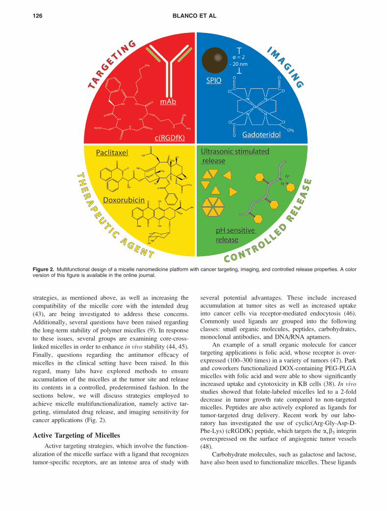
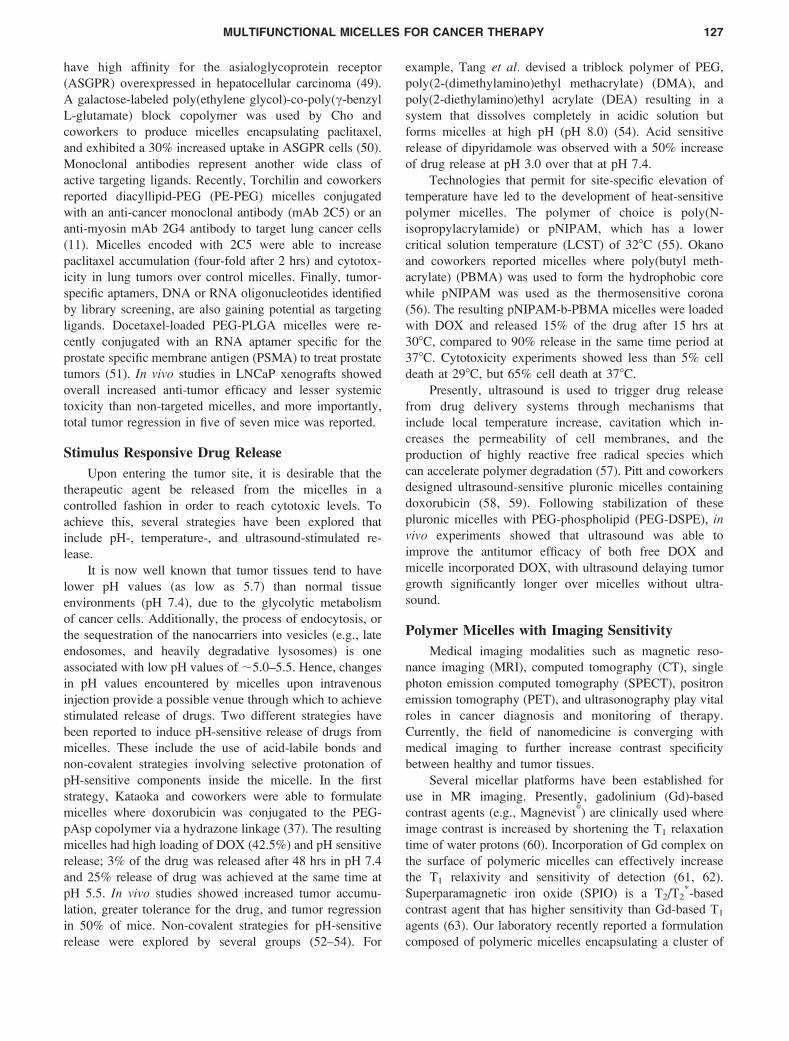
Figure 3. (A) Schematic of a multifunctional polymeric micelle. (B–C) Pre- and post-contrast coronal images of tumor-bearing mice by T2-weighted imaging (TR/TE¼4 s/40 ms), respectively. (D–E) Bioluminescence imaging of mouse bearing luciferase-transfected H1299 xenograftat days 0 and 7, respectively. DOX-loaded, 16% cRGD-micelles were injected via tail vein at days 0 and 3 (4 mg/kg DOX dose each time). (F)Antitumor efficacy data for PBS control, DOXO-SPIO micelles with 0 and 16% surface density of cRGD in subcutaneous A549 tumor xenograftsin nude mice. Each data point is averaged from 3 animals. A color version of this figure is available in the online journal.
128 BLANCO ET AL
developed by Kennedy and coworkers combined delivery of
doxorubicin and imaging of tumors via ultrasound (69). In
this strategy, consisting of DOX-containing PEG-PLLA
micelles and nanodroplets of perfluoropentane (PFP)
stabilized by an outer layer of block copolymer, ultrasound
triggered drug release from micelles through inertial
cavitation and facilitated entry into tumor cells by altering
cell permeability. Upon i.v. administration of the formula-
tion, MDA-MB 231 tumors not treated with ultrasound
showed a pattern of growth similar to control tumors.
Tumors undergoing administration of the micelles and
treated with ultrasound (30-sec treatment at 3 MHz) showed
significant regression. Additionally, within 4 hrs after
injection, ultrasonography revealed strong echoes within
the center of the tumor, further demonstrating in vivocoalescing of nanobubbles.
Recently, a multifunctional micellar platform that
incorporates a targeting ligand, pH-stimulated release of
DOX, and an MRI-visible agent was established in our
laboratory (70). In this design, DOX and a cluster of SPIO
nanoparticles (8 nm in diameter) were loaded into the cores
of PEG-PLA micelles, while a cRGD ligand on the micelle
surface (cRGD-DOX-SPIO micelles) was used for targeting
as shown in Figure 3A. The resulting targeted, multifunc-
tional micelles, measuring 45 6 8 nm in diameter, showed
increased uptake in vitro in avb3-overexpressing SLK
endothelial cells.
In in vivo studies, the multifunctional micelles showed
effective tumor targeting, imaging sensitivity, and antitumor
efficacy. Figure 3B shows a pre-contrast image obtained
using a T2-weighted (T2-w) scan on a 4.7 T MRI scanner.
The coronal image depicts the presence of an orthotopic
H1299 non-small cell lung tumor xenograft. Following an
i.v. injection of a solution of DOXO-SPIO micelles with
16% cRGD surface density (6.3 mg Fe/kg), a T2-w image
taken 15 hrs after injection demonstrates significant
darkening of the tumor (Fig. 3C), showing micelle
accumulation in tumors. The antitumor efficacy of the
micelle platform in the orthotopic H1299 tumor model was
monitored by bioluminescent imaging (BLI) of tumors
transfected with the firefly luciferase gene. Figure 3D
represents a BLI image of a representative mouse at day 0,
immediately before the micelle injection, showing a relative
luminescence unit (RLU) value of 1.2 3 106. At days 1 and
3, a solution of cRGD-encoded DOXO-SPIO micelles (4
mg DOX/kg) was injected intravenously. At day 7, BLI was
performed and the RLU value was found to be 5.6 3 105,
indicating an approximate 50% reduction in tumor size. In a
separate antitumor efficacy study, a subcutaneous A549
lung tumor xenograft was utilized (Fig. 3F). As in the case
of the orthotopic study, cRGD-encoded DOX-SPIO mi-
celles exhibited regression of tumor size (approximately
50% size reduction after 10 days). On the contrary, tumor
sizes from mice treated with cRGD-free DOX-SPIO
micelles did not change dramatically, with the PBS control
group showing a steady tumor growth, the size almost
doubling after 10 days.
Conclusions and Future Outlook
Polymeric micelles are emerging as a powerful, multi-
functional nanotherapeutic platform for cancer imaging and
therapeutic applications. Herein, we summarized recent
advances in micellar nanomedicine capable of targeting
tumors through the addition of targeting ligands while
delivering anti-neoplastic drugs with responsive release to
achieve maximal antitumor efficacy. Furthermore, micelles
incorporating imaging functions have been established for
diagnostic and monitoring purposes to assess therapeutic
efficacy. The incorporation of multiple functionalities in
polymeric micelles is a necessary step towards overcoming
the biological complexity and therapeutic challenges during
cancer chemotherapy. Solid tumors are known to have a
highly heterogeneous population of different cell types.
Molecular and phenotypic heterogeneity becomes a signifi-
cant challenge since not all cells within a given tumor will
respond to a single therapeutic agent. Additionally, as more
specific agents are developed to target tumors, selection of
tumor cells that can bypass the targeted pathway will lead to
adaptive resistance and become a cause of treatment failure.
As such, micellar nanomedicine that can deliver multiple
agents to target several key cancerous pathways will be the
next step in their evolution. To further overcome the
challenge of tumor heterogeneity, micelles with imaging
functions to characterize the phenotypic expressions within
solid tumors will be valuable in guiding the selection of
tumor-specific therapy. In addition, such imaging functions
allow the monitoring of treatment efficacy and enable the
subsequent modification of therapy to address adaptive
resistance. Such theranostic micellar nanomedicine holds
considerable promise to achieve personalized therapy of
cancer and to maximize the specificity and efficacy of
chemotherapy.
1. Duncan R. The dawning era of polymer therapeutics. Nat Rev Drug
Discov 2:347–360, 2003.
2. Ferrari M. Cancer nanotechnology: opportunities and challenges. Nat
Rev Cancer 5:161–171, 2005.
3. Peer D, Karp JM, Hong S, Farokhzad OC, Margalit R, Langer R.
Nanocarriers as an emerging platform for cancer therapy. Nat
Nanotechnol 2:751–760, 2007.
4. Croy SR, Kwon GS. Polymeric micelles for drug delivery. Curr Pharm
Des 12:4669–4684, 2006.
5. Nishiyama N, Kataoka K. Current state, achievements, and future
prospects of polymeric micelles as nanocarriers for drug and gene
delivery. Pharmacol Ther 112:630–648, 2006.
6. Sutton D, Nasongkla N, Blanco E, Gao J. Functionalized micellar
systems for cancer targeted drug delivery. Pharm Res 24:1029–1046,
2007.
7. Torchilin VP. Micellar nanocarriers: pharmaceutical perspectives.
Pharm Res 24:1–16, 2007.
8. Gros L, Ringsdorf H, Schupp H. Polymeric anti-tumor agents on a
MULTIFUNCTIONAL MICELLES FOR CANCER THERAPY 129
molecular and on a cellular-level. Angew Chem Int Ed Engl 20:305–
325, 1981.
9. Jones M, Leroux J. Polymeric micelles—a new generation of colloidal
drug carriers. Eur J Pharm Biopharm 48:101–111, 1999.
10. Torchilin VP. Structure and design of polymeric surfactant-based drug
delivery systems. J Control Release 73:137–172, 2001.
11. Torchilin VP, Lukyanov AN, Gao Z, Papahadjopoulos-Sternberg B.
Immunomicelles: targeted pharmaceutical carriers for poorly soluble
drugs. Proc Natl Acad Sci U S A 100:6039–6044, 2003.
12. Soga O, van Nostrum CF, Fens M, Rijcken CJ, Schiffelers RM, Storm
G, Hennink WE. Thermosensitive and biodegradable polymeric
micelles for paclitaxel delivery. J Control Release 103:341–353, 2005.
13. Moghimi SM, Hunter AC. Recognition by macrophages and liver cells
of opsonized phospholipid vesicles and phospholipid headgroups.
Pharm Res 18:1–8, 2001.
14. Torchilin VP. PEG-based micelles as carriers of contrast agents for
different imaging modalities. Adv Drug Deliv Rev 54:235–252, 2002.
15. Liu J, Zeng F, Allen C. In vivo fate of unimers and micelles of a
poly(ethylene glycol)-block-poly(caprolactone) copolymer in mice
following intravenous administration. Eur J Pharm Biopharm 65:309–
319, 2007.
16. Hashizume H, Baluk P, Morikawa S, McLean JW, Thurston G,
Roberge S, Jain RK, McDonald DM. Openings between defective
endothelial cells explain tumor vessel leakiness. Am J Pathol 156:
1363–1380, 2000.
17. Maeda H. The enhanced permeability and retention (EPR) effect in
tumor vasculature: the key role of tumor-selective macromolecular drug
targeting. Adv Enzyme Regul 41:189–207, 2001.
18. Li C, Wallace S. Polymer-drug conjugates: recent development in
clinical oncology. Adv Drug Deliv Rev 60:886–898, 2008.
19. Lee CC, MacKay JA, Frechet JM, Szoka FC. Designing dendrimers for
biological applications. Nat Biotechnol 23:1517–1526, 2005.
20. Torchilin VP. Recent advances with liposomes as pharmaceutical
carriers. Nat Rev Drug Discov 4:145–160, 2005.
21. Maeda H, Ueda M, Morinaga T, Matsumoto T. Conjugation of
poly(styrene-co-maleic acid) derivatives to the antitumor protein
neocarzinostatin: pronounced improvements in pharmacological prop-
erties. J Med Chem 28:455–461, 1985.
22. Greish K, Fang J, Inutsuka T, Nagamitsu A, Maeda H. Macromolecular
therapeutics: advantages and prospects with special emphasis on solid
tumour targeting. Clin Pharmacokinet 42:1089–1105, 2003.
23. Gabizon AA. Pegylated liposomal doxorubicin: metamorphosis of an
old drug into a new form of chemotherapy. Cancer Invest 19:424–436,
2001.
24. Kukowska-Latallo JF, Candido KA, Cao Z, Nigavekar SS, Majoros IJ,
Thomas TP, Balogh LP, Khan MK, Baker JR Jr. Nanoparticle targeting
of anticancer drug improves therapeutic response in animal model of
human epithelial cancer. Cancer Res 65:5317–5324, 2005.
25. Nakanishi T, Fukushima S, Okamoto K, Suzuki M, Matsumura Y,
Yokoyama M, Okano T, Sakurai Y, Kataoka K. Development of the
polymer micelle carrier system for doxorubicin. J Control Release 74:
295–302, 2001.
26. Matsumura Y, Hamaguchi T, Ura T, Muro K, Yamada Y, Shimada Y,
Shirao K, Okusaka T, Ueno H, Ikeda M, Watanabe N. Phase I clinical
trial and pharmacokinetic evaluation of NK911, a micelle-encapsulated
doxorubicin. Br J Cancer 91:1775–1781, 2004.
27. Gillies ER, Frechet JM. Dendrimers and dendritic polymers in drug
delivery. Drug Discov Today 10:35–43, 2005.
28. Gopin A, Ebner S, Attali B, Shabat D. Enzymatic activation of second-
generation dendritic prodrugs: conjugation of self-immolative den-
drimers with poly(ethylene glycol) via click chemistry. Bioconjug
Chem 17:1432–1440, 2006.
29. Greenwald RB, Choe YH, McGuire J, Conover CD. Effective drug
delivery by PEGylated drug conjugates. Adv Drug Deliv Rev 55:217–
250, 2003.
30. Liu J, Lee H, Huesca M, Young A, Allen C. Liposome formulation of a
novel hydrophobic aryl-imidazole compound for anti-cancer therapy.
Cancer Chemother Pharmacol 58:306–318, 2006.
31. Kamaly N, Kalber T, Ahmad A, Oliver MH, So PW, Herlihy AH, Bell
JD, Jorgensen MR, Miller AD. Bimodal paramagnetic and fluorescent
liposomes for cellular and tumor magnetic resonance imaging.
Bioconjug Chem 19:118–129, 2008.
32. Zalipsky S, Saad M, Kiwan R, Ber E, Yu N, Minko T. Antitumor
activity of new liposomal prodrug of mitomycin C in multidrug
resistant solid tumor: insights of the mechanism of action. J Drug
Target 15:518–530, 2007.
33. Yuan F, Leunig M, Huang SK, Berk DA, Papahadjopoulos D, Jain RK.
Microvascular permeability and interstitial penetration of sterically
stabilized (stealth) liposomes in a human tumor xenograft. Cancer Res
54:3352–3356, 1994.
34. McKee TD, Grandi P, Mok W, Alexandrakis G, Insin N, Zimmer JP,
Bawendi MG, Boucher Y, Breakefield XO, Jain RK. Degradation of
fibrillar collagen in a human melanoma xenograft improves the efficacy
of an oncolytic herpes simplex virus vector. Cancer Res 66:2509–2513,
2006.
35. Chen Q, Tong S, Dewhirst MW, Yuan F. Targeting tumor microvessels
using doxorubicin encapsulated in a novel thermosensitive liposome.
Mol Cancer Ther 3:1311–1317, 2004.
36. Li W, Huang Z, MacKay JA, Grube S, Szoka FC Jr. Low-pH-sensitive
poly(ethylene glycol) (PEG)-stabilized plasmid nanolipoparticles:
effects of PEG chain length, lipid composition and assembly conditions
on gene delivery. J Gene Med 7:67–79, 2005.
37. Bae Y, Nishiyama N, Fukushima S, Koyama H, Yasuhiro M, Kataoka
K. Preparation and biological characterization of polymeric micelle
drug carriers with intracellular pH-triggered drug release property:
tumor permeability, controlled subcellular drug distribution, and
enhanced in vivo antitumor efficacy. Bioconjug Chem 16:122–130,
2005.
38. Yoo HS, Park TG. Folate receptor targeted biodegradable polymeric
doxorubicin micelles. J Control Release 96:273–283, 2004.
39. Shuai X, Ai H, Nasongkla N, Kim S, Gao J. Micellar carriers based on
block copolymers of poly(epsilon-caprolactone) and poly(ethylene
glycol) for doxorubicin delivery. J Control Release 98:415–426, 2004.
40. Allen C, Maysinger D, Eisenberg A. Nano-engineering block
copolymer aggregates for drug delivery. Colloid Surface B 16:3–27,
1999.
41. Gaucher G, Dufresne MH, Sant VP, Kang N, Maysinger D, Leroux JC.
Block copolymer micelles: preparation, characterization and application
in drug delivery. J Control Release 109:169–188, 2005.
42. Kim SY, Shin IG, Lee YM. Preparation and characterization of
biodegradable nanospheres composed of methoxy poly(ethylene
glycol) and DL-lactide block copolymer as novel drug carriers. J
Control Release 56:197–208, 1998.
43. Liu J, Xiao Y, Allen C. Polymer-drug compatibility: a guide to the
development of delivery systems for the anticancer agent, ellipticine. J
Pharm Sci 93:132–143, 2004.
44. Bontha S, Kabanov AV, Bronich TK. Polymer micelles with cross-
linked ionic cores for delivery of anticancer drugs. J Control Release
114:163–174, 2006.
45. Xu P, Tang H, Li S, Ren J, Van Kirk E, Murdoch WJ, Radosz M, Shen
Y. Enhanced stability of core-surface cross-linked micelles fabricated
from amphiphilic brush copolymers. Biomacromolecules 5:1736–1744,
2004.
46. Sethuraman VA, Bae YH. TAT peptide-based micelle system for
potential active targeting of anti-cancer agents to acidic solid tumors. J
Control Release 118:216–224, 2007.
47. Ross JF, Chaudhuri PK, Ratnam M. Differential regulation of folate
receptor isoforms in normal and malignant tissues in vivo and in
established cell lines. Physiologic and clinical implications. Cancer 73:
2432–2443, 1994.
130 BLANCO ET AL
48. Silletti S, Kessler T, Goldberg J, Boger DL, Cheresh DA. Disruption of
matrix metalloproteinase 2 binding to integrin alpha vbeta 3 by an
organic molecule inhibits angiogenesis and tumor growth in vivo. Proc
Natl Acad Sci U S A 98:119–124, 2001.
49. Wands JR, Blum HE. Primary hepatocellular carcinoma. N Engl J Med
325:729–731, 1991.
50. Jeong YI, Seo SJ, Park IK, Lee HC, Kang IC, Akaike T, Cho CS.
Cellular recognition of paclitaxel-loaded polymeric nanoparticles
composed of poly(gamma-benzyl L-glutamate) and poly(ethylene
glycol) diblock copolymer endcapped with galactose moiety. Int J
Pharm 296:151–161, 2005.
51. Farokhzad OC, Cheng J, Teply BA, Sherifi I, Jon S, Kantoff PW,
Richie JP, Langer R. Targeted nanoparticle-aptamer bioconjugates for
cancer chemotherapy in vivo. Proc Natl Acad Sci U S A 103:6315–
6320, 2006.
52. Potineni A, Lynn DM, Langer R, Amiji MM. Poly(ethylene oxide)-
modified poly(beta-amino ester) nanoparticles as a pH-sensitive
biodegradable system for paclitaxel delivery. J Control Release 86:
223–234, 2003.
53. Stapert HR, Nishiyama N, Jiang DL, Aida T, Kataoka K. Polyion
complex micelles encapsulating light-harvesting ionic dendrimer zinc
porphyrins. Langmuir 16:8182–8188, 2000.
54. Tang Y, Liu SY, Armes SP, Billingham NC. Solubilization and
controlled release of a hydrophobic drug using novel micelle-forming
ABC triblock copolymers. Biomacromolecules 4:1636–1645, 2003.
55. Liu SQ, Tong YW, Yang YY. Thermally sensitive micelles self-
assembled from poly(N-isopropylacrylamide-co-N,N-dimethylacryla-
mide)-b-poly(D,L-lactide-co-glycolide) for controlled delivery of
paclitaxel. Mol Biosyst 1:158–165, 2005.
56. Chung JE, Yokoyama M, Yamato M, Aoyagi T, Sakurai Y, Okano T.
Thermo-responsive drug delivery from polymeric micelles con-
structed using block copolymers of poly(N-isopropylacrylamide)
and poly(butylmethacrylate). J Control Release 62:115–127, 1999.
57. Mitragotri S. Healing sound: the use of ultrasound in drug delivery and
other therapeutic applications. Nat Rev Drug Discov 4:255–260, 2005.
58. Gao ZG, Fain HD, Rapoport N. Controlled and targeted tumor
chemotherapy by micellar-encapsulated drug and ultrasound. J Control
Release 102:203–222, 2005.
59. Pruitt JD, Pitt WG. Sequestration and ultrasound-induced release of
doxorubicin from stabilized Pluronic P105 micelles. Drug Deliv 9:253–
258, 2002.
60. Sato H, Enmi J, Teramoto N, Hayashi T, Yamamoto A, Tsuji T, Naito
H, Iida H. Comparison of Gd-DTPA-induced signal enhancements in
rat brain C6 glioma among different pulse sequences in 3-Tesla
magnetic resonance imaging. Acta Radiol 49:172–179, 2008.
61. Nakamura E, Makino K, Okano T, Yamamoto T, Yokoyama M. A
polymeric micelle MRI contrast agent with changeable relaxivity. J
Control Release 114:325–333, 2006.
62. Zhang G, Zhang R, Wen X, Li L, Li C. Micelles based on
biodegradable poly(L-glutamic acid)-b-polylactide with paramagnetic
Gd ions chelated to the shell layer as a potential nanoscale MRI-visible
delivery system. Biomacromolecules 9:36–42, 2008.
63. Wang YX, Hussain SM, Krestin GP. Superparamagnetic iron oxide
contrast agents: physicochemical characteristics and applications in MR
imaging. Eur Radiol 11:2319–2331, 2001.
64. Ai H, Flask C, Weinberg B, Shuai X, Pagel MD, Farrell D, Duerk J,
Gao J. Magnetite-loaded polymeric micelles as ultrasensitive magnetic-
resonance probes. Adv Mater 17:1949–1952, 2005.
65. Trubetskoy VS, Gazelle GS, Wolf GL, Torchilin VP. Block-copolymer
of polyethylene glycol and polylysine as a carrier of organic iodine:
design of long-circulating particulate contrast medium for X-ray
computed tomography. J Drug Target 4:381–388, 1997.
66. Sumer B, Gao J. Theranostic nanomedicine for cancer. Nanomed 3:
137–140, 2008.
67. Bae Y, Jang WD, Nishiyama N, Fukushima S, Kataoka K. Multifunc-
tional polymeric micelles with folate-mediated cancer cell targeting and
pH-triggered drug releasing properties for active intracellular drug
delivery. Mol Biosyst 1:242–250, 2005.
68. Lee ES, Na K, Bae YH. Doxorubicin loaded pH-sensitive polymeric
micelles for reversal of resistant MCF-7 tumor. J Control Release 103:
405–418, 2005.
69. Rapoport N, Gao Z, Kennedy A. Multifunctional nanoparticles for
combining ultrasonic tumor imaging and targeted chemotherapy. J Natl
Cancer Inst 99:1095–1106, 2007.
70. Nasongkla N, Bey E, Ren J, Ai H, Khemtong C, Guthi JS, Chin SF,
Sherry AD, Boothman DA, Gao J. Multifunctional polymeric micelles
as cancer-targeted, MRI-ultrasensitive drug delivery systems. Nano Lett
6:2427–2430, 2006.
MULTIFUNCTIONAL MICELLES FOR CANCER THERAPY 131