mlebical !section. - europe pubmed central
TRANSCRIPT

Mlebical !section.
October 25, 1910.
Dr. J. MITCHELL BRUCE, President of the Section, in the Chair.
Multiple Cartilaginous Exostoses.
BY A. M. GOSSAGE, M.D., and E. ROCK CARLING, F.R.C.S.
THERE is a class of exostosis which differs from all others in thatthe tumours appear spontaneously in connexion with cartilage and aremultiple. At first cartilaginous, the tumours only become ossifiedlater, and throughout their period of growth are covered by a layer ofhyaline cartilage. They may be attached to any bone that is developedin cartilage, but are naturally never found connected with the mem-branous bones, such as those of the skull. In the long bones thegrowths take their origin from the epiphysial cartilages, though laterin life, with skeletal growth, they may become detached from them andappear to arise from the diaphyses. When growing from the epiphysialcartilages they have the same structure as the adjacent long bone,having a compact outer covering which encloses cancellous bone and acentral canal which communicates with the central canal of the bone.The tumours may be very numerous or few; Chiari records over athousand in his patient, and one of ours had several score. In size theymay be as large as the foetal head, or so small as to be undiscoverableuntil the bone has been macerated after death. In shape they arevery variable, sometimes forming a sharp spine, in other cases beinghook-shaped, rounded, flattened, or irregularly tuberous. They mayalso lie in a depression of. the parent bone, and so be larger than wouldbe expected from external examination. They may be present before
N 9

2 Gossage & Carling: Multiple Cartilaginous Exostoses
birth (Drescher), but usually first appear in early childhood and increasein size and number up to the end of the period of skeletal growth, whenthe epiphyses all become attached to the diaphyses about the twenty-fifth year. After this the size nmay diminish, and in certain cases somehave even been said to have disappeared. Hartmann's photographs ofa case before and after an interval of twenty years proves the possibilityof retrogression. In rare cases the first appearance has been recordedas occurring at the age of 50.
As a rule, they do not cause the patient any pain or inconvenience,but sometimes pain may be produced by pressure on adjacent structures;exostoses of the feet may cause difficulty in walking and those on thepelvic bones may interfere with parturition (Drescher). It is theincreasing deformity that usually leads the patient to consult a medicalman. It is probable, though not perfectly certain, that the deformitiesare not mnerely a mechanical result of the pressure of the bony mass-of which the growth direction is always unilateral-but are also theexpression of irregular ossification throughout the intermediary cartilage.Sometimes dislocations of bones occur. This is seen particularly at theelbow, the head of the radius being displaced backwards. Other surgicalconditions arising in connexion with these outgrowths are due to pressureupon the bulb or spinal cord or peripheral nerves, and upon vessels withresultina erosion or aneurysmal formation. The growths are subject alsoto spontaneous necrosis.
Although the short and flat bones are affected, it is the long bonesthat suffer more particularly, and of these the upper end of the humerus,the distal end of the radius, and the articular ends of the femur, tibia,and fibula are most frequently concerned. Occurring usually near theepiphysial ends, the projection may sometimes be covered with synovialmembrane and may even become detached and appear as a loose body inthe adjacent joint.
As a general rule, the exostoses are best left alone, but where inter-vention is demanded by extraneous symptoms or lesions, removal iseasily effected by the chisel; the periosteum and the covering cap ofcartilage, where present, should be carefully excised. Difficulty maypossibly arise in reaching the base of the tumour for anatomical reasons,but with the help afforded by radiography this can usually be surmounted.The compact tissue of the exostoses in some of the cases that have beenoperated on has been found to pass through the compact covering ofthe bones to which they were attached, tapering towards the medulla;hence the growth has often to be scooped out of the parent bone.

Medical Section
Various hypotheses have been advanced as to the aetiology of thesegrowths; from isolated cases they have been regarded as consequencesof previous rheumatism, syphilis, or rickets, but the evidence in favourof any of these views is of the flimsiest character. Virchow ratherfavoured the idea of an intimate relation to rickets, because in that con-dition he was familiar with " islets " of cartilage; but, as Pels-Leusdenpoints out in his exhaustive study of the subject, these are always in themiddle of the bone and would more properly be advanced as an explana-tion of enchondromata. Of course, rickets may occur in a patientafflicted with multiple exostoses, as in Young's case; but this is no argu-ment that it is a causative agent. The greater incidence on males-calculated by Reinecke as thrice that in females-is a strong argumentagainst regarding rickets as the cause. Sinlilarly, there is no realevidence that congenital syphilis is a cause; yet it appears certain thatthe abnormality is initiated in intra-uterine life, and that the abnormalossification of the intermediary cartilage is not limited to the plane ofthe exostoses. The frequent association of disturbance in symmetry ofgrowth in the affected bone, generally in the direction of arrest, withconsequent shortening, but sometimes as a prolonged or excessiveactivity resulting in undue length, and occasionally as a local or generalexcess or diminution in total bulk of a bone, indicates that it is notmerely a- question of accidental "deviation" of undisciplined offspringby the normal mother cartilage. If this were the case there wouldhave been justification for the use of these tumours as examples ofCohnheim's " rest " theory of neoplasms. There are undoubtedly somerelationships between cartilaginous exostoses and multiple enchon-dromata-e.g., Weber and Liiwen found them conjoined-and of bothwith the peculiar disturbance of growth described by Ollier and knownas dyschondroplasia. In this condition, however, only one side of thebody is, as a rule, affected. Karewski did describe a case of multiplecartilaginous exostoses of the skull and face in association with facialhemiatrophy, and Kohler noted them in a case of partial gigantism.Again, they have been found sometimes (Reubsael, Kryger) with myositisossificans progressiva, and, where few in number, confusion of diagnosiswith other conditions may arise. For example, a good deal has beenwritten lately about exostoses or osteomata of the os calcis as a cause ofpainful heel. Some have been instances of true osteomata, but somecertainly have been the outcome of inflammatory processes, such asgonorrhceal effusion into the long plantar and other ligaments.
N-9a
3

4 Gossage & Carling: Multiple Cartilaginous Exostoses
From quite early times it had been noticed, especially in England,that the condition was hereditary, and we have collected records ofsixty-seven families in which more than one member has been attacked.It seems clear that some inherent abnormality of the intermediatecartilage, which causes it to develop in this strange fashion, is the chieffactor in the production of the tumours, and that other circumstancesand diseases only influence their development to a minor extent. Thisabnormal constitution of the cartilage forms a definite entity, which isfrequently handed down from parent to child. It must be acknowledgedthat in a large number of examples of this condition no history of theoccurrence of similar tumours in the sufferer's relatives can be obtained,and one is obliged to consider the possibility of the abnormality arisingde novo or of passing through several generations without manifestingitself. It should be specially noted that in sonme cases the tumours arefew and small, and it is possible that they may not attract the notice ofthe bearer or his relatives, and so an abnormal individual may bereckoned in the family genealogy as a normal one. For instance, themother of the B. family, fronm the Westnminster Hospital, stated that hertumours had quite disappeared; but on careful examination six exostoseswere found-five small ones up to the size of a pea, and a larger one asbig as a walnut-and in Roberg's family one woman had only one smallspinous exostosis on the lower end of the right radius.
Males are decidedly more frequently attacked than females; in the67 families we have collected there were 199 affected males and only 89females-i.e., a proportion of more than 2 to 1. In Tillmann's familythe sex is not mentioned. It is of interest that in multiple exostosesthere are examples of a skipped generation, and in nine such instancesthe condition was transmitted through unaffected females. In theliterature we have found recorded two examples of transmission throughunaffected males-(Roberg and Ziegler)-but in both instances thetruth of this transmission is very doubtful, whereas the transmissionthrough unaffected females seeins quite clear. Stanley's family is veryinteresting in this connexion: a normal woman had a child withmultiple exostoses by each of two separate normal husbands (see fig. 3).
An affected individual does not transmit the tendency to all of hisprogeny, but a certain proportion of them are quite normal, and thechildren of these normal individuals are normal in the majority of casesand do not hand on the family tendency to bony tumours. Unfortu-nately, the family histories are usually incomplete, but this point comesout in many of the genealogical tables-e.g., in the B. family here

Medical Section 5
recorded. In the 67 families there were 293 affected individuals, and139 unaffected; but the excess of the abnormal is evidently due to theincompleteness of the records, many authors omitting to mention thenormal members of the family. In 35 families the records are fairlycomplete, especially in the later generations. Taking these, and omittingthe earlier generations, we find 133 affected (83 male, 45 female, and
c'x ? xV
, ' '~~~~~~~~~~~~~~~~~~~~~~~~~~~~~C
FIG. 1.
H. family. MIultiple exostoses.
6P X ?Gnan(i
o~~~~
FIG. 2.
B. family. Mlultiple exostoses.
wx ? xY
FIG. 3.
Stanley, Medical Timies azd Gazette, 1857, ii, p. 39.
5 doubtful) and 135 unaffected, a close approximation to equality. Inthese more complete records the excess of males is not so great, and theproportions of the sexes are, we think, nearer to the truth. Males werefound in the 67 families to transmit the condition to their children in72 instances, whereas in 31 the condition was transmitted throughfemales. If, however, it is correct to say that the females can sometimes

6 Gossage & Carling: Multiple Cartilaginous Exostoses
transmit even when themselves unaffected, then probably a largenumber of the cases that arise apparently de novo are really inheritedthrough one or more generations of unaffected females, and the powerof transmission is probably represented equally in the two sexes. Theseverity of the affection varies much in different individuals, but it can-not be said that it is altered either in the direction of greater or lessseverity in the succeeding generations. An individual who has inheritedthe abnormality is just as likely, and no more, to be severely affected ashis parent or grandparent.
We have here to do with a condition which is handed down by anaffected person unchanged to about half his children bv a normal consort,and his abnormal offspring again transmit it unchanged to half theirprogeny. The normal members of the family as a rule do not pass onthe condition, but a certain number of the apparently normal femalescarry the abnormality, and can transmit it to half their children by anormal mate. The hereditary transmission conforms here fairly closelywith Mendel's law as worked out in animals or plants. Each somaticcell in the body may be regarded as double in its constitution, one partbeing derived from each parent. These parental constituents may besimilar or different, and the character of the somatic cell may be similarto both the parents, be like one parent only, or be a blend of the two.In the generative cells or gametes, these parental constituents separateor segregate, so that each gamete has only one of them, half the gametesbeing of the character derived from one parent and half of the characterfrom the other, and each zygote formed by the union of two gameteswhen developing into an embryo has the constituent derived from onlyone of its grandparents on each side. These parental constituents areknown as allelomorphs; and when in any cell they differ fromn oneanother, this difference may usually, if not always, be expressed as thepresence or absence of some special feature. For example, the colourof the human iris seems mainly to depend on the presence of somethingwhich causes the deposit of visible pigment in front of the iris when thevarious types of brown eye are developed, or the absence of this some-thing when the eyes belong to the various shades of blue. If only thefactor for presence be in the somatic cells, then the quality will alwaysshow itself. If both opposing factors, however, be present in all thesomatic cells, then it will depend on whether the factor for presence orthe factor for absence is the dominant one as to whether the particularquality is shown. As a rule, the allelomorph for presence is dominant,and always shows its attendant quality whenever it exists in the somatic

Medical Section
cells, whether it is pure there or mixed with the opposite allelomorphfor absence. Sometimes, however, the usually dominant factor may bepresent and yet not show its quality, so that an individual belonging toa family in which that quality exists may be able to transmit it to hisdescendants, although he himself does not exhibit it. Bateson hasshown that this incomplete dominance occurs with regard to extra toesin fowls, and this may explain some types of inheritance in humanbeings, such as in epidermolysis bullosa. In other cases the allelomorphfor presence may be dominant in one sex and recessive in the other, ashas been found to be the case with horns in sheep, which are dominantin the male and so always exhibited when the factor is present, butrecessive in the female, in whom horns only appear when both allelo-morphs are of the same kind-those which produce horns. Thus thepower to develop horns may be imparted by hornless females to some oftheir male offspring by a hornless male, but hornless males never trans-mit the power to develop horns. Colour blindness affords an example ofsimilar inheritance in man.
One can imagine a condition being completely dominant in one sexand incompletely dominant in the other. Under these circumstances,the one sex would be more affected than the other, and one wouldrather expect the affected individuals of the one sex to be more severelyattacked than those of the other. So far no example of this form ofinheritance seems to have been met with in animals or plants where itcould be thoroughly investigated by experimental breeding. This seems,however, the easiest explanation of the greater incidence of multipleexostoses in the male and the fact that it is not infrequently transmittedby normal females, but it is to be noted that we have no very strongevidence that the affected females are any less severely attacked thanthe males, although the very slight cases seem all females. Anotherexample of such a form of inheritance is possibly afforded by heemophilia.Bulloch has recently argued, with much plausibility, that females arenever affected by haemophilia properly so-called. Under these circum-stances, hammophilia would be a strictly sex-limited condition, likeDaltonism. If so, there is another condition-certainly less definitethan haemophilia-which is also characterized by an abnormal tendencyto bleed, and which is hereditary. Allowing for an exaggerated pro-clivity to class females as bleeders whose menstruation has been exces-sive or who have had epistaxis, this other haemorrhagic diathesis attacksfemales as well as males, but in fewer numbers and less severely, whileit is commonly transmitted through normal females belonging to the
7

8 Gossage & Carling: Multiple Cartilaginous Exostoses
abnormal families. Some two years ago- one of us (Dr. Gossage)suggested a similar explanation for cretinism, which attacks malesmore frequently and more severely than females, and in which, accord-ing to McCarrison, the mother of a cretin child frequently shows signsof thyroid deficiency. Dr. McCarrison's assistant, Sirdar Sahib KeharSingh, has since then carefully collected details from fourteen familiesat Gilgit, noting the occurrence of cretinism, goitre, myxcedema, deaf-mutism and tetany. The results of this careful and able piece of workshow that no such simple scheme of heredity can possibly prevail incretinism, if it be hereditary at all. Dr. Gossage's hearty thanks aredue to Sirdar Sahib Kehar Singh and to Major McCarrison for thecompletion of this investigation.
If this view be correct, that there is a deficiency in the number ofaffected females because the condition, while dominant in the male, isonly incomupletely doiinant in the female, then we ought to add asufficient number to the total of abnormal females to make it equal tothat of the males. This would make the grand total of abnormals 171to 135 normal-a decided excess of the abnormal. It is improbable,however, that all these thirty-five families are really complete, and it isespecially the normal members who tend to be left out. As a matterof fact, an excess over the expected number of abnormal individuals isfound in other conditions in man where we have reason to suppose thatthe abnormal condition is dominant to the normal. For example, thishas been shown to be the case by one of us with regard to tylosispalmaris et plantaris.
The following are some details of the cases on which this paper hasbeen founded, the cases having been examined by one of us or by one ofour colleagues. We beg to tender our thanks to our colleagues forpermission to use their notes:
Case 1.-James H., aged 9 in 1901, a fairly healthy looking boy, who wasalive and well when last heard of in 1909. The following exostoses werefound:-Chest: Two or three spines at the sternal end of the left clavicle,spines on second and third left ribs, and a small exostosis on the fifth right rib.Right arm: Bony swelling in front of lower end of radius (removed in theEast London Hospital for Children) and another at the outer side, largerswelling at the lower end of ulna. Left arm: Spiny process in front of greattuberosity of humerus, large bony swellings at 'lower ends of radius and ulna.Right leg: Exostoses about lower condyle of femur and near adductor tubercle(one of which was removed), exostosis at upper end of tibia (removed) and alsoa sharp, bony spine, an exostosis at lower end of fibula. Left leg: Exostoses

Medical Section 9
on condyles of femur, large exostosis on inner part of head of tibia and anotherat head of fibula, spine above external malleolus. In this case there were atleast twenty-two separate exostoses.
Case II.-Violet H., aged 4, sister of James H., was brought to the EastLondon Hospital for Children in 1903, suffering with general tuberculosis, fromwhich she died ten days later. Right arm: Exostosis on humerus near theshoulder-joint; an exostosis, stated to have been present at birth, over thelower third of ulna, large exostoses over lower ends of radius and ulna, smallexostosis on the little finger. Left arm: No exostoses noticed. Right leg: Anexostosis on the femur, just above the knee-joint. Left leg: An exostosis onthe inner side of the tibia, just below the knee-joint. Except the exostosis onthe ulna, none of these had been noticed until the age of 3, after which theywere all stated to have increased rapidly in size.
Case III.-Mrs. H., mother of Cases I and II, a healthy woman aged about45 when interviewed in 1909. She stated that she had never had similarlumps to those in her children, and no exostoses could be found on examina-tion. Her husband, her husband's relatives, and her own relatives had neverhad similar tumours. She gave a full family history, which is represented infig. 1. In this the abnormal individuals are marked black, the normal white,and sex is indicated in the usual way. (Several normal persons may be shownby a numeral in a white circle.)
Case IV.-Fred. B., aged 14 in 1902, a healthy boy in whom the osteomatahad first been noticed when aged 2; height, 4 ft. 3' in. Right leg: Toes free;small nodule head of fifth metatarsal; tarsus free; tibia-an irregular lump,1 in. from back to front, above inner malleolus; fibula-prominent lump belowinner side of bead, small nodule above back of external malleolus,'large boss,size of half a small orange, over front of head (probably from tibia); femur-one small growth 1V in. above external condyle, one from inner side of poplitealaspect, low elevation posterior aspect below great trochanter. Left leg': Toesfree; small nodule dorsal aspect of fifth metatarsal; small elevation outer sideof head of os calcis; tibia-large irregular mass posterior aspect of malleolus,smaller one anterior aspect lower end of tibia, smooth elQvation inner side ofhead; fibula--growth posterior aspect lower end, and discrete projectingnodule anterior aspect of head; femur-bifurcated mass on posterior aspect ofouter condyle, bifurcated mass anterior and inner aspect of inner condyle.Dorsal aspect of trunk: Small mass about 2 in. from head of twelfth right rib,rounded nodule outside angle of sixth rib; scapulm both winged by masses onanterior surface--on the right side a large, nodular, irregular mass, 2 in. by 2 in.by 1j in., growing from the ribs over which scapula rides, one nodule on spinalborder of right scapula and two near root of spine, two nodules on angle andspine of left scapula, and one on lower part of axillary border. Anterior aspectof trunk: Clavicles-nodules at outer ends of both, very small left, about size

10 Gossage & Carling: Multiple Cartilaginous Exostoses
of a bean right; two or three nodules on left sixth and seventh ribs. Rightarm: Tiny nodule on second phalanx of second finger and also on first phalanxof first finger; metacarpus free; one tiny nodule dorsal aspect trapezoid; radius-two tiny nodules and one as big as a marble at lower end (two dorsal andone ventral) ; ulna-one inner aspect lower extremity, whole hand deflectedtowards ulnar side; humerus -one in axilla and one just above deltoidinsertion. Left arm: One at the base of second phalanx of second finger, onenear the heads of both first and second metacarpals, one on anterior aspect oftrapezius; radius-two small ones at dorsal aspect of lower end and a largerprominent one at ventral aspect; one on inner side lower end of ulna; humerus-large one in axilla and one on outer aspect about middle of deltoid. Noneon the vertebree, pelvis, or other bones. Feet in good position and no impair-ment of movement. All the calvarial sutures are very prominently marked.
Case V.-Florence B., aged 12 in 1902, a healthy girl who had had scarletfever when aged 7; height, 4 ft. 3 in. Right leg: Toes and metatarsus free;small nodule inner dorsal aspect of base of cuboid; tibia-one 2 in. abovemalleolus and a prominent one 2 in. below inner aspect of head; fibula-diffuseone about head; femur-four at least about lower end, two in popliteal space,marked spine above external condyle, enlargement of posterior aspect of upperpart of shaft. Left leg: Toes free; metatarsus free; one nodule head of astragalus;tibia--two at lower end, one prominent one below inner side of head; fibula-discrete nodule lower end, diffuse swelling upper end; femur-several at lowerend, one large mass in outer part of popliteal space. Trunk: Ribs free; nodulesat inner and outer ends of clavicles; pelvis nodule over right posteriorsuperior spinous process and one at middle of left iliac crest; one on lastdorsal vertebra; scapulh-symmetrical bunches about bases of spines and anodule at middle of spine on right side (injury at 3 years). Right arm:Nodule dorsal aspect base of first phalanx of second finger and one or twoover heads of metacarpal bones; much ulnar deflection of hand, ? absence oflower epiphysis of ulna, large mass about lower end of ulna which is possiblycallus; humerus-one nodule beneath deltoid and one in axilla. Left arm:Nodule at base of second phalanx of fourth finger; fairly large nodule overdorsum of head of second metacarpal bone; nodule dorsal side of trapezoid;one, dorsal, lower end of radius and one on inner side of lower end of ulna;humerus -one anterior aspect of shaft at junction of upper and middlethirds; one small one in axilla; hook-shaped spine beneath deltoid. Headand neck nil. Now, in 1910, she is a healthy girl of short stature. Theexostoses are much the same, but she has a fresh one at the root of theterminal phalanx of the first finger of the left hand, which followed an injury(see figs. 5, 7 and 9).
Case VI.-Harriet B., aged 10 in 1902, had had rheumatic fever whenaged 5; height, 3 ft. 8 in. Right leg: Toes free; nodule on inner side of head

Medical Section 11
of os calcis; one on outer side base of fifth metatarsal bone; tibia--one aboveand behind malleolus and one on anterior aspect just below bead; fibula-largemass on outer side below head; femur at least six about lower end, largest inmiddle of popliteal space, one on anterior and one on posterior aspect of upperend of shaft. Left leg: Toes free; one nodule each over the heads of thesecond, third and fifth metatarsal bones and over the bases of the first and fifth;several nodules over the cuneiform, os calcis and astragalus; tibia-markedlynodular at lower end, an exostosis just below inner side of head; fibula-noduleover malleolus, mass below head on outer side; femur-several about inner,outer and anterior aspect of lower end, none in popliteal space. Anterior aspect
FIG. 4.
Dorothy B.-Note the distortion of the joint surface of the first phalanx ofthe right little finger, and the many irregularities of outline of the shafts of thephalanges.
of trunk: Several large and prominent nodules over the ribs, on the right sidein the anterior axillary line, on the left further forward; spines of vertebrae andpelvis free; slight lateral curvature of spine; scapulae-very large mass onanterior aspect of right, symmetrical nodules about vertebral borders and spinesof both. Right arm: Nodule on anterior aspect of head of first phalanx ofsecond finger; nodule near head of first metacarpal and also on dorsal aspectof os magnum; radius-nodule on outer and anterior aspect of lower end;

12 Gossage & Carling: Multiple Camtilaginous Exostoses
ulna free; humerus- one on outer side, upper end, beneath- deltoid, and one inaxilla. Left arin: Nodule at base of second phalanx of second finger and ofsecond plhalanx of tlhird finger; nodules, dorsal, at lower ends of third andfourth metacarpals ; carpus free; radius several on both anterior andposterior aspect of lower end; ulna one just above styloid process pos-teriorly; humerus-two beneath deltoid, one anterior and one posterior; onesmall one in axilla. Very slight ulnar deflection of lhands.
FIG. 5.
Florelnce B.-Note the ulnar deviation, the miiassive lower eild of the radius.The initerphalangeal joint of the iring finger became allkylosed after an attemiptto excise a spur-like osteomai.
Case VII.-Dorothy B., now aged 12. Slhe was examined in 1902, butmost of the notes have been unfortunately lost; still it can be definitely saidthat the tumours have increased in number and size in the past eight years.At present the description and location of her exostoses are as follows:-Right
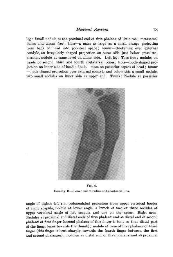
Medical Section 13
leg: Small nodule at the proximal end of first phalanx of little toe; metatarsalbones and tarsus free; tibia-a mass as large as a small orange projectingfrom back of head into popliteal space; femur-thickening over externalcondyle, an irregularly shaped projection on outer side just below great tro-chanter, nodule at same level on inner side. Left leg: Toes free; nodules onheads of second, third and fourth metatarsal bones; tibia-hook-shaped pro-jection on inner side of head; fibula-mass on posterior aspect of head; femur-hook-shaped projection over external condyle and below this a small nodule,two small nodules on inner side at upper end. Trunk: Nodule at posterior
FIG. 6.
Dorothy B.-Lower end of radius and shortelned ulna.
angle of eighth left rib, pedunculated projection from upper vertebral borderof right scapula, nodule at lower angle, a bunch of two or three nodules atupper vertebral angle of left scapula and one on the spine. Right arm:Nodules at proximal and distal ends of first phalanx and at distal end of secondphalanx of first finger (second phalanx of this finger is bent so that distal partof the finger leans towards the thumb); nodule at base of first phalanx of thirdfinger (this finger is bent sharply towards the fourth finger between the firstand second phalanges); nodules at distal end of first phalanx and at proximal

14 Gossage & Carling: Multiple Cartilaginous Exostoses
end of end phalanx of little finger (tbis finger is bent sharply outwards atjunction of first and second phalanges) ; there is also a nodule on the palmaraspect of proximal end of the first phalanx of thumb; nodule on inner side ofproximal end of first metacarpal bone ; there is a large mass over the distalend of the ulna and thickening of the outer side of the lower end of the radius;the upper end of the humerus is markedly thickened on both the outer andinner sides. Left arm: Nodules on distal ends of second phalanges of twomiddle fingers and on proximal end of first phalanx of third finger; nodules atdistal ends of first, third and fourth metacarpal bones; there is some thicken-ing of the lower end of the ulna, cbiefly on its radial side; nodule at upper end
0
FIG. 7.
Florelnce B.-Lower end of radius and ulnia.
of the humerus beneath the deltoid. Although there is very striking deformityof the right hand in this patient, there is no marked ulnar deviation of thehands as a whole like that in her sister Florence (Case V) (see figs. 4, 6and 8).
Case VIII.-Ernest B., aged 16 in 1902, a delicate, overgrown boy, whoshows no signs of osteomata. Is now, in 1910, stated to be strong and healthy;is married, and has a healthy female child aged 18 months; this child showsno signs of bony tumours.

Medical Section
Case IX.-Mrs. B., mother of the above and two other healthy children,who show no signs of exostoses, is now a woman aged 47. Neither her fathernor her mother, nor any of her mother's relatives, nor any of her five brothersand sisters or their children, have ever had lumps similar to those on her ownchildren. She stated that she herself, however, had had a few lumps about
FIG. 8.
Dorothy B.-Upper end of femur. The mass is definitely juxta-epiphysial.The periosteum of the shaft has apparently been irregularly active.
the fingers and shoulders when a young girl, but that these had disappeared.Her husband and his relatives have never had similar lumps. On examinationshe was found to be a healthy woman of rather low stature. In spite of her
15
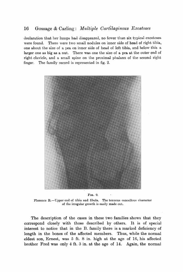
16 Gossage & Carling: Multiple Cartilaginous EJxostoses
declaration that her lumps had disappeared, no fewer than six typical exostoseswere found. There were two small nodules on inner side of head of right tibia,one about the size of a pea on inner side of head of left tibia, and below this alarger one as big as a nut. There was one the size of a pea at the outer end ofright clavicle, and a small spine on the proximal phalanx of the second rightfinger. The family record is represented in fig. 2.
FIG. 9.
Florence B.-Upper end of tibia and fibula. The tenuous cancellous characterof the irregular growth is easily made out.
The description of the cases in these two families shows that theycorrespond closely with those described by others. It is of specialinterest to notice that in the B. family there is a marked deficiency oflength in the bones of the affected members. Thus, while the normaleldest son, Ernest, was 5 ft. 8 in. high at the age of 16, his affectedbrother Fred was only 4 ft. 3 in. at the age of 14. Again, the normal
Medical Section
Edith B. at the age of 8 was described as head and shoulders taller thanher affected sister Harriet, aged 10. Similarly, Florence B., both at theage of 12 and now when fully grown, is decidedly below the normalheight. It is also of interest to note the varying intensity with whichthe individuals are affected, Mrs. B. having only six small tumours,Florence and Harriet B. being moderately affected, while in Fred andDorothy B. the growths are very large and numerous. We have investi-gated the family records as closely as possible, but have unfortunatelybeen unable personally to interview more than the nine members of thetwo families here described. The details as to the other members aretherefore less certain than we could wish, being dependent on the state-ments of Mrs. H. and Mrs. B. We would call attention to the largenumber of normal relatives claimed by both these women-a numberthat would not be expected according to the view advocated in thisarticle. It is of interest that Mrs. B.'s mother was German, andmultiple exostoses seem much commoner in Germany than in England.
The radiograms of the B. family, some of which are here reproduced,illustrate very well the gross disturbance of growth at the intermediarycartilage. It is apparent that there is much more than mere outgrowthof bone in various shapes and directions, such as might be inferred frominspection of macerated specimens. In some instances the nornmaldifferentiation into compact and cancellous tissue seem-s to be lostfor two or three inches at the extremities of the diaphysis. In manysituations, particularly at the ends of the metacarpals, and in the upperextremity of the humerus-not illustrated in the reproductions-thereis a marked transparency of the bone comparable to that seen in someforms of osteo-arthritis.
17
18 Gossage & Carling: Multiple Cartilaginous Exostoses
BIBLIOGRAPHY.
Hereditary Cases with Fairly Complete Family History.
Affected individuals marked black, lnormal white. Sex indicated in the usual way.
J; ? 0BESSEL-HAGEN. Arch. f. klimz. Chir., Berl., 1891, xli, p. 420... ... 3 2 5Idem ... ... ... ... ... ... ... 2 03COTES. Trans. Clin. Soc. Lond., 1890, xxiv, p. 228 ... 1 0 4DITTRICH. Inaugural Dissertation, Kiel, 1892 ..I ... ... 1 1 2FIsCHER. Deutsch. Zeitschr. f. Chir., Leipz., 1880, xii, pp. 34 and 357 3 0 3Idemn . ..... ... ... ... ... 4 o 1Idem ., ... ... ... ... ... ... ... 20 2FRANCIS. Lancet, 1902, i, p. 1765 ... ... ... ... 1 0 0GIBNEY. Amer. Journ. of Med. Sci., 1875, lxxii, p. 73 .. ... 3 2 3HEYMANN. Virchows Archir, Berl., 1886, civ, p. 145 ... ... 7 1 11HCEBER. Inaugural Dissertation, Munchen, 1901 ... ... ... 4 2 0JUNGMANN. Berl. klint. Wochenschr., 1902, xxxix, p. 890 ... ... 1 1 2KELLER. Inaugural Dissertation, Halle, 1901 ... ... ... 3 0 6
(Cf. also BRAUNE and CHOLEWA.)KIRMISSON. Rev. d'Orthop., 1905, vi, p. 245 ... ... ... 1 1 0KREMSER. Inaugural Dissertation, Mluncheni, 1893 ... ... 2 0 1LIPPERT. Detutsch. Arch. f. klin. Med., Leipz., 1903, lxxvi, p. 63 ... 2 4 17MARLE. Inaugural Dissertation, Berlin, 1868 ... ... ... 2 0 1Idern .. ... ... ... ... ... ... ... 1 4 4NASSE. Klin. Beitr., neue Folge, 124 ... ... ... 4 0 2
(Quoted by KNOOP, Inaugural Dissertation, Bonn, 1897.)PAGET. " Lectures on Tumours," 1851, p. 79 ... ... ... 1 0 3POORE. Lancet, 1873, ii, p. 771 ... ... ... ... ... 0 1 4PULLE. Niederland. Tijdschft. v. Geneeskunde, 1900, i, p. 890 ... 1 0 1REINECKE. Beitr. z. klin. Chir., Tiib., 1891, vii, p. 657 ... ... 3 2 9REULOS. Le Progres Med., 1885, ii, p. 71 ... ... ... ... 6 5 11RICHTER. Inaugural Dissertation, Giessen, 1908 ... ... ... 3 1 4ROBERG. Inaugural Dissertation, Bonn, 1886 ...I ... ... 3 5 5Idem ... ... ... ... ... ... ... 2 o1SPENGLER. Inaugural Dissertation, Strasburg, 1887 ... ... 8 0 6Idemn . .. .... ... ... ... ... 0 3 5STANLEY. Med. Times and Gaz., 1853, n.s. vii, p. 39 ... ... 2 0 1STOLZENBERG. Inaugural Dissertation, Greifswald, 1898 ... ... 2 3 2WEBER. Vircn,ows Archiv, 1866, xxxv, P. 501 ... ... ... 2 1 1H. family ... ... ... ... ... ... ... 2 2 5B. family ... ... ... ... ... ... ...1 4 8
(Total families- 34) ... 83 45 133To which must be added-
TILLMANN. Miinch. med. Wochenschr., 1898, p. 1073 .. ... 5 2
(35 families) ... 133 135

Medical Section 19
Hereditary Cases with IncomplWete Famlily Records.
AUSCH. Deutsch. med. Wochenschr., 1904, p. 1304.BENNEKE. Centralbl. f. Chir., Leipz., 1901, xxviii, p. 315.CHIARI. Prager med. Wochenschr., 1892, xvii, P. 403.CRUVEILHIER, quoted by BRAUNE. Inaugural Dissertation, Halle, 1882.DRESCHER. Inaugural Dissertation, Giessen, 1889.FISCHER. Loc. cit.HARTMANN. Archiv f. klin. Chir., Berl., 1893, xlv, p. 572.HASHIMOTO. Archiv f. klini. Chir., 1885, xxxii, p. 1.HERBST. Inaugural Dissertation, Berlin, 1890.KIENBOCK. Wien. klhn. Wochenschr., 1903, p. 109.LAWEN. Deutsch. Zeitschr. f. Chir., 1904, lxxv, p. 14.LIPPERT. Loc. cit.MACLEAN. Bristol Med.-Chir. Journ., 1890, viii, p. 217.MARLE. Loc. cit.NAST. Loc. cit.PELS-LEUSDEN. Deutsch. Zeitschr. f. Chir., 1907, lxxxvi, p. 434, who gives a complete
bibliography.PRICE. Lancet, 1868, i, p. 631.ROBERG. Loc. cit.SCHAEFER. Beitr. z. klin. Chir., TUb., 1901, xxxi, p. 228.SCHMIDT. Inaugural Dissertation, Griefswald, 1868.SCHOR, quoted by HERBST. St. Petersb. mied. Wochenschr., 1881, p. 327.SEIDEL. Centralbl. f. Chir., 1885, xii, p. 12.SONNENSCHEIN. Inaugural Dissertation, Berlin, 1873.STANLEY. "Diseases of Bones," 1849, p. 213.STARCK. Beitr. z. klin. Chir., 1902, xxxiv, p. 508.VILLEMIN. Rev. d'Orthop., 1903, iv, p. 49.ZIEGLER. Miunch. mned. Wochenschr., 1892, xxxix, p. 552.
Other References.
BATESON. "1 Reports to the Evolution Committee of Royal Society," 1905, ii, p. 114.GOSSAGE. Quart. Journ. of Med., 1908, i, p. 331.KAREWSKI. Berl. klin. Wochenschr., 1892, xxix, p. 257.KOHLER, quoted by PELS-LEUSDEN. Fortschr. a. d. Geb. d. Ri;ntgenstrahlen, 1905, viii,
Heft 1.KRYGER. Archiv f. klin. Chir., 1898, lvii, p. 859.MCCARRISON. Proc. Roy. Soc. Med., 1908, ii (Med. Sect.), p. 1.OLLIER. Cp. PAYR in WALLSTEIN U. WILM'S II Lehrbuch der Chirurgie," 1909, ii, part 2,
p. 403.REUBSAEL. These de Par., 1909.
20 Gossage & Carling: Multiple Cartilaginous Exostoses
DISCUSSION.
Mr. WILLIAM BATESON, F.R.S., thanked the Council for inviting himto hear the paper. He knew nothing of the subject beyond what he hadlearned from Dr. Gossage; but it was an admirable thing that Dr. Gossageshould have co-ordinated this evidence. Dr. Gossage had made a number ofother collections of a similar character, and this last was a most useful additionto the series. When they came to consider the system of heredity which thisparticular condition followed, the problem was a rather baffling one. The con-dition was evidently transmitted in a very complex manner, and was influencedpresumably by various factors present in the body besides the particular factorto which the disease itself was due. Nevertheless, he thought Dr. Gossage wasentitled, on the evidence he had put forward, to his view that the inheritancewas more or less that of a Mendelian dominant; but so many complicationsmust necessarily be introduced that, as Dr. Gossage had stated, this conclu-sion must at present be regarded as provisional. They were greatly in need ofthe data of new examples of cases in which heredity was sex-limited. Thebest-known case up to the present was colour-blindness, and there was alsohaemophilia; but beyond these they had no considerable evidence to work uponof sex-limited heredity in man, although in animals there were several instances.It was true that they now understood fairly well the lines which were followedby these systems of inheritance; but more examples would be welcome, espe-cially with regard to numerical effects. He conlfessed to a difficulty in thinkingthat the condition which formed the subject of the paper was a case of sex-limited inheritance comparable with that of colour-blindness. The point thatweighed particularly with him was the evidence as to inheritance from theaffected females. One would expect the daughters of affected females to beaffected in greater proportion than the daughters of unaffected females byaffected males, but there was no evidence at all of this from the genealogicaltables which Dr. Gossage had shown. He would like, while speaking to theSection, to take the opportunity of saying how greatly they were in need ofevidence of sex-limited conditions in which the limitation was to femalesrather than to males. It was possible that such things did not exist, butif they did exist students of heredity ought to begin to learn something aboutthem. Their study would greatly contribute tQ the general theory of heredity.The only disease to which it might possibly apply, he thought, was Graves'disease. He had examined the evidence as well as he could, and on the whole,though he did not think thaet Graves' disease could be brought within the sex-limited class, the mode of its descent was worth investigating.' He wasunaware of any other condition that seemed likely to come within the scope ofthat system of transmission. He was grateful to Dr. Gossage, who had madeadmirable collections on similar subjects at intervals, but he thought that thiswas one of the most important he had yet done.
I Evidence as to heredity in Graves' disease has been collected by Ernst Schultheiss,"Ub. Erblichkeit bei Morbus Basedowii," Inaugural Dissertation, Jena, 1909.

Medical Section
Dr. RUSSELL WELLS said he was by no means convinced that Mendelismapplied to questions of disease in the human subject. Possibly the class of casesmentioned might be instances of Mendelian inheritance, but he did not see thatthe tables put forward proved anything beyond the fact that the condition inquestion ran in families, and was probably transmitted by inheritance. If onetook the histories of the individual families given and examined them separately,it was noticeable how many of them did not seem to correspond to what thehypothesis of Mendel required. To take one instance, in more than one caseall the children had exostoses, though he did not gather that both parents wereabnormal. No doubt it was particularly difficult to get complete histories, andthey were indebted to Dr. Gossage and Mr. Carling for the great amount ofwork the paper represented; but he noted that even where they had fairlycomplete family histories they based their argument on the last generationalone. He would be glad if, in his reply, Dr. Gossage would point out allthe data this investigation supplied in support of the Mendelian hypothesis.Possibly he was somewhat biased because he had performed some experimentsin breeding rabbits, which he would probably never publish because circum-stances prevented them being complete, but which made him doubt whetherthe full Mendelian doctrine could be applied to the higher animal kingdom,as there was reason to believe it could to the vegetable. The year before lasthe crossed a Himalayan doe rabbit with an Angora buck. These two sorts ofrabbits are quite distinct varieties, breeding true; they are both albinos in thesense that they have pink eyes, but the Angora has long white hair all over,while the Himalaya, though chiefly white, has black ears, nose, paws, and tail,and its hair is short. The progeny of this union were all white rabbits withshort hair. Some died, but he saved sufficient to mate bucks with does of thesame litter; on the Mendelian hypothesis he expected to find in their progenysome with long hair and others with black points, but it was not so; all theyoung in this second generation were like their parents, completely white rabbitswith short fur. Some of this second generation were mated together, andtheir young ones again were all white rabbits with short fur. The interestingpoint is that this experiment resulted in the production of a race of rabbitsintermediate between the Angora and the Himalaya, having neither the long hairof the one nor the black points of the other, and not, as one would expect onthe Mendelian theory, some with one or other of these characteristics. Hecould not say what the numbers were, but he would try to ascertain them.
Mr. CARLING said he could not add anything to the aspect of the questionnow being discussed, and he disclaimed credit for the industry which had beendisplayed in collecting the family histories; that credit belonged to Dr. Gossage.But it might be of interest to show a specimen from the museum of theDreadnought Hospital, which illustrated many of the surgical points; the sizeof the tumours, their liability to compress important structures, the generaltendency of exostoses in the limbs to run upwards, and some of the difficultieswhich might be experienced in removing them. It was noteworthy that theman from whom the specimen was obtained died -of chondro-sarcoma. of therib. Whether it began in an exostosis could not now be said, but it was
N-9b
'21

22 Gossage & Carling: Multiple Cartilaginous Exostoses
interesting that multiple benign osteomata should be associated with terminalsarcoma of bone.
Professor BATESON remarked that the Himalaya rabbit was one of thestrangest problems which students of the Mendelian theory had to deal with,and any evidence about it would be most welcome.
Dr. GosSAGE, in reply, said that one of the points brought up byDr. Russell Wells was as to how far these families of multiple exostoses couldbe taken as evidence in favour of Mendelism. It was doubtful whether onecould take anything from human beings as evidence of Mendelism if one had tostart afresh, and had no other evidence of Mendelism at all. Mendelismdepended on evidence which was obtained from experimental work with animalsand plants in regard to which one could repeat the experiment until the proofseemed certain. Evidence from human beings could not be corrected like thatderived by experimental methods, because each marriage was haphazard; butone was justified in saying that when one found that an abnormal individualmated with a normal had half his children normal and half abnormal, and thatsimilar phenomena went on from generation to generation, it conformed withMendel's law. In multiple exostoses there was evidence that the families ofthe affected people when crossed with normals were, taking them as a whole,half of them abnormal and half of them normal. He said taking them as awhole, because one could not judge from a single family, in which it was a" toss up " whether a particular child would be normal or abnormal, and thetotal number of children was very limited. In the tables there were numbersrunning into hundreds; there were the actual figures of 133 abnormal and 135normal, drawn from a large number of families, and the collecting of largenumbers of families was the only way in which one could possibly put forwardevidence from human beings. But supposing it to be true that the femalecould often carry the abnormality without showing it, one had to add a con-siderable number of females to the abnormals, which made the abnormalssomewhat too numerous. In other conditions where the presumption that theyconformed with Mendelism was more justified, the same excess of abnormalswas obtained, and it was not difficult to explain, because if one tried to get afamily history from a woman of the less intelligent class it would be found thatshe remembered the abnormals but not the normals, sometimes even amongher own children, still more so among her brothers and sisters and otherrelatives. So the abnormals would be mentioned, while the normals tended tobe omitted; 171 and 135 were sufficiently near equality to justify the con-sideration that the condition was handed down more or less on Mendelian lines.As to the exact manner in which it was handed down, that was a very difficultproblem, one in regard to which human evidence could not possibly supply ananswer. The only way in which a working theory could be obtained as to themanner of the hereditary transmission would be by finding some such conditionin animals where it allowed of being submitted to exact experimental investi-gation. Multiple exostoses were not, properly speaking, a disease, except in sofar as they occasionally caused unpleasantness and the pressure of the tumoursgave discomfort. Many of the people were perfectly healthy, and so thecondition was better described as an abnormality.