molecular and functional analysis of two new mttp gene mutations
TRANSCRIPT

HAL Id: inserm-00813163http://www.hal.inserm.fr/inserm-00813163
Submitted on 15 Apr 2013
HAL is a multi-disciplinary open accessarchive for the deposit and dissemination of sci-entific research documents, whether they are pub-lished or not. The documents may come fromteaching and research institutions in France orabroad, or from public or private research centers.
L’archive ouverte pluridisciplinaire HAL, estdestinée au dépôt et à la diffusion de documentsscientifiques de niveau recherche, publiés ou non,émanant des établissements d’enseignement et derecherche français ou étrangers, des laboratoirespublics ou privés.
Molecular and functional analysis of two new MTTPgene mutations in an atypical case of
abetalipoproteinemia.Mathilde Di Filippo, Hervé Créhalet, Marie Elisabeth Samson-Bouma,Véronique Bonnet, Lawrence Aggerbeck, Jean-Pierre Rabès, Frederic
Gottrand, Gérald Luc, Dominique Bozon, Agnès Sassolas
To cite this version:Mathilde Di Filippo, Hervé Créhalet, Marie Elisabeth Samson-Bouma, Véronique Bonnet, LawrenceAggerbeck, et al.. Molecular and functional analysis of two new MTTP gene mutations in an atyp-ical case of abetalipoproteinemia.: Functional analysis of two new MTTP mutations. Journal ofLipid Research, American Society for Biochemistry and Molecular Biology, 2012, 53 (3), pp.548-55.<10.1194/jlr.M020024>. <inserm-00813163>

1
Molecular and functional analysis of two new MTTP gene mutations in an
atypical case of abetalipoproteinemia.
Mathilde Di Filippo [email protected] (1,2), Hervé Créhalet
[email protected] (1), Marie Elisabeth Samson-Bouma marie-
[email protected] (3), Véronique Bonnet veronique.bonnet@chu-
lyon.fr (1), Lawrence P. Aggerbeck [email protected] (4), Jean-Pierre
Rabès [email protected] (3,5,6), Frederic Gottrand
[email protected] (7), Gérald Luc [email protected]
(8), Dominique Bozon [email protected] (1), Agnès Sassolas
[email protected] (1,2)
Institutions
(1) Hospices Civils de Lyon, Centre de Biologie et de Pathologie Est, Département
de biochimie et biologie moléculaire, Bron cedex F-69677, France
(2) Université de Lyon, INSERM U1060, INSA de Lyon, INRA U1235, Univ Lyon-1,
Villeurbanne F-69621, Oullins F-69600, France
(3) INSERM U698, Université D Diderot, CHU X. Bichat Secteur C. Bernard, Paris,
75877, France
(4) INSERM UMR S-747, Université Paris Descartes, 75006 Paris
(5) Université Versailles Saint-Quentin-en-Yvelines, UFR de Médecine Paris Ile-de-
France Ouest, Guyancourt, 78280, France
(6) AP-HP, Hôpital Ambroise Paré, Service de Biochimie et Génétique Moléculaire,
Boulogne, 92104, France
2
(7) CHRU Lille, Hôpital Jeanne de Flandre, Département de Pédiatrie, Université
Lille Nord de France, Faculté de médecine, INSERM U995, IFR114, Lille, France
(8) Hôpital Universitaire de Lille, Service de Médecine Interne, Université Lille Nord
de France
Running head: Functional analysis of two new MTTP mutations
Corresponding author : Mathilde Di Filippo,
Hospices Civils de Lyon, Centre de Biologie et de Pathologie Est, Département de
biochimie et biologie moléculaire, Bron cedex F-69677, France
Tel. +33 4 72 11 89 94
Fax +33 4 27 85 59 00
E-mail: [email protected]
List abbreviations in the order cited
ABL abetalipoproteinemia
MTTP microsomal triglyceride transfer protein large subunit gene
MTP microsomal triglyceride transfer protein large subunit
PDI protein disulfide isomerase
P4HB prolyl 4-hydroxylase, beta polypeptide
ApoB apolipoprotein B
MCT medium chain triglyceride
PCSK9 proprotein convertase subtilisin/kexin type 9
ANGPTL3 angiopoietin-like 3
3
Abstract
Abetalipoproteinemia (ABL) is an inherited disease characterized by the
defective assembly and secretion of apolipoprotein B-containing lipoproteins caused
by mutations in the microsomal triglyceride transfer protein large subunit (MTP) gene
(MTTP). We report here a female patient with an unusual clinical and biochemical
ABL phenotype. She presented with severe liver injury, low levels of LDL-cholesterol,
subnormal levels of vitamin E, but only mild fat malabsorption and no retinitis
pigmentosa or acanthocytosis. Our objective was to search for MTTP mutations and
to determine the relationship between the genotype and this particular phenotype.
The subject exhibited compound heterozygosity for two novel MTTP
mutations: one missense mutation (p.Leu435His) and an intronic deletion (c.619-
5_619-2del). COS-1 cells expressing the missense mutant protein exhibited
negligible levels of MTP activity. In contrast, the minigene splicing reporter assay
showed an incomplete splicing defect of the intronic deletion with 26% of the normal
splicing being maintained in the transfected HeLa cells.
The small amount of MTP activity resulting from the residual normal splicing in
the patient explains the atypical phenotype observed. Our investigation provides an
example of a functional analysis of unclassified variations, which is an absolute
necessity for the molecular diagnosis of atypical ABL cases.
Supplementary key words: dyslipidemias, familial hypocholesterolemia,
chylomicrons, lipoprotein/assembly, hepatosteatosis, genetics, gene expression,
genotype-phenotype correlation, functional analysis, splicing

4
Abetalipoproteinemia (ABL; OMIM#200100) is an autosomal recessive
hypocholesterolemia usually detected during infancy due to failure to thrive, severe
diarrhoea and a lipid malabsorption syndrome. Sixty years ago, Bassen and
Kornzweig described this disease for the first time (1) and forty years later, the role of
MTP (microsomal triglyceride transfer protein large subunit) was discovered (2-5).
MTP forms a heterodimer with protein disulfide isomerase (PDI) also named prolyl 4-
hydroxylase beta polypeptide (P4HB) which is responsible of the assembly of
apolipoprotein B (ApoB) containing lipoproteins in the liver and the intestine.
Following the discovery of the first mutations, many mutations have been identified in
the microsomal triglyceride transfer protein large subunit gene (MTTP) in patients (3-
6). The identification of mutations in the MTTP gene in DNA from patients is
important for establishing the diagnosis of ABL in the context of two other hereditary
hypocholesterolemias (homozygous familial hypobetalipoproteinemia OMIM#107730
and Anderson’s disease/Chylomicron retention disease, OMIM#246700) which are
due to mutations in the APOB and SAR1B genes, respectively.
To date, mutations in the MTTP gene have been established in about 50
cases of ABL (7-12). In some instances, the prediction of altered MTP function is
readily made on the basis of premature stop codons, mutations in canonical splice
sites or frameshift mutations. In other cases, the functional consequences have been
difficult to predict (13-15), even if the mutations co-segregate with clinical phenotype.
The measurement of MTP activity (2, 3, 6, 11, 13, 16, 17) and the consequence of
intronic mutations (9, 11) have been studied in only a few cases due to the necessity
of additional intestinal or hepatic biopsies.
ABL patients are usually diagnosed during infancy and they exhibit marked
lipid malabsorption, very low levels of cholesterol and triglycerides, the absence of

5
ApoB in the plasma, the absence of chylomicrons after fat loading, and essential fatty
acid and fat-soluble vitamin deficiencies (particularly vitamin E). Hepatic steatosis
has not been reported uniformly (although it is frequent), and hepatic fibrosis or
cirrhosis has been reported in a few cases (18, 19). We report here an atypical case
of ABL presenting in childhood with severe liver injury, hypocholesterolemia
associated with a low (but not absent) levels of plasma ApoB and a subnormal level
of plasma vitamin E. Furthermore, the patient exhibited normal development into
adulthood. ABL was suspected because of the presence, at the ultrastructural level,
of large amounts of free lipid droplets accumulated in the cytoplasm of enterocytes
and hepatocytes in intestinal and liver biopsies.
The patient was found to be compound heterozygous for two novel mutations
in the MTTP gene, one intronic (c.619-5_619-2 del, from her mother) and the other a
missense mutation (p.Leu435His, from her father). The possible functional impact of
these two mutations was not readily apparent. Since no additional biopsy material
was available from the patient, two different assays were established. COS-1 cells
were transfected with wild type and several mutant MTTP cDNAs in order to evaluate
the impact of the missense mutation on MTP activity and HeLa cells were transfected
with wild-type and mutant minigenes to evaluate possible defects in splicing.
Material and methods
Patient (diagnosis and follow-up)
A 4 year-old girl was referred for an anicteric chronic hepatitis fortuitously
discovered 10 months previously. The level of blood transaminases were consistently
elevated and ranged from 5 to 7 times the normal values associated with
bilirubinemia in the absence of inflammation or hepatic insufficiency. Upon clinical
6
examination, the liver was moderately enlarged and firm, but there was no other sign
of chronic liver disease. The patient’s growth was normal. The patient’s parents
altered her diet to avoid fatty foods following episodes of diarrhoea in the first months
of life. The levels of total cholesterol and ApoB in the plasma were found to be low,
whereas the level of triglyceride was normal (Table1). The parents were not related
and they had normal levels of blood lipids.
A perendoscopic jejunal biopsy, performed as previously described (20),
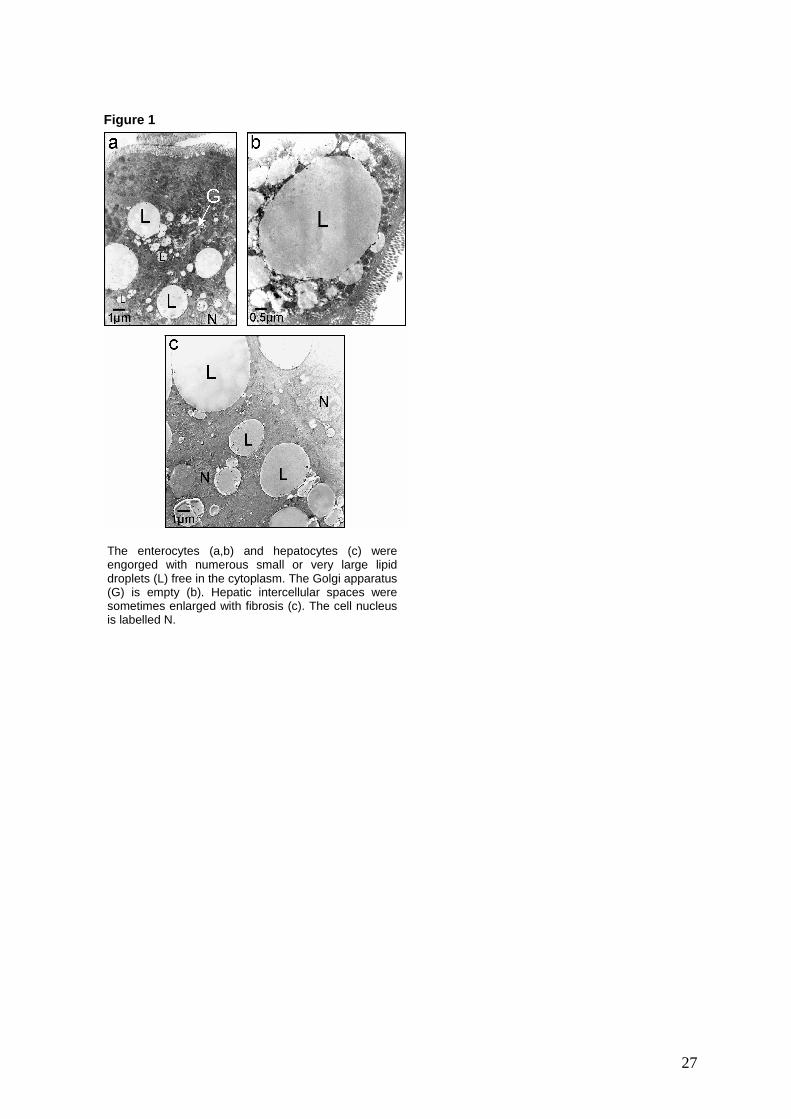
showed a white mucosa. Electron microscopic examination of the biopsy showed
enterocytes with many small or very large free lipid droplets scatterred throughout the
cytoplasm (Figure 1a, 1b). Examination by ultrasound showed that the liver was
moderately enlarged and slightly hyper-echogenic. A percutaneous liver biopsy
showed extensive fibrosis (scored F3 according to the METAVIR scoring system
(21)) which was associated with a massive micro- and macro-vesicular steatosis.
Large amounts of free lipid droplets were observed in the cytoplasm by electron
microscopy (Figure 1c). Abetalipoproteinemia was suspected although retinitis
pigmentosa and acanthocytosis were absent and the blood vitamin E level was only
slightly decreased. The fecal fat content, measured over a 3 day period, was also
normal (mean 2.8 g/24h, normal < 3g/24h). However, when the child consumed a
normal fat diet (33% fat), diarrhoea occurred and steatorrhoea was 24 g/day. After an
oral fat load (Table1), the level of serum triglycerides increased slightly and a small
amount of chylomicrons appeared in the plasma, suggesting that some intestinal lipid
absorption occurs accompanied by low levels of intestinal lipoprotein secretion.
A diet limited in saturated fatty acids (22% of total energy) and enriched with
medium chain triglycerides (MCT) was begun and resulted in the elimination of
diarrhoea, progressive improvement in liver function (transaminases 2 to 4 X normal

7
at the age of 7 years, 2 to 3 X normal at 14 years, normal values at 18 years of age),
the normalization of the levels of essential fatty acids and fat soluble vitamins, and
normal growth and puberty. At the age of 14 years, the patient no longer adhered to
the MCT diet and 36% of total energy intake was composed of fat. Hepatitis recurred
(transaminases increased to 150 to 200 X normal) but resolved when the MCT diet
was re-instituted.
The patient is now 22 years of age and is in good health. Fat soluble vitamins
are in the normal range without any supplementation. The levels of transaminases
are < 1.5 X normal and hepatic steatosis persists (16% of hepatic parenchyma as
determined by magnetic resonance imaging). There are no neurological,
ophthalmological or retinal abnormalities.
Blood samples, intestinal and liver biopsies were obtained from the patient
using the procedures and the experimental methods approved by INSERM (RBM
0256) and by a bioethics committee (Comité Consultatif de Protection des Personnes
dans la Recherche Biomédicale de Paris Bichat-Claude Bernard, Paris, France,
CCPPRB Bichat-C. Bernard-2003/05). Informed, written consent was obtained from
the patient’s parents.
Mutation analysis
Following the extraction of genomic DNA from the blood (Nucleon Bac3, GE
Healthcare®, Chalfont St. Giles, UK, http://www.gehealthcare.com), each of the
coding exons, and the flanking intronic junctions, of the MTTP, APOB, proprotein
convertase subtilisin/kexin type 9 (PCSK9) and angiopoietin-like 3 (ANGPTL3) genes
was amplified by PCR. The amplicons were sequenced directly with the BigDye®

8
Terminator v3.1Cycle Sequencing Kit on an ABI PRISM 3130 or 3730 DNA
sequencer (Applied Biosystems, Foster City, USA, www.appliedbiosystems.com).
In Silico Analysis
Analysis of the mutations was performed with Alamut v2.0 (Interactive
Software), Polyphen (http://genetics.bwh.harvard.edu/pph/) and SIFT
(http://sift.jcvi.org/www/SIFT_aligned_seqs_submit.html). Several algorithms were
used for computational scoring of 3’ splice site based on different concepts using
default parameter settings (Neural Network Splice Prediction (NNSplice) (22),
MaxEntScan (23), Splice site Finder Like, GeneSplicer (24), Human Splicing Finder
(HSF) (25)).
Protein expression
To evaluate the p.Leu435His protein expression, two assays were performed.
First, to measure wild type and mutant MTP activities, we transfected COS-1 cells
with the MTTP and P4HB cDNA. Second, to compare the production of wild type and
p.Leu435His, we transfected COS-1 cells with wild type and mutated MTTP cDNA C-
ter tagged with GFP.
MTP activity
A 2699 bp fragment containing the entire MTTP coding sequence, extending
from c.-5 to c.*9 (NM_000253.2) and a 1582 bp fragment containing the entire P4HB
coding sequence (from c.-14 to c.1527, NM_000918.3) were obtained by RT-PCR
from 1µg of human liver total RNA (Cat.No.636531, Clontech, Mountain View, U.S.A,
www.clontech.com) with the Transcriptor High Fidelity cDNA Synthesis Sample Kit
9
(Cat.No.05081963001 Roche Applied Science, Indianapolis, USA, www.roche-
applied-science.com).
The MTTP cDNA was inserted, with the In-Fusion Advantage PCR Cloning Kit
(Cat.No.639616, Clontech, Mountain View, U.S.A, www.clontech.com), into the Kpn I
site of the pBudCE4.1 expression vector (Cat.No.V532-20 Invitrogen, Carlsbad,
U.S.A, www.invitrogen.com) downstream of the human elongation factor 1�-subunit
promoter. The P4HB cDNA was inserted into the Hind III site of the same vector,
downstream of the human cytomegalovirus immediate-early promoter, allowing the
production of the two proteins from the same plasmid.
The p.Leu435His (c.1304T>A), p.Leu435Glu (c.1303_1305delinsGAA),
p.Leu435Val (c.1303C>G) and p.Cys194Stop (c.582C>A) mutants were constructed
from the wild type sequence with the QuickChange II XL Site-Directed Mutagenesis
Kit (Cat.No.200521 Stratagene, Agilent Technologies, Cedar Creek, USA,
www.agilent.com) according to the manufacturer’s instructions.
Transient expression of MTP and P4HB in COS-1 cells (Cat. No. CRL-1650
ATCC) was carried out by transfecting 6 µg of plasmid per T25 flask in the presence
of 9 µL of FuGENE® HD (Cat.No.4709691001 Roche Applied Science, Indianapolis,
USA, www.roche-applied-science.com), according to the manufacturer’s instructions.
COS-1 cells were harvested by trypsinization 48 hours post-transfection and
disrupted by sonication. Triglyceride transfer from donor to acceptor vesicles was
measured by a fluorescent-labelled method using a commercial kit (R100 MTP
activity, Chylos Inc, USA, www.chylos.com) according to the manufacturer’s
instructions and previously described fluorescent methods (26-28). The results are
expressed as % transfer/mg total proteins/h. The method was evaluated with
10
intestinal and hepatic biopsies and the MTP activities were in agreement with
previous published results (data not shown).
Visualisation of MTP in transfected cells
A fragment containing the MTTP cDNA, extending from c.1 to c.2682
(NM_000253.2) of each sequence (wild type, p.Cys194Stop, p.Leu435His) was
amplified from the previously obtained pBudCE4.1 vector. The MTTP cDNA was
inserted, with the In-Fusion Advantage PCR Cloning Kit into the KpnI site of
pAcGFP1-N1 expression vector (Cat.No.632469 Clontech, Mountain View, U.S.A,
www.clontech.com) between the immediate early promoter of the CMV (PCMV IE)
and the AcGFP1 cDNA allowing the fusion of the MTP and the GFP proteins. COS-1
cells were electroporated with 0.5 µg of MTP-AcGFP1 plasmid per 80 000 cells
(MicroPorator, DigitalBio Technology and Neon Transfection System 10µL Kit,
Cat.No. MPK1096, Invitrogen Carlsbad, U.S.A, www.invitrogen.com) according to the
manufacturer’s instructions (1050V, 30ms, 2 pulses). Intracellular fluorescence was
observed with a Nikon Eclipse TE 2000-U microscope 48h after transfection.
Minigene Splicing Reporter Assay
A 371 bp MTTP fragment (the last 147 bases of intron 5, 140 bp of exon 6,
and the first 84 bases of intron 6) was amplified from the patient’s DNA and inserted
(In-Fusion Advantage PCR Cloning Kit) into the Nde I restriction site of the pTB
minigene vector (29). The transfection of normal and mutant minigenes into HeLa
cells and RT-PCR procedures were as previously described (30).
The fluorescence of the ethidium bromide bands obtained following gel
electrophoresis of RT–PCR products was integrated under unsaturated conditions
11
(Quantity One® 1-D Analysis Software Cat. No170-9600, BIO-RAD, www.bio-
rad.com) to derive band intensities.
Metabolic labelling of intestinal biopsies
Intestinal biopsies from normal individuals and from the patient were placed
into organ culture and metabolically labelled with [35S] methionine as described
previously (31). After homogenization and solubilization, the labelled intestinal biopsy
extracts and the corresponding media were immunoprecipitated with polyclonal
antibodies against Apo B (Rabbit polyclonal antibodies to Apo B were the gifts of Dr
A. Mazur of Institut National de la Recherche Agronomique, Champanelle, France)
and MTP (31). For semi-quantitative analysis, densitometric analysis was performed
using the PC version of NIH Image software (Scion Image) after photography with a
computer-assisted camera (GS-800 Calibrated Densitometer, BIO-RAD). The values
were normalized with respect to the amount of TCA precipitable incorporated
material in the biopsies.
Results
Identification of mutations
Sequencing was performed on the genomic DNA of the patient and her
parents. Two novel variants of the MTTP gene were identified in the patient. The first,
inherited from her father, is a change from T to A at position c.1304 in exon 10
(Supplemental Data, Figure 1A) (Nucleotide numbering starts at A of the ATG
initiating codon and exon 1 is the first coding exon) which changes the amino acid
Leu 435 to a His. The second, inherited from her mother, is a 4 bp deletion in intron

12
5: c.619-5_619-2 del (Supplemental Data, Figure 1B). This deletion of a repeated
motif (TTTA) is upstream the acceptor site. These mutations were found neither in
our panel of 100 normal alleles from unrelated subjects nor in the 1000 genomes
database (32).
The patient is also heterozygous for a well described polymorphism
p.Gln95His inherited from her father. The frequency of this allele is between 5.4 and
6% in a healthy adult Caucasian population (England) and in a sample of 270
unrelated French Canadian men (6, 33) and 6.2% in a cohort of abetalipoproteinemia
(6).
The patient had no mutation in the APOB, PCSK9 or ANGTL3 genes.
Analysis of c.1304 T>A, p.Leu435His:
The missense mutation (p.Leu435His) is located in exon 10 and affects a
highly conserved residue in the middle �-helical domain of MTP. The substitution
changes a hydrophobic to a hydrophilic, basic residue (Grantham distance: 99 [0-
215]). This missense mutation is predicted by Polyphen to be “probably damaging”
with a score of 0.99 and by SIFT to “affect protein function”.
To determine whether the p.Leu435His missense mutation produces a protein
that is functional or not, we expressed, in COS-1 cells, the wild type MTP as well as
the p.Leu435His and several other mutants (p.Leu435Glu, p.Leu435Val and
p.Cys194Stop). These different mutations were tested in COS-1 cells to evaluate the
sensitivity and specificity of our MTP activity assay: p.Leu435Glu is expected to be
as severe as p.Leu435His as it changes the hydrophobic Leu to a hydrophilic acidic
Glu, whereas p.Leu435Val is expected to be a mild change (Leu and Val are both
hydrophobic and closely related amino acids). The introduction of a premature stop

13
codon at the position p.Cys194 is expected to lead to the complete abolition of MTP
activity since the ApoB and PDI binding sites are absent from the mutant. Further,
the homozygous p.Cys194Stop mutation has been reported in an ABL patient (8).
As shown in Figure 2, cells expressing p.Leu435His (the patient’s mutation),
p.Cys194Stop, or p.Leu435Glu have negligible levels of MTP activity (0.30 to 1.9%
TG transfer/30µg protein/h) as compared to cells expressing p.Leu435Val or the wild
type protein (24.15 to 27.25 % TG transfer/30µg protein/h respectively). The activity
of the p.Leu435His MTP is not significantly different from the p.Cys194Stop truncated
protein and, thus, must be considered as having a negligible MTP activity as
compared to the wild-type protein. RT-PCR of the MTTP and P4HB transcripts in
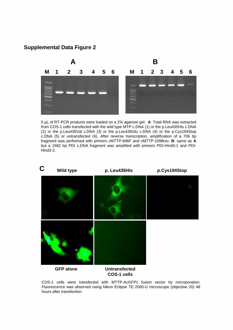
transfected COS-1 cells shows that the level of expression from each vector is
comparable for the wild type and the different mutant transfected cDNA
(Supplemental Data, Figure 2A and 2B). Fluorescent tagged MTP showed an
identical cellular distribution (Supplemental Data, Figure 2C).The p.Leu435His
mutant, therefore, is functionally defective.
Analysis of c.619-5_619-2del:
Splicing predictions
All the algorithms predicted an effect on splicing with a decrease in the score
for the acceptor site of intron 5 ranging from -10% for SSF-like to a complete
abolition for GeneSplicer, (-44% for MaxEnt Scan, -38.9% for NNSplice, -79% for
HSF). Given that all the algorithms predicted an effect on splicing, experimental
analysis of splicing was performed. HeLa cells transfected with wild type or mutant
minigenes were analyzed (size and sequence) by RT-PCR to determine the
presence of abnormal transcript processing of the mutant.

14
Minigene splicing reporter assay
Transfection of the wild type minigene into HeLa cells produced, as expected,
a 386 bp RT-PCR fragment (Figure 3, lanes 1, 2 3). In contrast, transfection of the
mutant c.619-5_619-2del minigene produced 2 different RT-PCR fragments: a small
amount of a fragment of normal size (386bp) and a large amount of a 248 bp product
(lanes 4, 5, 6). The sequence of the 386 bp PCR product includes exon 6 of MTTP
whereas exon 6 is skipped in the 248 bp product (Supplemental Data, Figure 3). This
result shows that c.619-5_619-2del mutation produces two differently spliced
transcripts: one containing exon 6 and the other without exon 6. Exon 6 skipping
would result in a protein containing the first 206 MTP amino acids but followed by 26
aberrant amino acids and a premature stop codon. In vivo, this mutant mRNA might
be targeted for nonsense mediated decay and degraded. By densitometry of the
agarose gel, the amount of normal splicing for the mutant minigene is estimated to be
26% of that of the wild type (Figure 3). These data indicate that the c.619-5_619-2del
is a splice site mutation which can lead to the skipping of exon 6. However, the effect
of this mutation on splicing is not complete and about 26% of the transcript is
correctly spliced.
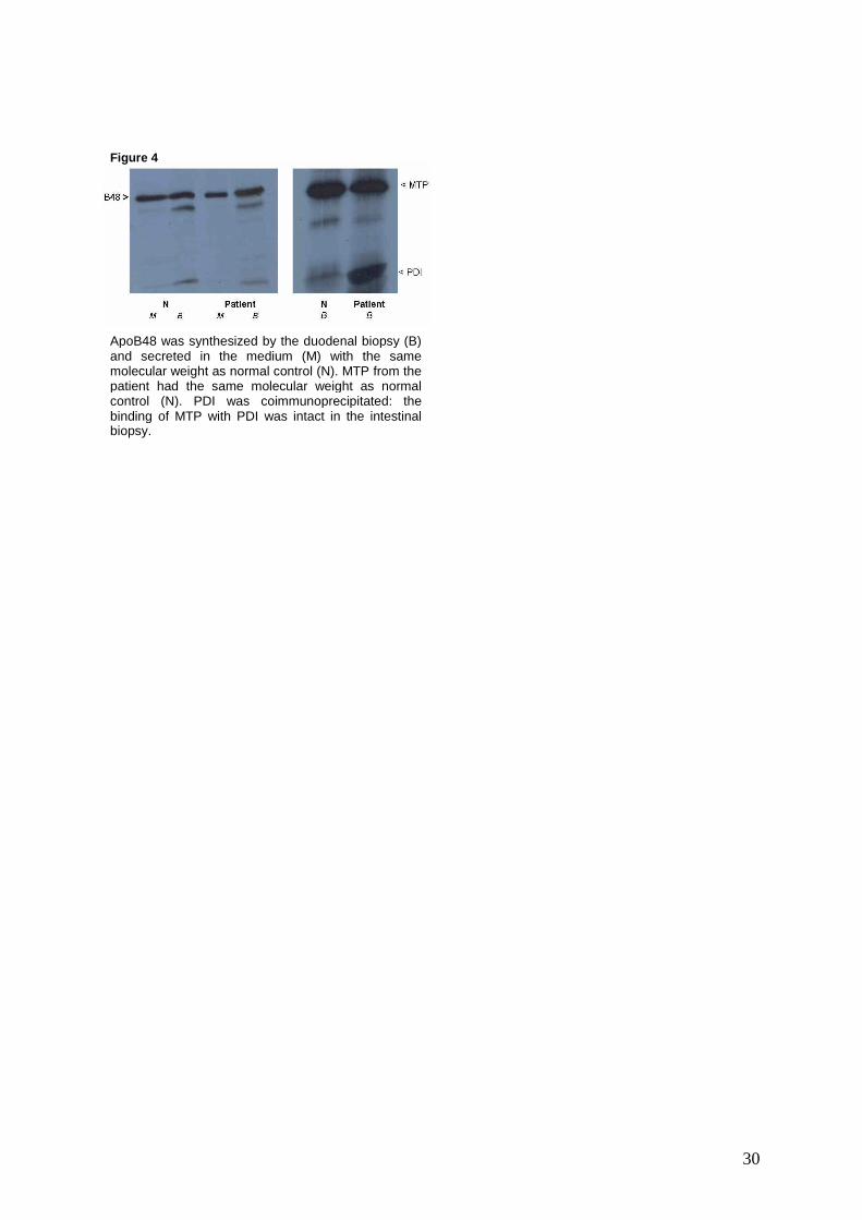
Biosynthesis of ApoB and MTP in intestinal organ culture
Immunoprecipitation with polyclonal antibodies to ApoB of the medium and the
total homogenate of the organ culture of intestinal biopsies from the patient showed
the synthesis of an ApoB48 identical in size to that of normal control subjects (Figure
4). Analysis by densitometry with correction for the amount of incorporated TCA

15
precipitable incorporated radioactivity showed that there is an intracellular retention
of ApoB48 (1.6 fold more) and a decreased ApoB48 secretion (1.3 fold less).
Immunoprecipitation with polyclonal antibodies to MTP of the total
homogenate of the organ culture of intestinal biopsies from the patient showed the
synthesis of a large subunit of identical in size to that of normal control subjects.
However, by densitometric analysis, there was 2.49 fold less MTP synthesized in the
patient’s biopsy as compared to that of the normal control subject after correction. A
protein band corresponding to the molecular mass of PDI co-immunoprecipitated with
the MTP in the patient as was observed for the normal control subject (identical in
amount as compared to that of the normal subject after correction).
Discussion
In this paper, we describe two new mutations in the MTTP gene: p.Leu435His
and a 4 bp deletion in intron 5: c.619-5_619-2 del. Since the impact of these
mutations was unclear and the phenotype was atypical, cellular functional tests were
used to further understand this new case of ABL.
The diagnosis of ABL is made readily if the following criteria are present during
the first months of life: severe diarrhoea, failure to thrive, undetectable vitamin E,
severe hypocholesterolemia and hypotriglyceridemia with undetectable amounts of
plasma ApoB, and the absence, post-prandially, of chylomicrons in the serum. Our
patient presented with a mild phenotype and lacked the major criteria for the
diagnosis of ABL. Surprisingly, the patient was referred only for anicteric chronic
hepatitis with major liver steatosis at 4 years of age. The other causes of
hypobetalipoproteinemias were ruled out because of the patient’s lipids profile and
normal lipid values of the parents (34-40). Furthermore, no mutation was found in the
16
PCSK9, ANGPTL3, APOB genes. The diagnosis of ABL was finally suspected
because of hypocholesterolemia and the typical aspect of intestinal and liver biopsies
suggesting a defect in the assembly of ApoB-containing lipoproteins.
Seven atypical cases of ABL have been reported (13-15, 19, 41, 42). For five
of these patients (Table 2) a molecular diagnosis of the mutation in MTTP was
established. However, a functional assay of the mutation was performed in only one
case (13). Two of the cases (cases 1 and 2 in Table 2) are very surprising. Both the
patients have deleterious mutations, however, the first patient exhibits a mild
phenotype (42) and the other is almost asymptomatic (13). Two other patients (cases
4 and 5 in Table 2) are homozygous for the same p.Ser590Ile mutation (14, 15).
Unfortunately the functional impact of this mutation has not been established. Only
the third case (19) (Table 2) resembles our case, clinically and biologically. This
patient exhibits compound heterozygosity for a splice-site and a missense mutation,
along with liver dysfunction and slightly decreased level of vitamin E.
The p.Leu435His mutation affects an amino-acid in helix 8 of the predicted �-
helical domain of MTP (43). Although this helix is not included in the major binding
site for PDI and ApoB (43-45), the MTP activity of the mutant protein is very low and
similar to that of “severe” mutations. The p.Leu435His mutation leads to a non-
functional protein probably by affecting the MTP folding. Misfolded MTP may be
targeted for degradation in the intestine or the liver; however, this recognition of
misfolded proteins cannot be observed in heterologous expression systems
(transfected COS-1 cells).
The c.619-5_619-2del is probably responsible for the mild phenotype as it
produces two transcripts. One is normal-sized and contains exon 6 of the MTTP. The
other transcript is aberrantly spliced and lacks exon 6. The phenotype may result

17
from the correctly spliced RNA which is able to produce an active MTP. Although
there may be some variability in the amounts of correctly spliced RNA between HeLa
cells and the liver or the intestine, the marked effect of the intronic deletion observed
in HeLa cells most likely reflects what might be expected in other cell types. A
possible molecular mechanism for this splicing anomaly, predicted by the in silico
analysis, is the shortening of the polypyrimidine tract by the deletion of one TTTA
repeat. Short polypyrimidine tracts have been showed to be associated with variable
levels of correctly spliced transcripts in the CFTR gene (46) thus providing evidence
for incomplete penetrance of some splicing mutations in disease.
In our patient, the lipid transfer activity of the MTP produced from the normally
spliced transcript may be sufficient to allow the assembly and secretion of a relatively
limited amount of ApoB-containing lipoproteins by liver and intestine and could
explain the presence of the lipid absorption (as shown by metabolic labelling
biosynthesis data) observed in the intestine of the patient. Both the intracellular
accumulation and the decreased secretion of ApoB48 from the intestine are
consistent with a partial defect in the assembly of ApoB which leads to a partial
defect in secretion of ApoB48 (unusual in ABL). In addition, the data also suggest
that a low amount of intestinal MTP was present with a normal molecular weight and
with an intact PDI binding domain. Finally, the presence of a limited amount of fat
absorption and lipoprotein secretion could explain the normal level of vitamin E and
the normal development with only a low fat diet as treatment. However, the residual
MTP activity is not sufficient to prevent the accumulation of lipids in the enterocytes
and the hepatocytes.
In conclusion, our study of this atypical case of abetalipoproteinemia shows
that the combination of molecular diagnosis and functional analysis resulted in a

18
definitive diagnosis of MTP deficiency in a patient when insufficient biopsy material
was available for the analysis of the MTP protein or RNA. Second, the functional
studies of the substitution of Leu435 to a charged amino acid (acid Glu or basic His)
highlight the requirement for this hydrophobic un-charged residue for MTP activity.
Third, in cases of unusual phenotype, the functional characterization of the MTTP
mutants allows a better understanding of the milder ABL phenotype.

19
Acknowledgments
We thanks S. Dumont (Service de Biochimie, CBE, HCL, Lyon), S. Faïna
(Service de Biochimie et Génétique Moléculaire, CHU A Paré, AP-HP et PIFO-
UVSQ, Boulogne) and M. Lannoy (INSERM U 698, Paris) for technical assistance; N
Verthier for her expertise in electron microscopy methods and JP Laigneau for his
expertise in illustration (INSERM, IFR 94 IRNM, Hôpital Necker-Enfants Malades,
Paris).

20
References
1. Bassen, F. A., and A. L. Kornzweig. 1950. Malformation of the erythrocytes in a case of atypical retinitis pigmentosa. Blood 5: 381-387. 2. Wetterau, J. R., L. P. Aggerbeck, M. E. Bouma, C. Eisenberg, A. Munck, M. Hermier, J. Schmitz, G. Gay, D. J. Rader, and R. E. Gregg. 1992. Absence of microsomal triglyceride transfer protein in individuals with abetalipoproteinemia. Science 258: 999-1001. 3. Sharp, D., L. Blinderman, K. A. Combs, B. Kienzle, B. Ricci, K. Wager-Smith, C. M. Gil, C. W. Turck, M. E. Bouma, D. J. Rader, and et al. 1993. Cloning and gene defects in microsomal triglyceride transfer protein associated with abetalipoproteinaemia. Nature 365: 65-69. 4. Shoulders, C. C., D. J. Brett, J. D. Bayliss, T. M. Narcisi, A. Jarmuz, T. T. Grantham, P. R. Leoni, S. Bhattacharya, R. J. Pease, P. M. Cullen, and et al. 1993. Abetalipoproteinemia is caused by defects of the gene encoding the 97 kDa subunit of a microsomal triglyceride transfer protein. Hum Mol Genet 2: 2109-2116. 5. Berriot-Varoqueaux, N., L. P. Aggerbeck, M. Samson-Bouma, and J. R. Wetterau. 2000. The role of the microsomal triglygeride transfer protein in abetalipoproteinemia. Annu Rev Nutr 20: 663-697. 6. Narcisi, T. M., C. C. Shoulders, S. A. Chester, J. Read, D. J. Brett, G. B. Harrison, T. T. Grantham, M. F. Fox, S. Povey, T. W. de Bruin, and et al. 1995. Mutations of the microsomal triglyceride-transfer-protein gene in abetalipoproteinemia. Am J Hum Genet 57: 1298-1310. 7. Zamel, R., R. Khan, R. L. Pollex, and R. A. Hegele. 2008. Abetalipoproteinemia: two case reports and literature review. Orphanet J Rare Dis 3: 19. 8. Chardon, L., A. Sassolas, B. Dingeon, L. Michel-Calemard, M. Bovier-Lapierre, P. Moulin, and A. Lachaux. 2009. Identification of two novel mutations and long-term follow-up in abetalipoproteinemia: a report of four cases. Eur J Pediatr 168: 983-989. 9. Najah, M., E. Di Leo, J. Awatef, L. Magnolo, J. Imene, E. Pinotti, M. Bahri, S. Barsaoui, I. Brini, M. Fekih, M. N. Slimane, and P. Tarugi. 2009. Identification of patients with abetalipoproteinemia and homozygous familial hypobetalipoproteinemia in Tunisia. Clin Chim Acta 401: 51-56. 10. Uslu, N., F. Gurakan, A. Yuce, H. Demir, and P. Tarugi. 2010. Abetalipoproteinemia in an infant with severe clinical phenotype and a novel mutation. Turk J Pediatr 52: 73-77. 11. Pons, V., C. Rolland, M. Nauze, M. Danjoux, G. Gaibelet, A. Durandy, A. Sassolas, E. Levy, F. Terce, X. Collet, and E. Mas. 2011. A severe form of abetalipoproteinemia caused by new splicing mutations of microsomal triglyceride transfer protein. Hum Mutat. 12. Sani, M. N., M. Sabbaghian, F. Mahjoob, A. B. Cefalu, M. R. Averna, and N. Rezaei. 2011. Identification of a novel mutation of MTP gene in a patient with abetalipoproteinemia. Ann Hepatol 10: 221-226. 13. Ohashi, K., S. Ishibashi, J. Osuga, R. Tozawa, K. Harada, N. Yahagi, F. Shionoiri, Y. Iizuka, Y. Tamura, R. Nagai, D. R. Illingworth, T. Gotoda, and N. Yamada. 2000. Novel mutations in the microsomal triglyceride transfer protein gene causing abetalipoproteinemia. J Lipid Res 41: 1199-1204. 14. Wang, J., and R. A. Hegele. 2000. Microsomal triglyceride transfer protein (MTP) gene mutations in Canadian subjects with abetalipoproteinemia. Hum Mutat 15: 294-295. 15. Al-Shali, K., J. Wang, F. Rosen, and R. A. Hegele. 2003. Ileal adenocarcinoma in a mild phenotype of abetalipoproteinemia. Clin Genet 63: 135-138. 16. Ricci, B., D. Sharp, E. O'Rourke, B. Kienzle, L. Blinderman, D. Gordon, C. Smith-Monroy, G. Robinson, R. E. Gregg, D. J. Rader, and et al. 1995. A 30-amino acid truncation

21
of the microsomal triglyceride transfer protein large subunit disrupts its interaction with protein disulfide-isomerase and causes abetalipoproteinemia. J Biol Chem 270: 14281-14285. 17. Rehberg, E. F., M. E. Samson-Bouma, B. Kienzle, L. Blinderman, H. Jamil, J. R. Wetterau, L. P. Aggerbeck, and D. A. Gordon. 1996. A novel abetalipoproteinemia genotype. Identification of a missense mutation in the 97-kDa subunit of the microsomal triglyceride transfer protein that prevents complex formation with protein disulfide isomerase. J Biol Chem 271: 29945-29952. 18. Black, D. D., R. V. Hay, P. L. Rohwer-Nutter, H. Ellinas, J. K. Stephens, H. Sherman, B. B. Teng, P. F. Whitington, and N. O. Davidson. 1991. Intestinal and hepatic apolipoprotein B gene expression in abetalipoproteinemia. Gastroenterology 101: 520-528. 19. Sakamoto, O., D. Abukawa, J. Takeyama, N. Arai, M. Nagano, H. Hattori, T. Egashira, N. Sakai, S. Yamashita, K. Iinuma, and T. Ohura. 2006. An atypical case of abetalipoproteinaemia with severe fatty liver in the absence of steatorrhoea or acanthocytosis. Eur J Pediatr 165: 68-70. 20. Bouma, M. E., I. Beucler, M. Pessah, C. Heinzmann, A. J. Lusis, H. Y. Naim, T. Ducastelle, B. Leluyer, J. Schmitz, R. Infante, and et al. 1990. Description of two different patients with abetalipoproteinemia: synthesis of a normal-sized apolipoprotein B-48 in intestinal organ culture. J Lipid Res 31: 1-15. 21. . 1994. Intraobserver and interobserver variations in liver biopsy interpretation in patients with chronic hepatitis C. The French METAVIR Cooperative Study Group. Hepatology 20: 15-20. 22. Reese, M. G., F. H. Eeckman, D. Kulp, and D. Haussler. 1997. Improved splice site detection in Genie. J Comput Biol 4: 311-323. 23. Yeo, G., and C. B. Burge. 2004. Maximum entropy modeling of short sequence motifs with applications to RNA splicing signals. J Comput Biol 11: 377-394. 24. Pertea, M., X. Lin, and S. L. Salzberg. 2001. GeneSplicer: a new computational method for splice site prediction. Nucleic Acids Res 29: 1185-1190. 25. Desmet, F. O., D. Hamroun, M. Lalande, G. Collod-Beroud, M. Claustres, and C. Beroud. 2009. Human Splicing Finder: an online bioinformatics tool to predict splicing signals. Nucleic Acids Res 37: e67. 26. Athar, H., J. Iqbal, X. C. Jiang, and M. M. Hussain. 2004. A simple, rapid, and sensitive fluorescence assay for microsomal triglyceride transfer protein. J Lipid Res 45: 764-772. 27. Rava, P., H. Athar, C. Johnson, and M. M. Hussain. 2005. Transfer of cholesteryl esters and phospholipids as well as net deposition by microsomal triglyceride transfer protein. J Lipid Res 46: 1779-1785. 28. Mirandola, S., S. Realdon, J. Iqbal, M. Gerotto, F. Dal Pero, G. Bortoletto, M. Marcolongo, A. Vario, C. Datz, M. M. Hussain, and A. Alberti. 2006. Liver microsomal triglyceride transfer protein is involved in hepatitis C liver steatosis. Gastroenterology 130: 1661-1669. 29. Baralle, D., and M. Baralle. 2005. Splicing in action: assessing disease causing sequence changes. J Med Genet 42: 737-748. 30. Crehalet, H., P. Latour, V. Bonnet, S. Attarian, P. Labauge, N. Bonello, R. Bernard, G. Millat, R. Rousson, and D. Bozon. 2010. U1 snRNA mis-binding: a new cause of CMT1B. Neurogenetics 11: 13-19. 31. Wetterau, J. R., and D. B. Zilversmit. 1986. Localization of intracellular triacylglycerol and cholesteryl ester transfer activity in rat tissues. Biochim Biophys Acta 875: 610-617. 32. . 2010. A map of human genome variation from population-scale sequencing. Nature 467: 1061-1073.
22
33. Berthier, M. T., P. Couture, A. Houde, A. M. Paradis, A. Sammak, A. Verner, J. P. Depres, C. Gagne, D. Gaudet, and M. C. Vohl. 2004. The c.419-420insA in the MTP gene is associated with abetalipoproteinemia among French-Canadians. Mol Genet Metab 81: 140-143. 34. Cohen, J., A. Pertsemlidis, I. K. Kotowski, R. Graham, C. K. Garcia, and H. H. Hobbs. 2005. Low LDL cholesterol in individuals of African descent resulting from frequent nonsense mutations in PCSK9. Nat Genet 37: 161-165. 35. Kotowski, I. K., A. Pertsemlidis, A. Luke, R. S. Cooper, G. L. Vega, J. C. Cohen, and H. H. Hobbs. 2006. A spectrum of PCSK9 alleles contributes to plasma levels of low-density lipoprotein cholesterol. Am J Hum Genet 78: 410-422. 36. Tarugi, P., M. Averna, E. Di Leo, A. B. Cefalu, D. Noto, L. Magnolo, L. Cattin, S. Bertolini, and S. Calandra. 2007. Molecular diagnosis of hypobetalipoproteinemia: an ENID review. Atherosclerosis 195: e19-27. 37. Abifadel, M., L. Bernier, G. Dubuc, G. Nuel, J. P. Rabes, J. Bonneau, A. Marques, M. Marduel, M. Devillers, A. Munnich, D. Erlich, M. Varret, M. Roy, J. Davignon, and C. Boileau. 2008. A PCSK9 variant and familial combined hyperlipidaemia. J Med Genet 45: 780-786. 38. Musunuru, K., J. P. Pirruccello, R. Do, G. M. Peloso, C. Guiducci, C. Sougnez, K. V. Garimella, S. Fisher, J. Abreu, A. J. Barry, T. Fennell, E. Banks, L. Ambrogio, K. Cibulskis, A. Kernytsky, E. Gonzalez, N. Rudzicz, J. C. Engert, M. A. DePristo, M. J. Daly, J. C. Cohen, H. H. Hobbs, D. Altshuler, G. Schonfeld, S. B. Gabriel, P. Yue, and S. Kathiresan. 2010. Exome sequencing, ANGPTL3 mutations, and familial combined hypolipidemia. N Engl J Med 363: 2220-2227. 39. Martin-Campos, J. M., R. Roig, C. Mayoral, S. Martinez, G. Marti, J. A. Arroyo, J. Julve, and F. Blanco-Vaca. 2011. Identification of a novel mutation in the ANGPTL3 gene in two families diagnosed of familial hypobetalipoproteinemia without APOB mutation. Clin Chim Acta. 40. Pisciotta, L., E. Favari, A. L. Magnolo, S. Simonelli, M. P. Adorni, R. Sallo, T. Fancello, I. Zavaroni, D. Ardigo, F. Bernini, L. Calabresi, G. Franceschini, P. Tarugi, S. Calandra, and S. Bertolini. 2011. Characterization of Three Kindred with Familial Combined Hypolipidemia Due to Loss of Function Mutations of ANGPTL3. Circ Cardiovasc Genet. 41. Di Leo, E., S. Lancellotti, J. Y. Penacchioni, A. B. Cefalu, M. Averna, L. Pisciotta, S. Bertolini, S. Calandra, C. Gabelli, and P. Tarugi. 2005. Mutations in MTP gene in abeta- and hypobeta-lipoproteinemia. Atherosclerosis 180: 311-318. 42. Al-Mahdili, H. A., A. J. Hooper, D. R. Sullivan, P. M. Stewart, and J. R. Burnett. 2006. A mild case of abetalipoproteinaemia in association with subclinical hypothyroidism. Ann Clin Biochem 43: 516-519. 43. Bradbury, P., C. J. Mann, S. Kochl, T. A. Anderson, S. A. Chester, J. M. Hancock, P. J. Ritchie, J. Amey, G. B. Harrison, D. G. Levitt, L. J. Banaszak, J. Scott, and C. C. Shoulders. 1999. A common binding site on the microsomal triglyceride transfer protein for apolipoprotein B and protein disulfide isomerase. J Biol Chem 274: 3159-3164. 44. Shoulders, C. C., T. M. Narcisi, J. Read, A. Chester, D. J. Brett, J. Scott, T. A. Anderson, D. G. Levitt, and L. J. Banaszak. 1994. The abetalipoproteinemia gene is a member of the vitellogenin family and encodes an alpha-helical domain. Nat Struct Biol 1: 285-286. 45. Mann, C. J., T. A. Anderson, J. Read, S. A. Chester, G. B. Harrison, S. Kochl, P. J. Ritchie, P. Bradbury, F. S. Hussain, J. Amey, B. Vanloo, M. Rosseneu, R. Infante, J. M. Hancock, D. G. Levitt, L. J. Banaszak, J. Scott, and C. C. Shoulders. 1999. The structure of vitellogenin provides a molecular model for the assembly and secretion of atherogenic lipoproteins. J Mol Biol 285: 391-408.

23
46. Rave-Harel, N., E. Kerem, M. Nissim-Rafinia, I. Madjar, R. Goshen, A. Augarten, A. Rahat, A. Hurwitz, A. Darvasi, and B. Kerem. 1997. The molecular basis of partial penetrance of splicing mutations in cystic fibrosis. Am J Hum Genet 60: 87-94.

24
Figure 1: Ultrastructure of intestinal and liver bi opsies after a 12-hour fast.
Figure 2: Triglyceride transfer activity of normal and mutant MTTP in COS-1 cells over a period
of 48h.
Figure 3: Size separation of RT-PCR products as det ermined by 2% agarose gel
electrophoresis :
Figure 4: Intestinal biosynthesis of ApoB48 and MTP in organ culture
25
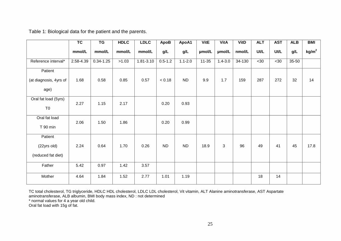
Table 1: Biological data for the patient and the parents.
TC
mmol/L
TG
mmol/L
HDLC
mmol/L
LDLC
mmol/L
ApoB
g/L
ApoA1
g/L
VitE
µmol/L
VitA
µmol/L
VitD
nmol/L
ALT
UI/L
AST
UI/L
ALB
g/L
BMI
kg/m 2
Reference interval* 2.58-4.39 0.34-1.25 >1.03 1.81-3.10 0.5-1.2 1.1-2.0 11-35 1.4-3.0 34-130 <30 <30 35-50
Patient
(at diagnosis, 4yrs of
age)
1.68 0.58 0.85 0.57 < 0.18 ND 9.9 1.7 159 287 272 32 14
Oral fat load (5yrs)
T0 2.27 1.15 2.17 0.20 0.93
Oral fat load
T 90 min 2.06 1.50 1.86 0.20 0.99
Patient
(22yrs old)
(reduced fat diet)
2.24 0.64 1.70 0.26 ND ND 18.9 3 96 49 41 45 17.8
Father 5.42 0.97 1.42 3.57
Mother 4.64 1.84 1.52 2.77 1.01 1.19 18 14
TC total cholesterol, TG triglyceride, HDLC HDL cholesterol, LDLC LDL cholesterol, Vit vitamin, ALT Alanine aminotransferase, AST Aspartate aminotransferase, ALB albumin, BMI body mass index, ND : not determined * normal values for 4 a year old child. Oral fat load with 15g of fat.

26
Table 2: Atypical mild cases of ABL reported in the literature
case 1 case 2 case 3 case 4 case 5
ref. (42) (13) (19) (15) (14)
mutation [c. 61+2T>C]+
[c.419-420insA]
homozygous
p.Asn780Tyr
[c. 61+1G>C]+
[p.Ile564Thr]
homozygous
p.Ser590Ile
homozygous
p.Ser590Ile
age at diagnosis
(year) 13 27 1.25 52 24
age at present
(year) 32 27 5 52 44
context of
diagnosis NA
during a routine
medical
examination
hepatomegaly +
liver dysfunction
during a routine
medical
examination
NA
malabsorption + 0 NA + +
liver
impairements
ALT, AST, GGT
increased mild fatty liver
hepatomegaly,
steatosis, fibrosis NA NA
neurological
impairements reduced reflexes 0 0 0 0
ophtalmological
impairements
minor
xerophtalmia,
abnormal dryness
of membrane of
the eyes
0 0 atypical retinitis
pigmentosa retinopathy
Vit E (µmol/L)
14 (RI: 12-36) on
replacement
therapy
<2.4 10 (RI: 18-34) 23 (RI: 12-46) NA
TC (mmol/L) 0.90 0.87 1.25 to 2.36 0.85 NA
TG (mmol/L) 0.50 0.03 0.11 to 1.14 0.06 NA
HDLC (mmol/L) 0.50 0.59 NA 0.68 0.56
LDLC (mmol/L) 0.07 NA NA 0.16 NA
ApoB (g/L) 0.06 0.006 < 0.007 NA NA
MTTP activity NA negligible NA NA NA
TC total cholesterol, TG triglyceride, HDLC HDL cholesterol, LDLC LDL cholesterol, Vit + present, 0 absent, NA not available, RI reference interval
27
The enterocytes (a,b) and hepatocytes (c) were engorged with numerous small or very large lipid droplets (L) free in the cytoplasm. The Golgi apparatus (G) is empty (b). Hepatic intercellular spaces were sometimes enlarged with fibrosis (c). The cell nucleus is labelled N.
Figure 1

28
Figure 2
0
5
10
15
20
25
30
Wild type L435V L435H L435E C194X Notransfection
% T
G tr
ansf
er /
30 µ
g pr
otei
n / h
Cell homogenates were used to measure triglyceride transfer from donor to acceptor vesicles using fluorescent-labelled method membranes. Columns and bars represents means +/- SD (n=2).

29
Figure 3
M: molecular weight marker, lanes 1, 2, 3: normal minigene, lane 4 5 6: mutant (c.619-5_619-2del) minigene.
30
ApoB48 was synthesized by the duodenal biopsy (B) and secreted in the medium (M) with the same molecular weight as normal control (N). MTP from the patient had the same molecular weight as normal control (N). PDI was coimmunoprecipitated: the binding of MTP with PDI was intact in the intestinal biopsy.
Figure 4

�������
�����A
�������
�BCDEF����������������
�������
�����A
�������������
�B�C�����C�� !�A�����
�����������
Supplemental Data 1 : Sequencing of genomic DNAFigure 1A : Sequencing of the exon 10 of the MTTP gene
Figure 1B : Sequencing of the intron 5 and exon 6 of the MTTP gene
M 1 2 3 4 5 6 M 1 2 3 4 5 6
5 µL of RT-PCR products were loaded on a 2% agarose gel. A: Total RNA was extractedfrom COS-1 cells transfected with the wild type MTP c.DNA (1) or the p.Leu435His c.DNA(2) or the p.Leu435Val c.DNA (3) or the p.Leu435Glu c.DNA (4) or the p.Cys194Stop c.DNA (5) or untransfected (6). After reverse transcription, amplification of a 706 bpfragment was performed with primers cMTTP-946F and cMTTP-1098rev. B: same as A but a 1582 bp PDI c.DNA fragment was amplified with primers PDI-Hind3-1 and PDI-Hind3-2.
A B
Supplemental Data Figure 2
C
COS-1 cells were transfected with MTTP-AcGFP1 fusion vector by microporation. Fluorescence was observed using Nikon Eclipse TE 2000-U microscope (objective 20) 48 hours after transfection.
Wild type p. Leu435His
GFP alone UntransfectedCOS-1 cells
p.Cys194Stop

Sequencing of the 248 bp RT- PCR product from minigene transfection :
pTB exon 3 Beginning of MTTP exon 7
End of MTTP exon 7 pTB exon 4
Sequencing of the 386 bp RT- PCR product from minigene transfection :
pTB exon 3 pTB exon 4
Supplemental Data Figure 3