molecular mechanisms of c-terminal eps15 homology domain
TRANSCRIPT
University of Nebraska Medical Center University of Nebraska Medical Center
DigitalCommons@UNMC DigitalCommons@UNMC
Theses & Dissertations Graduate Studies
Summer 8-18-2017
Molecular mechanisms of C-terminal Eps15 Homology Domain Molecular mechanisms of C-terminal Eps15 Homology Domain
containing (EHD) protein function containing (EHD) protein function
Kriti Bahl University of Nebraska Medical Center
Follow this and additional works at: https://digitalcommons.unmc.edu/etd
Part of the Biochemistry Commons, Cell Biology Commons, Molecular Biology Commons, and the
Structural Biology Commons
Recommended Citation Recommended Citation Bahl, Kriti, "Molecular mechanisms of C-terminal Eps15 Homology Domain containing (EHD) protein function" (2017). Theses & Dissertations. 213. https://digitalcommons.unmc.edu/etd/213
This Dissertation is brought to you for free and open access by the Graduate Studies at DigitalCommons@UNMC. It has been accepted for inclusion in Theses & Dissertations by an authorized administrator of DigitalCommons@UNMC. For more information, please contact [email protected].
Molecular mechanisms of C-terminal Eps15 Homology
Domain containing (EHD) protein function
By
Kriti Bahl
A DISSERTATION
Presented to the Faculty of
The Graduate College in the University of Nebraska
In Partial fulfillment of Requirements
For the degree of Doctor of Philosophy
Department of Biochemistry and Molecular Biology
Under the Supervision of Professor Steve Caplan
University of Nebraska Medical Center
Omaha, Nebraska
June, 2017
Supervisory Committee:
Richard MacDonald, Ph.D. Justin Mott, M.D., Ph.D.
Laurey Steinke, Ph.D.

I
TITLE
Molecular mechanisms of C-terminal Eps15 Homology Domain containing (EHD) protein function
BY
Kriti Bahl
APPROVED DATE Steve Caplan, Ph.D. June 23rd 2017 Richard MacDonald, Ph.D. June 23rd 2017 Justin Mott, M.D., Ph.D. June 23rd 2017
Laurey Steinke, Ph.D. June 23rd 2017
SUPERVISORY COMMITTEE
GRADUATE COLLEGE UNIVERSITY OF NEBRASKA
II
Molecular mechanisms of C-terminal Eps15 Homology Domain containing (EHD)
protein function
Kriti Bahl, Ph.D.
Advisor: Steve Caplan, Ph.D.
Endocytic trafficking is not only an essential process for the maintenance of
cellular homeostasis but also plays a vital role in regulating diverse cellular processes
such as signaling, migration and cell division. The C-terminal Eps 15 Homology Domain
proteins (EHD1-4) play pivotal roles in regulating distinct steps of endocytic trafficking.
Among the EHDs, EHD2 is disparate both in terms of sequence homology (70%) and its
subcellular localization at the caveolae. The crystal structure of EHD2 has been solved
and it contains an unstructured loop consisting of two proline-phenylalanine (PF) motifs:
KPFRKLNPF. However, the other paralogs EHD1, EHD3 and EHD4 contain a single
KPF or RPF motif, but no NPF motif. In this study, we sought to elucidate the precise
role of the two PF motifs of EHD2 in homo-dimerization, binding with the protein
partners, and subcellular localization. We demonstrated that an EHD2 NPF-to-NAF
mutant that mimics the homologous sequences of EHD1 and EHD3, lost its ability to
dimerize and bind to Syndapin2. However, it continues to localize primarily to the
cytosolic face of the plasma membrane. On the other hand, EHD2 NPF-to-APA mutants
maintained their ability to dimerize and bind to Syndapin2, but exhibited markedly
increased nuclear localization and decreased association with the plasma membrane.
Hence, the EHD2 NPF phenylalanine residue is crucial for EHD2 localization to the
plasma membrane, whereas the proline residue is essential for EHD2 dimerization and
binding. These studies also support the recently proposed model in which the EHD2 N-
terminal region may regulate the availability of the unstructured loop for interactions with
neighboring EHD2 dimers, thus promoting oligomerization. We further hypothesized that
the single PF motif of EHD1 might be responsible for both binding and localization

III
functions of EHD1. Indeed, the EHD1 RPF motif was required for dimerization,
interaction with MICAL-L1 and Syndapin2, as well as localization on tubular recycling
endosomes. Moreover, recycling assays demonstrated that EHD1 RPF-to-APA was
incapable of supporting normal receptor recycling. The biogenesis of tubular recycling
endosomes (TRE), their role in cargo-sorting and subsequently their vesiculation are
essential for receptor recycling. EHD proteins have been implicated in the bending and
fission of TRE, thus regulating endocytic recycling. Recent studies from our lab have
demonstrated that asparagine-proline-phenyalanine (NPF)-containing binding partners
of EHD1 and EHD3, such as molecules interacting with CasL-like1 (MICAL-L1) and
Syndapin2, are indispensable for TRE biogenesis. Also vital for TRE biogenesis is the
generation of phosphatidic acid (PA), an essential lipid component of TRE that serves as
a docking point for MICAL-L1 and Syndapin2. EHD1 and EHD3 have 86% amino acid
identity; they homo-and heterodimerize and partially co-localize to TRE. Despite
remarkable identity between EHD1 and EHD3, they have disparate mechanistic
functions. EHD1 induces membrane vesiculation, whereas EHD3 supports TRE
generation and/or stabilization by an unknown mechanism. While using
phospholipase D inhibitors (which block the conversion of glycerophospholipids to PA) to
deplete cellular TRE, we observed that, upon inhibitor washout, there was a rapid and
dramatic regeneration of TRE, as observed by immunostaining with MICAL-L1
antibodies. This “synchronized” TRE biogenesis system has enabled us to determine
that EHD3 is involved in the stabilization of TRE rather than in their biogenesis.
Moreover, we have identified residues Ala-519/Asp-520 in the EH domain of EHD1 and
Asn-519/ Glu-520 in the EH domain of EHD3 as being important for that dictating the
preference of these two paralogs for NPF-containing binding partners. Overall, we have
delineated a model to explain the atomic basis for understanding the differential roles of
EHD3 and EHD1 in stabilization and vesiculation of TRE, respectively.
IV
Table of Contents
Title Page ......................................................................................................................... I
Abstract ........................................................................................................................... II
Table of Contents .......................................................................................................... IV
Table of Figures ............................................................................................................ IX
List of Tables ................................................................................................................ XII
Abbreviations ............................................................................................................... XIII
Acknowledgements ..................................................................................................... XIX
CHAPTER I ..................................................................................................................... 1
Introduction .................................................................................................................. 1 1. Endocytic Trafficking ............................................................................................. 2
1.1 Overview ........................................................................................................ 2
2. Routes of Internalization ........................................................................................ 3
2.1 Clathrin-Mediated Endocytosis (CME) ........................................................... 6
2.2 Clathrin-Independent Endocytosis (CIE) ........................................................ 8
2.2.1 Caveolae-mediated pathway ................................................................... 8
2.2.2 Clathrin-Independent Carriers/GPI-AP-enriched early endosomal
compartment CLIC/GEEC………………………………………………………….9
2.2.3 Arf6 Associated Pathway ...................................................................... 10
2.2.4 Flotillin Dependent Pathway ................................................................. 11
2.2.5 Other routes .......................................................................................... 11
3. Sorting of Cargo at EE/ SE .................................................................................. 11
3.1 Sorting to the lysosomes for degradation ....................................................... 12
3.2 Sorting for Recycling ...................................................................................... 14
3.2.1 Significance of TREs in Recycling ......................................................... 15

V
3.3 Sorting for TGN .............................................................................................. 16
4. Regulators of Endocytic Trafficking ................................................................... 18
4.1 Regulation by Rab GTPases........................................................................... 19
4.1.1 Rab5 and the early endosome ................................................................ 23
4.1.2 Rab7and the maturation of the late endosome ........................................ 26
4.1.3 Rabs in Fast Recycling ........................................................................... 27
4.1.4 Rab11 and Slow Recycling ..................................................................... 28
4.2 Arf GTPases ...................................................................................................... 29
4.3 SNAREs ............................................................................................................ 30
5. C-terminal Eps15 Homology Domain containing proteins (EHDs) ..................... 32
5.1 Domain Architecture, Structure and Organization of EHD Proteins ................... 32
5.2 EH Domain ........................................................................................................ 37
5.3 Interaction Partners of EHD proteins.................................................................. 38
5.4 Distinct features of EHD proteins ....................................................................... 40
6. EHD1 ....................................................................................................................... 41
6.1 EHD1 as a vesiculator ....................................................................................... 44
7. EHD2 ....................................................................................................................... 46
7.1 Structure of EHD2.............................................................................................. 47
7.1.1 Homo-and Hetero-oligomerization of EHDs ............................................. 48
7.1.2 Role of KPFRKLNPF loop in EHD2 oligomerization ................................. 49
8. EHD3 ....................................................................................................................... 49
8.1 Curvature and tubule generation ........................................................................ 51
9. Conclusion ............................................................................................................. 55
II Materials and Methods ............................................................................................. 57
10. Materials and Methods ........................................................................................ 58
VI
10.1Recombinant DNA Constructs .................................................................... 58
10.2 Antibodies and Reagents ............................................................................ 59
10.3 Cell culture, transfections and SiRNA treatment ......................................... 60
10.4 Protein Purification ..................................................................................... 60
10.5 Immunobloting ............................................................................................ 61
10.6 GST pull-down and co-immunoprecipitation ............................................... 61
10.7 Yeast Two Hybrid ...................................................................................... 62
10.8 Transferrin uptake and recycling assays ..................................................... 63
10.9 CAY Inhibitor washout assay ...................................................................... 63
10.10 Isothermal Titration Calorimetry (ITC) ....................................................... 63
10.11 Confocal microscopy imaging ................................................................... 64
10.12 Structured Illumination Microscopy (SIM) imaging..................................... 64
10.13 Quantification of MICAL-L1-containing tubular recycling endosomes ....... 64
10.14 Statistical analysis..................................................................................... 65
10.15 Sequence homology and Identity Analysis ................................................ 65
CHAPTER III ................................................................................................................. 66
Role of the EHD2 Unstructured Loop in Dimerization, Protein Binding and Subcellular Localization……………………………………………………………………..66 11. Introduction.......................................................................................................... 67
12. Results ................................................................................................................. 69
12.1 Alteration of the EHD2 NPF motif to NAF impairs dimerization and binding
with interaction partners, but does not affect EHD2 localization ........................... 69
12.2 Modification of the EHD2 NPF motif to APA induces loss of PM localization,
but does not affect homo-dimerization and interactions with binding partners ..... 70
12.3 The phenylalanine residue of NPF motif plays a key role in the PM

VII
localization of EHD2. ........................................................................................... 72
12.4 Disruption of the EHD2 KPF motif induces relocalization of EHD2 to the
nucleus, but does not alter its oligomerization and partner binding ability ............ 72
12.5 A single EHD1 PF motif (RPF) controls its homo- and hetero-dimerization,
binding to interaction partners, and localization to Tubular Recycling Endosomes
(TRE) .................................................................................................................. 73
12.6 The NAF motif of EHD1 is dispensable for homo- or hetero-oligomerization,
and for its association with binding partners. ....................................................... 74
12.7 The EHD1 RPF motif is essential for receptor recycling ............................... 74
13. Discussion ........................................................................................................... 75
CHAPTER IV ............................................................................................................... 102 EHD3 Protein Is Required for Tubular Recycling Endosome Stabilization, and an
Asparagine-Glutamic Acid Residue Pair within Its Eps15 Homology (EH) Domain
Dictates Its Selective Binding to NPF Peptides………………………………………..102
14. Introduction........................................................................................................ 103 15. Results ............................................................................................................... 105 15.1 TRE can undergo biogenesis in the absence of EHD3. .............................. 105
15.2 EHD3 stabilizes TRE ................................................................................. 107
15.3 The EH domain is responsible for the differential function of EHD1 and EHD3
in vesiculation and TRE stabilization, respectively. ............................................ 108
15.4 Comparison of the binding affinity of the EHD1 EH domain and EHD3 EH
domain for a MICAL-L1 NPF peptide by isothermal titration calorimetry (ITC) ... 109
15.5 Identification of EH1 and EH3 residues responsible for their differential
interactions with NPF-containing partners. ........................................................ 110

VIII
15.6 The EHD3 NE519AD mutant is not competent to rescue the impaired
transferrin trafficking phenotype observed in EHD3 knockdown cells ................ 111
15.7 The number of acidic residues after the NPF motif does not dictate binding to
EHD3 or EHD1………………………………………………………………………...112
15.8 The binding difference between EHD1 and EHD3 is governed by residue
differences upstream of the NPF motif .............................................................. 113
15.9 Atomic basis for the differential interaction of EHD1 and EHD3 with NPF-
containing binding partners. .............................................................................. 114
16. Discussion ......................................................................................................... 114
CHAPTER V................................................................................................................ 139
Summary and Future Directions .............................................................................. 139
17. Summary ........................................................................................................... 140
18. Future Directions .............................................................................................. 142
18.1 Role of NPF motif of KPFRKLNPF loop in caveolar mobility ...................... 143
18.2 How does EHD2 link caveolae to the actin cytoskeleton? .......................... 143
18.3 Spatio-temporal regulation of EHD1 and EHD3 .......................................... 139
18.4 Identification of residues in EH1, which govern binding of EHD1 with
Rabankyrin-5 ..................................................................................................... 144
18.5 Structure of EH-3 ....................................................................................... 144
References ................................................................................................................. 145

IX
Table of Figures
CHAPTER 1
Figure 1.1: Pathways of endocytosis and endocytic recycling.…………………………….4
Figure 1.2: Schematic representation of steps of vesicle transport.……………………..20
Figure 1.3: The Rab switch and its circuitry.……………………………………………….24
Figure 1.4: Domain architecture of EHD proteins, structure of EHD2 and, solution
structure of EH domain of EHD1……………………………………………………………...34
Figure 1.5: Endocytic transport and regulatory proteins…………………………………..42
Figure 1.6: Model for biogenesis of tubular recycling endosomes.………………………53 CHAPTER 3
Figure 3.1: EHD protein domain architecture and sequence homology…………………77
Figure 3.2: EHD dimerization requires an intact α-helical region, but not the presence of
EHdomains……………………………………………………………………………………...79
Figure 3.3: Modification of the EHD2 NPF motif to NAF impairs dimerization and binding
with interaction partners, but does not affect EHD2 localization…………………………..81
Figure 3.4: Co-immunoprecipitation of EHD2 and mutant EHD2 proteins with
Syndapin2……………………………………………………………………………………….83
Figure 3.5: Modification of the EHD2 NPF motif to APA induces loss of plasma
membrane localization, but does not affect interactions with binding partners………….85
Figure 3.6: The NPF phenylalanine residue is responsible for the plasma membrane
localization of EHD2………………………………………………………………..................87
Figure 3.7: Wild-type and EHD2 NPF-to-NPY homo-dimerize and interact with
Syndapin2, whereas EHD2 NPF-to-NFP does not…………………………………………89

X
Figure 3.8: Disruption of the EHD2 KPF motif induces relocalization of EHD2 to the
nucleus, but does not alter its binding ability………………………………….…………….91
Figure 3.9: A single EHD1 PF motif (RPF) controls its homo- and hetero-dimerization,
binding to interaction partners, and localization to Tubular Recycling Endosomes (TRE)
…………………………………………………………………………………………………...93
Figure 3.10: The NAF motif of EHD1 is dispensable for homo- or hetero oligomerization,
and for its association with binding partners. ………………………………………………95
Figure 3.11: The EHD1 RPF motif is essential for receptor recycling.………………….97 Figure 3.12: Model for the role of the EHD2 unstructured KPFRKLNPF motif in
subcellular localization……………………………………………………………….............99
CHAPTER 4
Figure 4.1: TRE biogenesis occurs in the absence of EHD3........................................121
Figure 4.2. EHD3 stabilizes TRE…………………………………………………………..123
Figure 4.3. The EH domain is responsible for the differential function of EHD1 and
EHD3 in vesiculation and TRE stabilization, respectively……………………………….125
Figure 4.4. Comparison of the binding affinity of the EHD1 EH domain and EHD3 EH
domain for a MICAL-L1 NPF peptide by isothermal titration calorimetry………………127
Figure 4.5. Identification of EH1 and EH3 residues responsible for their differential
interactions with NPF-containing partners…………………………………………………129
Figure 4.6. Wild-type EHD3, but not the EHD3NE519AD mutant, rescues the impaired
transferrin trafficking phenotype observed in EHD3 knockdown cells………………….131
Figure 4.7. The number of acidic residues after the NPF motif does not discriminate
between binding to EHD1 or EHD3…………………………………………………………133
Figure 4.8. Residue differences upstream of the NPF motif govern binding difference
between EHD1 and EHD3…………………………………………………………………..135
XI
Figure 4.9. Atomic basis for the differential interaction of EHD1 and EHD3 with NPF-
containing binding partners………………………………………………………………….137

XII
List of Tables
CHAPTER I
Table I: Relationship between EHD protein and diseases ………............................56
CHAPTER III
Table 3.1: Comparison of wild-type EHD2 and mutants in homo-dimerization, Syndapin2-
binding and sub-cellular localization……………………………………………………101

XIII
Abbreviations
ACAP1 Arf GAP with coiled-coil ankyrin repeat and PH domains 1
AMPA 2-amino-3 (3-hydroxy-5-methyl-isoxazol-4-yl) propanoic acid
AMPPNP Adenylyl Imidodiphosphate
AP-2 Adaptor Protein-2
ARF ADP-Ribosylation Factor
ARH Autosomal Recessive Hypercholestrolemia Protein
B2AR β2 Adrenergic Receptor
BAR Bin-Amphiphysin-Rvs
BSA Bovine Serum Albumin
C-terminus Carboxyl-Terminal
C.elegans Caenorhabditis elegans
CCIE Clathrin and Caveolae-Independent Endocytosis
CCP Clathrin-Coated Pit
CCV Clathrin-Coated Vesicle
Cdc42 Cell Cycle Dependent 42
CI Clathrin-Independent

XIII
CIE Clathrin-Independent Endocytosis
CLASPs Clathrin-Associated Sorting Protein
CLICs Clathrin-Independent Tubulovesicular Intermediate
CME Clathrin-Mediated Endocytosis
COPI Coat Protein Complex I
CORVET Class C core vacuole/endosome tethering
cPLA2α Cytosolic Phospholipase A2-α
CSC Cargo-Selective-Complex
CTxB Cholera toxin B subunit
DAB2 Disabled Homolog 2
DAG Diacylglycerol
[DE] XXXL [LI] Dileucine-based Motifs
DMEM Dulbecco’s Modified Eagle Medium
dSTORM Direct Stochastic Optical Reconstruction Microscopy
EE Early Endosome
EGF Epidermal Growth Receptor
eGFPC3 Green Fluorescent Protein
EH Eps15
EHBP1 EHD2 Binding Protein1
EHD C-terminal Eps15 Homology domain containing Protein
ERC Endocytic Recycling Compartment
XIV
ESCRT Endosomal Sorting Complexes Required for Transport FBS Fetal Bovine Serum
FCH Fps/Fes/Fer/CIP4 Homology
FCHO FCH Domain Only
FIPI 5-Fluoro-2-indolyl des-chlorohalopemide
FRAP Fluorescence recovery after photobleaching
FYVE Fab1, YOTB, Vac1, and EEA1
GAP GTPase-activating protein
GDF GDI displacement factor
GDI GDP dissociation inhibitor
GDP Guanosine-5’-Diphosphate
GEEC GPI-AP-enriched Early Endosomal Compartment
GEF Guanyl nucleotide factor
GGA Golgi-localized γ-ear-containing ARF-binding
GPCR Gprotein-coupled receptor
GPI Glycosylphosphatidylinositol
GPI-AP GPI-anchor linked proteins
GRAF1 GTPase Regulator Associated with Focal Adhesion Kinase-1 GST Glutathione-S-transferase
GTP Guanosine-5’-Triphosphate
GTPase Guanosine-5’-Triphosphatase

XV
h Hour
Hrs Hepatocyte growth factor-regulated tyrosine kinase substrate
HOPS Homotypic fusion and protein sorting
Hsc70 Heat Shock Cognate 70
IF Immunofluorescence
IL-2 Interleukin-2
ILV Intraluminal Vesicle
IPTG Isopropyl β-D-1-thiogalactopyranoside
KD Knockdown
KD Binding constant
Km Michaelis Constant
LDLR Low-Density Lipoprotein Receptor
LE Late Endosome
LRP LDLR-Related Protein
MHC I Major Histocompatibility Complex I
MICAL-L1 Molecules Interacting with CasL-Like1
min Minute
MTOC Microtubule Organizing Center
MVB Multivesicular Bodies
N Amino-terminal
N-WASP Neural Wiskott-Aldrich syndrome protein
XVI
NgCAM Neuron-glia Cell Adhesion Molecule
NMR Nuclear Magnetic Resonance
NPF Asparagine-Proline-Phenylalanine
NSF N-ethylmaleimide-sensitive fusion protein
O.D. Optical Density
P-loop Phosphate Binding-Loop
PA Phosphatidic Acid
PAGE Polyacrylamide Gel Electrophoresis
PAST1 Putative Achaete-Scute Target 1
PDZ PSD-95, DLG and ZO-1
PF Proline-Phenylalanine
PH Plecksterin Homology
PI Phosphoinositide
PI (3,5) P2 Phosphatidylinositol 3,5 bisphosphate
PI (3)K Phosphatidylinositol-3-kinase
PI (3)P Phosphotidylinositol (3) phosphate
PIKfyve Phosphoinositide kinase
PIP2 Phophotidylinositol 4,5 bisphosphate
PIP5K Phosphatidylinositol 4,5 kinase
PLD Phospholipase D
PLD2 Phospholipase D2

XVII
PM Plasma Membrane
PRD Proline-rich Domain
PTB Phosphotyrosine-Binding
PX Phox-Homology
Rab11-FIPs Rab11-family Interacting Proteins
Rac1 Ras-related C3 Botulinum Toxin Substrate
RE Recycling Endosome
RILP Rab7-interacting Lysosomal Protein
RME-1 Receptor-Mediate Endocytosis-1
RTK Receptor Tyrosine Kinase
S. cerevisiae Saccharomyces cerevisiae
SDS Sodium Dodecyl Sulphate
SE Sorting Endosome
SH3 Src Homology Domain 3
STAM2 Signal-transducing Adaptor Molecule 2
SIM Structured Illumination Microscopy
SNAP-29 Synaptosome-associated Protein 29
SNAREs Soluble-NSF Attachment Protein
SNX Sorting nexin
SV40 Simian Virus 40
TBC1D10C TBC Domain member 10C

XVIII
TfR Transferrin receptor
TGN Trans-Golgi Network
TRE Tubular Recycling Endosome
TrkA Nerve Growth Factor Receptor
Tsg101 Tumor susceptibility gene 101
UIM Ubiquitin-interacting Motif
Vps Vacuolar protein sorting
WB Western blot
YXXϕ Tyrosine-based Motifs
XIX
Acknowledgements
I wish to express my sincere appreciation to those who have made significant
contribution to this dissertation and supported me in one way or another during this
amazing journey.
First and foremost, I would like to thank God for giving me the strength to
complete this Ph.D. program. Secondly, I would like to express my sincere gratitude to
my mentors, Dr. Steve Caplan and Dr. Naava Naslavsky, for their guidance, support,
patience, time, motivation and encouragement during my years of training. They have
been great mentors and have made these five years a productive and stimulating
experience.
I am fortunate to have been helped in different capacities by the past and present
members of my lab. I would also like to sincerely thank Dr. Sai Srinivas Panappakam
Giridharan and Dr. Shuwei Xie for their friendship and for training me in performing
experiments and analyzing results. I would also like to thank Trey Farmer for being an
amazing frend and junior in the lab. I would also like to thank Dr. Bishuang Cai, Dr. Jing
Zhang, Dr. Laura Simone, Dawn Katafiaz, Dr.James Reinecke. I would also like to give a
special thanks to Dr. Gaelle Spagnol and Andrew Trease from Dr. Sorgen’s laboratory
for helping me with protein purifications.
I am indebted to my supervisory committee members Dr. Richard MacDonald,
Dr. Justin Mott, and Dr. Laurey Steinke for their guidance, encouragement, and support.

XX
I am grateful to my comprehensive exam committee members Dr. Kaustabh Datta, Dr.
Xu Luo and Dr. Terrence Donohue for guiding me during the rigors of the
comprehensive exam process.
I would also like to thank everyone at the University of Nebraska Medical Center,
especially the Department of Biochemistry and Molecular Biology for their support and
collegial environment to pursue my graduate studies. I would specifically like to thank
Karen Hankins in the BMB office for being an amazing friend, and for her support in the
tumultuous times. I would also like to thank Graduate Studies at UNMC for funding.
Words are short to express my heartfelt thanks to my family for their unparalleled
love, help, and support. I would also like to especially thank my mom, who raised me
with a love of science, supported me in all my pursuits, and has been a constant source
of determination and inspiration in the last five years. Thanks to my Dad for always
believing in me and encouraging me to follow my dreams. To my sister Agrima and my
brother Vikram for being there for me always, showing me the importance of being a free
spirit and having a fresh perspective on every aspect of life.
I also want to thank my amazing friends here at UNMC, Sohini Roy, Swati
Surkar, Shreya Roy and Mary Anne Smith, for taking out time for me from their busy
schedules for talking to me, offering me advice, constantly encouraging me, warm hugs
and being there for me when I needed a friend. I also want to thank my friends back in
India, Soumya Menon, Ruchika Bharadwaj and Priyanka Arora for being just a text and
phone call away, despite the time difference between us.

XXI
Last but not the least, I would like to acknowledge the most important person in my life –
my husband and best friend, Suprit Gupta, for his endless love, humor, and
encouragement throughout this journey. Without him, I would have struggled to find the
inspiration, motivation, sanity, and bliss needed to complete this dissertation.
- 1 -
CHAPTER I
Introduction

- 2 -
1. Endocytic trafficking
1.1 Overview
The plasma membrane (PM) is a lipid bilayer that forms a permeability barrier
between the interior of the cells and the extracellular environment (Conner and Schmid,
2003). The PM not only regulates the selective transport of ions and macromolecules in
and out of the cell but also mediates communication with neighboring cells and the
extracellular environment. Hence, the composition of the PM needs to be tightly
regulated to generate appropriate responses to the cues from the extracellular
environment. The dynamic interplay between endocytic trafficking and exocytic events is
crucial for the precise regulation, and maintenance of the surface area and composition
of the PM (Doherty and McMahon, 2009).
Endocytosis or endocytic trafficking refers to the process of internalization of
receptors, proteins, and nutrients along with extracellular fluid enclosed in an
invaginated portion of the PM, which culminates in pinching off of the membrane to form
a vesicle (Conner and Schmid, 2003). The internalized lipids and proteins are returned
to the PM by the process of exocytosis. Internalization of PM containing lipids, proteins,
and receptors can occur through mechanistically diverse pathways regulated by distinct
molecular players (detailed explanation in section 2). Irrespective of the mode of
internalization, endocytosed cargo is packaged into a vesicle and delivered to a common
“sorting station” known as the sorting endosome (SE) or the early endosome (EE)
(Mayor et al., 1993; Mellman et al., 1996), where initial sorting events decide the fate of
incoming cargo (Hauotari and Helenius, 2011). From here, cargo destined for
degradation is transported to the late endosome (LE) and the lysosome, bound for
recycling to the PM, or transported to the trans-Golgi network (TGN). While the receptors

- 3 -
and some of the lipids are destined for recycling; the ligands, soluble proteins of the PM
are generally transported for degradation (Maxfield and MacGraw, 2004) (Fig 1.1).
Endocytic trafficking plays a vital role in regulating diverse processes, including
nutrient uptake, regulation of surface receptors, cellular signaling, cytokinesis (Schmid
and Conner, 2003; Skop et al., 2001), maintenance of cell polarity, cell adhesion and
migration (Wang et al., 2000; Caswell and Norman, 2008), and synaptic vesicle retrieval
in neurons (Kjaerulff et al., 2002). In addition, elegant studies have confirmed that
pathogens exploit distinct endocytic pathways for their internalization in the cell (Mercer
et al., 2010). Dysregulation of endocytic transport is related to diverse diseases including
cancer, neurodegeneration, and heart disease (Conner and Schmid 2003; Stein et al.,
2003). Thus, elucidation of the underlying mechanisms of endocytic trafficking will
ultimately provide novel avenues for developing innovative therapeutic strategies and
drug discovery.
2. Routes of Internalization
Internalization can occur through various mechanisms primarily governed by the
size of the molecules and particles that the cell uptakes. Small molecules including
amino acids, sugars and ions can enter through the channels and protein pumps.
However, the macromolecules are endocytosed through the membrane invaginations
and budding of the PM. Endocytosis is broadly classified into two main types based on
the size of the endocytic vesicle: 1) phagocytosis involves large particles (>250) nm
including microbial pathogens and cellular debris (Addermen and Underhill, 1999) 2)
pinocytosis involves the uptake of fluid and low-molecular-weight solutes (<150) nm

- 4 -
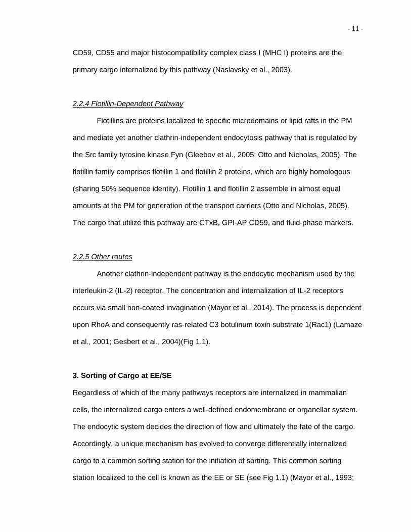
Figure 1.1. Pathways of endocytosis and endocytic recycling. Itinerary of cargo proteins and lipids internalized in cells by clathrin-mediated endocytosis (CME) (blue cargo) and clathrin-independent endocytosis (CIE) (red cargo) and subsequent routes of cargo to the early endosome (EE), endocytic recycling compartment (ERC) and recycling endosome (RE) is shown.

- 5 -
Figure 1.1

- 6 -
(Schmid and Conner, 2003). Pinocytosis, also known as “cell drinking” can be further
classified based on the endocytic machinery recruited by the cargo molecules into
clathrin-mediated endocytosis (CME) and clathrin-independent endocytosis (CIE). CIE is
further subdivided into caveolae-mediated endocytosis, clathrin-independent
endocytosis and caveolae-independent endocytosis (CCIE) (Conner and Schmid, 2003;
Mayor and Pagano,2007) (Mayor, Parton and Donaldson, 2014).
2.1 Clathrin-mediated endocytosis (CME)
CME is the most extensively characterized route of internalization from the PM.
The seminal discovery of clathrin and purification of clathrin-coated vesicles (CCVs) by
Barbara Pearse forms the basis of our current understanding of CME (Pearse, 1987).
Over the past 40 years, much work has shed light on the mechanism by which receptors
and their bound ligands are internalized into clathrin-coated pits (CCPs) and eventually
form the CCVs (Sorokin, 2004; Robinson, 2015). CCV formation is divided into a five-
stage process: initiation, cargo selection, coat assembly, scission, and uncoating. The
first stage of pit formation (CCP) is the assembly of a putative “nucleation module”
consisting of Fps/Fes/Fer/CIP4 homology (FCH) domain only (FCHO) proteins, EGFR
pathway substrate 15 (Eps15) and Intersectins at the PM (McMahon and Boucrot, 2011;
Boucrot and McMahon, 2011). Furthermore, recent studies have demonstrated, proteins
in the “nucleation module” are responsible for recruiting a tetrameric Adaptor Protein-2
(AP-2) that is a hub of interactions as it binds to both cargo and lipids in the PM
(preferentially phosphatidylinositol (4, 5) bisphosphate (PIP2)). AP-2 is a complex
composed of two large adaptin subunits-α and β2, one medium-μ2 and one small σ2
subunit (Owen et al., 2004). The AP-2 complex recognizes two types of motifs on the
cytoplasmic tail of receptors (cargo): tyrosine-based motifs with a consensus sequence
YXXΦ, where Y is a tyrosine residue, X stands for any amino acid residue and Φ is a

- 7 -
bulky hydrophobic amino acid residue, and the dileucine-based sorting signals with a
consensus sequence [aspartic acid-glutamic acid]-X stands for any amino acid residue-
X-X-leucine- [leucine-isoleucine] [DE] XXXL [LI] (Bonifacino and Traub, 2003). The
binding site for the tyrosine-based motifs is on the carboxyl terminus (C) of the μ2
domain (Ohno et al.,1995), while the α/σ2 hemi-complex and potential β2 subunit bind to
the dileucine-based sorting signal sequence (Chaudri et al., 2007; Doray et al., 2007).
There are other specialized adaptor proteins known as clathrin-associated sorting
proteins (“CLASPs”), that recognize diverse sorting signals on the respective cargo
receptors thus facilitating a large repertoire of distinct cargo to be endocytosed (Linton
and Bonifacino, 2013). For instance, cargo receptors such as the low-density lipoprotein
receptor (LDLR) have an phenyalanine-X stands for any amino acid residue-asparagine
-proline-X-tyrosine (FXNPXY) motif, that doesn’t directly bind to AP-2; hence the
cooperation with CLASPs containing a phospho-tyrosine binding (PTB) domain such as
disabled homolog 2 (Dab2) or autosomal recessive hypercholesterolemia protein (ARH)
contributes to the process (Maurer and Cooper, 2006; Hawryluk et al., 2006). Post-
translational modifications such as phosphorylation and ubiquitination can also recruit
adaptor proteins to receptor tails as in the case of binding of Epsin to the ubiquitinated
Epidermal Growth Factor Receptor (EGFR). Epsin is another example of a CLASP and it
primarily regulates EGFR internalization (Polo et al., 2002; Kazazic et al., 2009). Once
the cargo is selected and packaged by AP-2 or CLASPs, the assembly of the clathrin
coat is initiated. AP-2 and accessory proteins recruit clathrin on the nascent CCP.
Clathrin is a trimer of a dimers of three heavy chains and three light chains assembled
as a triskelion (three-legged) that has an intrinsic ability to form cage-like structures,
which facilitate membrane invagination (Kirchhausen, 2000). Next, accessory proteins
such as Bin-Amphiphysin-Rvs (BAR) containing proteins are recruited to generate and
stabilize the curvature of maturing CCPs (Quallmann B et. al, 2011). In addition, a large
- 8 -
and modular guanosine tri-phosphatases (GTPase) known as Dynamin, together with
other curvature sensing proteins including Amphiphysin, Endophilins and Sorting nexin 9
(SNX9), facilitates the release of CCVs from CCPs (Lee et al., 1999; Vallis et al., 1999;
van der Bliek et al., 1993; Yoshida et al., 2004). Dynamin oligomerizes as collar-like
structures around the neck of CCPs and undergoes guanosine tri-phosphate (GTP)
hydrolysis to mediate membrane fission and generate CCVs. After vesicle scission, the
clathrin coat is disassembled by the action of an adenosine tri-phosphatase (ATPase)
known as Heat shock cognate 70 (Hsc70) and its cofactor Auxillin (Braell et al., 1984;
Prasad et al., 1993; Ungewickell et al., 1995). LDL receptor and the iron-laden
transferrin receptor (Tf) are examples of signature cargo internalized by CME.
2.2 Clathrin-Independent Endocytosis (CIE)
CME is the predominant endocytic paradigm by which receptors are internalized;
however, cells utilize mechanisms beyond clathrin-coated pits, collectively known as
CIE. A common feature of CIE is their dependence upon cholesterol (Sandvik and
vanDeurs, 1994; Mayor and Pagano, 2007).
2.2.1 Caveolae-mediated pathway
The caveolae-mediated pathway is the best-characterized clathrin-independent
endocytic pathway (Rothberg et al., 1992). Caveolae are flask-shaped invaginations of
50-100 nm that are concentrated in microdomains of PM enriched in cholesterol,
sphingolipids, and phosphatidylinositol (4,5)-bisphosphate (PIP2) (Andereson 1998; Pitto
et al., 2000; Simone et al., 2014). Caveolin-1, an integral membrane protein that
oligomerizes as well as inserts itself as a loop in the PM to generate the framework of
caveolae (Bastini and Parton, 2010). At the PM, caveolin-1 recruits multimeric
complexes of cavin proteins (cavin 1-4) that aid in shaping and stabilizing the caveolar

- 9 -
invaginations (Hill et al., 2008; Hansen et al., 2009; et al., 2009. Additionally, Syndapin2
(also known as Pacsin2), a BAR domain-containing protein that can sense and modulate
membrane curvature, shapes the caveolar invagination (Senju et al., 2011 and Koch et
al., 2012). Syndapin2 also has a Src homology 3 (SH3) domain that facilitates its binding
to the proline-rich domain (PRD) of Dynamin and a tripeptide sequence containing,
asparagine-proline-phenylalanine (NPF) motif that mediates binding with C-terminal
Eps15 homology domain containing (EHD) protein 2 (EHD2). Recently, a series of
reports have firmly established the caveolar localization of EHD2 and caveolar
stabilization occurs in an adenosine triphosphate (ATP)-dependent manner. Previous
studies from our lab have shown that the localization of EHD2 on caveolae is
independent of its interaction with Syndapin2 and dependent on PIP2 in the PM (Moren
et al., 2012; Stoeber et al., 2012; Simone et al., 2013). Caveolae are specifically
enriched in certain cell types including smooth muscle cells, fibroblasts, adipocytes, and
endothelial cells (Parton and Simons, 2007). The cargo that are internalized in caveolin-
positive structures include simian virus 40 (SV40) virions, cholera toxin B subunit
(CTxB), and glycosylphosphatidylinositol (GPI)-linked proteins (Cheng et al., 2006;
Kirkham and Parton, 2005; Parton and Simons, 2007).
2.2.2 Clathrin-Independent Carriers/GPI-AP-enriched early endosomal compartment
(CLIC/GEEC)
Proteins that are attached to the membrane by GPI-anchor-linked proteins (GP1-
AP) are internalized independent of clathrin and caveolin coats however they require
cholesterol enriched microdomains (Sabharjanek et al., 2002). GPI-APs are internalized
through specialized EE-like structures termed, GPI-AP-enriched early endosomal
compartment (GEEC). They are formed by the fusion of cell surface-derived clathrin
independent (CI) tubulovesicular intermediates termed CLICs (Kirkham et al., 2005).
- 10 -
CLIC formation is regulated by small GTPases including cell cycle dependent 42
(Cdc42) and adenosine di-phosphate (ADP)-ribosylation factor1 (Arf1) (Kumari and
Mayor, 2008). These structures are Dynamin-independent and their budding mechanism
is not clear. However, recently a specific marker and regulator of these CLICs has been
identified, a protein called GTPase regulator associated with focal adhesion kinase-1
(GRAF1). GRAF1 has distinct domains capable of regulating membrane deformation, a
scission-BAR domain (membrane and curvature sensing), a pleckstrin homology (PH)
domain (directly interacts with PIP2 in PM) and an SH3 domain (which can bind to a
PRD domain in Dynamin) (Lundmark et al., 2008). Furthermore, previous work from our
lab has led to a model that suggests the formation of a vesiculation complex that
comprises Molecules Interacting with CAsL-Like1 (MICAL-L1) and C-terminal Eps15
homology domain containing (EHD) protein 1(EHD1) on tubular recycling endosome
(TRE) and supports TRE vesiculation (Cai et al., 2012; Cai et al., 2014). Thus, GRAF1
could also be a potential vesiculator of this pathway. Cargo that are internalized by this
pathway are GPI-APs, CTxB and fluid phase markers (Mayor and Pagano, 2007;
Doherty and MaMahon, 2009).
2.2.3 Arf6 Associated Pathway
Another clathrin-independent pathway for the internalization of cell surface
integral proteins lacking adaptor protein recognition sequences is associated with
ATPase, ADP-ribosylation factor 6 (Arf6). Arf6 is localized at the PM and it regulates the
flow of trafficking into and out of the cell, and the actin cytoskeleton at the PM. Arf6 is
responsible for activating phosphatidylinositol 4,5 kinase (PIP5K) for generation of PIP2.
The tubule-vacuolar carriers of cargo in this pathway are enriched in PIP2. Furthermore,
PIP2 can also stimulate the actin polymerization machinery and drive the endocytic
pathway. Thus, Arf6 indirectly regulates endocytic events through PIP2. The GPI-AP,
- 11 -
CD59, CD55 and major histocompatibility complex class I (MHC I) proteins are the
primary cargo internalized by this pathway (Naslavsky et al., 2003).
2.2.4 Flotillin-Dependent Pathway
Flotillins are proteins localized to specific microdomains or lipid rafts in the PM
and mediate yet another clathrin-independent endocytosis pathway that is regulated by
the Src family tyrosine kinase Fyn (Gleebov et al., 2005; Otto and Nicholas, 2005). The
flotillin family comprises flotillin 1 and flotillin 2 proteins, which are highly homologous
(sharing 50% sequence identity). Flotillin 1 and flotillin 2 assemble in almost equal
amounts at the PM for generation of the transport carriers (Otto and Nicholas, 2005).
The cargo that utilize this pathway are CTxB, GPI-AP CD59, and fluid-phase markers.
2.2.5 Other routes
Another clathrin-independent pathway is the endocytic mechanism used by the
interleukin-2 (IL-2) receptor. The concentration and internalization of IL-2 receptors
occurs via small non-coated invagination (Mayor et al., 2014). The process is dependent
upon RhoA and consequently ras-related C3 botulinum toxin substrate 1(Rac1) (Lamaze
et al., 2001; Gesbert et al., 2004)(Fig 1.1).
3. Sorting of Cargo at EE/SE
Regardless of which of the many pathways receptors are internalized in mammalian
cells, the internalized cargo enters a well-defined endomembrane or organellar system.
The endocytic system decides the direction of flow and ultimately the fate of the cargo.
Accordingly, a unique mechanism has evolved to converge differentially internalized
cargo to a common sorting station for the initiation of sorting. This common sorting
station localized to the cell is known as the EE or SE (see Fig 1.1) (Mayor et al., 1993;
- 12 -
Maxfield and McGraw, 2004). Rab5 is a key component of the cytosolic surface of EE,
and together with its effector Vps34/p150, forms a phosphatidylinositol 3-kinase (PI(3)K)
complex. Activation of PI(3)K complex leads to phosphorylation of phosphoinositidyl
inositol (PI) and its conversion to phosphatidylinositol (3) Phosphate (PI(3)P),
PtdIns(3)P, which is the most abundant phosphoinositide in the EE membrane. The
simultaneous presence of Rab5 and PtdIns(3)P helps to EE to initiate a signaling
cascade, which regulates the homotypic fusion in EE and motility of EE on actin and
microtubular tracks(Zerial and Mcbride, 2001; Behnia and Munro, 2005).
EE have a mildly acidic luminal pH (≈6.3-6.5), thereby facilitating the uncoupling
of ligands from their receptors within minutes of internalization. The uncoupling of
receptors from the ligands is the first step of sorting (Maxfield and McGraw, 2004).
Additionally, the EE is a highly dynamic structure with a strong propensity to undergo
homotypic fusion (Gruenberg et al., 1989). It is a highly complex and pleomorphic
organelle that consists of morphologically distinct elements, thin tubules (≈60 nm
diameter) and large vesicles (≈300-400 nm diameter) with membrane invaginations and
a multi-vesicular appearance. This precise subdomain morphology provides a platform
for efficient “geometric sorting” of the cargo (Mayor et al., 1993). The tubular elements
that have a high surface-to-volume ratio preferentially cluster cargo targeted for
recycling. On the other hand, cargo concentrated in large vesicles (that eventually form
multi-vesicular bodies (MVBs)) is shunted for degradation (Mellman, 1996). In most
cases, the receptors are recycled back to the PM for additional rounds of ligand binding
and the ligand is transported to lysosomes for degradation (Maxfield and McGraw,
2004). For instance, Tf and LDL receptors are recycled from EE, whereas the LDL itself
and EGFR coupled to its ligand are transported for degradation (Jovic et al., 2009).
3.1 Sorting to the lysosomes for degradation

- 13 -
EE is responsible for sorting the soluble ligands and signaling receptors for
degradation in a regulated manner. The soluble ligands are automatically sorted for
degradation as the EE matures into LE. However, the sorting of signaling receptors for
degradation in lysosomes requires specific sorting signals in the cytosolic domain. The
signaling receptors are targeted to the lysosomes through the LE in order to attenuate
their signals. Epidermal growth factor (EGF)-laden EGFR is a prototypic receptor
tyrosine kinase (RTK) having specific cytosolic domain recognized by the sorting
machinery for degradation (Haglund et al., 2003; Mosseson et al., 2003). The mono-
ubiquitinylation of one or more lysine residues in the cytoplasmic tail of EGFR serves as
an important intracellular sorting signal for the degradative pathway (Barriere et al.,
2006; Haglund et al., 2003; Huang et al., 2006; Levkowitz et al., 1999; Umebayashi et
al., 2008). The ubiquitylated receptors are recognized by several ubiquitin-interacting
motif (UIM)-containing proteins, endosomal sorting complexes required for transport-0
(ESCRT-0) component, hepatocyte growth factor regulated tyrosine kinase substrate
(Hrs), Eps15R, signal transducing adaptor molecule 2 (STAM2) and the ESCRT-I
component, tumor susceptibility gene 101 (Tsg101) (Raiborg et al., 2002). Hrs also
interacts with the flat clathrin lattice to form clustered Hrs microdomains for specialized
recognition of ubiquitylated membrane proteins and their efficient sorting for degradation
(Raiborg et al, 2002). Concomitantly, the ESCRT-I component, Tsg101, promotes the
recruitment of ESCRT-II complex and initiation of the budding process of the MVB. Upon
recruitment ESCRT-II initiates the oligomerization of the ESCRT-III complex on the
endosomal membrane (Raiborg et al., 2009). ESCRT-III complex sequesters the cargo
in nascent MVB and catalyzes the scission of MVB (Babst et al., 2002 a and b;
Bonifacino and Hurley, 2008; Schmidt and Ties, 2012). Once the MVB formation is
completed, Vps4, an ATPases associated with various cellular activities (AAA ATPase)
is recruited, which catalyzes the disassembly of ESCRT-III from the MVB membrane.

- 14 -
The disassembly of ESCRT-III is essential for termination of cargo sorting and MVB
release. Newly formed MVB fuses with either LEs or lysosomes and results in
degradation of EGFR and other sorted receptors (Shestakova et al., 2010). A recent
study from our lab has highlighted the role of C-terminal Eps15 homology domain
containing (EHD) protein 4 (EHD4) in the trafficking of receptors from EE to the
lysosomes (Sharma et al., 2008).
3.2 Sorting for Recycling
The majority of the internalized receptors from the surface of mammalian cells
are recycled back to the PM via EE. Simplistically, the recycling of receptors from EE
has been divided into two distinct pathways: “fast recycling” and “slow recycling”.
Recycling kinetics of TfR confirmed the existence of a faster route (t1/2 = 5 min) and a
slower route (t1/2 = 15-30 min) for recycling (Daro et al., 1996; Mayor et al., 1993). While
some of the receptors are directly returned to the PM from EE through the fast recycling
pathway, the majority of receptors traversing the slow recycling pathway are first
transported to an additional organelle, endocytic recycling compartment (ERC) localized
near the microtubular organizing center (MTOC) in the perinuclear area (Maxfield and
McGraw, 2004; Grant and Donaldson, 2009). Rab4 and Rab11 are the most prominent
markers for the fast and slow recycling processes, respectively (Van der Suljs et al.,
1992; Ulrich et al., 1996). The process of efficient recycling through the ERC is
coordinated through an elaborate network of endosomes known as TREs (detailed
explanation in section 3.2.1).
Previously, it was thought that no sorting motif is necessary for receptor
recycling. However, recent studies have demonstrated that a GTPase-activating protein
(GAP) for ADP ribosylation factor (Arf6), Arf GAP with coiled-coil ankyrin repeat and PH
domains 1 (ACAP1), serves as a sorting molecule involved in direct binding to two
- 15 -
phenylalanine-containing sorting motifs in the TfR; leucine-phenylalanine (LF) or
arginine-phenylalanine (RF) (Dai et al., 2004). Indeed, there was delay in the recycling of
TfR upon disruption of the binding between Tf and ACAP1 (Dai et al., 2004). Recent
studies have demonstrated that Sorting Nexin 17 (SNX17) directly binds to an
asparagine-X stands for any amino acid-proline-tyrosine (NPXY) motif in the cytoplasmic
domain of LDLR-related proteins (LRPs) and promotes LRP recycling (Van Kerkhof et
al., 2005). Another study demonstrated that SNX27 acts as an adaptor that links the coat
protein retromer to a prototypical G-protein coupled receptor (GPCR) cargo, the
β2 adrenergic receptor (B2AR), by recognizing its PDZ domain (named after the first
three proteins in which it was identified, PSD-95, DLG and ZO-1) to sort for recycling
(Lauffer et al., 2010). Thus, there are clearly instances where recycling can be dictated
by select sorting motifs.
3.2.1 Significance of TREs in Recycling
The composition, structure, and mode of functioning of ERC in endocytic
recycling are poorly understood, despite the importance of the recycling process. Recent
studies from our lab employing Structured Illumination Microscopy (SIM), dual channel
2D-direct Stochastic Optical Reconstruction Microscopy (dSTORM), and 3D dSTORM,
have shed new light on the ERC morphology and cargo segregation. The ERC is
composed of an array of dynamic, densely situated, yet independent, tubular and
vesicular recycling endosomes radiating from the MTOC (Xie et al., 2015). It has been
well established that the high surface area-to-volume ratio of tubular carriers effectively
serves to segregate the integral membrane proteins from the luminal content (Maxfield
and Macgraw, 2004). However, our recent studies suggest that the ERC maintains cargo
segregation acquired upon exit from the SE. Hence, the ERC serves as a focal point for
vesicular transport to the PM (Xie et al., 2015). TREs are crucial for the recycling of

- 16 -
internalized receptors and lipids. Previous studies from our lab have demonstrated that
MICAL-L1-decorated TREs can be generated from regions of SE that are enriched in a
Rab-5 effector, Rabenosyn-5, indicating that TREs are responsible for the movement of
CIE cargo from peripheral SE to the perinuclear ERC (Xie et al., 2015). Moreover,
current models from our lab support the finding that the fission of TREs leads to
formation of vesicle carriers (vesiculation) that carry recycled receptors back to the PM
(Cai et al., 2012; Cai et al., 2013; Cai et al., 2014).
Owing to the significance of TRE in endocytic recycling, multiple studies from our
lab have focused on identifying the molecular players involved in TRE generation,
fission, fusion, and function. Previous studies from our lab have demonstrated that
MICAL-L1 is a protein localized to the outer TRE leaflet (Sharma et al., 2009). MICAL-L1
acts as a hub that recruits and stabilizes a battery of proteins that directly impact
membrane shaping. For instance, the F-BAR domain containing protein Syndapin2
(Giridharan et al., 2013). MICAL-L1 also interacts with the C-terminal Eps15 homology
domain containing (EHD) protein 3 (EHD3) and EHD1 (Sharma et al., 2009; Kieken et
al., 2010), which are involved in TRE stabilization and vesiculation, respectively (Cai et
al., 2013; Bahl et al., 2016). Also crucial for TRE biogenesis is the high local
concentration of phosphatidic acid (PA), an essential lipid component of TRE tubules,
which binds and recruits MICAL-L1 and Syndapin2. Syndapin2 has an SH3 domain,
which mediates stable interaction with the PRD domain of MICAL-L1. Syndapin2 also
has an F-BAR domain that can sense and bend the membranes to induce tubulation
(Giridharan et al., 2013). EHD1 subsequently joins this complex on TRE, where it
interacts with both MICAL-L1 and Syndapin2 and initiates vesiculation, giving rise to
newly formed vesicles (Cai et al., 2012; Giridharan et al., 2013; Cai et al., 2013; Cai et
al., 2014).

- 17 -
3.3 Sorting for TGN
EEs not only serve as stations to sort receptors for recycling and degradation but
also function as a common junction that connects various endocytic and biosynthetic
routes. Trafficking from the EE to the biosynthetic routes is known as retrograde
transport. Retromer-mediated tubulation is required for the retrograde transport from EE
to the TGN and these retromer-mediated tubules are distinct from the TREs that
facilitate the recycling process from EE to ERC (Bonifacino and Rojas, 2006). Retromer
machinery is preferentially recruited to EEs which are maturing towards LE and contain
increasing concentrations of phosphatidylinositol 3,5 bisphosphate (PI(3,5)P2),
generated by phosphoinositide kinase (PIKfyve) (Rutherford et al., 2006) and an
increasing number of intralumenal vesicles (ILVs) (Arighi et al., 2004).
Pioneering studies by Seamen et al., 1998, in the yeast endolysosomal system
were instrumental in the identification of the protein coat “retromer”. Retromer was
shown to mediate the endosome-to-Golgi retrieval of the vacuolar hydrolase receptor
(Vps10p), the yeast functional equivalent of the mannose 6-phosphate (M6PR) receptor.
Retromer is a heteropentameric complex consisting of a sorting nexin (SNX) dimer
composed of SNX 1/2 and SNX 5/6 and a trimer consisting of Vps proteins namely,
Vps26, Vps29 and Vps35 (Bonifacino and Hurley, 2008; Bonifacino and Rojas, 2006;
Seaman, 2005; Rojas et al., 2007). The SNX protein dimer possesses a phox-homology
(PX) domain and a BAR domain. While the PX domain is involved in binding to PI(3)P
and other phosphoinositides in the EE membrane, the BAR domains (that can sense
and induce membrane curvature) mediate dimerization and attachment to the curved
membranes (Bonifacino and Rojas, 2006). The Vps26, 29 and 35 heterotrimer is
involved in recognizing cargo proteins and is therefore termed as the cargo-selective-
complex (CSC). Vps35 provides the interface for the recognition of cargo. Vps29 is
indispensable for the interaction of the CSC with the SNX dimer and functions as a

- 18 -
scaffold for retromer assembly by binding the carboxyl (C)-terminal half of Vps35. Vps26
binds to the amino (N)-terminal half of Vps35 (Seamen et al., 2007). The CSC, unlike the
SNX dimer, lacks lipid-binding domains; hence, its recruitment on the endosome is
dependent on the interaction of Rab7 with Vps35. Furthermore, Rab7/Vps35 interaction
not only stabilizes the CSC on the endosome membrane, but also synchronizes the
timing of cargo export with endosome maturation (explained in detail in section 3.1) (van
Weering et. al, 2012). The mechanism by which CSC recognizes cargo is also poorly
understood. However, it is well established that the cargo destined for retromer-
dependent sorting possesses at least one simple hydrophobic motif
phenylalanine/tryptophan-leucine-methionine/valine (F/W-L-M/V) (Seamen et al., 2007).
Recent studies have also implicated the involvement of Vps26 in recognizing cargo by a
FANSHY sorting signal and the role of SNX27 in cargo sorting (Fjorback et al., 2012;
Steinberg et al., 2013). The best-studied examples of cargo proteins transported through
the retromer-mediated pathway include the vacuolar hydrolase transport receptors,
Vps10 in yeast and M6PR in mammals (Bonifacino and Rojas 2006; Johannes and
Popoff 2008).
EHD1 is yet another important regulator of retromer-mediated transport. EHD1
colocalizes and interacts with Vps26 and Vps35 on retromer tubules, and affects the
retrieval of M6PR mediated by the retromer (Gokool et al., 2007). However, no direct
binding between EHD1 and the retromer has been detected. We have identified
Rabankyrin-5, a Rab5 effector, as a novel NPF-interaction partner for EHD1.
Rabankyrin-5 also interacts with Vps26 and Vps35 thus facilitating the association of
EHD1 with the retromer complex (Zhang et al., 2012 (a) and (b))(McKenzie et al., 2012).
EHD3, another member of the EHD protein family and the closest paralog of EHD1, also
mediates endosome-to-Golgi transport and retromer trafficking (Naslavsky et al., 2009).

- 19 -
4. Regulators of Endocytic Trafficking
The endosomal system is an elaborate and dynamic network of membrane-
bound organelles interconnected by vesicular “vehicles” that transport lipids and
proteins. The process of endosomal transport can be divided into four distinct steps:
vesicle budding from the donor organelle, transport, tethering with the acceptor organelle
and finally, fusion of the lipid bilayers of the vesicle and the acceptor organelle (Fig 1.2)
(Bonafacino and Glick, 2004). A conserved arsenal of regulatory proteins coordinates
these fission and fusion events.
The budding process is initiated by recruitment of coat proteins onto the donor
membrane to induce the formation of the vesicle. Coat subunits are involved
simultaneously in incorporating the cargo and deforming the donor membrane into the
budding vesicle (Cai et al., 2007). After budding, the vesicles are transported to their
destination donor compartments by molecular motors such as dynein, kinesin, and
myosin along cytoskeletal tracks (Hammer and Wu, 2002; Matanis et al., 2002; Short et
al., 2002). Once the vesicle reaches the acceptor organelle, tethering of the vesicle to
the acceptor organelle occurs by the joint action of tether complexes and Rab-GTPases
(Sztul and Lupashin, 2006; Whyte and Munro, 2002). The final step is fusion, which
involves the soluble NSF attachment protein receptor (SNAREs), where NSF stands
for N-ethyl-maleimide-sensitive fusion protein (Numrich and Ungermann, 2013).
4.1 Regulation by Rab GTPases
The Rab family of small Ras-like GTPases constitutes a critical group of
endocytic regulators. In mammalian cells, over 60 different members of the Rab family-
function as multifaceted regulators of distinct trafficking pathways. Rab-GTP-binding
proteins function as molecular switches, which are either in the guanosine triphosphate
- 20 -
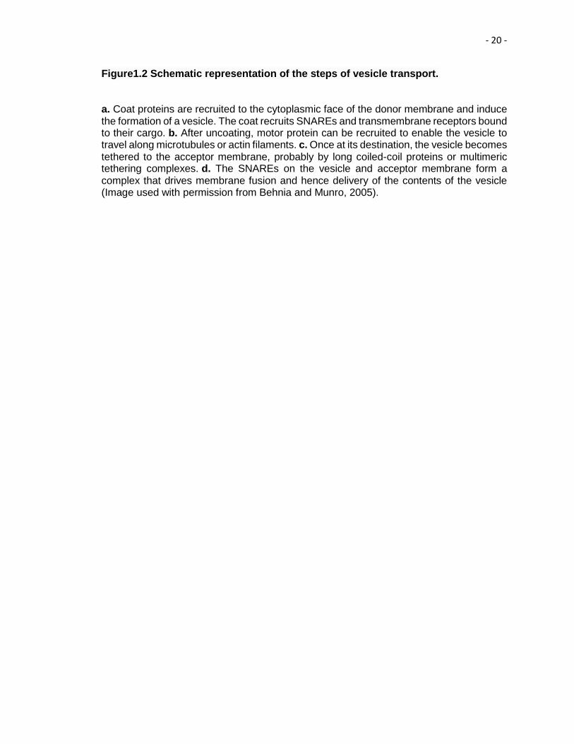
Figure1.2 Schematic representation of the steps of vesicle transport.
a. Coat proteins are recruited to the cytoplasmic face of the donor membrane and induce the formation of a vesicle. The coat recruits SNAREs and transmembrane receptors bound to their cargo. b. After uncoating, motor protein can be recruited to enable the vesicle to travel along microtubules or actin filaments. c. Once at its destination, the vesicle becomes tethered to the acceptor membrane, probably by long coiled-coil proteins or multimeric tethering complexes. d. The SNAREs on the vesicle and acceptor membrane form a complex that drives membrane fusion and hence delivery of the contents of the vesicle (Image used with permission from Behnia and Munro, 2005).

- 21 -
Figure 1.2

- 22 -
(GTP)-bound “active” form or the guanosine diphosphate (GDP)-bound “inactive” form
(Zerial and McBride, 2001; Grosshans et al., 2006). As shown in Fig 1.3, the membrane
association/dissociation of Rabs is intimately associated with the nucleotide/hydrolysis
cycle (“active” and “inactive” forms) and is regulated by specific cofactors. Exchange of
GDP with GTP is catalyzed by guanyl nucleotide exchange factors (GEFs), which
facilitate the release of GDP by inducing a conformational change in the Rab proteins
(Delparto et al., 2004). In the GTP-bound state, Rabs are stably associated with the
membrane via the membrane affinity endowed by the prenyl anchor. The prenyl anchor
is generated by the attachment of geranyl-geranyl groups to one or two cysteine
residues in the C-terminal cysteine-alanine-alanine-X stands for any amino acid (CAAX)
motif (Colicelli 2004; Lueng et al, 2006). GDP-dissociation inhibitor (GDI) maintains the
GDP-bound Rab in a soluble state by masking the C-terminal prenyl anchor. In order to
recruit Rab proteins to the membrane, the GDI has to be released by GDI displacement
factor (GDF). However, recent reports suggest that GEF may be sufficient to remove the
GDI, as well as function as a GDF (Itzen and Goody, 2011).
Different GTP-bound Rabs serve as distinct membrane scaffolds to coordinate
three major steps: vesicle budding, cytoskeletal transport, targeted docking and fusion.
Accordingly, Rabs sequentially interact with specific types of effector molecules,
including sorting adaptors, tethering factors, kinases, phosphatases and motor proteins,
to regulate each step in a spatiotemporal manner (Grosshans et al., 2006; Stein et al.,
2003). Therefore, Rabs act as membrane domain organizers that can locally change the
environment of the membrane (Miaczynska and Zerial, 2002). Once the individual
transport step catalyzed by a particular Rab is completed, the specific GAPs accelerate
GTP hydrolysis, converting the Rab protein to inactive GDP-bound state. The GDP-
bound Rabs can be extracted from the membrane by the effector GDI and recycled back
to the cytosol (Goody et al., 2005 and Grosshans et al., 2006)(Fig 1.3).

- 23 -
4.1.1 Rab5 and the early endosome
Rab5 is the best-characterized and most extensively studied Rab of the early
endocytic pathway (Zerial and McBride, 2001; Grosshans et al., 2006). Rab5 is the most
prominent identity marker and a regulator of the EE. Rab5 regulates the entry of cargo
from the PM to the EE, generates PI(3)P lipid, which is enriched on EE membrane
(Christoforidis et al., 1999; Murray et al., 2002), catalyzes homotypic fusion (Gorvel et
al., 1991), and facilitates the motility of EE on actin and microtubular tracks (Nielsen et
al., 1999; Pal et al., 2006). Rab5 is activated on the EE membrane by the recruitment of
its effector, Rabex-5 (Horiuchi et al., 1997). Another Rab5 effector, Rabaptin-5, further
promotes the GEF activity of Rabex-5, and these both together with Rab5 form a
complex known as the “Rab5 domain” (Stenmark et al., 2009). This complex is required
to establish a feedback loop for maintaining and stabilizing Rab5-GTP on the EE
membrane (Lippe et al., 2001). These transient but high levels of Rab5-GTP are
sufficient to recruit effector proteins to the EE, where they can carry out their specialized
functions in trafficking and sorting (Grosshans et al., 2006). Additionally, this complex
also triggers the rapid recruitment of other Rab5 effectors, PI(3)P-kinase/hVPS34/p150
(VPS34), and forms a complex. This complex generates PI(3)P, the most abundant
phosphoinositide in the EE membrane. The concomitant presence of Rab-GTP and
PI(3)P acts as a signal to recruit a spectrum of effector proteins such as EEA1 (Lawe et
al., 2000, 2002; Pfeffer, 2001) and Rabenosyn-5 (Nielsen et al., 2000).
EEA1 and Rabenosyn-5 bind to the PI(3)P-enriched membrane through their
FYVE (named after 4 cysteine-rich proteins-Fab1, YOTB, Vac1, and EEA1) zinc finger
domains (Nielsen et al., 2000) (Lawe et al., 2000; Nielsen et al., 2000; Stenmark and
Aasland, 1999). EEA1 interacts with SNARE proteins Syntaxin 13 (McBride et al., 1999)
- 24 -
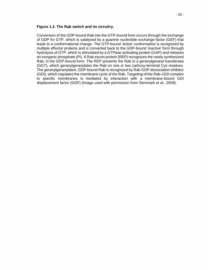
Figure 1.3. The Rab switch and its circuitry. Conversion of the GDP-bound Rab into the GTP-bound form occurs through the exchange of GDP for GTP, which is catalysed by a guanine nucleotide exchange factor (GEF) that leads to a conformational change. The GTP-bound 'active' conformation is recognized by multiple effector proteins and is converted back to the GDP-bound 'inactive' form through hydrolysis of GTP, which is stimulated by a GTPase-activating protein (GAP) and releases an inorganic phosphate (Pi). A Rab escort protein (REP) recognizes the newly synthesized Rab, in the GDP-bound form. The REP presents the Rab to a geranylgeranyl transferase (GGT), which geranylgeranylates the Rab on one or two carboxy-terminal Cys residues. The geranylgeranylated, GDP-bound Rab is recognized by Rab GDP dissociation inhibitor (GDI), which regulates the membrane cycle of the Rab. Targeting of the Rab–GDI complex to specific membranes is mediated by interaction with a membrane-bound GDI displacement factor (GDF) (Image used with permission from Stenmark et al., 2009).

- 25 -
Figure 1.3
- 26 -
and Syntaxin 6 (Simonsen et al., 1999), which regulate vesicular fusion at the EE. EEA1
also facilitates the fusion of transport vesicles with recycling endosomes (REs) and the
Golgi, respectively.
Rabenosyn-5 is regarded as a dual Rab5 and Rab4 effector, as it can bind to
both of these Rabs in their active states. Rabenosyn-5 also interacts with EHD1 and
EHD3. EHD1 regulates recycling of cargo from the perinuclear ERC back to the PM
(Caplan et al., 2002; Naslavsky et al., 2004) and EHD3 is involved in the transport of
cargo from EE to the ERC (Naslavsky et al., 2009). Thus, Rabenosyn-5 might serve as
the Rab5 effector that is the link between sorting events at the EE and the recycling of
cargo back to the PM either directly from EE (fast recycling) or via the ERC (slow
recycling).
Rabankyrin-5, another Rab5 effector is required for macropinocytosis and EE
fusion (Schnatwinkel et al., 2004). Our studies have identified a novel role of
Rabankyrin-5 in the regulation of retromer localization, which is contingent upon its
specific interaction with EHD1 (Zhang et al., 2012).
4.1.2 Rab7 and the maturation of late endosomes
Rab7 is the best-known regulator and organelle identifier of late endosomes (LE).
Although Rab7 localizes to both the EE and the LE/MVB, it regulates the late steps of
endocytic trafficking and lysosomal degradation (Wichhmann et al., 1992). The
prerequisites for the generation of a new LE are: the generation of a Rab7 domain, and
the removal of Rab5 (Rink et al., 2005; Vonderheit and Helenius, 2005). The current
model, which is known as the “Cascade Model” is used to describe the Rab5-to-Rab7
switch. The level of Rab5 on the EE membrane is not constant but fluctuates
dynamically. As the EEs grow in size due to frequent homotypic fusion events and
accumulation of cargo destined for degradation, the surface density of Rab5 increases

- 27 -
until it reaches its maximum concentration (Rink et al., 2005; Lakadamyali et al., 2006).
At this stage, a further increase in Rab5 levels triggers rapid recruitment of Rab7 on the
endosomal membrane via class C core vacuole/endosome tethering (CORVET)/
homotypic fusion and protein sorting (HOPS) complexes. Once Rab7 reaches a
threshold level, it starts to repress Rab5. Mon1-Ccz1, an evolutionarily preserved protein
complex, physically interacts as a stable pair and facilitates the Rab5-Rab7 exchange by
simultaneously displacing Rabex-5 and activating a GEF of Rab7 (Wang et al., 2002;
Kucharczyk et al., 2009; Kinchen and Ravichandran, 2010). VPS39, a HOPS complex
subunit acts as a GEF for Rab7. Additionally, Mon1/Ccz1 complex binds to Rab7 on the
membrane and controls the localization of Rab7 on LE and subsequent activation (Rink
et al., 2005; Hutanglang and Novick, 2011). Rab7-GTP also recruits its own effectors,
such as, Rab7-interacting lysosomal protein (RILP), which interacts with Rab7 on LEs
and lysosomes (Cantalupo et al., 2001). RILP then connects dynein-dynactin motor
complexes to Rab7-containing LEs and lysosomes (Jordens et al., 2001). Dynein
transports LE to the minus end of microtubules by the motor complexes towards the
lysosomes (Johansson et al., 2007).
4.1.3 Rabs in Fast Recycling
Rab4 is required for the fast recycling of Tf and glycophospholipids from the EE
to the PM (Sonnichsen et al., 2000; Maxfield and MacGraw, 2004). Rab4 regulates fast
recycling through interaction with various effector proteins, including Rabenosyn-5 and
Rabapatin-5/Rabex-5 complex (Vitale et al., 1998; de Renzis et al., 2002; Mattera and
Bonifacino, 2008). Recent studies have identified that Rab35 localizes to the PM as well
as to the EE, and is an important regulator of rapid recycling (Sato et al., 2008). Rab35
also associates with Arf6 and EHD1-positive TREs carrying cargo back to the PM. The
GAP for Rab35 is TBC domain member 10C (TBC1D10C) (Walseng et al., 2008).
- 28 -
Furthermore, Rab35 recruits the EHD1 binding protein MICAL-L1 to the Arf6 positive
recycling tubules (Rahajeng et al., 2012). Moreover, Rab35 recruits multiple Rabs,
Rab8, Rab13 and Rab36 at the recycling tubules through MICAL-L1 during nerve growth
factor (NGF)-induced neurite outgrowth (Kobayashi et al., 2014).
4.1.4 Rab11 and Slow Recycling
Slow recycling occurs through the ERC and is facilitated by the recycling
endosomes (REs) that originate from the ERC (Grant and Donaldson, 2009; Stenmark,
2009; van Ijzendoorn, 2006). Rab11 is the signature marker for the REs that originate
from the ERC. The Rab11-decorated REs selectively transport CME cargos from the
ERC to the PM and are distinct from the TREs that preferentially transport CIE cargoes
from the ERC to the PM. The Rab11-family interacting proteins (Rab11-FIPs) constitute
an evolutionarily conserved family of proteins (Hales et al., 2002; Prekeris et al., 2001).
There are several members in this family including FIP1, FIP2, FIP3 (Hales et al., 2001),
FIP4 (Wallace et al., 2002a), FIP5 (Prekeris et al., 2000), RCP (Wallace et al., 2002b;
Lindsay et al., 2002), Rabphilin-11/Rab11BP (Mammoto et al., 1999; Zeng et al., 1999).
Each of the FIPs is characterized by the presence of a highly conserved coiled-coiled
alpha-helical Rab11-binding domain at the C-terminus (Horgan and McCaffrey, 2009).
Rab11-FIP5 mediates transport from EEs to REs through binding with Kif3B, a
component of the kinesin II motor protein (Schonteich et al., 2008). We have recently
demonstrated in our studies, another Rab11 effector, Rab11-FIP2, which facilitates the
recycling of receptors through its specific interaction with EHD1 and EHD3 by its NPF
motifs (Naslavsky et al., 2009). Thus, Rab11-FIP2 serves as a link between EHDs and
Rab11. Furthermore, Rab11-FIP2 forms a ternary trafficking complex with Rab11, and
the motor protein myosin Vb for the regulation of the recycling process (Hales et al.,
2001; Hales et al., 2002).

- 29 -
4.2 Arf GTPases
The Arf family of proteins, like the Rabs, belongs to the Ras superfamily of small
GTPases (Jackson and Cassanova, 2000). The Arf proteins cycle between their active
GTP-bound states and inactive GDP-bound states like other GTPases. Exchange of
GDP for GTP is catalyzed by GEFs, and GTP hydrolysis to GDP is mediated by GAPs.
Several specific Arf GAPs and GEFs have been identified. The membrane tethering of
Arf proteins is facilitated by myristoylation at the N-terminus (Jackson and Cassanova,
2000; Randazzo and Hirsch, 2004).
In mammals, there are six members of the Arf family, which are divided into three
classes (Kahn et al., 2006). Class I Arf proteins (Arf1, Arf2, and Arf3) share 96%
sequence homology (Bonifacino and Glick, 2004). There are two Class II Arf proteins
Arf4 and Arf5; Arf5 has been implicated in early Golgi transport and in recruiting coat
components to trans-Golgi membrane (Claude et al., 1999; Takatsu et al., 2002). Class
III has a single member known as Arf6 that regulates endosomal trafficking and
structural organization of the cell surface (D’Souza Schorey et al., 1995; Grant and
Donaldson, 2009).
Arf1 and Arf6 are the most studied mammalian Arf proteins whose primary
localization in cells is at the Golgi and PM respectively. In cells, Arf1 reversibly
associates with Golgi membranes during its activated state, Arf1-GTP. Arf1-GTP recruits
coat protein complex I (COPI) on to pre-and cis-Golgi structures through the recruitment
of heterotetrameric complex (AP1, AP3, and AP4) and monomeric Golgi-localized γ-ear-
containing ARF-binding protein (GGA) on to TGN and endosomal membranes
(Bonifacino and Glick, 2004).
On the other hand, Arf6 is mainly localized at the PM and to some extent on the
endosomal membranes (Donaldson, 2003). Arf6 primarily functions in CIE-mediated
- 30 -
internalization and recycling. Arf6 promotes the recycling of MHCI molecules back to the
PM (Naslavsky et al., 2003). Moreover, Arf6 forms a ternary complex with Rab11,
Rab11-FIP3, and Rab11-FIP4 (Fielding et al., 2005), which regulates the recycling of β1-
integrin (Powelka et al., 2004). Arf6 regulates recycling at the molecular level by
activating phospholipase D2 (PLD2) that generates phosphatidic acid (PA) and
diacylglycerol (DAG), and PIP5K that generates PIP2 (Jonavic et al., 2006; Brown et al.,
2001). Furthermore, the Arf6 GTPase regulates EHD1-containing TRE. Indeed, the
cycling of Arf6 between its GTP-and GDP-bound forms impacts the localization of EHD1
on the tubular membranes (Caplan et al., 2002). Co-expression of the GTP-locked Arf6-
Q67L mutant with Myc-EHD1 led to altered EHD1 localization, loss of EHD1-associated
tubular structures and recruitment of EHD1 to enlarged Arf6 endosomes. Furthermore,
overexpression of Arf6-GEF, EFA6, or the Arf6-GAP, ACAP1 in HeLa cells, leads to loss
of EHD1 from the tubular membranes (Caplan et al., 2002). In addition, our recent
studies have demonstrated that PA is an essential component of TREs (Giridharan et
al., 2013). Moreover, PIP2 is primarily localized to the PM, where it mediates vesicular
fusion and recruits Arp2/Arp3-actin polymerization machinery (Santy and Cassanova,
2001; Vitale et al., 2002). Hence, Arf6 mediates the recycling process by regulating the
lipid dynamics of the membranes involved in recycling and the PM.
4.3 SNAREs
SNAREs are a highly conserved family of membrane proteins primarily
regulating vesicular fusion events in membrane trafficking pathways (Bennett et al.,
1995; Sollner, 1995). The hallmark of SNARE proteins is the presence of a heptad
SNARE motif: an evolutionarily conserved domain of 60-70 amino acids arranged to
form a coiled-coiled region (Fasshauer, 2003). A SNARE on the transport vesicle (v-
SNARE) pairs with its cognate SNARE-binding partner (t-SNARE) with the acceptor

- 31 -
membrane, which drives fusion and delivery of cargo (Chen and Scheller, 2001). The
interaction of cognate v-and t-SNAREs leads to the formation of a trans-SNARE
complex or SNAREpin, in which four SNARE motifs assemble as a twisted parallel four-
helix bundle. The formation of a SNAREpin is not only important for the specificity of the
initial interaction but also for bringing opposing membranes together and eventually
membrane fusion. The energy released during the formation of SNAREpin drives the
fusion of lipid bilayers (Lin and Scheller, 1997). After the fusion process is complete, the
remaining part of the SNARE complex on the fused membranes is called the cis-SNARE
complex. This complex undergoes recycling upon disassembly catalyzed by AAA
ATPase NSF and its cofactor soluble NSF attachment protein (SNAP) (Sollner et al.,
1993; Mayer et al., 1996; Hanson and Whiteheart, 2005).
The structural studies of SNAREs have revealed that the helical core contains
three highly conserved glutamine (Q) residues and one highly conserved arginine (R)
residue. Consequently, the SNARE proteins are classified as Qa-, Qb-, Qc- and R-
SNAREs. Four SNAREs, with one member each of Qa-, Qb-, Qc- and R-SNARE
subfamilies form a functional SNARE complex that drives membrane fusion (Fasshauer
et al., 1998; Bock et al., 2001). The Rab5 effector protein EEA1 can recruit Syntaxin13
and Syntaxin6 on the EE membrane (McBride et al., 1999; Simonsen et al., 1999).
Syntaxin13-Vps10p tail interactor 1 and (Vti1a)-Syntaxin6-VAMP4 are the key SNARE
complexes involved in the homotypic fusion of EEs (Brandhorst et al., 2006; Zwilling et
al., 2007). The SNARE complex Syntaxin 13-SNAP23/25-VAMP2/3 has been implicated
in the trafficking of cargo from RE to the PM (Kubo et al., 2015). The Syntaxin16-Vti1a-
Syntaxin 6-VAMP3/4 SNARE complex functions between EE and the TGN, whereas the
Syntaxin 16-Vti1a-Syntaxin 10-VAMP3 complex operates between LE and the TGN
(Ganley et al., 2008). The lysosomal degradation pathway involves Syntaxin4-SNAP23-
VAMP7 for the fusion of the LE and lysosomes to the PM (Williams et al., 2014).

- 32 -
5. C-terminal Eps15 Homology Domain containing proteins (EHDs)
A novel group of endosomal scaffolding proteins, the EHDs, regulates specific
steps of endocytic trafficking pathway. Mammalian cells express four EHD protein
paralogs, EHD1, EHD2, EHD3, EHD4 having a high level of amino acid sequence
similarity and showing about 70-86% identity (Naslavsky and Caplan, 2005). The
genomes of many invertebrate organisms, including Caenorhabditis elegans (C.
elegans) and Drosophila melanogaster, contain a single EHD family gene, most closely
resembling EHD1/EHD3. C. elegans homolog of EHD1, receptor mediated endocytosis-
1(RME-1), displays 67% identity with EHD1 and is a component of the recycling
machinery regulating the return of yolk receptors back to the PM (Grant et al., 2001).
Drosophila has a putative achaete-scute target 1 (PAST1) protein that is evolutionarily
closest to EHD2 displaying 70% homology and is involved in regulating endocytosis. The
flies lacking PAST1 are infertile and die prematurely (Olswang-Kutz et al., 2009). Recent
studies have further highlighted the role of PAST1 in the development of the neuro-
muscular junction and differentiation of rod and cone cells of the fly eye ommatidia
(Koles et al., 2015; Dorot et al., 2017).
5.1 Domain Architecture, Structure and Organization of EHD Proteins
The four EHD protein (EHD1-4) paralogs have conserved domain architecture.
The structural studies by McMahon’s group, which solved the first crystal structure of
mouse full-length EHD2 protein have been instrumental in our understanding of the
domain architecture and structure of EHD proteins (Daumke et al., 2007). The domain
architecture of EHD proteins consists of a nucleotide binding domain (G-domain)
enclosed between helical domains at the N-terminus, followed by a linker region, and a
- 33 -
C-terminal Eps15 homology (EH) domain (Fig. 1.4A) (Grant and Caplan, 2008;
Naslavsky and Caplan, 2011) (Fig 1.5).
One of the trademark characteristics of EHD proteins is their distribution to the
cytosolic face of long tubular endosomal membranes. Electron microscopy studies have
determined that the tubular structures containing EHD1 are 200 nm wide and up to 10
μm long (Caplan et al., 2002). Consistent with the high degree of homology between
EHD proteins, all four paralogs are capable of localizing to tubular membranes when
overexpressed. However, the extent of tubulation varies between different paralogs
(Blume et al., 2007; George et al., 2007). Both EHD1 and EHD3 localize primarily to
tubular membranes, which emanate from the perinuclear ERC and from the peripheral
SE, while EHD2 and EHD4 are mostly present on vesicular membranes with very few
tubules (Caplan et al., 2002; Naslavsky et al., 2006; Sharma et al., 2008). Since EHD
proteins lack a transmembrane domain, the recruitment of EHD proteins is dependent
upon EH domain lipid-binding ability. The positively charged lysine 483, which is very
well conserved, has been hypothesized to mediate the EH-domain/lipid-binding. Indeed,
charge reversal mutation of this lysine to glutamate (K483E) completely abrogates the
tubular localization of the mutant EHD1, resulting in redistribution to endosomal vesicles,
a phenotype similar to the truncation of the entire EH-domain (Naslavsky et al., 2007).
Sequence alignment analysis led to the prediction that EHD proteins have a putative
phosphate binding (P)-loop motif in the G domain, an ATP/GTP-binding site found in
GTPases such as Dynamin and Ras.

- 34 -
Figure 1.4 Domain architecture of EHD proteins, structure of EHD2 and, solution structure of EH domain of EHD1 (A) Domain architecture of EHD proteins consists of a N-terminal and central α-helices together form a single helical domain that mediates lipid binding (Daumke, et al. 2007), a dynamin-like G-domain binds ATP and serves as a platform for homo-dimerization and a C-terminal EH-domain exhibits preferential binding to NPF motifs. (B) The crystal structure of full-length mouse EHD2. EHD2 exists as a homo-dimer and the EH-domains of two monomers cross over to align themselves with the N-terminus of the opposing monomer (see the ribbon structure in red denoting a monomer). The primary lipid-binding site is on the opposite face from the EH-domains (Image used with permission from Daumke, et al. 2007). (C) Solution structure of EH domain of EHD1, stereoview of the ribbon diagram of the lowest energy EHD1 EH domain structure showing the location of the calcium binding site.

- 35 -
Figure 1.4

- 36 -
When the crystal structure of EHD2 was solved in the presence of the non-
hydrolysable ATP analog adenylyl imidodiphosphate (AMPPNP), it was confirmed that
EHDs bind to ATP instead of GTP (Lee et al., 2005; Daumke et al., 2007). Indeed, the
Km value also depicts the stronger preference of EHDs for ATP compared to GTP (Km
of 80 μM for ATP vs. no detectable Km for GTP-binding) (Daumke et al., 2007). The
functional importance of the nucleotide binding was demonstrated by endocytic assays
in C. elegans oocytes that displayed impaired yolk receptor recycling in the G65R
mutant. RME-1 nucleotide binding ability affects its oligomerization ability and
endosomal binding (Grant et al., 2001). Additionally, the equivalent mutation in
mammalian EHD1 of the critical glycine residue in the nucleotide-binding domain to
arginine (G65R) rendered the protein cytosolic, suggesting that membrane binding is
contingent upon nucleotide binding status of the EHDs (Caplan et al., 2002). Further
support for the interaction with membranes upon binding with the nucleotide, came from
fluorescence recovery after photobleaching (FRAP) studies, which displayed recovery of
EHD1 on tubular membranes within a few minutes upon photobleaching (Caplan et al.,
2002). The ability of EHD proteins to undergo nucleotide hydrolysis and membrane
fission has also been demonstrated in recent studies (Lee et al., 2005; Naslavsky et al.,
2007).
Even though EHD proteins bind ATP and not GTP, there are similarities with the
Dynamin superfamily GTPases. Similar to Dynamin, EHD2 has the ability to tubulate
negatively charged liposomes in vitro, forming spiral ring-like oligomers around lipid
tubules. ATP hydrolysis was also observed with purified mouse EHD2 and was
stimulated up to ten-fold in the presence of lipids (Daumke et al., 2007; Pant et al.,
2009). However, the rate of nucleotide hydrolysis of EHD2 is 600-fold slower as
compared to that of Dynamin. This slow rate of ATP hydrolysis facilitates conformational
changes leading to membrane destabilization and tubulation or release of vesicular
- 37 -
structures. Furthermore, another unique capability of EHD proteins to undergo homo-
dimerization and hetero-dimerization is invaluable for the trafficking process. The two
helical regions fold to form a coiled-coiled region that mediates lipid binding and
facilitates homo-and hetero-oligomerization of EHDs (Daumke et al., 2007). All the EHD
family members are capable of hetero-dimerization except EHD2. Moreover, binding of
the EHD proteins to the NPF-containing binding partners is also facilitated by
oligomerization in some cases (Naslavsky et al., 2006).
5.2 EH Domain
The EH domain present at the C-terminus is the hallmark of EHD proteins, since
this domain is generally present at the N-terminus of other proteins (Paoulazi et al.,
1998). The EH domain was first identified as three copies of ≈100 residues repeat
regions at the N-terminus of the epidermal growth factor receptor tyrosine kinase
substrate, Eps15 (Fazoli et al., 1993; Wong et al., 1995). EH domains are well
conserved evolutionarily in species as diverse as yeast and humans. Accumulating
evidence supports a high level of structural homology of EH domains as well. Each
domain contains two calcium-binding helix-loop-helix motifs (EF-hands) connected by a
short antiparallel β-sheet in between the loops, canonically only the second loop can
bind to calcium (Ca2+). However, the functional importance of the EH domain as a
protein interaction module was discovered when innovative nuclear magnetic resonance
(NMR) studies demonstrated that the EH domain of Eps15R has the ability to interact
with proteins that contain the tripeptide, NPF motif (Salcini et al., 1997 and De Beer et
al., 1998). The NPF residues attain a conformation of a type I β turn for binding and
access a conserved hydrophobic pocket that is deeply buried within the EH domain. This
allows close contact between the asparagine and a conserved tryptophan in the pocket
and provides stability of interaction (Kieken et al., 2007). It is not surprising that mutating

- 38 -
this tryptophan in EHD proteins (W485 in the case of EHD1) abrogates the binding with
the NPF-containing binding partners (Naslavsky et al., 2004).
In collaboration with Dr. Sorgen’s group, we solved the NMR solution structure of
the EH domain of EHD1 (Fig 1.4 C). As compared to the EH domains of Eps15 proteins,
the EH domain of EHD proteins has a highly positively charged surface area (Kieken et
al., 2007). Hence, the positively charged surface of the EH domain of EHD protein
predisposes the EHDs to preferentially bind with the proteins containing an NPF motif
followed by acidic residues (Kieken et al., 2010, Henry et al., 2010). Moreover, the NMR
solution structure was solved in the presence of bound MICAL-L1 peptide
(NPFEEEEED). The sequence of MICAL-L1 peptide provided advanced insight on the
stability of binding of the EH domain-NPF, which can be attributed to the fact that the
first two glutamate residues of the peptide are located close to the lysine residues of the
EH domain, thus favoring the formation of salt bridges (Kieken et al., 2010).
5.3 Interaction Partners of EHD proteins
About 30 different direct and indirect interaction partners of EHD proteins have
been reported. In most cases, the mode of interaction of the binding partners with the
EHDs is through the EH domain that preferentially binds to NPF-containing proteins
followed by acidic residues. Rabenosyn-5 (Naslavsky et al., 2004), Rab11-FIP2
(Naslavsky et al., 2006), Syndapin2 (Braun et al., 2005; Xu et al., 2004), and
synaptosome-associated protein 29 (SNAP29) (Xu et al., 2004) are examples of
interaction partners utilizing this mechanism of interaction with EHDs. Previously, using
mass spectrometry (MS) studies, we identified a novel binding partner of EHD1, known
as MICAL-L1 (Sharma et al., 2009). A common feature between Rabenosyn-5, Rab11-
FIP2, Syndapin2 and MICAL-L1 is that they all contain multiple NPF motifs (human
Rabenosyn-5 has five NPF motifs, mammalian Rab11-FIP2 has three NPF motifs,

- 39 -
Syndapin2 has three NPF motifs and MICAL-L1 has two NPF motifs). We also identified
a novel interaction partner Rabankyrin-5 for EHD1 (Zhang et al., 2012).
Despite the high level of identity between the EHDs, the EHD proteins display
different selectivity for binding to NPF-containing proteins. MICAL-L1 binds to EHD1 and
EHD3 but not to EHD2 and EHD4 (Sharma et al., 2009). Rabankyrin-5 binds only to
EHD1 and not to EHD2-4 (Zhang et al., 2012). Although the individual EHD proteins
interact with unique subsets of binding partners, EHDs can also bind to the same partner
in some cases. For instance, Syndapin2 is a protein with three different NPF motifs and
all four EHDs interact with Syndapin2 (Braun et al., 2005). Hence, Syndapin2 serves as
a unique model to identify the different amino acid residues surrounding its three
different NPF motif responsible for the binding selectivity of each EHD protein. Strikingly,
one intriguing factor governing differential binding to the NPF-containing binding partners
is the oligomerization state of the protein. For instance, oligomerization of EHD1 and
EHD3 is required for binding to Rab11-FIP2, but not for Rabenosyn-5 (Naslavsky et al.,
2006). More details of binding partner selectivity of EHD1 and EHD3 will be discussed
later, in Chapter IV.
Rabenosyn-5, Rab11-FIP2, Rabankyrin-5 and MICAL-L1 are all Rab effectors
and through EH domain-NPF interactions, EHD1 cross-talks with various Rabs. Thus,
Rabs and EHDs provide a network for endocytic regulation that is bridged by “Rab
effectors” (Naslavsky and Caplan, 2011; Zhang et al., 2012). SNAP29 forms a complex
with clathrin, AP-2, and EHD1, indicating its involvement in the endocytic machinery
(Rapaport et al., 2010). A current model holds that EHD1 is recruited to TRE
membranes by a complex formed by the stable interaction of MICAL-L1 and Syndapin2
to perform fission of TREs and give rise to newly formed vesicles (Giridharan et al.,
2013). We have further demonstrated that Rabankyrin-5 and EHD1 play an important
role in regulating localization of the retromer complex (Zhang et al., 2012).

- 40 -
The atomic and molecular mechanisms underlying this differential EHD partner
binding are not understood. We propose two models describing the binding partners to
understand the nuances of binding with the EHD proteins.
In the “Downstream” Number Model, we propose that a number of sequential
acidic residues are needed after the phenylalanine (F) of the NPF motif. Rabankyrin-5
has a motif composed of negatively charged residues at the +1 and +2 positions
(NPFED) but binds to EHD1 and not EHD3 (Schnatwinkel et al., 2004; Zhang et al.,
2012). On the other hand, MICAL-L1 has 6 negatively charged residues immediately
following its phenylalanine (NPFEEEEED), and it interacts robustly with both EHD1 and
EHD3.
In the “Upstream” Residue Model, we postulate that the residues just prior to the
NPF motif might dictate the selectivity of binding. MICAL-L1 has KPY residues
immediately upstream to the NPF motif that might allow more promiscuous binding than
the QSV upstream to the Rabankyrin-5 NPF motif (Schnatwinkel et al., 2004).
We have further discussed these models in Chapter IV for understanding the
fine-tuning of partner binding with EHD proteins. Moreover, the differences in the EH
domain could also be responsible for differential binding. Other interaction motifs have
also been observed, aside from the typical NPF-EH interface. One such atypical
interaction motif is the YPXL motif found in the RME-1 interaction partner, is the C.
elegans protein ALX-1/Alix, Alix the V domain (the structure of the residues spanning
360-702 amino acids of Alix has the shape of the letter “V”) of which binds to YPXL motif
in the extreme C-terminus of RME-1 (Lee et al., 2007; Shi et al., 2007).
5.4 Distinct features of EHD proteins
Despite having such high level of identity, EHD proteins have unique subcellular
localizations and regulate distinct steps of trafficking. EHD1 is localized mainly at the

- 41 -
tubular components of the endosomal recycling compartment (ERC), punctate
membranes and cytoplasm. The primary function of EHD1 is to carry the receptors from
ERC back to the PM (Grant et al., 2001; Caplan et al., 2002). EHD2 is recruited to the
PM by PIP2 and where it interacts with molecular components of caveolae and regulates
caveolar mobility (Daumke et al., 2007; Stoeber et al., 2012; Simone et al., 2013). EHD3
facilitates the trafficking of receptors from EE to the ERC and the Golgi (Galperin et al.,
2002; Nasvalsky et al., 2006). EHD4 localizes to the Rab5 and EEA1 positive EEs and
regulates the trafficking of receptors from EE to the ERC with EHD3 and primarily to the
lysosomes for degradation (Sharma et al., 2008) (Fig 1.5). EHD4 has also been
implicated in regulating nerve growth factor receptor (TrkA) internalization and
L1/neuron-glia cell adhesion molecule (NgCAM) trafficking in neuronal cells (Shao et al.,
2002; Yap et al., 2010). Given that EHD proteins have an indispensable role in the
regulation of trafficking process, it is not surprising that aberrant function or expression
of EHD proteins would lead to a host of diseases (Table 1).
6. EHD1
EHD1 is the best-characterized and most extensively studied member of the
EHD protein family. The primary role of EHD1 is to promote the recycling of receptors
from the ERC back to the PM (Caplan et al., 2002). EHD1 regulates the recycling of an
extensive array of receptors that have been internalized by both CME and CIE. Some of
the examples of receptors recycled by EHD1 are transferrin receptor (TfR) (Caplan et
al., 2002), major histocompatibility complex I (MHCI) (Jovic et al., 2009), glucose
transporter type 4 (GLUT4) receptor (Guilherme et al., 2004), and 2-amino-3 (3-hydroxy-
5-methyl-isoxazol-4-yl) propanoic acid (AMPA) receptors (Park et al., 2004).

- 42 -
Figure 1.5 Endocytic transport and regulatory proteins. EHD family members regulate endocytic trafficking at different steps. EHD1 regulates trafficking at the endocytic recycling compartment and also localizes to the retromer tubules during retrograde transport to the trans-Golgi network. Rab5 regulates the of entry cargo from plasma membrane to the early endosome. Rab4 and Rab11 regulate fast recycling and slow EHD2 regulates caveolin assembly and trafficking to the early endosome; whereas EHD3 regulates the transport from the Early Endosome to the Endocytic Recycling Compartment and to the trans-Golgi network; EHD4 and Rab7 regulate the movement from early endosome to the late endosome and lysosomes.

- 43 -
Figure 1.5

- 44 -
EHD1 localizes to a distinct array of tubular and vesicular membranes (Caplan et al.,
2002). The EH domain of EHD1 interacts with lipids and localizes EHD1 to the tubules.
Indeed, mutating lysine (K) at position 483 (present within the EH domain) affects the
binding of EHD1 to lipids and abrogates its tubular localization, thereby rendering the
protein cytosolic (Naslavsky et al., 2007a). Recent studies have demonstrated that the
loss of tubular localization leads to accumulation of cargo in the ERC. Thus, the function
of EHD1 is contingent upon localization to tubular endosomes. Recent studies from our
lab have highlighted the role of EHD1 in release of cargo-containing vesicles from the
TREs by the process of vesiculation.
6.1 EHD1 as a vesiculator
Vesiculation is the process of generation and release of vesicles from the tubular
endosomes. This process requires the recruitment of an array of proteins that
orchestrate the inward bending of the PM to form a deeply invaginated budding vesicle
and subsequently promote its fission (Ferguson and DeCamilli, 2012). One of the most
extensively studied proteins that directly catalyze the process of fission of vesicles is the
GTPase Dynamin. Accumulating evidence has suggested that GTP-bound Dynamin
assembles as helical polymers around the necks of invaginated membrane and upon
GTP hydrolysis, Dynamin polymer constricts and releases the budding endocytic vesicle
from the PM (Takei et al., 1995; Hinshaw and Schmid, 1995)
EHD proteins are thought to share common features with Dynamin. Indeed,
EHD2 forms spiral-like oligomers around the negatively charged liposomes and
stimulates ATP hydrolysis upon lipid binding, consequently tubulating the liposomes in
vitro (Daumke et al., 2007; Pant et al., 2009). This led to an initial conclusion that EHD
proteins can tubulate membranes. However, our studies in cells seem to suggest that
EHD proteins are not capable of generating tubules. Our initial evidence came from the

- 45 -
observation that EHD1 localizes to pre-existing tubular membranes (Jovic et al., 2009).
Furthermore, identification of MICAL-L1 as not only a novel direct interaction partner of
EHD1 but also as a protein that co-localizes with EHD1 on the TREs, has augmented
our understanding of TREs (Sharma et al., 2009). Interestingly, MICAL-L1 functions as a
membrane hub and is capable of interaction with a battery of proteins that have an
impact on TRE membrane shaping. MICAL-L1 is capable of direct interaction with
Syndapin2, which can induce membrane curvature and also directly interact with EHD1
(Braun et al., 2005). We also identified phosphatidic acid (PA) as a lipid indispensable
for the generation of TREs. Both MICAL-L1 and Syndapin2 preferentially bind to
membranes having a high concentration of PA. Subsequently, the MICAL-L1-PRD (2 of
the 14 PRDs) interacts with the SH3 domain of Syndapin2, and this stable interaction
leads to the generation of TREs. Upon completion of TRE generation, EHD1 is recruited
to the TRE membranes and it leads to the fission of vesicles containing cargo from the
TREs by the process of vesiculation (Giridharan et al., 2013). Evidence for this model
came from the knockdown of EHD1 in HeLa cells, which led to the generation of long,
more elaborate and extensive TREs in the cells in the absence of EHD1. Furthermore,
the addition of purified EHD1 to a novel semi-intact cell system in the presence of ATP
fostered the release of vesicles from TREs, reinforcing the notion of EHD1 as a
vesiculator (Cai et al., 2013). EHD4 also behaves as a vesiculator like EHD1 in a semi-
intact cell system. In agreement with our notion of EHD1 as a vesiculator, studies in
Lampetra fluviatilis have also demonstrated that the EHD ortholog, l-EHD1, is present on
presynaptic sites and co-operates with Dynamin2, to promote the release of synaptic
vesicles (Jakobsson et al., 2011).
Furthermore, we have previously shown that the lipid modifier cytosolic
phospholipase A2α (cPLA2α) cooperates with EHD1 in the vesiculation of GPI-AP-
containing endosomes, using endogenous CD59 as a cargo for GPI-APs. We

- 46 -
hypothesize that cPLA2α might act by locally modifying lipids and ultimately promoting
the membrane curvature, to generate a template for EHD1 to cleave vesicles (Cai et al.,
2012). Furthermore, we also discovered that TRE vesiculation is supported by a
synergistic role of GRAF1 and EHD1 (Cai, Xie et al. 2014). Chapter IV of this
dissertation describes the studies which have been performed to elucidate the molecular
and atomic basis of EHD1 function as a vesiculator.
7. EHD2
EHD2 is the most diverse member of the EHD family in terms of sequence
homology that is (>70%) and function. EHD2 is recruited to the PM by the lipid PIP2
(Simone et al., 2013). At the PM, EHD2 localizes to small cave-like structures known as
caveolae. The structure of caveolae comprises the integral membrane caveolin proteins
and cavin proteins coat it. A recent model by Stoeber et al., 2016 proposes that caveolin
and cavin proteins form a well-defined decahedron, a structural unit of the caveolae,
which provides stability and membrane confinement (Stoeber et al., 2016). In addition,
Syndapin2 is recruited to caveolae and functions in shaping the caveolae. Through its
NPF motif Syndapin2 binds to the EH domain of EHD2 (Braun et al, 2005); however, it is
not responsible for recruiting EHD2 to the caveolae. ATP and the unstructured loop
containing the sequence KPFRKLNPF are required for recruiting EHD2 to the caveolae.
EHD2 primarily functions in restricting the mobility of caveolae (Stober et al., 2012;
Moren et al., 2012). EHD2 is also present in the actin-rich regions of the PM and is
connected to the cytoskeleton. EHD2 is connected to the actin cytoskeleton by the
EHD2 binding protein 1 (EHBP1) (Guilherme et al., 2004). Further, it has been
hypothesized that EHD2 behaves as a vesiculator in the caveolae (similar to the role of
EHD1 in TREs and Dynamin in CME) and gives rise to caveolar vesicles. Hence, EHD2

- 47 -
regulates caveolar mobility by interaction with the actin cytoskeleton and possibly by
releasing caveolar vesicles (Stoeber et al., 2012).
7.1 Structure of EHD2
EHD2 crystallizes as a dimer; the dimerization is mediated by a highly conserved
hydrophobic interface. The crystal structure was solved as a dimer in the presence of
AMPPNP (Fig 1.4 B). The two G-domains along with the helical domains adopt a
compact scissor-shaped orientation, such that the C-termini of the two monomers cross
each other, orienting the EH domains on top of or on the same axis as the G-domains of
opposing monomers (Daumke et al., 2007). The stretch of polybasic residues close to
the helical tip is involved in lipid binding. EH domains were proposed to mediate homo-
oligomerization by binding to intrinsic NPF motifs in adjacent EHD2 dimers (Daumke et
al, 2007). This internal NPF is a part of a partially conserved unstructured loop with the
sequence KPFRKLNPF (Daumke et al., 2007).
Recent studies focusing on the structural analysis of EHD2 have highlighted the
role of N-terminus of EHD2 in governing the conformation of EHD2 and subsequently
targeting EHD2 to the caveolae. The N-terminus of EHD2 is a switch region and it exists
in two states, dependent upon the localization of EHD2. When EHD2 is in solution, it
exists in a highly ordered state that is stabilized by the interaction of its N-terminus with
the G domain specifically the KPFRKLNPF motif. This state is called the “auto-inhibited”
state. However, in the presence of membrane, there is a flexible state of the N-terminus
also known as the “open conformation”. The N-terminal residues are buried in a
hydrophobic pocket of the G domain in the crystal structure. In the presence of lipid
membranes, N-terminal residues relocate and insert into the lipid bilayer; hence, the
“open conformation” is a prerequisite for the membrane insertion of EHD2 and
conformational change in helical domains. Upon insertion of the N-terminus of EHD2 into

- 48 -
the membrane, the KPFRKLNPF loop is free and can interact with the EH domain of the
neighboring dimer, facilitating oligomerization (Shah et al., 2014; Alves Melo et al.,
2017). Furthermore, another recent study has firmly established that ATP binding is
indispensable for partial insertion of the N-terminus of EHD2 into the membrane
enabling G-domain-mediated oligomerization. Upon completion of this step, ATP
hydrolysis occurs and the EHD2 oligomers are disassembled, released and
subsequently EHD2 detaches from the membrane (Hornecke et al., 2017).
7.1.1 Homo-and Hetero-oligomerization of EHDs
C-terminal EHD proteins have a unique ability to homo- and hetero-oligomerize.
Initial evidence of EHD1 existing in a large oligomeric complex and not as monomers in
HeLa cells came from the sedimentation velocity analysis of EHD1 (Caplan et al., 2002).
In addition, hetero-oligomerization of EHD1 and EHD3 was demonstrated by yeast two-
hybrid analysis and overexpression studies (Galperin et al., 2002). A previous study from
our group has demonstrated the hetero-oligomerization of EHD1 and EHD3 with EHD4
in physiological conditions (Sharma et al., 2008). Interestingly, EHD2 is the only EHD
protein that is not capable of hetero-oligomerization with the other three paralogs
(George et al., 2007; Sharma et al., 2008). The coiled-coiled region in EHDs was
predicted to be the region of dimerization and oligomerization (Lee et al., 2005).
Interestingly, we were able to identify a conserved valine in EHD1, EHD2, and EHD3 in
the coiled-coiled region using Paircoil-coil prediction program
(http://paircoil.lcs.mit.edu/cgi-bin/paircoil) that is critical for mediating dimerization.
Mutation of this valine residue (at 203 positions in EHD1 and EHD3, and 297 positions in
EHD2) to a proline residue prevents dimerization and renders the protein insoluble
(Naslavsky et al., 2006; Bahl et al., 2015).

- 49 -
7.1.2 Role of KPFRKLNPF loop in EHD2 oligomerization
The crystal structure of EHD2 had shed new light on the putative region involved
in oligomerization. The crystal structure of EHD2 was solved as a dimer and it led to the
identification of a partially conserved unstructured loop KPFRKLNPF in the G-domain.
The unstructured loop has an intrinsic NPF motif, which mediates the oligomerization of
EHD2 dimers by interacting with the EH domain of the neighboring EHD2 dimer
(Daumke et al., 2007; Shah et al., 2014). This serves as a unique case of EH-NPF
interaction involved in mediating self-interaction, in addition, to the well-established role
of EH domain of EHDs in mediating interaction with NPF containing binding partners.
Structural data also demonstrate that EHD2 forms dimers through the previously
proposed G-domain hydrophobic interface (Daumke et al., 2007), but the conserved
KPFRKLNPF loop in the G domain plays a role in oligomerization (Moren et al, 2012).
Recent studies have also demonstrated the role of the KPF motif on caveolar targeting;
however, its exact function remains elusive. The current model holds that the assembly
of EHD2 into ring-like structures creates the scaffold that generates and/or stabilizes at
the membrane. As discussed in the previous section, oligomerization of EHD2 was
significantly enhanced in the presence of ATP, suggesting that the membrane-inserted
open conformation of EHD2 provides an optimized template for oligomerization, and the
KPFRKLNPF loop is available for EH-binding (Hornecke et al., 2017). Studies described
in Chapter III have defined the precise role of the KPFRKLNPF loop in homo-
oligomerization, localization to PM and binding to the interaction partners. We have also
delineated the function of the single RPF motif of EHD1 in homo and hetero-
oligomerization, localization on the TREs and binding to interaction partners.
8. EHD3

- 50 -
EHD3 shares the highest level of homology with EHD1, about 86% identity at the
amino acid level (Galperin et al., 2002). EHD3 is involved in the regulation of transport
from the EE to the ERC and the absence of EHD3 leads to failure of transport of cargo
to ERC (Naslavsky et al., 2006). Moreover, EHD3 is involved in retrograde transport
from the EE to the TGN and maintenance of Golgi morphology (Naslavsky et al., 2009).
Thus, it is obvious that absence of EHD3 leads to trapping of cargo in the EE, a
characteristic phenotype of EHD3 knock-down (KD) (Naslavsky et al., 2006). EHD3 is
capable of heterodimerizing with EHD1 and colocalizes with EHD1 on the tubulo-
vesicular endosomes (Galperin et al., 2002). Similar to EHD1, EHD3 binds to Rab
effectors, such as Rab11-FIP2, Rabenosyn-5, and MICAL-L1 (Naslavsky et al., 2004;
Naslavsky et al., 2006; Sharma et al., 2009).
The knockout mouse of EHD1 did not show a discernable phenotype and we
hypothesized that EHD3, being so similar to EHD1, would be responsible for
compensation of the protein (Rapaport et al., 2006). However, an additional mouse
model revealed that EHD1 is required for normal spermatogenesis and fertility of male
mice (Rainey et al., 2010). These differences in the phenotypes of two mouse models
might be subject to a different strain of the background mouse used for the crosses.
Since EHD3 has such a high level of homology with EHD1, one would expect
EHD1 and EHD3 to have a similar function. However, our knockdown studies
demonstrated that loss of EHD3 has an opposing effect on TREs, namely the loss of
TRE in cells as compared with EHD1. This finding was further corroborated by our semi-
intact cell system, wherein upon addition of purified EHD3, rapid induction of tubules
was observed (Cai et al., 2014). The in vitro experiments using purified liposomes also
demonstrated that EHD3 is required for membrane tubulation. However, liposomes
come with a caveat that they have an innate propensity towards tubulation (Henmi et al.,

- 51 -
2016). Although, our findings suggest that EHD3 is involved in tubulation, its precise role
is unclear.
Regardless of the disparate functions of EHD3 and EHD1, EHD3 does display
functional redundancy in some aspects of cellular regulation. For instance, EHD1 and
EHD3 are both required for the formation of ciliary vesicles (Lu et al., 2015).
8.1 Curvature and tubule generation
The generation of a tubule from a flat lipid bilayer requires the dynamic interplay
of proteins and lipids that sense and stabilize the local regions of membrane curvature
(McMahon and Gallop, 2006). The phospholipid bilayer can be deformed causing
positive or negative membrane curvature. There are five main categories: 1) changes in
lipid composition 2) influence of integral membrane proteins that have intrinsic curvature
or have curvature upon oligomerization 3) changes in cytoskeletal polymerization and
pulling of tubules by motor proteins 4) direct and indirect scaffolding of the bilayer and 5)
active amphipathic helix insertion into one leaflet of the bilayer (McMahon and Gallop,
2005).
One important class of membrane curvature sensor and generating proteins is
the BAR-domain containing protein. The BAR domain bears a dimeric, crescent-shaped
membrane binding region that senses curved membranes and generates a positive
curvature upon binding (Peter et al., 2004). Based on sequence homology and structural
analysis, BAR domain-containing proteins are classified into several types: classical
BAR, N-BAR, BAR-PH, PX-BAR, F-BAR and I-BAR (Frost et al., 2009; Suetsugu et al.,
2010). The BAR domain includes elongated homo-dimers of 6-alpha-helical coiled coils
and is often referred to as a crescent-shaped or banana–shaped structure (Frost et al.,
2008; Peter et al., 2004).

- 52 -
Recently, we have linked one of the three isoforms of Syndapin, Syndapin2, to
TRE biogenesis (Braun, et al., 2005; Giridharan et al., 2013). Syndapin2 also contains a
C-terminal SH3 domain (Kessels et al., 2004) along with three NPF motifs (Braun et al.,
2005), which serve as protein binding modules. Some of the Syndapin2 SH3 domain-
interacting proteins are: dynamin, synaptojanin, synapsin, Rac1 and neural Wiskott-
Aldrich syndrome protein (N-WASP) and MICAL-L1 (de Kreuk et al., 2011; Kessels and
Qualmann, 2004; Giridharan et al., 2013). Syndapin2 interaction with the EH domain of
EHD1 is mediated by its NPF motifs (Braun et al., 2005). The current proposed model
for the generation of TREs suggests that local high concentration of PA is a prerequisite
for the generation of tubules. Localized high concentration of PA leads to the formation
of microdomains and supports slight positive curvature (Faraudo and Travesse, 2007).
The F-BAR domain of Syndapin2 binds to the slightly curved surface and preferentially
recruits other TRE generation proteins MICAL-L1 and EHD3 (See Figure 1.5 for the
proposed model depicting the function of Syndapin2 in TRE biogenesis). Once TRE
biogenesis is completed, EHD1 is recruited to release cargo-containing vesicles by the
process of vesiculation. This biogenesis process is vital for cargo sorting into the
recycling pathways because depletion of MICAL-L1 and EHD1 results in significant
accumulation of cargo in the ERC (Giridharan et al., 2013). The function of EHD3 in the
process of TRE generation remains unclear. We hypothesize that there are two alternate
possibilities: 1) EHD3 can induce tubulation (the prerequisite for tubulation is the ability
to sense and generate curved membranes, for instance, functioning similar to BAR
domains) or 2) EHD3 stabilizes the newly synthesized MICAL-L1 and Syndapin2-
decorated TRE, according to our current understanding of TRE biogenesis (Giridharan
et al., 2013). Studies described in chapter IV of this dissertation test the two alternatives
to delineate the precise role of EHD3 in the process of TRE generation.

- 53 -
Figure1.6 Model for biogenesis of tubular recycling endosomes. (A) Phosphatidic acid is generated or enriched on membranes. (B) MICAL-L1 (via its CC domain) and Syndapin2 (via its F-BAR domain) are recruited to PA-enriched membranes. (C) The MICAL-L1 PXXP motifs interact with the SH3 domain of Syndapin2 to stabilize both proteins on the membranes (D) Next, facilitate the generation of tubular endosomes by Syndapin2. (E) Syndapin2 and MICAL-L1 bind to the EH domain of EHD1 via their NPF motifs and recruit EHD1 to these tubular membranes, potentially facilitating vesiculation (Image used with permission from Giridharan et al., 2013).
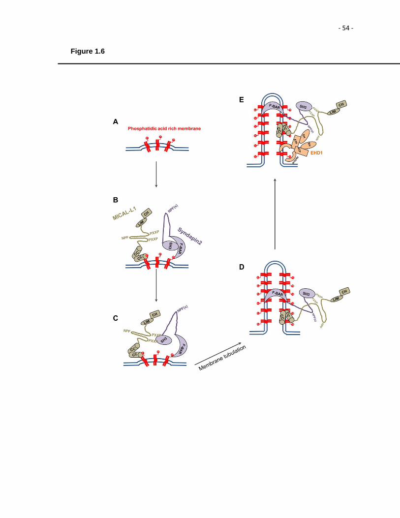
- 54 -
Figure 1.6

- 55 -
9. Conclusion
In recent years, there has been an intense interest in the EHD protein family and
many studies have focused on EHD proteins. Major advances have been made towards
gaining insight into structure-function relationship of EHD2. A partially conserved
unstructured loop, KPFRKLNPF loop in the G-domain, which is primarily responsible for
oligomerization and targeting of EHD2 to caveolae has been characterized. Strikingly,
EHD2 is the only family member that has 2 proline-phenyalanine (PF) (KPF and NPF)
motifs in this loop and with an internal NPF motif. However, the exact role of the 2PF
motifs in the unstructured loop remains unknown. In this dissertation, we focus on
delineating the precise role of 2PF motifs in oligomerization, targeting to the PM and
binding to the interaction partners.
EHD1 and EHD3 have the highest level of homology amongst the members of
the EHD family. Despite hetero-dimerizing and co-localizing at TRE membranes,
surprisingly, EHD1 and EHD3 have distinct, even opposing functions in the TRE
biogenesis/stabilization and vesiculation. EHD1 is involved in the release of vesicles by
the process of vesiculation and EHD3 has been implicated in the tubulation and/or
stabilization of TREs. In this dissertation, we set out to determine the precise
mechanistic role of EHD3 in TRE generation and/or stabilization. Furthermore, we aimed
to elucidate the molecular and atomic basis of the differential function of two such similar
proteins likely evolved through gene duplication and divergent evolution.

- 56 -
Table 1: Relationship between EHD proteins and disease
EHD protein Disease Abberant Phenotype
Reference
EHD1 and EHD3 Alzheimer’s Disease
Loss of EHD1or EHD3
Buggia-Prévot et al., 2013
EHD1 Non-small lung
cancer Overexpression Meng et al., 2016
EHD1 Breast Cancer Overexpression Tong et al., 2017
EHD1 Sickle Cell Anemia 4.9 fold increased expression
Ammann et al., 2009
EHD2 Breast Cancer Overexpression Yang et al., 2015
EHD3 Gliomas Overexpression Chukkapalli et al., 2014
EHD3 Small-cell lung carcinoma
Overexpression Taniwaki et al., 2006
EHD4 Systemic onset juvenile idiopathic
arthritis
Overexpression Allantaz et al., 2007

- 57 -
CHAPTER II
Materials and Methods
- 58 -
10 Materials and Methods
10.1 Recombinant DNA Constructs
The following constructs were used for studies conducted in chapter III and
chapter IV. Cloning of EHD1, EHD2, EHD3, EHD4, MICAL-L1, Rabankyrin-5, Syndapin2
into yeast two-hybrid vectors pGBKT7 and pGADT7, glutathione-S-transferase (GST)-
EH1 and GST-EH3 vectors as well as cloning of green fluorescent protein (GFP)-Myc–
EHD1, GFP-Myc-EHD2, GFP-Myc-EHD3 in expression vectors and their SiRNA-
resistant versions have been described previously (Caplan et al., 2002; Sharma et al.,
2009; Giridharan et al., 2013; Zhang et al., 2013). The SiRNA-resistant-GFP-Myc-
EHD3/EH1 chimera was generated, by cloning amino acid residues 1-434 of EHD3 and
435-450 of EHD1 in eGFP-C3 vector. Two-hybrid control vectors (Gal4ad-SV40 large T-
antigen and Gal4bd-p53) were purchased from (BD Biosciences Clontech, Palo Alto,
CA). The following constructs were generated for the studies described in chapter III
using QuikChange site-directed mutagenesis kit (Stratagene, La Jolla, CA): in the
pGADT7 vector (yeast-two hybrid vector), in the GFP-Myc-EHD1 and in the GFP-Myc-
EHD2 vectors (mammalian expression vectors); pGADT7 EHD2 and GFP-Myc-EHD2
(NPF-to-NAF), pGADT7 EHD2 and GFP-Myc-EHD2 (NPF-to-NPA), pGADT7 EHD2 and
GFP-Myc-EHD2 (NPF-to-APA), pGADT7 EHD2 and GFP-Myc-EHD2 (KPF-to-KAF),
pGADT7 EHD2 and GFP-Myc-EHD2 (KPF-to-APA), GFP-Myc-EHD2 (NPF-to-NPY),
GFP-Myc-EHD2 (NPF-to-NFP), pGADT7 EHD1(RPF-to-APA), pGADT7 EHD1(NAF-to-
NPF), pGADT7 EHD1(RPF-to-APA+NAF-to-NPF) and GFP-Myc-EHD1 (RPF-to-APA).
The following mutations were introduced by site-directed mutagenesis for studies
described in chapter IV: pGADT7 EHD3 (AHLL523PHLV), pGADT7 EHD3 (D459N),
pGADT7 EHD3 (M447T), pGADT7 EHD3 (KVAE523RHE), pGADT7 EHD3 (N519A),
pGDAT7 EHD3 (NE519AD), pGADT7 EHD1 (AD519NE), pGBKT7 MICAL-L1
- 59 -
(NPFDEEEEE to NPFDEEEEA), pGBKT7 MICAL-L1 (NPFDEEEEE to NPFDEEEAA),
pGBKT7 MICAL-L1 (NPFDEEEEE to NPFDEEAAA), pGBKT7 MICAL-L1 (NPFDEEEEE
to NPFDEAAAA), pGBKT7 MICAL-L1 (NPFDEEEEE to NPFDAAAAA), pGBKT7 MICAL-
L1 (NPFDEEEEE to NPFAAAAAA), pGADT7 Rabankyrin-5 (NPFEDV to NPFEDE),
pGADT7 Rabankyrin-5 (NPFEDV to NPFEDEE), pGADT7 Rabankyrin-5 (NPFEDV to
NPFEDEEE), pGADT7 Rabankyrin-5 (QSV to KPY), pGADT7 Rabankyrin-5 (QSV to
KSV), pGADT7 Rabankyrin-5 (QSV to QPV), pGADT7 Rabankyrin-5 (QSV to QSY) and
GST-EH3 (NE519AD).
10.2 Antibodies and Reagents
The primary antibodies used in the studies described in Chapter III and IV were:
the affinity-purified rabbit polyclonal antibodies directed against the C-terminus of human
EHD1 (DLPPHLVPPSKRRHE), EHD2 (VERGPDEAMEDGEEGSDDEA) (Anaspec,
Fremont, CA) have been described previously (Naslavsky et al., 2004). Mouse
polyclonal anti–MICAL-L1 (Novus Biologicals, Littleton, CO), rabbit polyclonal anti-
Syndapin2 used for immunofluroscence (IF) (Abgent, San Diego, CA) and for western
blot (WB) (ProteinTech, Chicago, IL), goat anti-GST conjugated to horseradish
peroxidase (HRP) (GE life sciences, Piscataway, NJ), mouse anti-Rabankyrin-5 (Novus
Biologicals, Littleton, CO), rabbit anti-HA (Signalway Antibody, Pearland, TX), rabbit anti-
HA (Bethyl, Montgomery, TX), and rabbit anti-caveolin1 (Cell Signaling Technology,
Danvers, MA). The following secondary antibodies were used in studies described in
Chapter III and Chapter IV: Alexa-Fluor-568-conjugated goat anti-mouse, Alexa-Fluor-
488-conjugated goat anti-mouse, Alexa-Fluor-568-conjugated goat anti-rabbit
antibodies, and Alexa-Fluor 568–labeled transferrin (Tf-568) (Life Technologies,
Carlsbard, CA). Goat anti-mouse HRP was obtained from (Jackson ImmunoResearch
Laboratories, West Grove, PA). Donkey anti-rabbit HRP was obtained from (GE Life
- 60 -
Sciences, Piscataway, NJ). CAY 10593 and CAY 10594 were purchased from (Cayman
Chemical Company, Ann Arbor, MI).
10.3 Cell culture, transfections and SiRNA treatment
The HeLa cervical cancer cell line (ATCC-CCL2; Manassas, VA) was maintained
in high-glucose Dulbecco’s modified Eagle medium (DMEM) supplemented with 10%
(v/v) fetal bovine serum (FBS) (Sigma-Aldrich, St.-Louis, MO), 200 U/ml penicillin, 200
μg/ml streptomycin. On-Target-plus SMART pool SiRNA for EHD3, MICAL-L1,
Syndapin2, and SiRNA duplexes for EHD1 (base pairs gaaagagatgcccaatgtc) and
synthesized by Dharmacon, Lafayette, CO were transfected using Lipofectamine
RNAiMax (Life Technologies, Carlsbad, CA) to perform knockdown studies as previously
described (Naslavsky et al., 2006 and Sharma et al., 2009) for 48 hour (h) or 72 h
depending on the efficiency of the SiRNA duplexes. For transfection, HeLa cells, plated
on 6-well plates, were transfected using 6 μl X-tremeGENE 9 (Roche Life Sciences,
Indianapolis, IN) or 6 μl Lipofectamine 2000 (Life Technologies, Carlsbad, CA) with 2 μg
of DNA for 16-18 h at 37°C according to manufacturer’s protocol.
10.4 Protein Purification
The EH domains of EHD1, EHD3 and EHDNE519AD mutant were cloned into
bacterial expression vector pGEX-6p-2 and transformed into BL-21 cells. A single colony
from the transformed cells was cultured overnight in 50 ml LB medium containing 10%
glucose. The next day, 20 ml of the culture was transferred to 2 L Super Broth medium
and grown at 37°C. When the optical density (O.D.) reached 0.6-0.7, the cells were
induced with 0.2 mM isopropyl β-D-1-thiogalactopyranoside (IPTG) and transferred to an
18°C incubator shaker for 3 h. Cells were then pelleted and lysed using Emulsiflex-C3
- 61 -
homogenizer (Avestin, Ottawa, Canada). The EH1, EH3 and EH3 (NE519AD) proteins
were purified from the lysate using the gluthathione-agarose resin.
10.5 Immunoblotting
For immunoblotting experiments described in both Chapter III and Chapter IV,
HeLa cells were harvested and lysed on ice for 30 minutes (min) in lysis buffer
containing 50 mM tris(hydroxymethyl)aminomethane(Tris), pH 7.4, 150 mM NaCl, 1%
Triton-X, 1.8 mg/ml iodoacetamide, and protease inhibitor mixture (Roche Life Sciences,
Indianapolis, IN). Total protein levels in the lysate were calculated by BioRad protein
assay reagent (Bio-Rad Laboratories, Hercules, CA) and normalized for equal protein
loading on gels. Protein samples were separated on 8%, 10% or 13% sodium dodecyl
sulphate (SDS)- polyacrylamide gel electrophoresis (PAGE) depending on the molecular
weight of the protein of interest. The separated proteins were transferred to
nitrocellulose membrane and detected by immunoblotting with appropriate antibodies.
10.6 GST pull-down and co-immunoprecipitation
For the co-immunoprecipitation experiments between EHD2 and Syndapin2,
HeLa cells were transiently transfected with either GFP-Myc-EHD2, or GFP-Myc-EHD2
mutants (NPF-to-NAF, NPF-to-APA, KPF-to-KAF, KPF-to-APA), lysed 24 h later in a
buffer containing 50 mM Tris-HCl (pH 7.4), 150 mM NaCl, 1% Triton-X, 1:1000 of 1
mg/ml protease inhibitor cocktail (Calbiochem, San Diego, CA) and subjected to
immunoprecipitation with antibodies against Syndapin2. Separated proteins were
transferred on to nitrocellulose membrane and then immunoblotted with anti-EHD2
antibodies. Input lane contains 5% of the total lysate immunoprecipitated.
In order to address the binding of EH domains of EHD1, EHD3, and EHD3
NE519AD proteins with Rabankyrin-5, in chapter IV, we performed GST pull-down
- 62 -
experiments: 50 μg of purified GST-fusion proteins EH1, EH3 or EH3 (NE519AD) were
incubated with GST beads, in buffer containing 20 mM Tris and 300 mM NaCl containing
0.1% Triton-X-100, leupeptin and incubated for 4 h as previously done (Giridharan et al.,
2013). GST beads were then washed 4X in 20 mM Tris and 300 mM NaCl containing
0.1% Triton-X-100 and leupeptin. HeLa cells were transiently transfected with HA-
Rabankyrin-5 for 24 h and lysed in buffer containing 50 mM Tris-HCl, pH 7.4, 150 mM
NaCl, 0.5% Triton-X-100, 1.8 mg/ml iodoacetamide and protease inhibitor cocktail. GST
beads were then incubated with the HeLa lysate overnight, washed 4X in 20 mM Tris
and 300 mM NaCl containing 0.1% Triton-X-100 and leupeptin, eluted with 4X sample
buffer, and analyzed by immunoblotting with the anti-HA antibody.
10.7 Yeast Two Hybrid
The yeast two-hybrid assay was utilizing the Saccharomyces cerevisiae (S.
cerevisiae) strain, AH109 (BD Biosciences Clontech, Palo Alto, CA) maintained on yeast
extract, peptone and dextrose (YPD) agar plates. A loop full of yeast was grown
overnight at 30°C with shaking at 250 rpm in liquid YPD medium. The transformation
was done by the lithium acetate procedure as described in the instructions for the
MATCHMAKER two-hybrid kit (BD Biosciences Clontech, Palo Alto, CA). For colony
growth assays AH109 co-transformants were streaked on plates lacking leucine and
tryptophan. Co-transformants were allowed to grow at 30°C, usually for three days, or
until the colonies were large enough for further assays. An average of three to four
colonies were selected and suspended in water, equilibrated to the same optical density
at 600 nm, and replated on plates lacking leucine and tryptophan (+HIS, -2 plates), as
well as plates also lacking histidine (-HIS, -3 plates). The interaction between p53 and
SV40 was used as positive control.

- 63 -
10.8 Transferrin uptake and recycling assays
The rate of recycling of transferrin (TfR) was measured for experiments
described in Chapter III and Chapter IV. HeLa cells were plated on 35-mm plates
containing five coverslips each. Cells were approximately 75% confluent by the time of
experiment. The cells were starved in DMEM containing 0.5% bovine serum albumin
(BSA) for 1 h. After starvation, the cells were “pulsed” with 1 μg/ml of Tf-568 for 15 min
and chased for 15 min in complete media, to allow the Tf-568 to be recycled. The
coverslips were then fixed in 4% (v/v) paraformaldehyde in PBS and imaged by confocal
microscopy.
10.9 CAY inhibitor washout assay
HeLa cells were grown on coverslips in 6-well plates and were subjected to
SiRNA treatment using SiRNA duplexes against Syndapin2, EHD1 and EHD3. After 48
h, the cells were subjected to100 μM PLD inhibitors (CAY10593 and CAY10594) for 30
min at 37°C. The cells were then washed 3X with complete medium and allowed to
recover after the washout of the inhibitors for 20 min, 1 h, 4 h, and 6 h. The coverslips
were fixed in 4% (v/v) paraformaldehyde in PBS and stained with anti-MICAL-L1
antibodies for the assessment of TRE.
10.10 Isothermal Titration Calorimetry (ITC)
Heat produced by the binding of MICAL-L1 peptide with the EH domains of
EHD1 and EHD3 was measured by ITC using the MicroCal iTC200 isothermal titration
calorimeter (Malvern, Worcestershire, UK). All proteins were equilibrated in 1X
phosphate-buffered saline (PBS) at pH 7.4 by overnight dialysis. ITC binding isotherms
were collected at 25°C by injecting 20 x 2 μL of peptide (930 μM) into a solution of each
of the EH domains (90 μM), representing a 1:10 molar ratio. The heat from each
- 64 -
injection was measured by integrating the area of the injection peak, corrected for the
background heat produced by the dilution of the peptide into the buffer, and plotted as a
function of the EH domain/MICAL-L1 peptide molar ratio. Dissociation constant (KD)
values were calculated by fitting the titration curves according to a single binding site
mode with Origin 7 software with ITC add-ons supplied by Malvern.
10.11 Confocal microscopy imaging
HeLa cells were grown on cover slips and fixed with 4% (v/v) paraformaldehyde
in PBS. The fixed cells were then incubated with primary antibodies prepared in staining
solution (0.2% (w/v) saponin and 0.5% (w/v) bovine serum albumin (BSA) in PBS) for 1
h at room temperature. After washing in PBS, the cells were incubated with the
appropriate fluorochrome-conjugated secondary antibody mixture in staining solution for
30 min at room temperature. Images were acquired with a Zeiss LSM 5 Pascal confocal
microscope (Carl Zeiss, Thornwood, NY) using a 63 X 1.4 numerical aperture (NA)
objective with appropriate filters.
10.12 Structured illumination microscopy (SIM) imaging
SIM images were collected on samples obtained as described above with a Zeiss
ELYRA PS.1 illumination system (Carl Zeiss, Thornwood, NY) using a 63 X objective
lens with a NA of 1.4 at room temperature as we have done previously (Reinecke et al.,
2014; Xie et al., 2015). Three orientation angles of the excitation grid were acquired for
each z-plane, with z-spacing of 110 nm between planes. SIM processing was performed
using the SIM module of the ZEN BLACK software (Carl Zeiss, Thornwood, NY). The
processed SIM images were then exported in TIFF format.
10.13 Quantification of MICAL-L1-containing tubular recycling endosomes
- 65 -
Tubular recycling endosomes (TREs) were quantified as described previously
(Cai et al., 2013). Briefly, using NIH ImageJ software, threshold was adjusted to reduce
the background in the image. The particle size was set between 5 μm2 and 150 μm2. All
MICAL-L1-containing particles (tubules and vesicles) in this range were counted. Ten
fields of images from each treatment were analyzed for statistical analysis.
10.14 Statistical analysis
The data sets (n=100 unless otherwise indicated) presented were collected from
three independent experiments and were analyzed by one-way ANOVA, with one star
for p < 0.01, and three stars for p < 0.05. The p-values are shown for each experiment.
10.15 Sequence homology and Identity Analysis
Sequence homology and identity between EHD1, EHD2, EHD3, MILCAL-L1,
Rabankyrin-5 and their respective mutants generated by site-directed mutagenesis, was
analyzed using AlignX program of Vector NTI. Vector NTI performs multiple sequence
alignments using a modified Clustal W algorithm.

- 66 -
CHAPTER III
Role of the EHD2 Unstructured Loop in Dimerization, Protein Binding and Subcellular Localization
The following chapter has been published in PLoS One. 2015 Apr 15;10(4):e0123710

- 67 -
11 Introduction
The C-terminal Eps15 homology domain-containing (EHD) proteins coordinate
various endocytic membrane regulatory events in mammalian cells (Naslavsky and
Caplan, 2011). All four EHDs (EHD1-4) share a common domain architecture that
includes three characteristic domains: 1) a Dynamin-like-G-domain that binds and
catalyzes hydrolysis of ATP (Caplan et al., 2002; Naslavsky et al., 2006; Lee et al.,
2005; Daumke et al., 2007; Simone et al., 2014), 2) a coiled-coiled domain n formed by
two helical regions that facilitates EHD oligomerization and lipid binding, 3) a C-terminal
Eps15 Homology (EH) domain with a positively charged electrostatic surface that
preferentially binds to proteins containing an asparagine-proline-phenylalanine (NPF)
motif followed by acidic residues (Kieken et al., 2007; Kieken et al., 2009; Kieken et al,
2010; Henry et al., 2010), that is a hallmark of these proteins. The most diverse EHD
both in terms of sequence homology and function is EHD2 (Marg et al., 2012). A series
of recent studies has established the involvement of EHD2 in regulating a variety of
important functions that include sarcolemmal repair (Marg et al., 2007), myogenesis
(Posey et al., 2011; Doherty et al., 2008), and control of Rac1 and the actin cytoskeleton
(Stoeber et al., 2012; Benjamin et al., 2011; Park et al., 2004; Guilherme et al., 2004).
Unlike EHD2, EHD1, EHD3 and EHD4, all of which play roles in regulating endocytic
transport from sorting and recycling endosomes (Naslavsky and Caplan, 2011; Grant
and Caplan, 2008; Naslavsky and Caplan, 2005). However, EHD2 is recruited to the
cytoplasmic interface of the plasma membrane (PM) by phosphatidylinositol 4,5
bisphosphate (PIP2) (Simone et al., 2013) where it interacts with caveolin and regulates
caveolar mobility (Benjamin et al., 2011; Hansen et al., 2011; Moren et al., 2012).
EHD2 was the first family member whose crystal structure was solved and the
structure indicates that this protein contains a partially conserved region with two proline-
phenylalanine (2 PF) motifs KPFRKLNPF in an unstructured flexible loop near the G-

- 68 -
domain (Daumke et al., 2007) (Fig 3.1A). This unstructured KPFRKLNPF region was
proposed to link EHD2 dimer pairs through interactions with neighboring EH domains
(Daumke et al., 2007). This is a unique situation where the NPF motif is intrinsic and the
NPF-EH interaction facilitates the self-interaction of the protein. Recent studies have
firmly established that N-terminus plays an indispensable role in targeting of EHD2 to the
membrane (Shah et al., 2014), and provide support for the notion that both PF motifs
play an important role in EHD2 localization and function (Moren et al., 2012). However,
the degree to which both of these closely situated PF motifs impact EHD2 function, and
particularly how each motif affects dimerization and interactions with binding partners
remains obscure. The function of the PF motifs in the unstructured loop in EHD proteins
is further complicated because while EHD2 has two PF motifs in its KPFRKLNPF
sequence, the other three EHD paralogs all contain only one PF motif (Fig 3.1B); either
a KPF or RPF motif at the N-terminal side of the unstructured loop. Indeed, both EHD1
and EHD3 have a NAF motif (instead of NPF), whereas EHD4 has a SRF motif. In this
study, we sought out to determine the significance of the two proline-phenylalanine
(2PF): KPF and NPF motifs of EHD2 for their ability to dimerize, interact with protein
partners and localize to caveolae. To test the role of the NPF motif, we generated an
EHD2 NPF-to-NAF mutant to mimic the homologous sequences of EHD1 and EHD3.
While the EHD2 NPF-to-NAF mutant lost its dimerization potential and protein binding
ability, this motif had little impact on its localization within the cell. On the other hand,
mutation of the NPF motif to APA had little or no impact on dimerization or partner
binding, but shunted EHD2 localization away from the PM primarily to the nucleus. We
further examined EHD1 as a representative of the other three more closely related
EHDs; we found that a single RPF motif of EHD1 is essential for (i) its dimerization, (ii)
interaction with protein partners, and (iii) its localization to tubular recycling endosomes.
Overall, our data suggests that the phenylalanine residue in the NPF motif is
- 69 -
crucial for EHD2 localization to the PM, whereas the proline residue is essential for
EHD2 dimerization and binding. These studies support the recently proposed model
suggesting that the EHD2 N-terminal region regulates the availability of the unstructured
loop for interactions with neighboring EHD2 dimers, thus promoting oligomerization.
12 Results
12.1 Alteration of the EHD2 NPF motif to NAF impairs dimerization and binding
with interaction partners, but does not affect EHD2 localization
EHD2 is the only member of EHD protein family that homo-dimerizes but does
not hetero-dimerize (Simone et al., 2013). This homo-dimerization potential may be
required for interaction with NPF-containing protein binding partners. Accordingly, our
goal was to first determine the requirement of the EHD2 NPF motif for homo-
dimerization (Fig 3.1A and 3.1B). By selective two-hybrid binding assay, we
demonstrated that while wild-type EHD2 proteins homo-dimerize, they were not capable
of interacting with wild-type EHD1, EHD3 or EHD4 (Fig 3.2A). On the other hand, when
the EHD2 NPF motif was perturbed by introduction of a point mutation to convert to a
NAF, the homo-dimerization was no longer observed. EHD2 binds to specific subsets of
NPF-containing protein partners through its EH domain. Indeed, wild-type EHD2
interacted with Syndapin2, but not MICAL-L1, even though both proteins contain NPF
motifs followed by acidic clusters (Fig 3.2B). In vivo, co-immunoprecipitation analysis
between EHD2 and Syndapin2 further confirmed the interaction (Fig 3.4A and Table
3.1). However, the EHD2 NPF-to-NAF mutant lost its ability to interact with Syndapin2,
suggesting that the EHD2 NPF motif is required for both homo-dimerization and for
protein partner binding. Despite these data in yeast two-hybrid binding studies, the NAF
mutant retained its ability to bind to Syndapin2 by co-immunoprecipitation, suggesting

- 70 -
the involvement of bridging proteins in the cells (Fig 3.4A and Table 3.1). In our previous
studies, we demonstrated that interaction of EHD2 with PIP2 is responsible for
recruitment of EHD2 to the PM and is independent of interaction with other binding
partners through its EH domain (Simone et al., 2013). Recent findings by Moren et al.
showed that EHD2 caveolar localization was independent of its protein-binding EH
domain, which supports our conclusions (Moren et al., 2012). Furthermore, using
selective yeast two-hybrid studies we have demonstrated that the EH domain is not
required for EHD2 (or EHD1) homo-dimerization (Fig 3.2A-3.2C). Accordingly, we
wanted to test the impact of EHD2 NPF-to-NAF modification on the localization of
protein to the PM. Confocal microscopy demonstrated that both wild type and EHD2
NPF-to-NAF primarily displayed PM localization (Fig 3.3C and 3.3D). We further
analyzed both the wild-type and EHD2 NPF-to-NAF mutant using Structured Illumination
Microscopy (SIM) and also observed both the wild-type and EHD2 NPF-to-NAF mutant
partially co-localized to structures positive for caveolin-1 (Fig3.3E-3.3H); (see insets,
asterisks indicate transfected cells). These data support the notion that the EHD2 NPF
motif plays an essential role in EHD2 dimerization and binding to NPF-containing
interaction partners. However, normal localization of EHD2 to the PM and colocalization
of EHD2 with caveolae are not contingent upon the homo-dimerization ability of EHD2.
12.2 Modification of the EHD2 NPF motif to APA induces loss of PM localization,
but does not affect homo-dimerization and interactions with binding partners.
Recent studies by Moren et al. demonstrated that upon mutating the coding
sequence of phenylalanine 128 of the EHD2 NPF motif (see Fig 3.1A) to that of alanine
(NPF-to-NPA), there is no significant effect on the subcellular localization of EHD2
(Moren et al., 2012). In order to recapitulate the homologous sequence found in both
EHD1 and EHD3, we decided to initially disrupt the NPF motif by substituting the proline

- 71 -
for alanine (NPF-to-NAF). Furthermore, we aimed at delineating the functional role of
EHD2 when the proline residue of its NPF motif is left intact. To this aim, we assessed
homo-dimerization of EHD2 NPF-to-APA mutants as well as its interaction with
Syndapin2 (Fig 3.5A and 3.5B). Surprisingly, unlike the EHD2 NPF-to-NAF mutant, the
NPF-to-APA mutants retained both homo-dimerization and binding to Syndapin2. On the
other hand, despite normal dimerization and binding, EHD2 NPF-to-APA displayed a
dramatic relocation to the nucleus (Fig 3.5D). Previous studies have demonstrated
EHD2 has a bipartite nuclear localization sequence and many cells display a portion of
their EHD2 in the nucleus (Fig 3.5C) (Pekar et al., 2012). Interestingly, the NPF-to-APA
mutant displayed primarily nuclear localization (Fig 3.5D). Accordingly, despite the yeast
two-hybrid binding data, we were unable to detect EHD2 NPF-to-APA interactions with
Syndapin2 by co-immunoprecipitation, likely due to the mislocalization of the mutant
exclusively to the nucleus (Fig 3.4A and Table 3.1). However, Moren et al. used a
subtler EHD2 NPF-to-NPA mutant (rather than NPF-to-APA) and did not report such
relocation to the nucleus (Moren et al., 2012). Consequently, we resolved to compare
the functional role of EHD2 NPF-to-APA mutant to EHD2 NPF-to-NPA. As demonstrated
in (Fig 3.6A and 3.6B), similar to the APA mutant, EHD2 NPF-to-NPA continued to
homo-dimerize and interact with Syndapin2. Furthermore, the subcellular distribution of
EHD2 NPF-to-NPA was also similar to that of EHD2 NPF-to-APA, with most of the
mutant localizing to the nucleus and diminished localization at the PM (compare Fig
3.6C and 3.6D). Taken together, these results suggested that the NPF phenylalanine
residue contributes to the localization of EHD2 at the PM, either by preserving EHD2 at
the PM, or by possibly preventing it from transporting to or exporting out of the nucleus.
However we cannot entirely reconcile the differences we observed in EHD2 NPF-to-NPA
localization (largely nuclear) compared to those of Moren et al. (Fig 3.6B; GFP-EHD2
F128A) (Moren et al., 2012), where EHD2 continued to localize at the caveolae on the

- 72 -
PM and a small fraction was localized to the nucleus (data not shown).
12.3 The phenylalanine residue of NPF motif plays a key role in the PM localization
of EHD2.
We then proceeded to analyze the precise role of phenylalanine 128. To this end,
we engineered an EHD2 NPF-to-NPY mutant. The rationale behind this mutant protein
was that tyrosine is structurally the closest residue to phenylalanine, with the only
difference being the additional hydroxyl group on the aromatic ring. Interestingly, the
EHD2 NPF-to-NPY mutant displayed an intermediate cellular distribution (compare Fig
3.7A with 3.7C and 3.7D). Although PM association was not abrogated, a significant
portion of the EHD2 NPF-to-NPY EHD2 mutant was observed in the nucleus. However,
the NPF-to-NPY mutant continued to homo-dimerize with wild-type EHD2 and maintains
binding with Syndapin2 (Fig 3.4A and Table 3.1). We further posited that the spatial
localization of phenylalanine 128 is essential for its function in facilitating EHD2
localization, primarily to the PM. Accordingly, we engineered EHD2 NPF-to-NFP by
swapping the proline and phenylalanine residues at positions 127 and 128. Although
some EHD2 was clearly localized to the PM, the majority of this NFP had a subcellular
localization in the nucleus (Fig 3.7F). Furthermore, the NPF-to-NFP mutant failed to
either homo-dimerize or interact with Syndapin2 (Fig 3.4A and Table 3.1). These data
firmly establish that the position of phenylalanine at residue 128 within the unstructured
loop is highly significant and is crucial for the localization to the PM.
12.4 Disruption of the EHD2 KPF motif induces relocalization of EHD2 to the
nucleus, but does not alter its oligomerization and partner binding ability.
Previous studies by Moren et al. have implicated the role of the KPF motif in the
unstructured loop of EHD2 (Fig 3.3A and 3.3B) in caveolar binding and stable

- 73 -
association of EHD2 with the membrane (Moren et al., 2012). However, the impact of
the KPF motif on EHD2 homo-dimerization and interactions with binding partners has
not been studied. Hence, we turned our attention to the KPF motif of EHD2 and
accordingly, we generated EHD2 KPF-to-APA or KPF-to-KAF mutants. EHD2 KPF-to-
APA or KPF-to-KAF mutants had no effect on dimerization or binding to Syndapin2 (Fig
3.9A–3.9D) by yeast two-hybrid binding assays. Both KPF mutants reduced the level of
EHD2 on the PM and increased the level of nuclear-localized EHD2 compared to wild-
type EHD2 (Fig 3.9E–3.9G). Furthermore, the co-immunoprecipitation experiments did
not detect binding between the mutants and Syndapin2, possibly because the mutants
are localized primarily to the nucleus (as opposed to the normal PM localization).
Overall, these studies help elucidate the complicated functional role for the unstructured
loop and its two PF motifs, which is in agreement with both the initial structural model
(Daumke et al., 2007; Moren et al., 2012) and the revised model (Shah et al., 2014)
proposed for dimer pair PF-EH domain interactions and EHD2 oligomerization (see
model for further elaboration Fig 3.12).
12.5 A single EHD1 PF motif (RPF) controls its homo- and hetero-dimerization,
binding to interaction partners, and localization to Tubular Recycling Endosomes
(TRE).
Given that EHD1 contains a single PF motif, namely the RPF motif that aligns
with the EHD2 KPF motif (see Fig 3.1B), we evaluated the role of this motif on EHD1
dimerization and partner binding. The ability to homo-and heterodimerize was lost in the
EHD1 RPF-to-APA mutant (Fig 3.10A). Furthermore, EHD1 RPF-to-APA had abrogated
binding to MICAL-L1, and decreased binding to Syndapin2 (Fig 3.10B). Moreover, the
typical subcellular localization of EHD1 to an array of tubular and vesicular recycling
endosomes (Fig 3.10C) was lost and the EHD1 RPF-to-APA mutant was mislocalized to

- 74 -
the cytoplasm (Fig 3.10D). These data suggest that the single RPF motif in EHD1 is
necessary and sufficient to successfully carry out functions that the dual NPF and KPF
motifs present in EHD2 execute.
12.6 The NAF motif of EHD1 is dispensable for homo- or hetero-oligomerization,
and for its association with binding partners.
Additionally, since EHD1 has a “non-functional” NAF motif aligned with the NPF
motif of EHD2 (depicted in Fig 3.1B) we hypothesized that the EHD1 NAF motif would
still function if its lone existing PF motif (RPF) was impaired. To begin with, we
engineered EHD1 NAF-to-NPF mutants as a control and showed that as expected,
homo- and hetero-dimerization along with MICAL-L1 and Syndapin2 binding remained
stable (Fig 3.11A and 3.11B). Next, we generated double mutants where the EHD1 RPF
motif was disrupted (RPF-to-APA) and the NAF motif was ‘corrected’ to NPF. Compared
to the EHD1 RPF-to-APA single mutants, which lost all of their dimerization and protein
interactions, the additional NAF-to-NPF mutation partially rescued homo- dimerization
(Fig 3.11C) and binding to MICAL-L1 (Fig 3.11D).
12.7 The EHD1 RPF motif is essential for receptor recycling.
To evaluate the functional role of the EHD1 RPF motif, we utilized a siRNA
approach combined with rescue by transient transfection. As demonstrated, EHD1-
siRNA treatment was successful in depleting more than 90% of EHD1 from HeLa cells
(Fig 3.12C). HeLa cells on coverslips were either mock-treated or treated with EHD1-
siRNA and subjected to “pulse-chase” experiments with fluorochrome-labeled transferrin
(Tf-568). After the pulse with Tf-568, both mock- and EHD1-siRNA treated cells
exhibited a similar subcellular distribution of internalized Tf-568 (Fig 3.12C). However,
after a 20 min chase, mock-treated cells had recycled most of their internalized Tf-568

- 75 -
back to the PM, whereas EHD1 depleted cells exhibited significant accumulation of Tf in
the ERC (compare Fig 3.12B with control Fig 3.12A). This accumulation was ‘rescued’ in
EHD1-depleted cells where a siRNA-resistant form of the wild-type EHD1 (siEHD1) had
been transfected (Fig 3.12D and 3.12E; see yellow borders for transfected cells).
However, when an EHD1 RPF-to-APA mutant (that is mislocalized to the cytoplasm)
was reintroduced into EHD1-depleted cells, the Tf and its receptor remained
accumulated in the perinuclear region of the cell (Fig 3.12F and 3.12G; see yellow
borders for transfected cells). Quantification of these data (from 3 independent
experiments) demonstrated that wild-type EHD1 rescued the recycling defect in > 85%
of the EHD1-depleted cells whereas the RPF-to-APA mutant displayed less than 15%
rescue (Fig 3.12H).
13 Discussion
Despite being the first EHD protein whose crystal structure was solved (Daumke
et al., 2007), and important structural characterizations of its interaction with membranes
(Shah et al., 2014) and its recruitment by PI(4,5)P2 to the PM (Simone et al., 2013),
nonetheless many of the mechanistic aspects by which EHD2 functions have remained
obscure. In this study we have identified intriguing differences in the function of the
proline and phenylalanine residues of the NPF motif that is found within the
KPFRKLNPF region of the unstructured loop present in the G-domain of EHD2 (Fig
3.1A). Our data suggest that the phenylalanine is crucial for localization of EHD2 to the
PM. Indeed, mutation of this residue leads to the translocation and accumulation of
EHD2 in the nucleus. On the other hand, mutation of the proline residue of this same
NPF motif not only leads to loss of homo-oligomerization and presumed loss of
oligomerization, but also loss of protein partner binding without impacting localization to
the PM. Given the elegant structural studies that propose the PF-EH domain interactions

- 76 -
for EHD2 oligomerization (Shah et al., 2014), how does our data contribute to the current
model? Shah and colleagues recently proposed that upon membrane sensing, the EHD2
N-terminus disrupts its interaction with the unstructured loop in the G-domain and moves
away (illustrated in Fig 3.11A–3.11B), rendering the KPFRKLNPF unstructured loop in a
conformation that is competent for interactions with EH domains of nearby EHD2 dimer
pairs, thus promoting oligomerization and caveolar localization (Fig 3.11C) (Shah et al.,
2014).
Based on this model, we hypothesize that the N-terminus of the EHD2-NPF-to-
NPA mutant (Fig 3.11D), even in the presence of the membrane, may not completely
detach from the G-domain regions flanking the unstructured loop and thus fail to insert
properly into the PM. We rationalize that under conditions where membrane binding is
partially impaired, the string of lysine residues in the EHD2 helical domain, which
perform the dual function of being membrane contact sites (Daumke et al., 2007) and a
bipartite nuclear localization sequence (Pekar et al., 2012), are now free to act in the
latter capacity and facilitate the transport of EHD2 to the nucleus. The support for this
notion also comes from previous studies that have described the role of EHD2 as a
transcriptional repressor (Pekar et a., 2012). On the other hand, we predict that with the
EHD2 NPF-to-NAF mutant, the EHD2 N-terminus is nonetheless freed from the G-
domain to interact with the PM. However, despite its correct localization to the PM, the
NAF motif abrogates EH binding and consequently the dimerization and oligomerization
potential is impaired. Overall, our study provides support and additional novel details that
further clarify the complex structural model of EHD2 localization, membrane binding,
protein binding, dimerization, oligomerization, and function.
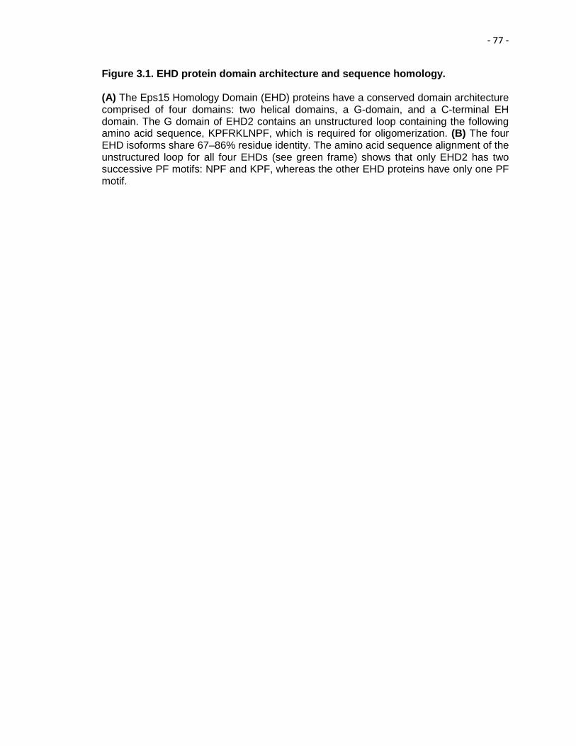
- 77 -
Figure 3.1. EHD protein domain architecture and sequence homology. (A) The Eps15 Homology Domain (EHD) proteins have a conserved domain architecture comprised of four domains: two helical domains, a G-domain, and a C-terminal EH domain. The G domain of EHD2 contains an unstructured loop containing the following amino acid sequence, KPFRKLNPF, which is required for oligomerization. (B) The four EHD isoforms share 67–86% residue identity. The amino acid sequence alignment of the unstructured loop for all four EHDs (see green frame) shows that only EHD2 has two successive PF motifs: NPF and KPF, whereas the other EHD proteins have only one PF motif.

- 78 -
Figure 3.1

- 79 -
Figure 3.2 EHD dimerization requires an intact α-helical region, but not the presence of EH domains. (A) Schematic diagram of EHD2 domain organization. (B–C) Yeast were co-transformed with Gal4bd fusion constructs: Gal4bd-p53 (control), EHD2 (wt), EHD2 (ΔEH) along with Gal4ad-SV40 (control), EHD2 (wt), EHD2 (ΔEH), EHD1 (wt), EHD1 (ΔEH), and EHD1. All co-transformants were plated on non-selective (+HIS) and selective (-HIS) media.

- 80 -
Figure 3.2

- 81 -
Figure 3.3. Modification of the EHD2 NPF motif to NAF impairs dimerization and binding with interaction partners, but does not affect EHD2 localization. (A) S. cerevisae yeast were co-transformed with the following Gal4 binding domain (Gal4bd) fusion constructs (see Materials and Methods): p53 (control), -Syndapin-2 and -MICAL-L1. Gal4 activating domain (Gal4ad) fusion constructs were either-SV40 (control), -EHD2 (wt) or -EHD2 (NPF-to-NAF). Cotransformants were plated on non-selective (+HIS) and selective (-HIS) agar plates. (B) As in (A), co-transformation was with Gal4bd-p53 (control), -EHD2 (wt), -EHD1 (wt), -EHD3 (wt), and -EHD4 (wt) and with Gal4ad-SV40 (control), -EHD2 (wt) and -EHD2 (NPF-to-NAF). (C–H) HeLa cells were grown on coverslips, transfected with GFP-myc-EHD2 (wt) (C, E, G) or with GFP-myc-EHD2 (NAF-to-NPF) (D, F, H) and fixed. (C–D) are transfected cells imaged by confocal microscopy, whereas (E–H) does Structured Illumination Microscopy (SIM) obtain micrographs. Asterisks indicate transfected cells. Bar;10 μm.

- 82 -
Figure 3.3

- 83 -
Figure 3.4 Co-immunoprecipitation of EHD2 and mutant EHD2 proteins with Syndapin2. (A and B) HeLa cells were transfected with either GFP-Myc-EHD2, or GFP-Myc-EHD2 mutants (NPF-to-NAF, NPF-to-APA, KPF-to-KAF, KPF-to-APA), lysed 24 h later and subjected to immunoprecipitation with antibodies against Syndapin2. Separated proteins were transferred onto nitrocellulose and then immunoblotted with anti-EHD2 antibodies. Input contains 5% of the total lysate immunoprecipitated.

- 84 -
Figure 3.4
- 85 -
Figure 3.5. Modification of the EHD2 NPF motif to APA induces loss of plasma membrane localization, but does not affect interactions with binding partners. (A) S. cerevisae yeast were co-transformed with thefollowing Gal4bd fusion constructs: Gal4bd-p53 (control), -Syndapin-2 and -MICAL-L1 along with Gal4ad fusion constructs Gal4ad-SV40 (control), -EHD2 (wt.) and -EHD2 (NPF-to-APA). (B) As in (A) cotransformationwas with Gal4bd fusion constructs: Gal4bd-p53 (control), -EHD2 (wt) along with Gal4ad fusion constructs: Gal4ad-SV40 (control), -EHD2 (wt) and -EHD2 (NPF-to-APA). Co-transformants from A-B were plated on non-selective (+HIS) and selective (-HIS) agar plates. (C–D) HeLa cells were grown on coverslips, transfected with GFP-myc-EHD2 (C) or GFP-myc-EHD2 NPF-to-APA (D). Bar; 10 μm.

- 86 -
Figure 3.5

- 87 -
Figure 3.6. The NPF phenylalanine residue is responsible for the plasma membrane localization of EHD2. (A) S. cerevisae yeast were co-transformed with the following Gal4bd fusion constructs: Gal4bd-p53 (control), -Syndapin-2 and -MICAL-L1 along with Gal4ad fusion constructs: Gal4ad-SV40 (control), -EHD2 (wt) and -EHD2 (NPF-to-NPA). (B) As in (A), yeast were co-transformed with the Gal4bd fusion constructs: Gal4bd-p53 (control), -EHD2 (wt) alongwith Gal4ad fusion constructs: Gal4ad-SV40 (control), -EHD2 (wt.) and -EHD2 (NPF-to-NPA). Co-transformants were plated on non-selective (+HIS) andselective (-HIS) agar plates. (C–I) HeLa cells were grown on coverslips and transfected with: (C) GFP-myc-EHD2 (wt), (D) GFP-myc-EHD2 (NPF-to-NPA), (E) GFP-myc-EHD2 (NPF-to-NPY), (F) GFP-myc-EHD2 (NPF-to-NPA), (G) GFP-myc-EHD2 (NPF-to-NFP). Bar;10 μm.

- 88 -
Figure 3.6
- 89 -
Figure 3.7. Wild-type and EHD2 NPF-to-NPY homo-dimerize and interact with Syndapin2, whereas EHD2 NPF-to-NFP does not. (A and B) S. cerevisae yeast were co-transformed with the following Gal4bd fusion constructs: Gal4bd-p53 (control), -EHD2 (wt), -MICAL-L1, and -Syndapin-2 along with Gal4ad-SV40 (control), -EHD2 (wt), -EHD2 NPY, and EHD2 NFP. Co-transformants in were plated on non-selective (+HIS) and selective (-HIS) agar plates.
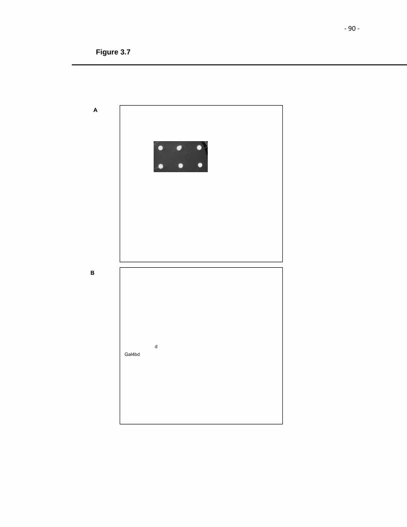
- 90 -
Figure 3.7

- 91 -
Figure 3.8. Disruption of the EHD2 KPF motif induces relocalization of EHD2 to the nucleus, but does not alter its binding ability. (A–D) S.cerevisae yeast were co-transformed with the following Gal4bd fusion constructs: Gal4bd-p53 (control), -Syndapin-2 and -MICAL-L1 along with Gal4ad fusion constructs: Gal4ad-SV40 (control), -EHD2 (wt), -EHD2 (KPF-to-APA) and—EHD2 (KPF-to-KAF). Co-transformants were plated on non-selective (+HIS) and selective (-HIS) media. (E–H) HeLa cells were grown on coverslips, transfected with GFP-myc-EHD2 (wt) (E), GFP-myc-EHD2 (KPF-to-APA) (F), or GFP-myc-EHD2 (KPF-to-KAF) (G) and analyzed by confocal microscopy. Bar;10 μm.

- 92 -
Figure 3.8

- 93 -
Figure 3.9. A single EHD1 PF motif (RPF) controls its homo- and hetero-dimerization, binding to interaction partners, and localization to Tubular Recycling Endosomes (TRE). (A) S. cerevisae yeast were co-transformed with Gal4bd fusion constructs: Gal4bd-p53 (control), -Syndapin-2 and-MICAL-L1 along with Gal4ad-SV40 (control), -EHD1 (wt.) and -EHD1 (RPF-to-APA). (B) As in (A), yeast were co-transformed with Gal4bd-p53 (control),-EHD1 (wt), -EHD3 (wt), -EHD4 (wt), along with Gal4ad-SV40 (control), -EHD1 (wt) and EHD1 (RPF-to-APA). Co-transformants from A-B were plated on non-selective (+HIS) and selective (-HIS) agar plates. (C–D) HeLa cells were grown on coverslips, transfected with GFP-myc-EHD1 (wt) (C), GFP-myc-EHD1 (RPF-to-APA) (D, arrows point to TRE). Bar; 10 μm.

- 94 -
Figure 3.9

- 95 -
Figure 3.10. The NAF motif of EHD1 is dispensable for homo- or hetero oligomerization, and for its association with binding partners. (A) S. cerevisae yeast were co-transformed with the following Gal4bd fusion constructs: Gal4bd-p53 (control), -MICAL-L1, and -Syndapin-2 along with Gal4ad-SV40 (control), -EHD1 (wt.) and -EHD1 (NAF to NPF). (B) As in (A), yeast were co-transformed withGal4bd-p53 (control), -EHD1 (wt), -EHD2 (wt), -EHD3 (wt) and -EHD4 (wt), along with Gal4ad-SV40 (control), -EHD1 (wt) and -EHD1 (NAF-to-NPF). (C) As in (A), yeast were co-transformed with Gal4bd-p53 (control), -EHD1 (wt), -EHD3 (wt), and -EHD4 (wt), along with Gal4ad-SV40 (control), -EHD1 (wt) and -EHD1 (NAF-to-NPF). (D) Yeast were co-transformed with Gal4bd-p53 (control), -Syndapin-2, and -MICAL-L1, along with Gal4ad-SV40 (control), EHD1 (wt) and EHD1 (NAF-to-NPF). Co-transformants in A-D were plated on non-selective (+HIS) and selective (-HIS) agar plates.

- 96 -
Figure 3.10

- 97 -
Figure 3.11. The EHD1 RPF motif is essential for receptor recycling. (A–B) HeLa cells grown on coverslips were mock-treated (A) or treated with EHD1-siRNA (B–F). For A–B, After a 30 min. serum-starvation, cells were allowed to uptake Tf-568 for 15 min., and then chased in complete media for 15 min. to permit Tf- 568 recycling to the PM. (C) Immunoblot analysis of Mock- or EHD1-siRNA-treated cells depicting levels of actin (control) or EHD1. (D–G) During the EHD1-siRNA treatment, cells were transfected with the siRNA-resistant EHD1 constructs: siRNA-res-GFP-myc-EHD1 (wt) in (D), and siRNA-res-GFP-myc- EHD1 (RPF-to-APA) in (F). Following serum-starvation, Tf-568 was internalized for 15 min , and chased in complete media for 15 min. Note the typical concentration of Tf in the ERC in untransfected cells treated with EHD1-siRNA. (H) Quantitative analysis of 100 cells from 3 independent experiments as in (E and G). Cells that recycled Tf-568 (“empty of Tf”) were scored and calculated as % of total cell number, and portrayed with standard error bars. Stars represent significance of p < 0.01 (one star) or p < 0.05 (three stars) for one-way ANOVA tests. Bar; 10 μm.

- 98 -
Figure 3.11
- 99 -
Figure 3.12. Model for the role of the EHD2 unstructured KPFRKLNPF motif in subcellular localization. (A) In the absence of PIP2-containing membrane, the N-termini interact with regions flanking the unstructured loops (U.L.) of EHD2 dimer pairs. (B) PIP2-binding by the N-terminus facilitates a-8 helical binding to the membrane and frees the U.L. (C) the U.L. NPF motif is now capable of interacting with an EH domain of a neighboring EHD2 dimer pair, inducing oligomerization. (D) For EHD2 NPF-to-NPA mutants, we hypothesize that the N-terminus may not insert into membranes, thus preventing the U.L. from oligomerizing and maintaining the lysine residues on α-8 helix free to serve as a nuclear localization sequence.

- 100 -
Figure 3.12

- 101 -
Table 3.1 Comparison of wild-type EHD2 and mutants in homo-dimerization, Syndapin2-binding and sub-cellular localization.
- 102 -
Chapter IV
EHD3 Protein Is Required for Tubular Recycling Endosome Stabilization, and an Asparagine-Glutamic
Acid Residue Pair within Its Eps15 Homology (EH) Domain Dictates Its Selective Binding to NPF Peptides
The following chapter has been published in JBC. 2016 Jun 24; 291(26):13465-78

- 103 -
14 Introduction
Endocytic trafficking entails internalization, sorting, degradation, and recycling of
macromolecules in mammalian cells (Conner and Schmid, 2003). These processes are
not only essential for the maintenance of cellular homeostasis, but also key for the
regulation of a variety of cellular events, including nutrient uptake, cell adhesion, cell
migration, cell polarity, cytokinesis, and signal transduction. In this process, receptors
encounter ligands, the ligand-receptor complexes are internalized and enter peripheral
sorting endosomes (SE). While some receptors are fated for degradation, others are
sent back to plasma membrane to partake in subsequent rounds of internalization (Jovic
et al., 2009). The latter process is known as endocytic recycling, which can occur
through distinct routes (Grant and Donaldson, 2009). Recycling of receptors can occur
either directly from the SE known as fast recycling, or indirectly in a process known as
slow recycling (Hao and Maxfield, 2011). For slow recycling process, receptors are first
trafficked to a transitory perinuclear organelle adjacent to the microtubule-organizing
center (MTOC), known as the endocytic recycling compartment (ERC) (Maxfield and
McGraw, 2004).
The ERC maintains cargo segregation, acquired upon exit from the SE, and
serves as a focal point for vesicular transport to the plasma membrane (Xie et al., 2016).
The Rab family of small GTP-binding family proteins has been characterized as a key
group of endocytic regulatory proteins. Rabs interact with specific effectors to promote
SNARE-based membrane fusion. Although each pathway requires a multitude of
regulatory proteins, Rab4 and Rab11 are among the best-characterized Rabs involved in
fast and slow recycling, respectively (van der Sluijs et al., 1992; Ullrich et al., 1996). The
ERC is comprised of an array of dynamic, densely situated yet largely independent,
tubular and vesicular recycling endosomes (Jovic et al., 2009). Efficient recycling via the
ERC relies on the integrity of an elaborate network of elongated, non-symmetrical

- 104 -
endosomes known as tubular recycling endosomes (TRE) (Jovic et al., 2009).
Furthermore, TREs are defined as tubular endosomes with lengths of up to 10 μm and
diameters of up to 200 nm. Some of the TRE also originate from the SE and carry cargo
towards the ERC (Xie et al., 2015). Current models hold that fission of TRE containing
receptors facilitates the formation of vesicle carriers that carry receptors to be recycled
back to the plasma membrane (Cai et al., 2012; Cai et al., 2013; Cai et al., 2014;
Daumke et al., 2007).
Because of the significance of TRE in membrane recycling, a growing number of
studies have addressed a family of proteins known as the C-terminal Eps15 homology
domain (EHD1–4) proteins that have been implicated in TRE generation and fission and
control membrane recycling (Naslavsky and Caplan, 2011). EHD1–4 are hetero/
homodimeric ATPases that oligomerize and influence endocytic trafficking by promoting
the bending and/or fission of endosomes. Despite their high level of amino acid identity
(70–86%), the EHD proteins display unique subcellular localizations and regulate distinct
steps of endocytic trafficking. EHD3 and EHD1 are the most closely related members of
the EHD protein family. While EHD1 induces the vesiculation of TRE, EHD3 supports
the process of membrane tubulation (Cai et al., 2013). The hallmark of EHDs is their C-
terminal Eps15 Homology (EH) domain (Grant and Caplan, 2008; Naslavsky and
Caplan, 2005). These EH domains contain a positively charged electrostatic surface that
preferentially binds to proteins containing NPF motifs followed by acidic residues (Kieken
et al., 2007; Kieken et al., 2009; Kieken et al., 2010). Over the last decade, we have
identified a variety of important EHD interaction partners, including molecules interacting
with CasL-like1 (MICAL-L1) and Syndapin2 (Giridharan et al., 2013; Sharma et al.,
2009). Both MICAL-L1 and Syndapin2 are essential for TRE biogenesis, and impaired
recruitment of either protein to membranes causes a failure of TRE biogenesis and
impaired recycling (Giridharan et al., 2013; Sharma et al., 2009). Indeed, TRE

- 105 -
biogenesis involves the recruitment of MICAL-L1 and Syndapin2 to membranes that
have a high local concentration of phosphatidic acid, a lipid essential for TRE biogenesis
(Giridharan et al., 2013; Sharma et al., 2009). MICAL-L1 and Syndapin2 stably interact
with each other via the Syndapin2 Src homology 3 (SH3) domain and proline rich
domain (PRD) of MICAL-L1. The MICAL-L1-Syndapin2 interaction leads to membrane
bending and tubulation. EHD3 is subsequently recruited to these membranes through
the interaction of its EH domain with the NPF motifs of MICAL-L1 and/or Syndapin2. A
recent model holds that EHD1, thereafter, joins this complex on TRE, where it binds to
both MICAL-L1 and Syndapin2, possibly replacing EHD3 within the complex to perform
fission and give rise to newly formed vesicles (Cai et al., 2013). Given the 86% amino
acid identity between EHD1 and EHD3, and their disparate cellular functions, these
findings frame new questions of outstanding biological significance. What is the
mechanism by which two such remarkably similar proteins play opposing roles in the
generation and vesiculation of TRE? What is the specific role of EHD3 in the process of
TRE biogenesis? Does EHD3 directly promote membrane bending as proposed or via its
interaction with Syndapin2 (Daumke et al., 2013)? Or does EHD3 play a role in
stabilization of MICAL-L1-Syndapin2 complexes on the TRE membranes? Here we
demonstrate that EHD3 is dispensable for TRE biogenesis but that it serves to stabilize
these membrane structures after their generation. Moreover, we characterize the
molecular and atomic bases for the differential interactions of EHD1 and EHD3 with
binding partners, providing new insights into their differential roles in vesiculation and
tubulation, respectively.
15 Results
15.1 TRE can undergo biogenesis in the absence of EHD3.

- 106 -
We have previously delineated the model required for TRE biogenesis
(Giridharan et al., 2013) and our previous studies have lent support for the disparate
functions of EHD3 and EHD1 in the tubulation and vesiculation of TRE, respectively (Cai
et al., 2013). Furthermore, our prior studies have established a strong consensus for the
function of EHD1 as membrane vesiculator (Cai et al., 2012; Cai et al., 2013; Cai et al.,
2014; Jakobsson et al., 2011), recruited by MICAL-L1 and Syndapin2, with the latter
proteins directly implicated in TRE biogenesis (Giridharan et al., 2013). Although, both
MICAL-L1 and Syndapin2 interact directly with EHD3, the mechanistic role of EHD3 in
the process of TRE biogenesis is not well understood (Cai et al., 2013; Naslavsky and
Caplan, 2011). Because chronic depletion of EHD3 leads to a loss of MICAL-L1 and
Syndapin2 containing TRE, we hypothesized two potential distinct roles for EHD3 in
TRE biogenesis: 1) EHD3 is directly required for membrane bending and TRE
generation (together with Syndapin2 and MICAL-L1), or 2) EHD3 is not required for TRE
biogenesis but instead serves to stabilize TRE when they have been generated. To
address the definite functional role of EHD3 in TRE biogenesis, we took advantage of
our recent observation that, upon treatment of cells with 100 μM phospholipase D (PLD),
inhibitors CAY 10593 and CAY 10594 (that inhibit the two mammalian PLD isoforms
PLD1 and PLD2, respectively) for 30 minutes (min) at 37°C, TRE were depleted
(Giridharan et al., 2013). Remarkably, upon washout of the inhibitors, we observed a
rapid, synchronized de-novo regeneration of TRE (“burst”), providing us with a system to
temporally monitor the role of EHD3 in acute TRE biogenesis. However, it was not
feasible to repeat these findings using a different PLD inhibitor, 5-fluoro-2-indolyl des-
chlorohalopemide (FIPI) because previous studies indicated that washout of this PLD
inhibitor led to only 29% recovery of PLD activity 1 h after removal (Su et al., 2009). As
depicted in Fig 4.1, 75% of mock-treated cells contained some visible MICAL-L1
decorated TRE (Mock; the pretreatment, mean tubule area is quantified in Fig. 4.2 B).
- 107 -
We have demonstrated in our previous studies that upon PLD inhibitor treatment, TRE
were almost entirely depleted from cells. Interestingly, we made an important discovery
that within a 20 min washout of the inhibitor, there was a dramatic recovery of TRE
levels that returned to (and exceeded) baseline levels (Fig. 4.1, Mock; quantified in Fig.
4.2B). In comparison, as anticipated, upon Syndapin2 knockdown (efficiency of
knockdown calculated at 90%, Fig. 1B), no TRE were observed either at pretreatment,
after inhibitor treatment, or even following washout (20 min to 1 h). Therefore, this further
validated the essential role of Syndapin2 in TRE biogenesis (Fig. 4.1,Syndapin2-KD;
quantified in Fig. 4.2B). On the other hand, although no TRE were observed upon
chronic EHD3 knockdown (efficiency of knockdown calculated at ≈85%, Fig. 4.1B), or
following PLD inhibitor treatment, a dramatic recovery of TRE was documented following
inhibitor washout (20 min to 1 h). This indicated that TRE can be generated even in the
absence of EHD3 (Figs. 4.1 and 4.2, EHD3-KD; quantified in Fig 4.2B). Upon EHD1
knockdown in pretreated cells (efficiency of knockdown calculated at 90%, Fig. 4.1B), we
observed increased TRE, consistent with its role in TRE vesiculation (Fig. 4.1, EHD1-
KD; quantified in Fig. 4.2B). As expected, PLD inhibitors depleted cellular TRE in EHD1
knockdown cells, but upon inhibitor washout, TRE levels were recovered (Fig. 4.1),
(EHD1-KD; quantified in Fig. 4.2B). Collectively, these data suggest that Syndapin2 is
required for the generation of MICAL-L1-decorated TRE, but EHD3 (and EHD1) is not
required.
15.2 EHD3 stabilizes TRE
EHD3 interacts directly with both MICAL-L1 and Syndapin2. This interaction has
lent support to the notion that EHD3 may be required to stabilize MICAL-L1 and
Syndapin2 on the membranes, consequently stabilizing TRE. To assess the role of
EHD3 in TRE stabilization over time, we monitored TRE levels 1–6 h after PLD inhibitor
- 108 -
washout in the absence of EHD3 (Fig. 4.2). In mock-treated cells, for up to 6 h after PLD
inhibitor washout, the mean TRE levels continued to increase (Mock, quantified in Fig.
4.2B). In cells lacking Syndapin2, as anticipated, no TRE generation was observed at
any point of time (Syndapin2 KD, quantified in Fig. 4.2 B). Surprisingly, despite the
recovery of TRE generation in EHD3-depleted cells immediately following PLD inhibitor
washout, within 1–4 h after washout, the TRE levels in these cells declined to
pretreatment levels (EHD3-KD, quantified in Fig. 4.2B). These data suggest that
although EHD3 is dispensable for TRE generation, it is required for continued stability of
tubules within the cell. It is important to note, however, that these data do not provide
direct information on the TRE half-life, because TRE could be undergoing multiple cycles
of biogenesis and vesiculation in the course of recovery. On the other hand, in the
absence of EHD1, as anticipated, the mean TRE area continued to increase 4–6 h after
washout, further validating the role for EHD1 in TRE vesiculation (EHD1-KD, quantified
in Fig. 4.2B). The transient recovery of TRE in EHD3 knockdown cells immediately
following PLD inhibitor washout, followed by loss of TRE over 4–6 h time point post-
washout and are consistent with a role for EHD3 in stabilization of TRE but indicate that
this protein is not involved directly in TRE biogenesis.
15.3 The EH domain is responsible for the differential function of EHD1 and EHD3
in vesiculation and TRE stabilization, respectively.
Despite 86% amino acid identity between EHD3 and EHD1, remarkably these
proteins display distinct cellular functions. Given that their ATP hydrolysis domains are
highly conserved, and that the two proteins differentially interact with partners through
their EH domains, we hypothesized that the EH domains may determine whether these
proteins affect TRE stabilization or vesiculation. To evaluate the role of the EH domain in
the stabilization or vesiculation of TREs, we knocked down EHD3 or EHD1 in HeLa cells

- 109 -
and respectively, rescued the expression with wild-type EHD3 (Fig. 4.3, A and B), wild-
type EHD1 (Fig. 4.3, A and C), or a chimera of EHD3 containing the EH domain of EHD1
(EHD3-EH1, Fig 4.3D) and assessed their effect on mean TRE length. Endogenous
Syndapin2 served as a bona fide marker for TRE. Average TRE length is quantified in
Fig. 4.3E, which demonstrates that, although wild-type EHD3 localized to long TRE that
were positive for Syndapin2 (Fig. 4.3B and 4.3E), EHD1 was associated with shorter and
more fragmented TRE (Fig. 4.3C, quantified in Fig. 4.3E). Surprisingly, upon the
expression of the EHD3-EH1 chimera, the TRE observed were not only shorter than
those observed for wild-type EHD3, but also for those observed for wild-type EHD1 (Fig.
4.3D, quantified in Fig. 4.3E). These data support the notion that the EH domains are the
prime determinants of differential EHD function.
15.4 Comparison of the binding affinity of the EHD1 EH domain and EHD3 EH
domain for a MICAL-L1 NPF peptide by isothermal titration calorimetry (ITC)
Because both EHD1 and EHD3 interact with some of the same NPF-containing
proteins, such as MICAL-L1 (Sharma et al., 2009; Sharma et al., 2010) and Syndapin2
(Braun et al., 2005), we hypothesized that their EH domains may have different affinities
for these binding partners. Our rationale was that, initially, EHD3 might reside on TRE to
stabilize these structures. Subsequently, EHD1, with a potentially higher affinity for
MICAL-L1 and Syndapin2, might interact with these proteins and replace EHD3 in the
complex. Eventually, EHD1 might play a more predominant role on the TRE, possibly
leading to the onset of vesiculation. Accordingly, we used ITC to test the affinity of EH1
and EH3 for a MICAL-L1 NPF peptide. As demonstrated, the KD for EH1 binding was
calculated at 23.2 ± 3.2 μM (Fig. 4.4), very close to that observed previously for an NPF
peptide from Rabankyrin-5 (Zhang e al., 2012). Interestingly, the KD for EH3 binding is
not significantly different from that of EH1 and was measured at 17.8 ± 3.7 μM (Fig. 4.4).

- 110 -
Nevertheless, based on these data, we cannot rule out different binding affinities for full-
length EHD proteins or differences in affinity resulting from additional in vivo interactions
and/or localizations. However, it appears unlikely that the recruitment of EHD1 and
EHD3 to TRE is governed by simple differences in the binding affinity.
15.5 Identification of EH1 and EH3 residues responsible for their differential
interactions with NPF-containing partners.
Given the role of the EH domain in dictating EHD1 versus EHD3 function, we set
out to determine the specific residues in the EH domain that mediate differential function.
On comparing the amino acid sequences of EH1 and EH3, we identified fourteen distinct
residues between EH1 and EH3 and further divided them into six potential stretches of
non-conserved amino acid residues for analysis (Fig. 4.5A, white highlights). As readout
of differential function, we assessed the ability of EHD1, EHD3, and the substitution
mutants to interact with binding partners using a selective yeast two-hybrid assay. For
instance, wild-type EHD1 normally interacts with both Rabankyrin-5 and MICAL-L1,
whereas wild-type EHD3 binds only to MICAL-L1 (Sharma et al., 2009; Zhang et al.,
2012; Sharma et al., 2010) (Fig. 4.5B). We methodically swapped all the six non-
conserved regions of EH3 with those found in EH1 (Fig. 4.5A), and then tested whether
the EHD3 mutant protein displayed gain-of-function binding to Rabankyrin-5 (Fig. 4.5C).
As expected, the selective two-hybrid binding assay showed that wild-type EHD1 bound
to both MICAL-L1 and Rabankyrin-5, whereas EHD3 interacted exclusively with MICAL-
L1 (Fig. 4.5C, dashed red rectangle). Of the six substitution mutations to render the EH3
domain similar to EH1, remarkably, only the NE519AD change resulted in a gain-of-
function binding to Rabankyrin-5 (Fig. 4.5C, dashed red rectangle). Additionally, each of
the single mutants (EHD3 N519A and EHD3 E520D) were also capable of inducing gain-
of-binding of EHD3 to Rabankyrin-5 (Fig. 4.5E). We performed complementary GST
- 111 -
pulldown experiments to demonstrate that the GST-EH3 NE519AD mutant gained the
ability to bind to Rabankyrin-5 in vitro. However, the binding was not as robust as that
seen with wild-type GST-EH1 (Fig. 4.5D). This suggested that other residues in EH1
might carry out additional regulation. Indeed, the reverse substitution mutation in EHD1
(EHD1 AD519NE, Fig. 4.5E) was insufficient to cause a loss of binding to Rabankyrin-5.
This observation further hints at involvement of additional residues in the EH domain of
EHD1 in regulating the interaction between EHD1 and Rabankyrin-5.
15.6 The EHD3 NE519AD mutant is not competent to rescue the impaired
transferrin trafficking phenotype observed in EHD3 knockdown cells.
Given that the EHD3 NE519AD mutant is capable of binding to an EHD1-specific
interaction partner (Rabankyrin-5), we next asked whether the mutant EHD3 retains
EHD3 function, or if the NE-to-AD mutations render it incapable of carrying out the
function of EHD3. To test this, we used previously characterized functional assays in
which labeled transferrin (Tf-568) is internalized continuously for 20 min in the presence
or absence of EHD3 (Naslavsky et al., 2006). As depicted in the representative
micrographs, in mock treated cells, internalized Tf-568 displayed clear accumulation at
the ERC (Fig. 4.6, A, C, and F) regardless of whether wild-type EHD3 or EHD3
NE519AD was transfected (Fig. 4.6, B–D and E–G). However, upon EHD3 knockdown,
consistent with our previous observation (Naslavsky et al., 2006), Tf-568 failed to reach
the ERC and instead was maintained in the periphery in somewhat enlarged endosomal
structures (Fig. 4.6, I and L; untransfected cells without yellow borders). Reintroduction
of wild-type EHD3 restored the function of EHD3, allowing Tf-568 to accumulate at the
ERC (Fig. 4.6I transfected cells with yellow borders and arrows indicating ERC
accumulation). However, introduction of EHD3 NE519AD did not restore EHD3 function
because transfected cells failed to show ERC accumulation and the Tf-568 remained

- 112 -
largely in the periphery (Fig. 4.6L, transfected cells with yellow borders). These data
from three independent experiments were scored and quantified (Fig. 4.6, graph). In
mock-treated cells, Tf-568 reached the ERC in almost 90% of cells. Upon EHD3
knockdown, less than 15% of the cells displayed Tf-568 at the ERC. Whereas, when
wild-type EHD3 was reintroduced into the cells 75% of the cells had perinuclear Tf-568.
However, upon transfection the EHD3 NE519AD mutant, only about 20% of the cells
displayed Tf-568 at the ERC suggesting the mutant failed to rescue transferrin trafficking
(Fig. 4.6, graph). These data led us to argue that residues 519 and 520 of EHD3 not only
regulate its binding to NPF-containing protein partners, but also dictate its function in
vivo.
15.7 The number of acidic residues after the NPF motif does not dictate binding to EHD3 or EHD1
Having identified residues in the EH domain responsible for the binding
selectivity to Rabankyrin-5, we next wanted to assess the residues in Rabankyrin-5 and
MICAL-L1 that mediate selective binding to EHDs. Previous studies from our laboratory
and others identified a requirement for an NPF motif, followed by acidic residues for
binding to EHDs (Kieken et al., 2007; Kieken et al., 2009; Kieken et al., 2010; Henry et
al., 2010). Because the MICAL-L1 NPF motif is followed by six acidic residues (and
binds to both EHD1 and EHD3), whereas the Rabankyrin-5 motif is followed by only two
acidic residues (and binds only to EHD1), we postulated that for binding, EHD3 might
require a NPF motif followed by more than two acidic residues (Fig. 4.7A). To this end,
we first engineered Rabankyrin-5 proteins containing three, four, or five acidic residues
after its single NPF motif. As shown by selective yeast two-hybrid binding assays (Fig.
4.7B), wild-type Rabankyrin-5 and the mutants with additional acidic residues all bound
to EHD1 as anticipated. However, in the case of EHD3, even in a Rabankyrin-5 mutant
- 113 -
with five acidic residues following its NPF motif, no gain-of-binding was observed (Fig.
4.7B). We then decided to test whether reducing the number of acidic residues following
the NPF motif of MICAL-L1 would lead to loss of binding to EHD3 (Fig. 4.7C). As
demonstrated, although both EHD1 and EHD3 displayed loss of binding to MICAL-L1 in
the absence of any acidic residues (NPFAAAAAA), EHD3 continued to bind to MICAL-
L1 even when it contained only a single acidic residue after its NPF motif. On the other
hand, EHD1 showed decreased binding to MICAL-L1 with only a single acidic residue
after its NPF motif (NPFEAAAAA). Overall, these data suggest that the number of acidic
residues following the NPF motif is not the factor governing binding selectivity of EHD1
and EHD3.
15.8 The binding difference between EHD1 and EHD3 is governed by residue
differences upstream of the NPF motif.
Because our studies suggest that the binding selectivity of MICAL-L1 and
Rabankyrin-5 for EHD proteins does not lie distal to the NPF motifs, we addressed the
possibility that residues upstream of the NPF motif might dictate binding selectivity. We
first hypothesized that the KPY residues immediately upstream of the MICAL-L1 NPF
motif (Fig. 4.8A) might be responsible for more promiscuous binding than the QSV
upstream of the Rabankyrin-5 NPF motif. Accordingly, we generated a mutant of
Rabankyrin-5 with the QSV residues substituted with KPY. Next, we tested the binding
of wild-type MICAL-L1, wild-type Rabankyrin-5, and a Rabankyrin-5 mutant containing
KPYNPF instead of QSVNPF to EHD1 and EHD3 (in Fig. 4.6B). As expected,
Rabankyrin-5 bound exclusively to EHD1, whereas MICAL-L1 bound to both EHD1 and
EHD3. However, replacing the QSV with KPY upstream of the Rabankyrin-5 NPF motif
led to a gain of binding to EHD3 (Fig. 4.8B). To further delineate the amino acid
requirements upstream of the NPF motif for binding to EHD3, we then mutated individual
- 114 -
residues in the Rabankyrin-5 QSV motif (Fig. 4.8C). Substituting QSV to either KSV or
QSY prior to the Rabankyrin-5 NPF motif was sufficient to induce gain of binding to
EHD3 (in addition to EHD1). However, modifying QSV to QPV did not lead to gain of
binding to EHD3. Indeed, even binding to EHD1 was lost. Together, these data suggest
that although NPF followed by at least one to two acidic residues is required for EHD
binding, the residues upstream of the NPF motif dictate the “fine-tuning” of binding to
individual EHD proteins.
15.9 Atomic basis for the differential interaction of EHD1 and EHD3 with NPF-
containing binding partners.
Since changing the Rabankyrin-5 QSVNPF motif to QSYNPF was sufficient to
allow gain-of-function binding to EH3, we examined the three-dimensional structure of
the EH1 domain and its binding to the NPF motif of MICAL-L1 (Kieken et al., 2007;
Kieken et al., 2009; Kieken et al., 2010). The EH1 binding pocket is predicted to interact
with the tyrosine of the NPF peptide via hydrogen bonds with glycine 494. We
hypothesized that the valine residue of the Rabankyrin-5 QSVNPF motif, being a
branched-carbon-chain containing amino acid, may display a weakened interaction with
glycine 494. Accordingly, we predicted that including the non-branched alanine in the
NPF peptide, and changing QSVNPF to QSANPF might allow gain-of-function binding to
EH3. Indeed, as shown in Fig. 4.9A, yeast two-hybrid binding assays demonstrated that
mutating the Rabankyrin-5 NPF motif from QSVNPF to QSANPF led to binding to EHD3.
16 Discussion
The EHDs are membrane curvature-sensing proteins that can induce membrane
bending (Daumke et al., 2007), and are involved in promoting vesiculation through their
intrinsic ATPase activity (Daumke et al., 2007; Naslavsky et al., 2006; Lee et al., 2005)

- 115 -
(Cai et al., 2012; Cai et al., 2013; Cai et al., 2014; Jakobsson et al., 2011). Indeed,
chronic knockdown of EHD3 (over 48 h) leads to lack of cellular TRE, as determined by
immunostaining with antibodies for either endogenous MICAL-L1 or Syndapin2,
seemingly supporting a role for EHD3 in TRE biogenesis (Cai et al., 2013). Since we
examined the TRE after chronic EHD3 loss, such studies do not allow us to differentiate
between two distinct possibilities: failure of TRE to undergo biogenesis in the absence of
EHD3, or loss of TRE stability over time upon EHD3 depletion. Recycling tubules (van
Weering and Cullen, 2014) enhance the efficiency of membrane sorting by providing a
high surface-to-volume ratio, and thus play an important role in endosomal trafficking
(Maxfield and McGraw, 2004). Indeed, TRE are required for efficient recycling of a
variety of receptor cargos, including integrins and major histocompatibility complex class
I (MHCI) proteins, which often take tens of minutes to recycle (Jovic et al., 2009; Caplan
et al., 2002; Jovic et al., 2007). MICAL-L1-, Syndapin2-, and EHD-decorated TRE
display greater stability than most endosomal tubules (Sharma et al., 2009). Typically,
we have observed that ≈75% of non-synchronized cells at steady state contain TRE
decorated by endogenous MICAL-L1. Although we do not understand why all cells do
not display such TRE, recent work from our laboratory leads us to suggest the possibility
that the cell cycle is a factor in TRE biogenesis. Fittingly, it was recently shown that, in
the course of mitosis, cells dramatically decrease their endocytic recycling (Boucrot et
al., 2007). Although we can only speculate that 25% of unsynchronized cells lacking
TRE may be entering mitosis, whether this is indeed the case, merits further
investigation.
In this study, we describe a unique method of inducing acute TRE biogenesis
that has allowed us to address the mechanistic role of EHD3. Having observed that
inhibition of phosphatidic acid generation with PLD inhibitors causes TRE depletion from
cells, we also noticed that washout of the inhibitors promotes a rapid recovery (burst) of

- 116 -
TRE within 20–30 min that goes on for several hours. Taking advantage of the acute and
“synchronized” TRE biogenesis under these conditions, for the first time, we were able to
determine that EHD3 is not required for TRE generation (unlike MICAL-L1 and
Syndapin2 (Giridharan et al., 2013)). Because these experiments were performed under
conditions where 85–95% of EHD3 was depleted, and because we have seen that
chronic EHD3 knockdown of even 50% efficiency leads to loss of TRE after 48 h, TRE
recovery following washout in EHD3-depleted cells is unlikely to result from residual
EHD3. On the other hand, although EHD3 depletion did not prevent TRE generation
upon inhibitor washout, the TRE remained stable for only 1–2 h, suggesting that EHD3
has a role in TRE stabilization. However, even though upon EHD3 depletion, TRE
generation was possible after inhibitor washout, the TRE remained stable for only 1–2 h,
suggesting that EHD3 has a role in TRE stabilization.
We do not anticipate that the very same tubules are maintained for many hours,
but instead that overall TRE stability is enhanced in the presence of EHD3. We have
demonstrated previously that TRE-decorating proteins such as EHD1 and
MICAL-L1 can remain stably associated for 5–10 min (Sharma et al., 2009), measuring
the TRE life span is difficult because of dynamic fusion and fission events that may
occur at the TRE tips or along the length of the tubule. Although we cannot pin down the
precise mechanism for TRE stabilization by EHD3, we rationalize that the ability of
EHD3 to interact with both MICAL-L1 and Syndapin2 via NPF-EH interactions, it
physically stabilizes these two proteins on TRE membranes, preventing their
dissociation and degradation.
Another open question is what differences in NPF-containing proteins, such as
MICAL-L1 and Rabankyrin-5, determine their ability to interact with different EHD
proteins. Our initial hypothesis was that the number of acidic residues following the NPF
motif might influence the ability to bind to certain EHDs. Indeed, six acidic residues

- 117 -
follow the first NPF motif of MICAL-L1, and it interacts with both EHD1 and EHD3. On
the other hand, Rabankyrin-5 has only two acidic residues after its NPF motif, and it
binds only to EHD1. However, neither reducing the number of acidic residues following
the NPF motif of MICAL-L1, nor increasing the number of acidic residues following the
NPF motif of Rabankyrin-5 had any impact on the binding pattern to EHD1 and EHD3.
This indicates that, although NPF motifs flanked by acidic residues are necessary for
EHD binding (Kieken et al., 2010; Henry et al., 2010), the precise number of acidic
residues is not a factor in fine-tuning the binding to select EHDs.
The remarkable likeness of EHD1 and EHD3 raises interesting questions of how
two proteins that share 86% identity carry out such disparate functions. Given that their
ATP hydrolysis domains are nearly identical, we predicted that the distinct function of
these EHD proteins may be attributed to differences in their EH domains, possibly
resulting in binding to distinct subsets of interaction partners. Although both EHD1 and
EHD3 bind to MICAL-L1 (Sharma et al., 2009) and Syndapin2 (Braun et al., 2005),
Rabankyrin-5 binds exclusively to EHD1 (Zhang et al., 2012). Hence, binding to
Rabankyrin-5 is a distinguishing factor between EHD1 and EHD3. We were able to map
this differential binding to a pair of residues (Ala-519/Asp-520 for EHD1 and Asn-
519/Glu-520 for EHD3). Remarkably, of the entire 534 EHD residues, a simple reversal
of EHD3 Asn-519/Glu-520 to AD (as found in EHD1) led to its ability to bind to
Rabankyrin-5. Notably, the switching of EHD3 residues 519 and 520 from NE to AD had
a direct functional effect on EHD3. Depletion of EHD3 normally causes a failure of
internalized receptors, such as the transferrin, to reach the perinuclear ERC from
peripheral sorting endosomes (Fig. 4.6, I and L). Remarkably, the NE-to-AD EHD3
mutant failed to rescue this phenotype (as did wild-type EHD3), highlighting the
significance of these residues functionally. Nonetheless, it remains to be determined
what connection, if any, exists between Rabankyrin-5 binding and the membrane
- 118 -
trafficking functions of these EHD proteins.
The possible explanation for the significance of these two residues is provided by
the examination of the EH1 NMR solution structure. Both Ala-519 and Asp-520 reside
within 5 Å of Leu-487, likely forming a hydrogen bond. Although Leu-487 does not
localize to the surface of the EH1 binding pocket, it does compose the back part (helix 3)
of the binding pocket (helix 3 of the helix 2-loop-helix 3 pocket) (Fig. 4.9B). Indeed, such
an interaction might alter the alignment of amino acids that make direct contact with the
NPF peptide, the Lys-486 and the critical Trp-485. Thus, we hypothesize that hydrogen
bonds between Ala-519/Asp-520 and Leu-487 impact the crucial Trp-485 and serve to
stabilize the pocket in a conformation that maintains affinity for both the MICAL-L1
KPYNPF motif and the Rabankyrin-5 QSVNPF motif. Indeed, it appears as though
reversal of Asn-519 of EHD3 to Ala or Glu-520 to Asp was sufficient to induce gain of
binding to Rabankyrin-5. However, the lack of the Ala-519/Asp-520 residues in the
EHD3 EH domain and the absence of the upstream tyrosine prior to the Rabankyrin-5
peptide NPF motif to interact with Gly-494 likely lead to low binding affinity between
EHD3 and the peptide. It is intriguing that, upon mutating EHD1 from AD to NE (at
residues 519/520), we did not discern a loss of EHD1 binding to Rabankyrin-5 as we
initially anticipated (Fig. 4.5E). Although further experimentation will be needed to fully
understand the reason for this anomaly, it is clear that other residues that are distinct
from EHD3 within the EHD1 EH domain must play a role in maintaining EH1 binding
pocket affinity for the Rabankyrin-5 NPF peptide.
Surprisingly, we observed that the residues immediately upstream of the NPF
motif are able to dictate binding to select EHDs. The QSVNPFED motif of Rabankyrin-5
bound to EHD1 but not EHD3. However, when mutated to KPYNPFED (the KPY is
upstream to the MICAL-L1 NPF motif), binding was observed with both EHD1 and
EHD3. What is the atomic mechanism to explain the differential binding? Based on our

- 119 -
previous NMR solution structure of EH1 with NPF-containing peptides (Kieken et al.,
2007; Kieken et al., 2009; Kieken et al., 2010), there are hydrogen bonds between
glycine 494 of the binding pocket and the tyrosine (and potentially lysine) residue of the
KPYNPF motif of MICAL-L1. We anticipate that a similar mechanism explains the
binding between the QSVNPF motif of Rabankyrin-5 and EH1. In the case of EH3, it is
plausible that the weakened affinity of the pocket (resulting from Asn-519/Glu-520
instead of Ala-519/Asp-520) causes a failure of EH3 binding to QSVNPF. However, we
also considered that the branched valine residue of the QSVNPF peptide might create a
geometry that is not conducive to hydrogen bonds with the backbone at Gly-494. Indeed,
mutation of the Rabankyrin-5 to QSANPF without the branched valine induces a gain of
binding to EH3.
Although Rabankyrin-5 binding only serves as a model to illustrate how subtle
residue changes can lead to differential binding, and additional studies will need to be
done to determine how such binding might affect EHD function in vesiculation and/or
TRE stabilization, our findings provide a unique window to understand how two proteins
that were likely generated through gene duplication evolved in higher eukaryotes to
perform distinct functions. Based on our findings in previous studies by our group and
others and the current study, a basic model for TRE function is emerging. TRE are
essential for efficient recycling (Jovic et al., 2009) and appear to regulate trafficking
steps both from sorting endosomes to the ERC (Xie et al., 2016) and from the ERC to
the plasma membrane (Caplan et al., 2002). The biogenesis of TRE occurs when
phosphatidic acid is generated on membranes by one of several pathways (Cai et al.,
2012; Giridharan et al., 2013; Xie et al., 2014). The phosphatidic acid binding proteins
MICAL-L1 and Syndapin2 are then recruited to the membrane, where they interact.
Syndapin2 also has an F-BAR domain that induces membrane curvature (Giridharan et
al., 2013). Although the loss of TRE by chronic depletion of EHD3 initially suggested that

- 120 -
this protein is required for TRE biogenesis, and a recent study maintains that EHD3
binds directly to phosphatidic acid and induces curvature (Henmi et al., 2016), this study
reveals that EHD3 is not required for TRE biogenesis and likely plays a role in stabilizing
TRE, possibly by strengthening the MICAL-L1-Syndapin2 interactions. Following cargo
sorting within the TRE, the most likely scenario, based on various studies supporting a
role for EHD1 in TRE fission (Cai et al., 2013; Cai et al., 2014), is that TRE stability is
decreased by a temporal replacement of EHD3 by EHD1. The mechanism and specific
triggers for this EHD switch and recruitment of EHD1 remain unknown and appear to be
unrelated to EHD affinity for the MICAL-L1 NPF motif because both EH1 and EH3 bind
with similar affinities (Fig. 4.4). We speculate that post-translational modifications, such
as EHD3 sumoylation (Cabasso et al., 2015) and potentially, desumoylation may
regulate this process. Alternatively, specific signaling triggers through receptors (i.e.,
epidermal growth factor receptor) may also promote EHD1 replacement of EHD3 and
TRE fission (Reinecke et al., 2015; Reinecke et al., 2014). Ultimately, TRE are cleaved,
either at their tips or potentially along the entire membrane, and the resulting vesicles
are transported along microtubules back to the plasma membrane.
In summary, we provide a deeper understanding of the mechanism of EHD3
function. First, we have demonstrated that EHD3 is not required for TRE biogenesis but
instead is involved in maintenance of TRE stability over time. Next, we characterized the
differential function of EHD1 and EHD3 and found important differences in EH domain
residues that led to differential binding with NPF-containing proteins. These residues
likely promote optimal hydrogen bonds with hydrophobic residues immediately upstream
of the NPF motifs. Although future studies will be needed to determine how differential
binding partners control TRE vesiculation versus stability, these results provide novel
insight into the atomic mechanisms of EHD function.

- 121 -
Figure 4.1. TRE biogenesis occurs in the absence of EHD3. A, using MICAL-L1 as a marker, TRE biogenesis was assessed in mock-treated cells (first column), Syndapin2 knockdown cells (second column), EHD3 knockdown cells (third column), and EHD1 knockdown cells (fourth column). Top row, pretreatment shows TRE status under the corresponding knockdown conditions. Center row, 30-min treatment with PLD inhibitors (100 µM CAY 10593 and CAY 10594 for 30 min at37°C). Bottom row, 30-min treatment with PLD inhibitors followed by 20-min washout of the inhibitor. A representative field of cells (in the top half of each micrograph) is also depicted by an inverted-contrast image (bottom) to better visualize TRE. Note that there is initiation of TRE formation (burst) at the 20-minwashout for all treatments except Syndapin2-KD. Scale bar=10µm. B, immunoblot analysis depicting the efficiency of knockdown for endogenous EHD1 and Syndapin2 and transfected EHD3. Actin is shown as a loading control.

- 122 -
Figure 4.1
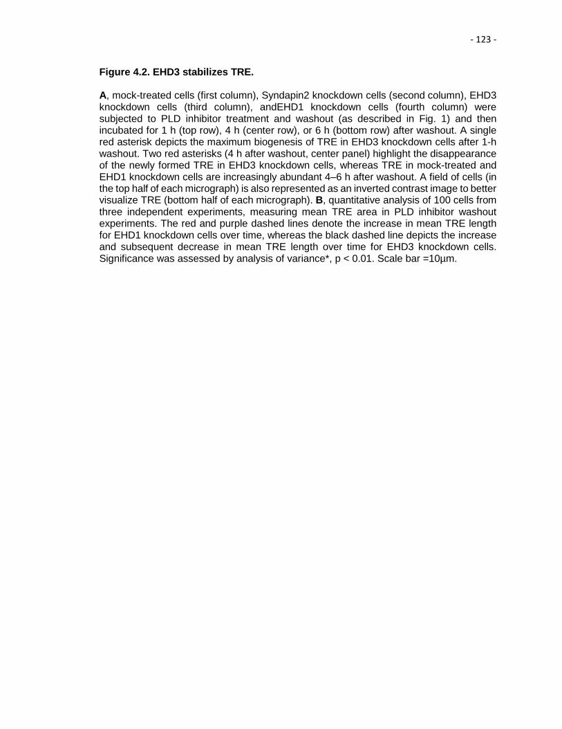
- 123 -
Figure 4.2. EHD3 stabilizes TRE. A, mock-treated cells (first column), Syndapin2 knockdown cells (second column), EHD3 knockdown cells (third column), andEHD1 knockdown cells (fourth column) were subjected to PLD inhibitor treatment and washout (as described in Fig. 1) and then incubated for 1 h (top row), 4 h (center row), or 6 h (bottom row) after washout. A single red asterisk depicts the maximum biogenesis of TRE in EHD3 knockdown cells after 1-h washout. Two red asterisks (4 h after washout, center panel) highlight the disappearance of the newly formed TRE in EHD3 knockdown cells, whereas TRE in mock-treated and EHD1 knockdown cells are increasingly abundant 4–6 h after washout. A field of cells (in the top half of each micrograph) is also represented as an inverted contrast image to better visualize TRE (bottom half of each micrograph). B, quantitative analysis of 100 cells from three independent experiments, measuring mean TRE area in PLD inhibitor washout experiments. The red and purple dashed lines denote the increase in mean TRE length for EHD1 knockdown cells over time, whereas the black dashed line depicts the increase and subsequent decrease in mean TRE length over time for EHD3 knockdown cells. Significance was assessed by analysis of variance*, p < 0.01. Scale bar =10µm.

- 124 -
Figure 4.2
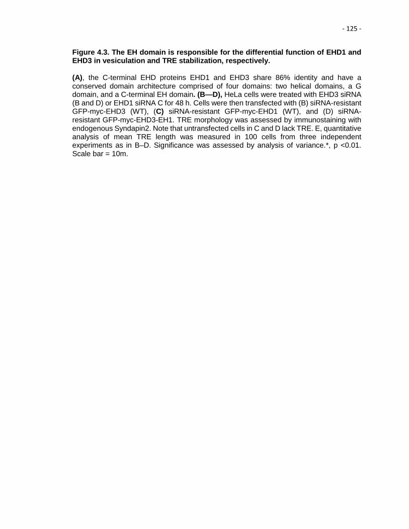
- 125 -
Figure 4.3. The EH domain is responsible for the differential function of EHD1 and EHD3 in vesiculation and TRE stabilization, respectively. (A), the C-terminal EHD proteins EHD1 and EHD3 share 86% identity and have a conserved domain architecture comprised of four domains: two helical domains, a G domain, and a C-terminal EH domain. (B—D), HeLa cells were treated with EHD3 siRNA (B and D) or EHD1 siRNA C for 48 h. Cells were then transfected with (B) siRNA-resistant GFP-myc-EHD3 (WT), (C) siRNA-resistant GFP-myc-EHD1 (WT), and (D) siRNA-resistant GFP-myc-EHD3-EH1. TRE morphology was assessed by immunostaining with endogenous Syndapin2. Note that untransfected cells in C and D lack TRE. E, quantitative analysis of mean TRE length was measured in 100 cells from three independent experiments as in B–D. Significance was assessed by analysis of variance.*, p <0.01. Scale bar = 10m.
- 126 -
Figure 4.3

- 127 -
Figure 4.4. Comparison of the binding affinity of the EHD1 EH domain and EHD3 EH domain for a MICAL-L1 NPF peptide by isothermal titration calorimetry. Solutions containing 90 µM purified EH domains of EHD1 or EHD3 were injected with 930 µM MICAL-L1 peptide, and ITC binding isotherms were collected. KD values obtained by fitting the titration curves are indicated for each EH domain.

- 128 -
Figure 4.4
- 129 -
Figure 4.5. Identification of EH1 and EH3 residues responsible for their differential interactions with NPF-containing partners. A, comparison of EH1 and EH3, exhibiting homologous (green) and non-homologous (white) residues. Six key regions containing non-conserved residues chosen for analysis are depicted. B, schematic showing the interaction of EHD1 with both Rabankyrin-5 and MICAL-L1, whereas EHD3 interacts only with MICAL-L1. C, the S.cerevisiae yeast strain AH109 was co-transformed with the following Gal4bd fusion constructs: Gal4bd-p53 (control), MICAL-L1 and Rabankyrin-5 along with Gal4ad fusion constructs Gal4ad-SV40 (control), Gal4ad-EHD1 (WT), Gal4ad-EHD3 (WT), Gal4ad-EHD3 (NE519AD), Gal4ad-EHD3 (AHLL523PHLV), Gal4ad-EHD3(D459N), Gal4adEHD3(M447T), Gal4ad-EHD3(A437V), and Gal4ad-EHD3(KVAE523RHE). D, HeLa cells were transfected with HA-Rabankyrin-5 (R-5) and lysed after 24 h. The GST-EH domains indicated were used to pull down Rabankyrin-5 from HeLa cell lysates. The protein pulled down was detected by immunoblotting with anti-HA, and anti-GST was used as a control for equal loading of proteins. The immunoblot shown is a representative of four individual experiments. E, the S.cerevisiae yeast strain AH109 was co-transformed with the following Gal4bd fusion constructs: Gal4bd-p53 (control), MICAL-L1, and Rank-5 along with the Gal4ad fusion constructs Gal4ad-SV40 (control), Gal4ad-EHD1 (WT), Gal4ad-EHD3 (WT), Gal4ad-EHD3 (NE519AD), Gal4ad-EHD1(AD519NE), Gal4ad-EHD3(N519A), and Gal4ad-EHD3(E519D). Co-transformants from C and E were plated on non-selective (+HIS) and selective (-HIS) agar plates. Dotted white lines indicate where two different scanned agar plates have been compiled into the same image. The dashed red rectangle in C shows the gain of binding by the EHD3 NE519AD mutant. A representative from four experiments is depicted.

- 130 -
Figure 4.5

- 131 -
Figure 4.6. Wild-type EHD3, but not the EHD3NE519AD mutant, rescues the impaired transferrin trafficking phenotype observed in EHD3 knockdown cells. HeLa cells were either mock-treated (A–G) or treated with EHD3 siRNA oligonucleotides (H–M) for 48 h. After the first 24 h, cells were transfected with either WT GFP-EHD3 (B–D and H–J) or GFP-EHD3 NE519AD (E–G and K–M) and then subjected to a 20-min uptake with Tf-568 prior to fixation and microscopic analysis. Mock-treated cells typically display accumulation of Tf-568 in the perinuclear ERC (A, C, and F). Upon EHD3 knockdown, Tf-568 failed to reach the ERC (I, non-labeled cells). I, wild-typeGFP-EHD3-transfectedcells are marked with a yellow border, and arrows highlight the rescued Tf-568 trafficking and arrival at the ERC.L, yellow borders indicate EHD3 KD cells transfected withGFP-EHD3NE519AD. Arrows are not shown because Tf-568 does not reach the ERC. The graph (top right) displays quantification of the percentage of cells in which Tf reaches the ERC (within 20 min), comparing wild-type and EHD3 NE519 AD-transfected cells. At least 50 cells from each treatment were scored from three individual experiments. Confirmation was done by “blind scoring.” Significance was assessed by analysis of variance. *, p<0.01.

- 132 -
Figure 4.6
- 133 -
Figure 4.7. The number of acidic residues after the NPF motif does not discriminate between binding to EHD1 or EHD3. A, comparison of the number of acidic residues (blue letters) following the NPF motif (red letters) of MICAL-L1 (NPFDEEEEEE) and Rabankyrin-5 (NPFED). B, the S. cerevisiae yeast strain AH109 was co-transformed with the following Gal4bd fusion constructs: Gal4bd-p53 (control), Gal4bd-Rabankyrin-5 (WT) (NPFEDV), Gal4bd-Rabankyrin-5(NPFEDV to NPFEDE), Gal4bd-Rabankyrin-5 (NPFEDV to NPFEDEE), and Rabankyrin-5 (NPFEDV to NPFEDEEE) along with the Gal4ad fusion constructs Gal4ad-SV40 (control), Gal4ad-EHD1 (WT), and Gal4ad-EHD3 (WT). Co-transformants were plated on non-selective (+ HIS) and selective (- HIS) agar plates. C, the S. cerevisiae yeast strain AH109 was co-transformed with the following Gal4bd fusion constructs: Gal4bd-p53 (control), Gal4bd-MICAL-L1 (WT) (NPFDEEEEE), Gal4bd-MICAL-L1 (NPFDEEEEE to NPFDEEEEA), Gal4bd-MICAL-L1(NPFDEEEEE to NPFDEEEAA), Gal4bd- MICAL-L1(NPFDEEEEE to NPFDEEAAA), Gal4bd-MICAL-L1(NPFDEEEEE to NPFDEAAAA), Gal4bd-MICAL-L1(NPFDEEEEE to NPFDAAAAA), and Gal4bd- MICAL-L1(NPFDEEEEE to NPFAAAAAA) along with the Gal4ad fusion constructs Gal4ad-SV40 (control), Gal4ad-EHD1 (WT) and Gal4ad-EHD3 (WT). A representative from four experiments is depicted.

- 134 -
Figure 4.7

- 135 -
Figure 4.8. Residue differences upstream of the NPF motif govern binding difference between EHD1 and EHD3. A, comparison of the residues upstream of the NPF motif (green) between MICAL-L1 (KPY) and Rabankyrin-5 (QSV). B, the S. cerevisiae yeast strain AH109 was co-transformed with the following Gal4bd fusion constructs: Gal4bd-p53 (control) and Gal4bd-EHD1 (WT), Gal4bd-EHD3 (WT) along with the Gal4ad fusion constructs Gal4ad- SV40 (control), Gal4ad-MICAL-L1 (WT), Gal4ad-Rabankyrin-5 (WT), and Gal4ad-Rabankyrin-5 (QSV to KPY). C, the S. cerevisiae yeast strain AH109 was co-transformed with the following Gal4bd fusion constructs: Gal4bd-p53 (control), Gal4bd-EHD1 (WT), or Gal4bd-EHD3 (WT) along with the Gal4ad fusion constructs Gal4ad-SV40 (control), Gal4ad-MICAL-L1 (WT) and Gal4ad- Rabankyrin-5 (WT), Gal4ad-Rabankyrin-5 (QSV to KPY), Gal4ad-Rabankyrin-5 (QSV to KSV), Gal4ad-Rabankyrin-5 (QSV to QPV), and Gal4ad-Rabankyrin-5 (QSV to QSY). Co-transformants were plated on non-selective (+ HIS) and selective (- HIS) agar plates. A representative from four experiments is depicted

- 136 -
Figure 4.8

- 137 -
Figure 4.9. Atomic basis for the differential interaction of EHD1 and EHD3 with NPF-containing binding partners. A, the S. cerevisiae yeast AH109 strain was co-transformed with the following Gal4bd fusion constructs: Gal4bd-p53 (control), Gal4bd-EHD1 (WT), and Gal4bd-EHD3 (WT) along with the Gal4ad fusion constructs Gal4ad-SV40 (control), Gal4ad-MICAL-L1 (WT) and Rabankyrin-5(WT), and Gal4ad-Rabankyrin-5 (QSV to QSV). Co-transformants were plated on non-selective (+ HIS) and selective (- HIS) agar plates. A representative from four experiments is depicted. B, model of the EHD1 EH domain with the MICAL-L1 KPYNPFEEEEED peptide, based on the NMR solution structure. Red depicts oxygen atoms in the Asp-520 side chain.

- 138 -
Figure 4.9

- 139 -
Chapter V
Summary and Future Directions
- 140 -
17 Summary
Endocytic trafficking, which involves the internalization, sorting, degradation and
recycling of macromolecules, is an integral process for maintaining cellular homeostasis
and the regulation of diverse cellular processes such as signaling, migration, and cell
division. The C-terminal Eps 15 Homology Domain proteins (EHD1-4) play an important
role in regulating distinct steps of endocytic trafficking. EHD1, EHD3 and EHD4 are
localized to intracellular tubular/vesicular membranes (Naslavsky and Caplan, 2011). In
contrast, EHD2 localizes to the cytoplasmic interface of the plasma membrane,
specifically the plasma membrane invaginations, caveolae. While EHD1, EHD3 and
EHD4 are capable of forming hetero-oligomers, EHD2 exclusively forms homo-oligomers
(Simone et al., 2013). Indeed, EHD2 is the most distinct of the four EHDs.
The crystal structure of EHD2 has been solved and it contains a partially
conserved unstructured loop consisting of two proline-phenylalanine (PF)
motifs: KPFRKLNPF (Daumke et al., 2007). Despite EHD2 having nearly 70% amino
acid identity with its paralogs, EHD1, EHD3 and EHD4 (Naslavsky and Caplan, 2011),
the latter proteins contain only a single KPF or RPF motif, but no NPF motif in their
unstructured loop. We wanted to delineate the precise role of the KPFRKLNPF
unstructured loop, in dimerization, protein binding, and subcellular localization.
Overall, in this study, we have demonstrated that the phenylalanine residue of
EHD2 NPF is crucial for the localization of EHD2 to the plasma membrane and the
cavolae. On the other hand, the proline residue is essential for the EHD2 dimerization
and binding to NPF-containing protein partners. Furthermore, these studies support the
recently proposed model, in which the EHD2 N-terminal region may regulate the
availability of the unstructured loop for interactions with neighboring EHD2 dimers, thus
promoting oligomerization. Interestingly, EHD1 has a single RPF motif that aligns with
the KPF motif of EHD2. This led to the hypothesis that the RPF motif might be crucial for

- 141 -
binding and localization functions of EHD1. Indeed, EHD1’s single RPF motif has a
critical role in dimerization, binding to MICAL-L1 and Syndapin2, and localization to the
tubular recycling endosomes (TRE). Moreover, recycling assays demonstrated that
EHD1 RPF-to-APA was incapable of supporting normal receptor recycling.
The elaborate network of dynamic lipid membranes known as TRE orchestrates
the process of endocytic recycling in mammalian cells (Jovic et al., 2009). Electron
microscopy studies have determined that the tubular structures containing EHD1 are
200 nm wide and up to 10 μm long (Caplan et al, 2002). The EHD proteins have been
implicated in the bending and fission of TREs, thus regulating endocytic recycling (Cai et
al., 2013). EHD proteins have a C-terminal EH domain that preferentially interacts with
proteins containing an asparagine-proline-phenylalanine (NPF) motif followed by acidic
residues (Kieken et al., 2010; Henry et al., 2010). We had previously demonstrated that
NPF-containing EHD1 interaction partners, such as molecules interacting with CasL-
Like1 (MICAL-L1) and Syndapin2, are essential for TRE biogenesis (Sharma et al.,
2009; Giridharan et al., 2013). Also crucial for TRE biogenesis is the generation of
phosphatidic acid (PA). PA is an essential lipid component of TRE that serves as a
docking point for MICAL-L1 and Syndapin2 (Giridharan et al., 2013). EHD1 and EHD3
have 86% amino acid identity; they homo- and hetero-dimerize, and partially co-localize
to TRE (Galperin et al., 2002). Despite their remarkable identity, they have distinct
mechanistic functions: EHD1 induces membrane vesiculation, whereas EHD3 supports
TRE biogenesis and/or stabilization by an unknown mechanism. While using
phospholipase D inhibitors (which block the conversion of glycerophospholipids to PA) to
deplete cellular TRE, we observed that upon inhibitor washout there was a rapid and
dramatic regeneration of MICAL-L1-marked TRE. Using this “de-novo synchronized”
TRE biogenesis system, we determined that EHD3 is involved in the stabilization of TRE
rather than their biogenesis. Moreover, we identified the residues Ala-519/Asp-520 of

- 142 -
EHD1 and Asp-519/Glu-520 of EHD3, governing the selectivity of these two paralogs for
NPF-containing binding partners, and we presented a model to explain the atomic
mechanism and provide new insight into their differential roles in vesiculation and
tubulation, respectively.
In addition, our study has broader scientific relevance. It serves as a general
model to resolve how highly similar proteins, which are likely to have evolved through
gene duplication, have diverged within mammalian cells to carry out distinct, but related
functions.
18 Future Directions
18.1 Role of NPF motif of KPFRKLNPF loop in caveolar mobility
Recent studies have demonstrated that EHD2 forms dimers with the previously
proposed G-domain interface (Daumke et al., 2007) and the conserved KPFRKLNPF
loop in the G domain plays a role in oligomerization. Furthermore, EHD2 is assembled
into oligomeric ring-like structures (Melo et al., 2017). These structures create a scaffold
that generates and/or stabilizes membrane curvature. Another study demonstrated the
contribution of ATP in enhancing the oligomerization of EHD2 (Hoerneke et al., 2017).
Recent evidence also supports a role of EHD2 in controlling the movement of small
vesicles under the plasma membrane that are enriched in sphingolipids and cholesterol
and known as caveolae (Stoeber et al., 2012; Moren et al., 2012). Moreover, regulating
the formation of functional EHD2 oligomers at the membrane plays a key role in
maintaining caveolae in a static and immobile state (Melo et al., 2017). We have recently
demonstrated that EHD2 NPF to NAF mutants display impaired dimerization of EHD2
and binding with interaction partners. The effect of this mutant on caveolar mobility
remains unknown. We will test whether the EHD2 NAF is required to regulate caveolar

- 143 -
mobility or more precisely, their stabilization. Our readout will use transfected GFP-
caveolin1 to measure the mobility of caveolae by fluorescence recovery after
photobleaching (FRAP), as we have previously done (Pelkmans et al., 2005; Sharma et
al., 2009).
18.2 How does EHD2 link caveolae to the actin cytoskeleton?
Our previous studies have suggested that the binding of EHD2 to PIP2 connects
caveolae to the actin microfilaments (Simone et al., 2013). Additionally, EHD2 interacting
proteins may contribute to the tethering of caveolae to actin. Indeed, we show that
caveolin1 is dispersed across the plasma membrane in EHBP1-depleted cells,
suggesting that EHBP1 may co-operate with EHD2 in linking caveolae to actin
microfilaments (Simone et al., 2013). However, the dimerization of EHD2 is not required
for binding to EHBP1. The multifaceted relationship between the unstructured loop,
localization of caveolae and the binding partners EHBP1 and Syndapin2 remains
unclear. We will focus on understanding the connection between EHD2 oligomerization
and actin cytoskeleton.
18.3 Spatio-temporal regulation of EHD1 and EHD3
EHD1 and EHD3 are capable of hetero-dimerization; it is possible that EHD1 is
on TRE, but initially inactive until it receives a signal to induce vesiculation. This is a
competing idea to the one proposing temporal binding, and if necessary, we will address
potential post-translational modifications. The role of SUMOylation of EHD3 on TRE has
already been addressed (Cabasso et al., 2015). We will address the effect of
deSUMOylation of EHD3 and phosphorylation of EHD1. There is also a possibility that
since EHD1 binds to a wider range of NPF-containing proteins than EHD3, including
Rabankyrin-5 (Zhang et al., 2012), and that such proteins might ‘sequester’ EHD1 in vivo
- 144 -
(away from TRE) and prevent it from competing with EHD3 for TRE binding. To this aim,
we will knock-down Rabankyrin-5 and determine whether the TRE become shorter,
fewer, and/or less elaborate.
18.4 Identification of Residues in EH1 that govern binding of EHD1 with
Rabankyrin-5
We have previously demonstrated that upon mutating EHD1, from AD to NE (at
residues 519/520), did not discern a loss of EHD1 binding to Rabankyrin-5, as we
initially anticipated. It is clear that other residues that are distinct from EHD3 within the
EHD1 EH domain must play a role in maintaining EH1 binding pocket affinity for the
Rabankyrin-5 NPF peptide. In order to evaluate the role of other residues in the EH1
binding pocket, we will generate double mutants of the six cassettes of the residues that
are distinct between EH1 and EH3. Our readout will be the loss-of-binding of EHD1 with
Rabankyrin-5.
18.5 Structure of EH-3
In order to gain insight into the structure-function relationship of EH3, we plan to
do structural studies of EH3 with MICAL-L1 NPF peptide, which we used previously. In
that regard, the solution structure of EH domain of EHD1 has already been solved
(Kieken et al., 2010). The chemical shift resonances for the EH3 have been assigned
(Spagnol et al., 2014). Solution of the structures of EH3 domains will be extremely useful
in analyzing the mechanisms responsible for the functional role played by EHD3 and its
localization.
- 145 -
References
• Aderem, A., & Underhill, D. M. (1999). Mechanisms of phagocytosis in
macrophages. Annual Review of Immunology, 17, 593-623.
doi:10.1146/annurev.immunol.17.1.593 [doi]
• Allantaz, F., Chaussabel, D., Stichweh, D., Bennett, L., Allman, W., Mejias, A.,
Pascual, V. (2007). Blood leukocyte microarrays to diagnose systemic onset
juvenile idiopathic arthritis and follow the response to IL-1 blockade. The Journal
of Experimental Medicine, 204(9), 2131-2144. doi:jem.20070070 [pii]
• Anderson, R. G. (1998). The caveolae membrane system. Annual Review of
Biochemistry, 67, 199-225. doi:10.1146/annurev.biochem.67.1.199 [doi]
• Babst, M., Katzmann, D. J., Estepa-Sabal, E. J., Meerloo, T., & Emr, S. D.
(2002)(a). Escrt-III: An endosome-associated heterooligomeric protein complex
required for mvb sorting. Developmental Cell, 3(2), 271-282. doi:S1534-
5807(02)00220-4 [pii]
• Babst, M., Katzmann, D. J., Snyder, W. B., Wendland, B., & Emr, S. D. (2002)(b).
Endosome-associated complex, ESCRT-II, recruits transport machinery for
protein sorting at the multivesicular body. Developmental Cell, 3(2), 283-289.
doi:S1534-5807(02)00219-8 [pii]
• Bahl, K., Naslavsky, N., & Caplan, S. (2015). Role of the EHD2 unstructured loop
in dimerization, protein binding and subcellular localization. PloS One, 10(4),
e0123710. doi:10.1371/journal.pone.0123710 [doi]
• Bahl, K., Xie, S., Spagnol, G., Sorgen, P., Naslavsky, N., & Caplan, S. (2016).
EHD3 protein is required for tubular recycling endosome stabilization, and an
asparagine-glutamic acid residue pair within its Eps15 homology (EH) domain

- 146 -
dictates its selective binding to NPF peptides. The Journal of Biological
Chemistry, 291(26), 13465-13478. doi:10.1074/jbc.M116.716407 [doi]
• Barriere, H., Nemes, C., Lechardeur, D., Khan-Mohammad, M., Fruh, K., &
Lukacs, G. L. (2006). Molecular basis of oligoubiquitin-dependent internalization
of membrane proteins in mammalian cells. Traffic (Copenhagen, Denmark), 7(3),
282-297. doi:TRA384 [pii]
• Bastiani, M., & Parton, R. G. (2010). Caveolae at a glance. Journal of Cell
Science, 123(Pt 22), 3831-3836. doi:10.1242/jcs.070102 [doi]
• Behnia, R., & Munro, S. (2005). Organelle identity and the signposts for
membrane traffic. Nature, 438(7068), 597-604. doi:nature04397 [pii]
• Bennett, M. K. (1995). SNAREs and the specificity of transport vesicle
targeting. Current Opinion in Cell Biology, 7(4), 581-586. doi:0955-
0674(95)80016-6 [pii]
• Blume, J. J., Halbach, A., Behrendt, D., Paulsson, M., & Plomann, M. (2007).
EHD proteins are associated with tubular and vesicular compartments and
interact with specific phospholipids. Experimental Cell Research, 313(2), 219-
231. doi:S0014-4827(06)00423-X [pii]
• Bock, J. B., Matern, H. T., Peden, A. A., & Scheller, R. H. (2001). A genomic
perspective on membrane compartment organization. Nature, 409(6822), 839-
841. doi:10.1038/35057024 [doi]
• Bonifacino, J. S., & Glick, B. S. (2004). The mechanisms of vesicle budding and
fusion. Cell, 116(2), 153-166. doi:S0092867403010791 [pii]
• Bonifacino, J. S., & Hurley, J. H. (2008). Retromer. Current Opinion in Cell
Biology, 20(4), 427-436. doi:10.1016/j.ceb.2008.03.009 [doi]
- 147 -
• Bonifacino, J. S., & Rojas, R. (2006). Retrograde transport from endosomes to
the trans-golgi network. Nature Reviews.Molecular Cell Biology, 7(8), 568-579.
doi:nrm1985 [pii]
• Bonifacino, J. S., & Traub, L. M. (2003). Signals for sorting of transmembrane
proteins to endosomes and lysosomes. Annual Review of Biochemistry, 72, 395-
447. doi:10.1146/annurev.biochem.72.121801.161800 [doi]
• Boucrot, E., & McMahon, H. T. (2011). Nucleation of clathrin-coated pits at work.
Medecine Sciences : M/S, 27(2), 122-125. doi:10.1051/medsci/2011272122 [doi]
• Braell, W. A., Schlossman, D. M., Schmid, S. L., & Rothman, J. E. (1984).
Dissociation of clathrin coats coupled to the hydrolysis of ATP: Role of an
uncoating ATPase. The Journal of Cell Biology, 99(2), 734-741.
• Brandhorst, D., Zwilling, D., Rizzoli, S. O., Lippert, U., Lang, T., & Jahn, R.
(2006). Homotypic fusion of early endosomes: SNAREs do not determine fusion
specificity. Proceedings of the National Academy of Sciences of the United
States of America, 103(8), 2701-2706. doi:0511138103 [pii]
• Braun, A., Pinyol, R., Dahlhaus, R., Koch, D., Fonarev, P., Grant, B. D., . . .
Qualmann, B. (2005). EHD proteins associate with syndapin I and II and such
interactions play a crucial role in endosomal recycling. Molecular Biology of the
Cell, 16(8), 3642-3658. doi:E05-01-0076 [pii]
• Brown, F. D., Rozelle, A. L., Yin, H. L., Balla, T., & Donaldson, J. G. (2001).
Phosphatidylinositol 4,5-bisphosphate and Arf6-regulated membrane traffic. The
Journal of Cell Biology, 154(5), 1007-1017. doi:10.1083/jcb.200103107 [doi]
• Buggia-Prevot, V., Fernandez, C. G., Udayar, V., Vetrivel, K. S., Elie, A.,
Roseman, J., . . . Thinakaran, G. (2013). A function for EHD family proteins in
unidirectional retrograde dendritic transport of BACE1 and alzheimer's disease

- 148 -
abeta production. Cell Reports, 5(6), 1552-1563.
doi:10.1016/j.celrep.2013.12.006 [doi]
• Cabasso, O., Pekar, O., & Horowitz, M. (2015). SUMOylation of EHD3 modulates
tubulation of the endocytic recycling compartment. PloS One, 10(7), e0134053.
doi:10.1371/journal.pone.0134053 [doi]
• Cai, B., Caplan, S., & Naslavsky, N. (2012). cPLA2alpha and EHD1 interact and
regulate the vesiculation of cholesterol-rich, GPI-anchored, protein-containing
endosomes. Molecular Biology of the Cell, 23(10), 1874-1888.
doi:10.1091/mbc.E11-10-0881 [doi]
• Cai, B., Giridharan, S. S., Zhang, J., Saxena, S., Bahl, K., Schmidt, J. A., . . .
Caplan, S. (2013). Differential roles of C-terminal Eps15 homology domain
proteins as vesiculators and tubulators of recycling endosomes. The Journal of
Biological Chemistry, 288(42), 30172-30180. doi:10.1074/jbc.M113.488627 [doi]
• Cai, B., Xie, S., Caplan, S., & Naslavsky, N. (2014). GRAF1 forms a complex
with MICAL-L1 and EHD1 to cooperate in tubular recycling endosome
vesiculation. Frontiers in Cell and Developmental Biology, 2, 22.
doi:10.3389/fcell.2014.00022 [doi]
• Cai, B., Xie, S., Liu, F., Simone, L. C., Caplan, S., Qin, X., & Naslavsky, N.
(2014). Rapid degradation of the complement regulator, CD59, by a novel
inhibitor. The Journal of Biological Chemistry, 289(17), 12109-12125.
doi:10.1074/jbc.M113.547083 [doi]
• Cai, H., Reinisch, K., & Ferro-Novick, S. (2007). Coats, tethers, rabs, and
SNAREs work together to mediate the intracellular destination of a transport
vesicle. Developmental Cell, 12(5), 671-682. doi:S1534-5807(07)00152-9 [pii]
- 149 -
• Campelo, F., Fabrikant, G., McMahon, H. T., & Kozlov, M. M. (2010). Modeling
membrane shaping by proteins: Focus on EHD2 and N-BAR domains. FEBS
Letters, 584(9), 1830-1839. doi:10.1016/j.febslet.2009.10.023 [doi]
• Cantalupo, G., Alifano, P., Roberti, V., Bruni, C. B., & Bucci, C. (2001). Rab-
interacting lysosomal protein (RILP): The Rab7 effector required for transport to
lysosomes. The EMBO Journal, 20(4), 683-693. doi:10.1093/emboj/20.4.683
[doi]
• Caplan, S., Naslavsky, N., Hartnell, L. M., Lodge, R., Polishchuk, R. S.,
Donaldson, J. G., & Bonifacino, J. S. (2002). A tubular EHD1-containing
compartment involved in the recycling of major histocompatibility complex class I
molecules to the plasma membrane. The EMBO Journal, 21(11), 2557-2567.
doi:10.1093/emboj/21.11.2557 [doi]
• Chaudhuri, R., Lindwasser, O. W., Smith, W. J., Hurley, J. H., & Bonifacino, J. S.
(2007). Downregulation of CD4 by human immunodeficiency virus type 1 nef is
dependent on clathrin and involves direct interaction of nef with the AP2 clathrin
adaptor. Journal of Virology, 81(8), 3877-3890. doi:JVI.02725-06 [pii]
• Chen, Y. A., & Scheller, R. H. (2001). SNARE-mediated membrane
fusion. Nature Reviews.Molecular Cell Biology, 2(2), 98-106.
doi:10.1038/35052017 [doi]
• Cheng, Z. J., Singh, R. D., Marks, D. L., & Pagano, R. E. (2006). Membrane
microdomains, caveolae, and caveolar endocytosis of sphingolipids. Molecular
Membrane Biology, 23(1), 101-110. doi:K7006VL47M683150 [pii]
• Christoforidis, S., Miaczynska, M., Ashman, K., Wilm, M., Zhao, L., Yip, S. C., . . .
Zerial, M. (1999). Phosphatidylinositol-3-OH kinases are Rab5 effectors. Nature
Cell Biology, 1(4), 249-252. doi:10.1038/12075 [doi]

- 150 -
• Chukkapalli, S., Amessou, M., Dekhil, H., Dilly, A. K., Liu, Q., Bandyopadhyay,
S., . . . Kandouz, M. (2014). Ehd3, a regulator of vesicular trafficking, is silenced
in gliomas and functions as a tumor suppressor by controlling cell cycle arrest
and apoptosis. Carcinogenesis, 35(4), 877-885. doi:10.1093/carcin/bgt399 [doi]
• Colicelli, J. (2004). Human RAS superfamily proteins and related
GTPases. Science's STKE : Signal Transduction Knowledge
Environment, 2004(250), RE13. doi:10.1126/stke.2502004re13 [doi]
• Conner, S. D., & Schmid, S. L. (2003). Regulated portals of entry into the
cell. Nature, 422(6927), 37-44. doi:10.1038/nature01451 [doi]
• Dai, J., Li, J., Bos, E., Porcionatto, M., Premont, R. T., Bourgoin, S., . . . Hsu, V.
W. (2004). ACAP1 promotes endocytic recycling by recognizing recycling sorting
signals. Developmental Cell, 7(5), 771-776. doi:S1534-5807(04)00362-4 [pii]
• Daro, E., van der Sluijs, P., Galli, T., & Mellman, I. (1996). Rab4 and cellubrevin
define different early endosome populations on the pathway of transferrin
receptor recycling. Proceedings of the National Academy of Sciences of the
United States of America, 93(18), 9559-9564.
• Daumke, O., Lundmark, R., Vallis, Y., Martens, S., Butler, P. J., & McMahon, H.
T. (2007). Architectural and mechanistic insights into an EHD ATPase involved in
membrane remodelling. Nature, 449(7164), 923-927. doi:nature06173 [pii]
• de Beer, T., Carter, R. E., Lobel-Rice, K. E., Sorkin, A., & Overduin, M. (1998).
Structure and asn-pro-phe binding pocket of the Eps15 homology
domain. Science (New York, N.Y.), 281(5381), 1357-1360.
• de Beer, T., Hoofnagle, A. N., Enmon, J. L., Bowers, R. C., Yamabhai, M., Kay,
B. K., & Overduin, M. (2000). Molecular mechanism of NPF recognition by EH
domains. Nature Structural Biology, 7(11), 1018-1022. doi:10.1038/80924 [doi]

- 151 -
• de Renzis, S., Sonnichsen, B., & Zerial, M. (2002). Divalent rab effectors regulate
the sub-compartmental organization and sorting of early endosomes. Nature Cell
Biology, 4(2), 124-133. doi:10.1038/ncb744 [doi]
• Delprato, A., Merithew, E., & Lambright, D. G. (2004). Structure, exchange
determinants, and family-wide rab specificity of the tandem helical bundle and
Vps9 domains of rabex-5. Cell, 118(5), 607-617. doi:10.1016/j.cell.2004.08.009
[doi]
• Doherty, G. J., & McMahon, H. T. (2009). Mechanisms of endocytosis. Annual
Review of Biochemistry, 78, 857-902.
doi:10.1146/annurev.biochem.78.081307.110540 [doi]
• Doherty, G. J., Ahlund, M. K., Howes, M. T., Moren, B., Parton, R. G., McMahon,
H. T., & Lundmark, R. (2011). The endocytic protein GRAF1 is directed to cell-
matrix adhesion sites and regulates cell spreading. Molecular Biology of the
Cell, 22(22), 4380-4389. doi:10.1091/mbc.E10-12-0936 [doi]
• Donaldson, J. G. (2003). Multiple roles for Arf6: Sorting, structuring, and
signaling at the plasma membrane. The Journal of Biological Chemistry, 278(43),
41573-41576. doi:10.1074/jbc.R300026200 [doi]
• Doray, B., Lee, I., Knisely, J., Bu, G., & Kornfeld, S. (2007). The gamma/sigma1
and alpha/sigma2 hemicomplexes of clathrin adaptors AP-1 and AP-2 harbor the
dileucine recognition site. Molecular Biology of the Cell, 18(5), 1887-1896.
doi:E07-01-0012 [pii]
• Dorot, O., Steller, H., Segal, D., & Horowitz, M. (2017). Past1 modulates
drosophila eye development. PloS One, 12(1), e0169639.
doi:10.1371/journal.pone.0169639 [doi]

- 152 -
• Faraudo, J., & Travesset, A. (2007). Phosphatidic acid domains in membranes:
Effect of divalent counterions. Biophysical Journal, 92(8), 2806-2818. doi:S0006-
3495(07)71085-8 [pii]
• Fasshauer, D. (2003). Structural insights into the SNARE
mechanism. Biochimica Et Biophysica Acta, 1641(2-3), 87-97.
doi:S0167488903000909 [pii]
• Fasshauer, D., Sutton, R. B., Brunger, A. T., & Jahn, R. (1998). Conserved
structural features of the synaptic fusion complex: SNARE proteins reclassified
as Q- and R-SNAREs. Proceedings of the National Academy of Sciences of the
United States of America, 95(26), 15781-15786.
• Ferguson, S. M., & De Camilli, P. (2012). Dynamin, a membrane-remodelling
GTPase. Nature Reviews.Molecular Cell Biology, 13(2), 75-88.
doi:10.1038/nrm3266 [doi]
• Fielding, A. B., Schonteich, E., Matheson, J., Wilson, G., Yu, X., Hickson, G. R., .
. . Gould, G. W. (2005). Rab11-FIP3 and FIP4 interact with Arf6 and the exocyst
to control membrane traffic in cytokinesis. The EMBO Journal, 24(19), 3389-
3399. doi:7600803 [pii]
• Fjorback, A. W., Seaman, M., Gustafsen, C., Mehmedbasic, A., Gokool, S., Wu,
C., . . . Andersen, O. M. (2012). Retromer binds the FANSHY sorting motif in
SorLA to regulate amyloid precursor protein sorting and processing. The Journal
of Neuroscience : The Official Journal of the Society for Neuroscience, 32(4),
1467-1480. doi:10.1523/JNEUROSCI.2272-11.2012 [doi]
• Francis, M. K., Holst, M. R., Vidal-Quadras, M., Henriksson, S., Santarella-
Mellwig, R., Sandblad, L., & Lundmark, R. (2015). Endocytic membrane turnover
at the leading edge is driven by a transient interaction between Cdc42 and

- 153 -
GRAF1. Journal of Cell Science, 128(22), 4183-4195. doi:10.1242/jcs.174417
[doi]
• Frost, A., Unger, V. M., & De Camilli, P. (2009). The BAR domain superfamily:
Membrane-molding macromolecules. Cell, 137(2), 191-196.
doi:10.1016/j.cell.2009.04.010 [doi]
• Galperin, E., Benjamin, S., Rapaport, D., Rotem-Yehudar, R., Tolchinsky, S., &
Horowitz, M. (2002). EHD3: A protein that resides in recycling tubular and
vesicular membrane structures and interacts with EHD1. Traffic (Copenhagen,
Denmark), 3(8), 575-589. doi:tra030807 [pii]
• Galperin, E., Benjamin, S., Rapaport, D., Rotem-Yehudar, R., Tolchinsky, S., &
Horowitz, M. (2002). EHD3: A protein that resides in recycling tubular and
vesicular membrane structures and interacts with EHD1. Traffic (Copenhagen,
Denmark), 3(8), 575-589. doi:tra030807 [pii]
• Ganley, I. G., Espinosa, E., & Pfeffer, S. R. (2008). A syntaxin 10-SNARE
complex distinguishes two distinct transport routes from endosomes to the trans-
golgi in human cells. The Journal of Cell Biology, 180(1), 159-172.
doi:10.1083/jcb.200707136 [doi]
• George, M., Ying, G., Rainey, M. A., Solomon, A., Parikh, P. T., Gao, Q., . . .
Band, H. (2007). Shared as well as distinct roles of EHD proteins revealed by
biochemical and functional comparisons in mammalian cells and C.
elegans. BMC Cell Biology, 8, 3. doi:1471-2121-8-3 [pii]
• George, M., Ying, G., Rainey, M. A., Solomon, A., Parikh, P. T., Gao, Q., . . .
Band, H. (2007). Shared as well as distinct roles of EHD proteins revealed by
biochemical and functional comparisons in mammalian cells and C.
elegans. BMC Cell Biology, 8, 3. doi:1471-2121-8-3 [pii]

- 154 -
• Gesbert, F., Sauvonnet, N., & Dautry-Varsat, A. (2004). Clathrin-lndependent
endocytosis and signalling of interleukin 2 receptors IL-2R endocytosis and
signalling. Current Topics in Microbiology and Immunology, 286, 119-148.
• Giridharan, S. S., Cai, B., Naslavsky, N., & Caplan, S. (2012). Trafficking
cascades mediated by Rab35 and its membrane hub effector, MICAL-
L1. Communicative & Integrative Biology, 5(4), 384-387. doi:10.4161/cib.20064
[doi]
• Giridharan, S. S., Cai, B., Vitale, N., Naslavsky, N., & Caplan, S. (2013).
Cooperation of MICAL-L1, syndapin2, and phosphatidic acid in tubular recycling
endosome biogenesis. Molecular Biology of the Cell, 24(11), 1776-90, S1-15.
doi:10.1091/mbc.E13-01-0026 [doi]
• Glebov, O. O., Bright, N. A., & Nichols, B. J. (2006). Flotillin-1 defines a clathrin-
independent endocytic pathway in mammalian cells. Nature Cell Biology, 8(1),
46-54. doi:ncb1342 [pii]
• Gokool, S., Tattersall, D., & Seaman, M. N. (2007). EHD1 interacts with retromer
to stabilize SNX1 tubules and facilitate endosome-to-golgi retrieval. Traffic
(Copenhagen, Denmark), 8(12), 1873-1886. doi:TRA652 [pii]
• Goody, R. S., Rak, A., & Alexandrov, K. (2005). The structural and mechanistic
basis for recycling of rab proteins between membrane compartments. Cellular
and Molecular Life Sciences : CMLS, 62(15), 1657-1670. doi:10.1007/s00018-
005-4486-8 [doi]
• Gorvel, J. P., Chavrier, P., Zerial, M., & Gruenberg, J. (1991). Rab5 controls
early endosome fusion in vitro. Cell, 64(5), 915-925. doi:0092-8674(91)90316-Q
[pii]

- 155 -
• Grant, B. D., & Caplan, S. (2008). Mechanisms of EHD/RME-1 protein function in
endocytic transport. Traffic (Copenhagen, Denmark), 9(12), 2043-2052.
doi:10.1111/j.1600-0854.2008.00834.x [doi]
• Grant, B. D., & Donaldson, J. G. (2009). Pathways and mechanisms of endocytic
recycling. Nature Reviews.Molecular Cell Biology, 10(9), 597-608.
doi:10.1038/nrm2755 [doi]
• Grosshans, B. L., Ortiz, D., & Novick, P. (2006). Rabs and their effectors:
Achieving specificity in membrane traffic. Proceedings of the National Academy
of Sciences of the United States of America, 103(32), 11821-11827.
doi:0601617103 [pii]
• Gruenberg, J., Griffiths, G., & Howell, K. E. (1989). Characterization of the early
endosome and putative endocytic carrier vesicles in vivo and with an assay of
vesicle fusion in vitro. The Journal of Cell Biology, 108(4), 1301-1316.
• Guilherme, A., Soriano, N. A., Bose, S., Holik, J., Bose, A., Pomerleau, D. P., . . .
Czech, M. P. (2004). EHD2 and the novel EH domain binding protein EHBP1
couple endocytosis to the actin cytoskeleton. The Journal of Biological
Chemistry, 279(11), 10593-10605. doi:10.1074/jbc.M307702200 [doi]
• Haglund, K., Di Fiore, P. P., & Dikic, I. (2003). Distinct monoubiquitin signals in
receptor endocytosis. Trends in Biochemical Sciences, 28(11), 598-603.
doi:S0968-0004(03)00229-9 [pii]
• Haglund, K., Di Fiore, P. P., & Dikic, I. (2003). Distinct monoubiquitin signals in
receptor endocytosis. Trends in Biochemical Sciences, 28(11), 598-603.
doi:S0968-0004(03)00229-9 [pii]
• Hales, C. M., Griner, R., Hobdy-Henderson, K. C., Dorn, M. C., Hardy, D.,
Kumar, R., . . . Goldenring, J. R. (2001). Identification and characterization of a

- 156 -
family of Rab11-interacting proteins. The Journal of Biological
Chemistry, 276(42), 39067-39075. doi:10.1074/jbc.M104831200 [doi]
• Hales, C. M., Vaerman, J. P., & Goldenring, J. R. (2002). Rab11 family
interacting protein 2 associates with myosin vb and regulates plasma membrane
recycling. The Journal of Biological Chemistry, 277(52), 50415-50421.
doi:10.1074/jbc.M209270200 [doi]
• Hammer, J. A.,3rd, & Wu, X. S. (2002). Rabs grab motors: Defining the
connections between rab GTPases and motor proteins. Current Opinion in Cell
Biology, 14(1), 69-75. doi:S0955067401002964 [pii]
• Hansen, C. G., Bright, N. A., Howard, G., & Nichols, B. J. (2009). SDPR induces
membrane curvature and functions in the formation of caveolae. Nature Cell
Biology, 11(7), 807-814. doi:10.1038/ncb1887 [doi]
• Hanson, P. I., & Whiteheart, S. W. (2005). AAA+ proteins: Have engine, will
work. Nature Reviews.Molecular Cell Biology, 6(7), 519-529. doi:nrm1684 [pii]
• Hawryluk, M. J., Keyel, P. A., Mishra, S. K., Watkins, S. C., Heuser, J. E., &
Traub, L. M. (2006). Epsin 1 is a polyubiquitin-selective clathrin-associated
sorting protein. Traffic (Copenhagen, Denmark), 7(3), 262-281. doi:TRA383 [pii]
• Henmi, Y., Oe, N., Kono, N., Taguchi, T., Takei, K., & Tanabe, K. (2016).
Phosphatidic acid induces EHD3-containing membrane tubulation and is required
for receptor recycling. Experimental Cell Research, 342(1), 1-10.
doi:10.1016/j.yexcr.2016.02.011 [doi]
• Henry, G. D., Corrigan, D. J., Dineen, J. V., & Baleja, J. D. (2010). Charge effects
in the selection of NPF motifs by the EH domain of EHD1. Biochemistry, 49(16),
3381-3392. doi:10.1021/bi100065r [doi]
- 157 -
• Hill, M. M., Bastiani, M., Luetterforst, R., Kirkham, M., Kirkham, A., Nixon, S. J., .
. . Parton, R. G. (2008). PTRF-cavin, a conserved cytoplasmic protein required
for caveola formation and function. Cell, 132(1), 113-124.
doi:10.1016/j.cell.2007.11.042 [doi]
• Hinshaw, J. E., & Schmid, S. L. (1995). Dynamin self-assembles into rings
suggesting a mechanism for coated vesicle budding. Nature, 374(6518), 190-
192. doi:10.1038/374190a0 [doi]
• Horiuchi, H., Lippe, R., McBride, H. M., Rubino, M., Woodman, P., Stenmark, H.,
. . . Zerial, M. (1997). A novel Rab5 GDP/GTP exchange factor complexed to
rabaptin-5 links nucleotide exchange to effector recruitment and
function. Cell, 90(6), 1149-1159. doi:S0092-8674(00)80380-3 [pii]
• Huang, F., Kirkpatrick, D., Jiang, X., Gygi, S., & Sorkin, A. (2006). Differential
regulation of EGF receptor internalization and degradation by multiubiquitination
within the kinase domain. Molecular Cell, 21(6), 737-748. doi:S1097-
2765(06)00120-1 [pii]
• Huotari, J., & Helenius, A. (2011). Endosome maturation. The EMBO
Journal, 30(17), 3481-3500. doi:10.1038/emboj.2011.286 [doi]
• Hutagalung, A. H., & Novick, P. J. (2011). Role of rab GTPases in membrane
traffic and cell physiology. Physiological Reviews, 91(1), 119-149.
doi:10.1152/physrev.00059.2009 [doi]
• Itzen, A., & Goody, R. S. (2011). GTPases involved in vesicular trafficking:
Structures and mechanisms. Seminars in Cell & Developmental Biology, 22(1),
48-56. doi:10.1016/j.semcdb.2010.10.003 [doi]

- 158 -
• Jackson, C. L., & Casanova, J. E. (2000). Turning on ARF: The Sec7 family of
guanine-nucleotide-exchange factors. Trends in Cell Biology, 10(2), 60-67.
doi:S0962-8924(99)01699-2 [pii]
• Jakobsson, J., Ackermann, F., Andersson, F., Larhammar, D., Low, P., & Brodin,
L. (2011). Regulation of synaptic vesicle budding and dynamin function by an
EHD ATPase. The Journal of Neuroscience : The Official Journal of the Society
for Neuroscience, 31(39), 13972-13980. doi:10.1523/JNEUROSCI.1289-11.2011
[doi]
• Johannes, L., & Popoff, V. (2008). Tracing the retrograde route in protein
trafficking. Cell, 135(7), 1175-1187. doi:10.1016/j.cell.2008.12.009 [doi]
• Johansson, M., Rocha, N., Zwart, W., Jordens, I., Janssen, L., Kuijl, C., . . .
Neefjes, J. (2007). Activation of endosomal dynein motors by stepwise assembly
of Rab7-RILP-p150Glued, ORP1L, and the receptor betalll spectrin. The Journal
of Cell Biology, 176(4), 459-471. doi:jcb.200606077 [pii]
• Jordens, I., Fernandez-Borja, M., Marsman, M., Dusseljee, S., Janssen, L.,
Calafat, J., . . . Neefjes, J. (2001). The Rab7 effector protein RILP controls
lysosomal transport by inducing the recruitment of dynein-dynactin
motors. Current Biology : CB, 11(21), 1680-1685. doi:S0960-9822(01)00531-0
[pii]
• Jovanovic, O. A., Brown, F. D., & Donaldson, J. G. (2006). An effector domain
mutant of Arf6 implicates phospholipase D in endosomal membrane
recycling. Molecular Biology of the Cell, 17(1), 327-335. doi:E05-06-0523 [pii]
• Jovic, M., Kieken, F., Naslavsky, N., Sorgen, P. L., & Caplan, S. (2009). Eps15
homology domain 1-associated tubules contain phosphatidylinositol-4-phosphate
and phosphatidylinositol-(4,5)-bisphosphate and are required for efficient

- 159 -
recycling. Molecular Biology of the Cell, 20(11), 2731-2743.
doi:10.1091/mbc.E08-11-1102 [doi]
• Jovic, M., Naslavsky, N., Rapaport, D., Horowitz, M., & Caplan, S. (2007). EHD1
regulates beta1 integrin endosomal transport: Effects on focal adhesions, cell
spreading and migration. Journal of Cell Science, 120(Pt 5), 802-814.
doi:jcs.03383 [pii]
• Jovic, M., Sharma, M., Rahajeng, J., & Caplan, S. (2010). The early endosome:
A busy sorting station for proteins at the crossroads. Histology and
Histopathology, 25(1), 99-112. doi:10.14670/HH-25.99 [doi]
• Kahn, R. A., Cherfils, J., Elias, M., Lovering, R. C., Munro, S., & Schurmann, A.
(2006). Nomenclature for the human arf family of GTP-binding proteins: ARF,
ARL, and SAR proteins. The Journal of Cell Biology, 172(5), 645-650.
doi:jcb.200512057 [pii]
• Kazazic, M., Bertelsen, V., Pedersen, K. W., Vuong, T. T., Grandal, M. V.,
Rodland, M. S.,Madshus, I. H. (2009). Epsin 1 is involved in recruitment of
ubiquitinated EGF receptors into clathrin-coated pits. Traffic (Copenhagen,
Denmark), 10(2), 235-245. doi:10.1111/j.1600-0854.2008.00858.x [doi]
• Kessels, M. M., Dong, J., Leibig, W., Westermann, P., & Qualmann, B. (2006).
Complexes of syndapin II with dynamin II promote vesicle formation at the trans-
golgi network. Journal of Cell Science, 119(Pt 8), 1504-1516. doi:jcs.02877 [pii]
• Kieken, F., Jovic, M., Tonelli, M., Naslavsky, N., Caplan, S., & Sorgen, P. L.
(2009). Structural insight into the interaction of proteins containing NPF, DPF,
and GPF motifs with the C-terminal EH-domain of EHD1. Protein Science : A
Publication of the Protein Society, 18(12), 2471-2479. doi:10.1002/pro.258 [doi]

- 160 -
• Kieken, F., Sharma, M., Jovic, M., Giridharan, S. S., Naslavsky, N., Caplan, S., &
Sorgen, P. L. (2010). Mechanism for the selective interaction of C-terminal Eps15
homology domain proteins with specific asn-pro-phe-containing partners. The
Journal of Biological Chemistry, 285(12), 8687-8694.
doi:10.1074/jbc.M109.045666 [doi]
• Kinchen, J. M., & Ravichandran, K. S. (2010). Identification of two evolutionarily
conserved genes regulating processing of engulfed apoptotic
cells. Nature, 464(7289), 778-782. doi:10.1038/nature08853 [doi]
• Kirchhausen, T. (1999). Adaptors for clathrin-mediated traffic. Annual Review of
Cell and Developmental Biology, 15, 705-732.
doi:10.1146/annurev.cellbio.15.1.705 [doi]
• Kirchhausen, T. (2000). Clathrin. Annual Review of Biochemistry, 69, 699-727.
doi:69/1/699 [pii]
• Kirkham, M., Fujita, A., Chadda, R., Nixon, S. J., Kurzchalia, T. V., Sharma, D.
K., . . . Parton, R. G. (2005). Ultrastructural identification of uncoated caveolin-
independent early endocytic vehicles. The Journal of Cell Biology, 168(3), 465-
476. doi:jcb.200407078 [pii]
• Kjaerulff, O., Verstreken, P., & Bellen, H. J. (2002). Synaptic vesicle retrieval: Still
time for a kiss. Nature Cell Biology, 4(11), E245-8. doi:10.1038/ncb1102-e245
[doi]
• Koch, D., Westermann, M., Kessels, M. M., & Qualmann, B. (2012).
Ultrastructural freeze-fracture immunolabeling identifies plasma membrane-
localized syndapin II as a crucial factor in shaping caveolae. Histochemistry and
Cell Biology, 138(2), 215-230. doi:10.1007/s00418-012-0945-0 [doi]

- 161 -
• Koles, K., Messelaar, E. M., Feiger, Z., Yu, C. J., Frank, C. A., & Rodal, A. A.
(2015). The EHD protein Past1 controls postsynaptic membrane elaboration and
synaptic function. Molecular Biology of the Cell, 26(18), 3275-3288.
doi:10.1091/mbc.E15-02-0093 [doi]
• Kubo, K., Kobayashi, M., Nozaki, S., Yagi, C., Hatsuzawa, K., Katoh, Y., . . .
Nakayama, K. (2015). SNAP23/25 and VAMP2 mediate exocytic event of
transferrin receptor-containing recycling vesicles. Biology Open, 4(7), 910-920.
doi:10.1242/bio.012146 [doi]
• Kucharczyk, R., Hoffman-Sommer, M., Piekarska, I., von Mollard, G. F., & Rytka,
J. (2009). The saccharomyces cerevisiae protein Ccz1p interacts with
components of the endosomal fusion machinery. FEMS Yeast Research, 9(4),
565-573. doi:10.1111/j.1567-1364.2009.00515.x [doi]
• Kumari, S., & Mayor, S. (2008). ARF1 is directly involved in dynamin-
independent endocytosis. Nature Cell Biology, 10(1), 30-41. doi:ncb1666 [pii]
• Lamaze, C., Dujeancourt, A., Baba, T., Lo, C. G., Benmerah, A., & Dautry-
Varsat, A. (2001). Interleukin 2 receptors and detergent-resistant membrane
domains define a clathrin-independent endocytic pathway. Molecular Cell, 7(3),
661-671. doi:S1097-2765(01)00212-X [pii]
• Lapierre, L. A., Kumar, R., Hales, C. M., Navarre, J., Bhartur, S. G., Burnette, J.
O., . . . Goldenring, J. R. (2001). Myosin vb is associated with plasma membrane
recycling systems. Molecular Biology of the Cell, 12(6), 1843-1857.
• Lauffer, B. E., Melero, C., Temkin, P., Lei, C., Hong, W., Kortemme, T., & von
Zastrow, M. (2010). SNX27 mediates PDZ-directed sorting from endosomes to
the plasma membrane. The Journal of Cell Biology, 190(4), 565-574.
doi:10.1083/jcb.201004060 [doi]

- 162 -
• Lee, A., Frank, D. W., Marks, M. S., & Lemmon, M. A. (1999). Dominant-negative
inhibition of receptor-mediated endocytosis by a dynamin-1 mutant with a
defective pleckstrin homology domain. Current Biology : CB, 9(5), 261-264.
doi:S0960-9822(99)80115-8 [pii]
• Lee, D. W., Zhao, X., Scarselletta, S., Schweinsberg, P. J., Eisenberg, E., Grant,
B. D., & Greene, L. E. (2005). ATP binding regulates oligomerization and
endosome association of RME-1 family proteins. The Journal of Biological
Chemistry, 280(17), 17213-17220. doi:M412751200 [pii]
• Leung, K. F., Baron, R., Ali, B. R., Magee, A. I., & Seabra, M. C. (2007). Rab
GTPases containing a CAAX motif are processed post-geranylgeranylation by
proteolysis and methylation. The Journal of Biological Chemistry, 282(2), 1487-
1497. doi:M605557200 [pii]
• Levkowitz, G., Waterman, H., Zamir, E., Kam, Z., Oved, S., Langdon, W. Y., . . .
Yarden, Y. (1998). c-Cbl/Sli-1 regulates endocytic sorting and ubiquitination of
the epidermal growth factor receptor. Genes & Development, 12(23), 3663-3674.
• Lin, R. C., & Scheller, R. H. (1997). Structural organization of the synaptic
exocytosis core complex. Neuron, 19(5), 1087-1094. doi:S0896-6273(00)80399-
2 [pii]
• Lin, S. X., Grant, B., Hirsh, D., & Maxfield, F. R. (2001). Rme-1 regulates the
distribution and function of the endocytic recycling compartment in mammalian
cells. Nature Cell Biology, 3(6), 567-572. doi:10.1038/35078543 [doi]
• Lindsay, A. J., Hendrick, A. G., Cantalupo, G., Senic-Matuglia, F., Goud, B.,
Bucci, C., & McCaffrey, M. W. (2002). Rab coupling protein (RCP), a novel Rab4
and Rab11 effector protein. The Journal of Biological Chemistry, 277(14), 12190-
12199. doi:10.1074/jbc.M108665200 [doi]

- 163 -
• Lippe, R., Miaczynska, M., Rybin, V., Runge, A., & Zerial, M. (2001). Functional
synergy between Rab5 effector rabaptin-5 and exchange factor rabex-5 when
physically associated in a complex. Molecular Biology of the Cell, 12(7), 2219-
2228.
• Lu, Q., Insinna, C., Ott, C., Stauffer, J., Pintado, P. A., Rahajeng, J., . . .
Westlake, C. J. (2015). Early steps in primary cilium assembly require
EHD1/EHD3-dependent ciliary vesicle formation. Nature Cell Biology, 17(3), 228-
240. doi:10.1038/ncb3109 [doi]
• Lundmark, R., Doherty, G. J., Howes, M. T., Cortese, K., Vallis, Y., Parton, R. G.,
& McMahon, H. T. (2008). The GTPase-activating protein GRAF1 regulates the
CLIC/GEEC endocytic pathway. Current Biology : CB, 18(22), 1802-1808.
doi:10.1016/j.cub.2008.10.044 [doi]
• Mammoto, A., Ohtsuka, T., Hotta, I., Sasaki, T., & Takai, Y. (1999).
Rab11BP/Rabphilin-11, a downstream target of rab11 small G protein implicated
in vesicle recycling. The Journal of Biological Chemistry, 274(36), 25517-25524.
• Marg, A., Schoewel, V., Timmel, T., Schulze, A., Shah, C., Daumke, O., &
Spuler, S. (2012). Sarcolemmal repair is a slow process and includes
EHD2. Traffic (Copenhagen, Denmark), 13(9), 1286-1294. doi:10.1111/j.1600-
0854.2012.01386.x [doi]
• Matanis, T., Akhmanova, A., Wulf, P., Del Nery, E., Weide, T., Stepanova, T., . . .
Hoogenraad, C. C. (2002). Bicaudal-D regulates COPI-independent golgi-ER
transport by recruiting the dynein-dynactin motor complex. Nature Cell
Biology, 4(12), 986-992. doi:10.1038/ncb891 [doi]
- 164 -
• Mattera, R., & Bonifacino, J. S. (2008). Ubiquitin binding and conjugation
regulate the recruitment of rabex-5 to early endosomes. The EMBO
Journal, 27(19), 2484-2494. doi:10.1038/emboj.2008.177 [doi]
• Maurer, M. E., & Cooper, J. A. (2006). The adaptor protein Dab2 sorts LDL
receptors into coated pits independently of AP-2 and ARH. Journal of Cell
Science, 119(Pt 20), 4235-4246. doi:jcs.03217 [pii]
• Maxfield, F. R., & McGraw, T. E. (2004). Endocytic recycling. Nature
Reviews.Molecular Cell Biology, 5(2), 121-132. doi:10.1038/nrm1315 [doi]
• Mayor, S., & Pagano, R. E. (2007). Pathways of clathrin-independent
endocytosis. Nature Reviews.Molecular Cell Biology, 8(8), 603-612. doi:nrm2216
[pii]
• Mayor, S., Parton, R. G., & Donaldson, J. G. (2014). Clathrin-independent
pathways of endocytosis. Cold Spring Harbor Perspectives in Biology, 6(6),
10.1101/cshperspect.a016758. doi:10.1101/cshperspect.a016758 [doi]
• Mayor, S., Presley, J. F., & Maxfield, F. R. (1993). Sorting of membrane
components from endosomes and subsequent recycling to the cell surface
occurs by a bulk flow process. The Journal of Cell Biology, 121(6), 1257-1269.
• McBride, H. M., Rybin, V., Murphy, C., Giner, A., Teasdale, R., & Zerial, M.
(1999). Oligomeric complexes link Rab5 effectors with NSF and drive membrane
fusion via interactions between EEA1 and syntaxin 13. Cell, 98(3), 377-386.
doi:S0092-8674(00)81966-2 [pii]
• McKenzie, J. E., Raisley, B., Zhou, X., Naslavsky, N., Taguchi, T., Caplan, S., &
Sheff, D. (2012). Retromer guides STxB and CD8-M6PR from early to recycling
endosomes, EHD1 guides STxB from recycling endosome to golgi. Traffic

- 165 -
(Copenhagen, Denmark), 13(8), 1140-1159. doi:10.1111/j.1600-
0854.2012.01374.x [doi]
• McMahon, H. T., & Boucrot, E. (2011). Molecular mechanism and physiological
function. Nature Reviews.Molecular Cell Biology, 12(8), 517-533.
doi:10.1038/nrm3151 [doi]
• McMahon, H. T., & Gallop, J. L. (2005). Membrane curvature and mechanisms of
dynamic cell membrane remodelling. Nature, 438(7068), 590-596.
doi:nature04396 [pii]
• McMahon, K. A., Zajicek, H., Li, W. P., Peyton, M. J., Minna, J. D., Hernandez, V.
J., . . . Anderson, R. G. (2009). SRBC/cavin-3 is a caveolin adapter protein that
regulates caveolae function. The EMBO Journal, 28(8), 1001-1015.
doi:10.1038/emboj.2009.46 [doi]
• Mellman, I. (1996). Membranes and sorting. Current Opinion in Cell Biology, 8(4),
497-498. doi:S0955-0674(96)80026-3 [pii]
• Melo, A. A., Hegde, B. G., Shah, C., Larsson, E., Isas, J. M., Kunz, S., . . .
Daumke, O. (2017). Structural insights into the activation mechanism of dynamin-
like EHD ATPases. Proceedings of the National Academy of Sciences of the
United States of America, doi:201614075 [pii]
• Meng, Q., Xing, Y., Ren, T., Lu, H., Xi, Y., Jiang, Z., . . . Cai, L. (2016).
Mammalian Eps15 homology domain 1 promotes metastasis in non-small cell
lung cancer by inducing epithelial-mesenchymal
transition. Oncotarget, doi:10.18632/oncotarget.11220 [doi]
• Miaczynska, M., & Zerial, M. (2002). Mosaic organization of the endocytic
pathway. Experimental Cell Research, 272(1), 8-14. doi:10.1006/excr.2001.5401
[doi]

- 166 -
• Mintz, L., Galperin, E., Pasmanik-Chor, M., Tulzinsky, S., Bromberg, Y., Kozak,
C. A., . . . Horowitz, M. (1999). EHD1--an EH-domain-containing protein with a
specific expression pattern. Genomics, 59(1), 66-76.
doi:10.1006/geno.1999.5800 [doi]
• Moren, B., Shah, C., Howes, M. T., Schieber, N. L., McMahon, H. T., Parton, R.
G., . . . Lundmark, R. (2012). EHD2 regulates caveolar dynamics via ATP-driven
targeting and oligomerization. Molecular Biology of the Cell, 23(7), 1316-1329.
doi:10.1091/mbc.E11-09-0787 [doi]
• Murray, J. T., Panaretou, C., Stenmark, H., Miaczynska, M., & Backer, J. M.
(2002). Role of Rab5 in the recruitment of hVps34/p150 to the early
endosome. Traffic (Copenhagen, Denmark), 3(6), 416-427. doi:tra030605 [pii]
• Naslavsky, N., & Caplan, S. (2005). C-terminal EH-domain-containing proteins:
Consensus for a role in endocytic trafficking, EH? Journal of Cell Science, 118(Pt
18), 4093-4101. doi:118/18/4093 [pii]
• Naslavsky, N., & Caplan, S. (2011). EHD proteins: Key conductors of endocytic
transport. Trends in Cell Biology, 21(2), 122-131. doi:10.1016/j.tcb.2010.10.003
[doi]
• Naslavsky, N., Boehm, M., Backlund, P. S.,Jr, & Caplan, S. (2004). Rabenosyn-5
and EHD1 interact and sequentially regulate protein recycling to the plasma
membrane. Molecular Biology of the Cell, 15(5), 2410-2422.
doi:10.1091/mbc.E03-10-0733 [doi]
• Naslavsky, N., McKenzie, J., Altan-Bonnet, N., Sheff, D., & Caplan, S. (2009).
EHD3 regulates early-endosome-to-golgi transport and preserves golgi
morphology. Journal of Cell Science, 122(Pt 3), 389-400. doi:10.1242/jcs.037051
[doi]

- 167 -
• Naslavsky, N., Rahajeng, J., Chenavas, S., Sorgen, P. L., & Caplan, S. (2007).
EHD1 and Eps15 interact with phosphatidylinositols via their Eps15 homology
domains. The Journal of Biological Chemistry, 282(22), 16612-16622.
doi:M609493200 [pii]
• Naslavsky, N., Rahajeng, J., Sharma, M., Jovic, M., & Caplan, S. (2006).
Interactions between EHD proteins and Rab11-FIP2: A role for EHD3 in early
endosomal transport. Molecular Biology of the Cell, 17(1), 163-177. doi:E05-05-
0466 [pii]
• Naslavsky, N., Weigert, R., & Donaldson, J. G. (2003). Convergence of non-
clathrin- and clathrin-derived endosomes involves Arf6 inactivation and changes
in phosphoinositides. Molecular Biology of the Cell, 14(2), 417-431.
doi:10.1091/mbc.02-04-0053 [doi]
• Naslavsky, N., Weigert, R., & Donaldson, J. G. (2004). Characterization of a
nonclathrin endocytic pathway: Membrane cargo and lipid
requirements. Molecular Biology of the Cell, 15(8), 3542-3552.
doi:10.1091/mbc.E04-02-0151 [doi]
• Nielsen, E., Severin, F., Backer, J. M., Hyman, A. A., & Zerial, M. (1999). Rab5
regulates motility of early endosomes on microtubules. Nature Cell Biology, 1(6),
376-382. doi:10.1038/14075 [doi]
• Numrich, J., & Ungermann, C. (2014). Endocytic rabs in membrane trafficking
and signaling. Biological Chemistry, 395(3), 327-333. doi:10.1515/hsz-2013-0258
[doi]
• Ohno, H., Stewart, J., Fournier, M. C., Bosshart, H., Rhee, I., Miyatake, S., . . .
Bonifacino, J. S. (1995). Interaction of tyrosine-based sorting signals with
clathrin-associated proteins. Science (New York, N.Y.), 269(5232), 1872-1875.

- 168 -
• Olswang-Kutz, Y., Gertel, Y., Benjamin, S., Sela, O., Pekar, O., Arama, E., . . .
Segal, D. (2009). Drosophila Past1 is involved in endocytosis and is required for
germline development and survival of the adult fly. Journal of Cell
Science, 122(Pt 4), 471-480. doi:10.1242/jcs.038521 [doi]
• Otto, G. P., & Nichols, B. J. (2011). The roles of flotillin microdomains--
endocytosis and beyond. Journal of Cell Science, 124(Pt 23), 3933-3940.
doi:10.1242/jcs.092015 [doi]
• Owen, D. J., Collins, B. M., & Evans, P. R. (2004). Adaptors for clathrin coats:
Structure and function. Annual Review of Cell and Developmental Biology, 20,
153-191. doi:10.1146/annurev.cellbio.20.010403.104543 [doi]
• Pal, A., Severin, F., Lommer, B., Shevchenko, A., & Zerial, M. (2006). Huntingtin-
HAP40 complex is a novel Rab5 effector that regulates early endosome motility
and is up-regulated in huntington's disease. The Journal of Cell Biology, 172(4),
605-618. doi:jcb.200509091 [pii]
• Pant, S., Sharma, M., Patel, K., Caplan, S., Carr, C. M., & Grant, B. D. (2009).
AMPH-1/Amphiphysin/Bin1 functions with RME-1/Ehd1 in endocytic
recycling. Nature Cell Biology, 11(12), 1399-1410. doi:10.1038/ncb1986 [doi]
• Paoluzi, S., Castagnoli, L., Lauro, I., Salcini, A. E., Coda, L., Fre', S., . . .
Cesareni, G. (1998). Recognition specificity of individual EH domains of
mammals and yeast. The EMBO Journal, 17(22), 6541-6550.
doi:10.1093/emboj/17.22.6541 [doi]
• Parton, R. G., & Simons, K. (2007). The multiple faces of caveolae. Nature
Reviews.Molecular Cell Biology, 8(3), 185-194. doi:nrm2122 [pii]
• Peter, B. J., Kent, H. M., Mills, I. G., Vallis, Y., Butler, P. J., Evans, P. R., &
McMahon, H. T. (2004). BAR domains as sensors of membrane curvature: The

- 169 -
amphiphysin BAR structure. Science (New York, N.Y.), 303(5657), 495-499.
doi:10.1126/science.1092586 [doi]
• Pitto, M., Brunner, J., Ferraretto, A., Ravasi, D., Palestini, P., & Masserini, M.
(2000). Use of a photoactivable GM1 ganglioside analogue to assess lipid
distribution in caveolae bilayer. Glycoconjugate Journal, 17(3 -4), 215-222.
• Polo, S., Sigismund, S., Faretta, M., Guidi, M., Capua, M. R., Bossi, G., . . . Di
Fiore, P. P. (2002). A single motif responsible for ubiquitin recognition and
monoubiquitination in endocytic proteins. Nature, 416(6879), 451-455.
doi:10.1038/416451a [doi]
• Popoff, V., Mardones, G. A., Bai, S. K., Chambon, V., Tenza, D., Burgos, P. V., .
. . Johannes, L. (2009). Analysis of articulation between clathrin and retromer in
retrograde sorting on early endosomes. Traffic (Copenhagen, Denmark), 10(12),
1868-1880. doi:10.1111/j.1600-0854.2009.00993.x [doi]
• Posey, A. D.,Jr, Pytel, P., Gardikiotes, K., Demonbreun, A. R., Rainey, M.,
George, M., . . . McNally, E. M. (2011). Endocytic recycling proteins EHD1 and
EHD2 interact with fer-1-like-5 (Fer1L5) and mediate myoblast fusion. The
Journal of Biological Chemistry, 286(9), 7379-7388.
doi:10.1074/jbc.M110.157222 [doi]
• Powelka, A. M., Sun, J., Li, J., Gao, M., Shaw, L. M., Sonnenberg, A., & Hsu, V.
W. (2004). Stimulation-dependent recycling of integrin beta1 regulated by ARF6
and Rab11. Traffic (Copenhagen, Denmark), 5(1), 20-36. doi:150 [pii]
• Prasad, K., Barouch, W., Greene, L., & Eisenberg, E. (1993). A protein cofactor
is required for uncoating of clathrin baskets by uncoating ATPase. The Journal of
Biological Chemistry, 268(32), 23758-23761.

- 170 -
• Prekeris, R., Davies, J. M., & Scheller, R. H. (2001). Identification of a novel
Rab11/25 binding domain present in eferin and rip proteins. The Journal of
Biological Chemistry, 276(42), 38966-38970. doi:10.1074/jbc.M106133200 [doi]
• Prekeris, R., Klumperman, J., & Scheller, R. H. (2000). A Rab11/Rip11 protein
complex regulates apical membrane trafficking via recycling
endosomes. Molecular Cell, 6(6), 1437-1448. doi:S1097-2765(00)00140-4 [pii]
• Rahajeng, J., Giridharan, S. S., Cai, B., Naslavsky, N., & Caplan, S. (2010).
Important relationships between rab and MICAL proteins in endocytic
trafficking. World Journal of Biological Chemistry, 1(8), 254-264.
doi:10.4331/wjbc.v1.i8.254 [doi]
• Raiborg, C., & Stenmark, H. (2002). Hrs and endocytic sorting of ubiquitinated
membrane proteins. Cell Structure and Function, 27(6), 403-408.
• Randazzo, P. A., & Hirsch, D. S. (2004). Arf GAPs: Multifunctional proteins that
regulate membrane traffic and actin remodelling. Cellular Signalling, 16(4), 401-
413. doi:S0898656803001918 [pii]
• Rapaport, D., Auerbach, W., Naslavsky, N., Pasmanik-Chor, M., Galperin, E.,
Fein, A., . . . Horowitz, M. (2006). Recycling to the plasma membrane is delayed
in EHD1 knockout mice. Traffic (Copenhagen, Denmark), 7(1), 52-60.
doi:TRA359 [pii]
• Rink, J., Ghigo, E., Kalaidzidis, Y., & Zerial, M. (2005). Rab conversion as a
mechanism of progression from early to late endosomes. Cell, 122(5), 735-749.
doi:S0092-8674(05)00697-5 [pii]
• Robinson, M. S. (2015). Forty years of clathrin-coated vesicles. Traffic
(Copenhagen, Denmark), 16(12), 1210-1238. doi:10.1111/tra.12335 [doi]

- 171 -
• Roland, J. T., Kenworthy, A. K., Peranen, J., Caplan, S., & Goldenring, J. R.
(2007). Myosin vb interacts with Rab8a on a tubular network containing EHD1
and EHD3. Molecular Biology of the Cell, 18(8), 2828-2837. doi:E07-02-0169 [pii]
• Rotem-Yehudar, R., Galperin, E., & Horowitz, M. (2001). Association of insulin-
like growth factor 1 receptor with EHD1 and SNAP29. The Journal of Biological
Chemistry, 276(35), 33054-33060. doi:10.1074/jbc.M009913200 [doi]
• Salcini, A. E., Confalonieri, S., Doria, M., Santolini, E., Tassi, E., Minenkova, O., .
. . Di Fiore, P. P. (1997). Binding specificity and in vivo targets of the EH domain,
a novel protein-protein interaction module. Genes & Development, 11(17), 2239-
2249.
• Sandvig, K., & van Deurs, B. (1994). Endocytosis without clathrin. Trends in Cell
Biology, 4(8), 275-277. doi:0962892494902119 [pii]
• Santy, L. C., & Casanova, J. E. (2001). Activation of ARF6 by ARNO stimulates
epithelial cell migration through downstream activation of both Rac1 and
phospholipase D. The Journal of Cell Biology, 154(3), 599-610.
doi:10.1083/jcb.200104019 [doi]
• Sato, M., Grant, B. D., Harada, A., & Sato, K. (2008). Rab11 is required for
synchronous secretion of chondroitin proteoglycans after fertilization in
caenorhabditis elegans. Journal of Cell Science, 121(Pt 19), 3177-3186.
doi:10.1242/jcs.034678 [doi]
• Sato, M., Sato, K., Liou, W., Pant, S., Harada, A., & Grant, B. D. (2008).
Regulation of endocytic recycling by C. elegans Rab35 and its regulator RME-4,
a coated-pit protein. The EMBO Journal, 27(8), 1183-1196.
doi:10.1038/emboj.2008.54 [doi]

- 172 -
• Schnatwinkel, C., Christoforidis, S., Lindsay, M. R., Uttenweiler-Joseph, S., Wilm,
M., Parton, R. G., & Zerial, M. (2004). The Rab5 effector rabankyrin-5 regulates
and coordinates different endocytic mechanisms. PLoS Biology, 2(9), E261.
doi:10.1371/journal.pbio.0020261 [doi]
• Schonteich, E., Wilson, G. M., Burden, J., Hopkins, C. R., Anderson, K.,
Goldenring, J. R., & Prekeris, R. (2008). The Rip11/Rab11-FIP5 and kinesin II
complex regulates endocytic protein recycling. Journal of Cell Science, 121(Pt
22), 3824-3833. doi:10.1242/jcs.032441 [doi]
• Seaman, M. N. (2007). Identification of a novel conserved sorting motif required
for retromer-mediated endosome-to-TGN retrieval. Journal of Cell
Science, 120(Pt 14), 2378-2389. doi:120/14/2378 [pii]
• Senju, Y., Itoh, Y., Takano, K., Hamada, S., & Suetsugu, S. (2011). Essential role
of PACSIN2/syndapin-II in caveolae membrane sculpting. Journal of Cell
Science, 124(Pt 12), 2032-2040. doi:10.1242/jcs.086264 [doi]
• Shah, C., Hegde, B. G., Moren, B., Behrmann, E., Mielke, T., Moenke, G., . . .
Langen, R. (2014). Structural insights into membrane interaction and caveolar
targeting of dynamin-like EHD2. Structure (London, England : 1993), 22(3), 409-
420. doi:10.1016/j.str.2013.12.015 [doi]
• Shao, Y., Akmentin, W., Toledo-Aral, J. J., Rosenbaum, J., Valdez, G., Cabot, J.
B., . . . Halegoua, S. (2002). Pincher, a pinocytic chaperone for nerve growth
factor/TrkA signaling endosomes. The Journal of Cell Biology, 157(4), 679-691.
doi:10.1083/jcb.200201063 [doi]
• Sharma, M., Giridharan, S. S., Rahajeng, J., Naslavsky, N., & Caplan, S. (2009).
MICAL-L1 links EHD1 to tubular recycling endosomes and regulates receptor

- 173 -
recycling. Molecular Biology of the Cell, 20(24), 5181-5194.
doi:10.1091/mbc.E09-06-0535 [doi]
• Sharma, M., Jovic, M., Kieken, F., Naslavsky, N., Sorgen, P., & Caplan, S.
(2009). A model for the role of EHD1-containing membrane tubules in endocytic
recycling. Communicative & Integrative Biology, 2(5), 431-433.
• Shestakova, A., Hanono, A., Drosner, S., Curtiss, M., Davies, B. A., Katzmann,
D. J., & Babst, M. (2010). Assembly of the AAA ATPase Vps4 on ESCRT-
III. Molecular Biology of the Cell, 21(6), 1059-1071. doi:10.1091/mbc.E09-07-
0572 [doi]
• Shin, J., Monti, S., Aires, D. J., Duvic, M., Golub, T., Jones, D. A., & Kupper, T.
S. (2007). Lesional gene expression profiling in cutaneous T-cell lymphoma
reveals natural clusters associated with disease outcome. Blood, 110(8), 3015-
3027. doi:blood-2006-12-061507 [pii]
• Short, B., Preisinger, C., Schaletzky, J., Kopajtich, R., & Barr, F. A. (2002). The
Rab6 GTPase regulates recruitment of the dynactin complex to golgi
membranes. Current Biology : CB, 12(20), 1792-1795. doi:S0960-
9822(02)01221-6 [pii]
• Simone, L. C., Caplan, S., & Naslavsky, N. (2013). Role of phosphatidylinositol
4,5-bisphosphate in regulating EHD2 plasma membrane localization. PloS
One, 8(9), e74519. doi:10.1371/journal.pone.0074519 [doi]
• Simone, L. C., Naslavsky, N., & Caplan, S. (2014). Scratching the surface: Actin'
and other roles for the C-terminal Eps15 homology domain protein,
EHD2. Histology and Histopathology, 29(3), 285-292. doi:10.14670/HH-29.285
[doi]

- 174 -
• Simonsen, A., Gaullier, J. M., D'Arrigo, A., & Stenmark, H. (1999). The Rab5
effector EEA1 interacts directly with syntaxin-6. The Journal of Biological
Chemistry, 274(41), 28857-28860.
• Skop, A. R., Bergmann, D., Mohler, W. A., & White, J. G. (2001). Completion of
cytokinesis in C. elegans requires a brefeldin A-sensitive membrane
accumulation at the cleavage furrow apex. Current Biology : CB, 11(10), 735-
746. doi:S0960-9822(01)00231-7 [pii]
• Sollner, T. (1995). SNAREs and targeted membrane fusion. FEBS
Letters, 369(1), 80-83. doi:0014-5793(95)00594-Y [pii]
• Sollner, T., Bennett, M. K., Whiteheart, S. W., Scheller, R. H., & Rothman, J. E.
(1993). A protein assembly-disassembly pathway in vitro that may correspond to
sequential steps of synaptic vesicle docking, activation, and fusion. Cell, 75(3),
409-418. doi:0092-8674(93)90376-2 [pii]
• Sonnichsen, B., De Renzis, S., Nielsen, E., Rietdorf, J., & Zerial, M. (2000).
Distinct membrane domains on endosomes in the recycling pathway visualized
by multicolor imaging of Rab4, Rab5, and Rab11. The Journal of Cell
Biology, 149(4), 901-914.
• Sorkin, A. (2004). Cargo recognition during clathrin-mediated endocytosis: A
team effort. Current Opinion in Cell Biology, 16(4), 392-399.
doi:10.1016/j.ceb.2004.06.001 [doi]
• Stein, M. P., Dong, J., & Wandinger-Ness, A. (2003). Rab proteins and endocytic
trafficking: Potential targets for therapeutic intervention. Advanced Drug Delivery
Reviews, 55(11), 1421-1437. doi:S0169409X03001613 [pii]
• Stoeber, M., Schellenberger, P., Siebert, C. A., Leyrat, C., Helenius, A., &
Grunewald, K. (2016). Model for the architecture of caveolae based on a flexible,

- 175 -
net-like assembly of Cavin1 and caveolin discs. Proceedings of the National
Academy of Sciences of the United States of America, 113(50), E8069-E8078.
doi:1616838113 [pii]
• Stoeber, M., Stoeck, I. K., Hanni, C., Bleck, C. K., Balistreri, G., & Helenius, A.
(2012). Oligomers of the ATPase EHD2 confine caveolae to the plasma
membrane through association with actin. The EMBO Journal, 31(10), 2350-
2364. doi:10.1038/emboj.2012.98 [doi]
• Sztul, E., & Lupashin, V. (2006). Role of tethering factors in secretory membrane
traffic. American Journal of Physiology.Cell Physiology, 290(1), C11-26.
doi:290/1/C11 [pii]
• Takei, K., Mundigl, O., Daniell, L., & De Camilli, P. (1996). The synaptic vesicle
cycle: A single vesicle budding step involving clathrin and dynamin. The Journal
of Cell Biology, 133(6), 1237-1250.
• Taniwaki, M., Daigo, Y., Ishikawa, N., Takano, A., Tsunoda, T., Yasui, W., . . .
Nakamura, Y. (2006). Gene expression profiles of small-cell lung cancers:
Molecular signatures of lung cancer. International Journal of Oncology, 29(3),
567-575.
• Tong, D., Liang, Y. N., Stepanova, A. A., Liu, Y., Li, X., Wang, L., . . . Vasilyeva,
N. V. (2017). Increased Eps15 homology domain 1 and RAB11FIP3 expression
regulate breast cancer progression via promoting epithelial growth factor receptor
recycling. Tumour Biology : The Journal of the International Society for
Oncodevelopmental Biology and Medicine, 39(2), 1010428317691010.
doi:10.1177/1010428317691010 [doi]

- 176 -
• Ullrich, O., Reinsch, S., Urbe, S., Zerial, M., & Parton, R. G. (1996). Rab11
regulates recycling through the pericentriolar recycling endosome. The Journal of
Cell Biology, 135(4), 913-924.
• Umebayashi, K., Stenmark, H., & Yoshimori, T. (2008). Ubc4/5 and c-cbl
continue to ubiquitinate EGF receptor after internalization to facilitate
polyubiquitination and degradation. Molecular Biology of the Cell, 19(8), 3454-
3462. doi:10.1091/mbc.E07-10-0988 [doi]
• Ungewickell, E. (1999). Clathrin: A good view of a shapely leg. Current Biology :
CB, 9(1), R32-5. doi:S0960-9822(99)80040-2 [pii]
• Vallis, Y., Wigge, P., Marks, B., Evans, P. R., & McMahon, H. T. (1999).
Importance of the pleckstrin homology domain of dynamin in clathrin-mediated
endocytosis. Current Biology : CB, 9(5), 257-260. doi:S0960-9822(99)80114-6
[pii]
• van der Bliek, A. M., Redelmeier, T. E., Damke, H., Tisdale, E. J., Meyerowitz, E.
M., & Schmid, S. L. (1993). Mutations in human dynamin block an intermediate
stage in coated vesicle formation. The Journal of Cell Biology, 122(3), 553-563.
• Van Der Sluijs, P., Hull, M., Zahraoui, A., Tavitian, A., Goud, B., & Mellman, I.
(1991). The small GTP-binding protein rab4 is associated with early
endosomes. Proceedings of the National Academy of Sciences of the United
States of America, 88(14), 6313-6317.
• van Ijzendoorn, S. C. (2006). Recycling endosomes. Journal of Cell
Science, 119(Pt 9), 1679-1681. doi:119/9/1679 [pii]
• van Kerkhof, P., Lee, J., McCormick, L., Tetrault, E., Lu, W., Schoenfish, M., . . .
Bu, G. (2005). Sorting nexin 17 facilitates LRP recycling in the early
endosome. The EMBO Journal, 24(16), 2851-2861. doi:7600756 [pii]

- 177 -
• van Weering, J. R., Sessions, R. B., Traer, C. J., Kloer, D. P., Bhatia, V. K.,
Stamou, D., . . . Cullen, P. J. (2012). Molecular basis for SNX-BAR-mediated
assembly of distinct endosomal sorting tubules. The EMBO Journal, 31(23),
4466-4480. doi:10.1038/emboj.2012.283 [doi]
• Vitale, G., Rybin, V., Christoforidis, S., Thornqvist, P., McCaffrey, M., Stenmark,
H., & Zerial, M. (1998). Distinct rab-binding domains mediate the interaction of
rabaptin-5 with GTP-bound Rab4 and Rab5. The EMBO Journal, 17(7), 1941-
1951. doi:10.1093/emboj/17.7.1941 [doi]
• Vonderheit, A., & Helenius, A. (2005). Rab7 associates with early endosomes to
mediate sorting and transport of semliki forest virus to late endosomes. PLoS
Biology, 3(7), e233. doi:04-PLBI-RA-0777R2 [pii]
• Wallace, D. M., Lindsay, A. J., Hendrick, A. G., & McCaffrey, M. W. (2002). The
novel Rab11-FIP/Rip/RCP family of proteins displays extensive homo- and
hetero-interacting abilities. Biochemical and Biophysical Research
Communications, 292(4), 909-915. doi:10.1006/bbrc.2002.6736 [doi]
• Wallace, D. M., Lindsay, A. J., Hendrick, A. G., & McCaffrey, M. W. (2002).
Rab11-FIP4 interacts with Rab11 in a GTP-dependent manner and its
overexpression condenses the Rab11 positive compartment in HeLa
cells. Biochemical and Biophysical Research Communications, 299(5), 770-779.
doi:S0006291X02027201 [pii]
• Walseng, E., Bakke, O., & Roche, P. A. (2008). Major histocompatibility complex
class II-peptide complexes internalize using a clathrin- and dynamin-independent
endocytosis pathway. The Journal of Biological Chemistry, 283(21), 14717-
14727. doi:10.1074/jbc.M801070200 [doi]

- 178 -
• Wang, C. W., Stromhaug, P. E., Shima, J., & Klionsky, D. J. (2002). The Ccz1-
Mon1 protein complex is required for the late step of multiple vacuole delivery
pathways. The Journal of Biological Chemistry, 277(49), 47917-47927.
doi:10.1074/jbc.M208191200 [doi]
• Wang, E., Brown, P. S., Aroeti, B., Chapin, S. J., Mostov, K. E., & Dunn, K. W.
(2000). Apical and basolateral endocytic pathways of MDCK cells meet in acidic
common endosomes distinct from a nearly-neutral apical recycling
endosome. Traffic (Copenhagen, Denmark), 1(6), 480-493. doi:tra010606 [pii]
• Whyte, J. R., & Munro, S. (2002). Vesicle tethering complexes in membrane
traffic. Journal of Cell Science, 115(Pt 13), 2627-2637.
• Wichmann, H., Hengst, L., & Gallwitz, D. (1992). Endocytosis in yeast: Evidence
for the involvement of a small GTP-binding protein (Ypt7p). Cell, 71(7), 1131-
1142. doi:S0092-8674(05)80062-5 [pii]
• Williams, K. C., McNeilly, R. E., & Coppolino, M. G. (2014). SNAP23, Syntaxin4,
and vesicle-associated membrane protein 7 (VAMP7) mediate trafficking of
membrane type 1-matrix metalloproteinase (MT1-MMP) during invadopodium
formation and tumor cell invasion. Molecular Biology of the Cell, 25(13), 2061-
2070. doi:10.1091/mbc.E13-10-0582 [doi]
• Wong, W. T., Schumacher, C., Salcini, A. E., Romano, A., Castagnino, P.,
Pelicci, P. G., & Di Fiore, P. P. (1995). A protein-binding domain, EH, identified in
the receptor tyrosine kinase substrate Eps15 and conserved in
evolution. Proceedings of the National Academy of Sciences of the United States
of America, 92(21), 9530-9534.
• Xie, S., Bahl, K., Reinecke, J. B., Hammond, G. R., Naslavsky, N., & Caplan, S.
(2016). The endocytic recycling compartment maintains cargo segregation

- 179 -
acquired upon exit from the sorting endosome. Molecular Biology of the
Cell, 27(1), 108-126. doi:10.1091/mbc.E15-07-0514 [doi]
• Xie, S., Naslavsky, N., & Caplan, S. (2014). Diacylglycerol kinase alpha regulates
tubular recycling endosome biogenesis and major histocompatibility complex
class I recycling. The Journal of Biological Chemistry, 289(46), 31914-31926.
doi:10.1074/jbc.M114.594291 [doi]
• Yang, X., Ren, H., Yao, L., Chen, X., & He, A. (2015). Role of EHD2 in migration
and invasion of human breast cancer cells. Tumour Biology : The Journal of the
International Society for Oncodevelopmental Biology and Medicine, 36(5), 3717-
3726. doi:10.1007/s13277-014-3011-9 [doi]
• Yap, C. C., Lasiecka, Z. M., Caplan, S., & Winckler, B. (2010). Alterations of
EHD1/EHD4 protein levels interfere with L1/NgCAM endocytosis in neurons and
disrupt axonal targeting. The Journal of Neuroscience : The Official Journal of the
Society for Neuroscience, 30(19), 6646-6657. doi:10.1523/JNEUROSCI.5428-
09.2010 [doi]
• Yoshida, Y., Kinuta, M., Abe, T., Liang, S., Araki, K., Cremona, O., . . . Takei, K.
(2004). The stimulatory action of amphiphysin on dynamin function is dependent
on lipid bilayer curvature. The EMBO Journal, 23(17), 3483-3491.
doi:10.1038/sj.emboj.7600355 [doi]
• Zeng, J., Ren, M., Gravotta, D., De Lemos-Chiarandini, C., Lui, M., Erdjument-
Bromage, H., . . . Sabatini, D. D. (1999). Identification of a putative effector
protein for rab11 that participates in transferrin recycling. Proceedings of the
National Academy of Sciences of the United States of America, 96(6), 2840-
2845.
- 180 -
• Zerial, M., & McBride, H. (2001). Rab proteins as membrane organizers. Nature
Reviews.Molecular Cell Biology, 2(2), 107-117. doi:10.1038/35052055 [doi]
• Zhang, J., Naslavsky, N., & Caplan, S. (2012). EHDs meet the retromer:
Complex regulation of retrograde transport. Cellular Logistics, 2(3), 161-165.
doi:10.4161/cl.20582 [doi]
• Zhang, J., Naslavsky, N., & Caplan, S. (2012)(a). EHDs meet the retromer:
Complex regulation of retrograde transport. Cellular Logistics, 2(3), 161-165.
doi:10.4161/cl.20582 [doi]
• Zhang, J., Reiling, C., Reinecke, J. B., Prislan, I., Marky, L. A., Sorgen, P. L., . . .
Caplan, S. (2012)(b). Rabankyrin-5 interacts with EHD1 and Vps26 to regulate
endocytic trafficking and retromer function. Traffic (Copenhagen,
Denmark), 13(5), 745-757. doi:10.1111/j.1600-0854.2012.01334.x [doi]
• Zhang, X. M., Ellis, S., Sriratana, A., Mitchell, C. A., & Rowe, T. (2004). Sec15 is
an effector for the Rab11 GTPase in mammalian cells. The Journal of Biological
Chemistry, 279(41), 43027-43034. doi:10.1074/jbc.M402264200 [doi]
• Zwilling, D., Cypionka, A., Pohl, W. H., Fasshauer, D., Walla, P. J., Wahl, M. C.,
& Jahn, R. (2007). Early endosomal SNAREs form a structurally conserved
SNARE complex and fuse liposomes with multiple topologies. The EMBO
Journal, 26(1), 9-18. doi:7601467 [pii]