mouse models of cancer - welcome | institute for … of mouse models of cancer • viral infection...
TRANSCRIPT

Mouse Models of Cancer
PATHG4500 Cellular and Molecular Biology of Cancer October 21, 2015 Ken Olive

Early History of mice in research
• Mice were originally studied by European zoologists as a means to test Galton’s law of ancestral inheritance (latter half of the 19th century)
• “Fancy” mice were bred with unusual traits that could be quantified to test genetic laws.
• With the re-discovery of Mendel’s laws of inheritance in 1900 by Carl Correns, Hugo De Vries, Erich Von Tschermak-Seysenegg, the mouse was repurposed to test whether Mendelian genetics applied to mammals
• In 1902, Lucien Cuenot confirmed that coat color inheritance followed Medelian inheritance patterns.
• In 1904, Cuenot also proved the presence of two alleles at the same genetic locus.
Lucien Cuenot

Early History of mice in research
• In 1908, William Earnst Castle established the Bussey institute at Harvard as a center for mammalian genetics. Castle and his undergraduate, Clarence Cook Little, expanded coat color studies to 9 genetic loci, and demonstrated the first lethal phenotype (Ay) in mammals. Inbred strains were established.
• As a grad student, C.C. Little joined the lab of Earnst Edward Tyzzer at Harvard, and studied the transplantation of spontaneous tumors between mice of different strains.
• From this work, Little and Tyzzer (1916) accurately predicted some basic concepts in immunology, specifically the multi-gene basis for immune recognition of “self”.
• C.C. Little went on to become the President of the University of Michigan, and later to found the Jackson Laboratories in Maine.
William Earnst Castle
Clarence Cook Little
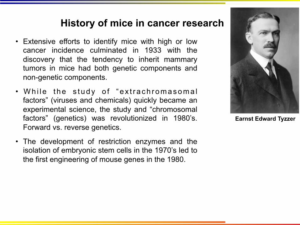
History of mice in cancer research
• Extensive efforts to identify mice with high or low cancer incidence culminated in 1933 with the discovery that the tendency to inherit mammary tumors in mice had both genetic components and non-genetic components.
• W h i l e t h e s t u d y o f “ e x t r a c h r o m a s o m a l factors” (viruses and chemicals) quickly became an experimental science, the study and “chromosomal factors” (genetics) was revolutionized in 1980’s. Forward vs. reverse genetics.
• The development of restriction enzymes and the isolation of embryonic stem cells in the 1970’s led to the first engineering of mouse genes in the 1980.
Earnst Edward Tyzzer

Uses of Mouse Models of Cancer
• Studies of cancer gene function
(Mouse +/- Gene X)
• A source of genetically defined cells of many types
(Isolate cell lines from different tissues)
• Understand the biology of specific diseases
(Engineer mice with a type of cancer, and study how it works)
• Understand the role of environment in disease and its interaction with genetics
• Understand the interactions of drugs with cancer
(Pharmacology and molecular biology of tumors treated with drugs)
• As final efficacy screens before clinical trials (pre-clinical trials)
• Better interpret the results of clinical trials (co-clinical and post-clinical trials)

Advantages/Disadvantages of Cancer Models Model Advantages Disadvantages
Humans Most accurate Least manipulable Extremely slow generation time (16 – 40 years) Genetically outbred
Bacteria Extremely fast growth (30 minutes) Powerful genetic tools Powerful biochemical techniques
Haploid, unicellular Great evolutionary divergence Not useful for efficacy screens
Yeast Very fast growth (100 minutes) Eukaryotic cells Powerful genetic and biochemical techniques
Unicellular Significant evolutionary divergence Not useful for efficacy screens
Mammalian Cell Culture
Fast growth (18 - 24 hours) Accurate genetics (especially in human cells) Powerful genetic and biochemical techniques
2D growth is different than 3D growth Only one or a few cell types (no stroma) No immune system Often inaccurate in predicting response to therapy
Drosophila Multicellular Reasonably fast growth (7 days) Powerful genetic tools
Significant evolutionary divergence Differences in organ composition/structure Not useful for efficacy screens
Zebrafish Vertebrate Optically clear for longitudinal studies Powerful genetic techniques
Few antibodies available Difficult to interpret histopathology Very slow generation time (3-4 months)
Mice Mammal Fairly accurate genetics Powerful genetic tools Accurate histopathology of diseases
Slow generation time (9 weeks) Mice are not small, furry people

Classes of Mouse Models of Cancer • Viral infection models
Infection with natural or engineered viruses • Chemical induced carcinogen models
Treatment with cancer-causing chemicals
• Transplantation Models Implantation/injection with tumor tissues or cells
• Engraftment Models Incorporation of tumorigenic cells into normal tissues
• Transgenic Models Randomly inserting tumor-promoting DNA into the mouse genome
• Genetically Engineered Models Targeted manipulation of specific sites in the mouse genome
• Somatic Engineering Models Targeted manipulation of specific sites in the mouse
+
+
++
+
+

Chemical Carcinogen Models
• 1775- Sir Percival Pott, a London surgeon, noticed that chimney sweepers frequently developed a peculiar form of scrotal cancer. He ascribed it to frequent, direct contact with coal tar. This launched 125 years of research in to the chemical basis of cancer.
• Treatment of mice with carcinogens is the basis of numerous mouse models of cancer.
Skin – 7,12-dimethylbenz[α]anthracene (DMBA) + 2-O-tetradecanoylphorbol-13-acetate (TPA)
Lung – Nitrosamines Liver – vinyl chloride Breast – N-Nitroso-N-methylurea (NMU) Colon – dimethylhydrazine (DMH)
Nitrosamines Bladder – Aromatic Amines
Br J Dermatol 149(5):960-967, 2003
DMBA/TPA

Chemical Carcinogen Models
Advantages – Very quick, cheap, and easy to use
Some models use common environmental carcinogens
Disdvantages – Carcinogen models are limited by the fact that it is challenging to identify the precise mutations induced by a chemical in a given cell or tumor.
Uses – The understanding that carcinogens act by mutating genes has diminished their use in cancer research in favor of direct genetic approaches
Most common models today seek to replicate exposure to complex environmental carcinogens such as cigarette smoke

Viral Infection Models • Cancer-causing viruses such as the Rous Sarcoma Virus (RSV) were initially
discovered in chickens in 1911 by Peyton Rous (Nobel Prize, 1966) at Rockefeller U.
• Mouse Mammary Tumor Virus (MMTV) was the first mouse virus, isolated at Jackson labs as the “non-chromosomal factor” that caused mammary tumors in the C3H strain of mice. Transmitted through milk- pups fostered to mothers of other strains did not develop breast tumors. (John Joseph Bittner, 1936)
• Some viruses cause cancer via random integration in certain cells
• Some viruses carry cellular oncogenes • Abelson murine leukemia virus - Abl • Moloney murine sarcoma virus – Raf • FBJ osteosarcoma virus - Fos • Friend murine leukemia virus – c-fms
• This was predicted by Ed Scolnick in 1973, and proven by Harold Varmus*, Mike Bishop*, Peter Vogt in 1976 (*1989 Nobel Prize in Physiology/Medicine)
• The finding that viruses simply regulate endogenous genes diminished interest. However, Gardasil (Merck) is a highly effective vaccine against HPV induced cervical cancer.
• Engineered viruses now used routinely in the laboratory to induce genetic changes that lead to cancer.

Transplantation Models
Ectopic – Implanted into a different organ than the original (typically sub-cutaneous or kidney capsule)
Orthotopic – Implanted into the analogous organ of the original tumor
Advantages – Typically cheap and fast Typically easy to use Not covered by patents
Disadvantages – Inaccurate histopathology compared to human tumors
Lack of step-wise progression through pre-neoplastic stages Evolution of tumor cells during passaging Requirement for angiogenesis to support the newly transplanted tumor Historically poor ability to predict response to therapy in humans
Tumor cells or tissues transplanted into a host mouse • Pioneered in the 1960’s and 1970’s

Transplantation Models Cell line based xenografts
Ectopic – Implanted into a different organ than the original (typically sub-cutaneous or kidney capsule)
Orthotopic – Implanted into the analogous organ of the original tumor
Advantages – Very easy to maintain cell lines indefinitely; Very fast to generate large numbers of xenograft tumors Relative homogeneity of tumors makes it easy to detect differences Comparatively cheap
Disadvantages – Inaccurate histopathology compared to human tumors Lack of an immune system Selection of cells during adaptation to growth on plastic Evolution of tumor cells during culture Historically poor ability to predict response to therapy in humans

Transplantation Models
Cell line based allografts
Advantages – Very easy to maintain cell lines indefinitely; Very fast to generate large numbers of xenograft tumors Relative homogeneity of tumors makes it easy to detect differences Comparatively cheap Intact immune system
Disadvantages – Mouse cells rather than human
Inaccurate histopathology compared to human tumors Selection of cells during adaptation to growth on plastic Evolution of tumor cells during culture Historically poor ability to predict response to therapy in humans

Transplantation Models
Patient-derived xenografts (PDX models)
Advantages – Neoplastic cells are human and may better interact with drugs designed for human proteins
Transplant both neoplastic cells and stroma Each PDX in a mouse represents the specific human patient No adaptation acquired during cell culture May be serially passaged in mice
Disadvantages – Lack of an immune system
Stromal cells are rapidly depleted over 1-3 passages Requirement for angiogenesis to support the newly transplanted tumor Requires a major infrastructure to acquire and maintain human tumors Relatively new- unclear how accurate they are

Engraftment Models Organ reconstitution models- cell engraft and create a normal organ, from which focal cancer then emerges
Visbal et al, 2011, 352 (1): 116–127

Engraftment Models
Mammary reconstitution- (Kuperwasser et al, 2004; PNAS 101(14):4966-71) • clear the fat pad from a recipient mouse • Isolate mammary epithelial cells from donor mice • Genetically manipulate donor cells (lentiviruses) • Inject mammary epithelial cells into cleared fat pad • Mammary adenocarcinoma
Liver reconstitution- (Scott Lowe lab) • Isolate fetal liver cells • Genetically manipulate donor cells • Inject fetal liver cells into newborn liver • Hepatocellular carcinoma
Hematopoietic reconstitution- (many laboratories) • Isolate fetal hematopoietic cells • Genetically manipulate donor cells • Inject fetal liver cells into newborn liver • Lymphoma, myeloid leukemias
Advantages – Step-wise progression through pre-neoplastic stages Can engineer the donor cells to study gene function or add reporters Relatively rapid and cheap Amenable to powerful genetic screening approaches
Disadvantages – Lack of an immune system when using human donor cells
Only available for a few organ systems: breast, liver, hematopoietic

Engineering Cancer in Mice
• Mice can be genetically engineered to develop cancer
• Use the human disease as a guide
• Mutations found in cancer can be programmed into mice
• Not all mice that develop cancer are “good” models of human disease
• Compare tumors in mice to human tumors- validation
Embryonic Stem Cell Blastocyst
Mutant Embryonic Stem Cell Injection Chimera
Genetically Engineered
Mouse
Engineering a Mouse
This video has been kindly provided by Johannes Wilbertz, from the Karolinska Institute, in Stockholm (Sweden) and belongs to a large collection of educational videos available from the International Society for Transgenic Technologies (ISTT) web site. https://www.youtube.com/watch?v=1m9kQuKXxEA

Engineered mice
Recipient blastocyst strain Chimera
Donor ES cell strain

Genetically Engineered mice
Advantages – Step-wise progression through pre-neoplastic stages Can engineer specific mutations to study gene function or add reporters Well established and understood technology Amenable to powerful genetic screening approaches Powerful genetic tools for imaging and other appliations Tumors develop in the presence of an intact immune system Can model both the neoplastic component and stroma cells Indications that the “best” engineered models are more accurate in predicting
the response of human tumors to therapy
Disadvantages – Mice are not people; mouse cells are not human cells Laboratory strains fail to represent the genetic diversity of human population Mouse tumors typically grow very fast relative to human tumors Complicated engineering strategies typically have drawabacks Relatively slow and expensive Requires a dedicated infrastructure

Transgenic Models
Uses – Demonstrate what happens when you overexpress gene X Used to create tissue specific gene regulatory elements for more complex strategies
Advantages – Easy to engineer. Comparatively rapid and cheap
Disadvantages – Random site of integration can lead result in positional effects Overexpression of cDNA lacks the physiological gene regulation
Randomly integrate DNA constructs into ES cells
cDNA transgenic CMV cDNA
tissues-specific transgenic
TSP cDNA
BAC transgenic Intact Gene
BAC transgenic Intact Promotor cDNA

Transgenic Models
Modern transgenic techniques allow for more subtle genetic control
shRNA knockdown U6 siRNA Advantages: Rapid way to learn what reduced expression of a gene does. May mimic drug ac9on. Disadvantages: Never get zero expression.
LenDviral transgenic LenD LTR cDNA Advantages: Extremely rapid genera9on of transgenic lines Disadvantages: Random integra9on. Can be mul9copy. Silencing of viral LTR promoters is common

Genetically Engineered Mouse Models
Uses – Precisely engineer gene loss, gene addition, subtle mutations, reporter constructs into specific places within the mouse genome
Advantages – Physiological gene regulation
Disadvantages – Slow and more difficult to carry out Targeted mutations can cause embryonic lethal phenotypes
Targeted integration of DNA into specific sites within the genome through homologous recombination
Mouse genomic locus
Area to be targeted
TargeDng construct
5’ homology arm 3’ homology arm
5’ homology arm 3’ homology arm NeoR
PosiDve selecDon
HSVTK
NegaDve selecDon

Types of GEM Models
Targeted Mutant
Wild-type locus V IV II I Pro.
Knockin mouse Pro. Foreign Gene
V IV II I Pro. *
Knockout mouse I Pro. NeoR V
Uses: Loss of function mutations in tumor suppressor genes
p53 -/- mice: lymphomas, sarcomas Rb +/- mice: pituitary tumors
Examples
Uses: Reporter alleles for developmental biology Gene function analysis
SHH-β-Gal: marking SHH+ cells K-rasH-Ras: ID differences between
K-ras and H-ras
Uses: Targeting subtle mutations into mice
p53 R172H/- : lymphomas, sarcomas and carcinomas

* V II I Pro. IV II
*
Pro. I II
II V IV
V IV II I Pro. *
Latent mice rely on stochastic recombination in vivo
Uses – Spontaneous tumor models
Advantages – Random cells are mutated, surrounded by wild-type cells.
Disadvantages – Don’t know which cells underwent recombination Different cell types have different rates of recombination Allele is null prior to recombination
*
K-rasLA1 Lung Cancer Model

Advanced genetic tools enable construction of complex gene regulation strategies
Recombinases – Enzymes that catalyze site specific recombination
Inducible alleles – Genes or proteins sensitive to the presence of a specific chemical
Transoposons – Mobile genetic elements who excision and reinsertion into the genome
is catalyzed by an enzyme (transposase) Reporters – Genes that allow visualization through microscopy, histochemistry, or in
vivo imaging

Cre is a site-specific bacterial recombinase
Cre
LoxP (44bp)
LoxP (44bp)
LoxP sites are conventionally represented as:
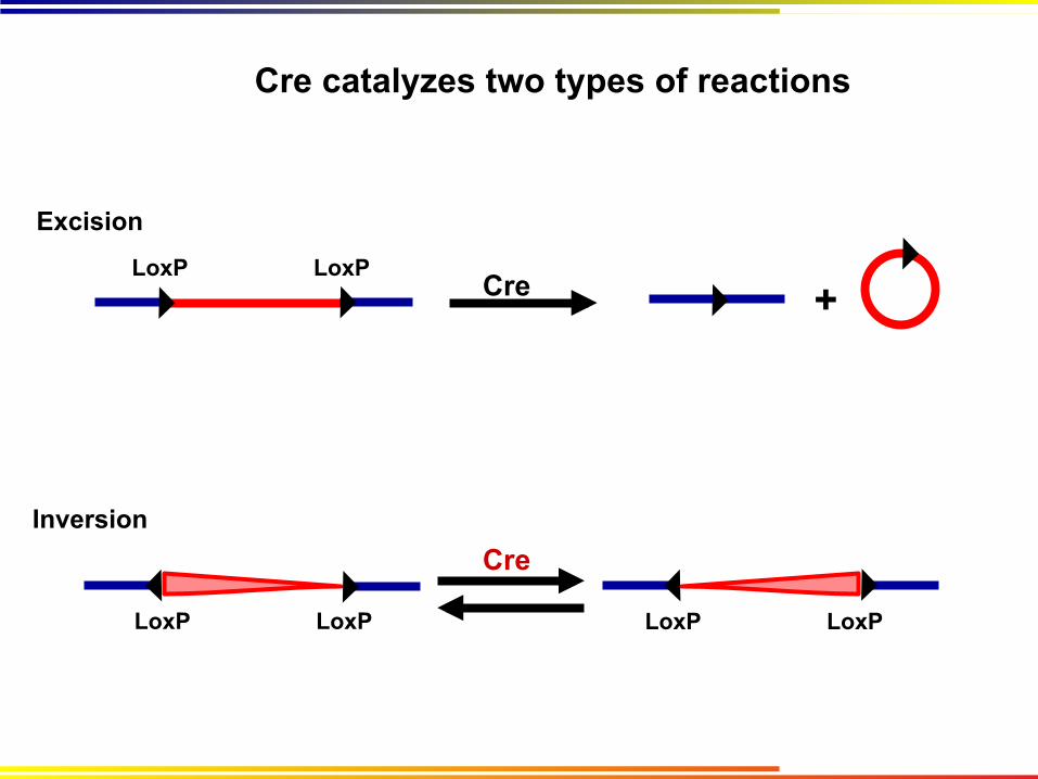
LoxP LoxP Cre +
Excision
Inversion Cre
LoxP LoxP
Cre catalyzes two types of reactions
LoxP LoxP

Unidirectional Inversion (10-fold weighting towards forward reaction)
Incompatible Sites
Not seen:
Variant Lox sites have been identified with useful properties
Cre
Lox66 Lox71 Lox72 LoxP
LoxP LoxP
Lox2722 Lox2722

Conditional mouse models
Conditional knockout V IV II I Pro.
Before After Before After Non-targeted Cells Targeted Cells
+/+ +/+ +/+ +/-
Cre/lox technology is widely used to create conditional alleles
Uses – Conditional loss of gene function in mice
Advantages – Spatial control can be achieved by expressing Cre from a tissue specific promoter Temporal control can be achieved by expressing Cre from a stage specfic
promoter, or by using inducible alleles to control Cre expression Gene is COMPLETELY lost from recombined cell
Disadvantages – Requires two alleles: conditional knockout, and Cre allele Because deletion is a binary event, even very low-level expression of Cre yields a 100% deletion

Conditional mouse models
= LoxP site
Conditional activatable
Conditional mutant
V IV II I Pro.
LSL
V IV II I Pro.
LSL
*
Before After Before After Non-targeted Cells Targeted Cells
+/- +/- +/- +/+
+/- +/- +/- +/m
LSL = LoxP-‐STOP-‐LoxP
Uses – Conditional expression of wild-type or mutant proteins under endogenous regulation
Advantages – Pysiological control of gene expression Spatial and temporal restriction of gene expression
Disadvantages – Requires two alleles: conditional knockout, and Cre allele Allele is null prior to recombination Sensitive to low-level Cre expression
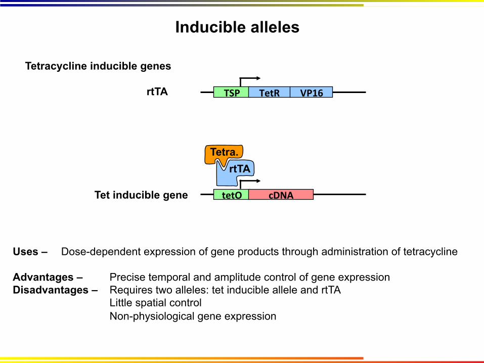
Inducible alleles
Uses – Dose-dependent expression of gene products through administration of tetracycline
Advantages – Precise temporal and amplitude control of gene expression Disadvantages – Requires two alleles: tet inducible allele and rtTA
Little spatial control Non-physiological gene expression
Tetracycline inducible genes
TSP TetR
tetO cDNA
rtTA Tetra.
VP16 rtTA
Tet inducible gene

Inducible alleles
Uses – Regulation of protein activity through administration of Tamoxifen
Advantages – Can control time and place of expression Tighter than tet-induced systems
Disadvantages – Tamoxifen is expensive Targeted protein must tolerate an N- or C-terminal fusion
Tamoxifen inducible alleles
TSP cDNA ERT2 Protein
ER
Protein ER
Tam
Sequestered
Protein ER
Tam

Reporter alleles
Activate expression of a marker gene under given conditions
Cre reporter: Activated when LoxP sites are removed Pathway reporter: Activated proportional to the activity of a transcription factor Fusion protein: Reporter protein is directly fused to another protein of interest
Reporter genes are typically optically based (visible, near-infrared) May be imaged by luminescence, fluorescence Instruments include 2-photon microscopy and whole animal imaging
May also include reporters for MRI, PET, and SPECT

Putting it together: the Brainbow Mouse
Livet et. al, 2007. Nature 450: 56 - 62
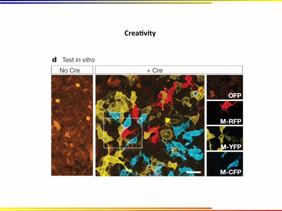
CreaDvity

CreaDvity
The coming revolution…
• CRISPR = Clustered Regularly Interspaced Short Palindromic Repeats • Cas = CRISPR-associated • Used by bacteria/archaea as an “adaptive immune defense” • Involves targeted degradation of foreign nucleic acids using short RNA and Cas9. http://www.youtube.com/watch?v=M739wgbcKuA


Theme and variation


SAISR provides access to imaging technology & expertise
Vevo 2100 Ultrasound
IVIS Quantum FX Micro CT
Bruker 9.4T MRI
IVIS Spectrum

Ultrasound is a non-invasive imaging technique
cm scale

Small Animal Imaging SR
High resolution ultrasound

Tomography can be used to reconstruct 3D tumor volumes
PancreaDc Tumor

Duodenum
Duo. Duo.
Duo.
PDA
Pancreas
Tissue Map B-mode Non-linear MIP
Contrast ultrasound shows pancreatic tumors are poorly perfused

High resolution ultrasound applications
B-mode: anatomical images and movies; quantitative measurements Guided injections: real-time image-guided injections of adult tissues and embyros Color Doppler: directionality of blood flow Power Doppler: magnitude of blood flow Pulsed Wave Doppler: Dynamics of blood flow M-mode: cardiac imaging; quantititative measurements of cardiac function 3D reconstruction: tomography of individual B-mode, Doppler, or contrast images
Color Doppler Power Doppler

Optical imaging of engineered alleles
SAISR Core
Spectral unmixing of 4 different fluorophores Luminescence imaging- of engineered
alleles and cell lines
Fluorescence imaging- of engingeered alleles and cell lines
Spectral unmixing- of different fluorophores
3D luminescent imaging- based on differential absorption of wavelengths through tissues
Co-registration with other modalities…

Optical imaging of engineered alleles
SAISR Core
Luminescence Fluorescence

Co-registration of optical + CT data

Micro CT Imaging is ideal for bone and lung
Brenner Lab/SAISR
Sagittal Coronal
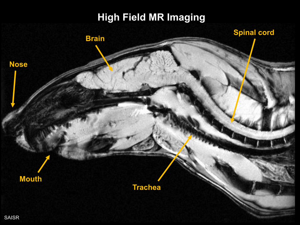
High Field MR Imaging
Nose
Brain
Mouth Trachea
Spinal cord
SAISR