multistage nanoparticle delivery system for deep ... · multistage nanoparticle delivery system for...
TRANSCRIPT

Corrections
EVOLUTIONCorrection for “Genome sequences of the human body louse andits primary endosymbiont provide insights into the permanentparasitic lifestyle,” by Ewen F. Kirkness, Brian J. Haas, WeilinSun, Henk R. Braig, M. Alejandra Perotti, John M. Clark, SiHyeock Lee, Hugh M. Robertson, Ryan C. Kennedy, EranElhaik, Daniel Gerlach, Evgenia V. Kriventseva, ChristineG. Elsik, Dan Graur, Catherine A. Hill, Jan A. Veenstra, BrianWalenz, José Manuel C. Tubío, José M. C. Ribeiro, Julio Rozas,J. Spencer Johnston, Justin T. Reese, Aleksandar Popadic, MartaTojo, Didier Raoult, David L. Reed, Yoshinori Tomoyasu, EmilyKrause, Omprakash Mittapalli, Venu M. Margam, Hong-Mei Li,Jason M. Meyer, Reed M. Johnson, Jeanne Romero-Severson,Janice Pagel VanZee, David Alvarez-Ponce, Filipe G. Vieira,Montserrat Aguadé, Sara Guirao-Rico, Juan M. Anzola, KyongS. Yoon, Joseph P. Strycharz, Maria F. Unger, Scott Christley,Neil F. Lobo, Manfredo J. Seufferheld, NaiKuan Wang, GregoryA. Dasch, Claudio J. Struchiner, Greg Madey, Linda I. Hannick,Shelby Bidwell, Vinita Joardar, Elisabet Caler, Renfu Shao,Stephen C. Barker, Stephen Cameron, Robert V. Bruggner,Allison Regier, Justin Johnson, Lakshmi Viswanathan, TerryR. Utterback, Granger G. Sutton, Daniel Lawson, Robert M.Waterhouse, J. Craig Venter, Robert L. Strausberg, May R.Berenbaum, Frank H. Collins, Evgeny M. Zdobnov, and BarryR. Pittendrigh, which appeared in issue 27, July 6, 2010, of ProcNatl Acad Sci USA (107:12168–12173; first published June 21,2010; 10.1073/pnas.1003379107).The authors note that the author name Emily Krause should
have appeared as Emily Kraus. The corrected author lineappears below. The online version has been corrected.
Ewen F. Kirknessa,1, Brian J. Haasa,2, Weilin Sunb, Henk R.Braigc, M. Alejandra Perottid, John M. Clarke, Si Hyeock Leef,Hugh M. Robertsonb, Ryan C. Kennedyg,h, Eran Elhaiki,Daniel Gerlachj,k, Evgenia V. Kriventsevaj,k, Christine G.Elsikl,3, Dan Grauri, Catherine A. Hillm, Jan A. Veenstran,Brian Walenza, José Manuel C. Tubíoo, José M. C. Ribeirop,Julio Rozasq, J. Spencer Johnstonr, Justin T. Reesel,Aleksandar Popadics, Marta Tojot, Didier Raoultu, David L.Reedv, Yoshinori Tomoyasuw,4, Emily Krausw, OmprakashMittapallix, Venu M. Margamm, Hong-Mei Lib, Jason M.Meyerm, Reed M. Johnsonb, Jeanne Romero-Seversong,y,Janice Pagel VanZeem, David Alvarez-Ponceq, Filipe G.Vieiraq, Montserrat Aguadéq, Sara Guirao-Ricoq, Juan M.Anzolal, Kyong S. Yoone, Joseph P. Strycharze, Maria F.Ungerg,y, Scott Christleyg,h, Neil F. Lobog,y, Manfredo J.Seufferheldz, NaiKuanWangaa, Gregory A. Daschbb, Claudio J.Struchinercc, Greg Madeyg,h, Linda I. Hannicka, ShelbyBidwella, Vinita Joardara, Elisabet Calera, Renfu Shaodd,Stephen C. Barkerdd, Stephen Cameronee, Robert V.Bruggnerg,h, Allison Regierg,h, Justin Johnsona, LakshmiViswanathana, Terry R. Utterbacka, Granger G. Suttona,Daniel Lawsonff, Robert M. Waterhousej,k, J. CraigVentera, Robert L. Strausberga, May R. Berenbaumb,Frank H. Collinsg,y, Evgeny M. Zdobnovj,k,gg,1,and Barry R. Pittendrighb,1,5
www.pnas.org/cgi/doi/10.1073/pnas.1103909108
PHYSICSCorrection for “Formation of a crystal nucleus from liquid,” byTakeshi Kawasaki and Hajime Tanaka, which appeared in issue32, August 10, 2010, of Proc Natl Acad Sci USA (107:14036–14041; first published July 27, 2010; 10.1073/pnas.1001040107).The authors note that Fig. 6 appeared incorrectly. The cor-
rected figure and its legend appear below. This error does notaffect the conclusions of the article.
www.pnas.org/cgi/doi/10.1073/pnas.1104042108
Fig. 6. Crystal nucleation dynamics. (A) Temporal change of the number ofcrystal nuclei for a system of N = 4,096 (SI Text). From the rate of the increasein the number of crystal nuclei, we estimated the crystal nucleation fre-quency I. The numbers in the figure indicate the volume fraction ϕ. (B) Thevolume fraction ϕ dependence of the reduced crystal nucleation frequency Irfor our work, the numerical estimate by Auer and Frenkel (15), and theexperimental work by Sinn et al. (17). Curves are guides to the eye. We alsoshow the results for three different system sizes (N = 1,024, 4,096, and16,834), which indicate few finite size effects for N ≥ 4,096. Here we use thevolume fraction ϕ estimated with σeff = 1.0953σ. Here BD stands for Brown-ian Dynamics simulations of the WCA system and HS stands for event-drivenMolecular Dynamics simulations of the hard sphere system.
www.pnas.org PNAS | April 12, 2011 | vol. 108 | no. 15 | 6335–6336
CORR
ECTIONS

MEDICAL SCIENCES, CHEMISTRYCorrection for “Multistage nanoparticle delivery system for deeppenetration into tumor tissue,” by Cliff Wong, TriantafyllosStylianopoulos, Jian Cui, John Martin, Vikash P. Chauhan, WenJiang, Zoran Popovi�c, Rakesh K. Jain, Moungi G. Bawendi, andDai Fukumura, which appeared in issue 6, February 8, 2011 of
Proc Natl Acad Sci USA (108:2426–2431; first published January18, 2011; 10.1073/pnas.1018382108).The authors note that Fig. 2 and its corresponding legend ap-
peared incorrectly. This error does not affect the conclusions of thearticle. The correctedfigure and its corrected legend appearbelow.
www.pnas.org/cgi/doi/10.1073/pnas.1104327108
CA B D
E F G H
Incubation with230 ng (0.16 μM)of MMP-2
0 2 4 6 8 10 12
0
20
40
60
80
100
Qu
an
tum
Do
tsR
ele
as
ed
(%)
Time (h)
Incubation for 12 hours
0 50 100 150 200 250
0
20
40
60
80
100
Qu
an
tum
Do
tsR
ele
as
ed
(%)
MMP-2 (ng)
Before MMP-2Incubation
10-2 10-1 100 101 102 103 104
0.0
0.2
0.4
0.6
0.8
D = 6.1 x 10⁻⁸
cm2/s
G(τ
)
τ (ms)
After 230 ngMMP-2 Incubation
10-2 10-1 100 101 102 103 104
0.0
0.2
0.4
0.6
G(τ
)
τ (ms)
D = 4.4 x 10⁻⁷
cm2/s
0 50 100 150 2000
25
50
Nu
mb
er
of
Pa
rtic
les
Diameter (nm)
1 10 100 1000 10000
0
10
20
30
%M
as
s
Diameter (nm)
Day 1Day 48
540 570 600
Flu
ores
cenc
e(A
U)
λ (nm)
Fig. 2. QDGelNP physical and in vitro characterization. (A) Epifluorescence image of QDGelNPs on a silicon substrate at 100×magnification. (Scale bar: 5 μm.)(B) DLS distribution of QDGelNP on day 1 and day 48 after synthesis and storage at 4 °C. (C) SEM image of QDGelNPs at 15,000× magnification. (Scale bar: 1μm.) (C Inset) SEM image of individual QDGelNP at 35,000× magnification. (Scale bar: 100 nm.) (D) Histogram of QDGelNPs’ size distribution from imageanalysis of SEM image. (E and F) Kinetics of MMP-2–induced QD release from QDGelNPs. (E) QD-release curve from incubation of 0.1 mg of QDGelNPs with230 ng (0.16 μM) of MMP-2. (F) QD release from incubation of 0.1 mg of QDGelNPs for 12 h with varying amounts of MMP-2. (G and H) FCS cross-correlogramsof QDGelNPs before (G) and after (H) incubation with MMP-2.
6336 | www.pnas.org

Multistage nanoparticle delivery system for deeppenetration into tumor tissueCliff Wonga, Triantafyllos Stylianopoulosb, Jian Cuia, John Martinb, Vikash P. Chauhanb, Wen Jiangb, Zoran Popovica,Rakesh K. Jainb, Moungi G. Bawendia,1, and Dai Fukumurab,1
aDepartment of Chemistry, Massachusetts Institute of Technology, Cambridge, MA 02139; and bEdwin L. Steele Laboratory for Tumor Biology, Department ofRadiation Oncology, Massachusetts General Hospital and Harvard Medical School, Boston, MA 02114
Contributed by Moungi G. Bawendi, December 7, 2010 (sent for review October 8, 2010)
Current Food and Drug Administration-approved cancer nano-therapeutics, which passively accumulate around leaky regions ofthe tumor vasculature because of an enhanced permeation andretention (EPR) effect, have provided only modest survival bene-fits. This suboptimal outcome is likely due to physiological barriersthat hinder delivery of the nanotherapeutics throughout thetumor. Many of these nanotherapeutics are ≈100 nm in diameterand exhibit enhanced accumulation around the leaky regions ofthe tumor vasculature, but their large size hinders penetration intothe dense collagen matrix. Therefore, we propose a multistagesystem in which 100-nm nanoparticles “shrink” to 10-nm nanopar-ticles after they extravasate from leaky regions of the tumor vas-culature and are exposed to the tumor microenvironment. Theshrunken nanoparticles can more readily diffuse throughout thetumor’s interstitial space. This size change is triggered by pro-teases that are highly expressed in the tumor microenvironmentsuch as MMP-2, which degrade the cores of 100-nm gelatin nano-particles, releasing smaller 10-nm nanoparticles from their surface.We used quantum dots (QD) as a model system for the 10-nmparticles because their fluorescence can be used to demonstratethe validity of our approach. In vitro MMP-2 activation of themultistage nanoparticles revealed that the size change was effi-cient and effective in the enhancement of diffusive transport. Invivo circulation half-life and intratumoral diffusion measurementsindicate that our multistage nanoparticles exhibited both the longcirculation half-life necessary for the EPR effect and the deep tu-mor penetration required for delivery into the tumor’s densecollagen matrix.
drug delivery | cancer therapy | nanomedicine
Nanoparticles (NPs) have offered new approaches to the de-livery of cancer therapeutics (1–5). Doxil (≈100 nm PEGy-
lated liposomal form of doxorubicin) and Abraxane (≈130 nmalbumin-bound paclitaxel nanoparticle) are two examples ofFood and Drug Administration-approved nanoparticle-basedtherapeutics for solid tumors; their large size compared withconventional cancer therapeutics allows them to preferentiallyaccumulate in solid tumors by the enhanced permeation andretention (EPR) effect (6), thus reducing normal tissue toxicity.However, despite the improved pharmacokinetic properties (7)and reduced adverse effects, these drugs have provided onlymodest survival benefits (1, 8–10). This shortcoming is likelyattributed to the physiological barriers imposed by the abnormaltumor vasculature and the dense interstitial matrix—a complexassembly of collagen, glycosaminoglycans, and proteoglycans—which hinder delivery of the drug throughout the entire tumor insufficient concentration (11, 12).Systemic delivery of therapeutics to the tumor is a three-step
process: blood-borne delivery to different regions of the tumor,transport across the vessel wall, and passage through the in-terstitial space to reach the tumor cells (13). Abnormalities inthe tumor vasculature lead to highly heterogeneous vascularperfusion throughout the tumor. The microvascular density ishigh at the invasive edge of the tumor, but sometimes the tumorcenter is unperfused, preventing delivery of therapeutics to this
region. However, the tumor center’s hostile microenvironment(low pH and low pO2) harbors the most aggressive tumor cells,and the tumor will regenerate if these cells are not eliminated.Moreover, exposure of the cancer cells to sublethal concentra-tion of the therapeutic agent may facilitate the developmentof resistance.Hyperpermeability of the abnormal vasculature and lack of
functional lymphatics lead to elevated levels of interstitial fluidpressure (IFP) (14, 15). This interstitial hypertension, in turn,reduces convective transport across the vessel wall and into theinterstitial space, leaving diffusion as the primary mode for drugtransport to the poorly perfused regions. Large 100-nm nano-particles are suitable for the EPR effect (6) but have poor dif-fusion in the dense collagen matrix of the interstitial space (16,17), resulting in restrictive nanoparticle accumulation aroundtumor blood vessels and little penetration into the tumor pa-renchyma. In the case of Doxil for example, the liposomal par-ticles are trapped close to the tumor vasculatures. Although thesmall size (≈400 MW) of doxorubicin, which is released from theliposomes, seemingly allow rapid diffusion, doxorubicin cannotmigrate far from the particles because of avid binding to DNAand sequestration in acidic endosomes of perivascular tumorcells (18, 19), resulting in heterogeneous therapeutic effects.Consequently, we propose a multistage approach in which
nanoparticles change their size to facilitate transport by adaptingto each physiological barrier. The original 100-nm nanoparticlespreferentially extravasate from the leaky regions of the tumorvasculature. After extravasation into tumor tissue, the nano-particles shrink to 10 nm, significantly lowering their diffusionalhindrance in the interstitial matrix (20) and allowing penetrationinto the tumor parenchyma. These smaller nanoparticles canpotentially be used as nanocarriers for therapeutics that are re-leased as the particles penetrate deep into the tumor (Fig. S1).Surface PEGylation of the small nanoparticles allows them todiffuse smoothly in the interstitial matrix by reducing the bind-ing, sequestration, and metabolism that hinder the transport oftherapeutic agents (21, 22). Furthermore, 10-nm nanoparticlesare not cleared from the tumor as rapidly as much smaller mo-lecular species because of their larger size.To achieve this size-shrinking property, a large nanoparticle
should be triggered to release smaller nanoparticles after ex-travasation into the tumor. Several nanoparticles have beendesigned to release their contents remotely via an externalstimulus (light, heat, ultrasound, magnetic field), but their use to
Author contributions: C.W., T.S., R.K.J., M.G.B., and D.F. designed research; C.W., T.S., J.C.,J.M., V.P.C., and W.J. performed research; Z.P. contributed new reagents/analytic tools;C.W., T.S., J.C., J.M., V.P.C., W.J., R.K.J., M.G.B., and D.F. analyzed data; and C.W., T.S.,R.K.J., M.G.B., and D.F. wrote the paper.
R.K.J. received commercial research grants from Dyax, AstraZeneca, and MedImmune;consultant fees from AstraZeneca/MedImmune, Dyax, Astellas-Fibrogen, Regeneron, Gen-zyme, MorphoSys, and Noxxon Pharma; and a speaker honorarium from Genzyme. R.K.J.owns stock in SynDevRx. No reagents or funding from these companies were used inthese studies. There is no significant financial or other competing interest in the work.1To whom correspondence may be addressed. E-mail: [email protected] or [email protected].
This article contains supporting information online at www.pnas.org/lookup/suppl/doi:10.1073/pnas.1018382108/-/DCSupplemental.
2426–2431 | PNAS | February 8, 2011 | vol. 108 | no. 6 www.pnas.org/cgi/doi/10.1073/pnas.1018382108
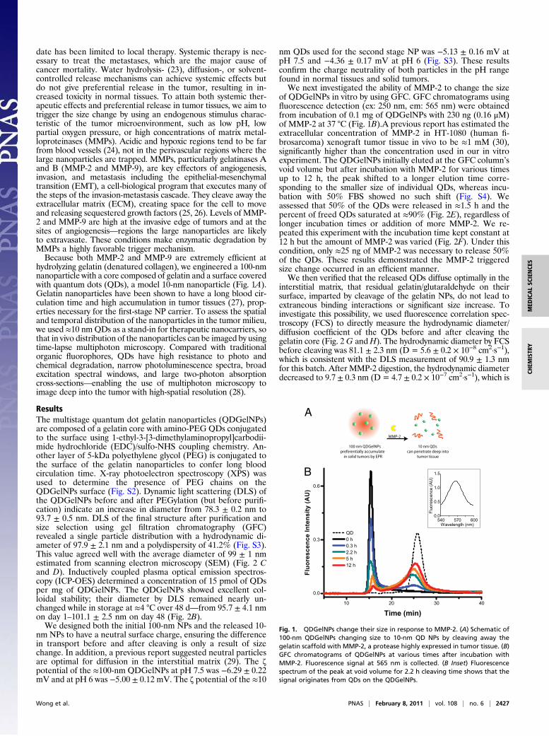
date has been limited to local therapy. Systemic therapy is nec-essary to treat the metastases, which are the major cause ofcancer mortality. Water hydrolysis- (23), diffusion-, or solvent-controlled release mechanisms can achieve systemic effects butdo not give preferential release in the tumor, resulting in in-creased toxicity in normal tissues. To attain both systemic ther-apeutic effects and preferential release in tumor tissues, we aim totrigger the size change by using an endogenous stimulus charac-teristic of the tumor microenvironment, such as low pH, lowpartial oxygen pressure, or high concentrations of matrix metal-loproteinases (MMPs). Acidic and hypoxic regions tend to be farfrom blood vessels (24), not in the perivascular regions where thelarge nanoparticles are trapped. MMPs, particularly gelatinases Aand B (MMP-2 and MMP-9), are key effectors of angiogenesis,invasion, and metastasis including the epithelial-mesenchymaltransition (EMT), a cell-biological program that executes many ofthe steps of the invasion-metastasis cascade. They cleave away theextracellular matrix (ECM), creating space for the cell to moveand releasing sequestered growth factors (25, 26). Levels of MMP-2 and MMP-9 are high at the invasive edge of tumors and at thesites of angiogenesis—regions the large nanoparticles are likelyto extravasate. These conditions make enzymatic degradation byMMPs a highly favorable trigger mechanism.Because both MMP-2 and MMP-9 are extremely efficient at
hydrolyzing gelatin (denatured collagen), we engineered a 100-nmnanoparticle with a core composed of gelatin and a surface coveredwith quantum dots (QDs), a model 10-nm nanoparticle (Fig. 1A).Gelatin nanoparticles have been shown to have a long blood cir-culation time and high accumulation in tumor tissues (27), prop-erties necessary for the first-stage NP carrier. To assess the spatialand temporal distribution of the nanoparticles in the tumor milieu,we used ≈10 nmQDs as a stand-in for therapeutic nanocarriers, sothat in vivo distribution of the nanoparticles can be imaged by usingtime-lapse multiphoton microscopy. Compared with traditionalorganic fluorophores, QDs have high resistance to photo andchemical degradation, narrow photoluminescence spectra, broadexcitation spectral windows, and large two-photon absorptioncross-sections—enabling the use of multiphoton microscopy toimage deep into the tumor with high-spatial resolution (28).
ResultsThe multistage quantum dot gelatin nanoparticles (QDGelNPs)are composed of a gelatin core with amino-PEG QDs conjugatedto the surface using 1-ethyl-3-[3-dimethylaminopropyl]carbodii-mide hydrochloride (EDC)/sulfo-NHS coupling chemistry. An-other layer of 5-kDa polyethylene glycol (PEG) is conjugated tothe surface of the gelatin nanoparticles to confer long bloodcirculation time. X-ray photoelectron spectroscopy (XPS) wasused to determine the presence of PEG chains on theQDGelNPs surface (Fig. S2). Dynamic light scattering (DLS) ofthe QDGelNPs before and after PEGylation (but before purifi-cation) indicate an increase in diameter from 78.3 ± 0.2 nm to93.7 ± 0.5 nm. DLS of the final structure after purification andsize selection using gel filtration chromatography (GFC)revealed a single particle distribution with a hydrodynamic di-ameter of 97.9 ± 2.1 nm and a polydispersity of 41.2% (Fig. S3).This value agreed well with the average diameter of 99 ± 1 nmestimated from scanning electron microscopy (SEM) (Fig. 2 Cand D). Inductively coupled plasma optical emission spectros-copy (ICP-OES) determined a concentration of 15 pmol of QDsper mg of QDGelNPs. The QDGelNPs showed excellent col-loidal stability; their diameter by DLS remained nearly un-changed while in storage at ≈4 °C over 48 d—from 95.7 ± 4.1 nmon day 1–101.1 ± 2.5 nm on day 48 (Fig. 2B).We designed both the initial 100-nm NPs and the released 10-
nm NPs to have a neutral surface charge, ensuring the differencein transport before and after cleaving is only a result of sizechange. In addition, a previous report suggested neutral particlesare optimal for diffusion in the interstitial matrix (29). The ζpotential of the ≈100-nm QDGelNPs at pH 7.5 was −6.29 ± 0.22mV and at pH 6 was −5.00 ± 0.12 mV. The ζ potential of the ≈10
nm QDs used for the second stage NP was −5.13 ± 0.16 mV atpH 7.5 and −4.36 ± 0.17 mV at pH 6 (Fig. S3). These resultsconfirm the charge neutrality of both particles in the pH rangefound in normal tissues and solid tumors.We next investigated the ability of MMP-2 to change the size
of QDGelNPs in vitro by using GFC. GFC chromatograms usingfluorescence detection (ex: 250 nm, em: 565 nm) were obtainedfrom incubation of 0.1 mg of QDGelNPs with 230 ng (0.16 μM)of MMP-2 at 37 °C (Fig. 1B).A previous report has estimated theextracellular concentration of MMP-2 in HT-1080 (human fi-brosarcoma) xenograft tumor tissue in vivo to be ≈1 mM (30),significantly higher than the concentration used in our in vitroexperiment. The QDGelNPs initially eluted at the GFC column’svoid volume but after incubation with MMP-2 for various timesup to 12 h, the peak shifted to a longer elution time corre-sponding to the smaller size of individual QDs, whereas incu-bation with 50% FBS showed no such shift (Fig. S4). Weassessed that 50% of the QDs were released in ≈1.5 h and thepercent of freed QDs saturated at ≈90% (Fig. 2E), regardless oflonger incubation times or addition of more MMP-2. We re-peated this experiment with the incubation time kept constant at12 h but the amount of MMP-2 was varied (Fig. 2F). Under thiscondition, only ≈25 ng of MMP-2 was necessary to release 50%of the QDs. These results demonstrated the MMP-2 triggeredsize change occurred in an efficient manner.We then verified that the released QDs diffuse optimally in the
interstitial matrix, that residual gelatin/glutaraldehyde on theirsurface, imparted by cleavage of the gelatin NPs, do not lead toextraneous binding interactions or significant size increase. Toinvestigate this possibility, we used fluorescence correlation spec-troscopy (FCS) to directly measure the hydrodynamic diameter/diffusion coefficient of the QDs before and after cleaving thegelatin core (Fig. 2G andH). The hydrodynamic diameter by FCSbefore cleaving was 81.1 ± 2.3 nm (D = 5.6 ± 0.2 × 10⁻⁸ cm2·s−1),which is consistent with the DLS measurement of 90.9 ± 1.3 nmfor this batch. After MMP-2 digestion, the hydrodynamic diameterdecreased to 9.7 ± 0.3 nm (D = 4.7 ± 0.2 × 10⁻⁷ cm2·s−1), which is
MMP-2
100 nm QDGelNPspreferentially accumulate
in solid tumors by EPR
10 nm QDscan penetrate deep into
tumor tissue
A
B
10 20 30 40
0.0
0.3
0.6
FluorescenceIntensity
(AU)
Time (min)
QD0 h0.3 h2.2 h5 h12 h
540 570 6000.0
0.5
1.0
1.5
Fluorescence(AU)
Wavelength (nm)
Fig. 1. QDGelNPs change their size in response to MMP-2. (A) Schematic of100-nm QDGelNPs changing size to 10-nm QD NPs by cleaving away thegelatin scaffold with MMP-2, a protease highly expressed in tumor tissue. (B)GFC chromatograms of QDGelNPs at various times after incubation withMMP-2. Fluorescence signal at 565 nm is collected. (B Inset) Fluorescencespectrum of the peak at void volume for 2.2 h cleaving time shows that thesignal originates from QDs on the QDGelNPs.
Wong et al. PNAS | February 8, 2011 | vol. 108 | no. 6 | 2427
MED
ICALSC
IENCE
SCH
EMISTR
Y

the size of individual QDs, indicating the size increase of the re-leased QDs from gelatin/glutaraldehyde fragments was negligible.We next evaluated whether the size change observed in GFC
and FCS enhances diffusive transport in dense collagen envi-ronments resembling those in solid tumors. To simulate the in-terstitial matrix of a solid tumor, we prepared a collagen gel ina capillary tube at 0.74% (7.4 mg/mL) concentration, similarto the reported estimate of 9.0 ± 2.5 mg/(mL interstitial matrix)for interstitial collagen in both human colon adenocarcinoma(LS174T) and murine mammary carcinoma (MCalV) implantedin mouse dorsal chambers (17, 31). The collagen gel penetrationof the QDGelNPs before and after cleaving with MMP-2 wascompared with a noncleavable, PEGylated, and QD-coated silicananoparticles (32) control (Diam. = 105.6 ± 0.8 nm, ζ potentialat pH 7.5 = −3.9 ± 0.2 mV) designed to behave like QDGelNPsbefore cleaving. A mixture of SilicaQDs and QDGelNPs (beforeor after cleaving) were placed in contact with the gel and in-cubated for 12 h. Infiltration of both particles into the collagenwas determined using multiphoton microscopy with simulta-neous second-harmonic generation (SHG) imaging of fibrillarcollagen (Fig. 3). The SilicaQDs and QDGelNPs before cleavingboth had negligible penetration and were excluded from thecollagen matrix (Fig. S5). However, after cleavage of QDGelNPswith MMP-2, the freed QDs were able to penetrate over a mil-limeter into the gel. By fitting the concentration profile of thecleaved QDGelNPs to a one-dimensional diffusion model (33,34), we obtained a diffusion coefficient of 2.3 × 10⁻⁷ cm2·s−1, thesame diffusion coefficient obtained for individual QDs in thecollagen gel before conjugation to the gelatin NP. The resultingdiffusion coefficient ratio (D/Do, where Do is diffusion coefficientof freed QDs in solution obtained by FCS) in the collagen matrixis 0.49. This value agrees well with the expected value for D/Doof ≈0.52 derived from previous reports (Materials and Methods)(29, 31). This result indicates that the diffusion coefficient ofreleased QDs in dense collagen increases to that of ≈10 nmparticles and any residual gelatin/glutaraldehyde fragmentsremaining on the surface do not impede their diffusion.
To test whether tumor secreted MMP-2 can change the size ofQDGelNPs in vivo, we intratumorally coinjected QDGelNPs andSilicaQDs in the HT-1080 tumor implanted in the dorsal skin-foldwindow chamber of severe combined immunodeficient (SCID)mice. The HT-1080 tumor model was selected because of itsreported high MMP-2 activity, which we confirmed by in situgelatin zymography on a tumor tissue section (Fig. S6). Multi-photon microscopy revealed a marked increase in QDGelNPspenetration into surrounding tumor tissue as compared with thenoncleavable SilicaQDs control, confirming a substantial en-hancement in interstitial transport associated with size change(Fig. 4 and Fig. S7). At 6 h postinjection, the QDGelNPs hadpenetrated up to ≈300 μm from the injection site while the Sili-caQDs control exhibited little or no dissemination from its initiallocation. We fitted the concentration profile to a model for sub-stances diffusing from a spherical source to obtain an effectivediffusion coefficient of ≈2.2 × 10⁻⁸ cm2·s−1 inside the tumor (Fig.S8). This value is ≈10% the diffusion coefficient obtained in thecollagen gel, which can be explained by the increased time neededto cleave the particles, the tortuosity of the interstitial space in-duced by cellular obstacles (35), and the possibly higher collagenconcentration in the HT-1080 tumor than in the gel we prepared.We next determined the QDGelNPs’ blood half-life (t1/2β) to
show that the QDGelNPs are not rapidly removed from circulationby the reticuloendothelial system. We systemically administered tonontumor bearingmice a mixture of theQDGelNPs and SilicaQDsby retro-orbital injection and measured the decrease in fluores-cence from both particles in the blood over time. The SilicaQDsexhibited a blood half-life of 12.9 ± 2.4 h, whereas the QDGelNPshad a half-life of 22.0 ± 3.4 h (Fig. S9). The difference in the half-lives may be due to variations in the QDGelNPs’ surface chemistrythat make it less immunogenic compared with SilicaQDs. Theseresults established that QDGelNPs possess both the long circula-tion half-life and large 100-nm size necessary for preferential ex-travasation from the leaky regions of the tumor vasculature as wellas the deep interstitial penetration of a 10-nm particle required fordelivery to the tumor’s poorly accessible regions.
CB D
E F G H
Incubation with230 ng (0.16 μM)of MMP-2
0 2 4 6 8 10 12
0
20
40
60
80
100
Quantum
DotsReleased(%)
Time (h)
Incubation for 12 hours
0 50 100 150 200 250
0
20
40
60
80
100
Quantum
DotsReleased(%)
MMP-2 (ng)
After 230 ng MMP-2 Incubation
10-2 10-1 100 101 102 103 1040.0
0.2
0.4
0.6
0.8
D = 6.1 x 10⁻⁸ cm2/s
G(τ)
τ (ms)
Before MMP-2Incubation
10-2 10-1 100 101 102 103 1040.0
0.2
0.4
0.6
G(τ)
τ (ms)
D = 4.4 x 10⁻⁷ cm2/s
A
0 50 100 150 2000
25
50
Num
berofParticles
Diameter (nm)1 10 100 1000 10000
0
10
20
30
%Mass
Diameter (nm)
Day 1Day 48
540 570 600
Fluorescence(AU)
λ (nm)
Fig. 2. QDGelNP physical and in vitro characterization. (A) Epifluorescence image of QDGelNPs on a silicon substrate at 100×magnification. (Scale bar: 5 μm.)(B) DLS distribution of QDGelNP on day 1 and day 48 after synthesis and storage at 4 °C. (C) SEM image of QDGelNPs at 15,000× magnification. (Scale bar:1 μm.) (C Inset) SEM image of individual QDGelNP at 35,000× magnification. (Scale bar: 100 nm.) (D) Histogram of QDGelNPs’ size distribution from imageanalysis of SEM image. (E and F) Kinetics of MMP-2–induced QD release from QDGelNPs. (E) QD-release curve from incubation of 0.1 mg (0.16 μM) ofQDGelNPs with 230 ng of MMP-2. (F) QD release from incubation of 0.1 mg of QDGelNPs for 12 h with varying amounts of MMP-2. (G and H) FCS cross-correlograms of QDGelNPs before (G) and after (H) incubation with MMP-2.
2428 | www.pnas.org/cgi/doi/10.1073/pnas.1018382108 Wong et al.

DiscussionWe have established three main criteria for the multistageQDGelNPs: the size of nanoparticles must change from 100 nm to10 nm, their surface before and after MMP-2 cleavage needs to bewell PEGylated and neutral, and their sensitivity to cleavageshould be at least as high as other reported MMP-2 probes (36–38). Satisfying these three criteria simultaneously presented sev-eral design and synthetic challenges. However, by optimizing thecoupling scheme and the degree of glutaraldehyde cross-linking,we were able to meet the desired criteria for this system whilepreserving the simplicity in design to ensure scalability.Because both the carrier and released nanoparticles need to be
highly PEGylated and neutral, encapsulation strategies that relyon hydrophobic or charged interactions are unsuitable. Using QDswith a sticky surface (low coverage of PEG) allowed for encap-sulation inside the gelatin core, but the particle after cleavageshowed a broad size distribution by GFC, indicating binding togelatin/glutaraldehyde fragments. An alternative strategy is tocovalently attach the QDs onto the gelatin NP surface. Althoughthis approach is susceptible to interparticle cross-linking, throughcareful optimization and using EDC/sulfo-NHS chemistry toconjugate amino-PEG QDs to the carboxylic acid groups on the
gelatin NP surface, we produced nearly monodisperse QDGelNPsthat released cleaved QDs without size increase.By controlling the length and degree of glutaraldehyde poly-
merization used to cross-link the gelatin nanoparticles, we wereable to optimize the size of the QDs after cleaving and their rateof release while maintaining particle stability. The method forgelatin nanoparticle synthesis developed in ref. 39 producedgelatin NPs with long extended networks of glutaraldehyde ontheir surface to maintain particle stability in aqueous solution.However, when the same scheme was applied to our design, theQDs remained covalently attached to this large glutaraldehydepolymer after MMP-2 degradation, resulting in QDs that weresignificantly larger upon release. To produce released QDswithout augmenting their size, we constructed a network of nu-merous short cross-links on the gelatin particles instead. Becauseglutaraldehyde readily self-polymerizes in solution, we usednearly monomeric glutaraldehyde (grade I) for consistent for-mation of short glutaraldehyde polymer cross-links. In addition,using grade I glutaraldehyde improved the MMP-2 cleaving rateconsiderably such that the release is as sensitive as previouslyreported MMP-2 probes. By modifying the degree of cross-linking on the gelatin nanoparticle, we optimized the QDGelNPs
A B CD
0 250 500 7500.0
0.2
0.4
0.6
0.8
1.0
NormalizedFluorescence( AU)
Distance (µm)
SilicaQDQDGelNPbefore MMP-2
H
0 1000 20000.0
0.2
0.4
0.6
0.8
1.0
No rmalizedFluor escence(AU)
Distance (μm)
SilicaQDQDGelNPafter MMP-2
D = 2.3 x 10⁻⁷ cm2/s
F GE
BeforeMMP-2
AfterMMP-2
SilicaQDs QDGelNPs SHG
Fig. 3. Diffusion of SilicaQDs and QDGelNPs (before and after MMP-2 cleavage) in a collagen gel. (A and B) Fluorescence images of SilicaQDs (A) andQDGelNPs before MMP-2 cleavage (B) penetrating into the collagen gel. (C) Second-harmonic generation (SHG) signal shows the corresponding location ofthe collagen matrix. (Scale bars: 125 μm.) (D) Normalized intensity profile of SilicaQDs and QDGelNPs in collagen gel. (E and F) Fluorescence images of Sil-icaQDs (E) and QDGelNPs after MMP-2 cleavage (F) penetrating into the collagen gel. (G) SHG signal shows the corresponding location of the collagen matrix.(Scale bars: 125 μm.) (H) Normalized intensity profile of SilicaQDs and QDGelNPs after MMP-2 cleavage in collagen gel. Black line displays theoretical intensityprofile for particles with diffusion coefficient of 2.3 × 10⁻⁷ cm2·s−1.
Wong et al. PNAS | February 8, 2011 | vol. 108 | no. 6 | 2429
MED
ICALSC
IENCE
SCH
EMISTR
Y

to be highly responsive to MMP-2 degradation while preservingparticle stability in storage for at least 48 d.Our FBS incubation experiment suggested the QDGelNPs are
stable in serum conditions, but additional studies will be neededto verify that the QDGelNPs are not degraded in the blood bycirculating MMPs (40) and other proteases. Circulating MMPs,however, have been reported to be inhibited by serum proteinssuch as α2-macroglobulins that entrap the MMPs (36, 41). Thedegree of PEGylation on the surface of the QDGelNPs mayneed to be optimized such that there is minimal cleaving andopsonization in the blood while achieving the desired releaserate in tumor tissue. However, MMP levels may vary in differenttumor types/individuals, causing inconsistent behavior. A strengthof our method is the potential for customized delivery of nano-particles by using genomic and molecular data to achieve optimaldelivery for a particular patient. For example, high levels of tumor-specific urinary MMPs measured noninvasively in an individualcan be indicative of high levels of the MMPs in plasma andtumor tissues (42). This information can help us customize theQDGelNPs for lower sensitivity to proteases (e.g., by increasingthe particles’ cross-linking) to minimize degradation in the bloodwhile maintaining the same desired release rate in the tumor. Inaddition, we can design a series of customized nanoparticles thatare activated by a variety of tumor-associated proteases—such ascathepsin B and urokinase-type plasminogen activator (uPA). Bymeasuring and modeling the levels of various tumor-associatedproteases in circulation, the primary tumor, and the metastaticsites, a personalized combination and dosage of protease-activatednanoparticles, may then be administered to achieve an optimaltherapeutic effect.Multistage nanoparticle systems provide additional tunability
in the spatial control of delivery to solid tumors. Because of themultiple physiological barriers that a therapeutic agent mustencounter, a multistage nanoparticle system can enhance pene-tration by changing its size, charge, shape, flexibility, and/orsurface coating to suit the transport across each barrier. We canextend the utility of this approach by incorporating additionalstages into the QDGelNPs to guide its delivery into tumor cellsor even subcellular compartment(s). Such an approach may notonly improve the efficacy of current anticancer drugs but alsomake available drugs that have been abandoned because of de-livery problems. As interest in increasingly sophisticated deliverysystems grows, we face a corresponding challenge to synthesizenanostructures of increasing complexity. Innovative schemes willbe necessary to assemble nanocomponents in complex config-urations that keep the synthesis simple yet functional so thatproduction can be scalable and cost-effective.We have provided a proof-of-principle demonstration that
a size-changing nanoparticle can facilitate delivery into the densecollagen matrix of a tumor. Ultimately, the QDs will be replacedwith a 10-nm nanocarrier of cancer therapeutics, and its antitu-
mor efficacy and survival benefits will be compared with that ofconventional drugs and other nanoparticle-based treatments.The loading capacity per particle for 10-nm nanocarrier is lim-ited. Therefore, not only the penetration depth but also sufficientquantity of nanoparticles needs to be delivered.
Materials and MethodsSynthesis of Gelatin Nanoparticle. Detailed information regarding synthesis oftheQDGelNP can be found in SIMaterials andMethods. Gelatin nanoparticleswere prepared from a modification of the two-step desolvation methoddeveloped in ref. 39. Gelatin type A (0.625 g) was dissolved in 12.5 mL ofdeionized (DI) water at 40 °C. Acetone (12.5 mL) was added to the solution at6 mL/min. Exactly 1 min after the addition was completed, the supernatantcontaining the low molecular weight gelatin fraction was removed. DI water(12.5 mL) was added to the remaining precipitate and heated again to 40 °Cuntil dissolution. Half the solution was removed, and the pH of theremaining half was adjusted to 2.7. Under constant stirring at 600 rpm and40 °C, 20.75 mL of acetone was added at 1 mL/min. After the acetone ad-dition was completed, 30 μL of 50% glutaraldehyde solution (Grade I) dilutedin 1 mL of acetone was added to the gelatin solution at 0.05 mL/min to cross-link the particles. Subsequently, the solution was kept at 40 °C and 600 rpmstir rate for 7.5 h. The acetone was then removed. The remaining solutionwas filtered through a 0.2-μm syringe filter. A 1 M glycine solution (0.2 mL)was added, and the solution was stored overnight at 4 °C. A 1-mL solution ofthe gelatin particles was injected into a Superose 6 GL 10/300 column (GEHealthcare) for GFC purification. The peak eluting at the void volume wascollected with 0.5-mL fractions. This purification procedure was repeated oncemore, and the first concentrated fractions from both GFC runs were combined.
Synthesis of QDGelNP. The 1-mL gelatin nanoparticle solution was combinedwith 20 μL of 8 μM PEG QDs. After stirring for 1 h and eventually changingthe pH to 6, 0.4 mg (2.1 μmol) of EDC and 0.4 mg (1.9 μmol) of N-hydrox-ysulfosuccinimide (sulfo-NHS) was dissolved in 50 μL of DI water and thenadded to the gelatin nanoparticle/QD mixture. The reaction proceeded for3 h. Afterward, a solution of mPEG amine 5 kDa (20 mg, ≈4 μmol) dissolvedin 50 μL of DI water was added to the gelatin/QD solution. Then, an addi-tional solution of EDC (0.4 mg) and sulfo-NHS (0.4 mg) was added. After 2 h,the pH was adjusted to 8 and stirring continued for 1 h. A 1 M glycine so-lution (50 μL) was added to quench the reaction. After 30 min, the resultingmixture was filtered through a 0.2-μm syringe filter and then purified byusing GFC with the Superose 6 column. The peak eluting at the void volumewas collected with 0.5-mL fractions and the first concentrated fraction wasused for further experiments.
Collagen Gel Diffusion. Collagen hydrogels were prepared by mixing thefollowing components in order on ice: 141.75 μL of 8.6 mg/mL rat tail col-lagen I (354249; BD Biosciences), 3.8 μL of 1 M sodium hydroxide, and 19.5 μLof 0.17 M EDTA. The final concentration of collagen was 7.38 mg/mL andEDTA was 20 mM. After vortexing, the gel was added to partially filla microslide capillary tube (Vitrocom no. 2540), then incubated overnight at37 °C. QDGelNPs (0.1 mg) were incubated with 230 ng of activated MMP-2for 12 h in 50 mM Hepes, 2 mM CaCl2. At the end of 12 h, EDTA was added togive a final concentration of 20 mM. A 20-μL mixture of the QDGelNPs eitherbefore or after incubation with MMP-2 and SilicaQDs was added into thecapillary tube and placed in contact with the surface of the collagen gel. Theconcentration of the two particles and sensitivity of the avalanche photo-diodes (APD) were adjusted so that both particles gave similar signal in-tensities. The sample was left in ≈295 K for 12 h and then imaged by using amultiphoton laser scanning microscope. Image analysis was performed byusing ImageJ. The concentration profile for the QDGelNPs after cleaving wasfitted to the following one-dimensional model to obtain the diffusion co-efficient in the collagen gel (33):
Cðx; tÞ α erfc
x
2ffiffiffiffiffiffiffiffiffiffiDeff t
p!;
where erfc is the complementary error function. The nonlinear curve fittingwas performed by using fminsearch in Matlab. The diffusion coefficient ratio(D/Do) was compared with reported values in refs. 31 and 29. In ref. 29, D/Do
was found to be ≈0.35 for an 11.2 nm QD in 9.37 mg/mL collagen gel. In ref.31, a value of ≈0.95 for D/Do was obtained for a ≈10-nm particle in 2.4 mg/mL collagen gel. Values for D/Do obtained for higher concentrations ofcollagen gel from ref. 31 were not used because these concentrations wereprepared by centrifuging low-concentration gels, and these results did not
A B C
D E F
QDGelNPs
SilicaQDs
6 hours3 hours1 hour
Fig. 4. In vivo images of QDGelNPs and SilicaQDs after intratumoral coin-jection into the HT-1080 tumor. QDGelNPs imaged 1 (A), 3 (B), and 6 h (C)after injection. SilicaQDs imaged 1 (D), 3 (E), and 6 h (F) after injection. (Scalebar: 100 μm.)
2430 | www.pnas.org/cgi/doi/10.1073/pnas.1018382108 Wong et al.

match gels prepared directly from high-concentration solutions. By simplelinear interpolation of these two values, we obtained a D/Do for ≈10 nm NPin 7.38 mg/mL collagen of 0.52.
Intravital Multiphoton Microscopy. All animal procedures were done by fol-lowing the guidelines of the Public Health Service Policy on Humane Care ofLaboratory Animals and approved by the Institutional Animal Care and UseCommittee of the Massachusetts General Hospital. Human fibrosarcoma HT-1080 cellswere implanted in the dorsal skin of SCIDmice for in vivo imaging (35,43). When tumors reached 5 mm in diameter, we injected a 1-μL mixture ofQDGelNPs and SilicaQDs (≈0.05 μL/min) into the tumor at constant pressure byusing a glass micropipette connected to a syringe filled with silicone oil.
Images were obtained with a custom-built multiphoton microscope byusing a Ti:Sapphire laser (Mai-Tai Broadband; Spectra-Physics) at 900 nm,a 20× (0.5 N.A.; Olympus) water-immersion objective, and photon-countingphotomultiplier tubes (H7421-40; Hamamatsu). Detection of QDGelNPs wasperformed via a 530DF100 emission filter and SilicaQDs via a 610DF75emission filter. Collagen fibers were imaged with second-harmonic gener-ation (16, 33, 44) via a 450DF100 emission filter. The laser power was set to500 mW. Three-dimensional image stacks containing 21 images of 5-μmthickness were obtained wherever fluorescence intensity from the injectedparticles was detected. A maximum intensity z projection of each coloredstack generated a 2D image. Images of consecutive adjacent regions in the xand y directions were combined into a montage, generating a single imageof the entire injection site.
The intensity profiles along the dotted lines in Fig. S7 was extracted byusing ImageJ and then normalized such that the backgrounds (a “dark”region from all three time-lapse images) had the same intensity. The back-
ground was subtracted, and the resulting profiles were fitted (Fig. S8) to themodel for diffusing substance initially distributed uniformly through asphere of radius a (34) to obtain the diffusion coefficient:
Cðr; tÞ ¼ 12Co
�erf
a� r
2ffiffiffiffiffiDt
p
þ erfaþ r
2ffiffiffiffiffiDt
p�−Co
r
ffiffiffiffiffiffiffiffiffiffiffiffiffiffiffiffiffiffiffiffiffiffiffiffiffiffiffiffiffiffiffiffiffiffiffiffiffiffiffiffiffiffiffiffiffiffiffiffiffiffiffiffiffiffiffiffiffiffiffiffiffiffiffiffiffiffiffiffiffiffiffiffiffiffiffiffiffiffiffiffiDtπ
"exp
(−ða− rÞ24Dt
)− exp
(ðaþ rÞ24Dt
)#vuut ;
where Co is the initial concentration in the sphere. It should be noted thatthe diffusion coefficient obtained in collagen gel was obtained at ≈295 K,whereas the in vivo experiment was measured at the slightly higher bodytemperature of ≈310 K.
ACKNOWLEDGMENTS. We thank Patrick Boisvert for assistance with SEMimaging, Yves Boucher for help with zymography, Peigen Huang for thedirection of gnotobiotic animal facility, Julia Kahn for animal model pre-paration, Eve Smith for cancer cell line preparation, Lisa Marshall forassistance with FCS, Li Miao for administrative support, Debby Pheasantfor assistance with DLS, and Elisabeth Shaw for assistance with XPS. Wethank Juwell Wu for assistance with image analysis and helpful discussions.This research was supported by US National Cancer Institute Grants R01-CA126642 (to R.K.J. and M.G.B.), R01-CA085140, R01-CA115767 (to R.K.J.),P01-CA080124 (to R.K.J. and D.F.), R01-CA096915 (to D.F.); MIT-Harvard NIHCenter for Cancer Nanotechnology Excellence Grant 1U54-CA119349 (to M.G.B.); MIT DCIF Grants CHE-980806 and DBI-9729592; by ISN Grant W911NF-07-D-0004 (to M.G.B.); and The Susan G. Komen Foundation Grant KG091281(to T.S.). R.K.J. is a recipient of Innovator Award W81XWH-10-1-0016 fromthe US Department of Defense Breast Cancer Research Program.
1. Jain RK, Stylianopoulos T (2010) Delivering nanomedicine to solid tumors. Nat RevClin Oncol 7:653–664.
2. Schroeder A, Levins CG, Cortez C, Langer R, Anderson DG (2010) Lipid-basednanotherapeutics for siRNA delivery. J Intern Med 267:9–21.
3. Farokhzad OC, et al. (2006) Targeted nanoparticle-aptamer bioconjugates for cancerchemotherapy in vivo. Proc Natl Acad Sci USA 103:6315–6320.
4. Duncan R (2006) Polymer conjugates as anticancer nanomedicines. Nat Rev Cancer 6:688–701.
5. Davis ME, et al. (2010) Evidence of RNAi in humans from systemically administeredsiRNA via targeted nanoparticles. Nature 464:1067–1070.
6. Perrault SD, Walkey C, Jennings T, Fischer HC, Chan WCW (2009) Mediating tumortargeting efficiency of nanoparticles through design. Nano Lett 9:1909–1915.
7. Choi HS, et al. (2007) Renal clearance of quantum dots. Nat Biotechnol 25:1165–1170.8. Winer EP, et al. (2004) Failure of higher-dose paclitaxel to improve outcome in
patients with metastatic breast cancer: Cancer and leukemia group B trial 9342. J ClinOncol 22:2061–2068.
9. Gradishar WJ, et al. (2005) Phase III trial of nanoparticle albumin-bound paclitaxelcompared with polyethylated castor oil-based paclitaxel in women with breast cancer.J Clin Oncol 23:7794–7803.
10. O’Brien MER, et al. (2004) Reduced cardiotoxicity and comparable efficacy in a phaseIII trial of pegylated liposomal doxorubicin HCl (CAELYX/Doxil) versus conventionaldoxorubicin for first-line treatment of metastatic breast cancer. Ann Oncol 15:440–449.
11. Jain RK (1998) Delivery of molecular and cellular medicine to solid tumors. J ControlRelease 53:49–67.
12. Jain RK (2008) Taming vessels to treat cancer. Sci Am 298:56–63.13. Jain RK (1999) Transport of molecules, particles, and cells in solid tumors. Annu Rev
Biomed Eng 1:241–263.14. Jain RK, Baxter LT (1988) Mechanisms of heterogeneous distribution of monoclonal
antibodies and other macromolecules in tumors: Significance of elevated interstitialpressure. Cancer Res 48:7022–7032.
15. Boucher Y, Baxter LT, Jain RK (1990) Interstitial pressure gradients in tissue-isolatedand subcutaneous tumors: Implications for therapy. Cancer Res 50:4478–4484.
16. McKee TD, et al. (2006) Degradation of fibrillar collagen in a human melanomaxenograft improves the efficacy of an oncolytic herpes simplex virus vector. CancerRes 66:2509–2513.
17. Netti PA, Berk DA, Swartz MA, Grodzinsky AJ, Jain RK (2000) Role of extracellularmatrix assembly in interstitial transport in solid tumors. Cancer Res 60:2497–2503.
18. Ouar Z, et al. (2003) Inhibitors of vacuolar H+-ATPase impair the preferentialaccumulation of daunomycin in lysosomes and reverse the resistance to anth-racyclines in drug-resistant renal epithelial cells. Biochem J 370:185–193.
19. Primeau AJ, Rendon A, Hedley D, Lilge L, Tannock IF (2005) The distribution of theanticancer drug Doxorubicin in relation to blood vessels in solid tumors. Clin CancerRes 11:8782–8788.
20. Pluen A, et al. (2001) Role of tumor-host interactions in interstitial diffusion ofmacromolecules: Cranial vs. subcutaneous tumors. Proc Natl Acad Sci USA 98:4628–4633.
21. Jain RK (1987) Transport of molecules in the tumor interstitium: A review. Cancer Res47:3039–3051.
22. Minchinton AI, Tannock IF (2006) Drug penetration in solid tumours. Nat Rev Cancer6:583–592.
23. Gref R, et al. (1994) Biodegradable long-circulating polymeric nanospheres. Science263:1600–1603.
24. Helmlinger G, Yuan F, Dellian M, Jain RK (1997) Interstitial pH and pO2 gradients insolid tumors in vivo: High-resolution measurements reveal a lack of correlation. NatMed 3:177–182.
25. Roy R, Zhang B, Moses MA (2006) Making the cut: Protease-mediated regulation ofangiogenesis. Exp Cell Res 312:608–622.
26. Deryugina EI, Bourdon MA, Reisfeld RA, Strongin A (1998) Remodeling of collagenmatrix by human tumor cells requires activation and cell surface association of matrixmetalloproteinase-2. Cancer Res 58:3743–3750.
27. Kaul G, Amiji M (2004) Biodistribution and targeting potential of poly(ethyleneglycol)-modified gelatin nanoparticles in subcutaneous murine tumor model. J DrugTarget 12:585–591.
28. Stroh M, et al. (2005) Quantum dots spectrally distinguish multiple species within thetumor milieu in vivo. Nat Med 11:678–682.
29. Stylianopoulos T, et al. (2010) Diffusion of particles in the extracellular matrix: Theeffect of repulsive electrostatic interactions. Biophys J 99:1342–1349.
30. Hatakeyama H, et al. (2007) Development of a novel systemic gene delivery system forcancer therapy with a tumor-specific cleavable PEG-lipid. Gene Ther 14:68–77.
31. Ramanujan S, et al. (2002) Diffusion and convection in collagen gels: Implications fortransport in the tumor interstitium. Biophys J 83:1650–1660.
32. Popovic Z, et al. (2010) A nanoparticle size series for in vivo fluorescence imaging.Angew Chem Int Ed Engl 49:8649–8652.
33. Clauss MA, Jain RK (1990) Interstitial transport of rabbit and sheep antibodies innormal and neoplastic tissues. Cancer Res 50:3487–3492.
34. Crank J (1979) The Mathematics of Diffusion (Oxford Univ Press, New York).35. Chauhan VP, et al. (2009) Multiscale measurements distinguish cellular and interstitial
hindrances to diffusion in vivo. Biophys J 97:330–336.36. Chau Y, Tan FE, Langer R (2004) Synthesis and characterization of dextran-peptide-
methotrexate conjugates for tumor targeting via mediation by matrix metalloproteinaseII and matrix metalloproteinase IX. Bioconjug Chem 15:931–941.
37. Harris TJ, Maltzahn GV, Derfus AM, Ruoslahti E, Bhatia SN (2006) Proteolytic actuationof nanoparticle self-assembly. Angew Chem Int Ed Engl 45:3161–3165.
38. Bremer C, Tung C-H, Weissleder R (2001) In vivo molecular target assessment of matrixmetalloproteinase inhibition. Nat Med 7:743–748.
39. Coester CJ, Langer K, van Briesen H, Kreuter J (2000) Gelatin nanoparticles by twostep desolvation—a new preparation method, surface modifications and cell uptake.J Microencapsul 17:187–193.
40. Zucker S, Lysik RM, Zarrabi MH, Moll U (1993) M(r) 92,000 type IV collagenase isincreased in plasma of patients with colon cancer and breast cancer. Cancer Res 53:140–146.
41. Woessner JF, Nagase H (2000) Matrix Metalloproteinases and TIMPs (Oxford UnivPress, Oxford).
42. Roy R, Yang J, Moses MA (2009) Matrix metalloproteinases as novel biomarkers andpotential therapeutic targets in human cancer. J Clin Oncol 27:5287–5297.
43. Leunig M, et al. (1992) Angiogenesis, microvascular architecture, microhemodynamics,and interstitial fluid pressure during early growth of human adenocarcinoma LS174T inSCID mice. Cancer Res 52:6553–6560.
44. Brown EB, et al. (2001) In vivo measurement of gene expression, angiogenesis andphysiological function in tumors using multiphoton laser scanning microscopy. NatMed 7:864–868.
Wong et al. PNAS | February 8, 2011 | vol. 108 | no. 6 | 2431
MED
ICALSC
IENCE
SCH
EMISTR
Y