myeloperoxidase targets oxidative host attacks to ... file2o2, which included 4 µm s-1 due to...
TRANSCRIPT
In the format provided by the authors and unedited.
Myeloperoxidase targets oxidative host attacks to Salmonella and prevents
collateral tissue damage
Nura Schürmann, Pascal Forrer, Olivier Casse, Jiagui Li, Boas Felmy, Anne-Valérie Burgener, Nikolaus Ehrenfeuchter, Wolf-Dietrich Hardt, Mike Recher, Christoph Hess, Astrid Tschan-
Plessl, Nina Khanna and Dirk Bumann
Supplementary Information
Table of Contents
Supplementary Note –Computational model parameters..………………….….. …………..…….2
Supplementary Figures……………………………….……………….…………………….……..5
Supplementary References……..……………………………………….…………………………8
© 2017 Macmillan Publishers Limited, part of Springer Nature. All rights reserved.
SUPPLEMENTARY INFORMATIONVOLUME: 2 | ARTICLE NUMBER: 16268
NATURE MICROBIOLOGY | DOI: 10.1038/nmicrobiol.2016.268 | www.nature.com/naturemicrobiology 1
Supplementary Note
Computational model parameters
We build a diffusion-reaction model based on a previous neutrophil phagosome model1 combined
with experimental data on Salmonella protective enzyme expression as obtained by ex vivo
proteomics2, estimated bacterial ROS production3,4, adjusted vacuole volume based on electron
microscopy5, and updated reaction kinetics and diffusion constants based on recent reports. Our
combined model included four compartments (host cell cytosol, phagosome lumen, Salmonella
periplasm, Salmonella cytosol), 8 small molecules, 10 host and bacterial enzymes, and 27
different reactions including generation and interconversion of various reactive oxygen species
(ROS), their diffusion across the phagosomal membrane and the two Salmonella membranes, as
well as ROS detoxification by Salmonella superoxide dismutases, catalases, and peroxidases with
quantitative abundance data based on our ex vivo proteomics results13.
Salmonella / phagosome geometry
Salmonella shape was approximated as a cylinder with a length of 2 µM and a diameter of 0.8
µM (ccdb.wishartlab.com/CCDB/intron_new.html). We assumed an outer membrane area of
5.8x10⁻¹² m2, an inner membrane area of 5.4x10⁻¹² m2, a periplasm volume of 5.7x10⁻¹⁷ l, and a
cytoplasm volume of 8.3x10⁻¹⁶ l. The phagosomal membrane was assumed to enclose one single
Salmonella5. The distance between phagosomal membrane and Salmonella outer membrane was
set to 200 nm based on TEM images5. This yielded a phagosomal membrane area of 1.1x10⁻¹¹ m2,
a total phagosome volume of 2.59x10⁻¹⁵ l and a phagosome lumen of 1.7x10⁻¹⁵ l (excluding the
volume occupied by Salmonella).
Generation of reactive oxygen species
Salmonella was assumed to endogenously generate O2·- in periplasm and cytoplasm at rates of 3
µM s-1 and 5 µM s-1, respectively3; and 10 µM s-1 H2O2 in the cytoplasm based on a total
generation of 14 µM s-1 H2O2, which included 4 µM s-1 due to dismutation of endogenously
generated O2·- 4. Neutrophil phagosomes were assumed to generate O2
·- at rates of 5.2x10⁻³ mol l-1
s-1 in the phagosome lumen1, and to contain 1 mM MPO (normal levels)1. Rates for the various
reactions of MPO and chloride concentration (100 mM) were used as reported1.
Membrane permeability for reactive oxygen species
For superoxide in the protonated form HO2·, membrane permeability was set to 9x10⁻⁶ m s-1 for
all three membranes (phagosomal membrane, Salmonella outer and inner membranes)6, while the
deprotonated form was assumed to permeate poorly (< 10-9 m s-1) based on reported values for
liposomes6.
For H2O2, a membrane permeability of 3.2x10⁻⁵ m s-1 was assumed based on the reported
permeability of intact E. coli (1.6x10⁻⁵ m s-1; two membranes)7. The phagosomal membrane was
assumed to have the same permeability based on the range of reported values for mammalian
membranes8.
OCl- was predicted to have a short reach (33 nm) based on phagosomal protein concentrations1,
reaction rate constants9, and its diffusion coefficient10. This short reach prevented leakage of
OCl-
through membranes.
Spontaneous dismutation of superoxide
Superoxide dismutation depends on its protonation state. The total dismutation rate is the sum of
rates of two different mechanisms, (i) HO2· + O2
·- + H2O → H2O2 + O2 + HO- with a second order
rate constant kAB = 8.5x10⁷ M-2 s-1; (ii) 2 HO2· → H2O2 + O2, kAA = 7.6x10⁵ M-2 s-1. The relative
proportions of HO2· + O2
·- were calculated based on its pKa of 4.88 11,12. We assumed pH 7.4 in
the neutrophil phagosome lumen and Salmonella periplasm in macrophages.
Detoxification of reactive oxygen species by Salmonella enzymes
Absolute Salmonella in vivo enzyme copy numbers were obtained from our recent study13:
SodCI, 39’800 ± 8’300 copies per Salmonella cell; SodCII, 370 ± 200; SodA, 4’000 ± 1’000;
SodB, 10’200 ± 2’100; KatG, 2’600 ± 500; KatE and KatN, below detection limit; Tpx, 22’000 ±
4’000; AhpC, 15’200 ± 3’500; AhpF, 1’000 ± 400; TsaA, 21’700 ± 3’600.
We used the following kinetic parameters for these enzymes: SodCI, SodCII, kcat/KM = 4x10⁹ M-
1s-1 diffusion-limited14; SodA, SodB, kcat/KM = 7x10⁹ M-1s-1 diffusion-limited15; KatG, kcat =
14’000 s-1, KM = 5.9 mM16; Tpx, kcat = 76 s-1, KM = 1.7 mM17; AhpC (and its paralog TsaA), kcat
= 55.1 s-1, KM = 1.4 µM18; AhpF, kcat = 25.5 s-1, KM = 14.3 µM18).
g
% P
ositi
ve c
ells
AnnexinVhi PIlo AnnexinVhi PIhi
0.1 1 10 1000
50
100
200 uM
3uM
% In
hibi
tion
ABAH (μM)
intact cellslysed cells
a
Oxygen consumption (pmol)
H2O
2re
leas
e(p
mol
)
0 1000 2000 30000
100
200
300d
H2O
2re
leas
e (p
mol
)
MPXIMPXI
MP
O a
ctiv
ity(A
.U.)
r = 0.60P = 0.0012
r = - 0.67P = 0.0003
h
e
O2
con
sum
pti
on
(p
mo
l)
- ABAH DPI
O2
con
sum
pti
on
(p
mo
l)
- ABAH DPI
Salmonella Candida
DPI
H2O
2re
leas
e (p
mol
)
AB - DPI- DPI
MP
O a
ctiv
ity(A
.U.)
AB -- DPI
**
Normal Def.Normal Def.
**cSalmonella Candida
AP
F f
luor
esce
nce
(A.U
.)AB- DPI AB- DPI
* *
b
AB- DPI
H2O
2re
leas
e (p
mol
)
AB- DPI
MP
O a
ctiv
tity
(A.U
.)
*i
Salm Cand PMA
CD
66 M
FI
f
AB- DPI
CD
63 M
FI
MP
O in
ng/
ml
AB- DPI AB- DPI AB- DPI
Contr
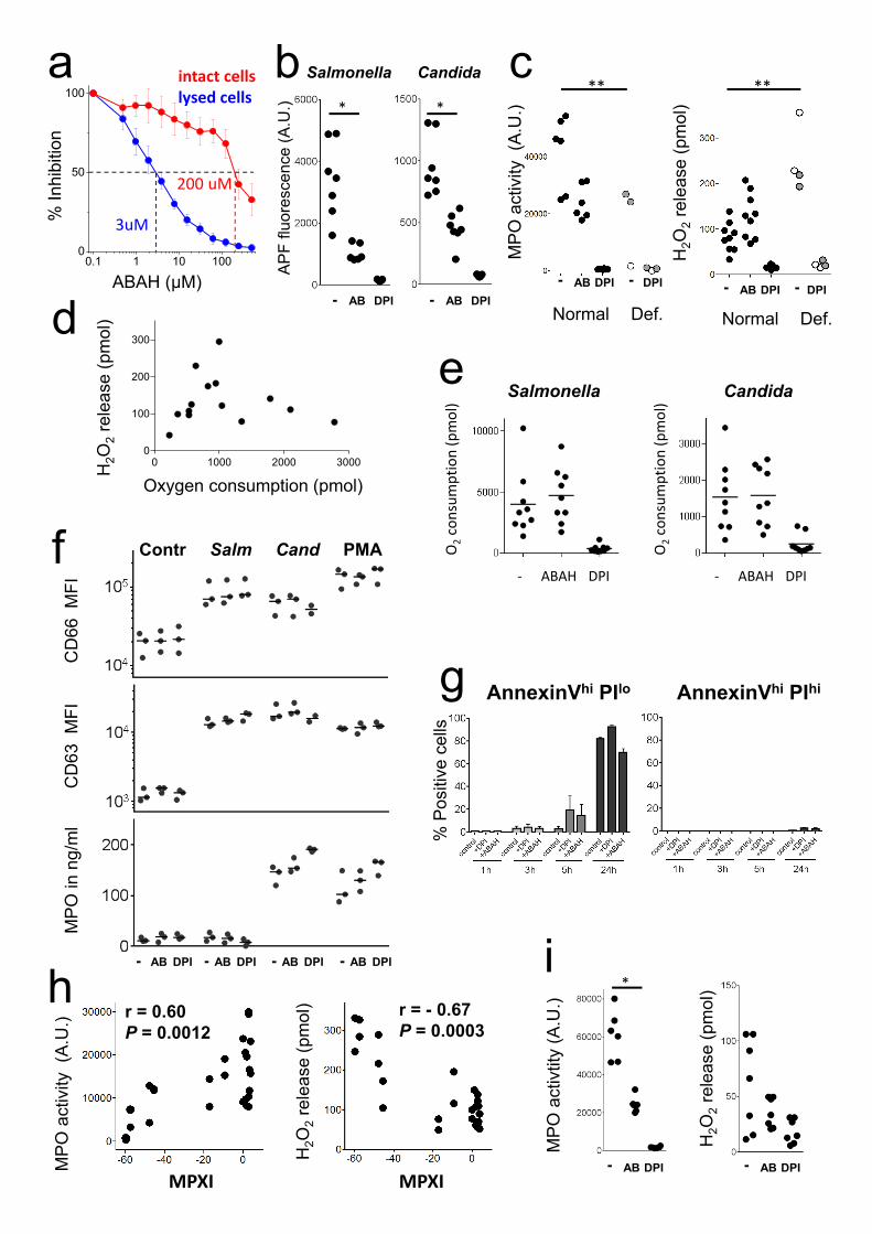
Supplementary Figure 1: Reactive oxygen species generation and leakage of human
neutrophils in vitro.
a, Inhibition of myeloperoxidase activity by ABAH in intact or lysed neutrophils. Data (means and
standard deviations of six technical triplicates) from one representative experiment out of two are
shown. The dotted lines indicate 50% activity and the corresponding ABAH concentrations for lysed
or intact cells, respectively. b, MPO activity measured as APF oxidation in neutrophils from seven
different normal donors after 60 min stimulation in presence/absence of the MPO inhibitor ABAH (AB) or
the NADPH oxidase inhibitor DPI (Wilcoxon test; *, P< 0.05). c, MPO activity and H2O2 release of
neutrophils from eight different normal donors (black circles), two partially deficient donors (grey), and
two severely deficient donors (empty circles) during stimulation with Candida in presence/absence of the
MPO inhibitor ABAH (AB) or the NADPH oxidase inhibitor DPI (Kruskal-Wallis multiple comparisons
test; **, P< 0.01). d, Relationship between oxygen consumption and H2O2 release in neutrophils
stimulated with heat-killed Candida for 75 min. Mean values of triplicate wells containing 100’000
neutrophils are shown. e, Impact of inhibiting myeloperoxidase with ABAH, or NADPH oxidase with
DPI, on oxygen consumption rate after stimulation with heat-killed Salmonella and Candida. f, Impact of
inhibitors on neutrophil degranulation. Cells from three different donors were stimulated with PBS
(Contr), heat-killed Salmonella (Salm), Candida (Cand), or PMA for 75 min in presence/absence of the
MPO inhibitor ABAH (AB) or the NADPH oxidase inhibitor DPI. Surface expression of degranulation
markers CD63 and CD66 was measured by flow cytometry. MPO release to the extracellular medium was
quantified using ELISA. Different stimuli triggered the three degranulation processes to a various degree,
but all three assays demonstrated no impact of inhibitors ABAH and DPI on neutrophil degranulation. g,
Impact of 500 μM ABAH and 10 μM DPI on neutrophil viability. The fraction of cells undergoing
apoptosis [annexinVhi propidium iodide (PI)lo, left] and cells undergoing necrosis (annexinVhi PIhi, right)
at various times after inhibitor addition is shown. Means and standard deviations for four donors measured
in one experiment are shown. In all other experiments of this study, cells were exposed to these inhibitors
for a maximum of 75 min. h, Relationship between donor mean peroxidase index (MPXI) and HOCl
generation (left) or H2O2 release (right) after stimulation with live or heat-killed Salmonella (r, Spearman
correlation coefficient). Each dot represents data for an individual human donor. i, MPO activity and H2O2
release of neutrophils from seven different normal donors during stimulation with 1 nM PMA in
presence/absence of the MPO inhibitor ABAH (AB) or the NADPH oxidase inhibitor DPI (Wilcoxon test;
*, P< 0.05).
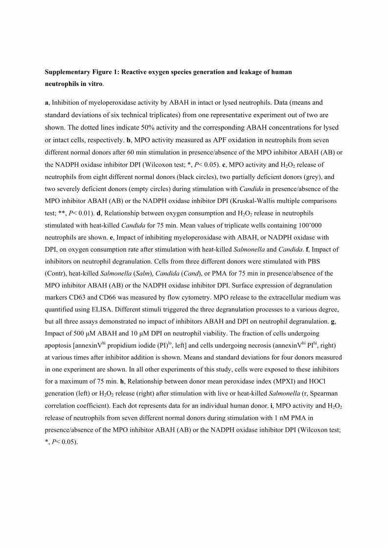
Supplementary Figure 2: Lipid peroxidation in uninfected spleen as detected by
an antibody to 4-hydroxynonenal (4-HNE).
Representative micrograph of a spleen cross-section from one out of four uninfected MPO-
deficient mice. CD11bhi cells populate the red pulp. 4-HNE signals are undetectable in these
areas, whereas some background staining is visible in the white pulp areas (which are
negative for CD11b). The scale bar represents 500 μm.
CD11b 4-HNE
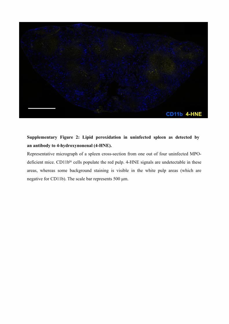
Supplemental References
1 Winterbourn, C. C., Hampton, M. B., Livesey, J. H. & Kettle, A. J. Modeling the reactions of superoxide and myeloperoxidase in the neutrophil phagosome: implications for microbial killing. J Biol Chem 281, 39860-39869 (2006).
2 Burton, N. A. et al. Disparate impact of oxidative host defenses determines the fate of Salmonella during systemic infection in mice. Cell host & microbe 15, 72-83, doi:10.1016/j.chom.2013.12.006 (2014).
3 Imlay, J. A. in EcoSal (ed R. III.; Kaper Curtiss, J.B.; Squires, C.L.; Karp, P.D.; Neidhardt, F.C.; Slauch, J.M.) Ch. Modul 5.4.4, (ASM Press, 2009).
4 Seaver, L. C. & Imlay, J. A. Are respiratory enzymes the primary sources of intracellular hydrogen peroxide? J Biol Chem 279, 48742-48750 (2004).
5 Beuzon, C. R. et al. Salmonella maintains the integrity of its intracellular vacuole through the action of SifA [published erratum appears in EMBO J 2000 Aug 1;19(15):4191]. EMBO J. 19, 3235-3249 (2000).
6 Korshunov, S. S. & Imlay, J. A. A potential role for periplasmic superoxide dismutase in blocking the penetration of external superoxide into the cytosol of Gram-negative bacteria. Mol Microbiol 43, 95-106 (2002).
7 Seaver, L. C. & Imlay, J. A. Hydrogen peroxide fluxes and compartmentalization inside growing Escherichia coli. J Bacteriol 183, 7182-7189 (2001).
8 Makino, N., Sasaki, K., Hashida, K. & Sakakura, Y. A metabolic model describing the H2O2 elimination by mammalian cells including H2O2 permeation through cytoplasmic and peroxisomal membranes: comparison with experimental data. Biochimica et biophysica acta 1673, 149-159, doi:10.1016/j.bbagen.2004.04.011 (2004).
9 Storkey, C., Davies, M. J. & Pattison, D. I. Reevaluation of the rate constants for the reaction of hypochlorous acid (HOCl) with cysteine, methionine, and peptide derivatives using a new competition kinetic approach. Free radical biology & medicine 73, 60-66, doi:10.1016/j.freeradbiomed.2014.04.024 (2014).
10 Kundrat, P., Bauer, G., Jacob, P. & Friedland, W. Mechanistic modelling suggests that the size of preneoplastic lesions is limited by intercellular induction of apoptosis in oncogenically transformed cells. Carcinogenesis 33, 253-259, doi:10.1093/carcin/bgr227 (2012).
11 Behar, D., Czapski, G., Rabani, J., Dorfman, L. M. & Schwarz, H. A. Acid dissociation constant and decay kinetics of the perhydroxyl radical. The Journal of Physical Chemistry 74, 3209-3213, doi:10.1021/j100711a009 (1970).
12 Aurudi, R. L. R., A.B. Reactivity of HO2/O-2 radicals in aqueous solution. J Phys Chem Ref
Data 14, 1041–1100 (1985). 13 Steeb, B. et al. Parallel exploitation of diverse host nutrients enhances salmonella virulence.
PLoS Pathog 9, e1003301 (2013). 14 Stroppolo, M. E. et al. Single mutation at the intersubunit interface confers extra efficiency to
Cu,Zn superoxide dismutase. FEBS Lett 483, 17-20 (2000). 15 Bull, C. & Fee, J. A. Steady-state kinetic studies of superoxide dismutases: properties of the
iron containing protein from Escherichia coli. Journal of the American Chemical Society 107, 3295-3304, doi:10.1021/ja00297a040 (1985).
16 Meir, E. & Yagil, E. Further characterization of the two catalases inEscherichia coli. Current Microbiology 12, 315-319, doi:10.1007/bf01567889 (1985).
17 Baker, L. M. & Poole, L. B. Catalytic mechanism of thiol peroxidase from Escherichia coli. Sulfenic acid formation and overoxidation of essential CYS61. J Biol Chem 278, 9203-9211 (2003).
18 Poole, L. B. Bacterial defenses against oxidants: mechanistic features of cysteine-based peroxidases and their flavoprotein reductases. Arch Biochem Biophys 433, 240-254 (2005).