neuroimaging in alzheimer’s disease emily altmananthony grigas sue kotchgeorgine pindar elizabeth...
TRANSCRIPT

Neuroimaging in Alzheimer’s Disease
Emily Altman•Anthony Grigas
Sue Kotch•Georgine Pindar
Elizabeth Thompson

Alzheimer’s DiseaseDefined
Alois Alzheimer 100 years ago41
Chronic neurodegenerative disorder characterized by the pathological accumulation of insoluble intraneuronal and extraneuronal proteins in the brain and neuronal degeneration
The most common cause of degenerative dementia in individuals over 6511

Alzheimer’s DiseaseAppearance
Progressive alterations in functional status secondary to cognitive deficits42
Warning signs Progression beginning with short-term
memory deficits or “forgetfulness.”

Alzheimer’s DiseaseThe Facts12
One of the principal causes of disability and decreased Quality of Life among older adults
4th leading cause of death due to disease in people over 65
In 2000: 4.5 million people with AD in the US Barring a cure: 13.2 million people will have
AD in 2050 Cost of treatment in US is $100 billion/year

Alzheimer’s DiseaseThe Facts12
Prognosis: inexorable decline and eventual death
Primary risk factors: age, family hx, genetic markers, female gender after 80, HTN, diabetes, obesity, ↑cholesterol
Possible risk factors: head injury, depression, low TSH, folate deficiency, low educational attainments

Alzheimer’s DiseaseThe Facts12
Possible protective factors: Homozygous APOE2, fish consumption, omega-3 fatty acid, high educational level, regular exercise, NSAID therapy, moderate ETOH intake, vitamins C, E, B6, B12, folate
Alzheimer’s DiseaseHistology 101
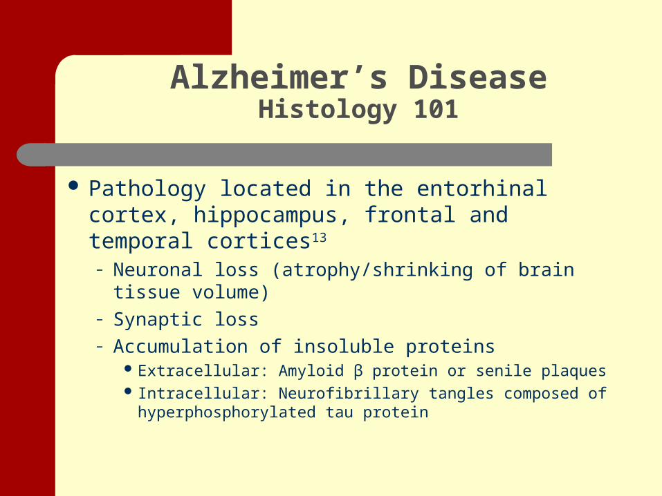
Pathology located in the entorhinal cortex, hippocampus, frontal and temporal cortices13
– Neuronal loss (atrophy/shrinking of brain tissue volume)
– Synaptic loss– Accumulation of insoluble proteins
Extracellular: Amyloid β protein or senile plaques Intracellular: Neurofibrillary tangles composed of
hyperphosphorylated tau protein
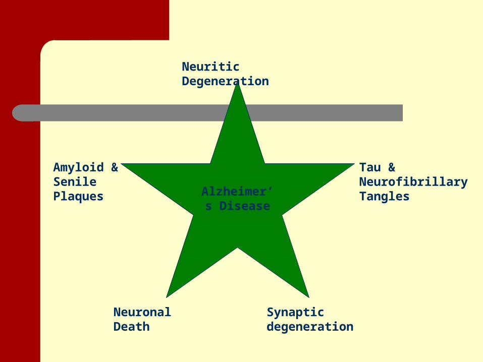
Neuritic Degeneration
Tau & Neurofibrillary Tangles
Synaptic degenerationNeuronal Death
Amyloid & Senile Plaques
Alzheimer’s Disease

Pathogenesis of Alzheimer’s Disease
Unknown– Amyloid cascade hypothesis– Tau hypothesis– Apoptosis hypothesis– Pathologic return to cell cycle– Attrition hypothesis– Genetic influences– Vascular theory

Current Pharmacological Interventions
Currently two classes of compounds– Cholinesterase inhibitors
Aricept
– A Partial NMDA receptor antagonist Memantine
Symptom management

Diagnosing Alzheimer’s Disease
Clinical Diagnosis Autopsy Neuroimaging
- Description
- New horizons
- Prognostic value?

Diagnosing Alzheimer’s DiseaseClinical Diagnosis
Clinical diagnosis requires 3 elements43:- Insidious onset of anterograde memory loss- Progressive decline in memory and mental functions as measured by neurocognitive tests- Cognitive and laboratory tests exclude all other differential diagnoses

Diagnosing Alzheimer’s DiseaseClinical Diagnosis
Laboratory tests to rule out other possible sources of symptoms
- Reversible dementia: nutritional deficiencies, normal-pressure hydrocephalus, subdural hematoma, hypothyroidism, brain tumor, hyponatremia, drug toxicity, alcoholism, infection12
- Non-reversible dementia: Parkinson’s Disease, HIV/AIDS, organic brain disease, cerebrovascular disease53

Diagnosing Alzheimer’s DiseaseClinical Diagnosis
Clinically diagnosed Alzheimer’s may be preceded by a stage of “mild cognitive impairment” (MCI), in which loss of memory and function does not yet meet the criteria for an Alzheimer’s diagnosis43,26

Post-mortem examination
- Autopsy is the only way to definitively identify Alzheimer’s pathology (plaques and tangles)12
- Examiners track the incidence and locations of neuritic plaques and neurofibrillary tangles53
- Disease process is considered more advanced if there are more plaques and tangles, and if they have spread out of the limbic/thalamic regions into the cortex27
Diagnosing Alzheimer’s DiseaseAutopsy

Diagnosing Alzheimer’s DiseaseAutopsy
Post-mortem examination- Uses CERAD scale and Braak staging to quantify
tangles and plaques27
- Sample taken in coronal sections around temporal lobe, hippocampus, entorhinal cortex53
Clinical diagnosis correlates well with autopsy diagnosis (74-90%)43

Diagnosing Alzheimer’s DiseaseNeuroimaging - Description
PET scan:
- F-18 isotope attached to fluorodeoxyglucose (FDG)
- studies brain metabolism of glucose
- provides good test to distinguish Alzheimer’s pathology from vascular dementia
(sensitivity 95%, specificity 71%)26
- H215O used to measure “regional cerebral blood
flow”26, an indirect measure of oxygen usage.

Diagnosing Alzheimer’s DiseaseNeuroimaging - Description
MRI- Structural MRI used to detect brain atrophy, especially in the entorhinal cortex, hippocampal region, medial temporal lobe, and posterior cingulate gyrus.
- Functional (fMRI) tracks blood flow during a task with better spatial resolution than PET, as well as no ionizing radiation. However, it is much more expensive.

Diagnosing Alzheimer’s DiseaseNeuroimaging - Description
MRI- MRI to study cerebral perfusion
- contrast media- arterial spin-labeled
SPECT- Provides perfusion info using tracer chemicals- Most useful in early stages of Alzheimer’s

Diagnosing Alzheimer’s DiseaseNeuroimaging – New horizons
Synchrotron X-ray fluorescence of metals
- Fe and Zn more concentrated in
plaques than normal tissue
- can be detected in very small amounts16

Diagnosing Alzheimer’s DiseaseNeuroimaging – Prognostic value
When combined with genetic testing (for APOE4 allele), FDG PET and fMRI can identify atypical activation patterns in high-risk, asymptomatic persons14,27.
In some cases, onset of symptoms may be predicted up to 3 years in advance.26
But with these research areas so new, and the tests so expensive, is neuroimaging really a good tool to fight Alzheimer’s?...

Use of Neuroimaging in Alzheimer’s Disease
The use of neuroimaging in AD is limited for many reasons including:
– Limited research– Existence of reliable cognitive exams– Once clinical signs present, already irreversible brain
damage– MRI is a costly test– Unable to see plaques and tangles on imaging– Common to see some imaging results with AD that also are
present with different types of dementia

Use of Neuroimaging in Alzheimer’s Disease
Limited Research
No neuroimaging or lab markers now exist for reliable presymptomatic diagnosis of AD12.
The exact mechanism by which brain degeneration results from neurofibrillary tangles and amyloid plaques remain incompletely understood41.
Use of Neuroimaging in Alzheimer’s Disease
Reliable Cognitive Exams
A probable diagnosis of AD can be established pre mortem with a confidence interval of ~80% based on the following clinical criteria:– Medical history– Physical exam– Lab tests– Radiology– Neuropsychological evaluation16
Use of Neuroimaging in Alzheimer’s Disease
Irreversible Brain Damage
By the time individuals show signs of cognitive impairment due to AD or MCI as a precursor to AD, they already have irreversible brain pathology.
The prodromal (preclinical) phase of AD includes a period of disease pathology that is clinically undetectable or uncertain43.

Use of Neuroimaging in Alzheimer’s Disease
Similar Imaging Results
Neurofibrillary tangles can be found in AD and other forms of dementia. They can be seen in the absence of plaques, while the same amount of plaques can be found in both people with AD and non-pathologic elderly. Plaques are specific to AD but may also occur in the absence of dementia36.

Diagnostic Imaging ForDifferential Diagnosis
COMMON TYPES OF DEMENTIA Alzheimer's Disease Vascular Dementia Dementia with Lewy Bodies

Neuroimaging Forthe
Diagnosis of Alzheimer’s Disease
Alzheimer's Disease– Structural MRI
Greater atrophy of hippocampus and parahippocampus than in DLB3
Atrophy of the entorhinal cortex is present in MCI27
Volume change of entorhinal cortex27
– Normal aging <1%– Early AD 4%

Neuroimaging Forthe
Diagnosis of Alzheimer’s Disease
Alzheimer's Disease– Functional Studies
Cerebral metabolism– PET
Cerebral perfusion– SPECT– PET– MRI

Neuroimaging Forthe
Diagnosis of Alzheimer’s Disease
Alzheimer's Disease– Activation Studies27
PET– Water marked with radioactive oxygen measures cerebral
regional blood flow (rCBF).– The rCBF increases paralled to the increase in oxygen
need, which corresponds to the increase in synaptic activity in the part of the brain being activated.
MRI– Greater spatial and temporal resolution than PET– No ionizing radiation
Neuroimaging Forthe
Diagnosis of Alzheimer’s Disease
Alzheimer's Disease– Regional density as measured by PET27
Compounds being developed that detect amyloid deposition and neurofibrillary tangles
Not readily available yet Further studies need to be done
Neuroimaging for the
Diagnosis of Vascular Dementia
Vascular Dementia– Initially thought to be caused by recurrent strokes– No pathological criteria exist for the diagnosis– “Cerebrovascular disease resulting in dementia”– MRI or CT Scan
Subcortical infarcts Ischemic changes Leukoaraiosis

Diagnostic Imaging forthe
Diagnosis of Dementia with Lewy Bodies
Dementia with Lewy Bodies (DBL)– Protein lumps found in the nuclei
Cortical or subcortical
– PET Hypometabolism29

Diagnostic Imaging forthe
Diagnosis of Dementia with Lewy Bodies
DLB– Supportive Features29
MRI or CT Scan– Preservation of medial temporal lobe structures– Hippocampal and parahippocampal volumes are significantly
larger when compared to AD SPECT
– Atrophy of putamen and occipiatl hypoperfusion PET
– Hypometabolism Low uptake on MIBG myocardial scintigraphy Prominent slow wave activity on EEG with temporal lobe
transient sharp waves
Neuroimaging forthe
Diagnosis of “Mixed Dementia”
Mixed Dementia– AD vs VaD
Combined pathology very common Autopsy final determination

Use of Neuroimaging Results Prognosis
Prognosis = very difficult to predict/evaluate on an individual level– Density of amyloid plaques: poor correlation with
severity of dementia– Vague trend toward ↓medial temporal lobe
volume as severity of dementia increases– ↑NF tangles seems to correlate with ↓cognitive
skills, but very late in disease progression

Use of Neuroimaging Results Selection of Intervention/Management
fMRI (Functional MRI)– Has been used in research to measure changes in brain
function as it is related to cognition.– Used as an indirect measure of neural activity as it
examines changes in blood oxygenation signal intensity.– Treatment strategies may be considered based on results
found on the group level, however clinicians will need to identify neurotransmitter system dysfunction in each individual.
– Currently not used frequently in clinical setting due to cost, unable to detect all plaques and tangles in AD, unable to detect pre-symptomatic AD, and damage already done.
Use of Neuroimaging Results Selection of
Intervention/Management
Pharmacological Intervention Current pharmacological treatment
approaches are based upon staging of AD.– Mild to Moderate AD
Acetylcholinesterase Inhibitors (Donepezil, Galantamine, Rivastigmine, Tacrine)
– Moderate to Severe AD NMDA antagonists (Memantine)

Use of Neuroimaging Results Selection of Intervention/Management
Acetylcholinesterase Inhibitors– Helps to delay the disease process in terms of
cognitive function and improves activities of daily living.
– Enhances cholinergic activity by inhibiting the enzyme acetylcholinesterase which metabolizes the neurotransmitter acetylcholine rapidly and improves cognitive function.

Use of Neuroimaging Results Selection of Intervention/Management
NMDA Antagonists– Improves functional capacity, global clinical status, and
cognitive domains in advanced stages of AD.– Attaches to NMDA receptors in the brain and regulates
glutamate allowing for the appropriate chemical environment for the brain to process, store, and retrieve information for learning and memory.
– Regulates glutamate to control the balance of calcium and prevent further nerve cell death.

Limitations Seen in MRI Studies
Small sample sizes No effective disease stage classification used for experimental group “MRI protocol” varies widely
– No uniformity/standardization of imaging acquisition and processing– Variety of choice of structures are used– Tracing methods vary, are often done by hand and are very labor
intensive and complex – +/- head size corrections– +/- establishment of interrater reliability for methods– Lack rater-independent objective measures of brain anatomy and
function Often lack post-mortem confirmation of “probable AD” diagnosis of
the experimental group What is the MCID for volume change?

Points to Ponder
We are treating this disease totally in the dark Think complement, not compete Biases of neuropsychological (cognitive) tests
used in the diagnosis of AD Need a preclinical diagnosis…then what? The pathologic burden of AD begins decades
before diagnosis
Discussion Questions
1. We are in an era of “pre-emptive” medical testing. Especially as clients age, they are advised to get early-detection tests even in the absence of any clinical symptoms (i.e. mammograms.) Do you think pre-emptive MRIs (or other imaging) are warranted to look for signs of Alzheimer’s?
2. Clinically speaking, would you treat a client with stroke differently if they manifest no symptoms, but their MRI also shows changes suggestive of Alzheimer’s? A client with hip fracture (and a positive MRI)? A client with elective knee replacement and “no health problems” (but a positive MRI)?
3. Despite an abundance of research on the use of MRI in the diagnosis of AD, in “real life” medical clinics, the use of MRI imaging is not the standard of care. Knowing the definition of Alzheimer’s disease (on a histological/tissue level), knowing how it is currently diagnosed in neurology clinics, and understanding a little about MR imaging, discuss why MR imaging is not yet the standard of care or routinely used in diagnosing patients with Alzheimer’s disease.

Thank You

References
1 Adak S, Illouz K, Gorman W, et al. Predicting the rate of cognitive decline in aging and early Alzheimer disease. Neurology. 2004;63(1):108-114.
2 Allen RS, Kwak J, Lokken KL, Haley WE. End-of-life issues in the context of Alzheimer’s disease. Alzheimer’s Care Quarterly. 2003; 4(4): 312-30.
3 Bondi MW, Houston WS, Eyler LT, Brown GG. fMRI evidence of compensatory mechanisms in older adults at genetic risk for Alzheimer disease. Neurology. 2005; 64(3): 501-8.
4 Bottino CM, et al. Volumetric MRI measurements can differentiate Alzheimer’s disease, mild cognitive impairment, and normal aging. Int Psychogeriatr. 2002;14(1): 59-72.
5 Bowler JV. Vascular cognitive impairment. Neuro in Practice. 2005; 76 (supp V): 35-44.
6 Burns JM et al. White matter lesions are prevalent but differentially related with cognition in aging and early Alzheimer’s disease. Arch Neurol. 2005; 63(12): 1870-6.
7 Callen DJ, et al. Beyond the hippocampus: MRI volumetry confirms widespread limbic atrophy in AD. Neurology. 2001; 57(9): 1669-74.
8 Chetelat G, Baron JC. Early diagnosis of Alzheimer’s disease: contribution of structural neuroimaging. Neuroimage. 2003; 18(2): 525-41.

References
9 Clarfield AM. The decreasing prevalence of reversible dementias: an updated meta-analysis. Archives of Internal Medicine. 2003; 163(18): 2219-29.
10 deLeon MJ, et al. MRI and CSF studies in the early diagnosis of Alzheimer’s disease. J Intern Med. 2004 ; 256(3): 205-23.
11 Denihan A, et al. CT measurement of medial temporal lobe atrophy in Alzheimer’s disease, vascular dementia, depression and paraphrenia. Int J Geriatr Psychiatry. 2000; 15(4): 306-12.
12 Desai AK, Grossberg GT. Diagnosis and treatment of Alzheimer’s disease. Neurology. 2005; 64(Suppl3): S34-S39.
13 Du AT, Schuff N, et al. Atrophy rates of entorhinal cortex in AD and normal aging. Neurology. 2003; 60(3): 481-486.
14 Fleisher AS, Houston WS, Eyler LT, et al. Identification of Alzheimer disease by functional magnetic resonance imaging. Archives of Neurology. 2005; 62(12): 1881-8.
15 Fox NC, Scahill RI, et al. Correlation between rates of brain atrophy and cognitive decline in AD. Neurology. 1999; 52(8):1687-1689.
16 Fradinger EA, Bitan G. En route to early diagnosis of Alzheimer’s disease—are we there yet? Trends Biotechnol. 2005 ; 23(11): 531-3.
17 Frisoni GB, et al. Radial width of the temporal horn: a sensitive measure in Alzheimer disease. ANJR Am J Neuroradiol. 2002; 23(1): 35-47.

References
18 Frisoni GB, Filippi M. Multiple sclerosis and Alzheimer disease through the looking glass of MR imaging. Am J Neuroradiol. 2005; 26(10): 2488-91.
19 Galton CJ, et al. A comparison of the Addenbrooke’s Cognitive Examination (ACE), conventional neuropsychological assessment, and simple MRI-based medial temporal lobe evaluation in the early diagnosis of Alzheimer’s disease. Cogn Behav Neurol. 2005; 18(3): 144-50.
20 Garrido GEJ, Furuie SS, et al. Relation between medial temporal atrophy and functional brain activity during memory processing in Alzheimers diseas: a combined MRE and SPECT study. J. Neurol Neurosurg Psychiatry. 2002; 73: 508-516.
21 Hampel H, et al. Age transformation of combined hippocampus and amygdala volume improves diagnostic accuracy in Alzheimer’s disease. J Neurol Sci. 2002; 194(1): 15-19.
22 Jack CR, et al. Medial temporal atrophy on MRI in normal aging and very mild Alzheimer’s disease. Neurology. 1997; 49: 786-794.
23 Jack CR, Slomkowski M, Gracon S, et al. MRI as biomarker of disease progression in a therapeutic trial of milameline for AD. Neurology. 2003; 60(2): 253-260.
24 Killiany RJ, et al. Use of structural magnetic resonance imaging to predict who will get Alzheimer’s disease. Ann Neurol. 2000; 47(4): 430-9.

References
25 LeBlanc AC. The role of apoptotic pathways in Alzheimer’s disease neurodegeneration and cell death. Current Alzheimer Research. 2005; 2(4): 389-402.
26 Masdeu JC, Zubeita JL, Arbizu J. Neuroimaging as a marker of the onset and progression of Alzheimer’s disease. J Neurol Sci. 2005; 236(1-2): 55-64.
27 Massoud F, et al. The role of routine laboratory studies and neuroimaging in the diagnosis of dementia: a clinicopathological study. J Am Geriatr Soc. 2000; 48: 1204-1210.
28 McKeith IG, et al. Diagnosis and management of dementia with Lewy bodies. Neurology. 2005; 65: 1863-72.
29 Mizuno K, Wakai M, Takeda A, Sobue G. Medial temporal atrophy and memory impairment in early stage of Alzheimer’s disease: an MRI volumetric and memory assessment study. J Neurol Sci. 2000; 173(1): 18-24.
30 Nagy Z. The last neuronal division: a unifying hypothesis for the pathogenesis of Alzheimer’s disease. J Cell Mol Med. 2005; 9(3): 531-41.
31 No authors. In search of Alzheimer’s disease. Harv Ment Health Lett. 2005; 22(4): 3-5.
32 Norfray JF. Alzheimer’s disease: neuropathologic findings and recent advances in imaging. AJR Am J Roentgen. 2004; 182(1): 3-13.

References
33 Petersen RC, et al. Memory and MRI-based hippocampal volumes in aging and AD. Neurology. 2000; 54(3): 581-7.
34 Sclan SG, Kanowski S. Alzheimer’s disease: stage-related interventions. Lippincott’s Case Management. 2001; 6(2): 48-60.
35 Selkoe DJ. By the way, doctor. Is there a brain scan that can specifically diagnose Alzheimer’s disease? Harv Health Lett. 2005; 30(12): 8.
36 Serretti A, Artioli P, Quartesan R, De Ronchi D. Genes involved in Alzheimer’s disease, a survey of possible candidates. Journ of Alzheimer’s Disease. 2005; 7: 331-53.
37 Schuff N, et al. Changes of hippocampal N-acetyl aspartate and volume in Alzheimer’s disease. A proton MR spectroscopic imaging and MRI study. Neurology. 1997; 49(6): 1513-21.
38 Silbert LC, Quinn JF, Moore MM, et al. Changes in premorbid brain volume predict Alzheimer’s disease pathology. Neurology. 2003; 61(4): 487-92.
39 Smith GB. Case management guidelines: Alzheimer disease and other dementias. Lippincott’s Case Management. 2002; 7(2): 77-84.
40 Stoub TR, et al. MRI predictors of risk of incident Alzheimer disease. A longitudinal study. Neurology. 2005; 64: 1520-1524.
References
41 Trojanowski JQ, Lee VM. Rous-Whipple Award Lecture. The Alzheimer’s brain: finding out what’s broken tells us how to fix it. Am J Pathology. 2005; 167(5): 1183-8.
42 Tsuang DW et al. Genetic association between APOE*4 allele and Lewy bodies in Alzheimer diease. Neurology. 2005; 64(3):509-513.
43 Zamrini E, De Santi S, Tolar M. Imaging is superior to cognitive testing for early diagnosis of Alzheimer’s disease. Neurobiol of Aging. 2004; 25(5): 685-91.
44 Rombouts et al. Alterations in Brain Activation during cholinergic enhancement with rivastigmine in Alzheimer’s Disease. J of Neurology. 2002; 73(6): 665-671.
45 Wilcock, G et al. A Long-Term Comparison of Galantamine and Donepezil in the Treatment of Alzheimer’s Disease. Adis International. 2003; 20(10): 777-789.
46 Wilkinson, D et al. Cholinesterase Inhibitors used in the Treatment of Alzheimer’s Disease: The relationship between Pharmacological effects and Clinical Efficacy. Drugs & Aging. 2004; 21(7): 453-478.

References
47 Plosker, G et al. Memantine: A Pharmacoeconomic Review of its Use in Moderate-to-Severe Alzheimer’s Disease. Pharmacoeconomics. 2005; 23(2): 193-206.
48 Rutger, G et al. Cholinergic challenge in Alzheimer’s patients and mild cognitive impairment differentially affects hippocampal activation – a pharmacological fMRI study. Brain. 2006; 129(1): 141-157.
49 www.alzheimers.org50 www.uptodate.com51 www.alz.org52 Barber R et al. A comparison of medial and lateral temporal lobe atrophy in
dementia with lewy bodies and alzheimer’s disease: magnetic resonance imaging volumetric study. Dement Geriatr Cogn Disord. 2001;12:198-205.
53 www.alzforum.org