neuron, vol. 24, 49–65, september, 1999, copyright 1999 by ... · neuron). this dissociation may...
TRANSCRIPT

Neuron, Vol. 24, 49–65, September, 1999, Copyright 1999 by Cell Press
Neuronal Synchrony: A Versatile Code Reviewfor the Definition of Relations?
temporal relations requires the joint evaluation of re-sponses from more than one neuron, only experimentsthat permit simultaneous measurements of responses
Wolf Singer*Max-Planck-Institute for Brain ResearchDeutschordenstrasse 4660528 Frankfurt from multiple units are considered. These include multi-Federal Republic of Germany electrode recordings from multiple individual cells, but
also measurements of local field potentials (LFPs) andelectroencephalographic (EEG) or magnetoencephalo-graphic (MEG) recordings. The signals of these latterMost of our knowledge about the functional organizationmethods reflect the average activity of large cell popula-of neuronal systems is based on the analysis of the firingtions. Because this activity leads to measurable signalpatterns of individual neurons that have been recordedfluctuations only if it is sufficiently synchronized, theseone by one in succession. This approach permits as-global recordings provide valuable information aboutsessment of event-related variations in discharge rate,the temporal relations between responses.but it precludes detection of any covariations in the
amplitude or timing of distributed responses if theseTwo Complementary Binding Strategiescovariations result from internal neuronal interactionsDiscussions about the putative functional role of syn-rather than from time locking to stimulus or motorchrony focus on the question of whether it can serve asevents.a mechanism to bind distributed neuronal activity. ToAs it is likely that internal coordination of distributedprovide an adequate background for the examination ofresponses is functionally as relevant as stimulus-this question, I shall first deal with some general, imple-induced coordination, multielectrode recordings are in-mentation-independent aspects of binding operations.creasingly being used to analyze internally generated
As the Gestalt psychologists pointed out, our cogni-covariations of firing patterns. More than a decade ago,we used this method to reveal that neurons in the visual tive systems have the tendency to interpret objects andcortex tend to synchronize their discharges with a preci- events as related if they are contiguous in space or time,sion in the millisecond range when activated with a sin- or if they exhibit similarities in certain feature domains.gle contour (Gray and Singer, 1987, Soc. Neurosci., ab- Thus, contours that touch one another, have similar con-stract; 1989), whereas they fail to do so when activated trast, or move with the same speed in the same directionby different contours moving in different directions (Gray (common fate) are more likely to be perceived as compo-et al., 1989; Engel et al., 1991c). In addition, these stimu- nents of the same object than spatially distant contourslus-induced, context-dependent synchronization phe- or contours that have no features in common. Likewise,nomena were found to be associated with a conspicu- events that coincide in time are interpreted with greaterous oscillatory modulation of cell firing in a frequency probability as related than events separated in time. Atrange between 30 and 50 Hz, the so-called g frequency early stages of sensory processing, spatial relations andrange. Two aspects make this synchronization interest- relations in feature space are represented by the ampli-ing. First, it results from internal coordination of spike tude and the topological relations of activation foci intiming and is not simply caused by stimulus-locked ordered maps. Temporal relations, however, are repre-changes in discharge rate. Second, synchronization sented by the relative timing of responses. In order toprobability changes in a systematic way when the per- accomplish perceptual grouping, the distributed re-ceptual coherence of stimulus constellations is modified. sponses of feature-selective cells need to be boundThus, this type of synchrony is not a trivial reflection of
together at some stage of processing. Evidence indi-anatomical connectivity such as shared input through bi-
cates that this is achieved in two complementary ways.furcating axons, but instead results from context-depen-One strategy is binding of responses by convergencedent, dynamic interactions within the cortical network.of axonal projections. Axons of cells whose responsesThe evidence for an internal coordination of spikeshould be bound are made to converge onto a commontiming raises the question of whether it serves a functiontarget cell at the next-higher processing level. If thein cortical processing or whether it is merely an epiphe-threshold of this binding unit is appropriately adjusted,nomenon. The goal of this paper is to review theoreticalits response signals the specific conjunction of featuresarguments and data that relate to this issue. The firstto which the feeding cells are tuned. We shall addresspart examines how well the nervous system, and in par-this grouping strategy as “binding by convergence” orticular the cerebral cortex, can distinguish between syn-“binding by conjunction cells.” This coding principle ischronous and asynchronous responses, and whetheralso known as “labeled line coding” because the re-any significance is attributed to precisely synchronizedsponses of a given unit have a fixed label attached todischarge patterns when these are coordinated by ex-them; they always signal the same conjunction of inputternal events, e.g., by the synchronous onset of sensorysignals. The complementary strategy for response bind-stimuli. In the second part, data are reviewed from ex-ing relies on dynamic selection and grouping of re-periments that were designed to examine putative func-sponses. Here, responses are bound by jointly enhanc-tions of internally generated synchronization. As the as-
sessment of internally generated, non-stimulus-locked ing their saliency relative to other, nonbound responses.Enhanced responses have a stronger impact on down-stream processes than nonenhanced responses and* E-mail: [email protected].

Neuron50
therefore dominate subsequent computations. Thus, the ground elements of ,10 ms still support perceptualgrouping (Leonards et al., 1996). Because responses ofresults of these computations will reflect the specific
configuration of features to which cells with enhanced neurons in the visual cortex follow the time course ofthe stimuli that evoke them, the results of these studiesresponses are tuned. We shall address this selection
and grouping strategy as “dynamic binding” and the suggest that synchronous responses are bound percep-tually while asynchronous responses are interpreted asassociated coding principle as “relational coding” or
“assembly coding,” because here the information about unrelated.It is noteworthy that the short offsets that support thisa particular conjunction is contained in the dynamically
adjustable configuration of the enhanced responses of segmentation are not perceptible, suggesting a dissoci-ation between the perceptibility of small differences indistributed neurons (for reviews of the extensive litera-
ture on labeled line and assembly coding, see Singer the time course of stimuli on the one hand and the effectof such differences on perceptual grouping on the other.and Gray, 1995, and other contributions in this issue of
Neuron). This dissociation may have to do with the fact that,in vision, the temporal cues supporting grouping as aThe topology of connections in cortical networks re-
flects these two grouping strategies and accounts well function of stimulus synchrony are mediated mainly bythe magnocellular pathway, while the other, nontempo-for perceptual grouping according to contiguity in eu-
clidian and feature space. The first strategy is imple- ral grouping cues are mediated by both the magno- andthe parvocellular pathway (Leonards and Singer, 1998).mented by the highly complex recombination of feedfor-
ward cortico-cortical connections. It leads to a large These two pathways interact at multiple levels but sub-serve somewhat different functions. The magnocellularvariety of conjunction-specific neurons, the complexity
of which increases as one proceeds along the pro- pathway is exquisitely sensitive to temporal featuresand can signal stimulus transients with high temporalcessing hierarchy. The second strategy appears to rely
on two other classes of cortico-cortical connections (re- resolution, while the parvocellular pathway operateswith low temporal but high spatial and spectral resolu-viewed by Singer, 1995; Phillips and Singer, 1997): (1)
reciprocal connections that link cells situated within the tion (for a review of the extensive literature, see Leo-nards and Singer, 1998). This functional dichotomy issame cortical area, as well as cells distributed across
different areas but occupying the same level in the pro- relevant in the present context because it is a likelybasis for the ability of the visual system to use bothcessing hierarchy; and (2) feedback connections that
reciprocate the feedforward connections. Together, temporal and spatial cues in parallel for perceptualgrouping. If within the same matrix of line elements onethese reciprocal cortico-cortical connections constitute
the large majority of synaptic inputs to cortical cells. figure is defined by the synchronous onset of elements(temporal cue), and another spatially overlapping figureFor the intraareal connections in primary visual cortex,
it is established that they preferentially couple neurons by differences in the orientation of the respective lineelements (spatial cue), either the temporally or the spa-that are nearby or that code for similar features (Gilbert
and Wiesel, 1989; Schmidt et al., 1997a, 1997b). Hence, tially defined figure is perceived depending on the rela-tive saliency of the two cues (temporal offset versusif these neurons are coactivated, they are more likely to
have the saliency of their responses enhanced jointly orientation difference) (Leonards and Singer, 1998). Thissuggests that spatial and temporal grouping cues areby cooperative interactions than are neurons that are
far apart or tuned to very dissimilar features. As a conse- processed in parallel and, if they conflict, the less salientcue is disregarded.quence, responses to contours that are contiguous in
euclidian and/or feature space have an enhanced proba- This ability of the visual system to rely on either spatialor temporal cues, if the two cues are in conflict, is ofbility of being processed jointly and, thus, of being
bound together. considerable functional relevance. On the one hand, itpermits binding of nontemporal features that are relatedbut attached to temporally dispersed elements. On theGrouping According to Temporal Cuesother hand, it allows segregation of features that areIn contrast to the numerous experimental and theoreti-unrelated but attached to temporally contiguous ele-cal studies devoted to the analysis of grouping opera-ments. The ability to base perceptual grouping on eithertions in the domain of spatial features, comparativelyspatial or temporal cues is also likely the cause of anfew studies have been devoted to the questions of (1)apparent conflict between the psychophysical studiesto what extent temporal contiguity of stimuli is exploitedthat support grouping based on temporal cues (seefor perceptual grouping and (2) through which neuronalabove) and a study that denies such a mechanism. Kipermechanisms such grouping could be achieved. Most ofet al. (1996) found that perception of figures defined bythe evidence regarding perceptual grouping by temporalspatial cues is not impaired if false temporal conjunc-cues comes from recent psychophysical studies on vi-tions are introduced at random by presenting selectedsion. These studies indicate that spatially distributedelements of the figure synchronously with elements ofcontour elements are bound perceptually and interpre-the background. Here, the temporal cues did not defineted as elements of a coherent figure if they appear ora figure and hence may have been simply discardedchange synchronously, while elements that follow differ-through competition.ent time courses are perceived as unrelated (Leonards
In summary, the results of psychophysical studieset al., 1996; Alais et al., 1998; Usher and Donnelly, 1998;suggest the following conclusions. First, informationLee and Blake, 1999). The temporal resolution of thisabout the temporal parameters of stimuli is transmittedgrouping mechanism is surprisingly high. Temporal off-
sets between the respective appearances of figure and over several processing stages with a precision in the

Review: Singer51
millisecond range. Second, asynchronies among spa- and across cortical areas, and even across the cerebralhemispheres and subcortical structures, can synchro-tially distributed responses of ,10 ms are exploitable
for perceptual grouping. Third, the mechanism that eval- nize their spike discharges on the basis of oscillationsin the g frequency range (reviewed by Singer and Gray,uates temporal relations among responses for percep-
tual grouping interprets synchronous responses as re- 1995; for more recent findings, see Livingstone, 1996;Brecht et al., 1998; Friedman-Hill et al., 1999; Maldonadolated and segregates them from responses that are
temporally offset. Fourth, temporal and nontemporal et al., 1999). This implies that dispersion of spike timingmust have remained below the duration of a half cycle,grouping cues are evaluated in parallel, the former being
conveyed mainly by the magnocellular pathway. i.e., below about 10 ms for cortical interactions andbelow 5 ms for retino-cortical transmission. The con-These findings raise the question of how temporal
grouping cues are evaluated at the neuronal level. In spicuous spike patterns described by Prut et al. (1998)in the prefrontal cortex point in the same direction.analogy to grouping mechanisms for nontemporal fea-
tures (see above), it would suffice that the synchronous As proposed by Abeles (1991) and recently again byShadlen and Newsome (1998), one possible way toresponses to simultaneously appearing or simultane-
ously changing stimuli are more salient, i.e., have a achieve such high temporal precision in neuronal signal-ing despite “slow” neurons is synchronization of dis-stronger joint impact on cells at subsequent processing
stages than the asynchronous responses to temporally charges across parallel channels, a special form of pop-ulation coding. In the proposed models, this is achieveddispersed stimuli. Two nonexclusive scenarios may be
considered. First, synchronously active cells might co- by cross-coupling parallel channels through divergingand converging axon collaterals. As demonstrated re-operate particularly effectively through cortico-cortical
connections and thereby increase their discharge rate. cently (Aertsen et al., 1996; Diesmann et al., 1997), suchsynfire chains (Abeles, 1991) have the interesting prop-Second, synchronous responses might by themselves
and without further amplification have a stronger impact erty that the synchronization of discharges across paral-lel channels does not decrease from one synaptic levelon cells at subsequent processing stages than cells
responding to temporally offset stimuli. to the next but may even increase if coupling is appropri-ately adjusted. The reason for this preservation of preci-In both cases, two prerequisites need to be fulfilled.
First, timing of discharges must be preserved across sion is that synchronized EPSP barrages are more effec-tive in triggering postsynaptic spikes than temporallypolysynaptic transmission chains with a precision in the
millisecond range. Second, neurons must be able to dispersed inputs (see below). The synchronous EPSPbarrages generated in such synfire chains elicit postsyn-differentiate between synchronous and asynchronous
input. Synchronous excitatory postsynaptic potentials aptic spikes with minimal latency jitter and hence cantransmit the temporal signature of stimuli with high pre-(EPSPs) must be more efficient than temporally dis-
persed EPSPs, and dispersions of ,10 ms must already cision over many synaptic stages. Note that what mat-ters for this temporal precision in transmission is themake a significant difference.rise time of the compound EPSPs rather than the passivemembrane time constant of the integrating neurons.Temporal Precision in Neuronal Transmission
Contrary to what one should expect from the long timeconstants of synaptic integration in central neurons (see, Synchronization Enhances Saliency of Responses
If synchronized responses are grouped because theye.g., Shadlen and Newsome, 1994), cortical networkscan operate with amazing temporal precision. In the are more salient than nonsynchronized responses, neu-
rons evaluating temporal grouping cues must respondauditory cortex of mammals, the spiking patterns ofsingle-cell responses to species-specific calls repro- differently to precisely synchronized and temporally dis-
persed barrages of EPSPs, and—as suggested by psy-duce with millisecond precision from trial to trial (De-Charms et al., 1998; Kilgard and Merzenich, 1998). Com- chophysics—dispersions of ,10 ms must be detect-
able. Again, at first glance, the long time constants ofparable accuracy is found in song birds for auditoryneurons responding to songs and for central motor neu- neuronal membranes seem incompatible with such dif-
ferential sensitivity to coincident and dispersed input,rons controlling the vocalization patterns (Yu and Mar-goliash, 1996; Doupe, 1997). In cat visual cortex, neu- but experimental observations suggest the contrary. In
hippocampal cultures, most of the spontaneously oc-rons faithfully follow flicker frequencies of up to 50 Hzand on occasion even up to 100 Hz (Rager and Singer, curring spikes are triggered by synchronously arriving
EPSPs rather than by the smaller and more numerous1998). Highly synchronous oscillatory discharges of reti-nal responses that reach oscillation frequencies of up temporally dispersed EPSPs, suggesting a privileged
role of synchronized activity in synaptic transmissionto 100 Hz are also transmitted reliably from the retinato primary visual cortex (Neuenschwander and Singer, (Stevens and Zador, 1998). The same conclusion is sug-
gested by in vivo intracellular recordings from pyramidal1996; Castelo-Branco et al., 1998a; Herculano et al.,1999; Neuenschwander et al., 1999) (Figure 1). Even cells of the monkey motor cortex (Matsumura et al.,
1996). Likewise, simultaneous recordings from coupledneurons in the medial temporal cortex (MT/V5) of ma-caque monkeys, which are at least four synaptic stages neuron triplets along thalamo-cortical (Alonso et al.,
1996; Usrey and Reid, 1999) and intracortical pathwaysaway from the retina, signal the time structure of tempo-rally modulated visual stimuli with a precision in the (Alonso and Martinez, 1998) in the visual system have
revealed that EPSPs synchronized within intervals be-millisecond range (Buracas et al., 1998). Further indica-tion of high temporal fidelity in neuronal transmission low 2 ms are more effective than EPSPs dispersed over
longer intervals.comes from evidence that neurons distributed within

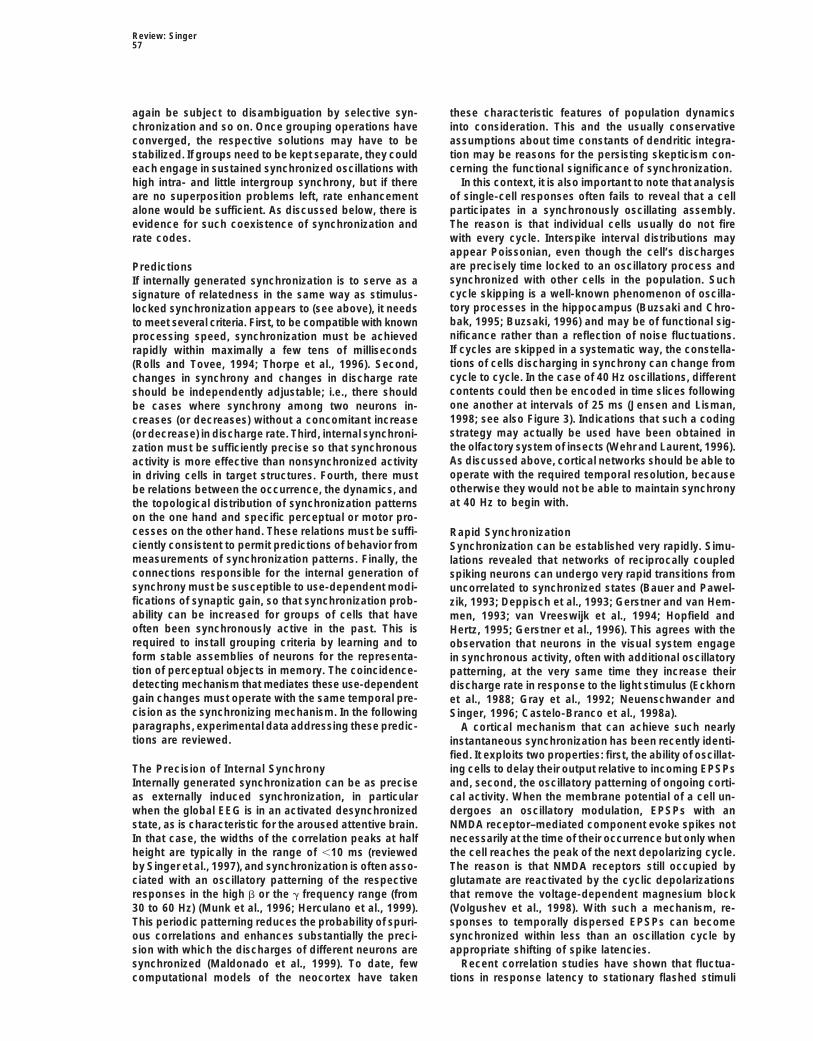
Neuron52
Figure 1. Precise Transmission of Temporal Signatures from the Retina to the Visual Cortex
(A and B) Synchronization between the retina, the lateral geniculate nucleus (LGN), and the cortex of oscillatory responses evoked by astationary stimulus (top right, inset). Responses were recorded simultaneously from the left retina (LRe), right LGN lamina A (RA), and leftarea 18 (LA18; top left, inset).(A) Autocorrelation functions. The onset of the stimulus evokes strong oscillatory patterning at all sites, at a frequency of 91 Hz.(B) Cross-correlation functions. Responses are correlated between all recorded pairs.(C) Correlated responses in area 17 following high-frequency flicker stimulation. Responses are recorded from two different sites in area 17.Responses are evoked by flicker stimuli at 20 Hz (top row) and 40 Hz (bottom row). Left columns show correlograms from simultaneouslyrecorded responses; right columns show correlograms computed across nonoverlapping response epochs. Comparison of the original andthe shifted correlograms indicates that the precise correlations among responses of distributed neurons are due to precise stimulus lockingof the responses ([A] and [B] are modified from Castelo-Branco et al., 1998a, and [C] is modified from Rager and Singer, 1998).
Evidence on a more global level for the enhanced cortical cells’ discharge was synchronized with othercortical cell groups projecting to the same collicular sitesaliency of synchronized activity has been obtained with
simultaneous recordings from several sites (area 18 and (Figure 2). Enhanced saliency of synchronized re-sponses can also be inferred from the tight correlationthe posterior mediolateral suprasylvian sulcus [PMLS])
in cat visual cortex and retinotopically corresponding between perception and the strength of neuronal re-sponse synchronization observed in experiments onsites in the superior colliculus (Brecht et al., 1998). The
impact of a particular group of cortical cells on target binocular rivalry in cats (Fries et al., 1997a) and humansubjects (Tononi and Edelman, 1998; Tononi et al., 1998)cells in the colliculus increased dramatically when the

Review: Singer53
according to saliency. The only difference is that, inthe case of enhanced contrast, saliency is increasedbecause of higher discharge rates rather than synchroni-zation.
A Role for Internally Generated SynchronyInternally generated response synchrony closely resem-bles that induced by synchronously presented stimuliwith respect to both its temporal precision and its mag-nitude (Rager and Singer, 1998). This raises the questionof whether the internally synchronized discharges affectprocessing in the same way as externally induced syn-chrony. If so, internal synchronization could serve tobind distributed activity according to grouping criteriaset by the brain itself, and the results of internal grouping
Figure 2. Dependence of Corticotectal Synchronization on Intracor-processes could be evaluated by the same neuronaltical Synchronization within and across Cortical Areasmechanisms that have evolved for the evaluation of ex-
(Left) Percentage of significant cortico-tectal correlations if corticalternally induced timing relations. Responses synchro-cells recorded from two sites in the same cortical area synchronizenized by internal interactions would undergo joint en-(gray column) or do not synchronize (black column) their responses.
(Right) Percentage of cortico-tectal interactions between A17 and hancement of their saliency and be treated as related bythe tectum if area 17 cells synchronize (gray column) or do not subsequent processing stages. Due to the high temporalsynchronize (black column) their responses with those of cells in precision with which cortical networks can distinguishthe lateral suprasylvian sulcus (modified from Brecht et al., 1998). synchronous from nonsynchronous events (see above),
internal synchronization could serve to define relationsbetween distributed responses with high temporal reso-(see also below). Finally, simulation studies also indicatelution, and could thus ideally complement selection andthat neurons with conventional integrate-and-fire prop-grouping operations based on sustained enhancementerties can be quite sensitive to the temporal dispersionof discharge rate. In principle, synchronization can en-of synaptic input. Large-scale simulations of biologicallyhance the impact of particular response constellationsinspired thalamocortical networks revealed that neuronson the basis of individual discharges. If, in addition, in-exhibited a strong tendency to engage in oscillatoryternally generated synchronization patterns can changefiring patterns and to synchronize their responses. Whenon a fast time scale, different relations could be definedsynchrony was artificially disrupted by introducing a jit-in rapid succession for successive segments of sus-ter in spike timing, transmission across polysynaptictained, temporally overlapping responses of distributedpathways was drastically reduced (Lumer et al., 1997a,neurons (Figure 3). Thus, if synchrony of discharges1997b).served as a signature of relatedness not only when in-There are several mechanisms, some of which haveduced from outside but also when generated internally,been identified only recently, that make synchronouslyit could be utilized in all processes where flexible andarriving EPSPs more efficient than temporally dispersedcontext-dependent selection and grouping of responsesEPSPs. First, because of their exponential decay, simul-are required on a fast time scale.taneous EPSPs summate more effectively than tempo-
rally dispersed EPSPs, and there is some evidence forsupralinear summation due to voltage-gated dendritic The Nature and Detectability of Internally
Generated Synchronyconductances. Second, firing threshold is sensitive tothe rising slope of the depolarization and lowers for fast- Synchronization is considered to be of internal origin if
two neurons exhibit a statistically significant covariationrising depolarizations (C. M. Gray, personal communica-tion). Third, the effect of EPSPs is dramatically enhanced in firing probability that cannot be attributed to stimulus-
locked covariations in discharge rate. Such episodeswhen these coincide with a back-propagating dendriticspike and hence with the input that generated this spike of synchronous firing can occur spontaneously, e.g.,
during the various sleep stages (Steriade, 1999); they(Larkum et al., 1999). All three mechanisms are sensitiveto dispersions of EPSPs in the range of a few milli- can appear during responses to sensory stimuli; or they
can be associated with cognitive processes such asseconds.In conclusion, both psychophysical and physiological focusing attention, analyzing complex patterns, storing
contents in short term memory, and preparing move-evidence indicates that neuronal networks are exqui-sitely sensitive to temporal relations among discharges ments (see below). These internally generated covaria-
tions of firing probability manifest themselves at differ-in input connections, assigning particular significanceto coincident, i.e., synchronous input. Synchronicity ent time scales ranging from a few milliseconds to
hundreds of milliseconds, as indicated by the variableserves as a tag of relatedness most likely because itcauses an increase in the saliency of the synchronized widths of the peaks in cross-correlograms. Often, syn-
chronization is associated with an oscillatory patterningresponses, which in turn favors their joint evaluation(binding) at subsequent processing stages. Conditions of the discharges, the frequency of these oscillations
covering a broad range and exhibiting a marked stateare thus comparable to those in which figure elementshave higher contrast than do elements of the back- dependence. Typically, synchronized EEG states such
as those that occur during drowsiness, deep sleep, andground: in that case, too, responses become grouped

Neuron54
Figure 3. Disambiguation of TemporallyOverlapping Assemblies by Synchronizationof Discharges in Selected Response Seg-ments
Despite the sustained and constant dis-charge rate (z30 Hz) of the ten depicted cells,the saliency of responses is transiently andrepeatedly increased for three different, spa-tially overlapping assemblies (A–C) by syn-chronization of the discharges of the sub-populations of cells constituting the threeassemblies. Note that the individual spiketrains appear nonoscillatory, while the spikedensity of the population response fluctuatesperiodically (lower continuous line). Note alsothat cells 5 and 7 are shared by two and cell3 is shared by all three assemblies. Becausecell 3 contributes spikes to more assembliesthan the others, its discharge frequency isslightly higher. In this case, the different as-semblies are interleaved with a rate of 40 Hz,and the integration interval for the evaluationof the population response is assumed to bearound 10 ms.
anesthesia are associated with broad correlation peaks event-related rate changes. Thus, even the coarsercorrelation patterns that can be described in terms ofand, if present, low oscillation frequencies (2–10 Hz);
activated, desynchronized EEG states that are charac- coherent rate fluctuations can only be assessed fromcorrelations of simultaneously recorded responses ifteristic of the awake, performing brain but also of para-
doxical sleep are associated with sharp correlation coherence is due to internal interactions. An exampleof such a case is the recent observation that the latenc-peaks (,15 ms) and high oscillation frequencies (30–60
Hz). In this state, synchronization exhibits maximal topo- ies of stimulus-induced rate increases of simultaneouslyrecorded cortical neurons covary due to internal coordi-logical specificity and can occur over particularly large
distances (Munk et al., 1996; Herculano et al., 1999). nation to a much higher degree than expected fromthe latency scatter of successively recorded responsesRelations between synchronization and changes in
discharge rate are variable. When synchronization oc- (Fries et al., 1998, Eur. J. Neurosci., abstract). This coor-dination in response timing could not have been dis-curs on a coarse time scale—i.e., when correlation
peaks are broader than the average interspike interval closed by studying the respective neurons one by one.Finally, it is to be expected that there are processes inof the respective cells’ discharges—synchronization
can be considered to be the result of internally produced the brain that are based on self-paced coordination ofboth the timing and the amplitude of distributed neu-covariations in discharge rate. However, when synchro-
nization occurs on a fast time scale with correlation ronal activity and are only loosely, if at all, time lockedto externally measurable events. These hidden but po-peaks narrower than the average interspike intervals, it
can be entirely independent of discharge rate. In this tentially important covariations of spike timing will alsobe detectable only by simultaneous recordings. Hence,case, changes in spike timing that leave average dis-
charge rate unaffected can lead to drastic changes in internally generated covariations in both the timing andthe amplitude of neuronal activity can only be disclosedsynchrony (see Figure 3 and Konig et al., 1996). Most
of the experimental results reviewed below show such with multicell recordings.independence. Note, however, that one could determinedischarge rates on a spike-by-spike basis, defining in-stant frequencies as the inverse of the respective in- Why Is There a Need for Dynamic Grouping
of Responses?terspike intervals. In that case, some of the synchronizedevents might be interpreted as the result of coherent, There are numerous instances in cortical processing
where dynamic selection and grouping of responses forultra-rapid rate fluctuations; but even then relations be-tween rate terms and synchrony can break down, be- further joint processing are required. Such is the case
whenever conjunctions have to be signaled for whichcause synchrony can be achieved by advancing a spikein one cell (rate increase) and delaying a spike in the there are no specific conjunction units. But there is a
deeper argument that suggests that dynamic groupingother (rate decrease).This potential independence of synchrony from rate is indispensable at all processing levels, even despite
the implementation of highly complex conjunction units.fluctuations is one reason why internally generated syn-chrony can only be assessed by direct correlation analy- This argument is derived from facts about the way corti-
cal networks encode features in general. It appears thatsis of simultaneously recorded neurons and not by com-paring rate fluctuations of successively recorded cells. cortex exploits the option of coarse coding in order to
economize on neuron numbers. The essence of thisAnother reason is that the internal coordination in-creases the covariance of rate changes above the level coding strategy is that contents are not encoded by the
responses of individual sharply tuned neurons but byexpected from comparison of successively recorded

Review: Singer55
specific constellations of graded responses of numer- first case, this is achieved through binding by conver-gence; in the second case, through dynamic bindingous broadly tuned neurons. The advantage is that the
number of different activity patterns that can be config- (Singer, 1995; 1999a; Phillips and Singer, 1997). Stereo-typed, frequently occurring, and particularly behavior-ured from a given set of neurons is much larger than
the number of neurons in the set. Hence, coarse coding ally relevant conjunctions would be represented by spe-cific binding units because this strategy is faster andis parsimonious with respect to neuronal numbers be-
cause a given neuron can participate in many different less susceptible to binding errors. However, becauseconjunction-specific neurons cannot exhaust the fullrepresentations when active in various constellations.
The constraint is, however, that neurons participating combinatorial space and cannot represent unantici-pated conjunctions, they should be recruitable into dy-in coarse coding must be broadly tuned. This, in turn, has
a price. Coarse codes may become ambiguous when namically configured population codes that representmeta-conjunctions for which there are no binding units.stimuli that overlap in euclidian or feature space, or in
both, need to be represented simultaneously (see also Thus, even conjunction units ought to be broadly tuned,in order to allow them to participate in coarse coding.Gray, 1999; von der Malsburg, 1999 [both in this issue
of Neuron]). Because of the broad tuning of cells partici- The idea is that this dual coding strategy is iteratedthroughout the processing hierarchy, whereby somepating in coarse coding, place codes alone may not be
sufficient to permit subsequent processing stages to feature conjunctions get represented explicitly by indi-vidual neurons and others by population codes. Whichdetect which of the many distributed responses ought to
be associated with which stimulus. Since coarse coding conjunctions are represented explicitly by the bindingunits at the various processing stages can be deducedappears to characterize all levels of processing, this
superposition problem is of a very general nature. Strat- from the rate-coded response properties of the respec-tive neurons. Evidence indicates that these conjunctionsegies to solve it can therefore be expected to be similar
across the various stages of the cortical processing become increasingly complex, abstract, and multimodalas one proceeds along the processing hierarchy, andhierarchy.
To illustrate, here is a concrete example of this general that at the interface between sensory and motor sys-tems their responses shift progressively from stimulus-problem. Due to broad tuning (coarse coding) of neurons
in primary visual cortex for position, orientation, length, related to movement-related features.At each processing level, novel, unanticipated con-width, contrast, and a few other features, a single elon-
gated contour evokes graded responses in a very large junctions or conjunctions for which specific bindingunits have not (yet) been implemented can then be repre-number of neurons. Because the response amplitude of
any one of these neurons is influenced by variations sented by additional population codes. Thus, despitethe implementation of ever more sophisticated bindingof the stimulus along any of these feature dimensions,
individual responses convey little specific information. units, flexible and context-dependent grouping (binding)operations remain necessary at each processing levelThe precise configuration of the stimulus can only be
deduced if a large number of the graded responses in order to represent relations that are not encoded byconjunction-specific neurons and in order to resolveevoked by the stimulus are evaluated jointly. If only a
single contour is present on a homogeneous back- superposition problems if they occur.In addition to their flexibility and virtually inexhaustibleground, this poses no difficulty. Superposition problems
arise, however, if several nearby or spatially overlapping coding capacity, dynamically bound population codeshave the further advantage that computational resultscontours are embedded in a textured background. In
that case, joint evaluation of responses needs to be can be represented at all stages of processing by popu-lation codes that have the same format. The onlyrestricted to responses evoked by the same contours.
A classical assumption is that there are neurons at changes concern the nature and complexity of the fea-ture conjunctions that are represented explicitly by thesubsequent processing levels that receive convergent
input in various constellations from subsets of broadly neurons forming the respective populations. Thus, dis-tributed representations can be mapped directly ontotuned low-level neurons, and that thereby acquire selec-
tivity for only the particular constellation of features that other distributed representations, which should facili-tate polymodal integration and sensory–motor coordi-characterizes one of the objects (binding by conver-
gence). In principle, population codes could be fully nation, thus circumventing bottleneck problems.In sensory–motor coordination, a vast number of dif-disambiguated by such conjunction-specific binding
units; but this solution is very expensive in terms of the ferent associations must be realized between sensoryrepresentations and motor programs (Roelfsema etnumber of required binding units if all possible conjunc-
tions were to be represented in this explicit way. One al., 1996). As the latter are in all likelihood distributedin nature, it would be quite uneconomical to first con-would require as many binding units as there are distin-
guishable population states. This is clearly not an attrac- vert distributed sensory representations into single-cellcodes, to then relay their output onto command neuronstive strategy because it sacrifices the main advantage
of coarse coding—the parsimonious use of neurons. that represent a particular movement, and then to recre-ate a distributed code from the output of these cells.It has been proposed, therefore, that the cerebral cor-
tex uses two coding strategies in parallel: first, partial Direct conversion of one distributed code into anotherappears as the more elegant solution, but again, it re-disambiguation of coarse population codes by the im-
plementation of conjunction units, and second, disam- quires dynamic binding.In conclusion, if relations are encoded both by con-biguation of population codes by dynamic, context-
dependent binding. Note that in both cases the essence junction-specific neurons and by dynamically associ-ated assemblies of such neurons, rapid and flexibleof the process is the representation of relations. In the

Neuron56
grouping operations have to be accomplished at all lev- the grouping problems encountered at each stage. Inels of cortical processing. Note, however, that the two primary visual cortex, these would be rather simple crite-strategies, although available at all stages, need not ria because neurons at this stage code for simple fea-always be used together. One can imagine conditions tures. All that needs to be accomplished here is thewhere the elementary features of a scene are all ex- grouping of responses that are likely to be generated byplicitly represented by conjunction-specific units, and the same contour. This would be the case for responseswhere the arrangement of contours poses no superpo- evoked by a continuous contour, by contour segmentssition problem, but where the objects defined by these that are collinear, or by contours that share similaritiesfeatures are novel and are not covered by conjunction- in any of the simple feature domains for which neuronsspecific cells of a higher order. In this case, dynamic in primary visual cortex are responsive. In areas special-grouping may only be necessary at higher levels of pro- ized for the analysis of other features—such as, for ex-cessing. Conversely, a highly familiar object could be ample, motion—grouping would have to occur ac-explicitly represented by a set of object-specific binding cording to motion parameters and so on. All theseunits—but this object could be embedded in a complex grouping operations could in principle occur in parallelbackground allowing for many unfamiliar conjunctions in the various cortical areas. Since all relevant areas arebetween elements of the figure and elements of the reciprocally coupled, preliminary grouping results couldbackground. In that case, extensive grouping operations themselves bias the grouping operations in other areaswould be required at peripheral levels of processing until a solution is found that is most consistent with theto associate the correct set of elementary conjunction set of low- and high-level grouping criteria that resideunits, but no dynamic grouping would be necessary at in the architecture of the various visual areas. Suchhigher levels provided there is no superposition problem dynamic, distributed grouping through iterative reentryat these levels. This possibility needs to be considered has been proposed as one of the core functions of corti-when designing experiments in search of binding mech- cal networks (Edelman, 1987, 1989). Analogous to theanisms. It should be noted that not every task requires proposed mechanism of attentional grouping, these dis-dynamic binding at all stages of processing. In some tributed grouping operations could be based on jointstudies on figure–ground discrimination, for example, enhancement of the firing rate of the selected neurons.animals are not forced to identify a particular figure and However, grouping of responses solely by joint ratemay respond if they simply detect an inhomogeneity in enhancement may engender problems. First, it can leada pattern. In such cases, it is likely that they opt for the to ambiguities. It may not always be easy to distinguishfastest strategy and rely on response changes of some whether rate increases are due to grouping or to varia-conjunction neurons, rather than go through the time- tions in stimulus properties, such as, for example, con-consuming process of dynamic feature binding. trast changes by uneven illumination. Second, in cases
where objects overlap in euclidian or feature space, onlyDynamic Grouping Mechanisms responses to a single object can be grouped at any oneOne proposal is that mechanisms of selective attention moment. Otherwise, it would be again unclear whichmay subserve dynamic binding of distributed responses of the selected responses belong to which popula-(see Ghose and Maunsell, 1999; Shadlen and Movshon, tion code. Because evaluation of nonsynchronized rate1999; Reynolds and Desimone, 1999; Wolfe, 1999 [all in changes requires integration of a minimal number ofthis issue of Neuron]). In vision it is assumed, for exam- EPSPs arriving successively from the selected cells, theple, that object-centered attention selectively enhances pace at which different populations can be defined bythe discharge rate of neurons responding to features of multiplexing is slow.the same contour or object, via top-down projections, Both problems could be alleviated by introducing in-and that the resulting joint increase of saliency leads to ternal synchronization as an additional grouping mecha-joint processing of the selected responses at subse-
nism. First, synchronization can bias the saliency of re-quent stages (see, e.g., Treisman, 1996). This mecha-
sponses independently of rate fluctuations. Second,nism encounters several difficulties if not supplemented
because it relies on coincidence detection rather thanby additional grouping operations. First, it presupposestemporal summation, synchronization can define rela-that higher centers “know” which of the peripheral re-tions with sufficiently high temporal precision to permitsponses code for the contours of a particular object.multiplexing.However, this information is only available once an ob-
ject is identified, and this is often possible only after theComplementarity of Rate Modulationpopulation codes at lower levels have been disambigu-and Synchronizationated. Thus, it must first be clarified whether the manyBecause grouping of responses by synchronization issimultaneous responses in a low-level area code forbound to enhance discharge rates of selected targetcoherent or disjunct contours before it can be decidedcells in areas receiving synchronized input, fast synchro-whether one has to do with one or another object. Sec-nization codes and more sustained rate codes couldond, because these top-down projections fan out overultimately coexist and perhaps even optimally comple-large cortical domains, it is difficult to see how the pro-ment one another. Sustained rate-coded input from ajections could enhance the responses of the cells codinggiven processing stage could be rapidly disambiguatedfor one out of several overlapping contours with theand bound through synchronization at other processingrequired topological specificity (Salin and Bullier, 1995).stages; this grouped activity would in turn lead to aThese problems can be alleviated if responses withinspecific pattern of sustained, rate-modulated responsesthe respective processing stages undergo a first, prelim-
inary grouping according to criteria that are adapted to at the following stage where these activity patterns can
Review: Singer57
again be subject to disambiguation by selective syn- these characteristic features of population dynamicsinto consideration. This and the usually conservativechronization and so on. Once grouping operations have
converged, the respective solutions may have to be assumptions about time constants of dendritic integra-tion may be reasons for the persisting skepticism con-stabilized. If groups need to be kept separate, they could
each engage in sustained synchronized oscillations with cerning the functional significance of synchronization.In this context, it is also important to note that analysishigh intra- and little intergroup synchrony, but if there
are no superposition problems left, rate enhancement of single-cell responses often fails to reveal that a cellparticipates in a synchronously oscillating assembly.alone would be sufficient. As discussed below, there is
evidence for such coexistence of synchronization and The reason is that individual cells usually do not firewith every cycle. Interspike interval distributions mayrate codes.appear Poissonian, even though the cell’s dischargesare precisely time locked to an oscillatory process andPredictionssynchronized with other cells in the population. SuchIf internally generated synchronization is to serve as acycle skipping is a well-known phenomenon of oscilla-signature of relatedness in the same way as stimulus-tory processes in the hippocampus (Buzsaki and Chro-locked synchronization appears to (see above), it needsbak, 1995; Buzsaki, 1996) and may be of functional sig-to meet several criteria. First, to be compatible with knownnificance rather than a reflection of noise fluctuations.processing speed, synchronization must be achievedIf cycles are skipped in a systematic way, the constella-rapidly within maximally a few tens of millisecondstions of cells discharging in synchrony can change from(Rolls and Tovee, 1994; Thorpe et al., 1996). Second,cycle to cycle. In the case of 40 Hz oscillations, differentchanges in synchrony and changes in discharge ratecontents could then be encoded in time slices followingshould be independently adjustable; i.e., there shouldone another at intervals of 25 ms (Jensen and Lisman,be cases where synchrony among two neurons in-1998; see also Figure 3). Indications that such a codingcreases (or decreases) without a concomitant increasestrategy may actually be used have been obtained in(or decrease) in discharge rate. Third, internal synchroni-the olfactory system of insects (Wehr and Laurent, 1996).zation must be sufficiently precise so that synchronousAs discussed above, cortical networks should be able toactivity is more effective than nonsynchronized activityoperate with the required temporal resolution, becausein driving cells in target structures. Fourth, there mustotherwise they would not be able to maintain synchronybe relations between the occurrence, the dynamics, andat 40 Hz to begin with.the topological distribution of synchronization patterns
on the one hand and specific perceptual or motor pro-cesses on the other hand. These relations must be suffi- Rapid Synchronizationciently consistent to permit predictions of behavior from Synchronization can be established very rapidly. Simu-measurements of synchronization patterns. Finally, the lations revealed that networks of reciprocally coupledconnections responsible for the internal generation of spiking neurons can undergo very rapid transitions fromsynchrony must be susceptible to use-dependent modi- uncorrelated to synchronized states (Bauer and Pawel-fications of synaptic gain, so that synchronization prob- zik, 1993; Deppisch et al., 1993; Gerstner and van Hem-ability can be increased for groups of cells that have men, 1993; van Vreeswijk et al., 1994; Hopfield andoften been synchronously active in the past. This is Hertz, 1995; Gerstner et al., 1996). This agrees with therequired to install grouping criteria by learning and to observation that neurons in the visual system engageform stable assemblies of neurons for the representa- in synchronous activity, often with additional oscillatorytion of perceptual objects in memory. The coincidence- patterning, at the very same time they increase theirdetecting mechanism that mediates these use-dependent discharge rate in response to the light stimulus (Eckhorngain changes must operate with the same temporal pre- et al., 1988; Gray et al., 1992; Neuenschwander andcision as the synchronizing mechanism. In the following Singer, 1996; Castelo-Branco et al., 1998a).paragraphs, experimental data addressing these predic- A cortical mechanism that can achieve such nearlytions are reviewed. instantaneous synchronization has been recently identi-
fied. It exploits two properties: first, the ability of oscillat-ing cells to delay their output relative to incoming EPSPsThe Precision of Internal Synchrony
Internally generated synchronization can be as precise and, second, the oscillatory patterning of ongoing corti-cal activity. When the membrane potential of a cell un-as externally induced synchronization, in particular
when the global EEG is in an activated desynchronized dergoes an oscillatory modulation, EPSPs with anNMDA receptor–mediated component evoke spikes notstate, as is characteristic for the aroused attentive brain.
In that case, the widths of the correlation peaks at half necessarily at the time of their occurrence but only whenthe cell reaches the peak of the next depolarizing cycle.height are typically in the range of ,10 ms (reviewed
by Singer et al., 1997), and synchronization is often asso- The reason is that NMDA receptors still occupied byglutamate are reactivated by the cyclic depolarizationsciated with an oscillatory patterning of the respective
responses in the high b or the g frequency range (from that remove the voltage-dependent magnesium block(Volgushev et al., 1998). With such a mechanism, re-30 to 60 Hz) (Munk et al., 1996; Herculano et al., 1999).
This periodic patterning reduces the probability of spuri- sponses to temporally dispersed EPSPs can becomesynchronized within less than an oscillation cycle byous correlations and enhances substantially the preci-
sion with which the discharges of different neurons are appropriate shifting of spike latencies.Recent correlation studies have shown that fluctua-synchronized (Maldonado et al., 1999). To date, few
computational models of the neocortex have taken tions in response latency to stationary flashed stimuli

Neuron58
can covary with a precision in the millisecond range for some of the basic Gestalt criteria according to whichthe visual system groups related features during scenesubsets of neurons located in different columns of thesegmentation. A consistent finding was that neuronsvisual cortex and even for neurons in different hemi-distributed across different columns within the same orspheres (Fries et al., 1997b, Soc. Neurosci., abstract).different visual areas, and even across hemispheres,This correlation between the very first discharges of asynchronized their responses with near-zero phase lagresponse appears to be based on the delay mechanismwhen activated with a single contour but fired indepen-identified in the slice experiments. Comparison betweendently when stimulated simultaneously with two differ-actual response latencies and immediately precedingent contours (Gray et al., 1989; Engel et al., 1991a,fluctuations of the local field potential revealed that the1991b, 1991c; Freiwald et al., 1995; Kreiter and Singer,response latency shifted as a function of the polarity of1996). This suggested that synchronization was the re-the preceding field potential fluctuation. This effect wassult of a context-dependent selection and grouping pro-confined to episodes in which the field potential oscilla-cess. Analysis of the dependence of synchrony on re-tions exhibited high power in the g frequency range,ceptive field and stimulus configurations revealed thatemphasizing the role of g oscillations in the coordinationthe probability and strength of response synchroniza-of response timing. In all likelihood, the spatiotemporaltion reflected elementary Gestalt criteria for perceptualpatterns of these fluctuations reflect the architecturegrouping such as continuity, proximity, similarity in theand the actual functional state of intra- and interarealorientation domain, collinearity, and common fate (re-association connections (Arieli et al., 1996). Thus, group-viewed by Singer et al., 1997; Gray, 1999 [this issue ofing by synchronization can be extremely fast and stillNeuron]). These early experiments were performed inoccur as a function of both the prewired associationalanesthetized animals, but more recent multielectrodedispositions and the current functional state of the corti-recordings from awake cats and monkeys indicate thatcal network.these synchronization phenomena are not artifacts ofanesthesia but are even more pronounced when theExternal versus Internal Synchronizationanimals are awake and attentive (Kreiter and Singer,Latency shifting by internal synchronization could com-1992, 1996; Frien et al., 1994; Fries et al., 1997a; Graypromise precise signaling of temporal stimulus features.and Viana Di Prisco, 1997; Friedman-Hill et al., 1999;Thus, responses that convey information about the pre-Maldonado et al., 1999) (see Figure 4). In none of thesecise timing of stimuli and that support grouping throughexperiments have systematic changes in synchroniza-externally imposed synchrony should be exempted fromtion probability been associated with systematic changesinternal latency adjustments. In the visual system, theof the neurons’ discharge rate.magno- and parvocellular pathways are kept separate
A particularly close correlation between neuronal syn-over the first few processing stages, and grouping cueschrony and perceptual grouping has recently been ob-provided by the two systems appear to be processedserved in experiments with plaid stimuli. These stimuliindependently and in parallel (see above). This supportsare well suited for the study of dynamic binding mecha-the possibility that internal synchronization mechanismsnisms because minor changes of the stimulus cause aact differentially on magno- and parvocellular pathwaysbinary switch in perceptual grouping. Two superim-and do not affect the former at those levels of processingposed gratings moving in different directions (plaid stim-where grouping is achieved according to external timinguli) may be perceived either as two surfaces, one beingand where precisely timed motor responses are pro-transparent and sliding on top of the other (componentgrammed. Since all sensory systems have developedmotion), or as a single surface, consisting of crossed
parallel pathways with differential sensitivity to temporalbars, that moves in a direction intermediate to the com-
(phasic) and nontemporal (sustained) stimulus features,ponent vectors (pattern motion) (Adelson and Movshon,
it may be a general strategy to use external, stimulus- 1982; Stoner et al., 1990). Which percept dominatesinduced and internal, self-generated timing relations in depends on the luminance of grating intersections, be-parallel for the grouping of responses. The advantage cause this variable defines the degree of transparencywould be that ultimately the relatedness of temporal and (Albright and Stoner, 1995). Component (or pattern) mo-nontemporal stimulus features could be expressed in tion is perceived when luminance conditions are com-the same format—namely, in the degree of synchrony— patible (or incompatible) with transparency (Figure 5A).and this should facilitate joint evaluation of temporal Thus, this is a case in which local changes in stimulusand nontemporal grouping cues at processing stages properties cause global changes in perceptual grouping.where both stimulus attributes have to be bound to- In the case of component motion, responses evoked bygether. However, to the best of our knowledge, this the two gratings must be segregated, and only re-possibility has not yet been investigated. sponses evoked by the contours of the same grating
must be grouped to represent one of the two surfaces;Relations between Response Synchronization in the case of pattern motion, responses to all contoursand Gestalt Rules for Perceptual Grouping must be bound together to represent a single surface.Based on the hypothesis that internal synchronization If this grouping of responses is initiated by selectiveof discharges could serve to group responses for joint synchronization, three predictions must hold (see Figureprocessing, a series of experiments has been performed 5B). First, neurons that prefer the direction of motion ofin the search for a correlation between dynamic changes one of the two gratings and have collinearly alignedof response synchronization and particular stimulus receptive fields should always synchronize their re-configurations. One prediction tested was that in early sponses because they respond always to contours that
belong to the same surface. Second, two neurons thatvisual areas, synchronization probability should reflect

Review: Singer59
Figure 4. Stimulus Dependence of Neuronal Synchronization in Area MT of the Visual Cortex of a Macaque Monkey Carrying Out a Fixation Task
Neuronal responses were obtained from two cell groups with different directional preferences. The figure shows cross-correlograms and peri-stimulus time histograms for four different stimulation conditions. The small insets indicate the receptive field locations (1 and 2) with respectto the fixation point (F) and the directional preference of the neurons (small arrows).(A) A single moving stimulus bar, whose direction of motion was intermediate between the neurons’ preferences, led to a pronouncedsynchronization of the two cell groups, as indicated by the central maximum in the cross-correlogram.(B) Presentation of two stimuli moving in the respective preferred directions of cell groups 1 and 2 abolishes synchronization.(C and D) The synchronization observed with a single stimulus does not depend on its particular orientation.(C) Changing orientation and direction of motion by 158 or (D) using one of the bars from the configuration in (B) had little influence onsynchronization. Scale bars for the peri-stimulus time histograms correspond to 40 spikes/s. The continuous line superimposed on thecorrelograms represents a damped cosine function that was fitted to the data to assess the significance of the correlogram modulation(modified from Kreiter and Singer, 1996).
are tuned to the motion directions of the two gratings likely to reveal only the static anisotropies in the networkof synchronizing connections. Such a problem may haveshould synchronize their responses in the case of pat-
tern motion because they then respond to contours of contributed to the negative results of a recent study thatfailed to show a relation between perceptual groupingthe same surface, but they should not synchronize in
the case of component motion because their responses and internal synchronization in monkey striate cortex(Lamme and Spekreijse, 1999).are then evoked by contours belonging to different sur-
faces. Third, neurons preferring the direction of pattern In the case of the plaid stimuli, predictions were testedwith multielectrode recordings from areas 18 and PMLSmotion should also synchronize only in the pattern and
not in the component motion condition. of the visual cortex of lightly anesthetized cats, after wehad confirmed with eye movement recordings in awakeAn important aspect of these predictions is that the
expected changes in synchrony differ for different cell cats that the animals distinguished between componentand pattern motion. Cross-correlation analysis of re-pairs, depending on the configuration of their receptive
fields. Thus, when searching for relations between syn- sponses from cell pairs distributed either within oracross areas 18 and PMLS confirmed all three predic-chrony and cognitive functions, it is not only crucial
to identify the processing stage where one assumes a tions. Cells synchronized their activity if they respondedto contours that are perceived as belonging to the sameparticular binding function to be accomplished but also
to select the appropriate cell pairs. Averaging data surface (Castelo-Branco et al., 1998b, Eur. J. Neurosci.,abstract) (Figure 5C). Analysis of the neurons’ dischargeacross cell pairs with different receptive field configura-
tions can mask dynamic changes in synchrony and is rate confirmed that most of the cells in these visual

Neuron60
to synchrony, variations in response amplitude failed toreflect the transition from component to pattern motioninduced by transparency manipulation. Dynamic changesin synchronization could, thus, serve to encode in acontext-dependent way the relations among the simul-taneous responses to spatially superimposed contoursand thereby bias their association with distinct surfaces.Future investigations have to clarify whether the popula-tions of differentially synchronized neurons alreadyserve as the final representations of the perceived sur-faces or whether, in the case of pattern motion, addi-tional assemblies are formed. These would then haveto consist of conjunction units tuned to the specificconstellations of superimposed gratings, and their re-sponses would have to be bound together to signal thatthey code for the same surface.
In all experiments referred to in this review, trials withdifferent stimulus configurations were interleaved ran-domly to counterbalance possible effects of slow driftsin central state that may cause covariations of dischargerate at a slow time scale. Moreover, phasic responsecomponents were excluded from correlation analysis toavoid artifactual correlations due to dynamic changesin discharge rate (Brody, 1999a, 1999b). Accordingly,the shift predictors (correlograms between responses tothe same stimulus configuration selected from differenttrials) were always flat, indicating that the changes inthe correlations were caused by context-dependentchanges of internal interactions and not by changesin stimulus-locked or state-dependent covariations ofdischarge rate.
The Anatomical SubstrateStudies involving lesions (Engel et al., 1991a) and devel-opmental manipulations (Lowel and Singer, 1992; Koniget al., 1993) indicate that the interactions responsiblefor these stimulus-specific synchronization phenomenaare mediated at least in part by cortico-cortical connec-tions that reciprocally link cells in the same cortical area,as well as cells distributed across different areas, andeven across the two hemispheres. The elementarygrouping criteria applied in early vision could thus reside
Figure 5. Neuronal Sychronization and the Bistable Perception of in the architecture of these association connections.Plaids
The evidence that intrinsic excitatory connections pref-(A) Two superimposed gratings that differ in orientation and drift in
erentially link neurons which code for features that tenddifferent directions are perceived either as two independently mov-to be grouped is consistent with this possibility (Ts’oing gratings (component motion) or as a single pattern drifting inand Gilbert, 1988; Gilbert and Wiesel, 1989; Malach etthe intermediate direction (pattern motion), depending on whether
the luminance conditions at the intersections are compatible with al., 1993; Schmidt et al., 1997a, 1997b).transparency.(B) Predictions on the synchronization behavior of neurons as a
Temporal Constraints of Use-Dependent Plasticityfunction of their receptive field configuration (left) and stimulationIf synchronization of responses serves as a mechanismconditions (right).to group responses, synchronizing connections must(C) Changes in synchronization behavior of two neurons recorded
simultaneously from areas 18 and PMLS that were activated with a be susceptible to use-dependent modifications. This isplaid stimulus under component (top graph) and pattern motion required in order to implement new grouping criteria(bottom graph) conditions. The two neurons preferred gratings with by learning and to stabilize assemblies representingorthogonal orientation (see receptive field configuration [top] and
previously experienced conjunctions. Synchronizationtuning curves obtained with component and pattern, respectively)probability must remain increased for groups of cellsand synchronized their responses only when activated with the pat-that have been forced previously to engage repeatedly intern stimulus (compare cross-correlograms on the right) (courtesy
of M. Castelo-Branco and S. Neuenschwander). highly synchronous firing, and the mechanism mediatingthese use-dependent changes in synchronization prob-ability must be capable of distinguishing between syn-chronous and nonsynchronous firing with a temporalareas respond preferentially to the component gratings
of the plaids (component-specific cells; Gizzi et al., 1990) resolution that is in the same range as the temporalprecision of observed synchrony. Both postulates haveand not to the pattern as a whole. However, in contrast

Review: Singer61
recently been confirmed. Herculano et al. (1997, Soc. state. Second, synchronization is selective and contextdependent, reflecting some of the elementary GestaltNeurosci., abstract) have shown that synchronization
probability increases between groups of neurons if criteria that determine perceptual grouping. Third, syn-chrony is due to intracortical interactions that are basedthese engage repeatedly in synchronous oscillatory fir-
ing in the g frequency range. This enhanced tendency on the network of reciprocal association connections.Fourth, the adaptive mechanisms supporting use-depen-of neurons to synchronize their responses to coherent
stimuli can be reduced again by having the same cells dent modifications of synchronization probability oper-ate with the required temporal resolution.engage repeatedly in oscillatory firing patterns that are
decorrelated. These increases and decreases of syn-chronization probability appear to depend upon the pre- Relations between Synchronization and Perceptioncise phase relations of the oscillatory discharges during While the experiments reviewed above had the goal toconditioning, because on a coarser time scale (.50 ms) identify basic characteristics of response synchroniza-the neurons’ activity overlapped completely in both con- tion, the studies described in the following paragraphsditioning paradigms. were aimed at establishing links between synchroniza-
Recent data from cortical slices support such a de- tion phenomena and behavior. A close relation betweenpendency of synaptic plasticity on precise timing be- response synchronization and perception has beentween pre- and postsynaptic discharges. Varying the found in cats who suffered from strabismic amblyopia,temporal relations between presynaptic and postsynap- a developmental impairment of vision, which results intic responses in simultaneously recorded coupled corti- suppression of the amblyopic eye, reduced visual acu-cal cells revealed that long-term potentiation (LTP) re- ity, and crowding. Crowding refers to the inability ofsults when the EPSP precedes the postsynaptic spike amblyopic subjects to identify target stimuli if these arewithin intervals of 10 ms or less, while the polarity of surrounded by nearby contours and is thought to resultthe modification reverses to long-term depression (LTD) from false binding of responses evoked by the targetas soon as the EPSP follows the spike (Markram et al., and the embedding background. Quite unexpectedly,1997; Zhang et al., 1998). Thus, shifts of a few millisec- the light responses of individual neurons in the primaryonds in the timing relations between pre- and postsyn- visual cortex of amblyopic cats were normal, and theaptic discharges suffice to invert the polarity of use- neurons continued to respond vigorously to stimuli thatdependent synaptic modifications. The mechanism the animals were unable to perceive through the ambly-permitting such precise evaluation of the temporal conti- opic eye. The only significant correlate of amblyopiaguity of pre- and postsynaptic responses is, with all detected in this study was a reduction of response syn-likelihood, the active dendritic response associated with chronization among neurons driven by the amblyopicthe back-propagating spike (Magee and Johnston, 1997). eye. This reduction in synchrony became particularly
Under the sustained activation conditions applied in pronounced when responses were evoked with gratingsthe in vivo experiments of Herculano et al. (1997, Soc. that had been identified in previous behavioral experi-Neurosci., abstract), the high temporal selectivity of syn- ments as too fine to be resolvable by the animal throughaptic modifications resulted most likely from the oscilla- the amblyopic eye (Roelfsema et al., 1994). Thus, im-tory modulation of the neuronal responses. That an os- paired synchrony is likely to account for at least somecillatory patterning of activity can effectively narrow the of the perceptual deficits: by reducing the saliency oftemporal window for coincidence detection has been responses, it could explain why signals from the ambly-shown in vitro. Experiments in slices of the hippocampus opic eye cannot compete successfully with the well-indicate that the polarity of synaptic gain changes de- synchronized responses from the normal eye and arepends critically on the phase relations between pre- and excluded from supporting perception when both eyespostsynaptic discharges if these overlap on a coarse are open. Recent recordings from area 21, a higher visual
area, have indeed shown that the poorly synchronizedtime scale but exhibit an oscillatory modulation (Huertaand Lisman, 1996). The same holds for the visual cortex. responses evoked from the amblyopic eye drive neurons
in area 21 less efficiently than the well-synchronizedPyramidal cells of rat visual cortex slices were made todischarge tonically at 20 Hz by injecting sinusoidally responses from the normal eye (Schroder et al., 1998,
Eur. J. Neurosci., abstract). Poor synchronization couldmodulated current through a patch pipette. Simultane-ously, EPSPs were evoked, also at 20 Hz, by electrical also be responsible for the reduction in visual acuity
and for crowding, because it is expected to impair dis-stimulation of excitatory afferents. Changing the phaserelations between pre- and postsynaptic activity re- ambiguation of responses evoked by closely apposed
stimuli.vealed that the stimulated input tended to undergo LTPwhen the EPSPs were coincident with the spikes, while A correlation between response synchronization and
perception has also been documented in experimentsafferents consistently underwent LTD when the EPSPsfell in the troughs of the membrane potential oscillations. on binocular rivalry that were again performed in strabis-
mic animals (Fries et al., 1997a). Perception in strabismicThus, although pre- and postsynaptic activation over-lapped completely on a coarse time scale, phase shifts subjects always alternates between the two eyes. This
can be exploited to investigate how neuronal responsesof ,20 ms between individual EPSPs and spikes re-versed the polarity of the synaptic modifications (Wes- to constant stimuli change if they pass from being se-
lected and perceived to being suppressed and excludedpatat et al., 1999).In conclusion, the synchronization mechanism identi- from perception and vice versa (Figure 6). The outcome
of these experiments was surprising, because the re-fied at early levels of visual processing shares essentialfeatures with the processes postulated for the dynamic sponses of neurons in areas 17 and 18 were not attenu-
ated when they were excluded from supporting percep-binding of responses. First, synchronization is preciseand rapidly established when the brain is in an activated tion. A close and highly significant correlation existed,

Neuron62
selection of responses for further processing is associ-ated with enhanced synchronization rather than in-creased firing. This agrees with rivalry experiments inawake, behaving monkeys, which showed no system-atic relation between the strength of visual responsesand perception in early visual areas but a clear correla-tion between perceptual suppression and loss of neu-ronal responses in higher visual areas (Logothetis andSchall, 1989b; Leopold and Logothetis, 1996; Sheinbergand Logothetis, 1997). This is what one expects if thesaliency of the responses from the two eyes is adjustedat early processing stages by modulating synchroniza-tion rather than discharge rates. These results, ofcourse, do not exclude additional response selectionby rate modulation. Thus, the outcome of rivalry canalso be biased by changing the contrast of the patternspresented to the two eyes, and this manifests itself inrate changes.
Global Interareal SynchronizationMore global synchronization phenomena, involving task-dependent coordination of the oscillatory activity ofwhole cortical areas, have been observed in cats trainedto perform a visually triggered motor response. The vi-sual, association, somatosensory, and motor areas in-volved in the execution of the task synchronized theiractivity in the g frequency range as soon as the animalsfocused their attention on the relevant stimulus; thestrength of synchronization among areas reflected pre-cisely the coupling of these areas by cortico-corticalconnections. Immediately after the appearance of thevisual stimulus, synchronization increased further, andthese coordinated activation patterns were maintained
Figure 6. Neuronal Synchronization under Conditions of Binocularuntil the task was completed. However, once the rewardRivalrywas available and the animals engaged in consumma-(A) Using two mirrors, different patterns were presented to the twotory behavior, these coherent patterns collapsed andeyes of strabismic cats. (B) through (E) show normalized cross-gave way to low-frequency oscillatory activity that didcorrelograms for two pairs of recording sites activated by the eye
that won (B and C) and lost (D and E) in interocular competition, not exhibit any consistent relations with regard to phaserespectively. Insets above the correlograms indicate stimulation and areal topology (Roelfsema et al., 1997). These re-conditions. Under monocular stimulation (B), cells driven by the sults suggest that an attention-related process had im-winning eye show a significant correlation, which is enhanced after
posed a coherent temporal pattern on the activity ofintroduction of the rivalrous stimulus to the other eye (C). The reversecortical areas required for the execution of the task. Asis the case for cells driven by the losing eye (compare conditionsdiscussed above, coordinated subthreshold modulation[D] and [E]). The white continuous line superimposed on the correlo-
grams represents a damped cosine function fitted to the data. RMA of membrane potential fluctuations acts like a temporalis the relative modulation amplitude of the center peak in the correlo- filter that permits rapid but selective synchronizationgram, computed as the ratio of peak amplitude over the offset of (Fries et al., 1997b, Soc. Neurosci., abstract). Here, at-correlogram modulation. This measure reflects the strength of syn-
tentional mechanisms, involving perhaps nonspecificchrony (modified from Fries et al., 1997a).thalamic projections (Ribary et al., 1991; Steriade, 1999),seem to exploit this option to prepare cortical areas forrapid synchronization, thereby accelerating subsequenthowever, between changes in the strength of responseselection and grouping of responses. If these anticipa-synchronization and the outcome of rivalry. Cells medi-tory synchronization patterns exhibit columnar specific-ating responses of the eye that won in interocularity, they could not only accelerate but also bias subse-competition increased the synchronization of their re-quent grouping as a function of expectancy. This thensponses upon presentation of the rivalrous stimulus tocould provide a mechanism for the well-known phenom-the other, losing eye, while the reverse was true for cellsenon that the outcome of grouping depends also ondriven by the eye that became suppressed. In addition,what one expects to see.there was a marked increase in the oscillatory modula-
tion of the synchronized discharges that supported per-ception. This agrees with the hypothesis (see above) Neuronal Synchrony and Motor Performance
In the motor cortex of primates, oscillatory modulationthat the outcome of a selection process can be stabilizedby having the selected group of neurons engage in sta- of activity in the g frequency range and synchronization
of responses both within and across hemispheres haveble, synchronized oscillatory response patterns. Thus, inprimary visual cortex, there are instances where internal been observed in association with self-paced grasping

Review: Singer63
movements or visually guided reaching tasks (Murthy about the association between a visual and a tactilestimulus, the visual stimulus evokes g activity over theand Fetz, 1996a, 1996b). Another study found recurringvisual cortex that is coherent with g activity over thephases of heightened discharge synchrony among si-somatosensory cortex contralateral to the stimulatedmultaneously recorded single cells in primate motor cor-finger, and this coherence breaks down again upon ex-tex that were precisely correlated with the animal’stinction (Miltner et al., 1999). Finally, evidence has beenexpectancy of a “go” signal (Riehle et al., 1997). Remark-obtained in an MEG study on binocular rivalry that activ-ably, these epochs of enhanced synchronization wereity evoked from the eye that dominates perception dur-not associated with measurable changes in the cells’ing rivalry is better synchronized than activity evokeddischarge rates, further evidence that synchrony andby the suppressed eye (Tononi and Edelman, 1998; To-discharge rate can be independent. A close relationnoni et al., 1998). This agrees with the rivalry experimentsbetween precisely timed discharge patterns and thein cats (Fries et al., 1997a).type of anticipated motor responses has also been de-
Also consistent with a functional role for synchronizedscribed by Prut et al. (1998) for simultaneously recordedg oscillations is the observation that these are closelyneurons in the frontal cortex of monkeys trained to per-related to arousal and attention. Synchronization of neu-form visually cued targeting movements. Finally, syn-ronal responses in the g frequency range does not occurchronous oscillatory discharge patterns were observedwhen the EEG exhibits low-frequency oscillations, as isin motion-sensitive visual areas of the monkey (MT/V5)the case in deep anesthesia or slow-wave sleep, but isin association with the preparation of visually guidedparticularly pronounced during activated “desynchro-eye movements. In this case, synchrony broke down,nized” states of the EEG (Munk et al., 1996; Herculanohowever, upon presentation of the stimulus (De Oliveiraet al., 1999). In human subjects, g oscillations are en-et al., 1997). While in these cases synchronization washanced in responses to attended as compared to nonat-mainly associated with movement preparation, recenttended stimuli (Tiitinen et al., 1993); they disappear instudies in monkeys trained to perform visually guideddeep anesthesia (Madler and Poppel, 1987) but are pres-targeting movements showed also a close correlationent during paradoxical sleep, during which dreaming—between patterns of discharge synchrony and the actuali.e., reactivation of memories—is thought to occur (Lli-direction of the arm movements (Ojakangas et al., 1997,nas and Ribary, 1993; Steriade, 1999).Soc. Neurosci., abstract; Donoghue et al., 1998; Hatso-
poulos et al., 1998). In these recordings, the strength ofManipulations of Synchronythe correlations varied with the direction of motion andThe results reviewed so far provide correlative evidenceprovided additional information about movement pa-for a role of internally generated response synchroniza-rameters.tion in neuronal processing, but do not prove that thenervous system attributes significance to the preciseStudies in Humanstemporal relations among discharges if these are gener-Correlates between the occurrence of synchronized os-ated by intrinsic interactions rather than by externalcillatory activity in the g frequency range and cognitivestimuli. To prove a causal relation requires the demon-processes have recently been observed in human sub-stration that selective manipulation of synchrony alters
jects with EEG and MEG recordings. In a series of pio-behavior. Recently, Brecht et al. (1997, Soc. Neurosci.,
neering EEG studies, evidence has been obtained thatabstract) obtained evidence from microstimulation ex-
the engagement in cognitive tasks requiring figure–ground periments in the optic tectum of awake cats that thedistinctions and feature binding (Tallon-Baudry et al., network responsible for the control of eye movements1996, 1997) or perceptual switching during perception is exquisitely sensitive to the synchronicity of dis-of bistable figures (Keil et al., 1999) or the retention of charges of neurons in the superior colliculus. Microstim-visual patterns in short-term memory (Tallon-Baudry et ulation of tectal neurons with a 50 Hz train of pulsesal., 1998, 1999) is associated with transient increases of evokes a targeting eye movement whose amplitude andg oscillations in cortical areas involved in the respective direction (vector) depend on the site of stimulation. Iftasks. Evidence is also available that oscillatory activity two spatially distant sites are stimulated with two syn-in the g frequency range is contingent with states of chronous train stimuli, the vector of the resulting eyefocused attention and with language processing (re- movement is the average of the vectors correspondingviewed by Pulvermuller et al., 1997). These findings have to the two sites. However, if the two trains are phaserecently been extended by the demonstration that dur- shifted by .5 ms, the vector of the resulting eye move-ing a face recognition task, populations of neurons do ment switches from the average to the sum of the indi-not only synchronize locally on the basis of g oscilla- vidual vectors. This indicates that changes in the syn-tions, but also phase lock across large distances with chronicity among two cell populations that dischargezero phase lag. A particularly interesting finding was periodically with constant frequency are detected bythat these large-scale synchronization patterns dissolve downstream networks and are converted into differentand settle into new configurations at exactly the time eye movement programs. Since tectal cell populationswhen subjects had recognized the pattern and prepared get entrained in synchronous oscillations by oscillatingthe execution of the motor response (Rodriguez et al., cell groups in the cortex (Brecht et al., 1998), this phase1999). sensitivity of the eye movement generator could con-
A close correlation has also been found between the tribute to the selection of targeting movements in theestablishment of coherent g oscillations across cortical presence of multiple overlapping objects. Again, thisareas and the acquisition of a visual–tactile association possibility has to be tested with natural stimuli and multi-
electrode recordings in the awake animal.in classical conditioning. Once subjects have learned

Neuron64
So far, the most direct evidence for a functional role permit inferences about stimulus configurations, per-ception, and motor performance that could not be drawnof precise timing relations among discharges in parallelfrom measurements of discharge rate alone.channels comes from the insect olfactory system. In a
As discussed above, grouping of responses throughsequence of studies on olfactory coding in locusts andsynchronization is likely to influence the discharge ratebees, Laurent and coworkers demonstrated, in analogyof neurons receiving input from the synchronously activeto similar conditions in the mammalian olfactory bulbcells. Thus, the results of dynamic binding operations(Freeman and Skarda, 1985), that neurons along theare to a certain extent reflected by rate changes at higherolfactory pathway engage in highly synchronous oscilla-levels of the processing hierarchy. However, becausetory activity (around 20 Hz) when stimulated with odors.there is no single cortical area that can be consideredFurther analysis revealed that (1) information about thethe final stage of processing, and because coarse codesmixture of odors is contained in the combination ofand distributed representations are common at all lev-spikes that are precisely synchronized across parallelels, the rate variations of individual cells can only reflectchannels—information that cannot be retrieved by con-partial solutions that most often require further specifi-sidering only the changes in discharge rate in the re-cation by dynamic grouping. Since analysis of thesespective channels (Laurent, 1996; Laurent et al., 1996;supervening grouping operations requires measure-MacLeod and Laurent, 1996; Wehr and Laurent, 1996);ments of temporal relations among distributed re-(2) odor discrimination deteriorates if these synchroniza-sponses, it seems unavoidable in the search for neuronaltion patterns are disturbed but rate modulation is unal-codes to complement single-cell analysis of rate changestered (Stopfer et al., 1997); and (3) target cells at higherwith multicellular analysis of temporal relations. Be-processing stages lose some of their odor specificity ifcause of the distributed nature of cortical processing,the responses of their inputs are desynchronized butthis analysis should probably not be restricted to individ-otherwise unchanged (MacLeod et al., 1998). Interest-ual, cytoarchitectonically defined areas but may haveingly, this oscillatory patterning of responses developsto be extended at least across all those areas that oc-only with repeated exposure to the same mixture ofcupy the same processing level. Moreover, this analysisodors, suggesting that it is the result of an active reorga-will eventually have to be based on more sophisticatednization of network interactions (M. Stopfer, personalstatistical procedures than pairwise correlations, be-communication). These studies provide the first evi-cause the dynamics of neuronal network interactionsdence, albeit not for the mammalian brain, that preciseare likely to evolve in a high-dimensional state space.synchronization of discharges in conjunction with anThis space of dynamic relations will have to be exploredoscillatory patterning of responses carries informationexperimentally in order to find out whether—and, if so,that is used for the segregation and identification ofto what extent—cortical networks exploit the option tosensory stimuli, and that the information conveyed bydefine relations among distributed activities by generat-these oscillations goes beyond that contained in theing self-paced, precisely timed covariations in firingdischarge rate of individual neurons.patterns.
Experiments exploring the limits of such a dynamicConclusionscoding strategy will have to be pursued at many different
The data reviewed in this paper indicate that neuronallevels, including both in vitro and in vivo studies. In vitro
networks including the cerebral cortex (1) transmit andpreparations are probably suited best to investigate the
process temporal signatures of spike trains with a preci- temporal limits of synaptic integration and plasticity,sion in the millisecond range; (2) respond more readily to identify the nature of dynamic interactions amongto synchronous than to asynchronous input; (3) treat reciprocally coupled neurons, and to analyze the mecha-synchronous discharges as related, at least when syn- nisms underlying oscillatory patterning and synchroni-chrony is induced by external stimulation; (4) synchro- zation. Irrespective of whether the correlation hypothe-nize responses with a precision in the millisecond range sis resists the test of time, these questions are importantindependently of external timing; and (5) possess mech- in their own right, as little is known to date about theanisms for the use-dependent modification of synaptic temporal dynamics of neuronal networks.efficacy that operate with the same high temporal reso- However, the currently debated question of whetherlution as the synchronizing mechanism. Together with synchronization serves binding can only be approachedthe numerous correlations between internally generated with multicellular recordings in behaving animals thatneuronal synchronization on the one hand, and percep- signal what they perceive or intend to do. In vision, thetual grouping rules, cognitive processes, and motor per- best tests are probably those that rely on patterns suchformance on the other, these data are consistent with as plaids or other bistable figures that can be perceivedthe hypothesis that neuronal networks exploit the option in different ways depending on how their elements getto encode information about the relatedness of re- bound, but other tasks requiring the establishment ofsponses by modulating not only the rate of individual, flexible, temporary conjunctions are suitable as well.conjunction-specific neurons but also by temporal coor- One attractive example is storage of ever-changing,dination of distributed responses. By adjusting the tem- novel conjunctions in working memory. In these in vivoporal relations among discharges of distributed neu- studies, it will be crucial to make educated guesses asrons, response segments could be selected with high to which neurons at which processing stage are likelytemporal resolution for further joint processing, thereby to be involved in solving the binding problem posed byestablishing temporary bonds in a highly dynamic and a particular task; and, as in the experiments with plaids,flexible way. This possibility is supported by the fact it needs to be considered that neuron pairs with different
receptive field constellations may exhibit very differentthat measurements of internally generated synchrony

Review: Singer65
synchronization behavior, excluding indiscriminate av-eraging across pairs. Furthermore, as discussed else-where (Singer et al., 1997), epochs of synchronized ac-tivity may be very brief in behaving animals and difficultto detect. However, this problem may be overcome bymore sophisticated techniques than simple cross-corre-lation, and by designing tasks that require more sus-tained binding.
The question remains as to what form of evidencewe are prepared to accept as conclusive. Most of theevidence for the functional significance of changes indischarge rates in single-cell studies is correlative innature: if changes in discharge rate correlate with othermeasurable events, they are commonly considered to bemeaningful in the respective context, and if a particularbehavior can be predicted from changes in activity, acausal relation is assumed. If the same criteria are ac-cepted for synchrony, then it should be possible to es-tablish evidence for or against the synchronization hy-pothesis at the same level of confidence as is foundacceptable for single cell studies—and some of the morerecent correlation studies come close. Much more chal-lenging are attempts to prove causal relations, becausethis requires manipulations that increase or disrupt syn-chrony without altering discharge rates. Electrical stimu-lation has been successfully applied to increase dis-charge rates (see Shadlen and Movshon, 1999 [this issueof Neuron]), but this strategy is inherently problematicbecause in addition to enhancing discharge rate electri-cal stimulation also synchronizes the activated cell pop-ulations. Still, this strategy seems worth pursuing, andmultisite stimulation with phase-adjusted train stimuli isone option. Also, it is to be expected that better under-standing of the mechanisms leading to synchronizationwill eventually provide finer tools to interfere specificallywith temporal coordination.
In conclusion, I believe that the theoretical implica-tions of the synchronization hypothesis and the dataavailable to date are of sufficient interest to motivatefurther examination. The application of the new methodsrequired to test the hypothesis will undoubtedly providenew insights into the dynamics of neuronal interactions.If it then turns out that the hypothesis falls short of thereal complexity—which is bound to be the case—wewill have learned something about the role of time inneuronal processing that we would not have learnedotherwise. Based on current evidence, I consider ithighly unlikely that self-paced temporal coordination ofdistributed activity will prove irrelevant for cortical pro-cessing; if it does, we nonetheless shall have made agreat step forward, because it is the unexpected resultthat contains maximal information.
References
A comprehensive reference list for all reviews can be found on pages111–125.

Neuron, Vol. 24, 111–125, September, 1999, Copyright 1999 by Cell Press
References for Reviews on the Binding Problem
Bair, W., and Koch, C. (1996). Temporal precision of spike trainsin extrastriate cortex of the behaving monkey. Neural Comput. 8,44–66.
Abeles, M. (1982a). Role of cortical neuron: integrator or coincidencedetector? Israel J. Med. Sci. 18, 83–92.
Abeles, M. (1982b). Studies of Brain Function, Volume 6, Local Corti-Bair, W., and O’Keefe, L.P. (1998). The influence of fixational eyecal Circuits: An Electrophysiological Study (Berlin: Springer-Verlag).movements on the response of neurons in area MT of the macaque.Abeles, M. (1991). Corticonics: Neural Circuits of Cerebral CortexVis. Neurosci. 15, 779–786.(Cambridge: Cambridge University Press).Bair, W., Koch, C., Newsome, W., and Britten, K. (1994). PowerAbeles, M., Prut, Y., Bergman, H., and Vaadia, E. (1994). Synchroni-spectrum analysis of bursting cells in area MT in the behaving mon-zation in neuronal transmission and its importance for informationkey. J. Neurosci. 14, 2870–2892.processing. Prog. Brain Res. 102, 395–404.Bair, W., Cavanaugh, J.R., and Movshon, J.A. (1997). ReconstructingAdam, H. (1998). Figur-Grund-Abtrennung mit ruckgekoppeltenstimulus velocity from neuronal responses in area MT. In AdvancesMerkmalsfeldern. PhD Thesis, Department of Physics and Astron-in Neural Information Processing Systems, M.C. Mozer, M.I. Jordan,omy, Ruhr University, Bochum, Germany.and T. Petsche, eds. (Cambridge, MA: MIT Press).
Adelson, E.H. (1993). Perceptual organization and the judgment ofBaldi, P., and Meir, R. (1990). Computing with arrays of coupledbrightness. Science 262, 2042–2044.oscillators: an application to preattentive texture discrimination.
Adelson, E.H. (1999). Lightness perception and lightness illusions. Neural Comput. 2, 458–471.In The Cognitive Neurosciences, M.S. Gazzaniga, ed. (Cambridge,
Ballard, D.H., Hinton, G.E., and Sejnowski, T.J. (1983). Parallel visualMA: MIT Press).computation. Nature 306, 21–26.
Adelson, E.H., and Movshon, J.A. (1982). Phenomenal coherence ofBarbas, H. (1988). Anatomic organization of basoventral and medio-moving visual patterns. Nature 300, 523–525.dorsal visual recipient prefrontal regions in the rhesus monkey. J.
Aertsen, A., Gerstein, G.L., Habib, M.K., and Palm, G. (1989). Dynam- Comp. Neurol. 276, 313–342.ics of neuronal firing correlation: modulation of “effective connectiv-
Barbas, H., and Pandya, D.N. (1989). Architecture and intrinsic con-ity.” J. Neurophysiol. 61, 900–917.nections of the prefrontal cortex in the rhesus monkey. J. Comp.
Aertsen, A., Diesmann, M., and Gewaltig, M.-O. (1996). Propagation Neurol. 286, 353–375.of synchronous spiking activity in feedforward neural networks. J.
Barlow, H.B. (1972). Single units and cognition: a neurone doctrinePhysiol. (Paris) 90, 243–247.
for perceptual psychology. Perception 1, 371–394.Aiple, F., and Kruger, J. (1988). Neuronal synchrony in monkey striate
Barlow, H.B. (1981). Critical limiting factors in the design of the eyecortex: interocular signal flow and dependency on spike rates. Exp.
and visual cortex. Proc. R. Soc. Lond. B Biol. Sci. 212, 1–34.Brain Res. 72, 141–149.
Barlow, H.B. (1985). The twelfth Bartlett memorial lecture: the roleAlais, D., Blake, R., and Lee, S.-H. (1998). Visual features that vary of single neurons in the psychology of perception. Quart. J. Exp.together over time group together over space. Nat. Neurosci. 1, Psychol. 37, 121–145.160–164.
Bauer, H.-U., and Pawelzik, K. (1993). Alternating oscillatory andAlbright, T.D., and Stoner, G.R. (1995). Visual motion perception. stochastic dynamics in a model for a neuronal assembly. PhysicaProc. Natl. Acad. Sci. USA 92, 2433–2440. D69, 380–393.Alonso, J.-M., and Martinez, L.M. (1998). Functional connectivity Baylis, G.C., Rolls, E.T., and Leonard, C.M. (1985). Selectivity be-between simple cells and complex cells in cat striate cortex. Nat. tween faces in the responses of a population of neurons in theNeurosci. 1, 395–403. cortex in the superior temporal sulcus of the monkey. Brain Res.,Alonso, J.-M., Usrey, W.M., and Reid, R.C. (1996). Precisely corre- 342, 91–102.lated firing in cells of the lateral geniculate nucleus. Nature 383, Baylis, G.C., Rolls, E.T., and Leonard, C.M. (1987). Functional subdi-815–819. visions of the temporal lobe neocortex. J. Neurosci. 7, 330–342.Andersen, R.A. (1995). Encoding of intention and spatial location in Beck, J. (1966). Perceptual grouping produced by changes in orien-the posterior parietal cortex. Cereb. Cortex 5, 457–469. tation and shape. Science 154, 538–540.Andersen, R.A. (1997). Multimodal integration for the representation Beck, J. (1967). Perceptual grouping produced by line figures. Per-of space in the posterior parietal cortex. Philos. Trans. R. Soc. Lond. cep. Psychophys. 2, 491–495.B Biol. Sci. 352, 1421–1428. Behrmann, M., and Tipper, S.P. (1994). Object-based attentionalAnderson, C.H., and Van Essen, D.C. (1987). Shifter circuits: a com- mechanisms: evidence from patients with unilateral neglect. In At-putational strategy for dynamic aspects of visual processing. Proc. tention and Performance, Volume 15, Conscious and NonconsciousNatl. Acad. Sci. USA 84, 6297–6301. Information Processing, C. Umilta and M. Moscovitch, eds. (Cam-
bridge, MA: MIT Press).Arbib, M. (1964). Brains, Machines and Mathematics (New York:McGraw-Hill). Bergen, J.R., and Adelson, E.H. (1988). Early vision and texture
perception. Nature 333, 363–364.Arguin, M., Cavanagh, P., and Joanette, Y. (1994). Visual featureintegration with an attention deficit. Brain Cogn. 24, 44–56. Bergen, J.R., and Julesz, B. (1983). Parallel versus serial processing
in rapid pattern discrimination. Nature 303, 696–698.Arieli, A., Sterkin, A., Grinvald, A., and Aertsen, A. (1996). Dynamicsof ongoing activity: explanation of the large variability in evoked Best, J., Reuss, S., and Dinse, H.R.O. (1986). Lamina-specific differ-cortical responses. Science 273, 1868–1871. ences of visual latencies following photic stimulation in the cat stri-
ate cortex. Brain Res. 385, 356–360.Ashby, F.G., Prinzmental, W., Ivry, R., and Maddox, W.T. (1996). Aformal theory of feature binding in object perception. Psychol. Rev. Biederman, I. (1981). On the semantics of a glance at a scene. In103, 165–192. Perceptual Organization, M. Kubovy and J. Pomerantz, eds. (Hills-
dale, NJ: Lawrence Erlbaum Associates).Assad, J., and Maunsell, J. (1995). Neuronal correlates of inferredmotion in primate posterior parietal cortex. Nature 373, 518–521. Biederman, I. (1987). Recognition by components: A theory of human
image understanding. Psychol. Rev. 94, 115–147.Bair, W. (1999). Spike timing in the mammalian visual system. CurrOpin. Neurobiol., in press. Biederman, I. and Kalocsai, P. (1997). Neurocomputational bases
of object and face recognition. Philos. Trans. R. Soc. Lond. B Biol.Bair, W. and Koch, C. (1995). Precision and reliability of neocorticalSci. 352, 1203–1219.spike trains in the behaving monkey. In The Neurobiology of Compu-
tation, B.J. Kluwer, ed. (New York: Academic Publishers). Bienenstock, E. (1995). A model of neocortex. Network 6, 179–224.

Neuron112
Bienenstock, E., and von der Malsburg, C. (1987). A neural network Bruce, C., Desimone, R., and Gross, C. (1981). Visual properties offor invariant pattern recognition. Europhys. Lett. 4, 121–126. neurons in a polysensory area in the superior temporal sulcus of
the macaque. J. Neurophysiol. 46, 369–384.Blake, R., and Yang, Y. (1997). Spatial and temporal coherence inperceptual binding. Proc. Natl. Acad. Sci. USA 94, 7115–7119. Bulthoff, H., and Edelman, S. (1992). Psychophysical support for a
two-dimensional view interpolation theory of object recognition.Booth, M., and Rolls, E. (1998). View-invariant representations ofProc. Natl. Acad. Sci. USA 89, 60–64.familiar objects by neurons in the inferior temporal visual cortex.
Cereb. Cortex 8, 510–523. Bulthoff, H.H., Edelman, S.Y., and Tarr, M.J. (1995). How are three-dimensional objects represented in the brain? Cereb. Cortex 3,Borg-Graham, L., Monier, C., and Fregnac, Y. (1996). Voltage-clamp247–260.measurement of visually-evoked conductances with whole-cell
patch recordings in primary visual cortex. J. Physiol. (Paris) 90, Buracas, G., Zador, A., DeWeese, M., and Albright, T. (1998). Efficient185–188. discrimination of temporal patterns by motion-sensitive neurons in
primate visual cortex. Neuron 20, 959–969.Borg-Graham, L.J., Monier, C., and Fregnac, Y. (1998). Visual inputevokes transient and strong shunting inhibition in visual cortical Burr, D. (1979). Acuity for apparent vernier offset. Vision Res. 19,neurons. Nature 393, 369–373. 835–837.Borisyuk, R.M., Borisyuk, G.N., and Kazanovich, Y.B. (1998). The Bush, P., and Sejnowski, T. (1996). Inhibition synchronizes sparselysynchronization principle in modelling of binding and attention. connected cortical neurons within and between columns in realisticMembr. Cell Biol. 11, 753–761. network models. J. Comput. Neurosci. 3, 91–110.Bosking, W.H, Zhang, Y., Schofield, B., and Fitzpatrick, D. (1997). Bushnell, M.C., Goldberg, M.E., and Robinson, D.L. (1981). Behav-Orientation selectivity and the arrangement of horizontal connec- ioral enhancement of visual responses in monkey cerebral cortex.tions in tree shrew striate cortex. J. Neurosci. 17, 2112–2127. I. Modulation in posterior parietal cortex related to selective visualBoussaoud, D., Ungerleider, L.G., and Desimone, R. (1990). Path- attention. J. Neurophysiol. 46, 755–772.ways for motion analysis: cortical connections of the medial superior Buzsaki, G. (1996). The hippocampo-neocortical dialogue. Cereb.temporal and fundus of the superior temporal visual area in the Cortex 6, 81–92.macaque. J. Comp. Neurol. 296, 462–495.
Buzsaki, G., and Chrobak, J.J. (1995). Temporal structure in spatiallyBouyer, J.J., Montaron, M.F., and Rougeul, A. (1981). Fast fronto- organized neuronal ensembles: a role for interneuronal networks.parietal rhythms during combined focused attentive behaviour and
Curr. Opin. Neurobiol. 5, 504–510.immobility in cat: cortical and thalamic localizations. Electroenceph-
Buzsaki, G., Leung, L.S., and Vanderwolf, C.H. (1983). Cellular basisalogr. Clin. Neurophysiol. 51, 244–252.of hippocampal EEG in the behaving rat. Brain Res. 6, 139–171.
Bradley, D.C., Chang, G.C., and Andersen, R.A. (1998). Encoding ofCaminiti, R., Ferraina, S., and Johnson, P.B. (1996). The sources ofthree-dimensional structure-from-motion by primate area MT neu-visual information to the primate frontal lobe: a novel role for therons. Nature 392, 714–717.superior parietal lobule. Cereb. Cortex 6, 319–328.Bragin, A., Jando, G., Nadasdy, Z., Hetke, J., Wise, K., and Buzsaki,Carney, T., Paradiso, M., and Freeman, R. (1989). A physiologicalG. (1995). Gamma (40–100 Hz) oscillation in the hippocampus of thecorrelate of the Pulfrich effect in cortical neurons of the cat. Visionbehaving rat. J. Neurosci. 15, 47–60.Res. 29, 155–165.Braitenberg, V. (1978). Cell assemblies in the cerebral cortex. InCarney, T., Silverstein, D., and Klein, S. (1995). Vernier acuity duringLecture Notes in Biomathematics, Volume 21, Theoretical Ap-image rotation and translation: visual performance limits. Vision Res.proaches in Complex Systems, R. Heim and G. Palm, eds. (Berlin:35, 1951–1964.Springer).Castelo-Branco, M., Neuenschwander, S., and Singer, W. (1998).Braitenberg, V., and Schuz, A. (1991). Anatomy of the Cortex: Statis-Synchronization of visual responses between the cortex, lateral ge-tics and Geometry (Berlin: Springer).niculate nucleus, and retina in the anesthetized cat. J. Neurosci. 18,Braun, J., and Julesz, B. (1998). Dividing attention at little cost:6395–6410.detection and discrimination tasks. Percept. Psychophys. 60, 1–23.Cave, K.R. (1999). The Feature Gate model of visual selection. Psy-Bravo, M., and Blake, R. (1990). Preattentive vision and perceptualchol. Res., in press.groups. Perception 19, 515–522.Cave, K.R., and Bichot, N.P. (1999). Visuo-spatial attention: beyondBrecht, M., Singer, W., and Engel, A.K. (1998). Correlation analysisa spotlight model. Psychonom. Bull. Rev., in press.of corticotectal interactions in the cat visual system. J. Neurophysiol.Cave, K.R., and Kosslyn, S.M. (1989). Varieties of size-specific visual79, 2394–2407.selection. J. Exp. Psychol. Gen. 118, 148–164.Bressler, S.L., Coppola, R., and Nakamura, R. (1993). Episodic multi-Cave, K.R., and Wolfe, J.M. (1990). Modeling the role of parallelregional cortical coherence at multiple frequencies during visual
task performance. Nature 366, 153–156. processing in visual search. Cogn. Psychol. 22, 225–271.
Briand, K.A., and Klein, R.M. (1987). Is Posner’s “beam” the same Chance, F., Nelson, S., and Abbott, L. (1999). Complex cells asas Triesman’s “glue”? On the relation between visual orienting and cortically amplified simple cells. Nat. Neurosci. 2, 277–282.feature integration theory. J. Exp. Psychol. Hum. Percept. Perform. Chelazzi, L., Miller, E.K., Duncan, J., and Desimone, R.C. (1993). A13, 228–241. neural basis for visual search in inferior temporal cortex. Nature 363,Britten, K.H., Shadlen, M.N., Newsome, W.T., and Movshon, J.A. 345–347.(1992). The analysis of visual motion: a comparison of neuronal and Chelazzi, L., Duncan, J., Miller, E.K., and Desimone, R. (1998). Re-psychophysical performance. J. Neurosci. 12, 4745–4765. sponses of neurons in inferior temporal cortex during memory-Britten, K.H., Newsome, W.T., Shadlen, M.N., Celebrini, S., and Mov- guided visual search. J. Neurophysiol. 80, 2918–2940.shon, J.A. (1996). A relationship between behavioral choice and the Cheng, K., Hasegawa, T., Saleem, K.S., and Tanaka, K. (1994). Com-visual responses of neurons in macaque MT. Vis. Neurosci. 13, parison of neuronal selectivity for stimulus speed, length, and con-87–100. trast in the prestriate visual cortex areas V4 and MT of the macaqueBrody, C.D. (1998). Slow covariations in neuronal resting poten- monkey. J. Neurophysiol. 71, 2269–2280.tialscan lead to artefactually fast cross-correlations in their spike Chino, Y., Shansky, M., Jankowski, W., and Banser, F. (1983). Effectstrains. J. Neurophysiol. 80, 3345–3351. of rearing kittens with convergent strabismus on development ofBrody, C.D. (1999a). Disambiguating different covariation types. receptive-field properties in striate cortex neurons. J Neurophysiol.Neural Comput. 11, 1527–1535. 50, 265–286.Brody, C.D. (1999b). Correlations without synchrony. Neural Com- Chino, Y.M., Smith, E.L., Wada, H., Ridder, W.H., Langston, A.L.,put. 11, 1537–1551. and Lesher, G.A. (1991). Disruption of binocularly correlated signals
alters the postnatal development of spatial properties in cat striateBrody, C.D. (1999c). Latency, excitability, and spike timing covaria-tions. Neural Comput., in press. cortical neurons. J. Neurophysiol. 65, 841–859.

References for Reviews113
Chrobak, J.J., and Buzsaki, G. (1998). Gamma oscillations in the De Oliveira, S.C., Thiele, A., and Hoffmann, K.P. (1997). Synchroniza-tion of neuronal activity during stimulus expectation in a directionentorhinal cortex of the freely behaving rat. J. Neurosci. 18, 388–398.discrimination task. J. Neurosci. 17, 9248–9260.Churchland, P.S., Ramachandran, V.S., and Sejnowski, T.J. (1994).Deppisch, J., Bauer, H.-U., Schillen, T.B., Konig, P., Pawelzik, K.,A critique of pure vision. In Large-Scale Neuronal Theories of theand Geisel, T. (1993). Alternating oscillatory and stochastic statesBrain, C. Koch and J.L. Davis, eds. (Cambridge, MA: MIT Press).in a network of spiking neurons. Network 4, 243–257.Cobb, S.R., Buhl, E.H., Halasy, K., Paulsen, O., and Somogyi, P.Desimone, R. (1991). Face-selective cells in the temporal cortex of(1995). Synchronization of neuronal activity in hippocampus by indi-monkeys. J. Cogn. Neurosci. 3, 1–8.vidual GABAergic interneurons. Nature 378, 75–78.Desimone, R., and Duncan, J. (1995). Neural mechanisms of selec-Cohen, A., and Ivry, R.B. (1989). Illusory conjunctions inside andtive visual attention. Annu. Rev. Neurosci. 18, 193–222.outside the focus of attention. J. Exp. Psychol. Hum. Percept. Per-
form. 15, 650–663. Desimone, R., and Schein, S. (1987). Visual properties of neuronsin area V4 of the macaque: sensitivity to stimulus form. J. Neurophys-Cohen, A., and Ivry, R.B. (1991). Density effects in conjunctioniol. 57, 835–868.search: evidence for coarse location mechanism of feature integra-
tion. J. Exp. Psychol. Hum. Percept. Perform. 17, 891–901. Desimone, R., and Ungerleider, L. (1986). Multiple visual areas inthe caudal superior temporal sulcus of the macaque. J. Comp. Neu-Colby, C.L., and Goldberg, M.E. (1999). Space and attention in pari-rol. 248, 164–189.etal cortex. Annu. Rev. Neurosci. 22, 319–349.Desimone, R., Albright, T.D., Gross, C.G., and Bruce, C. (1984).Colby, C.L., Duhamel, J.-R., and Goldberg, M.E. (1996). Visual, pre-Stimulus-selective properties of inferior temporal neurons in thesaccadic, and cognitive activation of single neurons in monkey lat-macaque. J. Neurosci. 4, 2051–2062.eral intraparietal area. J. Neurophysiol. 76, 2841–2852.Desimone, R., Schein, S.J., Moran, J., and Ungerleider, L.G. (1985).Connor, C.E., Gallant, J.L., Preddie, D.C., and Van Essen, D.C. (1996).Contour, color and shape analysis beyond the striate cortex. VisionResponses in area V4 depend on the spatial relationship betweenRes. 25, 441–452.stimulus and attention. J. Neurophysiol. 75, 1306–1308.Dev, P. (1975). Perception of depth surfaces in random-dot stereo-Connor, C., Preddie, D., Gallant, J., and van Essen, D. (1997). Spatialgrams: a neural model. Int. J. Man-Machine Stud. 7, 511–528.attention effects in macaque area V4. J. Neurosci. 17, 3201–3214.DeValois, R.L., and DeValois, K.K. (1988). Spatial Vision (New York:Connors, B.W., and Gutnick, M.J. (1990). Intrinsic firing patterns ofOxford University Press).diverse neocortical neurons. Trends Neurosci. 13, 99–104.DeValois, R.L., Albrecht, D.G., and Thorell, L.G. (1982a). Spatial fre-Connors, B.W., Gutnick, M.J., and Prince, D.A. (1982). Electrophysio-quency selectivity of cells in macaque visual cortex. Vision Res. 22,logical properties of neocortical neurons in vitro. J. Neurophysiol.545–559.48, 1302–1320.DeValois, R.L., Yund, E.W., and Hepler, N. (1982b). The orientationCorbetta, M., Miezin, F.M., Shulman, G.L., and Petersen, S.E. (1993).and direction selectivity of cells in macaque visual cortex. VisionA PET study of visuospatial attention. J. Neurosci. 13, 1202–1226.Res. 22, 531–544.
Corbetta, M., Shulman, G.L., Miezin, F.M., and Petersen, S.E. (1995).DeYoe, E.A., and Van Essen, D.C. (1985). Segregation of efferentSuperior parietal cortex activation during spatial attention shifts andconnections and receptive field properties in visual area V2 of thevisual feature conjunction. Science 270, 802–805.macaque. Nature 317, 58–61.
Cowey, A., and Gross, C.G. (1970). Effects of foveal prestriate andDeYoe, E.A., and Van Essen, D.C. (1988). Concurrent processinginferotemporal lesions on visual discrimination by rhesus monkeys.streams in monkey visual cortex. Trends Neurosci. 11, 219–226.Exp. Brain Res. 11, 128–144.Diesmann, M., Gewaltig, M.-O., and Aertsen, A. (1997). Cortical syn-Creutzfeldt, O.D., Garey, L.J., Kuroda, R., and Wolff, J.R. (1977). Thefire activity—a two dimensional state space analysis. In From Mem-distribution of degenerating axons after small lesions in the intactbrane to Mind: Proceedings of the 25th Gottinger Neurobiologyand isolated visual cortex of the cat. Exp. Brain Res. 27, 419–440.Conference, H. Wassle and N. Elsner, eds. (Stuttgart: Thieme-
Crewther, D., and Crewther, S. (1990). Neural site of strabismic Verlag).amblyopia in cats: spatial frequency deficit in primary cortical neu-
Dixon, P., and Shedden, J.M. (1987). Conceptual and physical differ-rons. Exp. Brain Res. 79, 615–622.
ences in the category effect. Percept. Psychophys. 42, 457–464.Crick, F. (1984). Function of the thalamic reticular complex: the
Dodwell, P.C., and Humphrey, G.K. (1992). A functional theory ofsearchlight hypothesis. Proc. Natl. Acad. Sci. USA 81, 4586–4590.
the McCollough effect. Psych. Rev. 97, 78–89.Crick, F., and Koch, C. (1990a). Some reflections on visual aware-
Donnelly, N., Humphreys, G.W., and Riddoch, M.J. (1991). Parallelness. Cold Spring Harbor Symp. Quant. Biol. 55, 953–962.
computation of primitive shape descriptions. J. Exp. Psychol. Hum.Crick, F., and Koch, C. (1990b). Towards a neurobiological theory Percep. Perform. 17, 561–570.of consciousness. Sem. Neurosci. 2, 263–275. Donoghue, J.P., Sanes, J.N., Hatsopoulos, N.G., and Gaal, G. (1998).Damasio, A.R. (1985). Disorders of complex visual processing: agno- Neural discharge and local field potential oscillations in primatesia, achromatopsia, Balint’s syndrome, and related difficulties of motor cortex during voluntary movement. J. Neurophysiol. 79,orientation and construction. In Principles of Behavioral Neurology, 159–173.M.M. Mesulam, ed. (Philadelphia: Davis). Doupe, A.J. (1997). Song- and order-selective neurons in the song-Das, A., and Gilbert, C.D. (1995). Receptive field expansion in adult bird anterior forebrain and their emergence during vocal develop-visual cortex is linked to dynamic changes in strength of cortical ment. J. Neurosci. 17, 1147–1167.connections. J. Neurophysiol. 74, 779–792. Dubner, R., and Zeki, S.M. (1971). Response properties and re-Das, A., and Gilbert, C.D. (1999). Topography of contextual modula- ceptive fields of cells in an anatomically defined region of the supe-tions mediated by short-range interactions in primary visual cortex. rior temporal sulcus. Brain Res. 35, 528–532.Nature 399, 655–661. Duncan, J. (1983). Category effects in visual search: a failure toDean, P. (1976). Effects of inferotemporal lesions on the behavior replicate the “oh-zero” phenomenon. Percept. Psychophys. 34,of monkeys. Psychol. Bull. 83, 41–71. 221–232.DeAngelis, G.C., Ghose, G.M., Ohzawa, I., and Freeman, R.D. (1999). Duncan, J. (1984). Selective attention and the organization of visualFunctional micro-organization of primary visual cortex: receptive information. J. Exp. Psychol. Gen. 113, 501–517.field analysis of nearby neurons. J. Neurosci. 19, 4046–4064. Eckes, C., and Vorbruggen, C.V. (1996). Combining data-driven anddeCharms, R., and Merzenich, M. (1996). Primary cortical represen- model-based cues for segmentation of video sequences. In WCNNtation of sounds by the coordination of action-potential timing. Na- 1996 (San Diego: Lawrence Erlbaum).ture 381, 610–613. Eckhorn, R., Bauer, R., Jordan, W., Brosch, M., Kruse, W., Munk,DeCharms, R.C., Blake, D.T., and Merzenich, M.M. (1998). Optimiz- M., and Reitboeck, H.J. (1988). Coherent oscillations: a mechanism
for feature linking in the visual cortex? Biol. Cybern. 60, 121–130.ing sound features for cortical neurons. Science 280, 1439–1443.

Neuron114
Eckhorn, R., Frien, A., Bauer, R., Woelbern, T., and Kehr, H. (1993). Folk, C.L., and Egeth, H. (1989). Does the identification of simplefeatures require serial processing? J. Exp. Psychol. Hum. Percept.High frequency 60–90 Hz oscillations in primary visual cortex ofPerform. 15, 97–110.awake monkey. Neuroreport 4, 243–246.
Foster, K.H., Gaska, J.P., Nagler, M., and Pollen, D.A. (1985). SpatialEdelman, G.M. (1978). Group selection and phasic reentrant signal-and temporal frequency selectivity of neurones in visual corticaling: a theory of higher brain function. In The Mindful Brain: Corticalareas V1 and V2 of the macaque monkey. J. Physiol. (Lond.) 365,Organization and the Group-Selective Theory of Higher Brain Func-331–363.tion. G.M. Edelman and V.B. Mountcastle, eds. (Cambridge, MA:
MIT Press). Franz, V., Gegenfurtner, K.R., Fahle, M., and Buelthoff, H.H. (1999).Grasping visual illusions: no evidence for a dissociation betweenEdelman, G.M. (1987). Neural Darwinism: The Theory of Neuronalperception and action. Psychol. Sci., in press.Group Selection (New York: Basic Books).Freeman, W.J. (1975). Mass Action in the Nervous System (NewYork:Edelman, G.M. (1989). The Remembered Present: A Biologoical The-Academic Press).ory of Consciousness (New York: Basic Books).Freeman, W.J., and Skarda, C.A. (1985). Spatial EEG-patterns, non-Edwards, D.P., Purpura, K.P., and Kaplan, E. (1995). Contrast sensi-linear dynamics and perception: the neo-Sherrington view. Braintivity and spatial frequency response of primate cortical neurons inRes. Rev. 10, 147–175.and around the cytochrome oxidase blobs. Vision Res. 35, 1501–
1523. Freiwald, W.A., Kreiter, A.K., and Singer, W. (1995). Stimulus depen-dent intercolumnar synchronization of single unit responses in catEggers, H., and Blakemore, C. (1978). Physiological basis of anarea 17. Neuroreport 6, 2348–2352.isometropic amblyopia. Science 201, 264–267.Friedman-Hill, S.R., Robertson, L.C., and Treisman, A. (1995). Pari-Elder, J., and Zucker, S. (1993). The effect of contour closure onetal contributions to visual feature binding: evidence from a patientthe rapid discrimination of two-dimensional shapes. Vision Res. 33,with bilateral lesions. Science 269, 853–855.981–991.Friedman-Hill, S., Maldonado, P.E., and Gray, C.M. (1999). TemporalEngel, A.K., Konig, P., Gray, C.M., and Singer, W. (1990). Stimulus-dynamics of neuronal activity in the striate cortex of alert macaque:dependent neuronal oscillations in cat visual cortex: inter-columnarI. Incidence and stimulus-dependence of oscillations. J. Neurosci.,interaction as determined by cross-correlation analysis. Eur. J. Neu-in press.rosci. 2, 588–606.Frien, A., Eckhorn, R., Bauer, R., Woelbern, T., and Kehr, H. (1994).Engel, A.K., Konig, P., Kreiter, A.K., and Singer, W. (1991a). Inter-Stimulus-specific fast oscillations at zero phase between visual ar-hemispheric synchronization of oscillatory neuronal responses ineas V1 and V2 of awake monkey. Neuroreport 5, 2273–2277.cat visual cortex. Science 252, 1177–1179.Fries, P., Roelfsema, P.R., Engel, A.K., Konig, P., and Singer, W.Engel, A.K., Kreiter, A.K., Konig, P., and Singer, W. (1991b). Synchro-(1997). Synchronization of oscillatory responses in visual cortex cor-nization of oscillatory neuronal responses between striate and extra-relates with perception in interocular rivalry. Proc. Natl. Acad. Sci.striate visual cortical areas of the cat. Proc. Natl. Acad. Sci. USAUSA 94, 12699–12704.88, 6048–6052.Fuji, H., Ito, H., Aihara, K., Ichinose, N., and Tsukada, M. (1996).Engel, A.K., Konig, P., and Singer, W. (1991c). Direct physiologicalDynamical cell assembly hypothesis-theoretical possibility ofevidence for scene segmentation by temporal coding. Proc. Natl.spatio-temporal coding in the cortex. Neural Network 9, 1303–1350.Acad. Sci. USA 88, 9136–9140.Fujita, I., Tanaka, K., Ito, M., and Cheng, K. (1992). Columns forEngel, A.K., Konig, P., and Singer, W. (1992a). Reply to: The func-visual features of objects in monkey inferotemporal cortex. Naturetional nature of neuronal oscillations. Trends Neurosci. 15, 387–388.360, 343–346.
Engel, A.K., Konig, P., Kreiter, A.K., Chillen, T.B., and Singer, W.Fukushima, K. (1980). Neocognitron: a self-organizing neural net-(1992b). Temporal coding in the visual cortex: new vista on integra-work model for a mechanism of pattern recognition unaffected bytion in the nervous system. Trends Neurosci. 15, 218–225.shift in position. Biol. Cybern. 36, 193–202.
Engel, A.K., Roelfsema, P.R., Fries, P., Brecht, M., and Singer, W.Gallant, J.L., Connor, C.E., Rakshit, S., Lewis, J.W., and Van Essen,(1997). Role of the temporal domain for response selection andD.C. (1996). Neural responses to polar, hyperbolic, and cartesian
perceptual binding. Cereb. Cortex 7, 571–582.gratings in area V4 of the macaque monkey. J. Neurophysiol. 76,
Eskandar, E.N., and Assad, J.A. (1999). Dissociation of visual motor 2718–2739.and predictive signals in parietal cortex during visual guidance. Nat.
Gattass, R., Sousa, A.P., and Gross, C.G. (1988). Visuotopic organi-Neurosci. 2, 88–93.
zation and extent of V3 and V4 of the macaque. J. Neurosci. 8,Fahle, M., and Koch, C. (1995). Spatial displacement, but not tempo- 1831–1845.ral asynchrony, destroys figural binding. Vision Res. 35, 491–494. Gawne, T.J., Kjaer, T.W., and Richmond, B.J. (1996). Latency: an-Fahle, M., and Poggio, T. (1981). Visual hyperacuity: spatio-temporal other potential code for feature binding in striate cortex. J. Neuro-interpolation in human vision. Proc. R. Soc. Lond. B Biol. Sci. 213, physiol. 76, 1356–1360.451–477. Geesaman, B.J., and Andersen, R.A. (1996). The analysis of complexFang, S.-P., and Wu, P. (1989). Illusory conjunctions in the percep- motion patterns by form/cue invariant MSTd neurons. J. Neurosci.tion of Chinese characters. J. Exp. Psychol Hum. Percept. Perform. 16, 4716–4732.15, 434–447. Geisler, W.S., and Albrecht, D.G. (1995). Bayesian analysis of identifi-Felleman, D.J., and Van Essen, D.C. (1987). Receptive field proper- cation performance in monkey visual cortex: nonlinear mechanismsties of neurons in area V3 of macaque monkey extrastriate cortex. and stimulus certainty. Vision Res. 35, 2723–2730.J. Neurophysiol. 57, 889–920. Georgopoulos, A.P., Ashe, J., Smyrnis, N., and Taira, M. (1992). TheFelleman, D.J., and van Essen, D.C. (1991). Distributed hierarchical motor cortex and the coding of force. Science 256, 1692–1695.processing in the primate cerebral cortex. Cereb. Cortex 1, 1–47. Gerstein, G.L., Bedenbaugh, P., and Aertsen, M.H. (1989). NeuronalFerrera, V.P., and Lisberger, S.G. (1995). Attention and target selec- assemblies. IEEE Trans. Biomed. Eng. 36, 4–14.tion for smooth pursuit eye movements. J. Neurosci. 15, 7472–7484. Gerstner, W., and van Hemmen, J.L. (1993). Coherence and incoher-Ferrera, V.P., Rudolph, K.K., and Maunsell, J.H.R. (1994). Responses ence in a globally coupled ensemble of pulse-emitting units. Phys.of neurons in the parietal and temporal visual pathways during a Rev. Lett. 7, 312–315.motion task. J. Neurosci. 14, 6171–6186. Gerstner, W., Kempter, R., Van Hemmen, J.L., and Wagner, H. (1996).Fetz, E., Toyama, K., and Smith, W. (1991). Synaptic interactions A neuronal learning rule for sub-millisecond temporal coding. Naturebetween cortical neurons. In Cerebral Cortex, A. Peters and E.G. 383, 76–78.Jones, eds. (New York: Plenum Press). Ghose, G.M., and Freeman, R.D. (1992). Oscillatory discharge in theFodor, J.A., and Pylyshyn, Z.W. (1988). Connectionism and cognitive visual system: does it have a functional role? J. Neurophysiol. 68,
1558–1574.architecture: a critical analysis. Cognition 28, 3–71.

References for Reviews115
Ghose, G.M., and Freeman, R.D. (1997). Intracortical connections Gray, C.M, Engel, A.K., Koenig, P., and Singer, W. (1990). Stimulus-dependent neuronal oscillations in cat visual cortex: receptive fieldare not required for oscillatory activity in the visual cortex. Vis.
Neurosci. 14, 963R–979R. properties and feature dependence. Eur. J. Neurosci. 2, 607–619.
Gray, C.M., Engel, A.K., Koenig, P., and Singer, W. (1992). Synchroni-Ghose, G.M., and Maunsell, J. (1999). Specialized representationsin visual cortex: a role for binding? Neuron 24, this issue, 79–85. zation of oscillatory neuronal responses in cat striate cortex: tempo-
ral properties. Vis. Neurosci. 8, 337–347.Ghose, G.M., and Ts’o, D.Y. (1997). Form processing modules inprimate area V4. J. Neurophysiol. 77, 2191–2196. Green, M. (1992). Visual search: detection, identification and local-
ization. Perception 21, 765–777.Gibson, J.R., and Maunsell, J.H.R. (1997). The sensory modalityspecificity of neural activity related to memory in visual cortex. J. Gregory, R.L. (1970). The Intelligent Eye (London: Weidenfield and
Nicholson).Neurophysiol. 78, 1263–1275.
Gilbert, C.D., and Wiesel, T.N. (1979). Morphology and intracortical Gross, C.G., Cowey, A., and Manning, F.J. (1971). Further analysisof visual discrimination deficits following foveal prestriate and infe-projections of functionally characterised neurones in the cat visual
cortex. Nature 280, 120–125. rotemporal lesions in rhesus monkeys. J. Comp. Physiol. Psychol.76, 1–7.Gilbert, C.D., and Wiesel, T.N. (1983). Clustered intrinsic connections
in cat visual cortex. J. Neurosci. 3, 1116–1133. Gross, C.G., Rocha-Miranda, C.E., and Bender, D.B. (1972). Visualproperties of neurons in inferotemporal cortex of the macaque. J.Gilbert, C.D., and Wiesel, T.N. (1989). Columnar specificity of intrin-Neurophysiol. 35, 96–111.sic horizontal and cortico-cortical connections in cat visual cortex.
J. Neurosci. 9, 2432–2442. Grossberg, S. (1995). The attentive brain. Am. Scientist 83, 438–449.
Grossberg, S. (1999a). The link between brain learning, attention,Givre, S.J., Arezzo, J.C., and Schroeder, C.E. (1995). Effects of wave-length on the timing and laminar distribution of illuminance-evoked and consciousness. Conscious. Cogn., in press.activity in macaque V1. Vis. Neurosci. 12, 229–239. Grossberg, S. (1999b). How does the cerebral cortex work? Spat.
Vis., in press.Gizzi, M.S., Newsome, W.T., and Movshon, J.A. (1983). Directionalselectivity of neurons in macaque MT. Invest. Ophthal. Vis. Sci. 24 Grossberg, S., and Somers, D. (1991). Synchronized oscillations(suppl.), 107. during cooperative feature linking in a cortical model of visual per-
ception. Neural Networks 4, 453–466.Gizzi, M.S., Katz, E., Schumer, R.A., and Movshon, J.A. (1990). Selec-tivity for orientation and direction of motion of single neurons in cat Haenny, P.D., Maunsell, J.H.R., and Schiller, P.H. (1988). State de-striate and extrastriate visual cortex. J. Neurophysiol. 63, 1529– pendent activity in monkey visual cortex. II. Retinal and extraretinal1543. factors in V4. Exp. Brain Res. 69, 245–259.Goldsmith, M. (1998). What’s in a location? Comparing object-based Hata, Y., Tsumoto, T., Sato, H., and Tamura, H. (1991). Horizontaland space-based models of feature integration in visual search. J. interactions between visual cortical neurones studied by cross-cor-Exp. Psychol. 127, 189–219. relation analysis in the cat. J. Physiol. 441, 593–614.Goldstone, R.L. (1998). Perceptual learning. Annu. Rev. Psychol. 49, Hatsopoulos, N.G., Ojakangas, C.L., Paninski, L., and Donoghue,585–612. J.P. (1998). Information about movement direction obtained from
synchronous activity of motor cortical neurons. Proc. Natl. Acad.Goodale, M. (1993). Visual pathways supporting perception and ac-tion in the primate cerebral cortex. Curr. Opin. Neurobiol. 3, 578–585. Sci. USA 95, 15706–15711.
Hayek, F.A. (1952). The Sensory Order (Chicago: University of Chi-Goodale, M., and Humphrey, G. (1998). The objects of action andperception. Cognition 67, 181–207. cago Press).
Hebb, D.O. (1949). The Organization of Behavior (New York: Wiley).Goodale, M.A., and Milner, A.D. (1992). Separate visual pathwaysfor perception and action. Trends Neurosci. 15, 20–25. Heeger, D. (1992). Normalization of cell responses in cat striate
cortex. Vis. Neurosci. 9, 181–197.Goodale, M.A., Milner, A.D., Jakobson, L.S., and Carey, D.P. (1991).A neurological dissociation between perceiving objects and grasp- Hellwig, B., Schuz, A., and Aertsen, A. (1994). Synapses on axoning them. Nature 349, 154–156. collaterals of pyramidal cells are spaced at random intervals: a Golgi
study in the mouse cerebral cortex. Biol. Cybern. 71, 1–12.Goodale, M.A., Meenan, J.P., Bulthoff, H.H., Nicolle, D.A., Murphy,K. J., and Racicot, C.I. (1994). Separate neural pathways for visual Herculano-Houzel, S., Munk, M.H., Neuenschwander, S., and Singer,analysis of object shape in perception and prehension. Curr. Biol. W. (1999). Precisely synchronized oscillatory firing patterns require4, 604–610. electroencephalographic activation. J. Neurosci. 19, 3992–4010.Goolkasian, P. (1988). Illusory conjunctions in the processing of Hess, R., Campbell, F., and Greenhalgh, T. (1978). On the nature ofclock times. J. Gen. Psychol. 115, 341–353. the neural abnormality in human amblyopia: neural aberrations and
neural sensitivity loss. Pflugers Arch. 377, 201–207.Gottlieb, J.P., Kusunoki, M., and Goldberg, M.E. (1998). The repre-sentation of visual salience in monkey parietal cortex. Nature 391, Hinton, G.E.A. (1981). A parallel computation that assigns canonical481–484. object-based frames of reference. In Proceedings of the Seventh
International Joint Conference on Artificial Intelligence, Volume 2Graham, N. (1989). Visual Pattern Analyzers (New York: Oxford Uni-versity Press). (Vancouver).
Hoffman, J.E. (1979). A two-stage model of visual search. Percept.Gray, C.M. (1994). Synchronous oscillations in neuronal systems:mechanisms and functions. J. Comput. Neurosci. 1, 11–38. Psychophys. 25, 319–327.
Hoffman, D.A., Magee, J.C., Colbert, C.M., and Johnston, D. (1997).Gray, C.M. (1999). The temporal correlation hypothesis of visualfeature integration: still alive and well. Neuron 24, this issue, 31–47. K1 channel regulation of signal propagation in dendrites of hippo-
campal pyramidal neurons. Nature 387, 869–875.Gray, C.M., and McCormick, D.A. (1996). Chattering cells: superficialpyramidal neurons contributing to the generation of synchronous Hommel, B. (1998). Event files: evidence for automatic integration
of stimulus-response episodes. Vis. Cogn. 5, 183–216.oscillations in the visual cortex. Science 274, 109–113.
Gray, C.M., and Singer, W. (1989). Stimulus-specific neuronal oscil- Hopfield, J.J., and Hertz, A.V.M. (1995). Rapid local synchronizationof action potentials: toward computation with coupled integrate-lations in orientation columns of cat visual cortex. Proc. Natl. Acad.
Sci. USA 86, 1698–1702. and-fire neurons. Proc. Natl. Acad. Sci. USA 92, 6655–6662.
Hoppensteadt, F.C., and Izhikevich, E.M. (1998). Thalamo-corticalGray, C.M., and Viana Di Prisco, G. (1997). Stimulus-dependent neu-ronal oscillations and local synchronization in striate cortex of the interactions modeled by weakly connected oscillators: could the
brain use FM radio principles? Biosystems 48, 85–94.alert cat. J. Neurosci. 17, 3239–3253.
Gray, C.M., Koenig, P., Engel, A.K., and Singer, W. (1989). Oscillatory Horwitz, G.D., and Newsome, W.T. (1999). Separate signals for targetselection and movement specification in the superior colliculus.responses in cat visual cortex exhibit inter-columnar synchroniza-
tion which reflects global stimulus properties. Nature 338, 334–337. Science 284, 1158–1161.
Neuron116
Houck, M.R., and Hoffman, J.E. (1986). Conjunction of color and form (1998). Mechanisms of directed attention in the human extrastriatecortex as revealed by functional MRI. Science 282, 108–111.without attention. Evidence from an orientation-contingent color
aftereffect. J. Exp. Psychol. Hum. Percept. Perform. 12, 186–199. Kastner, S., Pinsk, M.A., De Weerd, P., Desimone, R., and Unger-leider, L.G. (1999). Increased activity in human visual cortex duringHoward, I.P., and Rogers, B.J. (1995). Binocular Vision and Stereop-directed attention in the absence of visual stimulation. Neuron 22,sis (Oxford: Oxford University Press).751–761.Hubel, D., and Wiesel, T. (1962). Receptive fields, binocular interac-Kefalea, E. (1998). Object localization and recognition for a graspingtion and functional architecture in the cat’s visual cortex. J. Physiol.robot. In Proceedings of the 24th Annual Conference of the IEEE160, 106–154.Industrial Electronics Society (Aachen, Germany).Hubel, D., and Wiesel, T. (1965). Receptive fields and functionalKeil, K., Muller, M.M., Ray, W.J., Gruber, T., and Elbert, T. (1999).architecture in two nonstriate visual areas (18 and 19) of the cat. J.Human gamma band activity and perception of a Gestalt. J. Neu-Neurophysiol. 28, 229–289.rosci. 19, 7152–7161.Hubel, D.H., and Wiesel, T.N. (1968). Receptive fields and functionalKersten, D. (1999). High-level vision as statistical inference. In Thearchitecture of monkey striate cortex. J. Physiol. (Lond.) 195,Cognitive Neurosciences, M.S. Gazzaniga, ed. (Cambridge, MA: MIT215–243.Press).
Hubel, D.H., and Wiesel, T.N. (1974). Sequence regularity and geom-Kilgard, M.P., and Merzenich, M.M. (1998). Plasticity of temporaletry or orientation columns in the monkey striate cortex. J. Comp.information processing in the primary auditory cortex. Nat. Neurosci.Neurol. 158, 267–294.1, 727–731.
Huerta, P.T., and Lisman, J.E. (1996). Low-frequency stimulationKim, J.-N., and Shadlen, M.N. (1999). Neural correlates of a decisionat the throughs of Q-oscillation induces long-term depression ofin the dorsolateral prefrontal cortex of the macaque. Nat. Neurosci.previously potentiated CA1 synapses. J. Neurophysiol. 75, 877–884.2, 176–185.
Hummel, J.E., and Biederman, I. (1992). Dynamic binding in a neuralKiorpes, L., and McKee, S.P. (1999). Amblyopia and its neural basis.network for shape recognition. Psychol. Rev. 99, 480–517.Curr. Opin. Neurobiol., in press.
Hummel, J.E., and Stankiewicz, B.J. (1996). An architecture for rapid,Kiorpes, L., and Movshon, J.A. (1996). Amblyopia: a developmentalhierarchical structural description. In Attention and Performance,disorder of the central visual pathways. Cold Spring Harbor Symp.Volume XVI, T. Inui and J. McClelland, eds. (Cambridge, MA: MITQuant. Biol. 61, 39–48.Press).Kiorpes, L., Kiper, D., O’Keefe, L., Cavanaugh, J., and Movshon,Intraub, H. (1981). Identification and naming of briefly glimpsed vi-J. (1998). Neuronal correlates of amblyopia in the visual cortex ofsual scenes. In Eye Movement: Cognition and Visual Perception,macaque monkeys with experimental strabismus and anisometro-D.F. Fisher, R.A. Monty, and J.W. Senders, eds. (Hillsdale, NJ: Law-pia. J. Neurosci. 18, 6411–6424.rence Erlbaum Associates).Kiper, D.C., Gegenfurtner, K.R., and Movshon, J.A. (1996). CorticalIntraub, H. (1985). Visual dissociation: an illusory conjunction ofoscillatory responses do not affect visual segmentation. Vision Res.pictures and forms. J. Exp. Psychol. Hum. Percept. Perform. 11,36, 539–544.431–442.Kisvarday, Z.F., Toth, E., Rausch, M., and Eysel, U.T. (1997). Orienta-Ito, M., and Gilbert, C.D. (1999). Attention modulates contextualtion-specific relationship between populations of excitatory and in-
influences in the primary visual cortex of alert monkeys. Neuron 22,hibitory lateral connections in the visual cortex of the cat. Cereb.
593–604.Cortex 7, 605–618.
Ivry, R.B., and Prinzmetal, W. (1991). Effect of feature similarity onKobatake, E., and Tanaka, K. (1994). Neuronal selectivities to com-
illusory conjunctions. Percept. Psychophys. 49, 105–116.plex object features in the ventral visual pathway of the macaque
Jensen, O., and Lisman, J.E. (1998). An oscillatory short-term mem- cerebral cortex. J. Neurophys. 71, 856–857.ory buffer model can account for data on the Sternberg task. J. Kobatake, E., Wang, G., and Tanaka, K. (1998). Effects of shape-Neurosci. 18, 10688–10699. discrimination training on the selectivity of inferotemporal cells inJohnston, D., Magee, J.C., Colbert, C.M., and Christie, B.R. (1996). adult monkeys. J. Neurophysiol. 80, 324–330.Active properties of neuronal dendrites. Annu. Rev. Neurosci. 19, Koch, C. (1999). Biophysics of Computation (New York: Oxford Uni-165–186. versity Press).Jones, J., and Palmer, L. (1987). An evaluation of the two-dimen- Koch, C., and Poggio, T. (1999). Predicting the visual world: silencesional Gabor filter model of simple receptive fields in cat striate is golden. Nat. Neurosci. 2, 9–10.cortex. J. Neurophysiol. 58, 1233–1258.
Koch, C., and Ullman, S. (1985). Shifts in selective visual attention:Jonides, J., and Gleitman, H. (1972). A conceptual category effect in towards the underlying neural circuitry. Hum. Neurobiol. 4, 219–227.visual search: O as letter or digit. Percept. Psychophys. 12, 457–460. Koch, C., Rapp, M., and Segev, I. (1996). A brief history of timeJulesz, B. (1975). Experiments in the visual perception of texture. (constants). Cereb. Cortex 6, 93–101.Sci. Am. 232, 34–43. Koffka, K. (1935). Principles of Gestalt Psychology (New York: Har-Kahneman, D. (1973). Attention and Effort (Englewood Cliffs, NJ: court, Brace and World).Prentice-Hall). Koffka, K. (1969). The Task of Gestalt Psychology (Princeton:Kahneman, D., and Henik, A. (1981). Perceptual organization and Princeton University Press).attention. In Perceptual Organization, M. Kubovy and J. Pomerantz, Kohler, W. (1930). Gestalt Psychology (London: Bell and Sons).eds. (Hillsdale, NJ: Lawrence Erlbaum Associates).
Kohler, W., and Held, R. (1949). The cortical correlate of patternKahneman, D., Treisman, A., and Gibbs, B. (1992). The reviewing of vision. Science 110, 414–419.object files: object-specific integration of information. Cogn. Psy-
Konen, W., and von der Malsburg, C. (1993). Learning to generalizechol. 24, 175–219. from single examples in the dynamic link architecture. Neural Com-Kammen, D.M., Holmes, P.J., and Koch, C. (1989). Origin of oscilla- put. 5, 719–735.tions in visual cortex: feedback versus local coupling. In Models of Konen, W., Maurer, T., and von der Malsburg, C. (1994). A fastBrain Functions, R.M.J. Cotterill, ed. (Cambridge: Cambridge Uni- dynamic link matching algorithm for invariant pattern recognition.versity Press). Neural Networks 7, 1019–1030.Kanisza, G. (1979). The Organization of Vision (New York: Praeger). Konig, P., and Schillen, T.B. (1991). Stimulus-dependent assemblyKapadia, M.K., Ito, M., Gilbert, C.D., and Westheimer, G. (1995). formation of oscillatory responses: I. Synchronization. Neural Com-Improvement in visual sensitivity by changes in local context: paral- put. 3, 155–166.lel studies in human observers and in V1 of alert monkeys. Neuron Konig, P., Engel, A.K., and Singer, W. (1996). Integrator or coinci-15, 843–856. dence detector? The role of the cortical neuron revisted. Trends
Neurosci. 19, 130–137.Kastner, S., De Weerd, P., Desimone, R., and Ungerleider, L.G.

References for Reviews117
Konig, P., Engel, A.K., and Singer, W. (1995). Relation between oscil- Leonards, U., and Singer, W. (1998). Two segmentation mechanismslatory activity and long-range synchronization in cat visual cortex. with differential sensitivity for colour and luminance contrast. VisionProc. Natl. Acad. Sci. USA 92, 290–294. Res. 38, 101–109.
Konig, P., Engel, A.K., Lowel, S., and Singer, W. (1993). Squint affects Leonards, U., Singer, W., and Fahle, M. (1996). The influence ofsynchronization of oscillatory responses in cat visual cortex. Eur. temporal phase differences on texture segmentation. Vision Res.J. Neurosci. 5, 501–508. 36, 2689–2697.
Kovacs, I., and Julesz, B. (1993). A closed curve is much more than Leopold, D.A., and Logothetis, N.K. (1996). Activity changes in earlyan incomplete one: effect of closure in figure-ground segmentation. visual cortex reflect monkeys’ percepts during binocular rivalry. Na-Proc. Natl. Acad. Sci. USA 90, 7495–7497. ture 379, 549–553.Kreiter, A.K., and Singer, W. (1992). Oscillatory neuronal responses Leventhal, A.G., Thompson, K.G., Liu, D., Zhou, Y., and Ault, S.J.in the visual cortex of the awake macaque monkey. Eur. J. Neurosci. (1995). Concomitant sensitivity to orientation, direction, and color4, 369–375. of cells in Layers 2, 3, and 4 of monkey striate cortex. J. Neurosci.
15, 1808–1818.Kreiter, A.K., and Singer, W. (1996). Stimulus-dependent synchroni-zation of neuronal responses in the visual cortex of the awake ma- Levi, D., and Sharma, V. (1998). Integration of local orientation incaque monkey. J. Neurosci. 16, 2381–2396. strabismic amblyopia. Vision Res. 38, 775–781.Kruger, L.E. (1984). The category effect in visual search depends Levick, W.R., and Zacks, J.L. (1970). Responses of cat retinal gan-on physical rather than conceptual differences. Percept. Psy- glion cells to brief flashes of light. J. Physiol. (Lond.) 206, 677–700.chophys. 35, 558–564. Levitt, J.B., and Lund, J.S. (1997). Contrast dependence of contex-Kruger, J., and Aiple, F. (1988). Multimicroelectrode investigation of tual effects in primate visual cortex. Nature 387, 73–76.monkey striate cortex: spike train correlations in the infragranular Levitt, J.B., Kiper, D.C., and Movshon, J.A. (1994). Receptive fieldlayers. J. Neurophysiol. 60, 798–828. and functional architecture of macaque V2. J. Neurophysiol. 71,Kwak, H., Dagenbach, D., and Egeth, H. (1991). Further evidence 2517–2542.for a time-independent shift of the focus of attention. Percept. Psy- Levitt, J.B., Lund, J.S., and Yoshioka, T. (1996). Anatomical sub-chophys. 49, 473–480. strates for early stages in cortical processing of visual informationLades, M., Vorbruggen, J.C., Buhmann, J., Lange, J., von der Mals- in the macaque monkey. Behav. Brain Res. 76, 5–19.burg, C., Wurtz, R., and Konen, W. (1993). Distortion invariant object Li, C.-Y., and Li, W. (1994). Extensive integration field beyond therecognition in the dynamic link architecture. IEEE Trans. Comput. classical receptive field of cat striate cortical neurons-classification42, 300–311. and tuning properties. Vision Res. 34, 2337–2355.Lamme, V.A. (1995). The neurophysiology of figure-ground segrega- Li, Z. (1999a). Visual segmentation by contextual influences via intra-tion in primary visual cortex. J. Neurosci. 15, 1605–1615. cortical interactions in the primary visual cortex. Network Comput.Lamme, V.A.F., and Spekreijse, H. (1999). Neuronal synchrony does Neural Syst. 10, 187–212.not represent texture segregation. Nature 396, 362–366. Li, Z. (1999b). A neural model of contour integration in the primaryLampl, I., Reichova, I., and Ferster, D. (1999). Synchronous mem- visual cortex. Neural Comput., in press.brane potential fluctuations in neurons of the cat visual cortex. Neu- Lisberger, S.G., and Movshon, J.A. (1999). Visual motion analysisron 22, 361–374. for pursuit eye movements in area MT of macaque monkeys. J.Larkum, M.E., Zhu, J.J., and Sakmann, B. (1999). A new cellular Neurosci. 19, 2224–2246.mechanism for coupling inputs arriving at different cortical layers. Lisman, J.E. (1997). Bursts as a unit of neural information: makingNature 398, 338–341. unreliable synapses reliable. Trends Neurosci. 20, 38–43.Lashley, K.S., Chow, K.L., and Semmes, J. (1951). An examination Lisman, J.E., and Idiart, M.A. (1995). Storage of 7 6 2 short-termof the electrical field theory of cerebral integration. Psychol. Rev. memories in oscillatory subcycles. Science 267, 1512–1515.58, 128–136.
Livingstone, M.S. (1996). Oscillatory firing and interneuronal correla-Laufer, M., and Verzeano, M. (1967). Periodic activity in the visual tions in squirrel monkey striate cortex. J. Neurophysiol. 75, 2467–system of the cat. Vision Res. 7, 215–229. 2485.Laurent, G. (1996). Dynamical representation of odors by oscillating Livingstone, M.S., and Hubel, D.H. (1983). Specificity of cortico-and evolving neural assemblies. Trends Neurosci. 19, 489–496. cortical connections in monkey visual system. Nature 304, 531–534.Laurent, G., Wehr, M., and Davidowitz, H. (1996). Temporal represen- Livingstone, M.S., and Hubel, D.H. (1984). Anatomy and physiologytations of odors in an olfactory network. J. Neurosci. 16, 3837–3847. of a color system in the primate visual cortex. J. Neurosci. 4,Lavie, N., and Driver, J. (1996). On the spatial extent of attention in 309–356.object-based visual selection. Percept. Psychophys. 58, 1238–1251. Livingstone, M.S., and Hubel, D.H. (1988). Segregation of form, color,Lee, S.-H., and Blake, R. (1999). Visual form created solely from movement, and depth: anatomy, physiology, and perception. Sci-temporal structure. Science 284, 1165–1168. ence 240, 740–749.Lee, C., Rohrer, W.H., and Sparks, D.L. (1988). Population coding Llinas, R.R. (1988). The intrinsic electrophysiological properties ofof saccadic eye movements by neurons in the superior colliculus mammalian neurons: insights into central nervous system function.Nature 332, 357–360. Science 242, 1654–1664.Lee, D., Itti, L., Koch, C., and Braun, J. (1999). Attention activates Llinas, R., and Ribary, U. (1993). Coherent 40 Hz oscillation charac-winner-take-all competition among visual filters. Nat. Neurosci. 2, terizes dream state in humans. Proc. Natl. Acad. Sci. USA 90, 2078–375–381. 2081.Legendy, C. (1970). The brain and its information trapping device. Llinas, R.R., Grace, A.A., and Yarom, Y. (1991). In vitro neurons inIn Progress in Cybernetics, Volume 1, J. Rose, ed. (New York: Gor- mammalian cortical layer 4 exhibit intrinsic oscillatory activity indon and Breach). the 10- to 50-Hz frequency range. Proc. Natl. Acad. Sci. USA 88,
897–901.Lennie, P. (1981). The physiological basis of variations in visuallatency. Vision Res. 21, 815–824. Lowel, S., and Singer, W. (1992). Selection of intrinsic horizontal
connections in the visual cortex by correlated neuronal activity.Lennie, P., Krauskopf, J., and Sclar, G. (1990). Chromatic mecha-Science 255, 209–212.nisms in striate cortex of macaque. J. Neurosci. 10, 649–669.
Logothetis, N.K. (1998). Single units and conscious vision. Philos.Leon, M.I., and Shadlen, M.N. (1998). Exploring the neurophysiologyTrans. R. Soc. Lond. B Biol. Sci. 353, 1801–1818.of decisions. Neuron 21, 669–672.
Logothetis, N.K., and Pauls, J. (1995). Psychophysical and physio-Leonards, U., Singer, W., and Fahle, M. (1996). The influence oflogical evidence for viewer-centered object representations in thetemporal phase differences on texture segmentation. Vision Res.
36, 2689–2697. primate. Cereb. Cortex 5, 270–288.

Neuron118
Logothetis, N.K., and Schall, J.D. (1989a). Neuronal activity related Matsumura, M., Chen, D.-F., Sawaguchi, T., Kubota, K., and Fetz,E.E. (1996). Synaptic interactions between primate precentral cortexto motion perception in the middle temporal (MT) visual area of theneurons revealed by spike-triggered averaging of intracellular mem-macaque. In Neural Mechanisms of Visual Perception: Proceedingsbrane potentials in vivo. J. Neurosci. 16, 7757–7767.of the Retina Research Foundation, D.M.-K. Lam and C.D. Gilbert,
eds. (The Woodlands, TX: Portfolio Publishing Company). Maunsell, J.H.R., and Gibson, J.R. (1992). Visual response latenciesin striate cortex of the macaque monkey. J. Neurophysiol. 68, 1332–Logothetis, N.K., and Schall, J.D. (1989b). Neuronal correlates of1344.subjective visual perception. Science 245, 761–763.Maunsell, J.H.R., and Newsome, W.T. (1987). Visual processing inLogothetis, N.K., Pauls, J., and Poggio, T. (1995). Shape representa-monkey extrastriate cortex. Annu. Rev. Neurosci. 10, 363–401.tion in the inferior temporal cortex of monkeys. Curr. Biol. 5, 552–563.Maunsell, J.H., Sclar. G., Nealey, T.A., and DePriest, D.D. (1991).Luck, S.J., and Vogel, E.K. (1997). The capacity of visual workingExtraretinal representations in area V4 in the macaque monkey. Vis.memory for features and conjunctions. Nature 390, 279–281.Neurosci. 7, 561–573.Luck, S.J., Chelazzi, L., Hillyard, S.A., and Desimone, R. (1997a).Maunsell, J.H.R., Ghose, G.M., Assad, J.A., McAdams, C.J., Boud-Neural mechanisms of spatial selective attention in areas V1, V2,reau, C.E., and Noerager, B.D. (1999). Visual response latencies ofand V4 of macaque visual cortex. J. Neurophysiol. 77, 24–42.magnocellular and parvocellular LGN neurons in macaque monkeys.Luck, S.J., Girelli, M., McDermott, M.T., and Ford, M.A. (1997b).Vis. Neurosci. 16, 1–14.Bridging the gap between monkey neurophysiology and human per-McAdams, C.J., and Maunsell, J.H.R. (1999). Effects of attention onception: an ambiguity resolution theory of visual selective attention.orientation-tuning functions of single neurons in macaque corticalCogn. Psychol. 33, 64–87.area V4. J. Neurosci. 19, 431–441.Lumer, E.D., Edelman, G.M., and Tononi, G. (1997a). Neural dynamicMcClelland, J.L., and Mozer, M.C. (1986). Perceptual interactions inin a model of the thalamocortical system. I. Layers, loops and thetwo-word displays: familiarity and similarity effects. J. Exp. Psychol.emergence of fast synchronous rhythms. Cereb. Cortex 7, 207–227.Hum. Percept. Perform. 12, 18–35.Lumer, E.D., Edelman, G.M., and Tononi, G. (1997b). Neural dynam-McCollough, C. (1965). Color adaptation of edge-detectors in theics in a model of the thalamocortical system. II. The role of neuralhuman visual system. Science 149, 1115–1116.synchrony tested through perturbations of spike timing. Cereb. Cor-
tex 7, 228–236. McCulloch, W., and Pitts, W. (1943). A logical calculus of the ideasimmanent in nervous activity. Bull. Math. Biophys. 5, 115–133.Luthi, A., and McCormick, D.A. (1998). H-current: properties of a
neuronal and network pacemaker. Neuron 21, 9–12. McCormick, D.A., Connors, B.W., Lighthall, J.W., and Prince, D.A.(1985). Comparative electrophysiology of pyramidal and sparselyLytton, W.W., and Sejnowski, T.J. (1991). Simulations of corticalspiny stellate neurons of the neocortex. J. Neurophysiol. 54,pyramidal neurons synchronized by inhibitory interneurons. J. Neu-782–806.rophysiol. 66, 1059–1079.McLeod, P., Driver, J., and Crisp, J. (1988). Visual search for conjunc-MacLeod, K., and Laurent, G. (1996). Distinct mechanisms for syn-tions of movement and form is parallel. Nature 332, 154–155.chronization and temporal patterning of odor-encoding neural as-
semblies. Science 274, 976–979. McLeod, P., Driver, J., Dienes, Z., and Crisp, J. (1991). Filtering bymovement in visual search. J. Exp. Psychol. Hum. Percept. Perform.MacLeod, K., Backer, A., and Laurent, G. (1998). Who reads temporal17, 55–64.information contained across synchronized and oscillatory spike
trains? Nature 395, 693–698. Mel, B., and Fiser, J. (1999). Minimizing binding errors using learnedconjunctive features. Neural Comput., in press.Madler, C., and Poppel, E. (1987). Auditory evoked potentials indi-
cate the loss of neuronal oscillations during general anesthesia. Melssen, W.J., and Epping, W.J.M. (1987). Detection and estimationNaturwissenschaften 74, 42–43. of neural connectivity based on cross correlation analysis. Biol.
Cybern. 57, 403–414.Magee, J.C., and Johnston, D.A. (1997). A synaptically controlled,associative signal for Hebbian plasticity in hippocampal neurons. Merigan, W.H., and Maunsell, J.H. (1993). How parallel are the pri-Science 275, 209–213. mate visual pathways? Annu. Rev. Neurosci. 16, 369–402.Mainen, Z.F., and Sejnowski, T.J. (1995). Reliability of spike timing Michalski, A., Gerstein, G.L., Czarkowska, J., and Tarnecki, R. (1983).in neocortical neurons. Science 268, 1503–1506. Interactions between cat striate cortex neurons. Exp. Brain Res. 51,
97–107.Malach, R., Amir, Y., Harel, M., and Grinvald, A. (1993). Relationshipbetween intrinsic connections and functional architecture revealed Mikami, A., Newsome, W.T., and Wurtz, R.H. (1986). Motion selectiv-by optical imaging and in vivo targeted biocytin injections in primate ity in macaque visual cortex. I. Mechanisms of direction and speedstriate cortex. Proc. Natl. Acad. Sci. USA 90, 10469–10473. selectivity in extrastriate area MT. J. Neurophysiol. 55, 1308–1327.
Maldonado, P.E., Friedman-Hill, S.R., and Gray, C.M. (1999). Tempo- Miles, R., and Wong, R.K.S. (1986). J. Physiol. 380, 373–397.ral dynamics of neuronal activity in the striate cortex of alert ma- Miller, E.C., Gochin, P.M., and Gross, C.G. (1993). Suppression ofcaque: II. Short and long-range temporally-correlated activity. J. visual responses of neurons in inferior temporal cortex of the awakeNeurosci., in press. macaque monkey by addition of a second stimulus. Brain Res. 616,
25–29.Marcelja, S. (1980). Mathematical description of the responses ofsimple cortical cells. J. Optic. Soc. Am. 70, 1297–1300. Milner, P. (1974). A model for visual shape recognition. Psychol.
Rev. 81, 521–535.Margulis, M., and Tang, C.-M. (1998). Temporal integration canreadily switch between sublinear and supralinear summation. J. Milner, A.D., and Goodale, M.A. (1993). Visual pathways to percep-Neurophysiol. 79, 2809–2813. tion and action. Prog. Brain Res. 95, 317–337.Markram, H., Lubke, J., Frotscher, M., and Sakmann, B. (1997). Miltner, W.H.R., Braun, C, Arnold, M., Witte, H., and Taub, E. (1999).Regulation of synaptic efficacy by coincidence of postsynaptic APs Coherence of gamma-band EEG activity as a basis for associativeand EPSPs. Science 275, 213–215. learning. Nature 397, 434–436.Marr, D. (1982). Vision (San Francisco: Freeman). Minsky, M. (1961). Steps toward artificial intelligence. Proc. Inst.
Radio Engr. 49, 8–30.Marsalek, P., Koch, C., and Maunsell, J.H.R. (1997). On the relation-ship between synaptic input and spike output jitter in individual Missal, M., Vogels, R., and Orban, G. (1997). Responses of macaqueneurons. Proc. Natl. Acad. Sci. USA 94, 735–740. inferior temporal neurons to overlapping shapes. Cereb. Cortex 7,
758–767.Mason, A., Nicoll, A., and Stratford, K. (1991). Synaptic transmissionbetween individual pyramidal neurons of the rat visual cortex in Moore, G.P., Segundo, J.P., Perkel, D.H., and Levitan, H. (1970).vitro. J. Neurosci. 11, 72–84. Statistical signs of synaptic interactions in neurones. Biophys. J.
10, 876–900.Massad, A., Mertsching, B., and Schmalz, S. (1998). Combining mul-tiple views and temporal associations for 3-D object recognition. In Moran, J., and Desimone, R. (1985). Selective attention gates visual
processing in the extrastriate cortex. Science 229, 782–784.Proceedings of the ECCV 1998, Volume 2 Stockholm.

References for Reviews119
Motter, B.C. (1993). Focal attention produces spatially selective pro- Niebur, E., Kammen, D.M., and Koch, C. (1990). Phase-locking in1-D and 2-D networks of oscillating neurons. In Nonlinear Dynamicscessing in visual cortical areas V1, V2, and V4 in the presence of
competing stimuli. J. Neurophysiol. 70, 909–919. and Neuronal Networks, H.G. Schuster, ed. (Weinheim: VCH Pub-lishers).Motter, B.C., and Mountcastle, V.B. (1981). The functional properties
of the light-sensitive neurons of the posterior parietal cortex studied Niebur, E., Koch, C., and Rosin, C. (1993). An oscillation-basedmodel for the neuronal basis of attention. Vision Res. 33, 2789–2802.in waking monkeys: foveal sparing and opponent vector organiza-
tion. J. Neurosci. 1, 3–26. Nobre, A.C., Sebestyen, G.N., Gitelman, D.R., Mesulam, M.M.,Frackowiak, R.S., and Frith, C.D. (1997). Functional localization ofMountcastle, V.B. (1978). An organizing principle for cerebral func-
tion: the unit module and the distributed system. In The Mindful the system for visuospatial attention using positron emission tomog-raphy. Brain 120, 515–533.Brain, G.M. Edelman and V.B. Mountcastle, eds. (Cambridge, MA:
MIT Press). Nothdurft, H.C. (1992). Feature analysis and the role of similarity inpre-attentive vision. Percept. Psychophys. 52, 355–375.Movshon, J.A., Adelson, E.H., Gizzi, M.S., and Newsome, W.T.
(1985). The analysis of moving visual patterns. Exp. Brain Res. 11, Nothdurft, H.C. (1993). Faces and facial expression do not pop-out.117–151. Perception 22, 1287–1298.Movshon, J., Eggers, H., Gizzi, M., Hendrickson, A., Kiorpes, L., and Nowak, L.G., Munk, M.H., Girard, P., and Bullier, J. (1995a). VisualBoothe, R. (1987). Effects of early unilateral blur on the macaque’s latencies in areas V1 and V2 of the macaque monkey. Vis. Neurosci.visual system. III. Physiological observations. J. Neurosci. 7, 1340– 12, 371–384.1351. Nowak, L.G., Munk, M.H., Nelson, J.I., James, A.C., and Bullier, J.Mozer, M.C. (1983). Letter migration in word perception. J. Exp. (1995b). Structural basis of cortical synchronization. I. Three typesPsychol. Hum. Percept. Perform. 9, 531–546. of interhemispheric coupling. J. Neurophysiol. 74, 2379–2400.Mozer, M. (1991). The Perception of Multiple Objects: A Connec- Nowak, L.G., Munk, M.H., James, A.C., Girard, P., and Bullier, J.tionist Approach (Cambridge, MA: MIT Press). (1999). Cross-correlation study of the temporal interactions between
areas V1 and V2 of the macaque monkey. J. Neurophysiol. 81, 1057–Munk, M.H.J., Roelfsema, P.R., Konig, P., Engel, A.K., and Singer, W.(1996). Role of reticular activation in the modulation of intracortical 1074.synchronization. Science 272, 271–274. Nowlan, S., and Sejnowski, T. (1995). A selection model for motion
processing in area MT of primates. J. Neurosci. 15, 1195–1214.Murata, A., Gallese, V., Kaseda, M., and Sakata, H. (1996). Parietalneurons related to memory-guided hand manipulation. J. Neuro- Okada, K, Steffens J., Maurer, T., Hong, H., Elagin, E., Neven, H.,physiol. 75, 2180–2186. and von der Malsburg, C (1998). The Bochum/USC face recognition
system and how it fared in the FERET phase III test. In Face Recogni-Murthy, V.N., and Fetz, E.E. (1996a). Oscillatory activity in sensori-motor cortex of awake monkeys: synchronization of local field po- tion: From Theory to Applications, H. Wechsler, P.J. Phillips, V.
Bruce, F. Fogelman-Souli, and T.S. Huang, eds. (New York: Springer-tentials and relation to behavior. J. Neurophysiol. 76, 3949–3967.Verlag).Murthy, V.N., and Fetz, E.E. (1996b). Synchronization of neurons
during local field potential oscillations in sensorimotor cortex of O’Keefe, L.P., and Movshon, J.A. (1998). Processing of first- andsecond-order motion signals by neurons in area MT of the macaqueawake monkeys. J. Neurophysiol. 76, 3968–3682.monkey. Vis. Neurosci. 15, 305–317.Nakayama, K., and Silverman, G.H. (1986a). Serial and parallel pro-
cessing of visual feature conjunctions. Nature 320, 264–265. Oliva, A., and Schyns, P.G. (1997). Coarse blobs or fine edges?Evidence that information diagnosticity changes the perception ofNavon, D. (1975). Forest before trees: the precedence of globalcomplex visual stimuli. Cogn. Psychol. 34, 72–107.features in visual perception. Cogn. Psychol. 9, 353–383.Olshausen, B.A., Anderson, C.H., and Van Essen, D.C. (1993). ANeisser, U. (1963). Decision-time without reaction-time: experimentsneurobiological model of visual attention and invariant pattern rec-in visual scanning. Am. J. Psychol. 76, 376–385.ognition based on dynamic routing of information. J. Neurosci. 13,Neisser, U. (1967). Cognitive Psychology (New York: Appleton-Cen-4700–4719.tury-Crofts).Olshausen, B., Anderson, C., and Van Essen, D. (1995). A multiscaleNeisser, U., and Becklen, R. (1975). Selective looking: attending todynamic routing circuit for forming size- and position-invariant ob-visually specified events. Cogn. Psychol. 7, 480–494.ject representations. J. Comput. Neurosci. 2, 45–62.
Nelson, J.I., Salin, P.A., Munk, M.H.J., Arzi, M., and Bullier, J. (1992).Orban, G.A., Kennedy, H., and Bullier, J. (1986). Velocity sensitivitySpatial and temporal coherence in cortico-cortical connections: aand direction selectivity of neurons in areas V1 and V2 of the monkey:cross-correlation study in areas 17 and 18 in the cat. Vis. Neurosci.influence of eccentricity. J. Neurophysiol. 56, 462–480.9, 21–38.Pal, N.R., and Pal, S.K. (1993). A review of image segmentationNeuenschwander, S., and Singer, W. (1996). Long-range synchroni-techniques. Pattern Recog. Lett. 26, 1277–1294.zation of oscillatory light responses in the cat retina and lateralPalm, G. (1981). Towards a theory of cell assemblies. Biol. Cybern.geniculate nucleus. Nature 379, 728–733.39, 181–194.Neuenschwander, S., Castelo-Branco, M., and Singer, W. (1999).Palm, G. (1990). Cell assemblies as a guideline for brain research.Synchronous oscillations in the cat retina. Vision Res. 39, 2485–Concepts Neurosci. 1, 133–137.2497.Palm, G., Aertsen, A., and Gerstein, G.L. (1988). On the significanceNeven, H., and Aertsen, A. (1992). Rate coherence and event coher-of correlations among neuronal spike trains. Biol. Cybern. 59, 1–11.ence in the visual cortex: a neuronal model of object recognition.
Biol. Cybern. 67, 309–322. Parker, A.J., and Newsome, W.T. (1998). Sense and the single neu-ron: probing the physiology of perception. Annu. Rev. Neurosci. 21,Newsome, W.T., Britten, K.H., and Movshon, J.A. (1989). Neuronal227–277.correlates of a perceptual decision. Nature 341, 52–54.Perkel, D.H., Gerstein, G.L., and Moore, G.P. (1967). Neuronal spikeNicolelis, M.A.L., and Chapin, J.K. (1994). Spatiotemporal structuretrains and stochastic point processes. I. The single spike train. Bio-of somatosensory responses of many-neuron ensembles in the ratphys. J. 7, 391–418.ventral posterior medial nucleus of the thalamus. J. Neurosci. 14,
3511–3532. Perrett, D., and Oram, M. (1993). Neurophysiology of shape pro-cessing. Imag. Vis. Comput. 11, 317–333.Nicolelis, M.A., Ghazanfar, A.A., Stambaugh, C.R., Oliveira, L.M.,
Laubach, M., Chapin, J.K., Nelson, R.J., and Kaas, J.H. (1998). Simul- Perrett, D., and Oram, M. (1998). Visual recognition based on tempo-ral cortex cells: viewer-centred processing of pattern configuration.taneous encoding of tactile information by three primate cortical
areas. Nat. Neurosci. 1, 621–630. Z. Naturforsch. 53c, 518–541.
Perrett, D.I., Smith, P.A.J., Potter, D.D., Mistlin, A.J., Head, A.S.,Nicoll, A., and Blakemore, C. (1993). Single-fiber EPSPs in layer 5of rat visual cortex in-vitro. Neuroreport 4, 167–170. Milner, A.D., and Jeeves, M.A. (1984). Neurones responsive to faces
Neuron120
in the temporal cortex: studies of functional organization, sensitivity Pulvermuller, F., Birbaumer, N., Lutzenberger, W., and Mohr, B.(1997). High frequency brain activity: its possible role in attention,to identity and relation to perception. Hum. Neurobiol. 3, 197–208.perception and language processing. Prog. Neurobiol. 52, 427–445.Perrett, D.I., Smith, P.A.J., Potter, D.D., Mistlin, A.J., Head, A.S.,
Milner, A.D., and Jeeves, M.A. (1985). Visual cells in the temporal Purves, D., and LaMantia, A.-S. (1990). Number of ‘blobs’ in theprimary visual cortex of neonatal and adult monkeys. Proc. Natl.cortex sensitive to face view and gaze direction. Proc. R. Soc. Lond.
B Biol. Sci. 223, 293–317. Acad. Sci. USA 87, 5764–5767.
Qian, N., and Andersen, R.A. (1994). Transparent motion perceptionPerrett, D.I., Mistlin, A.J., and Chitty, A.J. (1987). Visual neuronesresponsive to faces. Trends Neurosci. 10, 358–364. as detection of unbalanced motion signals. II. Physiology. J. Neu-
rosci. 14, 7367–7380.Perrett, D., Oram, M., Harries, M., Bevan, R., Hietanen, J., Benson, P.,Quinlan, P.T., and Humphreys, G.W. (1987). Visual search for targetsand Thomas, S. (1991). Viewer-centred and object-centred coding ofdefined by combinations of color, shape, and size: an examination ofheads in the macaque temporal cortex. Exp. Brain Res. 86, 159–173.the task constraints on feature and conjunction searches. Percept.Peterhans, E., and von der Heydt, R. (1993). Functional organizationPsychophys. 41, 455–472.of area V2 in the alert macaque. J. Neurosci. 5, 509–524.Rafal, R.D. (1997). Balint syndrome. In Behavioral neurology andPeters, A., and Sethares, C. (1991). Organization of pyramidal neu-Neuropsychology, T.E. Feinberg and M.J. Farah, eds. (New York:rons in area 17 of monkey visual cortex. J. Comp. Neurol. 306, 1–23.McGraw-Hill).
Peters, A., and Yilmaz, E. (1993). Neuronal organization in area 17Rager, G., and Singer, W. (1998). The response of cat visual cortex toof cat visual cortex. Cereb. Cortex 3, 49–68.flicker stimuli of variable frequency. Eur. J. Neurosci. 10, 1856–1877.
Phillips, W.A., and Singer, W. (1997). In search of common founda-Raiguel, S.E., Lagae, L., Gulyas, B., and Orban, G.A. (1989). Re-tions for cortical computation. Behav. Brain Sci. 20, 657–722.sponse latencies of visual cells in macaque areas V1, V2 and V5.
Phillips, W.A., Hancock, P.J.B., Willson, N.J., and Smith, L.S. (1988). Brain Res. 493, 155–159.On the acquisition of object concepts from sensory data. In Neural
Rainer, G., Asaad, W.F., and Miller, E.K. (1998). Memory fields ofComputers. R. Eckmiller and C. von der Malsburg, eds. (Heidelberg:neurons in the primate prefrontal cortex. Proc. Natl. Acad. Sci. USASpringer).95, 15008–15013.
Phillips, P.J., Moon, H., Rizvi, S., and Rauss, P. (1998). The FERETERecanzone, G.H., Wurtz, R.H., and Schwarz, U. (1997). Responsesvaluation. In Face Recognition: From Theory to Applications. H.of MT and MST neurons to one and two moving objects in theWechsler, P.J. Phillips, V. Bruce, F. Fogelman-Souli, and T.S. Huang,receptive field. J. Neurophysiol. 78, 2904–2915.eds. (New York: Springer-Verlag).Reitboeck, H.J., Eckhorn, R., and Pabst, M. (1987). A model of figure/Platt, M.L., and Glimcher, P.W. (1997). Responses of intraparietalground separation based on correlated neural activity in the visualneurons to saccadic targets and visual distractors. J. Neurophysiol.system. In Synergetics of the Brain, H. Haken, ed. (New York:
78, 1574–1589.Springer).
Plenz, D., and Kitai, S.T. (1996). Generation of high-frequency oscil-Rensink, R.A., and Enns, J.T. (1995). Preemption effects in visual
lations in local circuits of rat somatosensory cortex cultures. J.search: evidence for low-level grouping. Psychol. Rev. 102, 101–130.
Neurophysiol. 76, 4180–4184.Reyes, A.D., and Fetz, E.E. (1993). Two modes of interspike interval
Poggio, T., and Edelman, S. (1990). A network that learns to recog-shortening by brief transient depolarizations in cat neocortical neu-
nize 3D objects. Nature 343, 263–266.rons. J. Neurophysiol. 69, 1661–1672.
Poggio, G.F., and Fischer, B. (1977). Binocular interaction and depth Reyes, A., Rubel, E., and Spain, W. (1994). Membrane propertiessensitivity in striate and prestriate cortex of behaving rhesus mon- underlying the firing of neurons in the avian cochlear nucleus. J.key. J. Neurophysiol. 40, 1392–1405. Neurosci. 14, 5352–5364.Poggio, T., Reichardt, W., and Hausen, W. (1981). A neuronal cir- Reyes, A., Rubel, E., and Spain, W. (1996). In vitro analysis of optimalcuitry for relative movement discrimination by the visual system of stimuli for phase-locking and time-delayed modulation of firing inthe fly. Naturwissenschaften 68, 443–466. avian nucleus laminaris neurons. J. Neurosci. 16, 993–1007.Polat, U., Sagi, D., and Norcia, A. (1997). Abnormal long-range spatial Reynolds, J.H., and Desimone, R. (1999). The role of neural mecha-interactions in amblyopia. Vision Res. 37, 737–744. nisms of attention in solving the binding problem. Neuron 24, thisPolat, U., Mizobe, K., Pettet, M.W., Kasamatsu, T., and Norcia, A.M. issue, 19–29.(1998). Collinear stimuli regulate visual responses depending on a Reynolds, J., Chelazzi, L., and Desimone, R. (1999). Competitivecell’s contrast threshold. Nature 391, 580–584. mechanisms subserve attention in macaque areas V2 and V4. J.Pomerantz, J. (1981). Perceptual organization in information pro- Neurosci. 19, 1736–1753.cessing. In Perceptual Organization, M. Kubovy and J. Pomerantz, Rhodes, P., and Gray, C.M. (1994). Simulations of intrinsically burst-eds. (Hillsadle, NJ: Lawrence Erlbaum Associates). ing neocortical pyramidal neurons. Neural Comput. 6, 1086–1110.Potter, M. (1975). Meaning in visual search. Science 187, 565–566. Ribary, U., Joannides, A.A., Singh, K.D., Hasson, R., Bolton, J.P.R.,Potter, M.C., and Levy, E.I. (1969). Recognition memory for a rapid Lado, F., Mogilner, A., and Llinas, R. (1991). Magnetic field tomogra-sequence of pictures. J. Exp. Psychol. 81, 10–15. phy of coherent thalamocortical 40 Hz oscillations in humans. Proc.
Natl. Acad. Sci. USA 88, 11037–11041.Pouget, A., and Sejnowski, T.J. (1997). A new view of hemineglectbased on the response properties of parietal neurones. Philos. Riehle, A., Grun, S., Diesmann, M., and Aertsen, A. (1997). SpikeTrans. R. Soc. Lond. B Biol. Sci. 352, 1449–1459. synchronization and rate modulation differentially involved in motor
cortical function. Science 278, 1950–1953.Prinzmetal, W. (1981). Principles of feature integration in visual per-ception. Percep. Psychophys. 30, 330–340. Riesenhuber, M., and Poggio, T. (1998a). Just one view: invariances
in inferotemporal cell tuning. In Advances in Neural Information Pro-Prinzmetal, W., and Keysar, B. (1989). Functional theory of illusorycessing Systems, Volume 10, M. Jordan, M. Kearns, and S. Solla,conjunctions and neon colors. J. Exp. Psychol. Gen. 118, 165–190.eds. (Cambridge, MA: MIT Press).Prinzmetal, W., Presti, D.E., and Posner, M.I. (1986). Does attentionRiesenhuber, M., and Poggio, T. (1998b). Modeling Invariances inaffect visual feature integration? J. Exp. Psychol. Hum. Percept.Inferotemporal Cell Tuning, Technical Report 1629 (Cambridge, MA:Perform. 12, 361–369.MIT Artificial Intelligence Laboratory).Prinzmetal, W., Henderson, D., and Ivry, R. (1995). Loosening theRiesenhuber, M., and Poggio, T. (1999a). Are cortical models reallyconstraints on illusory conjunctions: assessing the roles of exposurebound by the “binding problem”? Neuron 24, this issue, 87–93.duration and attention. J. Exp. Psychol. Hum. Percept. Perform. 21,
1362–1375. Riesenhuber, M., and Poggio, T. (1999b). Hierarchical models ofobject recognition in cortex. Nat. Neurosci., in press.Prut, Y., Vaadia, E., Bergman, H., Haalman, I., Slovin, H., and Abeles,
M. (1998). Spatiotemporal structure of cortical activity: properties Rizzolatti, G., Fogassi, L., and Gallese, V. (1997). Parietal cortex:from sight to action. Curr. Opin. Neurobiol. 7, 562–567.and behavioral relevance. J. Neurophysiol. 7, 2857–2874.

References for Reviews121
Robertson, L., Treisman, A., Freidman-Hill, S., and Grabowecky, M. Salin, P.A., and Bullier, J. (1995). Corticocortical connections in thevisual system: structure and function. Physiol. Rev. 75, 107–154.(1997). The interaction of spatial and object pathways: evidence
from Balint’s syndrome. J. Cogn. Neurosci. 9, 254–276. Sanes, J.N., and Donoghue, J.P. (1993). Oscillations in local fieldpotentials of the primate motor cortex during voluntary movement.Rock, I., and Brosgole, L. (1964). Grouping based on phenomenal
proximity. J. Exp. Psychol. 67, 531–538. Proc. Natl. Acad. Sci. USA 90, 4470–4474.
Sato, T. (1989). Interactions of visual stimuli in the receptive fieldsRockel, A.J., Hiorns, R.W., and Powell, T.P.S. (1980). The basicuniformity in structure of the neocortex. Brain 103, 221–244. of inferior temporal neurons in awake monkeys. Exp. Brain Res. 77,
23–30.Rockland, K.S. (1997). Elements of cortical architecture: hierarchyrevisited. In Cerebral Cortex: Extrastriate Cortex in Primate, K. Rock- Schein, S.J., and Desimone, R. (1990). Spectral properties of V4
neurons in the macaque. J. Neurosci. 10, 3369–3389.land, J. Kaas, and A. Peters, eds. (New York: Plenum PublishingCorporation). Schein, S.J., Marrocco, R.T., and de Monasterio, F.M. (1982). Is
there a high concentration of color-selective cells in area V4 ofRockland, K.S., and Lund, J.S. (1982). Widespread periodic intrinsicconnections in the tree shrew visual cortex. Science 215, 1532–1534. monkey visual cortex? J. Neurophysiol. 47, 193–213.
Schiller, J., Schiller, Y., Stuart, G., and Sakmann, B. (1997). CalciumRockland, K.S., and Van Hoesen, G.W. (1994). Direct temporal-occipital feedback connections to striate cortex (V1) in the macaque action potentials restricted to distal apical dendrites of rat neocorti-
cal pyramidal neurons. J. Physiol. 505, 605–616.monkey. Cereb. Cortex 4, 300–313.
Rodman, H.R., and Albright, T.D. (1989). Single-unit analysis of pat- Schmidt, K.E., Goebel, R., Lowel, S., and Singer, W. (1997a). Theperceptual grouping criterion of collinearity is reflected by anisotro-tern-motion selective properties in the middle temporal visual area
(MT). Exp. Brain Res. 75, 53–64. pies of connections in the primary visual cortex. Eur. J. Neurosci.9, 1083–1089.Rodriguez, E., George, N., Lachaux, J.-P., Martinerie, J., Renault, B.,
and Varela, F.J. (1999). Perception’s shadow: long-distance gamma Schmidt, K.E., Kim, D.-S., Singer, W., Bonhoeffer, T., and Lowel, S.(1997b). Functional specificity of long-range intrinsic and interhemi-band synchronization of human brain activity. Nature 397, 430–433.spheric connections in the visual cortex of strabismic cats. J. Neu-Roe, A.W., and Ts’o, D.Y. (1995). Visual topography in primate V2:rosci. 17, 5480–5492.multiple representation across functional stripes. J. Neurosci. 15,
3689–3715. Schmolesky, M.T., Wang, Y., Hanes, D.P., Thompson, K.G., Leutgeb,S., Schall, J.D., and Leventhal, A.G. (1998). Signal timing across theRoelfsema, P.R., and Singer, W. (1998). Detecting connectedness.macaque visual system. J. Neurophysiol. 79, 3272–3278.Cereb. Cortex 8, 385–396.Schroeder, C.E., Mehta, A.D., and Givre, S.J. (1998). A spatiotempo-Roelfsema, P.R., Konig, P., Engel, A.K., Sireteanu, R., and Singer,ral profile of visual system activation revealed by current sourceW. (1994). Reduced synchronization in the visual cortex of cats withdensity analysis in the awake macaque. Cereb. Cortex 8, 575–592.strabismic amblyopia. Eur. J. Neurosci. 6, 1645–1655.Schuster, H.G., and Wagner, P. (1990). A model for neuronal oscilla-Roelfsema, P.R., Engel, A.K., Konig, P., and Singer, W. (1996). Thetions in the visual cortex. 2. Phase description of the feature depen-role of neuronal synchronization in response selection: A biologicallydent synchronization. Biol. Cybern. 64, 83–85.plausible theory of structured representations in thevisual cortex.
J. Cogn. Neurosci. 8, 603–625. Schwarz, C., and Bolz, J. (1991). Functional specificity of the long-range horizontal connections in cat visual cortex: a cross-correlationRoelfsema, P.R., Engel, A.K., Konig, P., and Singer, W. (1997). Visuo-study. J. Neurosci. 11, 2995–3007.motor integration is associated with zero time-lag synchronization
among cortical areas. Nature 385, 157–161. Schwindt, P.C., and Crill, W.E. (1995). Amplification of synaptic cur-rent by persistent sodium conductance in apical dendrite of neocor-Rolls, D.T., and Tovee, M.J. (1994). Processing speed in the cerebraltical neurons. J. Neurophysiol. 74, 2220–2224.cortex and the neurophysiology of visual masking. Proc. R. Soc.
Lond. B Biol. Sci. 257, 9–15. Seidemann, E., and Newsome, W.T. (1999). Effect of spatial attentionon the responses of area MT neurons. J. Neurophysiol. 81, 1783–Rolls, E., and Tovee, M. (1995). The responses of single neurons in1794.the temporal visual cortical areas of the macaque when more than
one stimulus is present in the receptive field. Exp. Brain Res. 103, Sejnowski, T.J. (1981). Skeleton filters in the brain. In Parallel Modelsof Associative Memory, G.E. Hinton and J.A. Anderson, eds. (Hills-409–420.dale, NJ: Lawrence Erlbaum Associates).Rosenblatt, F. (1961). Principles of Neurodynamics: Perceptions and
the Theory of Brain Mechanisms. (Washington, CD: Spartan Books). Sereno, M.I., and Allman, J.M. (1991). Cortical visual areas in mam-mals. In The Neural Basis of Visual Function, A. Leventhal, ed. (NewRosenquist, A.C. (1985). Connections of visual cortical areas in theYork: MacMillan).cat. In Cerebral Cortex, A. Peters and E.G. Jones, eds. (New York:
Plenum Press). Sereno, A.B., and Maunsell, J.H.R. (1998). Shape selectivity in pri-mate lateral intraparietal cortex. Nature 395, 500–503.Rougeul, A., Bouyer, J.J., Dedet, L., and Debray, O. (1979). Fast
somato-parietal rhythms during combined focal attention and immo- Sestokas, A.K., and Lehmkuhle, S. (1988). Response variability ofX- and Y-cells in the dorsal lateral geniculate nucleus of the cat. J.bility in baboon and squirrel monkey. Electroenceph. Clin. Neuro-
physiol. 46, 310–319. Neurophysiol. 59, 317–325.
Shadlen, M.N., and Movshon, J.A. (1999). Synchrony unbound: aSaarinen, J. (1996a). Localization and discrimination of “pop-out”target. Vision Res. 36, 313–316. critical evaluation of the temporal bining hypothesis. Neuron 24, this
issue, 67–77.Saarinen, J. (1996b). Target localization and identification in rapidvisual search. Perception 25, 305–312. Shadlen, M.N., and Newsome, W.T. (1994). Noise, neural codes and
cortical organization. Curr. Opin. Neurobiol. 4, 569–579.Sagi, D., and Julesz, B. (1985). “Where” and “what” in vision. Science228, 1217–1219. Shadlen, M., and Newsome, W. (1995). Is there a signal in the noise?
Curr. Opin. Neurobiol. 5, 248–250.Saito, H., Yukie, M., Tanaka, K., Hikosaka, K., Fukada, Y., and Iwai,E. (1986). Integration of direction signals of image motion in the Shadlen, M.N., and Newsome, W.T. (1996). Motion perception:
seeing and deciding. Proc. Natl. Acad. Sci USA 93, 628–633.superior temporal sulcus of the macaque monkey. J. Neurosci. 6,145–157. Shadlen, M.N., and Newsome, W.T. (1998). The variable discharge
of cortical neurons: implications for connectivity, computation, andSakai, K., and Miyashita, Y. (1991). Neural organization for the long-term memory of paired associates. Nature 354, 152–155. information coding. J. Neurosci. 18, 3870–3896.
Shastri, L., and Ajjanagadde, V. (1993). From simple associations toSakai, K., and Miyashita, Y. (1994). Neuronal tuning to learned com-plex forms in vision. Neuroreport 5, 829–832. systematic reasoning. Behav. Brain Sci. 16, 417–494.
Sheinberg, D.L., and Logothetis, N.K. (1997). The role of temporalSakata, H., Taira, M., Murata, A., and Mine, S. (1995). Neural mecha-nisms of visual guidance of hand action in the parietal cortex of the cortical areas in perceptual organization. Proc. Natl. Acad. Sci. USA
94, 3408–3413.monkey. Cereb. Cortex 5, 429–438.

Neuron122
Shimojo, S., Silverman, G.H., and Nakayama, K. (1989). Occlusion Stopfer, M., Bhagavan, S., Smith, B.H., and Laurent, G. (1997). Im-paired odor discrimination on desynchronization of odor-encodingand the solution to the aperture problem for motion. Vision Res. 29,neural assemblies. Nature 390, 70–74.619–626.
Storm, J.F. (1990). Potassium currents in hippocampal pyramidalSillito, A.M., Grieve, K.L., Jones, H.L., Cuderio, J., and Davis, J.cells. Prog. Brain Res. 83, 161–187.(1995). Visual cortical mechanisms detecting focal orientation dis-
continuities. Nature 378, 492–496. Stryker, M.P. (1989). Cortical physiology: is grandmother an oscilla-tion? Nature 338, 297–298.Silverman, M.S., Grosof, D.H., DeValois, R.L., and Elfar, S.D. (1989).
Spatial-frequency organization in primate striate cortex. Proc. Natl. Stuart, G., and Sakmann, B. (1995). Amplification of EPSPs by axo-Acad. Sci. USA 86, 711–715. somatic sodium channels in neocortical pyramidal neurons. Neuron
15, 1065–1076.Simons, D.J., and Levin, D.T. (1997). Change blindness. TrendsCogn. Sci. 1, 261–268. Suzuki, S., and Cavanagh, P. (1995). Facial organization blocks ac-
cess to low-level features: an object inferiority effect. J. Exp. Psy-Singer, W. (1993). Synchronization of cortical activity and its putativechol. Hum. Percept. Perform. 21, 901–913.role in information processing and learning. Annu. Rev. Physiol. 55,
349–374. Szentagothai, J. (1973). Synaptology of the visual cortex. In Hand-book of Sensory Physiology VII/3B: Visual Centers in the Brain, R.Singer, W. (1994). Putative functions of temporal correlations inJung, ed. (New York: Springer-Verlag).neocortical processing. In Large-Scale Neuronal Theories of the
Brain, C. Koch and J.L. Davis, eds. (Cambridge, MA: MIT Press). Tallon-Baudry, C., and Bertrand, O. (1999). Oscillatory gamma activ-ity in humans and its role in object representation. Trends Cogn.Singer, W. (1995). Development and plasticity of cortical processingSci. 3, 151–162.architectures. Science 270, 758–764.Tallon-Baudry, C., Bertrand, O., Delpuech, C., and Pernier, J. (1996).Singer, W. (1999a). Response synchronization: a universal codingStimulus specificity of phase-locked and non-phase-locked 40 Hzstrategy for the definition of relations. In The Cognitive Neurosci-visual responses in human. J. Neurosci. 16, 4240–4249.ences, M.S. Gazzaniga, ed. (Cambridge, MA: MIT Press).Tallon-Baudry, C., Bertrand, O., Delpuech, C., and Pernier, J. (1997).Singer, W. (1999b). Neuronal synchrony: a versatile code for theOscillatory gamma band (30–70 Hz) activity induced by a visualdefinition of relations? Neuron 24, this issue, 49–65.search task in humans. J. Neurosci. 17, 722–734.
Singer, W., and Gray, C.M. (1995). Visual feature integration and theTallon-Baudry, C., Bertrand, O., Peronnet, F., and Pernier, J. (1998).temporal correlation hypothesis. Annu. Rev. Neurosci. 18, 555–586.Induced gamma band activity during the delay of a visual short-
Singer, W., Engel, A.K., Kreiter, A.K., Munk, M.H.J., Neuensch-term memory task in humans. J. Neurosci. 18, 4244–4254.
wander, S., and Roelfsema, P.R. (1997). Neuronal assemblies: ne-Tallon-Baudry, C., Kreiter, A.K., and Bertrand, O. (1999). Sustainedcessity, signature and detecability. Trends Cogn. Sci. 1, 252–261.and transient oscillatory responses in the gamma and beta bands
Skottun, B.C., Bradley, A., Sclar, G., Ohzawa, I., and Freeman, R.D. in a visual short-term memory task in humans. Vis. Neurosci., in(1987). The effects of contrast on visual orientation and spatial fre- press.quency discrimination: a comparison of single cells and behavior.
Tanaka, K. (1993). Neuronal mechanisms of object recognition. Sci-J. Neurophysiol. 57, 773–786.ence 262, 685–688.
Snyder, L.H., Batista, A.P., and Andersen, R.A. (1997). Coding ofTanaka, K. (1996). Inferotemporal cortex and object vision: stimulusintention in the posterior parietal cortex. Nature 386, 167–170.selectivity and columnar organization. Annu. Rev. Neurosci. 19,
Sompolinsky, H., Golomb, D., and Kleinfeld, D. (1990). Global pro- 109–139.cessing of visual stimuli in a neural network of coupled oscillators.
Tanaka, K., and Saito, H.-A. (1989). Analysis of motion of the visualProc. Natl. Acad. Sci. USA 87, 7200–7204.
field by direction, expansion/contraction, and rotation cells clus-Sperry, R.W., Miner, N., and Myers, R.E. (1955). Visual pattern per- tered in the dorsal part of the medial superior temporal area of theception following subpial slicing and tantalum wire implantations in macaque monkey. J. Neurophysiol. 62, 626–641.the visual cortex. J. Comp. Physiol. Psychol. 48, 50–58. Tanaka, K., Hikosaka, K., Saito, H., Yukie, M., Fukada, Y., and Iwai, E.Spitzer, H., Desimone, R., and Moran, J. (1988). Increased attention (1986a). Analysis of local and wide-field movements in the superiorenhances both behavioral and neuronal performance. Science 240, temporal visual areas of the macaque monkey. J. Neurosci. 6,338–340. 134–144.
Sporns, O., Gally, J.A., Reeke, G.N., and Edelman, G.M. (1989). Reen- Tanaka, M., Weber, H., and Creutzfeldt, O.D. (1986b). Visual proper-ties and spatial distribution of neurones in the visual associationtrant signaling among simulated neuronal groups leads to coherencyarea on the prelunate gyrus of the awake monkey. Exp. Brain. Res.in their oscillatory activity. Proc. Natl. Acad. Sci. USA 86, 7265–7269.65, 11–37.Sporns, O., Tononi, G., and Edelman, G.M. (1991). Modeling percep-Tanaka, K., Fukada, Y., and Saito, H.A. (1989). Underlying mecha-tual grouping and figure ground segregation by means of activenisms of the response specificity of expansion/contraction and rota-reentrant connections. Proc. Natl. Acad. Sci. USA 88, 129–133.tion cells in the dorsal part of the medial superior temporal area ofStemmler, M., Usher, M., and Niebur, E. (1995). Lateral interactionsthe macaque monkey. J. Neurophysiol. 62, 642–656.in primary visual cortex: a model bridging physiology and psycho-Tarr, M.J., and Bulthoff, H.H. (1995). Is human object recognitionphysics. Science 269, 1877–1880.better described by geon-structural-descriptions or by multiple-
Steriade, M., Timofeev, I., Durmuller, N., and Grenier, F. (1998). Dy-views? J. Exp. Psychol. Hum. Percept. Perform., in press.
namic properties of corticothalamic neurons and local cortical in-Theeuwes, J., and Kooi, J.L. (1994). Parallel search for a conjunctionterneurons generating fast rhythmic (30–40 Hz) spike-bursts. J. Neu-of shape and contrast polarity. Vision Res. 34, 3013–3016.rophysiol. 79, 483–490.Thomson, A.M., and Deuchars, J. (1997). Synaptic interactions inSteriade, M. (1999). Coherent oscillations and short-term plasticityneocortical local circuits: dual intracellular recordings in vitro. Cereb.in corticothalamic networks. Trends Neurosci. 22, 337–345.Cortex 7, 510–522.
Stevens, C.F., and Wang, Y. (1995). Facilitation and depression atThomson, A.M., and West, D.C. (1993). Fluctuations in pyramidal-
single central synapses. Neuron 14, 795–802. pyramidal excitatory postsynaptic potentials modified by presynap-Stevens, C.F., and Zador, A.M. (1998). Input synchrony and the tic firing pattern and postsynaptic membrane potential using pairedirregular firing of cortical neurons. Nat. Neurosci. 1, 210–217. intracellular recordings in rat neocortex. Neuroscience 54, 329–346.Stoner, G.R., and Albright, T.D. (1992). Neural correlates of percep- Thomson, A., Deuchars, J., and West, D. (1993). Single axon excit-tual motion coherence. Nature 358, 412–414. atory postsynaptic potentials in neocortical interneurons exhibit
pronounce paired pulse facilitation. Neuroscience 54, 347–360.Stoner, G.R., Albright, T.D., and Ramachandran, V.S. (1990). Trans-parency and coherence in human motion perception. Nature 344, Thorpe, S., Fize, D., and Marlot, C. (1996). Speed of processing in
the human visual system. Nature 381, 520–522.153–155.

References for Reviews123
Tiitinen, H., Sinkkonen, J., Reinikainen, K., Alho, K., Lavikainen, J., Treisman, A., and Gormican, S. (1988). Feature analysis in earlyvision: evidence from search asymmetries. Psychol. Rev. 95, 15–48.and Naatanen, R. (1993). Selective attention enhances the auditory
40-Hz transient response in humans. Nature 364, 59–60. Treisman, A., and Kanwisher, N.K. (1998). Perceiving visually-pre-sented objects: recognition, awareness, and modularity. Curr. Opin.Tipper, S.P., Weaver, B., Jerreat, L.M., and Burak, A.L. (1994). Ob-
ject-based and environment-based inhibition of return of visual at- Neurobiol. 8, 218–226.tention. J. Exp. Psychol. Hum. Percept. Perform. 20, 478–499. Treisman, A., and Sato, S. (1990). Conjunction search revisited. J.
Exp. Psychol. Hum. Percept. Perform. 16, 459–478.Tipper, S.P., and Weaver, B. (1998). The medium of attention: loca-tion-based, object-based, or scene-based? In Visual Attention, Vol- Treisman, A., and Schmidt, H. (1982). Illusory conjunctions in theume 8, R.D. Wright, ed. (Oxford: Oxford University Press). perception of objects. Cogn. Psychol. 14, 107–141.Tong, F., Nakayama, K., Vaughan, J.T., and Kanwisher, N. (1998). Treisman, A., and Souther, J. (1986). Illusory words: the roles ofBinocular rivalry and visual awareness in human extrastriate cortex. attention and of top-down constraints in conjoining letters to formNeuron 21, 753–759. words. J. Exp. Psychol. Hum. Percept. Perform. 12, 3–17.Tononi, G., and Edelman, G.M. (1998). Consciousness and complex- Treue, S., and Andersen, R.A. (1996). Neural responses to velocityity. Science 282, 1846–1851. gradients in macaque cortical area MT. Vis. Neurosci. 13, 797–804.Tononi, G., Sporns, O., and Edelman, G. (1992). Reentry and the Treue, S., and Maunsell, J.H.R. (1996). Attentional modulation ofproblem of integrating multiple cortical areas: simulation of dynamic visual motion processing in cortical areas MT and MST. Nature 382,integration in the visual system. Cereb. Cortex 2, 310–335. 539–541.Tononi, G., Srinivasan, R., Russell, D.P., and Edelman, G.M. (1998). Triesch, J., and von der Malsburg, C. (1996). Binding—a proposedInvestigating neural correlates of conscious perception by fre- experiment and a model. In Proceedings of the Proceedings of thequency-tagged neuromagnetic responses. Proc. Natl. Acad. Sci. International Conference on Artificial Neural Networks 1996 (NewUSA 95, 3198–3203. York: Springer-Verlag).Tootell, R.B.H., and Hamilton, S.L. (1989). Functional anatomy of the Tsal, Y. (1989). Do illusory conjunctions support feature integrationsecond visual area (V2) in the macaque. J. Neurosci. 9, 2620–2644. theory? A critical review of theory and findings. J. Exp. Psychol.
Hum. Percept. Perform. 15, 394–400.Tootell, R.B.H., and Taylor, J.B. (1995). Anatomical evidence for MTand additional cortical visual areas in humans. Cereb. Cortex 1, Ts’o, D., and Gilbert, C. (1988). The organization of chromatic and39–55. spatial interactions in the primate striate cortex. J. Neurosci. 8,
1712–1727.Tootell, R.B.H., Dale, A.M., Sereno, M.I., and Malach, R. (1996). Newimages from human visual cortex. Trends Neurosci. 19, 481–489. Ts’o, D.Y., Gilbert, C.D., and Wiesel, T.N. (1986). Relationships be-
tween horizontal interactions and functional architecture in cat stri-Tovee, M., and Rolls, E. (1992a). Oscillatory activity is not evidentin the primate temporal visual cortex with static stimuli. Neuroreport ate cortex as revealed by cross-correlation analysis. J. Neurosci. 6,
1160–1170.3, 369–372.
Tovee, M.J., and Rolls, E.T. (1992b). The functional nature of neu- Tsotsos, J.K. (1990). Analyzing vision at the complexity level. Behav.Brain Sci. 13, 423–445.ronal oscillations. Trends Neurosci. 15, 387.
Toyama, K., Kimura, M., and Tanaka, K. (1981a). Cross-correlation Tsotsos, J.K. (1995). Toward a computational model of visual atten-tion. In Early Vision and Beyond, T.V. Papathomas, ed. (Cambridge,analysis of interneuronal connectivity in cat visual cortex. J. Neuro-
physiol. 46, 191–201. MA: MIT Press).
Tsotsos, J.K., Culhane, S.N., Wai, W.Y.K., Lai, Y., Davis, N., andToyama, K., Kimura, M., and Tanaka, K. (1981b). Organization of catvisual cortex as investigated by cross-correlation techniques. J. Nuflo, F. (1995). Modeling visual attention via selective tuning. Artif.Neurophysiol. 46, 202–214. Intell. 78, 507–545.
Traub, R.D., Wong, R.K.S., Miles, R., and Michelson, H.B. (1991). A Ullman, S. (1996). High-Level Vision (Cambridge, MA: MIT Press).model of a CA3 hippocampal pyramidal neuron incorporating volt- Ungerleider, L., and Haxby, J. (1994). “What” and “Where” in theage-clamp data on intrinsic conductances. J. Neurophysiol. 66, human brain. Curr. Opin. Neurobiol. 4, 157–165.635–650.
Ungerleider, L.G., and Mishkin, M. (1982). Two cortical visual sys-Traub, R.D., Whittington, M.A., Stanford, I.M., and Jefferys, J.G. tems. In Analysis of Visual Behavior, D.J. Ingle, M.A. Goodale, and(1996). A mechanism for generation of long-range synchronous fast R.J.W. Mansfield, eds. (Cambridge, MA: MIT Press).oscillations in the cortex. Nature 383, 621–624.
Ungerleider, L.G., Gaffan, D., and Pelak, V.S. (1989). ProjectionsTreisman, A. (1982). Perceptual grouping and attention in visual from inferior temporal cortex to prefrontal cortex via the uncinatesearch for features and for objects. J. Exp. Psychol. Hum. Percept. fascicle in rhesus monkeys. Exp. Brain Res. 76, 473–484.Perform. 8, 194–214.
Usher, M., and Donnelly, N. (1998). Visual synchrony affects bindingTreisman, A. (1988). Features and objects: the fourteenth Bartlett and segmentation in perception. Nature 394, 179–182.memorial lecture. Quart. J. Exp. Psychol. 40A, 201–237.
Usrey, W.M., and Reid, R.C. (1999). Synchronous activity in theTreisman, A. (1991). Search, similarity and the integration of features visual system. Annu. Rev. Physiol. 61, 435–456.between and within dimensions. J. Exp. Psychol. Hum. Percept.
Vaadia, E., and Aertsen, A. (1992). Coding and computation in thePerform. 27, 652–676.cortex: single-neuron activity and cooperative phenomena. In Infor-
Treisman, A. (1992a). Perceiving and reperceiving objects. Am. Psy- mation Processing in the Cortex: Experiments and Theory, A. Aer-chol. 47, 862–875. tsen and V. Braitenberg, eds. (New York: Springer-Verlag).Treisman, A. (1992b). Spreading suppression or feature integration? Vaadia, E., Ahissar, E., Bergman, H., and Lavner, Y. (1991). Corre-A reply to Duncan and Humphreys. J. Exp. Psychol. Hum. Percept. lated activity of neurons: a neural code for higher brain functions?Perform. 18, 589–593. In Neuronal Cooperativity, J. Kruger, ed. (Berlin: Springer-Verlag).Treisman, A. (1993). The perception of features and objects. In Atten- Vaadia, E., Haalman, I., Abeles, M., Bergman, H., Prut, Y., Slovin,tion: Selection, Awareness and Control: A Tribute to Donald Broad- H., and Aertsen, A. (1995). Dynamics of neuronal interactions inbent, A. Baddeley and L. Weiskrantz, eds. (Oxford: Clarendon Press). monkey cortex in relation to behavioural events. Nature 373,Triesman, A. (1995). Modularity and attention: is the binding problem 515–518.real? Vis. Cogn. 2, 303–311. Van Essen, D.C., and Anderson, C.H. (1990). Information processingTreisman, A. (1996). The binding problem. Curr. Opin. Neurobiol. 6, strategies and pathways in the primate retina and visual cortex. In171–178. An Introduction to Neural and Electronic Networks, S.F. Zornetzer,
J.L. Davis, and C. Lau, eds. (New York: Academic Press).Treisman, A. (1998). Feature binding, attention and object percep-tion. Philos. Trans. R. Soc. Lond. B Biol. Sci. 353, 1295–1306. Van Essen, D.C., and Gallant, J.L. (1994). Neural mechanisms of
form and motion processing in the primate visual system. NeuronTreisman, A., and Gelade, G. (1980). A feature-integration theory ofattention. Cogn. Psychol. 12, 97–136. 13, 1–10.

Neuron124
Van Essen, D.C., and Zeki, S.M. (1978). The topographic organization Weisstein, N. (1973). Beyond the yellow Volkswagen detector andof rhesus monkey prestriate cortex. J. Physiol. (Lond.) 277, 193–226. the grandmother cell: a general strategy for the exploration of opera-
tions in human pattern recognition. In Contemporary Issues in Cog-Van Essen, D.C., Anderson, C.H., and Felleman, D.J. (1992). Informa-nitive Psychology: The Loyola Symposium, R.L. Solso, ed. (Washing-tion processing in the primate visual system: an integrated systemston, DC: Winston/Wiley).perspective. Science 255, 419–423.Wertheimer, M. (1923). Untersunchungen zur Lehre der Gestalt. Psy-van Vreeswijk, D., Abbott, L.F., and Ermentrout, G.B. (1994). Whenchol. Forschung 4, 301–350.inhibition not excitation synchronizes neural firing. J. Comput. Neu-
rosci. 1, 313–321. Wertheimer, M. (1955). Laws of organization in perceptual forms.In A Source Book of Gestalt Psychology, W.D. Ellis, ed. (London:Varela, F.J. (1995). Resonant cell assemblies: a new approach toRoutledge and Kegan Paul).cognitive functions and neuronal synchrony. Biol. Res. 28, 81–95.Wespatat, V., Tennigkeit, F., and Singer, W. (1999). Oscillations andVecera, S.P., and Farah, M.J. (1994). Does visual attention selectlong-term synaptic plasticity in rat visual cortex. In From Molecularobjects or locations? J. Exp. Psychol. Gen. 123, 146–160.Neurobiology to Clinical Neuroscience: Proceedings of the 1st Got-Vogels, R., and Orban, G.A. (1994). Activity of inferior temporal neu-tingen Conference of the German Neuroscience Society, Volume 1,rons during orientation discrimination with successively presented27th Gottingen Neurobiology Conference, N. Elsner and U. Eysel,gratings. J. Neurophysiol. 71, 1428–1451.eds. (Stuttgart: Thieme-Verlag).
Volgushev, M., Chistiakova, M., and Singer, W. (1998). ModificationWhittington, M.A., Traub, R.D., and Jefferys, J.G.R. (1995). Synchro-of discharge patterns of neocortical neurons by induced oscillationsnized oscillations in interneuron networks driven by metabotropicof the membrane potential. Neuroscience 83, 15–25.glutamate receptor activation. Nature 373, 612–615.
von der Heydt, R., and Dursteler, M.R. (1993). Visual search: mon-Wickelgren, W. (1969). Context-sensitive coding, associative mem-keys detect conjunctions as fast as features. Invest. Ophthalmol.ory, and serial order in (speech) behavior. Psychol. Rev. 76, 1–15.Vis. Sci. 34, 1288.Wilson, M., and Bower, J.M. (1992). Cortical oscillations and tempo-von der Malsburg, C. (1981). The correlation theory of brain function.ral interactions in a computer simulation of piriform cortex. J. Neuro-MPI Biophysical Chemistry, Internal Report 81–2. Reprinted in Mod-physiol. 67, 981–995.els of Neural Networks II (1994), E. Domany, J.L. van Hemmen, andWilson, M.A., and McNaughton, B.L. (1993). Dynamics of the hippo-K. Schulten, eds. (Berlin: Springer).campal ensemble code for space. Science 261, 1055–1058.von der Malsburg, C. (1985). Nervous structures with dynamicalWilson, F.A., Scalaidhe, S.P., and Goldman-Rakic, P.S. (1993). Dis-links. Ber. Bunsenges. Phys. Chem. 89, 703–710.sociation of object and spatial processing domains in primate pre-von der Malsburg, C. (1986). Am I thinking assemblies? In Proceed-frontal cortex. Science 260, 1955–1958.ings of the Trieste Meeting on Brain Theory, G. Palm and A. Aertsen,
eds. (Springer: Berlin). Wiskott, L. (1999). The role of topographical constraints in facerecognition. Pattern Recog. Lett. 20, 89–96.von der Malsburg, C. (1988). Pattern recognition by labeled graph
matching. Neural Networks 1, 141–148. Wiskott, L., and von der Malsburg, C. (1995). Face recognition bydynamic link matching. In Lateral Interactions in the Cortex: Struc-von der Malsburg, C. (1995). Binding in models of perception andture and Function (electronic book), J. Sirosh, R. Miikkulainen, andbrain function. Curr. Opin. Neurobiol. 5, 520–526.Y. Choe, eds., www.cs.utexas.edu/users/nn/web-pubs/htmlbook96.von der Malsburg, C. (1999). The what and why of binding: theWolfe, J.M. (1992). “Effortless” texture segmentation and “parallel”modeler’s perspective. Neuron 24, this issue, 95–104.visual search are not the same thing. Vision Res. 32, 757–763.von der Malsburg, C., and Bienenstock, E. (1987). A neural networkWolfe, J.M. (1994a). Guided Search 2.0: a revised model of visualfor the retrieval of superimposed connection patterns. Europhys.search. Psychon. Bull. Rev. 1, 202–238.Lett. 3, 1243–1249.Wolfe, J.M. (1994b). Visual search in continuous, naturalistic stimuli.von der Malsburg, C., and Buhmann, J. (1992). Sensory segmenta-Invest. Opthalmol. Visu. Sci. 35, 13–28.tion with coupled neural oscillators. Biol. Cybern. 67, 233–242.Wolfe, J.M. (1996). Extending Guided Search: why Guided Searchvon der Malsburg, C., and Reiser, K. (1995). Pose invariant objectneeds a preattentive “item map.” In Converging Operations in therecognition in a neural system. In Proceedings of the InternationalStudy of Visual Selective Attention, A. Kramer, G.H. Cole, and G.D.Conference on Artificial Neural Networks 1995, F. Fogelman-Souli,Logan, eds. (Washington, DC: American Psychological Association).J.C. Rault, P. Gallinari, and G. Dreyfus, eds. (New York: Springer-
Verlag). Wolfe, J.M. (1998). What do 1,000,000 trials tell us about visualsearch? Psychol. Sci. 9, 33–39.von der Malsburg, C., and Schneider, W. (1986). A neural cocktail-
party processor. Biol. Cybern. 54, 29–40. Wolfe, J., and Bennett, S. (1996). Preattentive object files: shapelessbundles of basic features. Vision Res. 37, 25–44.von der Malsburg, C., and Singer, W. (1991). Principles of cortical
network organization. In Neurobiology of Neocortex, P. Rakic and Wolfe, J.M., and Cave, K.R. (1999). The psychophysical evidenceW. Singer, eds. (New York: John Wiley). for a binding problem in human vision. Neuron 24, this issue, 11–17.von Helmholtz, H.L.F. (1925). Treatise on Physiological Optics (New Wolfe, J.M., and Gancarz, G. (1996). Guided Search 3.0: a model ofYork: Dover Press). visual search catches up with Jay Enoch 40 years later. In BasicWallis, G., and Rolls, E. (1997). A model of invariant object recogni- and Clinical Applications of Vision Science, V. Lakshminarayanan,tion in the visual system. Prog. Neurobiol. 51, 167–294. ed. (Dordrecht, Netherlands: Kluwer Academic).Wandell, B.A. (1999). Computational neuroimaging of human visual Wolfe, J.M., Cave, K.R., and Franzel, S.L. (1989). Guided Search: ancortex. Annu. Rev. Neurosci. 22, 145–173. alternative to the Feature Integration model for visual search. J. Exp.
Psychol. Hum. Percept. Perform. 15, 419–433.Wang, X.-J. (1999). Fast burst firing and short-term synaptic plastic-ity: a model of neocortical chattering neurons. Neuroscience 89, Wolfe, J.M., Chun, M.M., and Friedman-Hill, S.R. (1995). Making use347–362. of text on gradients: visual search and perceptual grouping exploit
the same parallel processes in different ways. In Early Vision andWang, X.-J., and Buzsaki, G. (1996). Gamma oscillation by synapticBeyond. T. Papathomas, C. Chubb, A. Gorea, and E. Kowler, eds.inhibition in a hippocampal interneuronal network model. J. Neu-(Cambridge, MA: MIT Press).rosci. 16, 6402–6413.Wurtz, R. (1997). Object recognition robust under translations, defor-Wang, D.L., Buhmann, J., and von der Malsburg, C. (1990). Patternmations, and changes in background. IEEE Trans. Pattern Anal.segmentation in associative memory. Neural Comput. 2, 94–106.Machine Intell. 19.Wang, Q., Cavanagh, P., and Green, M. (1994). Familiarity and pop-Yantis, S., and Gibson, B.S. (1994). Object continuity in apparentout in visual search. Percept. Psychophys. 56, 495–500.motion and attention. Can. J. Exp. Psychol. 48, 182–204.Wehr, M., and Laurent, G. (1996). Odour encoding by temporal se-Yen, S.-C., and Finkel, L.H. (1998). Extraction of perceptually salientquences of firing in oscillating neural assemblies. Nature 384,
162–166. contours by striate cortical networks. Vision Res. 38, 719–741.

References for Reviews125
Yen, S.-C., Menschik, E.D., and Finkel, L.H. (1999). Perceptualgrouping in striate cortical networks mediated by synchronizationand desynchronization. Neurocomputing, in press.
Yeshurun, Y., and Carrasco, M. (1998). Attention improves or impairsvisual performance by enhancing spatial resolution. Nature 396,72–75.
Yeshurun, Y., and Carrasco, M. (1999). Spatial attention improvesperformance in spatial resolution tasks. Vision Res. 39, 293–306.
Ylinen, A., Sik, A., Bragin, A., Nadasdy, Z., Jando, G., Szabo, I., andBuzsaki, G. (1995). Sharp wave-associated high-frequency oscilla-tion (200 Hz). in the intact hippocampus: network and intracellularmechanisms. J. Neurosci. 15, 30–46.
Yoshioka, T., Blasdel, G.G., Levitt, J.B., and Lund, J.S. (1996). Rela-tion between patterns of intrinsic lateral connectivity, ocular domi-nance, and cytochrome oxidase-reactive regions in macaque mon-key striate cortex. Cereb. Cortex 6, 297–310.
Young, M.P., and Yamane, S. (1992). Sparse population coding offaces in the inferotemporal cortex. Science 256, 1327–1331.
Young, M.P., Tanaka, K., Yamane, S. (1992). On oscillating neuronalresponses in the visual cortex of the monkey. J. Neurophysiol. 67,1464–1474.
Yu, A.C., and Margoliash, D. (1996). Temporal hierarchical controlof singing in birds. Science 273, 1871–1875.
Yuste, R., and Tank, D.W. (1996). Dendritic integration in mammalianneurons, a century after Cajal. Neuron 16, 701–716.
Zeki, S.M. (1978). Functional specialisation in the visual cortex ofthe rhesus monkey. Nature 274, 423–428.
Zeki, S. (1983). The distribution of wavelength and orientation selec-tive cells in different areas of monkey visual cortex. Proc. R. Soc.Lond. B Biol. Sci. 217, 449–470.
Zeki, S., and Shipp, S. (1988). The functional logic of cortical connec-tions. Nature 335, 311–317.
Zhang, X. (1999). Anticipatory inhibition: an intentional non-spatialmechanism revealed with the distractor previewing technique. PhDthesis, Princeton University, Princeton, NJ.
Zhang, L.I., Tao, H.W., Holt, C.E., Harris, W.A., and Poo, M. (1998). Acritical window for cooperation and competition among developingretinotectal synapses. Nature 395, 37–44.
Zipser, K., Lamme, V.A.F., and Schiller, P.H. (1996). Contextual Mod-ulation in Primary Visual Cortex. J. Neurosci. 16, 7376–7389.
Zohary, E., and Hochstein, S. (1989). How serial is serial processingin vision? Perception 18, 191–200.
Zohary, E., Shadlen, M.N., and Newsome, W.T. (1994). Correlatedneuronal discharge rate and its implications for psychophysical per-formance. Nature 370, 140–143.