niemeyer, mirkin (eds.): nanobiotechnology section ii ... · 3 kinesin molecule is able to perform...
TRANSCRIPT

1
Niemeyer, Mirkin (Eds.): NanoBiotechnology
Section II: Protein-based Nanostructures
Chapter 14
Biomolecular Motors Operating in Engineered Environments
by Stefan Diez*, Jonne H. Helenius, and Joe Howard*
Max Planck Institute for Molecular Cell Biology and Genetics
Pfotenhauerstr. 108
D- 01307 Dresden
Germany
* Corresponding Authors
Acknowledgements. We thank U. Queitsch for help with experiments on guidingmicrotubules, T. Pompe, R.M.M. Smeets, M.G.L. van den Heuvel, and C. Dekker for
fabricating microstructured channels, and F. Friedrich for assistance with the illustrations.
2
Overview
Recent advances in understanding how biomolecular motors work has raised the possibilitythat they might find applications as nanomachines. For example, they could be used as
molecule-sized robots that work in molecular factories where small, but intricate structuresare made on tiny assembly lines; that construct networks of molecular conductors andtransistors for use as electrical circuits; or that continually patrol inside "adaptive" materials
and repair them when necessary. Thus biomolecular motors could form the basis of bottom-upapproaches for constructing, active structuring and maintenance at the nanometer scale. Wewill review the current status of the operation of biomolecular motors in engineeredenvironments, and discuss possible strategies aimed at implementing them in
nanotechnological applications. We cite reviews whenever possible for the biochemical andbiophysical literature, and include primary references to the nanotechnological literature.
Biomolecular motors are the active workhorses of cells (Alberts, 1998). They are complexes
of two or more proteins that convert chemical energy, usually in the form of the high-energyphosphate bond of ATP, into directed motion. The most familiar motor is the protein myosinwhich moves along filaments, formed from the protein actin, to drive the contraction ofmuscle. It turns out that all cells, not just specialized muscle cells, contain motors that move
cellular components such as proteins, mitochondria and chromosomes from one part of thecell to another. These motors include relatives of muscle myosin (that also move along actinfilaments), as well as members of the kinesin and dynein families of proteins. The latter
motors move along another type of filament called the microtubule. The reason that motorsare necessary in cells is that diffusion is too slow to efficiently transport molecules fromwhere they are made, typically near the nucleus, to where they are used, often at the peripheryof the cell. For example, the passive diffusion of a small protein to the end of a 1-meter-long
neuron would take approximately 1000 years, yet kinesin moves it in week. This corresponds
to a speed of 1-2 µm/s, which is typical for biomolecular motors.(Howard, 2001) Actin
filaments and microtubules form a network of highways within cells, and localized cues areused to target specific cargoes to specific sites in the cell (Alberts, 2002). Using filaments andmotors, cells build highly complex and active structures on the molecular (nanometer) scale.Little imagination is needed to envisage employing biomolecular motors to build molecular
robots (Crichton, 2002).
Biomolecular motors are unusual machines that do what no man-made machines do: theyconvert chemical energy to mechanical energy directly rather than via an intermediate such as
heat or electrical energy. This is essential because the confinement of heat, for example, onthe nanometer scale is not possible because of its high diffusivity in aqueous solutions(Howard, 2001). As energy converters, biomolecular machines are highly efficient. Thechemical energy available from the hydrolysis of ATP is 100 x 10-21 J = 100 pN·nm (under
physiological conditions where the ATP concentration is 1 mM and the concentrations of theproducts ADP and phosphate are 0.01 mM and 1 mM, respectively). With this energy, a
3
kinesin molecule is able to perform an 8 nm step against a load of 6 pN (Howard, 2001). Theenergy efficiency is therefore nearly 50%. For the rotary motor F1F0-ATPase synthase whichuses the electrochemical gradient across mitochondrial and bacterial membranes to generateATP, the efficiency is reported to be between 80% and 100% (Kinosita et al., 1998; Soong et
al., 2000). The high efficiency demonstrates that, like other biological systems, the operationof biological motors has been optimized through evolution.
High efficiency is but one feature that makes biomolecular motors attractive for
nanotechnological applications. Other features are:
1) they are small and can therefore operate in a highly parallel manner,
2) they are easy to produce and can be modified through genetic engineering,
3) they are extremely cheap. For example, 20 x 109 kinesin motors can be acquired forone US cent from commercial suppliers (1 mg = 3.3 x 10¹⁵ motors cost $1500,Cytoskeleton, Inc., Colorado) and the price could be significantly decreased ifproduction were scaled up. And
4) a wide array of biochemical tools have been developed to manipulate these proteinsoutside the cell.
This review focuses on two broad categories of molecular motors. Linear motors generateforce as they move along intracellular filaments. In addition to myosin and kinesin mentionedabove, linear motors also include enzymes that move along DNA and RNA. Rotary motors
generate torque via the rotations of a central core within a larger protein complex. Theyinclude ATP synthase, mentioned above, as well as the motor that drives bacterial motility.Representatives of both categories have been used to manipulate molecules and nanoparticles.Mechanical and structural properties of relevant filaments are contained in Table 1 and those
of several associated motors in Table 2.
Filament Diamet
er
Strands
per
filament
Repeat
length
Persiste
nce
length
Young's
modulus
Maximum
length
Motors References
Actin filament 6 nm 2 5.5 nm 10 µm 2 GPa 100 µm Myosin (Sheterline et al., 1995)
Microtubule 25 nm ~13 8 nm 5 mm 2 GPa 10 cm Kinesin, Dynein (Nogales, 2000)
DNA 2 nm 2 0.34 nm 50 nm 1 GPa ~100 mm RNA Polymerase, DNA
helicase, Topoisomerase
(Bustamante et al.,
2003)
RNA 2 nm 2 0.34 nm 75 nm 1.5 GPa ~30 µm Ribosome (Hagerman, 1997)
Table 1. Physical attributes of actin filaments, microtubules, DNA and RNA. The persistence length (Lp)is related to the flexural rigidity (EI) by: Lp = EI / kT. where k is the Boltzmann constant and T isabsolute temperature. The Young's modulus (E) is calculated assuming that the filament is homogenousand isotropic. The repeat length describes the periodicity along a strand of the filament.

4
Motor Filament Size*
(nm)
Step size
(nm)
Maximum
speed (nm/s)
Maximum
force (pN)
Efficiency References
Myosin II Actin 16 5 30000 nm/s 10 pN 50% (Howard, 2001; Ruegg et al., 2002)
Myosin V Actin 24 36 300 nm/s 1.5 pN 50% (Mehta, 2001)
Conventional kinesin Microtubule 6 8 800 nm/s 6 pN 50% (Howard, 2001; Kull, 2000)
dynein Microtubule 24 6400 nm/s 6 pN (Burgess et al., 2003; Shingyoji et al., 1998)
T7 DNA polymerase
(exonuclease activity)
DNA 0.34 >100 bases 34 pN NA (Wuite et al., 2000)
RNA polymerase DNA 15 0.34 5 nm/s 25 pN NA (Forde et al., 2002; Wang et al., 1998)
Topoisomerase DNA up to 43
nm/turn
-- NA (Champoux, 2001; Strick et al., 2002)
Bacteriophage portal
motor
DNA 0.34 100 bps 57 pN (Smith et al., 2001)
Type IV pilus
retraction motor
pilus 1000 nm/s 110 pN (Maier et al., 2002; Merz et al., 2000)
F1-ATPase NA 8 x 14 120
degrees
8 rps 100 pN nm 80% (Soong et al., 2000)
Flagellar motor NA 45 300 rps ~550 pN nm (DeRosier, 1998)
Table 2. Values characterizing the operation of several important biomolecular motors. The filamentsalong which the linear motors operate are indicated in Table 1. The sizes refer to the motor domains.Dynamic parameters were determined by in vitro experiments at high ATP concentration. NA – notapplicable.
The general setups for studying motor proteins outside cells—the so-called motilityassays—are depicted in Figure 1. In the gliding assay, the motors are immobilized on a
surface and the filaments glide over the assembly (Figure 1A). In the stepping assay, thefilaments are laid out on the surface where they form tracks for the motors to move along(Figure 1B). In both assays, movement is observed under the light microscope usingfluorescence markers or high-contrast techniques. Variations on these assays have been used
to reconstitute linear motility on the four types of filaments—actin filaments, microtubules,DNA and RNA.
The gliding motility assay has provided detailed data on the directionality, speed and force
generation of purified molecular motors (Howard, 2001; Scholey, 1993). However, for use innanotechnological applications, the movement of gliding filaments has to be controllable inspace and time. For example, a simple application would be to employ a moving filament topickup cargo at point A, move it along a user-defined path to point B, and then release it.

5
A.
B.
C.
Figure 1. Biomolecular motor systems currently applicable for nanotechnological developments. ALinear transport of filaments by surface bound motor molecules (gliding assay). B Linear movement ofmotor proteins along filaments (stepping assay). C Rotation generated by a rotary motor.
A number of methods for the spatial and temporal control of filament movement have been
developed. Spatial control has been achieved using topographical features (Clemmens et al.,2003; Dennis et al., 1999; Hess et al., 2002b; Hess et al., 2001), chemical surfacemodifications (Hess et al., 2002b; Nicolau et al., 1999; Suzuki et al., 1997; Wright et al.,
2002), and a combination of both (Bunk et al., 2003; Hiratsuka et al., 2001; Mahanivong etal., 2002; Moorjani et al., 2003). Electrical fields (Asokan et al., 2003; Riveline et al., 1998;Stracke et al., 2002) and hydrodynamic flow (Prots et al., 2003; Stracke et al., 2000) have alsobeen used to direct the motion of gliding filaments. An example from our laboratory of
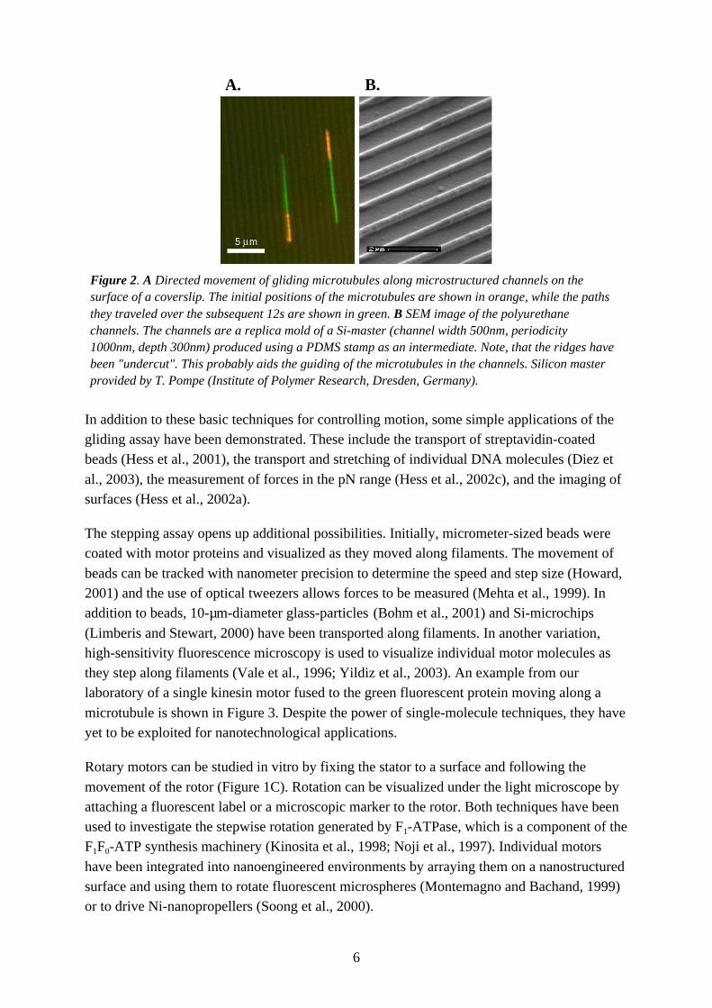
gliding microtubules that are guided by channels is shown in Figure 2. Temporal control hasbeen achieved by manipulating the ATP concentration (Bohm et al., 2000a; Hess et al., 2001).
6
A. B.
Figure 2. A Directed movement of gliding microtubules along microstructured channels on thesurface of a coverslip. The initial positions of the microtubules are shown in orange, while the pathsthey traveled over the subsequent 12s are shown in green. B SEM image of the polyurethanechannels. The channels are a replica mold of a Si-master (channel width 500nm, periodicity1000nm, depth 300nm) produced using a PDMS stamp as an intermediate. Note, that the ridges havebeen "undercut". This probably aids the guiding of the microtubules in the channels. Silicon masterprovided by T. Pompe (Institute of Polymer Research, Dresden, Germany).
In addition to these basic techniques for controlling motion, some simple applications of thegliding assay have been demonstrated. These include the transport of streptavidin-coatedbeads (Hess et al., 2001), the transport and stretching of individual DNA molecules (Diez et
al., 2003), the measurement of forces in the pN range (Hess et al., 2002c), and the imaging ofsurfaces (Hess et al., 2002a).
The stepping assay opens up additional possibilities. Initially, micrometer-sized beads werecoated with motor proteins and visualized as they moved along filaments. The movement of
beads can be tracked with nanometer precision to determine the speed and step size (Howard,2001) and the use of optical tweezers allows forces to be measured (Mehta et al., 1999). Inaddition to beads, 10-µm-diameter glass-particles (Bohm et al., 2001) and Si-microchips
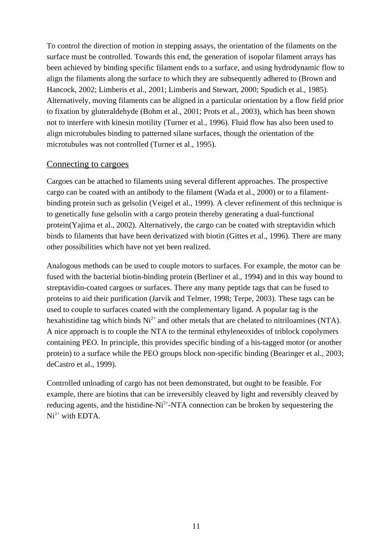
(Limberis and Stewart, 2000) have been transported along filaments. In another variation,high-sensitivity fluorescence microscopy is used to visualize individual motor molecules asthey step along filaments (Vale et al., 1996; Yildiz et al., 2003). An example from ourlaboratory of a single kinesin motor fused to the green fluorescent protein moving along a
microtubule is shown in Figure 3. Despite the power of single-molecule techniques, they haveyet to be exploited for nanotechnological applications.
Rotary motors can be studied in vitro by fixing the stator to a surface and following the
movement of the rotor (Figure 1C). Rotation can be visualized under the light microscope byattaching a fluorescent label or a microscopic marker to the rotor. Both techniques have beenused to investigate the stepwise rotation generated by F1-ATPase, which is a component of theF1F0-ATP synthesis machinery (Kinosita et al., 1998; Noji et al., 1997). Individual motors
have been integrated into nanoengineered environments by arraying them on a nanostructuredsurface and using them to rotate fluorescent microspheres (Montemagno and Bachand, 1999)or to drive Ni-nanopropellers (Soong et al., 2000).

7
Figure 3. Movement of a single kinesin molecule (labeled with the green fluorescent protein) along amicrotubule (red). Micrographs were acquired at the indicated times using total-internal-reflectionfluorescence microscopy.
Methods
There are many challenges in applying biomolecular motors to nanotechnology. Motility mustbe robust. It must be controlled both spatially and temporally. And the motors must be hitchedto and unhitched from their cargoes. This section summarizes key techniques towards these
ends.
General conditions for motility assays
Motility assays are performed in aqueous solutions that must fulfill a number of requirements.We will illustrate these requirements with the kinesin/microtubule system. Kinesin uses ATPas its fuel; the maximum speed is reached at ~0.5 mM, approximately equal to the cellular
concentration. Other nucleotides such as GTP, TTP and CTP can substitute for ATP, but thespeed is lower (Cohn et al., 1989). Motility also requires divalent cations, with magnesiumpreferred over calcium, and strontium and barium unable to substitute (Bohm et al., 1997).Optimal motility, assessed by gliding speed, occurs over a range of pH, between 6 and 9
(Bohm et al., 2000b; Cohn et al., 1989), and over a range of ionic strengths, between 50 mMand 300 mM (Bohm et al., 2000b). The speed increases with temperature, doubling for each10 ˚C between 5 ˚C and 50 ˚C (Bohm et al., 2000a; Kawaguchi and Ishiwata, 2001); motility
fails at higher temperatures. The force is independent of temperature between 15 ˚C and 35 ˚C(Kawaguchi and Ishiwata, 2000). When assays are performed in the middle of these ranges,motility is robust, and only a small drop in the mean velocities is seen after 3 hrs (Bohm et al.,

8
2000a; Bohm et al., 2000b). If fluorescent markers are used, an oxygen scavenging enzymesystem must be present to prevent photodamage. Many experimental details, including adiscussion of the densities of the motors, can be found in Scholey (1993)(Scholey, 1993).
Temporal control
Motors can be reversibly switched off and on by regulating the concentration of fuel, or byadding and removing inhibitors. The ATP concentration can be rapidly altered by flowing in anew solution. In such a setup, the kinesin-dependent movement of microtubules can bestopped within 1 s and restarted within 10 s (unpublished data from our lab). Similarly,
inhibitors such as AMP-PNP, a non-hydrolysable analogue of ATP (Schnapp et al., 1990),adociasulfate-2 a small molecule isolated from sponge (Sakowicz et al., 1998) and monastrol(Mayer et al., 1999) can be perfused to stop motility.
An alternative method to control energy supply is to use photoactivatable ATP. In thismethod, a flash of UV light is used to release ATP from a derivatized, non-functionalprecursor; an ATP-consuming enzyme is also present to return the ATP concentration to lowlevels following release. Using such a system, microtubule movement has been repeatedly
started and stopped (Hess et al., 2001), though the start-up and slow-down times were slow,on the order of minutes. The advantage of this method is that the solution in the flow cell doesnot have to be exchanged.
Fortuitously, many proteins possess natural regulatory mechanisms and, once understood,these might offer additional means to regulate the motors in vitro. Examples include theregulation of myosins by phosphorylation and calcium/calmodulin (Sellers and Goodson,1995) and the inhibition of kinesin by its cargo-binding "tail" domain (Coy et al., 1999).
Because such natural controls might not always be applicable in a synthetic environment,there is strong interest in the development of artificial control mechanisms for motor proteins.Towards this end, metal-ion binding sites have been genetically engineered into the F1-
ATPase motor. The binding of ions at the engineered site immobilizes the moving parts of themotor thus inhibiting its rotation (Liu et al., 2002). ATP driven rotation can be restored by theaddition of metal ion chelators. Clever genetic engineering of motors could provide temporalcontrol mechanisms switched by temperature, light, electrical fields or buffer composition.
Spatial control
In order to control the path along which filaments glide, a process that we call guiding, it isnecessary to restrict the location of active motors to specific regions of a surface. This can bedone by coating a glass or silicon surface with resist polymers such as PMMA, SU-8 or
SAL601 and using UV, electron beam or soft lithography to remove resist from definedregions (Bunk et al., 2003; Hiratsuka et al., 2001; Mahanivong et al., 2002; Moorjani et al.,2003; Nicolau et al., 1999; Riveline et al., 1998; Suzuki et al., 1997; Wright et al., 2002). The
motor-containing solution is then perfused across the surface. By choosing appropriateproperties of this solution—the concentration of motors, salts, other blocking proteins such as

9
casein and BSA, and detergents such as Triton X-100—motility can be restricted to either theunexposed, resist surface or to the exposed, underlying substrate. However, the interactions ofthe motors with these surfaces are not well understood. For example, myosin motility isprimarily restricted to the more hydrophobic resist surfaces while kinesin motility is primarily
restricted to the more hydrophilic non-resist surfaces. One limitation of this approach tobinding proteins to surfaces is that the motors tend to bind everywhere, so it is difficult toattain good contrast. A proven method to prevent motor binding is to coat a surface with
polyethylene oxide (PEO) (deCastro et al., 1999; Hess et al., 2002b). Techniques tospecifically bind motors and filaments to surfaces are summarized in the last section of the
Methods.
While chemical patterning can restrict movement of filaments to areas with a high density of
active motors, walking off the trails is not prevented. This was demonstrated by Hess et al.(Hess et al., 2002b) who showed that microtubules move straight across a boundary betweenhigh motor density (non-PEO) and low motor density (PEO) where they dissociate from thesurface. The problem with a purely chemical pattern is that if a rigid filament is propelled by
several motors along its length there is nothing to stop the motors at the rear from pushing thefilament across a boundary into an area of low motor density. Combining chemical andtopographic features, as occurs in the lithographic studies described above, leads to more
efficient guiding. For example, in the study of Moorjani et al. (Moorjani et al., 2003)filaments remained at the bottom of the channels formed in the SU-8 even when they collidedwith the walls at angles above 80 degrees. When the leading end of the microtubule hits thewall, the motors at the rear force the microtubule to bend into the region of high motor density
and in this way the motion is guided by the boundary (see Figure 4). The behavior ofmicrotubules colliding with the walls of channels imprinted in polyurethane has been studiedby Clemmens et al. (Clemmens et al., 2003). They found that the probability of a filament
being guided by the walls decreased as the approach angle increased. At high incident angles,guiding was not observed and instead the microtubules climbed the walls. Taken together,these studies show that a combination of chemical and topographic patterning is necessary forefficient guiding.
While it is possible to use chemical and topographical patterning to guide filaments—that isto restrict their movement to particular paths—it is more difficult to control the direction ofmovement along the path. The difficulty arises because the orientation in which motors bind
to a uniform surface is not controlled. Some motors will be oriented so that they propelfilaments in one direction along the path, whereas others will propel filaments in the oppositedirection. The reason that motors do not counteract each other is that filaments are polarstructures: the orientation of the proteins that form up the filaments is maintained all along the
length of the filament (see Figure 1). Because the motors bind stereospecifically to thefilament, they will exert force in only one direction. Thus, the orientation of the filamentdetermines its direction of motion; one end always leads.

10
Figure 4. Sequence of fluorescent images showing the kinesin driven, unidirectional movement of arhodamine-labeled microtubule (red) along a chemically and topographically structured Si-chip. Thebottom of the channels (green), whose depths are 300 nm, is coated with kinesin. The surroundingregions are blocked by polyethylene glycol. Collaboration with R.M.M. Smeets, M.G.L. van den Heuvel,and C. Dekker (Delft University of Technology, The Netherlands).
The direction of filament gliding can be controlled by the application of external forces. Actinfilaments and microtubules both possess negative net charges, and consequently, in thepresence of a uniform electric field, will experience a force directed towards the positive
electrode. It is possible to apply high enough electric fields to steer motor-driven filaments ina specified direction (Riveline et al., 1998; Stracke et al., 2002). Because the refractive indexof protein differs from that of water, filaments become electrically polarized in the presenceof an electric field, and consequently in a non-uniform field they move in the direction of
highest field strength. This so-called dielectrophoretic force has been used to direct thegliding of actin filaments on a myosin coated substrate (Asokan et al., 2003). It is evenpossible to manipulate a microtubule using optical gradients produced by focusing a laser
beam (i.e. an optical tweezers).(Felgner et al., 1997) Directional control of microtubulegliding has also been achieved using hydrodynamic flow fields (Bohm et al., 2001; Prots etal., 2003).
An alternative approach to directionality relies on more sophisticated guiding concepts. For
example, unidirectional movement of filaments can be achieved if guiding geometries basedon arrow and ratchet structures are employed (Hess et al., 2002b; Hiratsuka et al., 2001). Anexample of the unidirectional movement of a microtubule on a topographically and
chemically structured silicon chip is depicted in Figure 4 (unpublished results from ourlaboratory).
11
To control the direction of motion in stepping assays, the orientation of the filaments on thesurface must be controlled. Towards this end, the generation of isopolar filament arrays hasbeen achieved by binding specific filament ends to a surface, and using hydrodynamic flow toalign the filaments along the surface to which they are subsequently adhered to (Brown and
Hancock, 2002; Limberis et al., 2001; Limberis and Stewart, 2000; Spudich et al., 1985).Alternatively, moving filaments can be aligned in a particular orientation by a flow field priorto fixation by gluteraldehyde (Bohm et al., 2001; Prots et al., 2003), which has been shown
not to interfere with kinesin motility (Turner et al., 1996). Fluid flow has also been used toalign microtubules binding to patterned silane surfaces, though the orientation of themicrotubules was not controlled (Turner et al., 1995).
Connecting to cargoes
Cargoes can be attached to filaments using several different approaches. The prospectivecargo can be coated with an antibody to the filament (Wada et al., 2000) or to a filament-binding protein such as gelsolin (Veigel et al., 1999). A clever refinement of this technique isto genetically fuse gelsolin with a cargo protein thereby generating a dual-functional
protein(Yajima et al., 2002). Alternatively, the cargo can be coated with streptavidin whichbinds to filaments that have been derivatized with biotin (Gittes et al., 1996). There are manyother possibilities which have not yet been realized.
Analogous methods can be used to couple motors to surfaces. For example, the motor can befused with the bacterial biotin-binding protein (Berliner et al., 1994) and in this way bound tostreptavidin-coated cargoes or surfaces. There any many peptide tags that can be fused toproteins to aid their purification (Jarvik and Telmer, 1998; Terpe, 2003). These tags can be
used to couple to surfaces coated with the complementary ligand. A popular tag is thehexahistidine tag which binds Ni2+ and other metals that are chelated to nitriloamines (NTA).A nice approach is to couple the NTA to the terminal ethyleneoxides of triblock copolymers
containing PEO. In principle, this provides specific binding of a his-tagged motor (or anotherprotein) to a surface while the PEO groups block non-specific binding (Bearinger et al., 2003;deCastro et al., 1999).
Controlled unloading of cargo has not been demonstrated, but ought to be feasible. For
example, there are biotins that can be irreversibly cleaved by light and reversibly cleaved byreducing agents, and the histidine-Ni2+-NTA connection can be broken by sequestering theNi2+ with EDTA.

12
Outlook
The first steps have been made towards the operation of biomolecular motors in engineeredenvironments . However, many advances are necessary before these motors can be used in
nanotechnological applications such as working in molecular factories and building circuits.
An immediate task is to improve the spatial and temporal control over the motors. Bycombining improved surface techniques with the application of external electric, magnetic
and/or optical fields it should be possible, in the near future, to stretch and collide singlemolecules, to control cargo loading and unloading, and to sort and pool molecules.
A crucial longer-term goal is to control the position and orientation of motors with molecularprecision. This means placing motors with an accuracy of ~10 nm on a surface and
controlling their orientation within a few degrees. In this way both the location and thedirection of motion of filaments can be controlled. One approach to molecular patterning is to“decorate” filaments with stereospecifically bound motors. Once aligned along the filament
matrix, the motors can be transferred to another surface. This approach was taken by Spudichet al.(Spudich et al., 1985; Toyoshima et al., 1989) and should be followed up. A furtherdevelopment of this idea is to directly produce, perhaps by stamping a mold made with afilament, surfaces that have structures functionally similar to motor binding sites. An
alternative approach is to use dip-pen lithography (see Chapter 1.2 in this book) or other AFMtechniques(Jiang et al., 2003) to directly pattern motors on surfaces.
The robustness of motors must be increased. Motors operate only in aqueous solutions and
under a restricted range of solute concentrations and temperatures. While it is inconceivablethat protein-based motors could operate in a non-aqueous environment, two approaches toincreasing their robustness can be envisaged. First, motors could be purified fromthermophilic or halophilic bacteria some of which grow at temperatures up to 112 °C and
salinities above 5 M. There are extreme eukaryotes that grow up to 62 °C or 5 M NaCl. Thisapproach has already been taken for ATP synthase(Hazard and Montemagno, 2002), but notwith linear motors because no obvious homologues of myosins or kinesins have been found in
bacteria. Second, a genetic screening approach might reveal mutations that allow motors tooperate in less restrictive or different conditions. A longer-term goal is to use the designprinciples learnt from the study of biomolecular motors to build purely artificial nanomotorsthat can operate in air or vacuum. This is a daunting prospect and it is not even clear what
fuel(s) might be used. A potential way forward is to use chemical energy from a surface: forexample it was demonstrated that tin particles slide across copper surfaces driven by theformation of bronze alloy (Schmid et al., 2000). This is analogous to paraffin-driven toy
boats.
Besides the motor systems discussed so far, other biomechanical assemblies are goodcandidates for nanotechnological applications. Besides providing paths along which motorsmove, active biological filaments might find use in nanotechnological applications. The

13
pushing and pulling forces generated by the polymerization and depolymerization of actinfilaments and microtubules provide an alternative method of moving molecules(Dogteromand Yurke, 1997; Howard, 2001). This ability is of particular interest because bacteria possessactin-(van den Ent et al., 2002) and microtubule-like(Lowe and Amos, 1998) filaments, and as
mentioned above the proteins of extremophilic bacteria function in extreme environmentalconditions. Filaments and motors can also self-organize under certain conditions (Humphreyet al., 2002; Kruse and Julicher, 2000; Nedelec et al., 1997; Surrey et al., 1998). On a side
note, the flagellar filament in conjunction with the flagellar motors allow the bacteria to movein three dimensional liquid space(Ryu et al., 2000).
In addition to the motors that we have described so far, cells contain numerous biomolecularmachines that can also be thought of as motors (see e.g. Alberts, 2002 (Alberts, 2002)). These
machines use chemical energy to replicate DNA (DNA polymerases) and process it(recombinases, topoisomerases and endonucleases), to produce RNA (RNA polymerases)and splice it (spliceosomes), to make proteins (ribosomes) and fold them (chaperones) and
move them across membranes (translocases) and finally destroy them (proteasomes). Theenergy is provided by another group of machines that generate the electrochemical gradients(electron transport system, bacteriorhodopsin) used by the F1F0-ATP synthase to make ATP orby flagellar motors to propel bacteria. All these machines are candidates for
nanotechnological applications, and a recent report of the use of chaperones to maintainnanoparticles in solution (Ishii et al., 2003) is a step in this general direction.
We finish up by pointing out that the high order and nanometer-scale periodicity of DNA,
actin filaments and microtubules make them ideal scaffolds on which to erect 3-dimensionalnanostructures. While these features have been exploited to make DNA-based structures(Seeman, 2003) (see Chapter III), the use of DNA motors to address specific sites (based onnucleotide sequence) has not, to our knowledge, been realized. Some years ago it was
proposed that the regular lattice of microtubules might serve as substrates for molecularcomputing and information storage (Hameroff and Watt, 1982; Penrose, 2001). While theseideas seem crazy in the context of the living organism, they may be realizable for
biomolecular motors operating in engineered environments. At the moment, anything ispossible.

14
References
Alberts, B. (1998). The cell as a collection of protein machines: preparing the next generation ofmolecular biologists. Cell 92, 291-294.
Alberts, B. (2002). Molecular biology of the cell, 4th edn (New York, Garland Science).
Asokan, S. B., Jawerth, L., Carroll, R. L., Cheney, R. E., Washburn, S., and R. Superfine, R. (2003).Two-Dimensional Manipulation and Orientation of Actin-Myosin Systems with Dielectrophoresis.Nano Letters 3, 431-437.
Bearinger, J. P., Terrettaz, S., Michel, R., Tirelli, N., Vogel, H., Textor, M., and Hubbell, J. A. (2003).Chemisorbed poly(propylene sulphide)-based copolymers resist biomolecular interactions. NatMater 2, 259-264.
Berliner, E., Mahtani, H. K., Karki, S., Chu, L. F., Cronan, J. E., Jr., and Gelles, J. (1994).Microtubule movement by a biotinated kinesin bound to streptavidin-coated surface. J Biol Chem269, 8610-8615.
Bohm, K. J., Steinmetzer, P., Daniel, A., Baum, M., Vater, W., and Unger, E. (1997). Kinesin-drivenmicrotubule motility in the presence of alkaline-earth metal ions: indication for a calcium ion-dependent motility. Cell Motil Cytoskeleton 37, 226-231.
Bohm, K. J., Stracke, R., Baum, M., Zieren, M., and Unger, E. (2000a). Effect of temperature onkinesin-driven microtubule gliding and kinesin ATPase activity. Febs Letters 466, 59-62.
Bohm, K. J., Stracke, R., Muhlig, P., and Unger, E. (2001). Motor protein-driven unidirectionaltransport of micrometer-sized cargoes across isopolar microtubule arrays. IOP PublishingNanotechnology 12, 238-244.
Bohm, K. J., Stracke, R., and Unger, E. (2000b). Speeding up kinesin-driven microtubule gliding invitro by variation of cofactor composition and physicochemical parameters. Cell BiologyInternational 24, 335-341.
Brown, T. B., and Hancock, W. O. (2002). A polarized microtubule array for kinesin-powered-nanoscale assembly and force generation. Nano Letters 2, 1131-1135.
Bunk, R., Klinth, J., Montelius, L., Nicholls, I. A., Omling, P., Tagerud, S., and Mansson, A. (2003).Actomyosin motility on nanostructured surfaces. Biochemical and Biophysical ResearchCommunications 301, 783-788.
Burgess, S. A., Walker, M. L., Sakakibara, H., Knight, P. J., and Oiwa, K. (2003). Dynein structureand power stroke. Nature 421, 715-718.
Bustamante, C., Bryant, Z., and Smith, S. B. (2003). Ten years of tension: single-molecule DNAmechanics. Nature 421, 423-427.
Champoux, J. J. (2001). DNA topoisomerases: structure, function, and mechanism. Annu RevBiochem 70, 369-413.
Clemmens, J., Hess, H., Howard, J., and Vogel, V. (2003). Analysis of microtubule guidance in openmicrofabricated channels coated with the motor protein kinesin. Langmuir 19, 1738-1744.
Cohn, S. A., Ingold, A. L., and Scholey, J. M. (1989). Quantitative analysis of sea urchin egg kinesin-driven microtubule motility. J Biol Chem 264, 4290-4297.
Coy, D. L., Hancock, W. O., Wagenbach, M., and Howard, J. (1999). Kinesin's tail domain is aninhibitory regulator of the motor domain. Nat Cell Biol 1, 288-292.
Crichton, M. (2002). Prey (Toronto, Harper Collins).
deCastro, M. J., Ho, C. H., and Stewart, R. J. (1999). Motility of dimeric ncd on a metal-chelatingsurfactant: evidence that ncd is not processive. Biochemistry 38, 5076-5081.
15
Dennis, J. R., Howard, J., and Vogel, V. (1999). Molecular shuttles: directed motion of microtubulesslang nanoscale kinesin tracks. Nanotechnology 10, 232-236.
DeRosier, D. J. (1998). The turn of the screw: the bacterial flagellar motor. Cell 93, 17-20.
Diez, S., Reuther, C., Seidel, R., Mertig, M., Pompe, W., and Howard, J. (2003). Transporting andStretching Individual DNA Molecules by Kinesin and Microtubules. submitted.
Dogterom, M., and Yurke, B. (1997). Measurement of the force-velocity relation for growingmicrotubules. Science 278, 856-860.
Felgner, H., Frank, R., Biernat, J., Mandelkow, E. M., Mandelkow, E., Ludin, B., Matus, A., andSchliwa, M. (1997). Domains of neuronal microtubule-associated proteins and flexural rigidity ofmicrotubules. J Cell Biol 138, 1067-1075.
Forde, N. R., Izhaky, D., Woodcock, G. R., Wuite, G. J., and Bustamante, C. (2002). Usingmechanical force to probe the mechanism of pausing and arrest during continuous elongation byEscherichia coli RNA polymerase. Proceedings of the National Academy of Sciences of the UnitedStates of America 99, 11682-11687.
Gittes, F., Meyhofer, E., Baek, S., and Howard, J. (1996). Directional loading of the kinesin motormolecule as it buckles a microtubule. Biophys J 70, 418-429.
Hagerman, P. J. (1997). Flexibility of RNA. Annu Rev Biophys Biomol Struct 26, 139-156.
Hameroff, S. R., and Watt, R. C. (1982). Information processing in microtubules. J Theor Biol 98,549-561.
Hazard, A., and Montemagno, C. (2002). Improved purification for thermophilic F1F0 ATP synthaseusing n-dodecyl [beta]--maltoside. Archives of Biochemistry and Biophysics 407, 117-124.
Hess, H., Clemmens, J., Howard, J., and Vogel, V. (2002a). Surface imaging by self-propellednanoscale probes. Nano Letters 2, 113-116.
Hess, H., Clemmens, J., Matzke, C. M., Bachand, G. D., Bunker, B. C., and Vogel, V. (2002b).Ratchet patterns sort molecular shuttles. Applied Physics a (Materials Science Processing) A75,309-313.
Hess, H., Clemmens, J., Qin, D., Howard, J., and Vogel, V. (2001). Light-controlled molecularshuttles made from motor proteins carrying cargo on engineered surfaces. Nano Letters 1, 235-239.
Hess, H., Howard, J., and Vogel, V. (2002c). A piconewton forcemeter assembled from microtubulesand kinesins. Nano Letters 2, 1113-1115.
Hiratsuka, Y., Tada, T., Oiwa, K., Kanayama, T., and Uyeda, T. Q. P. (2001). Controlling thedirection of kinesin-driven microtubule movements along microlithographic tracks. BiophysicalJournal 81, 1555-1561.
Howard, J. (2001). Mechanics of Motor Proteins and the Cytoskeleton (Sunderland, MA, SinauerAssociates).
Humphrey, D., Duggan, C., Saha, D., Smith, D., and Kas, J. (2002). Active fluidization of polymernetworks through molecular motors. Nature 416, 413-416.
Ishii, D., Kinbara, K., Ishida, Y., Ishii, N., Okochi, M., Yohda, M., and Aida, T. (2003). Chaperonin-mediated stabilization and ATP-triggered release of semiconductor nanoparticles. Nature 423, 628-632.
Jarvik, J. W., and Telmer, C. A. (1998). Epitope tagging. Annu Rev Genet 32, 601-618.
Jiang, F., Khairy, K., Poole, K., Howard, J., and Muller, D. (2003). submitted.
Kawaguchi, K., and Ishiwata, S. (2000). Temperature dependence of force, velocity, and processivityof single kinesin molecules. Biochem Biophys Res Commun 272, 895-899.

16
Kawaguchi, K., and Ishiwata, S. (2001). Thermal activation of single kinesin molecules withtemperature pulse microscopy. Cell Motil Cytoskeleton 49, 41-47.
Kinosita, K., Jr., Yasuda, R., Noji, H., Ishiwata, S., and Yoshida, M. (1998). F1-ATPase: a rotarymotor made of a single molecule. Cell 93, 21-24.
Kruse, K., and Julicher, F. (2000). Actively contracting bundles of polar filaments. Phys Rev Lett 85,1778-1781.
Kull, F. J. (2000). Motor proteins of the kinesin superfamily: structure and mechanism. EssaysBiochem 35, 61-73.
Limberis, L., Magda, J. J., and Stewart, R. J. (2001). Polarized alignment and surface immobilizationof microtubules for kinesin-powered nanodevices. Nano Letters 1, 277-280.
Limberis, L., and Stewart, R. J. (2000). Toward kinesin-powered microdevices. IOP PublishingNanotechnology 11, 47-51.
Liu, H., Schmidt, J. J., Bachand, G. D., Rizk, S. S., Looger, L. L., Hellinga, H. W., and Montemagno,C. D. (2002). Control of a biomolecular motor-powered nanodevice with an engineered chemicalswitch. Nat Mater 1, 173-177.
Lowe, J., and Amos, L. A. (1998). Crystal structure of the bacterial cell-division protein FtsZ. Nature391, 203-206.
Mahanivong, C., Wright, J. P., Kekic, M., Pham, D. K., dos Remedios, C., and Nicolau, D. V. (2002).Manipulation of the motility of protein molecular motors on microfabricated substrates. BiomedicalMicrodevices 4, 111-116.
Maier, B., Potter, L., So, M., Seifert, H. S., and Sheetz, M. P. (2002). Single pilus motor forces exceed100 pN. Proceedings of the National Academy of Sciences of the United States of America 99,16012-16017.
Mayer, T. U., Kapoor, T. M., Haggarty, S. J., King, R. W., Schreiber, S. L., and Mitchison, T. J.(1999). Small molecule inhibitor of mitotic spindle bipolarity identified in a phenotype-basedscreen. Science 286, 971-974.
Mehta, A. (2001). Myosin learns to walk. J Cell Sci 114, 1981-1998.
Mehta, A. D., Rief, M., Spudich, J. A., Smith, D. A., and Simmons, R. M. (1999). Single-moleculebiomechanics with optical methods. Science 283, 1689-1695.
Merz, A. J., So, M., and Sheetz, M. P. (2000). Pilus retraction powers bacterial twitching motility.Nature 407, 98-102.
Montemagno, C., and Bachand, G. (1999). Constructing nanomechanical devices powered bybiomolecular motors. Nanotechnology 10, 225-231.
Moorjani, S. G., Jia, L., Jackson, T. N., and Hancock, W. O. (2003). Lithographically PatternedChannels Spatially Segregate Kinesin Motor Activity and Effectively Guide MicrotubuleMovements. Nano Letters 3, 633 -637.
Nedelec, F. J., Surrey, T., Maggs, A. C., and Leibler, S. (1997). Self-organization of microtubules andmotors. Nature 389, 305-308.
Nicolau, D. V., Suzuki, H., Mashiko, S., Taguchi, T., and Yoshikawa, S. (1999). Actin motion onmicrolithographically functionalized myosin surfaces and tracks. Biophysical Journal 77, 1126-1134.
Nogales, E. (2000). Structural insights into microtubule function. Annu Rev Biochem 69, 277-302.
Noji, H., Yasuda, R., Yoshida, M., and Kinosita, K., Jr. (1997). Direct Observation of the Rotation ofF-1-Atpase. Nature 386, 299-302.
Penrose, R. (2001). Consciousness, the brain, and spacetime geometry: an addendum. Some newdevelopments on the Orch OR model for consciousness. Ann N Y Acad Sci 929, 105-110.

17
Prots, I., Stracke, R., Unger, E., and Bohm, K. J. (2003). Isopolar microtubule arrays as a tool todetermine motor protein directionality. Cell Biology International 27, 251-253.
Riveline, D., Ott, A., Julicher, F., Winkelmann, D. A., Cardoso, O., Lacapere, J. J., Magnusdottir, S.,Viovy, J. L., Gorre-Talini, L., and Prost, J. (1998). Acting on actin: the electric motility assay.European Biophysics Journal with Biophysics Letters 27, 403-408.
Ruegg, C., Veigel, C., Molloy, J. E., Schmitz, S., Sparrow, J. C., and Fink, R. H. (2002). Molecularmotors: force and movement generated by single myosin II molecules. News Physiol Sci 17, 213-218.
Ryu, W. S., Berry, R. M., and Berg, H. C. (2000). Torque-generating units of the flagellar motor ofEscherichia coli have a high duty ratio. Nature 403, 444-447.
Sakowicz, R., Berdelis, M. S., Ray, K., Blackburn, C. L., Hopmann, C., Faulkner, D. J., and Goldstein,L. S. (1998). A marine natural product inhibitor of kinesin motors. Science 280, 292-295.
Schmid, A. K., Bartelt, N. C., and Hwang, R. Q. (2000). Alloying at surfaces by the migration ofreactive two-dimensional islands. Science 290, 1561-1564.
Schnapp, B. J., Crise, B., Sheetz, M. P., Reese, T. S., and Khan, S. (1990). Delayed start-up of kinesin-driven microtubule gliding following inhibition by adenosine 5'-[beta,gamma-imido]triphosphate.Proc Natl Acad Sci U S A 87, 10053-10057.
Scholey, J. M. (1993). Motility assays for motor proteins (San Diego, Academic Press).
Seeman, N. C. (2003). DNA in a material world. Nature 421, 427-431.
Sellers, J. R., and Goodson, H. V. (1995). Motor proteins 2: myosin. Protein Profile 2, 1323-1423.
Sheterline, P., Clayton, J., and Sparrow, J. (1995). Actin. Protein Profile 2, 1-103.
Shingyoji, C., Higuchi, H., Yoshimura, M., Katayama, E., and Yanagida, T. (1998). Dynein arms areoscillating force generators. Nature 393, 711-714.
Smith, D. E., Tans, S. J., Smith, S. B., Grimes, S., Anderson, D. L., and Bustamante, C. (2001). Thebacteriophage straight phi29 portal motor can package DNA against a large internal force. Nature413, 748-752.
Soong, R. K., Bachand, G. D., Neves, H. P., Olkhovets, A. G., Craighead, H. G., and Montemagno, C.D. (2000). Powering an inorganic nanodevice with a biomolecular motor. Science 290, 1555-1558.
Spudich, J. A., Kron, S. J., and Sheetz, M. P. (1985). Movement of Myosin-Coated Beads on OrientedFilaments Reconstituted from Purified Actin. Nature 315, 584-586.
Stracke, P., Bohm, K. J., Burgold, J., Schacht, H. J., and Unger, E. (2000). Physical and technicalparameters determining the functioning of a kinesin-based cell-free motor system. IOP PublishingNanotechnology 11, 52-56.
Stracke, R., Bohm, K. J., Wollweber, L., Tuszynski, J. A., and Unger, E. (2002). Analysis of themigration behaviour of single microtubules in electric fields. Biochemical and BiophysicalResearch Communications 293, 602-609.
Strick, T. R., Charvin, G., Dekker, N. H., Allemand, J. F., Bensimon, D., and Croquette, V. (2002).Tracking enzymatic steps of DNA topoisomerases using single-molecule micromanipulation.Comptes Rendus Physique 3, 595-618.
Surrey, T., Elowitz, M. B., Wolf, P. E., Yang, F., Nedelec, F., Shokat, K., and Leibler, S. (1998).Chromophore-assisted light inactivation and self-organization of microtubules and motors. ProcNatl Acad Sci U S A 95, 4293-4298.
Suzuki, H., Yamada, A., Oiwa, K., Nakayama, H., and Mashiko, S. (1997). Control of actin movingtrajectory by patterned poly(methyl methacrylate) tracks. Biophysical Journal 72, 1997-2001.
Terpe, K. (2003). Overview of tag protein fusions: from molecular and biochemical fundamentals tocommercial systems. Appl Microbiol Biotechnol 60, 523-533.

18
Toyoshima, Y. Y., Toyoshima, C., and Spudich, J. A. (1989). Bidirectional movement of actinfilaments along tracks of myosin heads. Nature 341, 154-156.
Turner, D., Chang, C. Y., Fang, K., Cuomo, P., and Murphy, D. (1996). Kinesin movement onglutaraldehyde-fixed microtubules. Analytical Biochemistry 242, 20-25.
Turner, D. C., Chang, C. Y., Fang, K., Brandow, S. L., and Murphy, D. B. (1995). Selective adhesionof functional microtubules to patterned silane surfaces. Biophysical Journal 69, 2782-2789.
Vale, R. D., Funatsu, T., Pierce, D. W., Romberg, L., Harada, Y., and Yanagida, T. (1996). Directobservation of single kinesin molecules moving along microtubules. Nature 380, 451-453.
van den Ent, F., Moller-Jensen, J., Amos, L. A., Gerdes, K., and Lowe, J. (2002). F-actin-likefilaments formed by plasmid segregation protein ParM. Embo J 21, 6935-6943.
Veigel, C., Coluccio, L. M., Jontes, J. D., Sparrow, J. C., Milligan, R. A., and Molloy, J. E. (1999).The motor protein myosin-I produces its working stroke in two steps. Nature 398, 530-533.
Wada, Y., Hamasaki, T., and Satir, P. (2000). Evidence for a novel affinity mechanism of motor-assisted transport along microtubules. Mol Biol Cell 11, 161-169.
Wang, M. D., Schnitzer, M. J., Yin, H., Landick, R., Gelles, J., and Block, S. M. (1998). Force andvelocity measured for single molecules of RNA polymerase. Science 282, 902-907.
Wright, J., Pham, D., Mahanivong, C., Nicolau, D. V., Kekic, M., and dos Remedios, C. G. (2002).Micropatterning of myosin on O-acryloyl acetophenone oxime (AAPO), layered with bovine serumalbumin (BSA). Biomedical Microdevices 4, 205-211.
Wuite, G. J., Smith, S. B., Young, M., Keller, D., and Bustamante, C. (2000). Single-molecule studiesof the effect of template tension on T7 DNA polymerase activity. Nature 404, 103-106.
Yajima, J., Alonso, M. C., Cross, R. A., and Toyoshima, Y. Y. (2002). Direct long-term observation ofkinesin processivity at low load. Curr Biol 12, 301-306.
Yildiz, A., Forkey, J. N., McKinney, S. A., Ha, T., Goldman, Y. E., and Selvin, P. R. (2003). MyosinV walks hand-over-hand: single fluorophore imaging with 1.5-nm localization. Science 300, 2061-2065.