obstruction of endotracheal tube with relevant … 2014/obstruction of...cardiopulmonary bypass...
TRANSCRIPT
Pediatric Anesthesia and Critical Care Journal 2014; 2(2):102-104 doi:10.14587/paccj.2014.21
Morei et al. Obstruction of endotracheal tube 102
Key points
Nasal intubation for cardiac surgery in children requiring systemic anticoagulation can lead to obstruction of the endo-tracheal tube by clot formation, requiring immediate tube replacement.
Obstruction of endotracheal tube with relevant respiratory acidosis during pediatric cardiac surgery
N. M. Morei1, H. E. Mungroop1, G. Michielon2 , T. W. L. Scheeren1
1Department of Anesthesiology, University Medical Center Groningen, University of Groningen, Gro-ningen, The Netherlands 2Department of Cardiothoracic Surgery, University Medical Center Groningen, University of Gronin-gen, Groningen, The Netherlands
Corresponding author: 1T. W. L. Scheeren, Department of Anesthesiology, University Medical Center Groningen, Uni-versity of Groningen, Groningen, The Netherlands. Email: [email protected]
Abstract
We describe a case of pediatric cardiac surgery in a 21-
days old baby, in whom a nasal endotracheal tube (ETT)
was inserted. At the end of surgery both ventilatory
pressures and end-tidal CO2 increased suggesting air-
way obstruction. Suctioning of the ETT lumen did not
relieve the problem, only ETT replacement did.
The ETT was almost completely obstructed with a clot,
leading to significant respiratory acidosis. We would
like to bring awareness of the possibility of ETT ob-
struction in pediatric cardiac surgery with nasal intuba-
tion and systemic anticoagulation, in which only tube
exchange relieved the problem.
Keywords: airway obstruction; pediatric cardiac surge-
ry, nasal intubation, systemic anticoagulation
Background
In congenital pediatric cardiac surgery, especially in
neonates, the patient is often intubated nasally in order
to prevent endotracheal tube malposition and accidental
spontaneous extubation in the intensive care unit.
However, nasal intubation together with the need for
systemic anticoagulation with heparin for cardiac surge-
ry is a known risk factor for endotracheal tube obstruc-
tion.
Patient case description
A 21-days old baby, length 50 cm, weight 3 kg, with the
diagnosis of total anomalous pulmonary venous return
(TAPVR) was scheduled for TAPVR repair. He was cy-
anotic, had respiratory distress and rapid breathing de-
spite supplementary oxygen breathing. The echocardi-
ography showed next to the TAPVR, in which all four
pulmonary veins drained abnormally to the right atrium
instead of the left atrium, dilatation and hypertrophy of
the right ventricle, an open foramen ovale with continu-
ous right to left shunt and a relatively small left atrium
and left ventricle, and a patent ductus arteriosus. On the
day of surgery anesthesia was induced with sevoflurane
through a breathing mask and intravenous injection of
sufentanil 2 microgram per kg, midazolam 0.3 mi-
crogram per kg and rocuronium 1.2 mg per kg followed
Pediatric Anesthesia and Critical Care Journal 2014; 2(2):102-104 doi:10.14587/paccj.2014.21
Morei et al. Obstruction of endotracheal tube 103
by a nasal intubation with a cuffed endotracheal tube
(ETT) sized 3 mm (internal diameter), distance 11.5 cm
from the nostril. The intubation was easy and succeeded
in one attempt. Auscultation of the lungs revealed a
good position of the ETT with symmetric bilateral ve-
sicular sounds. Anesthesia was maintained with midazo-
lam and sufentanil. An arterial line was inserted in the
right femoral artery and a central venous line in the right
internal jugular vein. The peak inspiratory pressure was
19 cmH2O, end-tidal carbon dioxide (ETCO2) was 5.0
kPa. Blood gas analysis showed a pH of 7.41, a PaO2 of
7.6 kPa, a PaCO2 of 6.1 kPa and an arterial oxygen satu-
ration of 92%. Sternotomy was performed and after
opening of the pericardium the patient was placed on
cardiopulmonary bypass using arterial cannulation of
the ascending aorta and venous cannulation of both the
superior and inferior vena cava. Patient was heparinized
(4 mg/kg) and surgery was performed during cardio-
pulmonary bypass for 3.5 hours and consisted of correc-
tion of total anomalous pulmonary venous connection,
according to the Tucker technique, ligation of the patent
ductus arteriosus, ligation of the vertical vein, and
patch-closure of the atrial septal defect using autologous
pericardial patch. During this procedure a period of 25
minutes of circulatory arrest was needed to reach the
collector of the pulmonary veins located posterior to the
pericardium. The patient was cooled to a oesophageal
temperature of 23° Celsius. After complete re-warming
of the patient, weaning from cardiopulmonary bypass
was uneventful. The systemic arterial blood pressure
was 65/35 mmHg while the pulmonary arterial pressure
was between 30-35 mmHg. The left atrial pressure was
between 8 and 12 mmHg. An epicardial echocardiogram
was performed, showing good size of the left atrial
chamber, and a good biventricular function. The anas-
tomotic side was identified and found to be free from
obstruction. Protaminsulfate was then administered (15
mg) and decannulation was achieved. During the hemo-
stasis period the peak inspiratory pressure increased
gradually from 22 to 44 cmH2O on the ventilator, the
ETCO2 varied between 4 and 7 kPa. After exclusion of
kinking and malposition of the endotracheal tube, suc-
tioning of the ETT for the presence of secretion or blood
was performed but was negative. On auscultation of the
lungs a vesicular breathing sound was heard with
wheezes on both sides. Manual ventilation of the lungs
revealed some resistance. An allergic reaction to prota-
mine was briefly considered but eventually not suspect-
ed because of the good hemodynamic picture. The blood
gas analysis showed a gradual increase of PaCO2 from
6.1 to 17.6 kPa with a decline of pH from 7.31 to 6.92.
Thus, the patient experienced a period of respiratory ac-
idosis through retention of CO2. The PaO2 and arterial
oxygen saturation (SaO2) remained adequate (30.4 kPa
and 99%, respectively). Cerebral oxygenation (rSO2) as
obtained by near-infrared spectroscopy (Invos 5100C,
Covidien, Dublin, Ireland) nicely followed the changes
in SaO2 but never decreased below 50%. After all failed
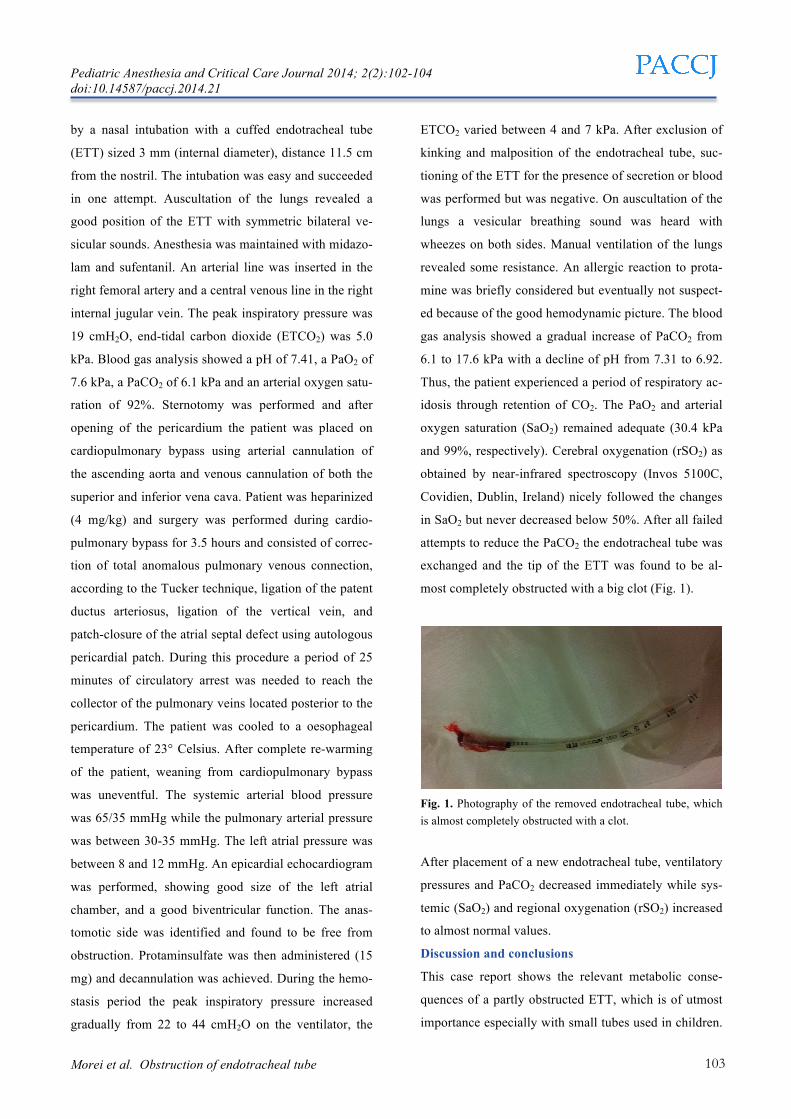
attempts to reduce the PaCO2 the endotracheal tube was
exchanged and the tip of the ETT was found to be al-
most completely obstructed with a big clot (Fig. 1).
Fig. 1. Photography of the removed endotracheal tube, which is almost completely obstructed with a clot.
After placement of a new endotracheal tube, ventilatory
pressures and PaCO2 decreased immediately while sys-
temic (SaO2) and regional oxygenation (rSO2) increased
to almost normal values.
Discussion and conclusions
This case report shows the relevant metabolic conse-
quences of a partly obstructed ETT, which is of utmost
importance especially with small tubes used in children.
Pediatric Anesthesia and Critical Care Journal 2014; 2(2):102-104 doi:10.14587/paccj.2014.21
Morei et al. Obstruction of endotracheal tube 104
Of note, suction of the ETT failed to relief the obstruc-
tion. The patient had an enormous drop in the pH to a
minimum of 6.92. This drop could have led to a cardiac
arrest if not identified and corrected in time. In addition,
the huge increase in PaCO2 did not correspond with an
equivalent increase in end-tidal CO2. While an increase
in peak airway pressure is a typical warning sign for
(partial) ETT obstruction,[1] the differential diagnosis
for high peak airway pressures and CO2 retention in-
cludes kinking, malposition and migration of the ETT,
obstruction of the tube filter with blood or secretion,
presence of long edema, atelectasis, pleural effusion,
bronchospasm, pneumothorax and an allergic reaction to
drug administration.
In this case it was hazardous to us that suctioning of the
ETT did not show any blood or secretion, perhaps be-
cause of the firm clot formation and the small diameter
of the tube.
In the literature there are few case reports over obstruc-
tion in the ETT by blood clot, mucus and even by over-
inflation of the cuff.[2-5]
Nasal intubation can cause damage to the mucosa of the
nasal cavity and turbinate. We conclude that nasal intu-
bation for cardiac surgery with full heparinization is a
risk factor for bleeding and ETT occlusion with clot,
which can lead to relevant respiratory acidosis
Disclosure and Acknowledgements
There is no financial support causing conflict of inter-
ests. The patient’s parents gave their consent to publish
this case report.
References
1. Kawati R, Lattuada M, Sjostrand U et al. Peak airway
pressure increase is a late warning sign of partial endo-
tracheal tube obstruction whereas change in expiratory
flow is an early warning sign. Anesth Analg 2005; 100:
889-893
2. Arney KL, Judson MA, Sahn SA. Airway obstruction
arising from blood clot: three reports and a review of the
literature. Chest 1999; 115: 293-300
3. Xue FS, Luo MP, Liao X et al. Delayed endotracheal
tube obstruction by mucus plug in a child. Chinese
Medical Journal 2009; 122: 870-872
4. Johnson KM, Lehman RE. Acute management of the
obstructed endotracheal tube. Respir Care 2012; 57:
1342-1344
5. Lim HK, Lee MH, Shim HY et al. Complete
obstruction of an endotracheal tube due to an
unexpected blood clot in a patient with a hemo-
pneumothorax after repositioning of the patient for
lumbar spine surgery. Korean Journal of Anesthesiology
2013; 64: 382-383