odorant receptors can mediate axonal identity and gene
TRANSCRIPT

HAL Id: hal-01393703https://hal-univ-bourgogne.archives-ouvertes.fr/hal-01393703
Submitted on 26 Sep 2017
HAL is a multi-disciplinary open accessarchive for the deposit and dissemination of sci-entific research documents, whether they are pub-lished or not. The documents may come fromteaching and research institutions in France orabroad, or from public or private research centers.
L’archive ouverte pluridisciplinaire HAL, estdestinée au dépôt et à la diffusion de documentsscientifiques de niveau recherche, publiés ou non,émanant des établissements d’enseignement et derecherche français ou étrangers, des laboratoirespublics ou privés.
Odorant receptors can mediate axonal identity and genechoice via cAMP-independent mechanisms
Kiavash Movahedi, Xavier Grosmaitre, Paul Feinstein
To cite this version:Kiavash Movahedi, Xavier Grosmaitre, Paul Feinstein. Odorant receptors can mediate axonal iden-tity and gene choice via cAMP-independent mechanisms. Open Biology, Royal Society, 2016, 6 (7),pp.160018. �10.1098/rsob.160018�. �hal-01393703�

on July 27, 2016http://rsob.royalsocietypublishing.org/Downloaded from
rsob.royalsocietypublishing.org
ResearchCite this article: Movahedi K, Grosmaitre X,
Feinstein P. 2016 Odorant receptors can
mediate axonal identity and gene choice via
cAMP-independent mechanisms. Open Biol. 6:
160018.
http://dx.doi.org/10.1098/rsob.160018
Received: 20 January 2016
Accepted: 1 July 2016
Subject Area:neuroscience/developmental biology
Keywords:olfactory receptor, axon guidance, olfaction,
gene choice, GPCR, neuronal wiring
Authors for correspondence:Kiavash Movahedi
e-mail: [email protected]
Paul Feinstein
e-mail: [email protected]
Electronic supplementary material is available
at http://dx.doi.org/10.1098/rsob.160018.
& 2016 The Authors. Published by the Royal Society under the terms of the Creative Commons AttributionLicense http://creativecommons.org/licenses/by/4.0/, which permits unrestricted use, provided the originalauthor and source are credited.
Odorant receptors can mediate axonalidentity and gene choice via cAMP-independent mechanisms
Kiavash Movahedi1,2,3, Xavier Grosmaitre4 and Paul Feinstein5
1Max Planck Institute of Biophysics, Max-von-Laue-Strasse 3, 60438 Frankfurt, Germany2Myeloid Cell Immunology Laboratory, VIB Inflammation Research Center, Ghent, Belgium3Laboratory of Cellular and Molecular Immunology, Vrije Universiteit Brussel, Brussels, Belgium4Centre des Sciences du Gout et de l’Alimentation, CNRS, INRA, Universite Bourgogne Franche-Comte,21000 Dijon, France5Department of Biological Sciences, Hunter College and The Graduate Center Biochemistry, Biology andBiopsychology and Behavioral Neuroscience Programs, CUNY, New York, NY, USA
KM, 0000-0002-0826-4399
Odorant receptors (ORs) control several aspects of cell fate in olfactory sen-
sory neurons (OSNs), including singular gene choice and axonal identity.
The mechanisms of OR-induced axon guidance have been suggested to prin-
cipally rely on G-protein signalling. Here, we report that for a subset of
OSNs, deleting G proteins or altering their levels of signalling does not
affect axonal identity. Signalling-deficient ORs or surrogate receptors that
are unable to couple to Gs/Golf still provide axons with distinct identities
and the anterior–posterior targeting of axons does not correlate with the
levels of cAMP produced by genetic modifications. In addition, we refine
the models of negative feedback by showing that ectopic ORs can be
robustly expressed without suppressing endogenous gene choice. In con-
clusion, our results uncover a new feature of ORs, showing that they can
instruct axonal identity and regulate olfactory map formation independent
of canonical G-protein signalling and cAMP production.
1. IntroductionOlfaction detects chemosensory stimuli with an enormous diversity in physico-
chemical properties. To accommodate this broad recognition, the olfactory
system employs a large repertoire of odorant receptors (ORs) [1,2]. OR genes
form the largest multi-gene family in mammals, with the mouse having
approximately 1100 functional receptors and approximately 200 pseudogenes
[3]. Signal transduction through the OR is derived by ligand binding (odour)
catalysing the bound heterotrimeric G-protein complex to exchange GDP for
GTP in the Ga subunit and the dissociation from the Gbg dimer. Ga bound
to GTP subsequently activates adenylyl cyclase 3 (Adcy3), inducing the pro-
duction of cyclic AMP (cAMP), which then binds to cyclic nucleotide-gated
ion channels and depolarizes the cell membrane [4]. While one would expect
that the OR’s main role would be to detect odorants and initiate signal trans-
duction, extensive evidence has shown that ORs also play pivotal roles in the
development of the olfactory system [5,6]. However, dissecting the pluralistic
roles of ORs has been challenging and their functions remain enigmatic.
Every olfactory sensory neuron (OSN) is thought to express only a single
OR gene from a single allele, which is referred to as singular expression [7,8].
OSNs expressing a given OR are scattered throughout the epithelium but are
confined to specific zones [9]. An important feature of the system is that the
expression of an OR seems to preclude the expression of additional OR alleles,
which is referred to as OR-induced feedback [10,11] and is shown to rely on
Adcy3, histone demethylase LSD1 and the unfolded protein response [12–15].

rsob.royalsocietypublishing.orgOpen
Biol.6:160018
2
on July 27, 2016http://rsob.royalsocietypublishing.org/Downloaded from
Axons from OSNs that express the same singular
expressed OR coalesce into two of the approximately 1800
glomeruli, typically one on the medial and one on the lateral
side of the olfactory bulb [16]. The OR itself plays an impor-
tant role in this impressive developmental task, since
mutations that alter single amino acids within an OR
sequence or its expression levels reroute axons and shift the
position of the glomerulus [5,6,17]. All developmental out-
comes by an OR can be substituted by surrogate receptors
such as the highly divergent b2-adrenergic receptor
[5,18,19]. These experiments have shown that glomeruli do
not form an invariant topographical map, which would
serve as ‘targets’ for ORs expressed on axons. Instead, glo-
meruli arise through homotypic interactions, in a context-
dependent manner [6]. How ORs and other chemosensory
receptors instruct an identity and regulate axonal interactions
and glomerular formation has remained unclear. One
hypothesis suggests that OR-induced wiring mechanisms
fully rely on Gsa and Golfa signalling [18]; ORs would
have an indirect role, one of regulating the expression levels
of conventional axon guidance and identity molecules. In
this model, each OR and OR polymorphism capable of gen-
erating unique glomeruli, would have a unique level of
basal, agonist-independent activity, which would alter the
transcriptional expression of axon guidance molecules such
as neuropilin-1. The altered levels of cell surface molecules
also regulate anterior–posterior (A-P) targeting of axons:
low levels of cAMP result in anterior glomerular positioning
and high levels of cAMP result in posterior glomerular posi-
tioning [20]. Following A-P positioning, agonist-dependent
Golfa signalling in mature OSNs assists in segregating
axons into unique glomeruli, by altering the expression
levels of cell adhesion molecules, of which Kirrel2, Kirrel3
and BIG2 result in adhesion (attraction of ‘like’ axons),
while EphA and ephrinA mediate contact-dependent repul-
sion (segregation of ‘non-like’ axons) [21–23]. Dorsoventral
positioning of glomeruli does not rely on OR signalling, but
instead correlates with the anatomical locations of OSNs in
the olfactory epithelium and was shown to rely on two
sets of repulsive ligands/receptors expressed by OSNs:
neuropillin2/Sema3F and Slits/Robo2 [24–26].
We now provide evidence for an extended model of
OR-mediated axonal wiring. Importantly, our results indicate
that ORs also regulate A-P targeting and axonal identity via
cAMP-independent mechanisms. In addition, our findings
also redefine concepts within the field of OR gene regulation.
2. Results2.1. Conditional deletion of Gsa does not affect the
axonal targeting of M71 OSNsWe aimed to investigate the role of Gsa in the axonal target-
ing of M71 OSNs. The olfactory epithelium is stratified such
that the basal stem cells reside beneath immature neurons
that are positive for growth-associated protein 43 (Gap43) and
these neurons are below mature neurons that are positive
for olfactory marker protein (Omp). Simultaneous in situ hybrid-
ization (ISH) using Gnas (Gsa), Gap43 and Omp probes
revealed detectable expression of Gnas in the basal stem cell
layer of the developing olfactory epithelium in postnatal
day (PD)6 animals that are Gnas-E1fl/fl (Gs WT, wild-type;
figure 1a and electronic supplementary material figure S1d ).
Coexpression of Gnas was observed in only a fraction of imma-
ture neurons expressing Gap43, when ORs are first expressed
and impart axonal identity to the axons (electronic sup-
plementary material, figure S1k). By contrast, Gnal (Golfa)
expression colocalized with Omp, indicating that it was
mainly expressed in mature neurons (electronic supplemen-
tary material, figure S1e). To directly test the role of Gsa
signalling in M71 OSNs, we sought to rely on the previously
described Gnas-E1fl/fl mice to obtain a conditional knockout
allele (cKO) for Gsa [27], since full Gsa KO animals are
embryonically lethal. First, we wished to confirm that
the Gnas-E1fl/fl mice could be used to delete Gs in OSNs.
To this end, we used a transgenic Cre line in which Cre
expression is driven by the olfactory epithelium specific
#123 promoter. The #123 promoter has been previously
shown to be active from an early developmental stage [28]
and #123-Cre mice were successfully used to abolish
Sema3F expression in immature OSNs [24]. We observed
that in #123-Cre mice, Cre was expressed in all zones and
within basal cells, immature and mature neurons (electronic
supplementary material, figure S1a) and reliably removed
the stop fragment in ROSA26-Stop-tauGFP reporter mice;
mRNA expression of the tauGFP reporter was now observed
in basal cells, immature and mature neurons (electronic sup-
plementary material, figure S1b,c). All olfactory glomeruli
appeared labelled by the ROSA26-tauGFP reporter after
Cre recombination (electronic supplementary material,
figure S1h). We next analysed the loss of Gsa from the entire
olfactory epithelium in the #123-Cre�Gnas-E1fl/fl (Gs cKO)
mice. The efficient excision of Gsa was readily observed in
the vomeronasal organ (VNO), where the ubiquitous
expression seen in WT mice was lost in Gs cKO animals
(figure 1c versus d). In the olfactory epithelium, Gnasexpression was strongly reduced in Gap43þ OSNs and was
mainly limited to the most basal cells (figure 1b and electronic
supplementary material, figure S1f ). Thus, this shows that the
Gnas-E1fl/fl mice can be used to eliminate Gsa expression in
OSNs. Of note, in the #123-Cre � Gnas-E1fl/fl mice, Gnalexpression was maintained in mature OSNs with no derepres-
sion in immature or basal cells (electronic supplementary
material, figure S1g). Remarkably, glomeruli appeared
normal in the #123-Cre � Gnas-E1fl/fl mice (electronic sup-
plementary material, figure S1i) and, by using OR reporters,
we observed that M72-LacZ and MOR23-LacZ glomeruli
were identical in pattern in both Gs WT and #123-Cre-
induced Gs cKO genetic backgrounds (figure 1e–h; electronic
supplementary material, figure S1j). Since deletion of Gs has
been previously shown to affect axonal targeting of OSNs
[18], this suggests that the #123-Cre promoter is not active
early enough during embryonic or neuronal development.
Next we set up a mosaic analysis of Gs WT and Gs cKO in
OSNs expressing the M71 OR. By using M71-Cre mice and
relying on the monoallelic expression pattern of OR genes,
we generated a mouse cross containing four different
mutant alleles in which M71 OSNs were now either:
(i) RFPþ and Gs WT or (ii) GFPþ and Gs cKO (figure 1i).No aberrations in axonal targeting were observed in the
mutant population of axons (figure 1j,k); all axons co-con-
verged and coalesced. Finally, we did not observe any
deficits in the glomerular formation of M71-GFP axons
in Gnal2/2 (Golf KO) animals (figure 1l; electronic sup-
plementary material, figure S1l) and no expression of Gsa

Gna
s-E
1fl/f
l =G
s W
T#
123-
Cre
× G
nas-
E1f1
/f1
=G
s cK
O
MOE VNOGnas Gnas GnasGap43 Omp
MOE
M71-CreM71-locus
Gnas locus
ROSA26 locus
M71M71
11
CAG-Stop-GFP
CreRFPM71-RFP
Gnas-E1fl/fl
R26-Stop-GFP
monoallelic expression M71
M71-CreM71-RFP
ROSA26-GFPGs cKOGs wt
×
×
VNO
WM dorsal
MOBGs WT/Gs cKO Gs WT/Gs cKO Gs WT/Gs cKO
Gs WT Gs cKO
Golf –/– M71-GFP
Golf KO
A
PLM
Gnas Gnas GnasGap43 Omp
Gnas Gap43 Omp M72-LacZ MOR23-LacZ
Gnas Gap43 Omp M72-LacZ MOR23-LacZ
(a1) (a2) (c1) (c2)
(b1) (b2)
( j1) ( j2)
(k)
(d1) (d2)
(e)
(i)
( f )
(g)
(h)
(l)
Figure 1. Conditional deletion of Gs in immature OSNs. (a – d) Three-colour ISH on coronal sections of PD6 MOE. Riboprobes were used against Gnas (red), Gap43 (green) andOmp (blue). (a1) MOE Gnas is expressed more basal than Gap43. (a2) Only a fraction of Gap43 cells colocalize with Gnas. (b1) Gs cKO mice, Gnas expression is no longerobserved in Gap43þ OSNs and remaining expression (b2) is more basal. (c1) Gnas is widely expressed in the VNO of Gnas-E1fl/fl mice (¼ Gs WT) and (c2) colocalizes withGap43 and Omp. (d1, d2) VNO of #123-Cre � Gnas-E1fl/fl mice (i.e. Gs cKO mice), Gnas expression is no longer observed. (e, f ) Representative images of X-gal-stainedmedial wholemounts of M72-LacZ OSNs in (e) Gs WT and ( f ) Gs cKO littermates. Bulbs were analysed for PD10 (Gs WT n ¼ 10; Gs cKO n ¼ 8) and three-week-old (3wo)animals (Gs WT n ¼ 10; Gs cKO n ¼ 6), no mistargeting was observed. (g,h) Representative images of X-gal-stained wholemounts of MOR23-LacZ OSNs in (g) Gs WT(n ¼ 10) and (h) Gs cKO (n ¼ 8) littermates (3wo). No mistargeting was observed. (i) Mice were crossed to obtain animals carrying all four of the indicated targetedalleles (i.e. quadruple mutant). In the quadruple mutant, M71 OSNs are either: (1) RFPþ and Gs WT or (2) GFPþ and Gs cKO. ( j1) Wholemount fluorescence of the dorsalbulb in a quadruple mutant described in (i) (6wo). RFPþ Gs WT (red) and GFPþ Gs cKO (green) axons converge and comingle (n . 10 mice); ( j2) High magnificationview of coalescing axons. (k) Coronal sections of the bulb of a quadruple mutant, showing an M71 glomerulus. Gs WT (red) and Gs cKO (green) axons converge andcoalesce. DAPI counterstain. (3wo) (l ) Representative wholemount fluorescence image of an M71-GFP glomerulus in a Golf KO mice (2wo). No mistargeting was observed(n ¼ 10 bulbs). MOB, main olfactory bulb; MOE, main olfactory epithelium. Scale bars, 50 mm (a2,b2,j2,l), 100 mm (c2,d2), 500 mm ( j1,e,g), 20 mm (k).
rsob.royalsocietypublishing.orgOpen
Biol.6:160018
3
on July 27, 2016http://rsob.royalsocietypublishing.org/Downloaded from
protein was observable in Golf KO M71-GFP OSNs (data
not shown).
2.2. A decrease in G-protein signalling, via theexpression of a dominant-negative Gs mutant,does not shift the position of M71 glomeruli
To corroborate our Gs cKO and Golf KO results, we envisaged
an alternative approach to inhibit basal levels of G-protein sig-
nalling by expressing a dominant-negative Gsa (dnGs) mutant
that has been reported to efficiently inhibit Gsa signalling [29].
This dnGs mutant is likely to compete with both Gsa and
Golfa, thereby reducing basal, agonist-independent G-protein
signalling regardless of whether OSNs are expressing Gsa or
Golfa. By contrast to the KO experiments, which is a gene abla-
tion in all cells, we wished to set up a competition between
cells that contained the dnGs and those that did not, similar
to the experiments done with the heterozygous deletion of
the X-linked Cnga2 gene in female mice [30,31]. This type of
experiment would cripple one population of cells by dnGs
and be directly compared to a WT population of cells in the
same animal. Thus, we developed a new approach, the
ROSA26 Mosaic or RoMo system, for mosaic analysis by com-
bining stochastic Cre recombination [32] with gene targeting
in the ROSA26 locus [33] (figure 2a). When using cell-type
specific promoters to drive Cre expression in RoMo-control
mice, we observed a mosaic population of cells, expressing
either Turquoise or Tomato (data not shown). This led us to
construct a second mouse strain, RoMo-dnGs, where the
expression of Tomato is now linked to the expression of a
dnGs gene via an internal ribosomal entry site (IRES)
sequence. Initially, we crossed RoMo-dnGs with OMP-Cre
mice to induce recombination in all mature OSNs. Again a
mosaic population of either Turquoiseþ or Tomato-dnGsþOSNs was observed in the VNO and main olfactory epi-
thelium (MOE) (figure 2b). No apparent segregation of
axons was observed in the olfactory glomeruli (data not
shown), possibly because the effects of dnGs occurred too
late in the development of OSNs. Therefore, to induce recom-
bination in immature OSNs, we crossed RoMo-dnGs mice
with our previously described #123-Cre mice. Mosaic
expression was observed in the MOE, but both populations
of axons converged and comingled into normal glomerular
patterns (figure 2c). Finally, we sought to investigate how
the expression of dnGs affects the identity and basal activity
of M71 expressing OSNs by crossing RoMo-dnGs to M71-
Cre mice. Mosaic expression was observed with axons that
fully converged and comingled (figure 2d ), indicating that
expression of dnGs did not change axonal identity or the glo-
merular position of M71 OSNs. This suggests that either the
inhibition of basal Gs activity did not affect M71 axonal iden-
tity or that the dnGs was not functional. It has been reported
that spontaneous OSN spiking is fully dependent on the
final Cre recombination products :RoMo-control
Turquoise Turquoise
Tomato +dominant-negative Gas
Tomato
RoMo-control
RoMo-dnGs
RoMo-dnGs
no fluorescence
WT ROSA26
no fluorescence
OMP-Cre × RoMo-dnGs
M71-Cre × RoMo-dnGs16 30
25
20
15
10
5
0
n.s.
14
12
10
8
6
4
2
0
–2M71-GFP
** n.s.
**
mea
n fi
ring
fre
quen
cy (
spik
es s
–1)
inst
anta
neou
s fr
eque
ncy
(spi
kes
s–1)
M71-Cre/Tomato Tomato-dnGs
M71-Cre/ M71-GFP M71-Cre/Tomato Tomato-dnGs
M71-Cre/
#123-Cre × RoMo-dnGs
VNO MOE
Turquoise
Turquoise
Turquoise
Tomato+ dnGs
Tomato+ dnGs
MOB
MOE MOB
Tomato+ dnGs
CAG
CAGloxP loxP lox2272 lox2272
loxN loxN
CAG
b-geo-STOP
1 2 3
b-geo-STOP
or
WPRE pA WPRE pA
WPRE pA
WPRE pA WPRE pA
WPRE pAIRES dnGs
WPRE
IRES dnGs
pA
tau-Turquoise tau-Turquoise
tau-TomatoCAG
CAG
CAGWPREpAtau-Tomato
tau-Turquoise tau-Tomato
tau-Tomatotau-Turquoise
(e) ( f )
(b)
(a1)
(a2)
(c)
(d )
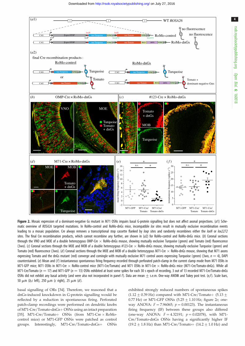
Figure 2. Mosaic expression of a dominant-negative Gs mutant in M71 OSNs impairs basal G-protein signalling but does not affect axonal projections. (a1) Sche-matic overview of ROSA26 targeted mutations. In RoMo-control and RoMo-dnGs mice, incompatible lox sites result in mutually exclusive recombination eventsleading to a mosaic population. Cre always removes a transcriptional stop cassette flanked by loxp sites and randomly recombines either the loxN or lox2272sites. The final Cre recombination products, which cannot recombine any further, are shown in (a2) for RoMo-control and RoMo-dnGs mice. (b) Coronal sectionsthrough the VNO and MOE of a double heterozygous OMP-Cre � RoMo-dnGs mouse, showing mutually exclusive Turquoise (green) and Tomato (red) fluorescence(3wo). (c) Coronal sections through the MOE and MOB of a double heterozygous #123-Cre � RoMo-dnGs mouse, showing mutually exclusive Turquoise (green) andTomato (red) fluorescence (3wo). (d ) Coronal sections through the MOE and MOB of a double heterozygous M71-Cre � RoMo-dnGs mouse, showing that M71 axonsexpressing Tomato and the dnGs mutant (red) converge and comingle with mutually exclusive M71 control axons expressing Turquoise (green) (3wo, n ¼ 4), DAPIcounterstained. (e) Mean and ( f ) instantaneous spontaneous firing frequency recorded through perforated patch-clamp in the current clamp mode from M71 OSNs inM71-GFP mice; M71 OSNs in M71-Cre � RoMo-control mice (M71-Cre/Tomato) and M71 OSNs in M71-Cre � RoMo-dnGs mice (M71-Cre/Tomato-dnGs). While allM71-Cre/Tomato (n ¼ 17) and M71-GFP (n ¼ 13) OSNs exhibited at least some spikes for each 30 s epoch of recording, 3 out of 13 recorded M71-Cre/Tomato-dnGsOSNs did not exhibit any basal activity (and were also not incorporated in panel f ). Data are mean+ s.e.m. One-way ANOVA and Tukey post test. (e,f ). Scale bars,50 mm (b,c left), 250 mm (c right), 25 mm (d ).
rsob.royalsocietypublishing.orgOpen
Biol.6:160018
4
on July 27, 2016http://rsob.royalsocietypublishing.org/Downloaded from
basal signalling of ORs [34]. Therefore, we reasoned that a
dnGs-induced knockdown in G-protein signalling would be
reflected by a reduction in spontaneous firing. Perforated
patch-clamp recordings were performed on dendritic knobs
of M71-Cre/Tomato-dnGsþOSNs using an intact preparation
[35]. M71-Cre/Tomatoþ OSNs (from M71-Cre � RoMo-
control mice) or M71-GFP OSNs were patched as control
groups. Interestingly, M71-Cre/Tomato-dnGsþ OSNs
exhibited strongly reduced numbers of spontaneous spikes
(1.12+0.39 Hz) compared with M71-Cre/Tomatoþ (5.13+0.77 Hz) or M71-GFP OSNs (5.25+ 1.10 Hz; figure 2e; one-
way ANOVA: F ¼ 7.96065; p ¼ 0.00123). The instantaneous
firing frequency (IF) between these groups also differed
(one-way ANOVA: F ¼ 4.32191; p ¼ 0.02078), with M71-
Cre/Tomato-dnGs OSNs having a significantly higher IF
(19.2+1.8 Hz) than M71-Cre/Tomatoþ (14.2+ 1.0 Hz) and

1 kb
M71 IRES caGs IRES tauGFP
M71
M71
M71
M71
M71
M71
M71
M71
M71M71M71M71
caGs
caGs caGs
IRES M71 IRES tauGFP
M71 M71 WT locus
M71-caGs-GFP
M71-caGs-GFP M71-M71-GFP M71-RFP/M71-caGs-GFP M71-RFP/M71-M71-GFP
M71-M71-GFP
Type 116/36
Type 220/36 14/14
(a)
(b) (c) (d) (e) ( f )
Figure 3. Coexpression of a constitutively active Gs mutant in M71 OSNs does not induce a posterior shift of glomeruli. (a) Schematic overview of the targeted M71mutations. (b,c) Wholemount intrinsic GFP fluorescence of the dorsal bulb (3wo), showing projection sites of lateral M71-caGs-GFP axons (green). (b) Glomerularformation is normal (16/36 bulbs). (c) Axons project to the correct A-P position, but do not converge into glomeruli (20/36 bulbs). (d ) Wholemount intrinsic GFPfluorescence in M71-M71-GFP mice (3wo). M71-M71-GFP axons always formed glomeruli (14/14 bulbs). (e) Wholemount intrinsic GFP and RFP fluorescence in M71-RFP � M71-caGs-GFP mice (3wo, n . 10). M71-RFP (red) and M71-caGs-GFP (green) axons converge, but segregate and form compartmentalized glomeruli.( f ) Wholemount GFP and RFP fluorescence in M71-RFP � M71-M71-GFP mice (3wo, n . 10). M71-RFP (red) and M71-M71-GFP (green) axons converge,but segregate and form compartmentalized glomeruli. Scale bars, 250 mm (b,c,d), 50 mm (e,f ).
rsob.royalsocietypublishing.orgOpen
Biol.6:160018
5
on July 27, 2016http://rsob.royalsocietypublishing.org/Downloaded from
M71-GFP neurons (13.7+ 1.3 Hz; figure 2f ), indicating that
M71-Cre/Tomato-dnGs OSNs have a higher tendency for
bursting. The basal and IF firing rates of M71-Cre/Tomatoþand M71-GFP OSNs were not different. The reduction in spon-
taneous firing of M71-Cre/Tomato-dnGs OSNs strongly
suggests that the dnGs inhibits basal, agonist-independent
G-protein signalling and cAMP production. Since axons of
Turquoiseþ control OSNs and Tomatoþ dnGs-affected
OSNs coalesced in the dorsal bulb, this suggests that crippling
basal G-protein signalling did not affect M71 axonal identity
or glomerular positioning.
2.3. Increasing G-protein signalling, via the expressionof a constitutively active Gs mutant, can modulatethe M71 axonal identity but not the resultingposition of its glomeruli
We have already addressed the consequences of downregu-
lating G-protein signalling. Now, we wished to assess how
axon targeting is affected by an increase in G-protein signal-
ling in M71 neurons. Therefore, we generated the M71-
caGs-GFP strain, in which the M71 locus was engineered to
coexpress M71 together with a constitutively active Gs
mutant (caGs) and a GFP reporter via a tricistronic mRNA
(figure 3a). In homozygous M71-caGs-GFP mice, 16 out 36
bulbs showed a lateral GFPþ glomerulus at an A-P position
comparable to that of WT M71 (figure 3b; Type 1 conver-
gence). Remarkably, in the remainder of bulbs (55%), axons
failed to converge into a glomerulus (figure 3c), indicating
that glomerular formation was disrupted. Importantly, by
crossing in the M71-RFP allele, we observed that M71-caGs-
GFP axons converged with M71-RFP axons (figure 3e).
This shows that when M71-caGs glomeruli were formed, they
were not posterior to WT M71 glomeruli. This is not because
of an intrinsic inability to shift M71 glomeruli posterior, since
we have previously reported that the M71::GFP fusion protein
induces a clear posterior shift [5]. However, we did observe
that M71-caGs-GFP and M71-RFP axons formed compartmen-
talized glomeruli (figure 3e), indicating a subtle change in
axonal identity. Together this shows that caGs co-expression
did not shift the position of the glomerulus, but did affect
axonal identity and glomerular formation.
The frequent failure to form M71-caGs-GFP glomeruli
suggests that OR-independent signalling via the caGs mutant
could disrupt glomerular formation. However, we cannot
exclude that the aberrant wiring resulted from the tricistronic
structure of the gene-targeted M71 locus. By generating the
M71-M71-GFP strain, which harbours another tricistronic
mutation in the M71 locus (figure 3a), we show that this is unli-
kely. In M71-M71-GFP mice, where instead of caGs we inserted
a second M71 coding region, the formation of GFPþ glomeruli
was always observed (figure 3d). Interestingly, M71-M71-GFP
and M71-RFP axons also often formed a compartmentalized
glomerulus (figure 3f ), suggesting that the change in M71
protein levels was sufficient to change the identity of the axons.
2.4. ORs can regulate A-P targeting via cAMP-independent mechanisms
Our results indicate that Gs/Golf signalling and cAMP pro-
duction are not the main drivers of A-P targeting in M71
OSNs. To further corroborate this, we wished to set up an
experimental system in which OSNs would express different
ORs but have the same level of G-protein signalling. If such
axons would not coalesce, this would be a clear indication

1 kb
M71(RDY)
M71(RDY)
MOR23(RDY)
IRES
IRES
IRES
IRES
IRES
tau-GFP
tau-LacZ
tau-Tomato
dorsal view
caGs
caGs
M71
M71-LacZ
M71(RDY)-caGs-GFP ×MOR23(RDY) Æ M71-caGs-Tomato
A
PLM
M71-RFP × M71(RDY) -caGs-GFPMOR23 Æ M71-LacZ ×
MOR23(RDY) Æ M71-caGs-Tomato
M71(RDY)-LacZ M71(RDY)-caGs-GFP Æ M71-caGs-Tomato
M71 WT locus
M71(RDY)-LacZ
M71(RDY)-caGs-GFP
MOR23(RDY) Æ M71-caGs-Tomato
MOR23(RDY)
(a)
(c)
(g1) (g2)
(d )
(h)
(e)
( f )
(b)
Figure 4. Signalling-deficient M71(RDY) and MOR23(RDY) ORs induce distinct neuronal identities and differentially regulate anterior-posterior targeting of axons.(a) Schematic overview of the targeted M71 mutations. (b,c) Dorsal view of X-gal-stained wholemounts of M71-LacZ (3wo) and M71(RDY)-LacZ (PD10) mice.(d ) Confocal image of a wholemount dorsal bulb from M71(RDY)-caGs-GFP and (e) MOR23(RDY)! M71-caGs-Tomato homozygous mice (PD10). Axons arevisualized by intrinsic GFP or Tomato fluorescence. Arrows indicate the main projection sites of lateral axons. ( f ) Wholemount fluorescence in M71(RDY)-caGs-GFP � MOR23(RDY)! M71-caGs-Tomato mice (PD10). M71(RDY)-caGs-GFP axons (green) project more posterior and do not fasciculate withMOR23(RDY)! M71-caGs-RFP axons (red). (g1, high-magnification view in g2) Wholemount fluorescence in M71-RFP � M71(RDY)-caGs-GFP mice (PD10).M71-RFP (red) and M71(RDY)-caGs-GFP (green) axons project to a similar A-P position. (h) Wholemounts of MOR23! M71-LacZ � MOR23(RDY)! M71-caGs-Tomato mice (PD10) were X-gal-stained (left) after confocal imaging (right). MOR23! M71-LacZ (blue, X-gal) and MOR23(RDY)! M71-caGs-Tomato(red, intrinsic fluorescence) axons project to a similar A-P position ( pigmentation is used to create anchor points for reference). Scale bars, 500 mm (b,c,h),250 mm (d,e,f,g1,g2).
rsob.royalsocietypublishing.orgOpen
Biol.6:160018
6
on July 27, 2016http://rsob.royalsocietypublishing.org/Downloaded from
of an OR-instructed identity, independent of the levels of
G-protein signalling. To achieve this, we thought to compare
M71 OSNs in which the coding sequence (CDS) was
exchanged with distinct signalling-deficient ORs and where
G-protein signalling was rescued via expression of the caGs
mutant. To induce signalling deficiency in ORs, we chose to
mutate the highly conserved DRY motif, which is critical
for regulating GPCR conformational states [36]. Mutating
the E/DRY residues into REY or RDY has been previously
shown to abolish G-protein signalling for Rhodopsin [37],
CXCR4 [38] and the rat I7 OR [20]. To confirm that these
mutations would indeed cause a loss of function phenotype,
we created a gene-targeted strain in which the M71 DRY
motif was replaced by RDY (D121R;R122D) along with
an IRES-taulacZ reporter, M71(RDY)-LacZ (figure 4a). In
contrast to M71-LacZ axons (figure 4b; electronic supplemen-
tary material, figure S2a), M71(RDY)-LacZ axons showed
poor outgrowth, typically did not reach the cribiform plate
and were not observed on the dorsal bulb (figure 4c; elec-
tronic supplementary material, figure S2b,c). Furthermore,
the number of M71(RDY)-LacZ neurons rapidly decreased
over time (electronic supplementary material, figure S2d )
and by PD21 their numbers were 10-fold lower when com-
pared with M71-LacZ neurons in age-matched animals
(electronic supplementary material, figure S2e). This dramatic
phenotype indicates that G-protein signalling in M71 OSNs is
necessary when competing with normal OSNs.
Now we were in a position to test two additional mouse
strains: M71(RDY)-caGs-GFP and MOR23(RDY)!M71-
caGs-Tomato, in which the RDY mutant OR is coexpressed
with a caGs to rescue G-protein signalling (figure 4a).
Furthermore, in the MOR23(RDY)!M71-caGs-Tomato
strain, the M71 CDS is replaced by a DRY! RDY mutant
of the MOR23 CDS. The M71(RDY)-caGs-GFP
and MOR23(RDY)!M71-caGs-Tomato OSNs thus express
different ORs—M71(RDY) versus MOR23(RDY)—but

rsob.royalsocietypublishing.orgOpen
Biol.6:160018
7
on July 27, 2016http://rsob.royalsocietypublishing.org/Downloaded from
should have the same level of G-protein signalling (derived
from the OR-independent caGs mutant). Coexpression of
caGs rescued axonal projections to the bulb for both
M71(RDY)-caGs-GFP and MOR23(RDY)!M71-caGs-
Tomato OSNs (figure 4d,e). The number of caGs-rescued
OSNs remained very low (electronic supplementary material,
figure S2e) and as a consequence glomerular formation was
often inefficient [39]. In mice that were only heterozygous
for one of the mutant alleles, axons projected to the expected
A-P region, but glomerular formation was often not observed
(e.g. Tomatoþ axons figure 4f,h). These findings are also in
line with what was seen for M71-caGs mice, where
expression of caGs often disrupted glomerular formation
(figure 3c). Importantly, however, M71(RDY)-caGs and
MOR23(RDY)!M71-caGs axons projected to fundamentally
different A-P regions (figure 4d,e). The differential identities
of M71(RDY)-caGs-GFP and MOR23(RDY)!M71-caGs-
Tomato axons were confirmed in a mixed cross, where coalesc-
ence was never observed between GFPþ and Tomatoþ axons,
and where GFPþ axons extended more posterior (figure 4f ).
Remarkably, M71(RDY) axons projected to the same A-P
region as WT M71 axons, as was seen in M71-RFP �M71(RDY)-caGs-GFP mice (figure 4g). Similarly,
MOR23(RDY)!M71-caGs-Tomato axons projected very
close to WT MOR23!M71-LacZ axons, which was anterior
to M71 (figure 4h). Together this shows that M71(RDY)-
caGs and MOR23(RDY)-caGs axons project to distinct A-P
regions on the bulb, very close to the targeting site of their
respective WT receptors. It is important to note that for
both populations the caGs is expressed from the same locus
with a similar tricistronic strategy. This suggests that ORs
can regulate axonal targeting via cAMP-independent
mechanisms.
2.5. G-protein signalling is critical for neuronalmaturation and competition
Are DRY! RDY mutations in ORs blocking G-protein
signalling? We observed that M71(RDY)-LacZ cells resided
very basal in the epithelium suggesting that these cells
were not mature and might not contain necessary signal
transduction components to assess G-protein signalling.
Thus, we used ISH to determine the percentage of Ompþmature, OmpþGap43þ intermediate and Gap43þ immature
cells in normal and RDY strains of mice: M71-LacZ,
M71(RDY)-LacZ, M71(RDY)-caGs-GFP and MOR23(RDY)!M71-caGs-Tomato OSNs (figure 5a). An almost complete
absence of Ompþ mature OSNs was observed in the
M71(RDY)-LacZ population (figure 5a). M71(RDY)-LacZ
OSNs also failed to upregulate Adcy3 expression, a key
component of the signalling machinery in mature OSNs
(figure 5b). By contrast, Ompþ and Adcy3þ mature
M71(RDY)-caGs and MOR23(RDY)!M71-caGs OSNs were
observed, albeit at a lower percentage as compared with
WT M71 (figure 5a,b). These results suggest that G-protein
signalling may be a checkpoint in OSN maturation. Alterna-
tively, the loss in G-protein signalling may render OSNs
uncompetitive and they may be quickly eliminated as they
mature, which would explain the rarely observed only
Ompþ expressing M71(RDY)-LacZ OSNs.
We next wished to perform physiology to assess OR func-
tionality. Since M71(RDY)-LacZ OSNs remained immature,
they would not be suitable for recordings as their signalling
machinery may not be functional. We therefore chose
MOR23(RDY)!M71-caGs-Tomato OSNs, on which we per-
formed perforated patch-clamp recordings. MOR23(RDY)!M71-caGs-Tomato neurons were stimulated with saturating
concentrations of lyral (an MOR23 ligand), acetophenone
(an M71 ligand), or a mixture of odorants that activates
66% of randomly patched OSNs in the MOE [40]. In our
analysis, we only considered OSNs that responded to a mix-
ture of IBMX and forskolin, which directly activates
adenylate cyclases and indicates whether the neurons are
mature. Of the 13 recorded OSNs that responded to
IBMX þ forskolin (out of 17 total), none responded to any
of the other stimulants, showing that there is no chemical
or mechanical activity in these neurons (figure 5c). In
addition, MOR23(RDY)!M71-caGs OSNs exhibited almost
no spontaneous activity, which is clearly present in WT
MOR23 or M71 OSNs (figure 5d,e). Four conclusions are
delineated: (i) DRY! RDY mutations uncouple ORs from
G-protein signalling; (ii) no other ORs were coexpressed in
these cells as MOR23(RDY)!M71-caGs OSNs never
responded to the mixture of odorants (gene choice remained
intact); (iii) spontaneous OSN spiking is indeed fully depen-
dent on the basal signalling of ORs; and (iv) caGs does not
elicit spontaneous spiking, indicating that the rescue of
MOR23(RDY) OSNs does not require electrical activity.
2.6. One neuron – two receptors: OSNs in O/E2-M71-GFP mice coexpress the M71 OR together withan endogenous receptor
Our results show that signalling-deficient ORs can regulate
the identity and A-P targeting of M71 axons, suggesting
that this did not rely on differences in OR-induced cAMP
levels. However, it may be argued that the M71(RDY) and
MOR23(RDY) coding sequences differentially affect mRNA
stability, thereby resulting in different caGs protein levels in
M71(RDY)-caGs compared to MOR23(RDY)-caGs OSNs. To
resolve these issues, we reasoned that forcing the coexpres-
sion of a WT OR in all OSNs may also rescue the
maturation and axonal projections of M71(RDY) neurons.
Furthermore, this might uniformly increase G-protein signal-
ling in all OSNs, allowing a direct comparison of the A-P
targeting of M71(RDY) with other ORs. Based on previous
experiments using ubiquitously activated ORs via the ttA/
tetO system [41–43], it was unclear whether it was a prioripossible to coexpress a second OR in all OSNs. In theory,
OR-mediated negative feedback mechanisms would prevent
endogenous gene expression or endogenous genes would
silence the expression of the ectopic OR [41,42].
In an attempt to express an OR in all OSNs, we started
from the MOR23 cDNA (containing exons 1–3, Tg3’D, see
[44]), where we removed the MOR23 promoter and MOR23coding region and replaced it with an M71-IRES-tauGFP-
ACNF cassette (figure 6a). This was subsequently cloned
into the O/E2 targeting vector (TV) [45] and used to replace
the first 6 exons of the O/E2 gene via gene targeting. In the
resulting O/E2-M71-GFP strain, expression of an M71-IRES-
tauGFP cDNA is placed under the control of the native
O/E2 promoter, which is prominently expressed in all OSNs
[45]. A widespread and bright intrinsic GFP fluorescence
was seen in the MOE and VNO of O/E2-M71-GFP mice

MOR23
Omp+ cell
taulacZ Gap43 Omp taulacZ Adcy3
Omp+Gap43+ cell
M71-LacZ
M71(RDY)-LacZ
M71(RDY)-caGs-GFP
MOR23(RDY) Æ M71-caGs-Tomato
MOR23(RDY) Æ M71-caGs
M71
MOR23(RDY) Æ M71-caGs MOR23(RDY) Æ M71-caGs MOR23 M71
20 mV5 s
odorant stimulas
Ringer
lyral 10–4 M
aceto 10–4 M
odorant mix10–5 M
IBMX + forskolin
20 pA1 s
80
*** *** ***
*** ***
*** *** ***
**
***
***
*
******
**
** **
60
40
765432
mea
n fi
ring
fre
quen
cy (
spik
ess–1
)
10
% o
f to
tal
20
0
80
60
40
% o
f to
tal
20
0Omp+ Omp+Gap43+ Gap43+
M71-LacZ
M71(RDY)-LacZ
M71(RDY)-caGs-GFP
MOR23(RDY) Æ M71-caGs-Tomato
Adcy3+
Gap43+ cell Adcy3+ cell Adcy3– cell
(b)(a)
(c) (d )
(e)
–67mV
Figure 5. Neuronal maturation and activity are lost in OSNs expressing G-protein coupling mutant ORs. (a) Three-colour ISH on coronal sections of the MOE (PD21).Probes were used against Omp (blue), Gap43 (green) and: taulacZ (red, tau is of bovine origin) for labelling M71-LacZ and M71(RDY)-LacZ OSNs; tauGFP for labellingM71(RDY)-caGs-GFP OSNs; tauTomato for labelling MOR23(RDY)! M71-caGs-Tomato OSNs (red). Examples are shown of a OMPþ, OMPþGap43þ and Gap43þM71 OSN. The percentage of these populations within the total counted cells (% of total) was quantified at PD21 (mean+ s.e.m., n ¼ 3). (b) Two-colour ISH oncoronal sections of the MOE. Riboprobes were used against Adcy3 (green) and OSN markers (red) as explained in (a). Examples are shown for Adcy3þ and Adcy32
M71 OSNs. The percentage of Adcy3þ and Adcy32 OSNs within the total counted cells were quantified at PD21 (mean+ s.e.m., n ¼ 3). (c) Representative tracesof patch-clamp recordings in a MOR23(RDY)! M71-caGs OSN under the voltage-clamp mode. Out of 17 recorded OSNs, 13 responded to IBMXþforskolin. How-ever, these cells did not respond to Ringer solution, lyral, acetophenone (aceto) or a mix of odorants. (d ) Representative traces of spontaneous activities recordedthrough perforated patch-clamp in the current clamp mode in MOR23(RDY)! M71-caGs-Tomato or MOR23-GFP or M71-GFP OSNs. Out of 21 recorded MOR23(RDY)! M71-caGs-Tomato OSNs, 14 did not exhibit any spontaneous action potentials. (e) Mean spontaneous firing frequency for MOR23(RDY)! M71-caGs (n ¼ 21),MOR23 (n ¼ 16) and M71 (n ¼ 13) OSNs. Data are mean+ s.e.m. One-way ANOVA and Tukey post test. Scale bars, 10 mm (a,b). One-way ANOVA and Newman –Keuls post test (a,b), *p , 0.05, **p , 0.01, ***p , 0.001.
rsob.royalsocietypublishing.orgOpen
Biol.6:160018
8
on July 27, 2016http://rsob.royalsocietypublishing.org/Downloaded from
(figure 6b–e). All OSNs appeared GFPþ, as did the glomer-
uli, which were homogeneously labelled and showed a
normal morphology (figure 6f ). Importantly, M71 mRNA
was robustly observed in the whole MOE and VNO of
O/E2-M71-GFP mice (figure 6g,h; electronic supplementary
material, figure S3a,c). O/E2 expression is typically observed
prior to Gap43, at a very immature neuronal stage of OSNs.
M71 expression from O/E2-M71-GFP mice was also seen in
this early OSN stage (figure 6g). The MOE of O/E2-M71-
GFP mice had a normal multi-layered structure and
thickness, comparable to that of O/E2 WT control animals
(electronic supplementary material, figure S3b,d). Interest-
ingly, it was evident that expression of M71 via the O/E2promoter did not suppress endogenous OR expression. ISH
showed coexpression of M71 with other unrelated class II
OR genes (figure 6i). Next, NanoString was used to assess
the expression of a random selection of OR genes in the
MOE of O/E2-M71-GFP(þ/2) and O/E2-WT littermates
(figure 6j ). Of the 19 OR genes tested, only one transcript
showed a significant but small downregulation, showing
that the majority of ORs had comparable expression levels
in the two groups. In addition, the zonal expression of

exon
M71 IRES
M71
O/E2 5¢ O/E2 3¢IRES
tau-GFP
tau-GFP
M71 IRES tau-GFP
WM medial view
MOE
MOB
VNO
MOE
VNO
MOE
NanoString O/E2-M71-GFP versus WT
0.5
0
log2
fol
d ch
ange
–0.5
2 4 6log2 expression
log 2
fold
cha
nge
8 10 12 14
DE genes
Olfr1507Olfr15
Olfr1511Olfr1508
Olfr1156Olfr78 Olfr73Olfr1509 Olfr124
Olfr309Olfr17Olfr160
Olfr2
Olfr1015Olfr6
Olfr1301
Olfr166
Olfr16
Olfr713
non-DE genespositive controlsnegative controls
M71 Gap43 Omp
M71 class II ORs
class II ORs
M71 Gap43 Omp
WM dorsal view A
A PD
V
P
M L
ACNF
ACNF targeting vector
WT O/E2 locus
O/E2-M71-GFP
O/E2promoter
1 2 3MOR23 MOR23 cDNA
CDS
(a)
(b) (c)
(d )
(g)
(h)
(i) ( j)
(e)
( f1) ( f2)
Figure 6. OSNs in O/E2-M71-GFP mice ubiquitously express M71 together with an endogenous OR. (a) Schematic of the O/E2-M71-GFP mutation. (b,c) Wholemount(WM) intrinsic GFP fluorescence of the medial and dorsal olfactory bulb in O/E2-M71-GFPþ/2 mice (3wo). (d – f ) Intrinsic GFP fluorescence in coronal sections ofthe (d ) MOE, (e) VNO and ( f1, high magnification in f2) MOB of O/E2-M71-GFPþ/2 mice (3wo). (g,h) Three-colour ISH against M71 (red), Gap43 (green), Omp(blue) in the (g) MOE and (h) VNO of O/E2-M71-GFPþ/2 mice (3wo). Arrow shows an example of an OSN expressing M71 before the onset of Gap43. (i) Two-colourISH in the MOE of O/E2-M71-GFPþ/2 mice, using probes against M71 (red) and a mix of four different class II OR (see the electronic supplementary material) genes(green and inset). Arrow shows an example of a double positive cell. ( j ) NanoString RNA analysis of whole olfactory mucosa samples collected from six WT and sixO/E2-M71-GFP heterozygous littermates (PD25), using a code set against 19 ORs. The values in the y-axis represent the log2 of the fold change of mutant versus WT(i.e. M value). Positive values represent an increase in expression, while negative values are a reduction. The values in the x-axis represent the log2 of the normalizedNanoString counts, which is a measure for both the expression level of the OR and the binding efficiency of the probe. To test for significance, tTreat analysis wasused, with a fold change threshold set at 1.3. Differentially expressed genes are indicated as green squares. Red stippled line: the average M value of all tested ORgenes; Yellow stippled lines: the chosen fold change threshold (set at 1.3, in log2 scale ¼ 0.38). Positive controls (cyan triangles) and negative controls (bluetriangles) for NanoString operation are distributed correctly as per manufacturer. Scale bars, 250 mm (b,c,f1), 50 mm (d,e,f2,g,i), 100 mm (h).
rsob.royalsocietypublishing.orgOpen
Biol.6:160018
9
on July 27, 2016http://rsob.royalsocietypublishing.org/Downloaded from

medial view
M71-LacZ × O/E2-M71-GFP P2-LacZ × O/E2-M71-GFPO/E2 WT O/E2 (+/–)
MOR23-LacZ × O/E2-M71-GFP
D
VA P
O/E2 WT O/E2 (+/–)
DM71-LacZ × O/E2-M71-GFP
O/E2 WT O/E2 (+/–)
O/E2 WT O/E2 (+/–)
(b)
(a) (c)
(d )
Figure 7. O/E2-M71 is co-expressed with endogenous OR genes. (a – d) Wholemount X-gal staining of medial or dorsal bulbs of the indicated crosses. Images onthe left are control O/E2 WT littermates, on the right are O/E2-M71-GFP heterozygous animals. M71-LacZ (3wo), MOR23-LacZ (3wo), P2-LacZ (3wo) and DM71-LacZ(5wo) mutations are heterozygous; a similar DM71-LacZ innervation was also observed in 8wo mice (data not shown). The number of half-bulbs (medial) with twoor more glomeruli were quantified for M71: 0/8 in O/E2 WT and 7/14 in O/E2 (þ/2); and MOR23: 0/8 in O/E2 WT and 3/6 in O/E2 (þ/2). Scale bars, 500 mm.
rsob.royalsocietypublishing.orgOpen
Biol.6:160018
10
on July 27, 2016http://rsob.royalsocietypublishing.org/Downloaded from
endogenous ORs in the MOE was not altered in O/E2-M71-
GFP mice, as illustrated by crossing the mice to the
M71-LacZ, MOR23-LacZ and P2-LacZ strains (figure 7a–c).
As reported previously, OSNs that choose an OR locus that
does not contain an OR gene—such as the DM71-LacZ
allele where the M71 CDS is deleted—coexpress additional
OR genes [5]. Therefore, the projection of lacZþ axons in
DM71-LacZ mice depends on the guidance properties of the
other ORs, resulting in a divergent pattern of projections
(figure 7d ). Interestingly, distributed axons entering multiple
glomeruli were also observed in DM71-LacZ mice crossed to
O/E2-M71-GFP (figure 7d ).
To investigate whether the ectopic M71 in O/E2-M71-
GFP mice is functional and contributes to signalling and
neuronal activity, we performed patch-clamp recordings on
the MOE of O/E2-M71-GFP mice. In addition, we also
crossed in the MOR23(RDY)!M71-caGs-Tomato mutation.
In the resulting cross, we patched the dendritic knobs of
random OSNs that express an unknown endogenous OR
(GFPþ Tomato2) or MOR23(RDY)!M71-caGs OSNs
(GFPþ Tomatoþ) and measured their responses to varying
concentrations of the M71 ligand acetophenone. All ran-
domly patched OSNs (n ¼ 10) and MOR23(RDY)!M71-caGs OSNs (n ¼ 10) responded to acetophenone
(figure 8a,b). While responses were heterogeneous, most
O/E2-M71-GFP neurons responded to concentrations above
1025 M acetophenone (figure 8d,e). Responses to acetophe-
none were not widespread in the normal WT MOE, as in
O/E2 WT littermates only 1 in 10 patched OSNs responded
(figure 8c). As a reference we also patched WT M71 OSNs
in M71-GFP mice and plotted a threshold frequency histo-
gram, which represents the percentage of cells that
responded to a specific threshold concentration (figure 8f ).
This showed that O/E2-M71-GFP neurons were less sensitive
to acetophenone than M71-GFP OSNs. Importantly, O/E2-
M71-GFP expression also rescued the spontaneous firing of
MOR23(RDY)!M71-caGs OSNs, which now exhibited
mean and instantaneous firing frequencies in the same
range as that of randomly patched OSNs (figure 8g,h).
Since without O/E2-M71-GFP expression MOR23(RDY)!M71-caGs OSNs exhibit no spontaneous firing (figure 5e),
this shows that the ectopic M71 contributes to basal, agonist-
independent, spiking in OSNs, while caGs does not.
2.7. O/E2-M71-GFP rescue experiments confirm that theA-P targeting of ORs and surrogate receptors canbe driven by cAMP-independent mechanisms
We wondered how the ectopic M71 expression would affect
M71-LacZ or MOR23!M71-LacZ axonal projections. In
O/E2-M71-GFP mutants, M71-LacZþ and MOR23!M71-
LacZ OSNs formed glomeruli in the correct A-P position
(figure 9a,b). However, M71 axons took altered routes on
the bulb and traversed through regions were they are nor-
mally excluded and often formed an additional glomerulus
at the same A-P position (figure 9a,b; electronic supplementary
material, figure S4).
We next tested the consequence of adding M71 expression
to signalling-deficient M71(RDY) OSNs. O/E2-M71-GFP
expression rescued axon outgrowth of M71(RDY)-LacZ
OSNs, which formed two points of coalescence on the
dorsal bulb (figure 9d,f ). An ectopic convergence point was
observed in an anteromedial region of the dorsal bulb,
where both medial and lateral projecting axons co-converged
(figure 9d, white arrow). The lateral M71(RDY) glomerulus
was observed at the same A-P region as WT M71 glomeruli
(figure 9d, black arrow), which was clearly more posterior
than MOR23!M71-LacZ or MOR23(RDY)!M71-caGs-
Tomato axons (figure 9b,c,e). Importantly, in O/E2-M71-
GFP mice, M71(RDY)-LacZ OSNs have lower levels of basal
G-protein signalling than MOR23!M71-LacZ OSNs or
MOR23(RDY)!M71-caGs-Tomato OSNs. Indeed, while in
M71(RDY)-LacZ OSNs only the O/E2-derived ectopic M71
lyral 10–4
odorant stimulus odorant stimulus odorant stimulus
O/E2-M71 MOR23(RDY)-caGsO/E2-M71
WT
O/E2-M71
O/E2-M71
–67 mV –67 mV
MOR23(RDY)-caGsO/E2-M71
MOR23(RDY)-caGsO/E2-M71
O/E2-M71
O/E2-M71
WT
MOR23(RDY)-caGsO/E2-M71
7 20
15
10
5
0
n.s.
n.s. n.s. n.s. n.s.
n.s.
6
5
4
3
2
mea
n fi
ring
fre
quen
cy(s
pike
ss–1
)
inst
anta
neou
s fi
ring
freq
uenc
y (s
pike
ss–1
)
1
0
aceto 10–7
150
100
50ampl
itude
(pA
)
0
10–7 10–6 10–5 10–4 10–7 10–6 10–5 10–4
10–7
80150
100
50ampl
itude
(pA
)
0
70
60
50
40
perc
enta
ge o
f ce
lls
30
20
10
0
concentration at threshold (M)concentration (M) concentration (M)
10–6 10–5 10–4
aceto 10–6
aceto 10–5
aceto 10–4
IBMX+
forskolin
lyral 10–4
aceto 10–7
aceto 10–6
aceto 10–5
aceto 10–4
IBMX+
forskolin
lyral 10–4
aceto 10–7
aceto 10–6
aceto 10–5
aceto 10–4
IBMX+
forskolin20 pA
1 s20 pA
1 s50 pA
1 s
(e) ( f )
(b)(a) (c)
(d )
(g) (h1) (h2)
M71-GFPMOR23(RDY)-caGs/O/E2-M71
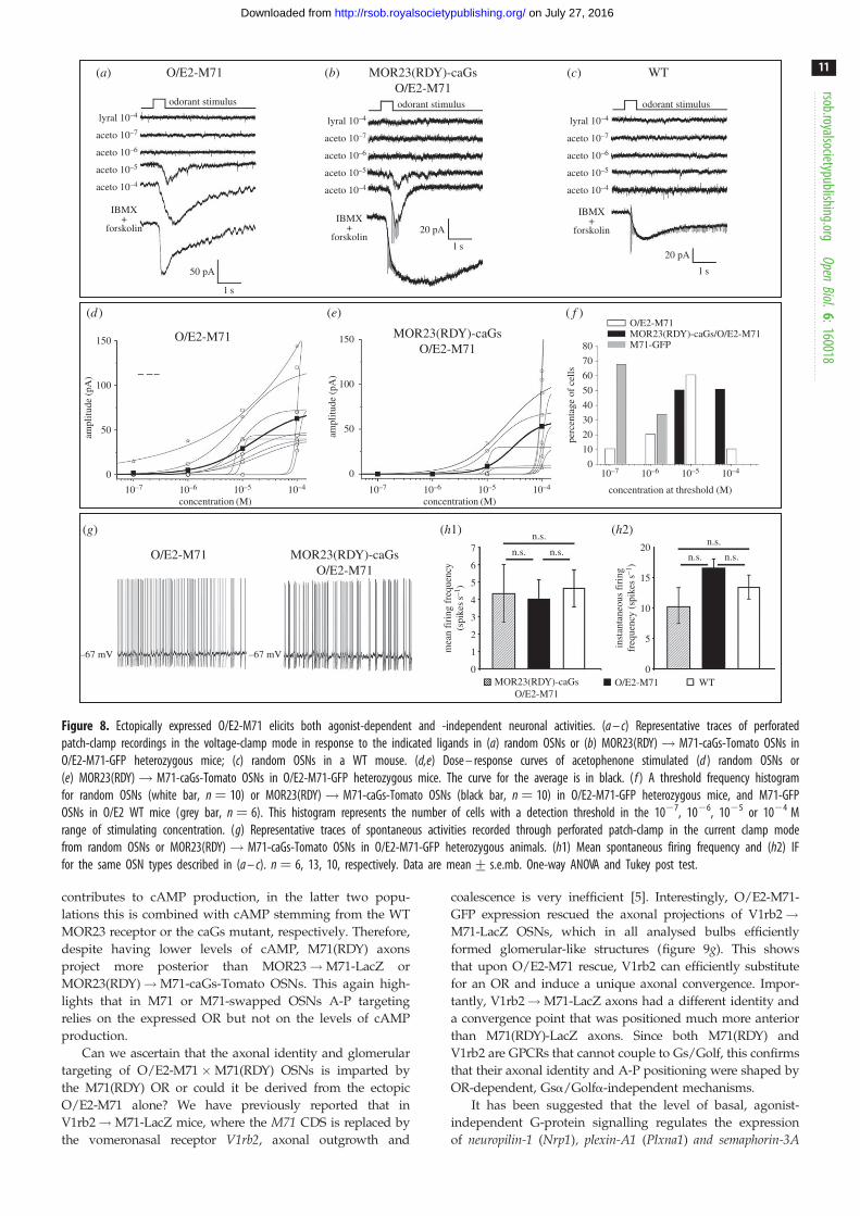
Figure 8. Ectopically expressed O/E2-M71 elicits both agonist-dependent and -independent neuronal activities. (a – c) Representative traces of perforatedpatch-clamp recordings in the voltage-clamp mode in response to the indicated ligands in (a) random OSNs or (b) MOR23(RDY)! M71-caGs-Tomato OSNs inO/E2-M71-GFP heterozygous mice; (c) random OSNs in a WT mouse. (d,e) Dose – response curves of acetophenone stimulated (d ) random OSNs or(e) MOR23(RDY)! M71-caGs-Tomato OSNs in O/E2-M71-GFP heterozygous mice. The curve for the average is in black. ( f ) A threshold frequency histogramfor random OSNs (white bar, n ¼ 10) or MOR23(RDY)! M71-caGs-Tomato OSNs (black bar, n ¼ 10) in O/E2-M71-GFP heterozygous mice, and M71-GFPOSNs in O/E2 WT mice (grey bar, n ¼ 6). This histogram represents the number of cells with a detection threshold in the 1027, 1026, 1025 or 1024 Mrange of stimulating concentration. (g) Representative traces of spontaneous activities recorded through perforated patch-clamp in the current clamp modefrom random OSNs or MOR23(RDY)! M71-caGs-Tomato OSNs in O/E2-M71-GFP heterozygous animals. (h1) Mean spontaneous firing frequency and (h2) IFfor the same OSN types described in (a – c). n ¼ 6, 13, 10, respectively. Data are mean+ s.e.mb. One-way ANOVA and Tukey post test.
rsob.royalsocietypublishing.orgOpen
Biol.6:160018
11
on July 27, 2016http://rsob.royalsocietypublishing.org/Downloaded from
contributes to cAMP production, in the latter two popu-
lations this is combined with cAMP stemming from the WT
MOR23 receptor or the caGs mutant, respectively. Therefore,
despite having lower levels of cAMP, M71(RDY) axons
project more posterior than MOR23!M71-LacZ or
MOR23(RDY)!M71-caGs-Tomato OSNs. This again high-
lights that in M71 or M71-swapped OSNs A-P targeting
relies on the expressed OR but not on the levels of cAMP
production.
Can we ascertain that the axonal identity and glomerular
targeting of O/E2-M71 �M71(RDY) OSNs is imparted by
the M71(RDY) OR or could it be derived from the ectopic
O/E2-M71 alone? We have previously reported that in
V1rb2!M71-LacZ mice, where the M71 CDS is replaced by
the vomeronasal receptor V1rb2, axonal outgrowth and
coalescence is very inefficient [5]. Interestingly, O/E2-M71-
GFP expression rescued the axonal projections of V1rb2!M71-LacZ OSNs, which in all analysed bulbs efficiently
formed glomerular-like structures (figure 9g). This shows
that upon O/E2-M71 rescue, V1rb2 can efficiently substitute
for an OR and induce a unique axonal convergence. Impor-
tantly, V1rb2!M71-LacZ axons had a different identity and
a convergence point that was positioned much more anterior
than M71(RDY)-LacZ axons. Since both M71(RDY) and
V1rb2 are GPCRs that cannot couple to Gs/Golf, this confirms
that their axonal identity and A-P positioning were shaped by
OR-dependent, Gsa/Golfa-independent mechanisms.
It has been suggested that the level of basal, agonist-
independent G-protein signalling regulates the expression
of neuropilin-1 (Nrp1), plexin-A1 (Plxna1) and semaphorin-3A

M71-LacZ × O/E2-M71-GFP
MOR23ÆM71-LacZ × O/E2-M71-GFP
MOR23(RDY) Æ M71-caGs-Tomato × O/E2-M71-GFP O/E2-M71-GFP versus WT NanoString profiling
2 4 6
OE2-M71 neurons behave as activity hypomorphs
Kirrel2
naris occlusion
CNGA2 KO
OE2-M71
Epha5 Efna5
DE genes
Efna5
Sema3f Sema3aNrp2
Kirrel3Plxna1
Plxna3
Epha5
Kirrel2
Nrp1
non-DE genespositive controlsnegative controls
0.5
0
–0.5
–1.0
8log2 expression
log 2
fold
cha
nge
10 12 14
M71(RDY)-LacZ × O/E2-M71-GFP
V1rb2 Æ M71-LacZ × O/E2-M71-GFPO/E2 WT O/E2 (+/–)
O/E2 WT O/E2 (+/–)
O/E2 WT
summary
dorsal anterior
dorsal posterior
MOR23 Æ M71-LacZMOR23ÆM71-LacZ × O/E2-M71MOR23 (RDY) Æ M71-caGs-TomatoMOR23 (RDY) Æ M71-caGs-Tomato
M71-LacZM71-LacZ × O/E2-M71M71(RDY)-LacZ × O/E2-M71M71(RDY)-caGs-GFPM71-caGs-GFP
× O/E2-M71
O/E2 (+/–)
O/E2 WT O/E2 (+/–)
8/8
O/E2 WT
A
P
M
MLA
P
PAD
VL
O/E2 (+/–) O/E2 WT
M71(RDY)-LacZ × O/E2-M71-GFP
O/E2 (+/–)dorsal view medial view
(e)
( f )
(b) (g)
(h)
(a)
(c)
(d )
Figure 9. O/E2-M71-GFP rescue experiments show that the anterior – posterior targeting of M71 OSNs is not correlated with the levels of G-protein signalling.(a,b,d,f,g) Wholemount X-gal staining of dorsal or medial bulbs of the indicated crosses (PD10 – PD21). Images on the left are control O/E2 WT littermates,on the right are O/E2-M71-GFPþ/2 heterozygous animals. Arrows highlight glomeruli and axonal projections, respectively. White arrows in (d) indicate ectopicanteromedial glomeruli. M71 mutations: (a,b,g) heterozygous; (c,d) homozygous. (c) Wholemount intrinsic GFP and Tomato fluorescence of the dorsal bulb. Arrowindicates the projection site of lateral MOR23(RDY)! M71-caGs axons (red). O/E2-M71-GFP intrinsic fluorescence is shown in green. (e) Schematic diagram of thedorsal bulb. Coloured dots represent approximate coordinates of individual lateral glomeruli of the indicated strains. Triangles represent the A-P domain to which thecaGs expressing OSNs of the indicated strains project. (h) NanoString RNA analysis of whole olfactory mucosa samples collected from six WT and six O/E2-M71-GFPheterozygous littermates (PD25). The y-axis represent the log2 of the fold change of mutant versus WT (i.e. M value), the x-axis represents the log2 of the normal-ized NanoString counts. To test for significance tTreat analysis was used, with a fold change threshold set at 1.1. Differentially expressed genes are indicated as greensquares. Red stippled line: the average M value of all tested OR genes. For reference, the yellow stippled lines are set at a fold change of 1.3 (or 0.38 in log2 scale).Positive controls (cyan triangles) and negative controls (blue triangles) for NanoString operation are distributed correctly as per manufacturer. As indicated in thetable, the changes in Kirrel2, Epha5 and Efna5 gene expression in O/E2-M71-GFP mice resemble the published gene expression changes in CNGA2 KO and narisoccluded animals. Scale bars, 500 mm.
rsob.royalsocietypublishing.orgOpen
Biol.6:160018
12
on July 27, 2016http://rsob.royalsocietypublishing.org/Downloaded from

rsob.royalsocietypublishing.orgOpen
Bio
13
on July 27, 2016http://rsob.royalsocietypublishing.org/Downloaded from
(Sema3a) and that these molecules are the main drivers of A-P
targeting [18]. To examine whether in O/E2-M71-GFP mice
the expression levels of these and other cell adhesion mol-
ecules were affected, we performed NanoString analysis on
the MOE of O/E2-M71-GFP mutant versus WT littermates
(figure 9h). There were no changes in the expression levels
of any of the neuropilin, semaphorin or plexin genes tested.
Surprisingly, we observed a significant downregulation of
Kirrel2 and Epha5 together with an increase of ephrin-A5 in
O/E2-M71-GFP mice (figure 9h). Remarkably, both signal-
ling impaired CNGA2 KO mice and activity reduced, naris
occluded animals are known to have this same expression
pattern [21]. This suggests that in O/E2-M71-GFP mice
there is a general reduction in the electrical activity of OSNs.
l.6:160018 3. DiscussionOur results provide a set of profound insights into how theOR regulates the developmental pathway of OSNs, starting
from regulation of gene choice, to OSN maturation and
finally to axonal identity and glomerular formation.
3.1. Negative feedback by OR genesForcing the widespread expression of an OR using trans-
genic approaches has not been straightforward, which has
contributed to shaping the current models of OR-induced
feedback. To achieve ectopic OR expression, several
groups have used the tetO-TTA system using immature/
mature neuronal TTA drivers [41–43]. These TTA-induced
transgenic ORs are able to suppress the expression of
endogenous ORs. Remarkably, it has been suggested that
the tetO-driven ORs themselves can be silenced by the
endogenous ORs, implying that OR coding regions are tar-
gets for feedback mediated suppression [41]. Remarkably,
all OSNs in O/E2-M71 mice robustly express M71 together
with an endogenous OR, while maintaining their specific
glomerular formation. This coexpression shows that OR
coding regions, including the 50 and 30 UTRs, are not targets
for feedback suppression, otherwise the endogenous OR
would have blocked O/E2-M71 expression. But why did
the O/E2-M71 OR, which was already expressed very
early in OSN development, not suppress the endogenous
OR? One explanation is that negative feedback does not sup-
press first choice, but only subsequent choices. The
projection pattern of DM71 OSNs—where first choice does
not lead to the expression of an OR—was similar in an O/
E2-M71-GFP background. This may indicate that OSNs
that have chosen to express the DM71 locus still choose a
second OR locus for expression, despite the ubiquitous
expression of M71 from the O/E2 promoter. An alternative
explanation is that second OR choice is blocked by O/E2-
M71, but its expression alone is not able to efficiently
coalesce DM71-LacZ axons into distinct glomeruli. Further
characterization of the expressed ORs in DM71 OSNs is
needed to resolve this.
An explanation for the lack of suppression is that the O/
E2-M71 expression levels do not reach a required threshold
level to induce feedback. This would imply that robust
expression levels that are sufficient to rescue axon out-
growth and neuronal maturation of OSNs are not
sufficient to induce negative feedback. It has recently been
proposed that stable OR gene expression requires the
recruitment of many cis- and trans-acting enhancers [15]. It
is therefore possible that gene choice remains open until
one OR locus is able to recruit a sufficient number of enhan-
cers and achieve a high threshold level of expression.
Interestingly, recent reports by Hanchate and colleagues
[46] have shown that developing OSNs transition from
expressing low levels of multiple ORs to expressing high
levels of a single OR. These results fit with the interpretation
that sub-threshold expression of an OR early in developing
OSNs does not block gene choice. Our results also suggest
that the expression of an OR is coupled to OSN maturation,
as described below.
3.2. G-protein signalling and the expression of afunctional OR promote OSN maturation
Our results indicate that OSNs expressing ORs with
G-protein coupling mutations in the DRY motif remained in
an immature state, were unable to send axons to the bulb
and were gradually eliminated from the epithelium. Co-
expression of a caGs was able to rescue OSN maturation,
with the reappearance of Ompþ Adcy3þ OSNs and axonal
projections on the bulb. One explanation for these obser-
vations is activity-dependent competition [30], which
would result in the rapid elimination of OSNs expressing
signalling-deficient ORs. However, no agonist-dependent or
-independent neuronal spiking was observed in caGs-rescued
OSNs. This suggests that the spatio-temporal dynamics of
cAMP production by the caGs did not reach spike threshold
levels. Furthermore, the total number of caGs-rescued OSNs
in the MOE remained very low, indicating that they were
still being eliminated. Together, this makes it unlikely that
the caGs-induced reappearance of mature OSNs was linked
to activity-dependent competition. One plausible explanation
for the observed caGs rescue is a slow or small accumulation
of cAMP in immature OSNs that result in a differentiation/
maturation signal to the nucleus. Therefore, the expression
of a functional OR that is capable of inducing G-protein
signalling may directly promote OSN maturation. Interest-
ingly, reports by Lomvardas and co-workers [13] have
shown that ORs induce Adcy3 expression via Perk and
ATF5 signalling. Adcy3 subsequently downregulates Lsd1
expression, thereby stabilizing OR gene choice [14]. Since
the expression of a functional OR, coupled to G-protein
signalling, promotes maturation and Adcy3 expression, this
could further help in stabilizing OR gene choice. An apparent
benefit would be an increased ability to filter out the many
dysfunctional OR genes in the repertoire, since signalling-
deficient ORs would be less efficient in inducing maturation
and Adcy3 upregulation.
The rescue of OSNs by O/E2-M71 is also likely to involve
a permissive signal that promotes OSN maturation or survi-
val. Surprisingly, however, OE2-M71 OSNs also showed
alterations in the expression of activity-dependent axon
guidance molecules that are normally suppressed or enhanced
in naris occlusion or CNG2A KO backgrounds [21]. Thus,
the rescue of axonal outgrowth in M71(RDY)!M71-LacZ
and V1rb2!M71-LacZ in O/E2-M71-GFP mice may be
further enhanced through a reduction in activity-dependent
competition, as has been observed in male CNGA2 KO mice
(2/0) or naris occluded CNGA2 (þ/2) female mice

rsob.royalsocietypublishing.orgOpen
Biol.6:160018
14
on July 27, 2016http://rsob.royalsocietypublishing.org/Downloaded from
[30]. Why would the ectopic expression of M71 result in a gen-
eral reduction of activity? We speculate that the ectopic M71
competes for space with the endogenous ORs in the cilia,
thereby reducing the sensitivity of OSNs for most odours
except M71 ligands. This would concomitantly result in a
global reduction in OSN activity.
We show that mosaic Gsa expression using M71-Cre does
not result in a differential A-P targeting of WT and Gs cKO
axons. This does not strictly mean that Gs is not involved
in the development or axonal targeting of M71 OSNs, but
rather that the levels of Gsa expression can vary in conver-
gent populations of axons. It also suggests that deleting Gs
shortly after the onset of OR expression does not lead to a
differential A-P targeting or segregation of WT and cKO
axons. It is possible that Gs deletion occurs too late, for
example that Gs protein carried over from the basal stem
state to immature OSNs is sufficient to support signalling.
In this regard, there are indications that Gs mRNA may
have a long half-life [47]. Also, if Cre recombination at the
Gnas locus is not very efficient, then Gs may be deleted
with some delay following Cre onset. In the absence of Gs,
low levels of Golf protein may rescue G-protein signalling.
However, Golf by itself was not critical, since Golf KO ani-
mals retained normal formation of M71 glomeruli in the
dorsal bulb consistent with previous reports for P2 [48] and
rI7 [18] glomeruli. While keeping some of the aforementioned
caveats in mind, based on our mosaic Cre-deletion exper-
iments, we speculate that leaky or low levels of Gs or Golf
protein are sufficient to support normal axonal targeting.
We favour the interpretation that Gs carried over from
basal cells to immature OSNs plays a role in kick-starting
signal transduction shortly after OSNs start to express an
OR. This would subsequently promote maturation and
further upregulation of the signal transduction machinery.
In line with this Nakashima and colleagues [18] have
shown that simultaneous deletion of both Gs and Golf results
in severe targeting defects.
3.3. A case for cAMP-independent mechanismsin regulating A-P targeting and axonal identityof M71 or M71-swapped OSNs
The molecular mechanism of how ORs and other chemosen-
sory receptors provide ‘self’ identity, thereby regulating
axonal interactions and glomerular formation in the olfactory
system, remains unclear. The state of OSN activity has often
been ascribed as having a role in axonal wiring [49]. One
model for axonal identity is based on OR-specific G-protein
signalling that would in turn provide discrete levels of
cAMP within each type of OSN [23]. The source of cAMP
production would be derived by Gsa signalling within
immature OSNs and Golfa signalling within mature OSNs.
Interestingly, we now have obtained results which show
that for M71 or M71-swapped OSNs, OR-induced A-P target-
ing also relies on cAMP-independent mechanisms. This
conclusion is based on various observations.
If A-P targeting would solely rely on differences in basal
cAMP levels, then OSNs with the same level of basal
G-protein signalling would project to the same A-P position,
irrespective of the expressed OR. Since every OR is suggested
to induce different basal activities, we devised a genetic swap
experiment, where we expressed different signalling-deficient
ORs from the same endogenous locus, and restored activity
via receptor-independent G-protein signalling. Importantly,
this showed that signalling-deficient M71(RDY) and
MOR23(RDY) ORs induced distinct identities and projected
axons to different A-P regions on the bulb, close to the cog-
nate WT receptor. These data show that even in the absence
of effective coupling of ORs to G proteins, M71(RDY) and
MOR23(RDY) ORs were still regulating A-P targeting.
Furthermore, M71(RDY) and MOR23(RDY)!M71 axons
did not coalesce, showing that they also had different identi-
ties. This suggests that both A-P targeting and axonal identity
can be regulated via cAMP-independent mechanisms. How-
ever, we cannot rule out small differences in mRNA
stability, leading to differential caGs protein levels, or that
the RDY mutants still possess some difficult to detect residual
activity. In this regard, the expression of caGs may provide a
permissive environment to allow OSN maturation and axon
outgrowth. Under this condition, the coupling of the
mutant ORs with the G proteins, albeit ineffective, may still
affect glomerular positioning. However, it is questionable if
the system would be sensitive enough to detect such small
variations in signalling. Moreover, a striking observation,
which cannot easily be explained by the aforementioned
arguments, was that M71(RDY) and MOR23(RDY) ORs sent
axons to the same A-P position as their respective WT
receptors.
To further investigate how subtle changes in G-protein
signalling would affect axonal identity and A-P targeting in
M71 OSNs, we also modified cAMP production through
the coexpression of a constitutively active or dominant-
negative Gsa mutant. Using a mosaic readout, neither was
found to induce a shift in the position of M71 glomeruli.
However, one cautionary note is that we do not yet know
to what extent the dnGs or caGs mutants modulate basal
cAMP production in OSNs. In the case of the dnGs mutant,
we used spontaneous electrical activity as a proxy for basal
cAMP signalling, which is based on previous observations
[34,50]. However, this does not allow us to ascertain the
exact level of cAMP inhibition, which will require new exper-
imental approaches. Our results with the Gsa mutant Q227L
are in contrast to the changes observed for the rI7 glomeruli,
where the same Gs mutant could shift the glomerular
position [20]. One explanation may be found in the hetero-
geneity of OSNs. The existence of different OSN cell types
that target to distinct domains on the bulb has been demon-
strated [51]. Our work is centred on OSNs that are able to
choose the endogenous M71 locus, while Imai and colleagues
[20] use the MOR23 minigene strategy, a transgene expressed
in OSNs that are able to choose the endogenous MOR23 locus.
Our results may suggest that the importance of G-protein sig-
nalling and cAMP production in axonal wiring may vary
depending on the OSN cell type. Alternatively, the difference
may simply be due to the transgenic versus gene-targeted
approach. The transgenic approach is sensitive to variations
in transcription levels, cell-type expression and RNA stability,
which can also affect the expression levels of caGs.
Additional evidence showing that ORs can regulate A-P
targeting via cAMP-independent mechanisms was provided
by the O/E2-M71 rescue experiments. These showed that
upon O/E2-M71 rescue, the A-P position of the lateral
M71(RDY) glomeruli closely matched that of WT M71,
which could not be explained by invoking the levels of G-
protein signalling and cAMP production. We also show

rsob.royalsocietypublishing.orgOpen
Biol.6:160018
15
on July 27, 2016http://rsob.royalsocietypublishing.org/Downloaded from
that upon O/E2-M71 rescue, the V1rb2 receptor can robustly
provide for axonal identity and the coalescence of axons.
Importantly, the M71(RDY) and V1rb2 glomeruli were in
completely different regions of the bulb. This shows that in
a permissive environment (via O/E2-M71 expression), the
M71(RDY) and V1rb2 genes regulate A-P targeting and
axonal coalescence. Since both receptors have a deficiency
in coupling to Gs/Golf, this suggests the involvement of
cAMP-independent mechanisms.
An important recent discovery was the existence of a
developmental critical period in olfactory map formation
[52,53]. Work by the Barnea laboratory has shown that
tetO-TTA-mediated ectopic expression of MOR28 in a
subset of OSNs results in the appearance of multiple trans-
genic MOR28 glomeruli. Interestingly, if transgene
expression is active during early development, endogenous
MOR28 axons are found to reroute to additional nearby glo-
meruli that are co-innervated by transgenic MOR28 axons.
This shows that the axonal targeting of OSNs is affected by
other OSNs that express the same OR, indicating non-cell
autonomous effects [52]. Interestingly, a striking feature of
the axonal projections in O/E2-M71 mice was that OSNs
expressing a given OR took altered routes on the bulb and
often formed multiple glomeruli (see the electronic sup-
plementary material, figure S4 for additional examples).
One hypothesis is that due to the global co-expression of
M71 in all OSNs, there are now M71-dependent homotypic
interactions between axons that express different endogenous
ORs. These interactions may allow axons to trail along novel
axonal tracts and thus re-route to nearby regions. If this
hypothesis were to be correct, it is likely that the M71-
induced homotypic interactions are not cAMP-dependent,
since the ectopic M71 expression is global and should affect
G-protein signalling to the same extent in all OSNs.
3.4. A case for cAMP/activity-dependent mechanismsin regulating axonal identity and glomerularformation
Importantly, in an O/E2-M71 background, M71(RDY) axonal
projections did not completely mirror those of WT M71.
Indeed, the medial M71(RDY) glomeruli were in a new ectopic
anterior position. Two independent groups have shown that in
Adcy3(2/2) animals, M71 and M72 axons form, besides the
expected glomeruli, an additional glomerulus in an anterome-
dial region [54,55]. Strikingly, the position of the anteromedial
Adcy3(2/2) M71 glomeruli closely resembles that of the
medial O/E2-rescued M71(RDY) glomeruli. This invites the
hypothesis that in the absence of normal G-protein-dependent
activity, additional glomeruli can be formed. This activity-
dependent exclusion may rely on specific adhesive or repul-
sive axon guidance molecules such as neuropilin, kirrel and
ephrins, the expression of which may be altered or lost in
activity mutants [54]. In this regard, the altered axonal projec-
tions and mistargeting observed in O/E2-M71 mice, may in
part be due to our observed changes in the expression of
Kirrel2, Epha5 and ephrin-A5.
Furthermore, we observed that M71 and M71(RDY)-caGs
axons had distinct identities. It is known that even single
amino acid substitutions can alter axonal identity [6]. Impor-
tantly, M71 and M71(RDY)-caGs have completely different
activity profiles, with M71(RDY)-caGs OSNs resembling
activity knockouts. This suggests that the differential
axonal identities of these OSNs are shaped by their neuronal
activity patterns. The importance of neuronal activity for
axonal identity and glomerular segregation has been pre-
viously reported. For example, blocking neuronal activity
via the forced expression of Kir2.1 leads to the erroneous
innervation of multiple glomeruli by neurons expressing
the same OR [53]. The segregation of M71 and M71-caGs
axons further fits this interpretation. CaGs-induced changes
in G-protein signalling may affect the expression of
adhesive/repulsive molecules and thereby change axonal
identity [23]. Furthermore, while in the presence of a WT
allele M71-caGs axons were consistently found to converge
with M71 glomeruli, in homozygous mutants glomerular
formation of M71-caGs OSNs was frequently disrupted.
While the mechanistic reasons are unclear, we speculate
that this is due to a reduction of neuronal survival leading
to fewer projections present needed to form stable glomeruli
[39]. Remarkably, a previous study has shown that the
expression of caGs via retroviral vectors can also induce
the convergence of axons expressing different ORs [56].
4. ConclusionIn conclusion, our results suggest that, besides activity-
dependent mechanisms, the A-P targeting and axonal
identity of OSNs is also regulated by mechanisms that do
not rely on cAMP production. That ORs can regulate axonal
wiring via cAMP-independent mechanisms is yet another
surprising feature of these highly pluralistic proteins. How
do ORs, which are present in olfactory axons [5,57], support
axonal wiring without relying on canonical G-protein signal-
ling? Based on our contextual model of axonal coalescence
[6], we previously proposed that a possible mechanism of
axonal identity is through ORs providing structural identity
(a key) to as yet unidentified cofactors, which would mediate
homotypic interactions. Recent work also suggests that ORs
can instruct adhesive properties in cells [58]. Alternatively,
other G-proteins that do not couple via the DRY motif or
non-canonical signalling pathways (e.g. Perk) may be
involved. These will be interesting starting points for future
investigations.
5. Material and methods5.1. Gene targeting and experimental animalsTargeting constructs for M71(RDY)-lacZ, M71(RDY)-caGs-GFP,
MOR23(RDY)!M71-caGs-Tomato and M71-M71-GFP were
generated by modifying the described M71 TV [5]. The O/E2-
M71-GFP targeting construct was generated by modifying the
described O/E2 TV [45]. The RoMo-control and RoMo-dnGs
targeting vectors were generated by modifying the described
Gateway-compatible ROSA26 locus targeting vectors [59].
5.1.1. M71(RDY)-lacZ, M71(RDY)-caGs-GFP, M71-caGs-GFP,MOR23(RDY)! M71-caGs-Tomato
Starting from the 9.2 kb M71 genomic sequence [5], we first
generated an M71(RDY)-ACNF and an MOR23(RDY)!M71-ACNF TV. For M71(RDY)-ACNF, this included four
point mutations in the M71 coding region (GACCGC!
rsob.royalsocietypublishing.orgOpen
Biol.6:160018
16
on July 27, 2016http://rsob.royalsocietypublishing.org/Downloaded from
CGCGAC), resulting in the D121R;R122D substitutions. For
the MOR23(RDY)!M71 swap, the 930 bp M71 coding
region was replaced with the 930 bp MOR23 coding region,
which again harboured four point mutations (GATCGT!CGTGAT) resulting in the same D121R;R122D substitutions.
An FseI and an AscI restriction site was inserted three nucleo-
tides after the stop codon of the M71(RDY) or MOR23(RDY)
CDS, and was followed by an ACNF cassette, which is a neoselection cassette, that is self-excised during transmission
though the male germ line [60]. The IRES-taulacZ, IRES-
caGs-IRES-tauGFP or IRES-caGs-IRES-tautdTomato cassettes
were subsequently inserted via directional cloning using
the FseI and AscI sites, generating the M71(RDY)-
taulacZ-ACNF, M71(RDY)-caGs-tauGFP-ACNF and the
MOR23(RDY)!M71-caGs-tautdTomato-ACNF targeting
vectors. The constitutively active Gas mutant (Q227L) is pre-
viously described [61]. The M71-caGs-GFP strain was derived
by screening targeted M71(RDY)-caGs-GFP embryonic stem
(ES) cell clones via restriction fragment length polymorphism
and sequencing to find clones in which the 5’ homologous
recombination event was after the DRY! RDY mutation.
For M71-M71-GFP, an IRES-M71-IRES-tauGFP-LNL cassette
was inserted via PacI, three nucleotides after the stop codon
of M71 CDS. The LNL neo cassette was removed by crossing
to EIIA-Cre mice [62].
5.1.2. O/E2-M71-GFP
The O/E2-M71-GFP targeting construct was generated by
ligating an AscI M71-IRES-tauGFP-ACNF fragment into the
O/E2 TV [45]. This AscI fragment was constructed using
the Tg3’D construct without the 405 bp MOR23 promoter
and with the 930 bp MOR23 CDS replaced with the 930 bp
M71 CDS followed by a PacI site. In addition, an IRES-
tauGFP-ACNF PacI cassette was cloned into the AscI cassette.
5.1.3. Romo-control and RoMo-dnGs
To generate the RoMo-control and RoMo-dnGs TVs, we modi-
fied the previously described attL4-pCAGG-loxP-Bgeo-3xpA-
loxP-attR1 Gateway-compatible entry clone (pCAGG entry
clone) [59], which was obtained via the BCCM/LMBP
Plasmid Collection. A loxN-taumTurquoise-WPRE-pA-
lox2272-loxN-tautdTomato-WPRE-pA-lox2272-attL3 (for
RoMo-control) or loxN-taumTurquoise-WPRE-pA-lox2272-
loxN-tautdTomato-IRES-dnGs-WPRE-pA-lox2272-attL3 (for
RoMo-dnGs) cassette was inserted into the pCAGG entry
clone via kpnI and NotI directional cloning, which also
removed the attR1 site. This entry clone was subsequently
inserted into the pROSA26-DV3 destination vector [59] pro-
vided by BCCM/LMBP using Gateway cloning (LR reaction
between attL3-attR3 and attL4-attR4). LR reactions were per-
formed using the Clonase Enzyme Mix (Invitrogen) following
the supplier’s instructions. b-geo-STOP is a b-galactosidase–neo-mycin fusion gene, followed by three pA sequences, that allows
for selection in ES cells and is expressed in the absence of Cre
recombination. The dominant-negative Gas mutant (a3b5/
G226A/A366S) is previously described [29] and was kindly
provided by Dr Catherine Berlot (Weis Center for Research,
USA). mTurquoise [63] was kindly provided by Dr Theodorus
W. J. Gadella (University of Amsterdam, The Netherlands). The
woodchuck hepatitis virus posttranscriptional regulatory
element (WPRE) was used to increase expression levels [64].
Gene targeting was performed in E14 ES cells as described
[17]. ES cells were injected into C57BL/6 blastocysts. Mice are
in a mixed 129/B6 background. All generated mice will be
made publicly available via the Jackson Laboratory:
M71(RDY)-lacZ: B6;129P2-Olfr151,tm38Mom./MomJ,
stock# 23669; M71(RDY)-caGs-GFP: B6;129P2-Olfr151,
tm39(Gnas*)Mom./MomJ, stock# 23672; MOR23(RDY)!M71-caGs-Tomato: B6;129P2-Olfr151,tm37 (Olfr16*,-Gnas*,-
tdTomato)Mom./MomJ, stock# 22789; M71-caGs-GFP:
B6;129P2-Olfr151,tm40(Gnas*)Mom./MomJ, stock# 24642;
M71-M71-GFP: B6;129P2-Olfr151,tm39(Olfr151)Mom./
MomJ, stock# 8092; O/E2-M71-GFP: B6;129P2-Ebf3,
tm1(Olfr151)Mom./MomJ, stock# 8094; RoMo-control:
B6;129P2-Gt(ROSA)26Sor,tm3Mom./MomJ; stock# 18670;
RoMo-dnGs: B6;129P2-Gt(ROSA)26Sor,tm2Mom./MomJ,
stock# 18669.
Previously described strains that were used in this study (see
also the electronic supplementary material, figure S5) are: M71-
LacZ, M71-GFP, MOR23!M71-LacZ, V1rb2!M71-LacZ,
M72-LacZ, DM71-LacZ [5], M71-RFP, M71-Cre, OMP-Cre [65],
P2-LacZ [17], MOR23-LacZ and MOR23-GFP [44]. #123-Cre
mice [24], Gnas-E1fl/fl mice [27], R26-STOP-tauGFP (ROSA26-
CAGS-tauGFP) mice [66] and Golf KO mice [48] were kindly
provided by Dr Yoshihiro Yoshihara, Dr Lee Weinstein, Dr
Ulrich Boehm and Dr Leonardo Belluscio, respectively.
5.2. Wholemount staining and imagingWholemount X-gal staining was performed as previously
described [17]. The A-P and D-V coordinates of X-gal-stained
glomeruli in wholemounts where determined using IMAGEJ
software. Intrinsic fluorescence in wholemounts was acquired
with an upright Zeiss LSM710 microscope. Images were col-
lected as z-stacks followed by maximum intensity projection to
a single image.
5.3. ISH and IHCMice were anaesthetized and perfused with 4% PFA in PBS.
Heads were post-fixed, decalcified, cryoprotected, frozen and
sectioned at 12 mm, as described [40]. IHC was performed as
described [40]. The following antibodies were used: chicken-
anti-bGal (Abcam, ab9361), chicken-anti-GFP (Abcam,
ab13970), rabbit-anti-Dsred (Clontech, 632496), donkey-anti-
rabbit AF555 (Invitrogen, A31572), goat-anti-chicken AF488
(Invitrogen, A11039). Multi-colour ISH was performed as pre-
viously described [67]. For riboprobes that were used, see the
electronic supplementary material, table S1. For quantifying
the percentage of OSNs at distinct maturation states in
figure 5a,b, every 10th section was collected from anterior to
posterior and 42 sections were analysed per mouse.
5.4. Patch-clamp recordingsAnimals were allowed access to food and water ad libitum
and were kept on a 12 L : 12 D cycle, with a constant
temperature.
Patch-clamp recordings were performed as described
earlier [35,68]. Briefly, male or female gene-targeted three to
five-weeks-old mice were anaesthetized by injection of keta-
mine HCl and xylazine (150 mg kg21 and 10 mg kg21 body
weight, respectively), and then decapitated. The head was
immersed in ice cold Ringer’s solution, which contained (in

rsob.royalsocietypublishing.orgOpen
Biol.6:160018
17
on July 27, 2016http://rsob.royalsocietypublishing.org/Downloaded from
millimolar): NaCl 124, KCl 3, MgSO4 1.3, CaCl2 2, NaHCO3
26, NaH2PO4 1.25, glucose 15; pH 7.6 and 305 mOsm. The
pH was kept at 7.4 by bubbling with 95% O2 and 5% CO2.
The nose was dissected out en bloc. The olfactory epithelium
attached to the nasal septum and the dorsal recess was
removed and kept in oxygenated Ringer. Right before start-
ing the recording session, the entire epithelium was peeled
away from the underlying bone and transferred to a record-
ing chamber with the mucus layer facing up. Oxygenated
Ringer was continuously perfused at room temperature.
OSNs’ dendritic knobs were visualized through an upright
microscope equipped with an Olympus DP72 camera and a
40� water-immersion objective. An extra 2-to-4� magnifi-
cation was achieved by a magnifying lens in the light path.
The GFPþ or tdTomatoþ labelled cells were visualized
under fluorescent illumination. Superimposition of the fluor-
escent and bright field images allowed identification of the
fluorescent cells under bright field, which directed the record-
ing pipettes. Electrophysiological recordings were controlled
by an EPC-10 USB amplifier combined with PATCHMASTER soft-
ware (HEKA Electronic, Germany). Perforated patch-clamp
was performed on the dendritic knobs by including 260 mM
nystatin in the recording pipette, which was filled with the fol-
lowing solution (in mM): KCl 70, KOH 53, methanesulfonic
acid 30, EGTA 5, HEPES 10, sucrose 70; pH 7.2 (KOH) and
310 mOsm. The junction potential was approximately 9 mV
and was corrected in all experiments off-line. For odorant-
induced transduction currents, signals were sampled at
20 kHz. Under voltage-clamp mode, the signals were initially
filtered at 10 kHz and then at 2.9 kHz.
A seven-barrel pipette was used to deliver stimuli by
pressure ejection through a picospritzer (Pressure System IIe,
Toohey Company, Fairfield, NJ, USA). The stimulus electrode
was placed approximately 20 mm downstream from the record-
ing site. Distance (approx. 20 mm) and pressure (20 psi) were
adjusted in order to minimize mechanical responses [69].
Single odorant stimuli were prepared in 0.5 M solution in
dimethyl sulfoxide (DMSO) and kept at –208C. Final solutions
were prepared before each experiment by adding Ringer. The
odorant mixture consists of 19 compounds in equal molar con-
centration [40,70]: heptanol, octanol, hexanal, heptanal, octanal,
heptanoic acid, octanoic acid, cineole, amyl acetate, (þ) limo-
nene, (2) limonene, (þ) carvone, (2) carvone, 2-heptanone,
anisaldehyde, benzaldehyde, acetophenone, 3-heptanone and
ethyl vanilline. Odorant mixture was prepared as a 0.1 M sol-
ution in DMSO and kept at 2208C; final solutions at 1025 M
for each odorant were prepared before each experiment by
adding Ringer. Forskolin, an activator of adenylyl cyclase,
was prepared as a 10 mM stock solution in DMSO. IBMX, an
inhibitor of phosphodiesterases, was prepared as a 100 mM
stock solution in DMSO. Final solution containing 200 mM of
IBMX and 20 mM of forskolin was prepared before each
experiment by adding Ringer.
Unless specified, all chemicals were from Sigma-Aldrich
(St-Quentin Fallavier, France). Lyral was provided as a gener-
ous gift from International Fragrances and Flavors (Dijon,
France).
Data were analysed using FITMASTER (HEKA). Maximum
amplitude of the response and kinetics characteristics was
measured. Dose–response curves were drafted and fitted
using ORIGIN software (OriginLabs). Statistical analysis
(ANOVA, non-paramteric Kolmogorov–Smirnov tests followed
by Mann–Whitney U-tests for two independent samples)
were performed using ORIGIN software (OriginLabs).
5.5. NanoString analysisTotal RNA extraction from whole olfactory mucosa was per-
formed as described [71]. One microgram of RNA was used
for each assay. Processing of raw counts and determination
of differential expression was performed as described [72].
The reference genes used were Omp (NM_011010.2),Gnal (NM_177137.4), Adcy3 (NM_001159537.1), Ano2(NM_153589.2) and Cnga2 (NM_007724.2). For the list of
examined ORs and axon guidance molecules, see the elec-
tronic supplementary material, table S2. The raw NanoString
counts for the various genes are included in the electronic
supplementary material.
Ethics. Mouse experiments were performed in accordance with theGerman, European and institutional guidelines (K.M.). All pro-cedures were approved by the Universite de Bourgogne ethiccommittee (X.G.). Other mice used in this study were bred and main-tained in the Laboratory Animal Facility of Hunter College, CUNY.The Hunter College IACUC approved all procedures. Animal careand procedures were in accordance with the Guide for the Careand Use of Laboratory Animals (NHHS Publication No. [NIH]85-23) (P.F.).
Authors’ contributions. K.M. designed the study, performed laboratoryexperiments and drafted the manuscript; X.G. performed laboratoryexperiments and revised the manuscript; P.F. performed laboratoryexperiments and drafted the manuscript.
Competing interests. We have no competing interests.
Funding. This work was supported by the Alexander von HumboldtFoundation and an Innoviris Attract grant (K.M.), the CentreNational de la Recherche Scientifique (X.G.), the Conseil Regionalde Bourgogne (X.G.), Research Centers in Minority Institutions Pro-gram grant from the National Institute on Minority Health andHealth Disparities (MD007599) and NIH SC1 GM088114 (P.F).
Acknowledgements. We are very grateful to Zhaodai Bai for blastocystinjections; Evelien Vaes for NanoString and statistical analysis;Masayo Omura for providing ISH probes and for constructive discus-sions; Florian Lindenblatt and Tobias Burbach for technicalassistance; and Anne Lefranc for animal care.
References
1. Buck L, Axel R. 1991 A novel multigene family mayencode odorant receptors: a molecular basis forodor recognition. Cell 65, 175 – 187. (doi:10.1016/0092-8674(91)90418-X)
2. Mombaerts P. 2004 Genes and ligands for odorant,vomeronasal and taste receptors. Nat. Rev. Neurosci.5, 263 – 278. (doi:10.1038/nrn1365)
3. Niimura Y, Matsui A, Touhara K. 2014 Extreme expansionof the olfactory receptor gene repertoire in Africanelephants and evolutionary dynamics of orthologousgene groups in 13 placental mammals. Genome Res. 24,1485 –1496. (doi:10.1101/gr.169532.113)
4. Firestein S. 2001 How the olfactory system makes sense ofscents. Nature 413, 211 – 218. (doi:10.1038/35093026)
5. Feinstein P, Bozza T, Rodriguez I, Vassalli A,Mombaerts P. 2004 Axon guidance of mouseolfactory sensory neurons by odorant receptors andthe beta2 adrenergic receptor. Cell 117, 833 – 846.(doi:10.1016/j.cell.2004.05.013)
6. Feinstein P, Mombaerts P. 2004 A contextual modelfor axonal sorting into glomeruli in the mouse

rsob.royalsocietypublishing.orgOpen
Biol.6:160018
18
on July 27, 2016http://rsob.royalsocietypublishing.org/Downloaded from
olfactory system. Cell 117, 817 – 831. (doi:10.1016/j.cell.2004.05.011)
7. Chess A, Simon I, Cedar H, Axel R. 1994 Allelicinactivation regulates olfactory receptor geneexpression. Cell 78, 823 – 834. (doi:10.1016/S0092-8674(94)90562-2)
8. Mombaerts P. 2004 Odorant receptor gene choice inolfactory sensory neurons: the one receptor-oneneuron hypothesis revisited. Curr. Opin. Neurobiol.14, 31 – 36. (doi:10.1016/j.conb.2004.01.014)
9. Buck LB. 1996 Information coding in the vertebrateolfactory system. Annu. Rev. Neurosci. 19, 517 – 544.(doi:10.1146/annurev.ne.19.030196.002505)
10. Serizawa S, Miyamichi K, Nakatani H, Suzuki M,Saito M, Yoshihara Y, Sakano H. 2003 Negativefeedback regulation ensures the one receptor-oneolfactory neuron rule in mouse. Science 302, 2088 –2094. (doi:10.1126/science.1089122)
11. Lewcock JW, Reed RR. 2004 A feedback mechanismregulates monoallelic odorant receptor expression.Proc. Natl Acad. Sci. USA 101, 1069 – 1074. (doi:10.1073/pnas.0307986100)
12. Colquitt BM, Markenscoff-Papadimitriou E, Duffie R,Lomvardas S. 2014 Dnmt3a regulates global geneexpression in olfactory sensory neurons and enablesodorant-induced transcription. Neuron 83, 823 –838. (doi:10.1016/j.neuron.2014.07.013)
13. Dalton RP, Lyons DB, Lomvardas S. 2013 Co-optingthe unfolded protein response to elicit olfactoryreceptor feedback. Cell 155, 321 – 332. (doi:10.1016/j.cell.2013.09.033)
14. Lyons DB, Allen WE, Goh T, Tsai L, Barnea G,Lomvardas S. 2013 An epigenetic trap stabilizessingular olfactory receptor expression. Cell 154,325 – 336. (doi:10.1016/j.cell.2013.06.039)
15. Markenscoff-Papadimitriou E, Allen WE, ColquittBM, Goh T, Murphy KK, Monahan K, Mosley CP,Ahituv N, Lomvardas S. 2014 Enhancer interactionnetworks as a means for singular olfactory receptorexpression. Cell 159, 543 – 557. (doi:10.1016/j.cell.2014.09.033)
16. Mombaerts P. 2006 Axonal wiring in the mouseolfactory system. Annu. Rev. Cell Dev. Biol. 22,713 – 737. (doi:10.1146/annurev.cellbio.21.012804.093915)
17. Mombaerts P, Wang F, Dulac C, Chao SK, Nemes A,Mendelsohn M, Edmondson J, Axel R. 1996Visualizing an olfactory sensory map. Cell 87, 675 –686. (doi:10.1016/S0092-8674(00)81387-2)
18. Nakashima A et al. 2013 Agonist-independent GPCRactivity regulates anterior-posterior targeting ofolfactory sensory neurons. Cell 154, 1314 – 1325.(doi:10.1016/j.cell.2013.08.033)
19. Jamet S, Bubnell J, Pfister P, Tomoiaga D, RogersME, Feinstein P. 2015 In vitro mutational analysis ofthe b2 adrenergic receptor, an in vivo surrogateodorant receptor. PLoS ONE 10, e0141696. (doi:10.10.1371/journal.pone.0141696)
20. Imai T, Suzuki M, Sakano H. 2006 Odorant receptor-derived cAMP signals direct axonal targeting.Science 314, 657 – 661. (doi:10.1126/science.1131794)
21. Serizawa S, Miyamichi K, Takeuchi H, Yamagishi Y,Suzuki M, Sakano H. 2006 A neuronal identity codefor the odorant receptor-specific and activity-dependent axon sorting. Cell 127, 1057 – 1069.(doi:10.1016/j.cell.2006.10.031)
22. Kaneko-Goto T, Yoshihara S, Miyazaki H, YoshiharaY. 2008 BIG-2 mediates olfactory axon convergenceto target glomeruli. Neuron 57, 834 – 846. (doi:10.1016/j.neuron.2008.01.023)
23. Sakano H. 2010 Neural map formation in the mouseolfactory system. Neuron 67, 530 – 542. (doi:10.1016/j.neuron.2010.07.003)
24. Takeuchi H et al. 2010 Sequential arrival and gradedsecretion of Sema3F by olfactory neuron axonsspecify map topography at the bulb. Cell 141,1056 – 1067. (doi:10.1016/j.cell.2010.04.041)
25. Cho JH, Kam JW, Cloutier JF. 2012 Slits and Robo-2regulate the coalescence of subsets of olfactorysensory neuron axons within the ventral region ofthe olfactory bulb. Dev. Biol. 371, 269 – 279.(doi:10.1016/j.ydbio.2012.08.028)
26. Cho JH, Lepine M, Andrews W, Parnavelas J, CloutierJF. 2007 Requirement for Slit-1 and Robo-2 in zonalsegregation of olfactory sensory neuron axons in themain olfactory bulb. J. Neurosci. 27, 9094 – 9104.(doi:10.1523/JNEUROSCI.2217-07.2007)
27. Chen M et al. 2005 Increased glucose tolerance andreduced adiposity in the absence of fastinghypoglycemia in mice with liver-specific Gs alphadeficiency. J. Clin. Invest. 115, 3217 – 3227. (doi:10.1172/JCI24196)
28. Hirata T, Nakazawa M, Yoshihara S, Miyachi H,Kitamura K, Yoshihara Y, Hibi M. 2006 Zinc-fingergene Fez in the olfactory sensory neurons regulatesdevelopment of the olfactory bulb non-cell-autonomously. Development 133, 1433 – 1443.(doi:10.1242/dev.02329)
29. Berlot CH. 2002 A highly effective dominantnegative as construct containing mutations thataffect distinct functions inhibits multiple Gs-coupledreceptor signaling pathways. J. Biol. Chem. 277,21 080 – 21 085. (doi:10.1074/jbc.M201330200)
30. Zhao H, Reed RR. 2001 X inactivation of the OCNC1channel gene reveals a role for activity-dependentcompetition in the olfactory system. Cell 104,651 – 660. (doi:10.1016/S0092-8674(01)00262-8)
31. Zheng C, Feinstein P, Bozza T, Rodriguez I,Mombaerts P. 2000 Peripheral olfactory projectionsare differentially affected in mice deficient in acyclic nucleotide-gated channel subunit. Neuron 26,81 – 91. (doi:10.1016/S0896-6273(00)81140-X)
32. Livet J, Weissman TA, Kang H, Draft RW, Lu J,Bennis RA, Sanes JR, Lichtman JW. 2007 Transgenicstrategies for combinatorial expression of fluorescentproteins in the nervous system. Nature 450, 56 – 62.(doi:10.1038/nature06293)
33. Soriano P. 1999 Generalized lacZ expression withthe ROSA26 Cre reporter strain. Nat. Genet. 21,70 – 71. (doi:10.1038/5007)
34. Connelly T, Savigner A, Ma M. 2013 Spontaneousand sensory-evoked activity in mouse olfactorysensory neurons with defined odorant receptors.
J. Neurophysiol. 110, 55 – 62. (doi:10.1152/jn.00910.2012)
35. Grosmaitre X, Vassalli A, Mombaerts P, ShepherdGM, Ma M. 2006 Odorant responses of olfactorysensory neurons expressing the odorant receptorMOR23: a patch clamp analysis in gene-targetedmice. Proc. Natl Acad. Sci. USA 103, 1970 – 1975.(doi:10.1073/pnas.0508491103)
36. Rovati GE, Capra V, Neubig RR. 2007 The highlyconserved DRY motif of class A G protein-coupledreceptors: beyond the ground state. Mol. Pharmacol.71, 959 – 964. (doi:10.1124/mol.106.029470)
37. Franke RR, Konig B, Sakmar TP, Khorana HG,Hofmann KP. 1990 Rhodopsin mutants that bindbut fail to activate transducin. Science 250,123 – 125. (doi:10.1126/science.2218504)
38. Tchernychev B et al. 2010 Discovery of a CXCR4agonist pepducin that mobilizes bone marrowhematopoietic cells. Proc. Natl Acad. Sci. USA 107,22 255 – 22 259. (doi:10.1073/pnas.1009633108)
39. Ebrahimi FA, Chess A. 2000 Olfactory neurons areinterdependent in maintaining axonal projections.Curr. Biol. 10, 219 – 222. (doi:10.1016/S0960-9822(00)00342-0)
40. Omura M, Grosmaitre X, Ma M, Mombaerts P. 2014The beta2-adrenergic receptor as a surrogateodorant receptor in mouse olfactory sensoryneurons. Mol. Cell Neurosci. 58, 1 – 10. (doi:10.1016/j.mcn.2013.10.010)
41. Nguyen MQ, Zhou Z, Marks CA, Ryba NJ, Belluscio L.2007 Prominent roles for odorant receptor codingsequences in allelic exclusion. Cell 131, 1009 – 1017.(doi:10.1016/j.cell.2007.10.050)
42. Fleischmann A et al. 2008 Mice with a ‘monoclonalnose’: perturbations in an olfactory map impair odordiscrimination. Neuron 60, 1068 – 1081. (doi:10.1016/j.neuron.2008.10.046)
43. Fleischmann A, Abdus-Saboor I, Sayed A, Shykind B.2013 Functional interrogation of an odorantreceptor locus reveals multiple axes oftranscriptional regulation. PLoS Biol. 11, e1001568.(doi:10.1371/journal.pbio.1001568)
44. Vassalli A, Rothman A, Feinstein P, Zapotocky M,Mombaerts P. 2002 Minigenes impart odorant receptor-specific axon guidance in the olfactory bulb. Neuron 35,681 – 696. (doi:10.1016/S0896-6273(02)00793-6)
45. Wang SS, Lewcock JW, Feinstein P, Mombaerts P,Reed RR. 2004 Genetic disruptions of O/E2 and O/E3genes reveal involvement in olfactory receptorneuron projection. Development 131, 1377 – 1388.(doi:10.1242/dev.01009)
46. Hanchate NK, Kondoh K, Lu Z, Kuang D, Ye X, Qiu X,Pachter L, Trapnell C, Buck LB. 2015 Single-celltranscriptomics reveals receptor transformationsduring olfactory neurogenesis. Science 350,1251 – 1255. (doi:10.1126/science.aad2456)
47. Gregersen LH, Schueler M, Munschauer M,Mastrobuoni G, Chen W, Kempa S, Dieterich C,Landthaler M. 2014 MOV10 Is a 5’ to 3’ RNA helicasecontributing to UPF1 mRNA target degradation bytranslocation along 3’ UTRs. Mol. Cell 54, 573 – 585.(doi:10.1016/j.molcel.2014.03.017)

rsob.royalsocietypublishing.orgOpen
Biol.6:160018
19
on July 27, 2016http://rsob.royalsocietypublishing.org/Downloaded from
48. Belluscio L, Gold GH, Nemes A, Axel R. 1998 Micedeficient in G(olf ) are anosmic. Neuron 20, 69 – 81.(doi:10.1016/S0896-6273(00)80435-3)
49. Zou DJ, Chesler A, Firestein S. 2009 How theolfactory bulb got its glomeruli: a just so story? Nat.Rev. Neurosci. 10, 611 – 618. (doi:10.1038/nrn2666)
50. Reisert J. 2010 Origin of basal activity inmammalian olfactory receptor neurons. J. Gen.Physiol. 136, 529 – 540. (doi:10.1085/jgp.201010528)
51. Bozza T, Vassalli A, Fuss S, Zhang JJ, Weiland B,Pacifico R, Feinstein P, Mombaerts P. 2009 Mappingof class I and class II odorant receptors toglomerular domains by two distinct types ofolfactory sensory neurons in the mouse. Neuron 61,220 – 233. (doi:10.1016/j.neuron.2008.11.010)
52. Tsai L, Barnea G. 2014 A critical period defined by axon-targeting mechanisms in the murine olfactory bulb.Science 344, 197 – 200. (doi:10.1126/science.1248806)
53. Ma L, Wu Y, Qiu Q, Scheerer H, Moran A, Yu CR.2014 A developmental switch of axon targeting inthe continuously regenerating mouse olfactorysystem. Science 344, 194 – 197. (doi:10.1126/science.1248805)
54. Col JA, Matsuo T, Storm DR, Rodriguez I. 2007Adenylyl cyclase-dependent axonal targeting in theolfactory system. Development 134, 2481 – 2489.(doi:10.1242/dev.006346)
55. Zou DJ, Chesler AT, Le Pichon CE, Kuznetsov A, PeiX, Hwang EL, Firestein S. 2007 Absence of adenylylcyclase 3 perturbs peripheral olfactory projections inmice. J. Neurosci. 27, 6675 – 6683. (doi:10.1523/JNEUROSCI.0699-07.2007)
56. Chesler AT, Zou DJ, Le Pichon CE, Peterlin ZA,Matthews GA, Pei X, Miller MC, Firestein S. 2007 AG protein/cAMP signal cascade is required for axonalconvergence into olfactory glomeruli. Proc. Natl
Acad. Sci. USA 104, 1039 – 1044. (doi:10.1073/pnas.0609215104)
57. Barnea G, O’Donnell S, Mancia F, Sun X, Nemes A,Mendelsohn M, Axel R. 2004 Odorant receptors onaxon termini in the brain. Science 304, 1468.(doi:10.1126/science.1096146)
58. Richard M, Jamet S, Fouquet C, Dubacq C, Boggetto N,Pincet F, Gourier C, Trembleau A. 2013 Homotypic andheterotypic adhesion induced by odorant receptors andthe beta2-adrenergic receptor. PLoS ONE 8, e80100.(doi:10.1371/journal.pone.0080100)
59. Nyabi O et al. 2009 Efficient mouse transgenesisusing Gateway-compatible ROSA26 locus targetingvectors and F1 hybrid ES cells. Nucleic Acids Res. 37,e55. (doi:10.1093/nar/gkp112)
60. Bunting M, Bernstein KE, Greer JM, Capecchi MR,Thomas KR. 1999 Targeting genes for self-excisionin the germ line. Genes Dev. 13, 1524 – 1528.(doi:10.1101/gad.13.12.1524)
61. Graziano MP, Gilman AG. 1989 Synthesis inEscherichia coli of GTPase-deficient mutants of Gsalpha. J. Biol. Chem. 264, 15 475 – 15 482.
62. Lakso M, Pichel JG, Gorman JR, Sauer B, Okamoto Y,Lee E, Alt FW, Westphal H. 1996 Efficient in vivomanipulation of mouse genomic sequences at thezygote stage. Proc. Natl Acad. Sci. USA 93,5860 – 5865. (doi:10.1073/pnas.93.12.5860)
63. Goedhart J, van Weeren L, Hink MA, Vischer NO, JalinkK, Gadella Jr TW. 2010 Bright cyan fluorescent proteinvariants identified by fluorescence lifetime screening.Nat. Methods 7, 137 – 139. (doi:10.1038/nmeth.1415)
64. Madisen L et al. 2010 A robust and high-throughput Cre reporting and characterizationsystem for the whole mouse brain. Nat. Neurosci 13,133 – 140. (doi:10.1038/nn.2467)
65. Li J, Ishii T, Feinstein P, Mombaerts P. 2004 Odorantreceptor gene choice is reset by nuclear transfer
from mouse olfactory sensory neurons. Nature 428,393 – 399. (doi:10.1038/nature02433)
66. Wen S, Gotze IN, Mai O, Schauer C, Leinders-ZufallT, Boehm U. 2011 Genetic identification of GnRHreceptor neurons: a new model for studying neuralcircuits underlying reproductive physiology in themouse brain. Endocrinology 152, 1515 – 1526.(doi:10.1210/en.2010-1208)
67. Ishii T, Omura M, Mombaerts P. 2004 Protocols fortwo- and three-color fluorescent RNA in situhybridization of the main and accessory olfactoryepithelia in mouse. J. Neurocytol. 33, 657 – 669.(doi:10.1007/s11068-005-3334-y)
68. Cadiou H, Aoude I, Tazir B, Molinas A, Fenech C,Meunier N, Grosmaitre X. 2014 Postnatal odorantexposure induces peripheral olfactory plasticity atthe cellular level. J. Neurosci. 34, 4857 – 4870.(doi:10.1523/JNEUROSCI.0688-13.2014)
69. Grosmaitre X, Santarelli LC, Tan J, Luo M, Ma M.2007 Dual functions of mammalian olfactorysensory neurons as odor detectors and mechanicalsensors. Nat. Neurosci. 10, 348 – 354. (doi:10.1038/nn1856)
70. Grosmaitre X, Fuss SH, Lee AC, Adipietro KA,Matsunami H, Mombaerts P, Ma M. 2009 SR1, amouse odorant receptor with an unusually broadresponse profile. J. Neurosci. 29, 14 545 – 14 552.(doi:10.1523/JNEUROSCI.2752-09.2009)
71. Khan M, Vaes E, Mombaerts P. 2011 Regulation ofthe probability of mouse odorant receptor genechoice. Cell 147, 907 – 921. (doi:10.1016/j.cell.2011.09.049)
72. Vaes E, Khan M, Mombaerts P. 2014 Statisticalanalysis of differential gene expression relative to afold change threshold on NanoString data of mouseodorant receptor genes. BMC Bioinform. 15, 39.(doi:10.1186/1471-2105-15-39)