on the fracture of human dentin: is it stress- or strain ...double notch_dentin).pdf · on the...
TRANSCRIPT
On the fracture of human dentin: Is it stress- orstrain-controlled?
R. K. Nalla,1 J. H. Kinney,2 R. O. Ritchie1
1Materials Sciences Division, Lawrence Berkeley National Laboratory, and Department of Materials Science andEngineering, University of California, Berkeley, California 947202Department of Preventive and Restorative Dental Sciences, University of California, San Francisco, California 94143
Received 10 September 2002; revised 30 January 2003; accepted 20 February 2003
Abstract: Despite substantial clinical interest in the frac-ture resistance of human dentin, there is little mechanisticinformation in archival literature that can be usefully used tomodel such fracture. In fact, although the fracture event indentin, akin to other mineralized tissues like bone, is widelybelieved to be locally strain-controlled, there has never beenany scientific proof to support this belief. The present studyseeks to address this issue through the use of a novel set ofin vitro experiments in Hanks’ balanced salt solution involv-ing a double-notched bend test geometry, which is designedto discern whether the critical failure events involved in theonset of fracture are locally stress- or strain-controlled. Suchexperiments are further used to characterize the notion of
“plasticity” in dentin and the interaction of cracks with thesalient microstructural features. It is observed that fracturein dentin is indeed locally strain-controlled and that thepresence of dentinal tubules does not substantially affectthis process of crack initiation and growth. The results pre-sented are believed to be critical steps in the development ofa micromechanical model for the fracture of human dentinthat takes into consideration the influence of both the mi-crostructure and the local failure mode. © 2003 Wiley Peri-odicals, Inc. J Biomed Mater Res 67A: 484–495, 2003
Key words: dentin; fracture; double-notched bend test; frac-tography; microstructure
INTRODUCTION
Dentin, the most abundant mineralized tissue in thehuman tooth, is located between the hard exteriorenamel and the softer interior pulp. There is verylimited understanding of its structural behavior, de-spite the fact that this is essential to the prediction ofhow microstructural alterations due to dentin pathol-ogies and their treatment can degrade the strengthand structural integrity of the tooth. Indeed, althoughthere are some five decades of research on the me-chanical properties of dentin (e.g., Refs. 1–16), there isstill little fundamental comprehension of some of thebasic questions that dictate its structural behavior.Notable among these is how fracture occurs in dentin.In particular, no micromechanical model for the frac-ture of dentin, which incorporates a local fracturecriterion with consideration of the role of the salientmicrostructural features, has ever been developed.
Such a model is essential for the understanding of howmicrostructural alterations can influence the tough-ness of dentin and thus affect the useful life of thetooth.
To date, only a handful of quantitative studies havebeen reported in archival literature on the fracturetoughness* of human dentin. The earliest of these wasby Rasmussen et al.10,11 who used a “work of fracture”(defined as the work per unit area to generate newcrack surface) to quantify the resistance to fracture.Unfortunately, the work of fracture, so defined, can behighly dependent on the geometry and sample size.Therefore, the results reported cannot be compared
Correspondence to: R. O. Ritchie; e-mail: [email protected] grant sponsor: National Institutes of Health, Na-
tional Institute for Dental and Craniofacial Research; con-tract grant number: P01DE09859.
© 2003 Wiley Periodicals, Inc.
*The fracture toughness is a fracture mechanics-based pa-rameter that is used to describe the onset of fracture. For anelastic body, it is defined at fracture either in terms of acritical value of the strain-energy release rate, Gc, defined asthe change in potential energy per unit increase in crackarea, or in terms of a critical value of the stress-intensityfactor, Kc � Q�app(�ac)
1/2, which characterizes the localstress and displacement fields ahead of a sharp crack (�app isthe applied stress, ac is the critical crack length, and Q is ageometry factor of order unity). Under linerar-elastic condi-tions for mode I (tensile opening) loading, Kc � (E�Gc)
1/2,where E� is the appropriate elastic modulus for plane stressor plane strain.
quantitatively with any subsequent measurements.However, their studies did suggest that the toughnessvaries with orientation. Specifically, the work of frac-ture tended to be lower for cracking perpendicular tothe dentinal tubular direction (i.e., in the plane of themineralized collagen fibrils), compared with all otherdirections. This finding suggests that crack bridgingby collagen fibrils could enhance the toughness alongdirections parallel to the tubule axes. Indeed, Ref. 10does indicate that crack propagation perpendicular tothe tubules is more energetically favorable, consistentwith the notion that there can be no fiber bridging inthat direction, although the excessive scatter in theirresults make definitive conclusions difficult.
A subsequent study by el Mowafy and Watts6 wasthe first to use fracture mechanics, using compact-tension specimens to measure the fracture toughness,Kc, of dentin. For an orientation parallel to the longaxis of the tubules, these authors reported a toughnessof Kc � 3.08 MPa�m (SD 0.33 MPa�m) for dentin,which was found to remain constant from 0° to 60°C.However, their experiments were conducted on ma-chined notches, rather than with atomically sharp(e.g., fatigue) precracked samples, and it is wellknown for a wide variety of materials including met-als, ceramics, and composites (e.g., Refs. 17–25) thatthe presence of a notch of finite radius, rather than asharp precrack, can lead to severe overestimates of thefracture toughness.
More recently, Iwamoto and Ruse13 reported frac-ture toughness values and an effect of tubule orienta-tion for human dentin, using the so-called notchlesstriangular prism specimen geometry. However, theaccuracy of these data may be deemed somewhatquestionable in light of the nonstandard nature of thetoughness test. Imbeni et al.,15 conversely, used fa-tigue-precracked three-point bend bar samples (nom-inally conforming to ASTM standards) to determinean accurate measure of the in vitro fracture toughnessof human dentin and to further assess the influence ofnotch acuity on such results. Measurements made inan orientation perpendicular to the tubules to deter-mine a worst-case value yielded a fracture toughnessof Kc � 1.79 MPa�m (SD 0.1). This value is consider-ably lower than the earlier el Mowafy et al.6 result,consistent with the observations of Imbeni et al.15 thattoughness values are significantly increased with in-creasing root radius.
The aim of the present study is to gain a furtherunderstanding of the factors that contribute to the invitro fracture properties in human dentin by discern-ing the nature of the microscopic local failure criteriafor the onset of cracking and characterizing how thesubsequent crack growth is affected by microstruc-ture.
BACKGROUND
Qualitatively, local fracture events that result inmacroscopic fracture can be described as either locallystress- or strain-controlled. This is an important dis-tinction in understanding the nature of fracture from arounded notch or sharp crack (as in a toughness test),because in the presence of any degree of plastic defor-mation or inelasticity, the maximum local strains are atthe crack or notch tip, whereas the maximum localstresses are ahead of the tip (Fig. 1).
For example, brittle fracture in metallic and ceramicmaterials (e.g., by cleavage cracking) is invariablymodeled as (tensile) stress-controlled, involving theunstable propagation of a microcrack, initiated whenthe local tensile stresses exceed some critical localfracture stress.26 Ahead of a sharp crack27,28 orrounded notch,29,30 the probability of this local eventoccurring is generally highest at the location of maxi-mum tensile (or hydrostatic) stress, which in the ab-sence of yielding is at the crack or notch tip (e.g., as inceramics). However, with inelastic deformation, somedegree of blunting occurs at a crack tip so that thelocation of the maximum stresses,29,31 and hence themost probable site for the initiation of fracture,32,33
moves ahead of the tip (e.g., as in metals). The locationof this site depends on several factors, including theapplied stress intensity K, yield strength �y, Young’smodulus, E�, and, in the case of a notch, its root radiusor included angle. For a sharp crack, it is located atroughly two crack-tip opening displacements from thecrack tip31 (i.e., at a distance on the order of K2/�yE�)(for a general description, see Refs. 34 and 35); for arounded notch, it is several orders of magnitude fur-ther away from the tip, essentially at, or just behind,the boundary of the plastic zone, [i.e., at a distance onthe order of (K/�y)2].29,30 Conversely, ductile fracturehas been modeled as locally strain-controlled, involv-ing the initiation and coalescence of voids. Ahead of asharp crack or rounded notch, this event is most likelyto occur at the location of maximum equivalent strain,which is at the crack or notch tip.35–37
From the foregoing discussion, it is apparent thatfor materials that display any degree of inelasticity,the locations of the microstructural cracking eventsthat precede the onset of macroscopic fracture aheadof a notch are a definitive indicator of whether thefracture is locally stress- or strain-controlled; strain-controlled fracture will initiate at the notch, whereasstress-controlled fracture will initiate ahead of thenotch.33,35,37–46
It should be noted here that the separation of thesites of peak tensile stress and plastic strain are thedirect consequence of some degree of inelasticity(“yielding”) at the notch tip. However, in dentin, suchyielding cannot be simply related to pure pressure-
FRACTURE OF HUMAN DENTIN 485

insensitive, shear-driven plasticity, as in metals, forwhich the notch-field solutions in Figure 1 were ex-plicitly derived. Although the precise nature of theinelastic constitutive behavior of dentin is not known,it clearly involves processes such as diffuse micro-cracking damage (e.g., Refs. 47 and 48), plasticity inthe highly extendable, low-modulus collagen fibrils(which would be sensitive to both tensile and shearstresses, akin to pressure-dependent yielding in tradi-tional polymeric materials), and poroelasticity (due tothe considerable fluid volume distributed in an openporous structure). Despite this, most theoretical mod-els for both deformation and fracture (e.g., Refs. 49and 50) in mineralized tissues, such as dentin andbone, simply use the von Mises criterion, which isbased on pressure-insensitive plasticity theory. How-ever, recent finite element results51 for inelastic defor-mation by both pressure-insensitive plasticity andpressure-sensitive microcracking indicate that the
notch stress and strain fields are qualitatively similar(i.e., the local stresses peak still ahead of the notch, andthe local strains peak at the notch root). It is in thisspirit that we use the notch-field solutions, although itmust be recognized that the precise quantitative de-tails of these fields have yet to be determined fordentin.
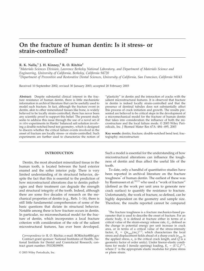
Given this difference between the notch stress andstrain distributions, the distinction between stress- orstrain-controlled fracture can be achieved through theuse of the double-notched four-point bend sample(Fig. 2), which has two important features to enablethe evaluation42–44,51–53:
• Rounded notches with a large root radius (� �200 �m) are used (rather than sharp cracks) tomaximize the difference between the locations ofmaximum stress and maximum strain ahead ofthe notch. Hill’s well-known logarithmic slip-line
Figure 1. (a) Hill’s logarithmic spiral slip-line field for a rounded notch in an elastic-plastic material.29, and the (b) tensilestress, �11, and (c) plastic strain, 11, distributions, based on slip-line field analysis29 and numerical computations,30 ahead ofsuch a notch. Also shown are schematic illustrations of possible (b) stress-controlled and (c) strain-controlled fracturemechanisms emanating from such notches. Note for stress-controlled mechanisms, the initial fracture event is ahead of thenotch, whereas it is at the notch root for strain-controlled mechanisms.
486 NALLA, KINNEY, AND RITCHIE
field solution29 for the notch in a perfectly plasticsolid [Fig. 1(a)] gives the location of the maxi-mum tensile stress (and maximum degree of tri-axiality) at the elastic-plastic interface. Subse-quent finite-element solutions for a linear work-hardening solid by Griffiths and Owen30 showthat this location is actually behind the elastic-plastic interface; depending on the nominal (far-field) applied stress, it is typically at distances of0.7–3 times the notch root radius ahead of thenotch [Fig. 1(b)]. Conversely, the maximumstrains are at the notch root and decrease mono-tonically with distance ahead of the notch.30
• Two notches in four-point bending are used be-cause there is a constant bending moment be-tween the inner two loading points, hence bothnotches see exactly the same stress and strainstates. For a nominally brittle material, such asdentin, this means that when unstable fractureensues from one notch, the other notch will beliterally at the point of instability, thereby “freez-ing in” the local microstructural events that pre-cede fracture. Thus, examination of the micro-structure below this unbroken notch enables anevaluation of how and where the critical crackingprocesses initiate and in doing so define whetherthe process is stress- or strain-controlled.
It might be noted here that even though the actual
measurement of the fracture toughness must involvefracture from a nominally atomically sharp crack, thisparticular experiment is best carried out with roundednotches because the distinction between the sites ofthe maximum tensile stress and strain substantiallydiminishes as the root radius, �3 0.29–31
Micromechanical models for fracture incorporatingsuch local failure criteria have been widely developedfor metallic systems [e.g., the so-called RKR model forstress-controlled cleavage fracture18 and the stress-state modified strain-controlled fracture model forductile (microvoid coalescence) fracture23–25]; how-ever, such deliberations have not been undertaken forthe fracture of biomaterials such as dentin.† This issomewhat surprising because the strain-based criteriahave been widely used to model such materials [e.g.,bone (e.g., Refs. 47, 54, and 55)]. Indeed, because den-tin displays marked “yielding” and postyield behav-ior, in the form of irrecoverable damage, as shownbelow in the section on Inelasticity or “Yielding” inDentin, a strain-based fracture criteria would appearto be most probable, although none of this has everbeen proved conclusively.
The prime objective of the current work is to use thephilosophy described above to determine systemati-cally the nature of the local fracture mechanisms inhuman dentin, by using the double-notched bendingtest described above. In addition, the aim is to addressseveral unanswered questions concerning the interac-tion of the crack with the microstructural features indentin, specifically: 1) whether the tubules actuallyaffect the macroscopic crack path, 2) the nature of theinteraction as the crack tip encounters a tubule, and 3)the role of the mineralized peritubular dentinal cuff inenhancing the toughness.15
It is believed that, in addition to the mechanisticunderstanding gained, this work is of importancefrom a clinical perspective. Notches resulting fromnatural caries, and possibly dental repair processes,are common in human teeth, and as such, a mecha-nistic understanding of dentin fracture from suchnotches may be considered critical for the develop-ment of a methodology for prediction of tooth failures.
EXPERIMENTAL PROCEDURES
Materials
Recently extracted human molars, sterilized with use ofgamma radiation after extraction,56 were used in the presentstudy. Sections (1.5–2.0 mm thick) were prepared from the
†Recent studies using the double-notched geometry haveshown that fracture in human cortical bone is consistentwith a strain-based criterion.51,53
Figure 2. (a) Schematic illustration of the double-notchedfour-point bend test used to discern whether fracture isstress- or strain-controlled. Between the inner two loadingpoints, the bending moment is constant; thus, when onenotch breaks, the other is “frozen” at a point just beforefracture instability. The region beneath this unbroken notch[as indicated by the arrow in (b)] is then carefully examinedto determine the site of the precursor microscopic eventsinvolved in the fracture process.
FRACTURE OF HUMAN DENTIN 487
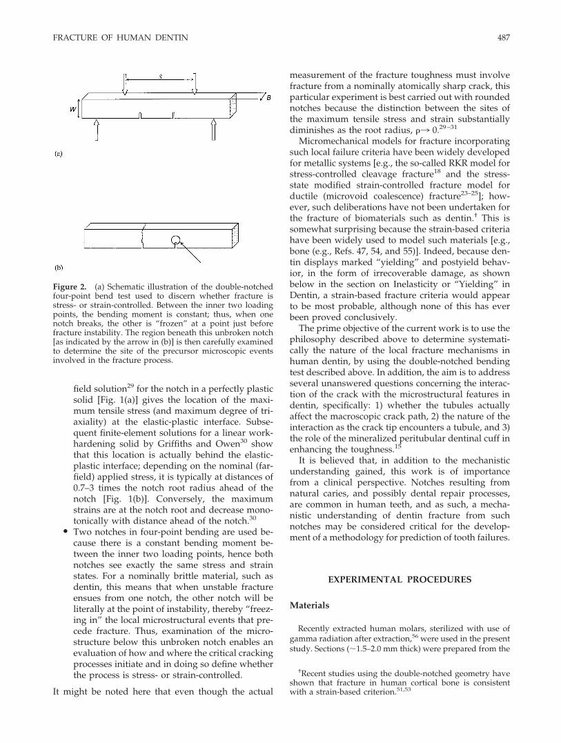
central portion of the crown and the root vertically throughthe tooth. The typical microstructure of dentin is shown inFigure 3, with the tubules being the most characteristicfeature; this structure is described in more detail below inthe section on Microstructure of Dentin. Although speci-mens were cut to define a specific orientation of the micro-structure with respect to the crack path, in actuality it isalmost impossible a priori to align the fracture plane pre-cisely with, for example, the tubule axes because, with theexception of the root, the tubules in dentin do not run astraight course from the enamel to the pulp. Rather, from thecervical margin through the crown, the tubules have a com-plex, S-shaped curvature.57 This is well illustrated by thescanning electron micrograph in Figure 4 showing the sideof a notch and the fracture surface of the crack emanatingfrom it; despite the fact that these two surfaces are mutuallyperpendicular, the orientation of the tubules appears to beidentical on the two planes. Consequently, the orientation of
the crack plane had to be determined from examination ofthe crack paths and the fracture surfaces.
Beams of dentin measuring roughly 1.0 � 1.0 � 12.0 mmwere then obtained from these sections by carefully furthersectioning using a slow-speed diamond saw and subsequentwet polishing up to a 600 grit finish. These beams were thenstored in Hanks’ balanced salt solution (HBSS) at ambienttemperature. Although mineral can be leached into solutionwhen storing dentin in deionized water, which can result inchanges in elastic properties with storage time, no suchchanges could be detected after a short-time storage inHBSS.58 However, the precise effects of storage solution onthe fracture properties have not been investigated per se.
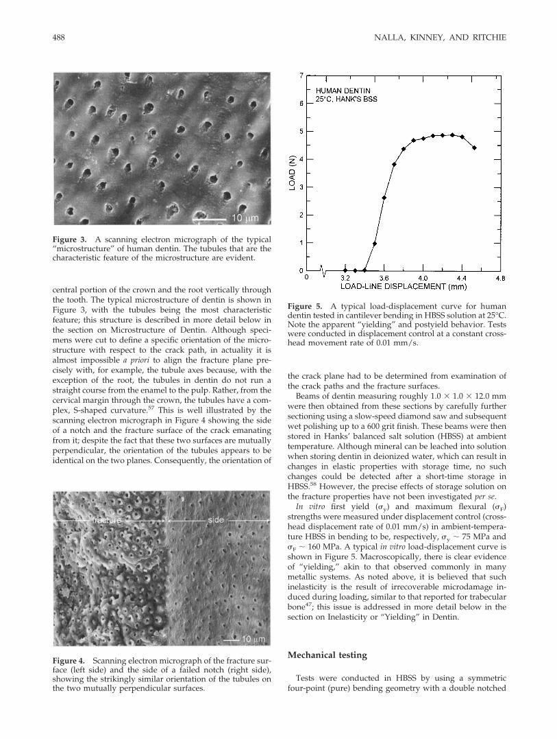
In vitro first yield (�y) and maximum flexural (�F)strengths were measured under displacement control (cross-head displacement rate of 0.01 mm/s) in ambient-tempera-ture HBSS in bending to be, respectively, �y 75 MPa and�F 160 MPa. A typical in vitro load-displacement curve isshown in Figure 5. Macroscopically, there is clear evidenceof “yielding,” akin to that observed commonly in manymetallic systems. As noted above, it is believed that suchinelasticity is the result of irrecoverable microdamage in-duced during loading, similar to that reported for trabecularbone47; this issue is addressed in more detail below in thesection on Inelasticity or “Yielding” in Dentin.
Mechanical testing
Tests were conducted in HBSS by using a symmetricfour-point (pure) bending geometry with a double notched
Figure 3. A scanning electron micrograph of the typical“microstructure” of human dentin. The tubules that are thecharacteristic feature of the microstructure are evident.
Figure 4. Scanning electron micrograph of the fracture sur-face (left side) and the side of a failed notch (right side),showing the strikingly similar orientation of the tubules onthe two mutually perpendicular surfaces.
Figure 5. A typical load-displacement curve for humandentin tested in cantilever bending in HBSS solution at 25°C.Note the apparent “yielding” and postyield behavior. Testswere conducted in displacement control at a constant cross-head movement rate of 0.01 mm/s.
488 NALLA, KINNEY, AND RITCHIE
configuration [Fig. 2(a)], as described above; this samplecreates a constant bending moment between the inner twoloading points, which in the present tests had a span S equalto 2–4 times the width W of the beam. Rounded notches, ofroot radius, � 200–300 �m, and depth, a 0.3–0.4 W, wereintroduced carefully with a slow-speed saw; care was takento maintain the specimens in a hydrated state throughoutthe specimen preparation process. The depths of bothnotches in each specimen were kept as identical as possibleto ensure similar stress/strain fields at the notch tips. A totalof 10 such tests were conducted. Conditions can be consid-ered to be nominally plane strain because, in accordancewith the ASTM Standard E-399 for fracture toughness test-ing, the thickness of the bend specimens was comparable to2.5 (Kc/�y)2 (1.4 mm).
All testing was conducted at ambient temperature on anELF� 3200 series voice coil-based mechanical testing ma-chine (EnduraTEC Inc., Minnetonka, MN). The bend barswere loaded to failure under displacement control at a con-stant cross-head movement rate of 0.02 mm/min in HBSS at25°C.
Microstructural characterization
The area around the unfractured notch in the fractureddouble-notched samples was examined by using a high-power optical microscope and (after coating with a gold-palladium alloy) with a scanning electron microscope (SEM)operating in the back-scattered electron mode. To minimizethe possibility of any damage during specimen preparation,the dentin beams were kept hydrated during all preparationand testing procedures, and for SEM imaging until the ac-tual coating was performed. Moreover, given the furtherpossibility of dehydration-induced cracking during the SEMimaging itself, some specimens were first observed in ahydrated condition in an environmental SEM and an atomic-force microscope to verify that the cracking configurationsobserved were not artifacts of the experimental procedure.However, the micrographs presented in this article wereobtained from a conventional SEM because this providedthe maximum resolution. In addition, postfailure observa-tions of the fracture surfaces of the broken ligaments wereconducted by using the same techniques.
RESULTS AND DISCUSSION
Microstructure of dentin
Human dentin is a hydrated composite of nanocrys-talline carbonated apatite mineral (45% by volume),type I collagen fibrils (30% by volume), and fluid(25% by volume). The mineral component is distrib-uted in the form of 5-nm-thick crystallites in a scaffoldcreated by the fibrils (typically 50- to 100-nm diame-ter). The most distinctive feature of this “microstruc-ture” is a unique distribution of 1- to 2-�m-diameter
cylindrical tubules (Fig. 3) that run from the dentin-enamel junction (DEJ) to the soft pulp in the interior ofthe tooth. These dentinal tubules are actually the pathsof the odontoblast cells during tooth formation. Theyare surrounded by a collar of highly mineralized peri-tubular dentin ( 1 �m thick) and are embeddedwithin a matrix of mineralized collagen (intertubulardentin). The mineralized collagen fibrils form a planarfelt-like structure oriented perpendicular to the tu-bules.59 The tubules are randomly displaced about aperiodic lattice60 but with a distribution that dependson location within the tooth (e.g., Ref. 8). The interac-tion of these tubules with cracks in the dentin is ofobvious interest from the perspective of an under-standing of the failure of dentin; this issue is ad-dressed below in the section on Double-Notch Exper-iments.
Inelasticity or “yielding” in dentin
Because the concept of the double-notched four-point bend test to distinguish between stress- andstrain-controlled fracture is based on the premise ofinelasticity or plastic yielding, it is important to estab-lish that inelastic deformation does indeed occur inhuman dentin. Although such deformation is wellunderstood for traditional materials (e.g., in the formof dislocation activity in metals), the mechanism of“yielding” in dentin is far less characterized, but it canbe considered in terms of regions of “microdamage,”as in bone (e.g., Ref. 47). Moreover, the nonlinear,nonrecoverable nature of the load/displacementcurve measured for dentin in the current work (Fig. 5)is indicative that some form of “plastic” yielding doesindeed occur.
With respect to localized yielding in the presence ofcracks, the notion of a plastic zone is invariably used,where the stresses locally exceed the “yield” strength,�y, of the material. The plastic-zone size can beroughly estimated from continuum arguments (e.g.,Ref. 34) to scale with the square of the ratio of thefracture toughness to the “yield” strength; specifically,at the onset of fracture, the maximum dimension isapproximately ry 1/2� (Kc/�y)2, which in dentin ison the order of 100 �m.# The boundary of this zone isdrawn around a typical crack in human dentin inFigure 6(a). It is evident that within this zone [Fig. 6 (c)and (d)], “microdamage” can be seen in the form ofcracked dentinal tubule cuffs, whereas no such dam-age is apparent outside the zone [Fig. 6(b)]. Thus, the
#Note that the relationship used to estimate the plastic-zone size, ry 1/2� (Kc/�y)2, pertains roughly to the max-imum extent of the zone in plane strain and the forwardextent in plane stress. Thus, it is a reasonable approximationfor all stress states.
FRACTURE OF HUMAN DENTIN 489

existence of a nonlinear load/displacement curve andformation of a “microdamage” zone in the high-stressregions surrounding a crack are strong evidence thatdentin does display inelastic deformation, consistentwith the notion of microcracking of the peritubularcuffs. It is appreciated, however, that other factors,e.g., “plasticity” in the collagen fibers, may also play arole.
Double-notch experiments
Results from the double-notched four-point bendtests, which were used to detect the microstructural
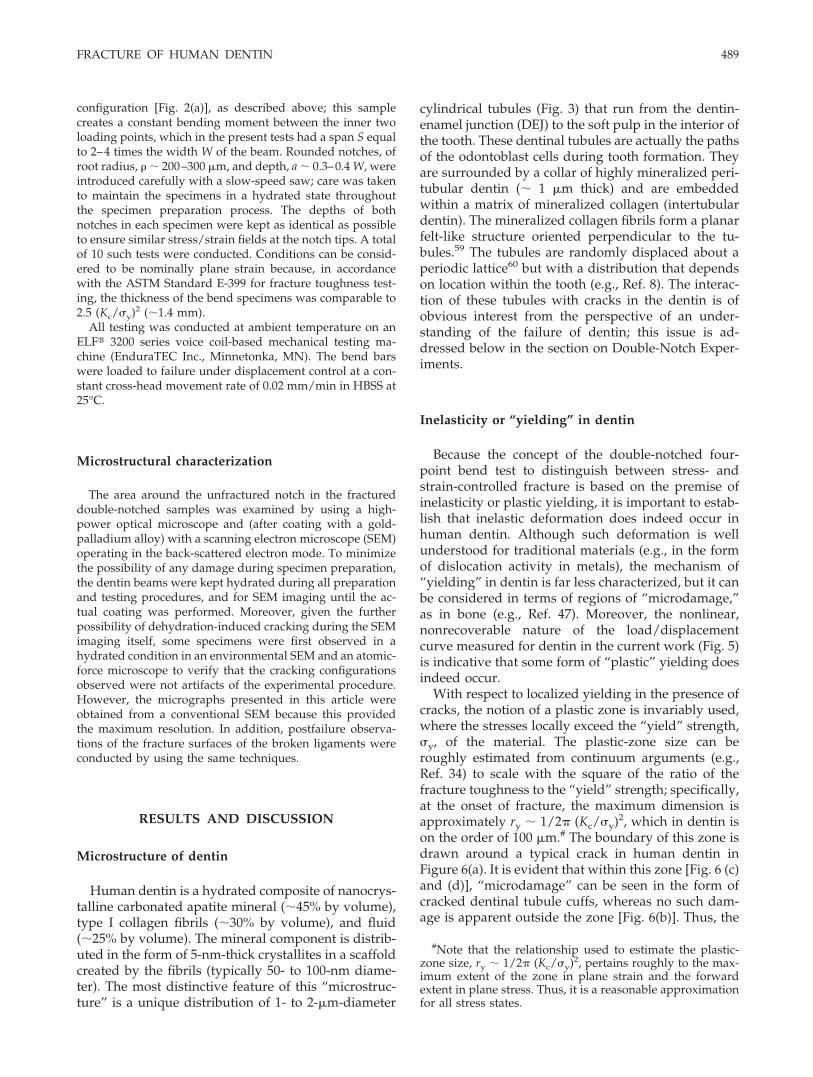
events before macroscopic fracture in dentin speci-mens, are shown in the macroscopic images in Figure7, before and after failure at one of the notches, and inthe microscopic SEM images of the crack paths inFigures 8 and 9. Macroscopic and microscopic imagesof the fracture surfaces are shown in Figure 10. Asdescribed above, when one notch breaks, the other is“frozen” at a point immediately preceding unstablefracture. Examination in the scanning electron micro-scope of this region, marked by the white circle inFigure 7(b), clearly indicates that all precursor cracksform directly at the notch root, generally with evi-dence of multiple initiation, as shown in Figure 8. Toprovide absolute proof that these cracks actually
Figure 6. Scanning electron micrographs of the concept of a “yielding” or “damage” zone is shown, based on the regionswhere the peritubular cuffs are cracked. (a) An overview of a crack emanating from a notch, with the nominal damage zoneindicated as a dotted line based on the continuum solution for the “plastic-zone” size (see text), and high magnificationmicrographs of areas (b) outside the zone, (c) inside the zone, and (d) at the notch tip. Note the extensive cracking ofperitubular cuffs in the “damage zone” in (c) and (d) (indicated by white arrows).
490 NALLA, KINNEY, AND RITCHIE
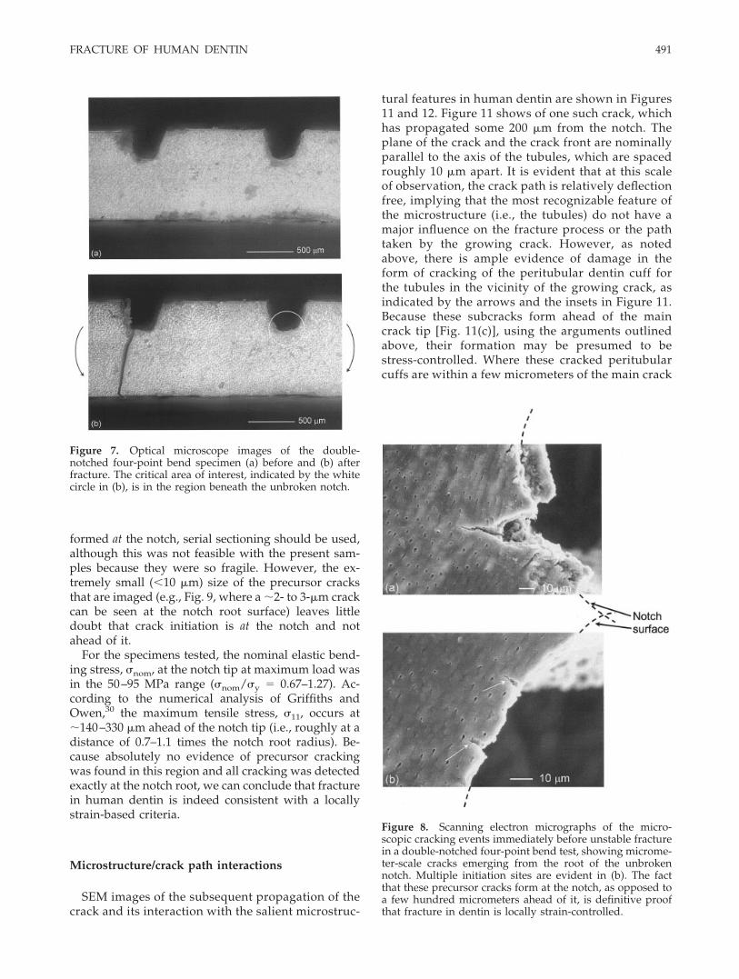
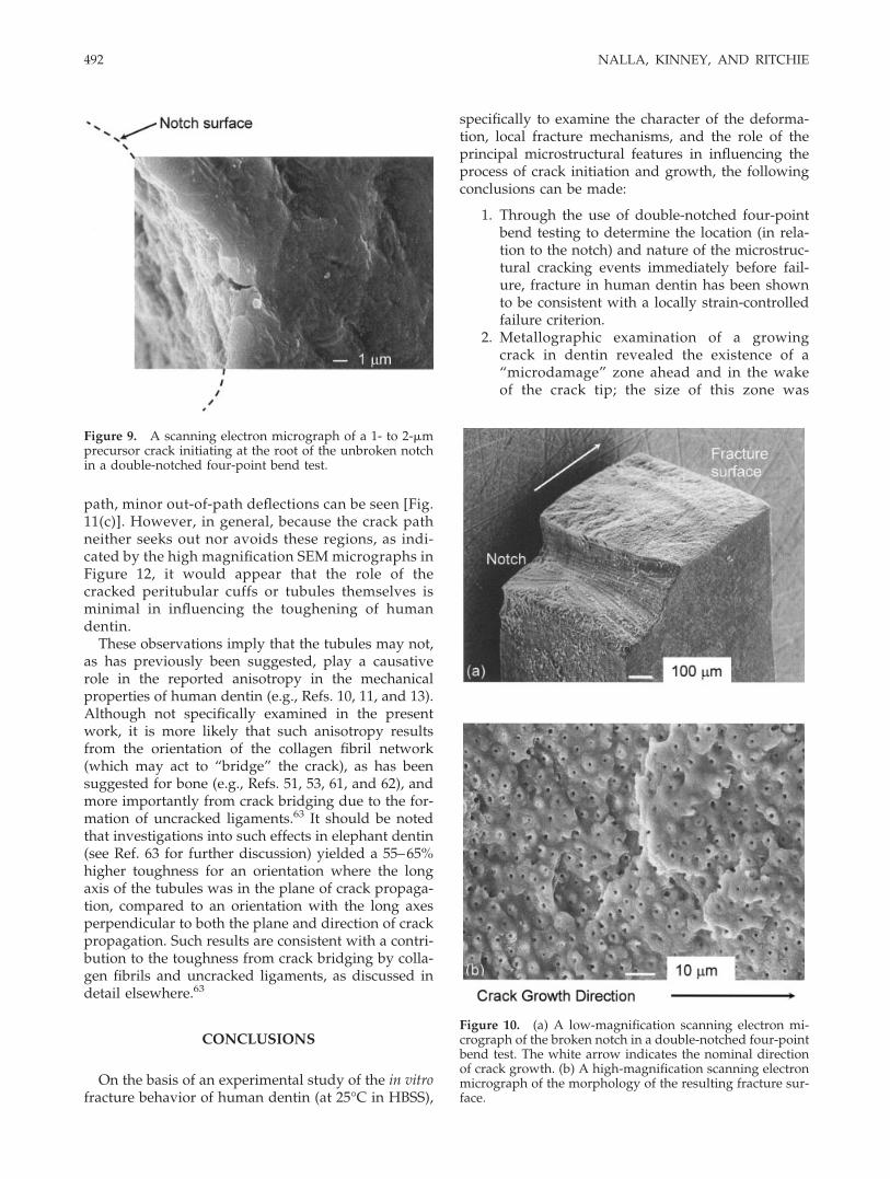
formed at the notch, serial sectioning should be used,although this was not feasible with the present sam-ples because they were so fragile. However, the ex-tremely small (�10 �m) size of the precursor cracksthat are imaged (e.g., Fig. 9, where a 2- to 3-�m crackcan be seen at the notch root surface) leaves littledoubt that crack initiation is at the notch and notahead of it.
For the specimens tested, the nominal elastic bend-ing stress, �nom, at the notch tip at maximum load wasin the 50–95 MPa range (�nom/�y � 0.67–1.27). Ac-cording to the numerical analysis of Griffiths andOwen,30 the maximum tensile stress, �11, occurs at140–330 �m ahead of the notch tip (i.e., roughly at adistance of 0.7–1.1 times the notch root radius). Be-cause absolutely no evidence of precursor crackingwas found in this region and all cracking was detectedexactly at the notch root, we can conclude that fracturein human dentin is indeed consistent with a locallystrain-based criteria.
Microstructure/crack path interactions
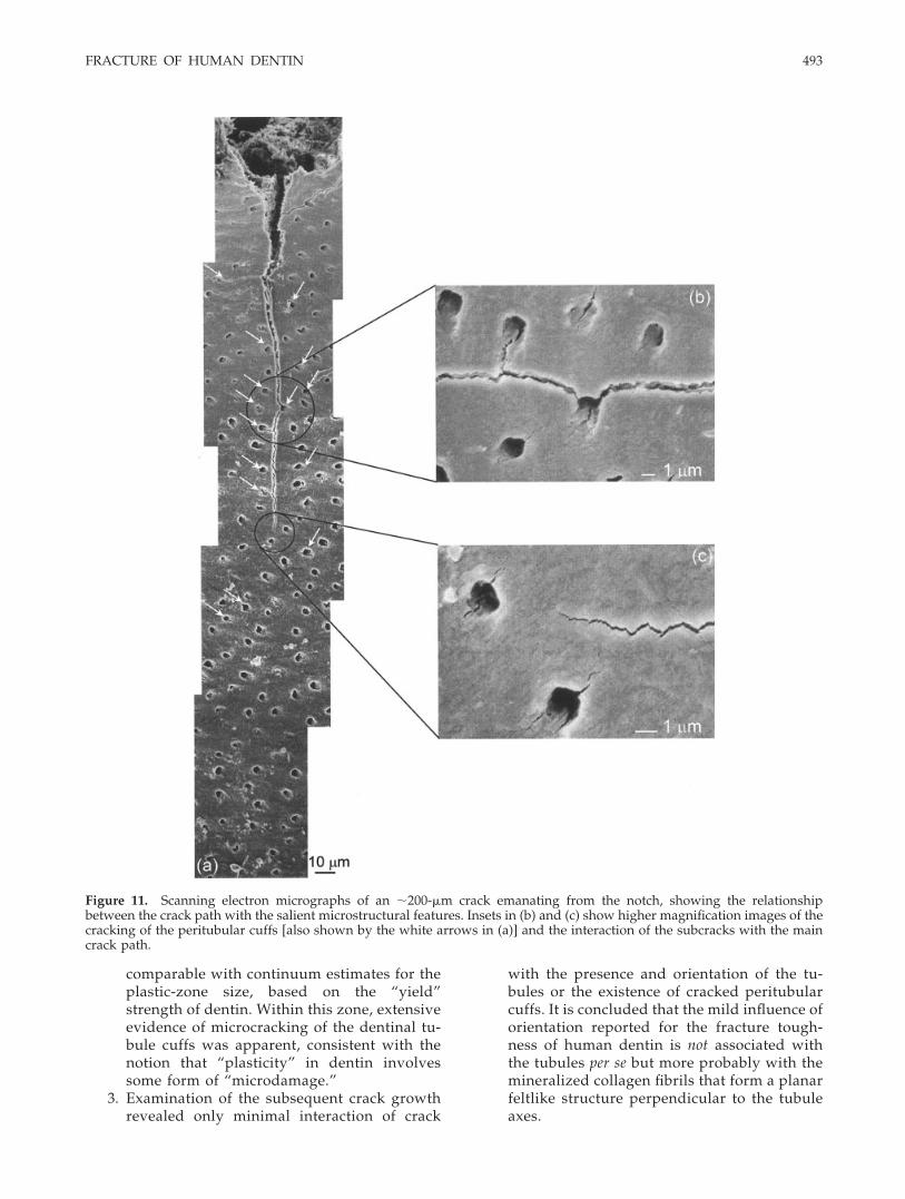
SEM images of the subsequent propagation of thecrack and its interaction with the salient microstruc-
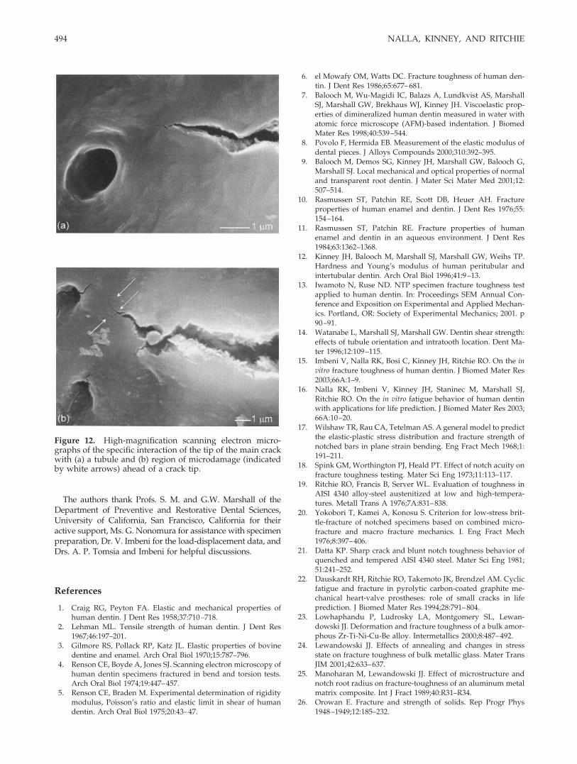
tural features in human dentin are shown in Figures11 and 12. Figure 11 shows of one such crack, whichhas propagated some 200 �m from the notch. Theplane of the crack and the crack front are nominallyparallel to the axis of the tubules, which are spacedroughly 10 �m apart. It is evident that at this scaleof observation, the crack path is relatively deflectionfree, implying that the most recognizable feature ofthe microstructure (i.e., the tubules) do not have amajor influence on the fracture process or the pathtaken by the growing crack. However, as notedabove, there is ample evidence of damage in theform of cracking of the peritubular dentin cuff forthe tubules in the vicinity of the growing crack, asindicated by the arrows and the insets in Figure 11.Because these subcracks form ahead of the maincrack tip [Fig. 11(c)], using the arguments outlinedabove, their formation may be presumed to bestress-controlled. Where these cracked peritubularcuffs are within a few micrometers of the main crack
Figure 8. Scanning electron micrographs of the micro-scopic cracking events immediately before unstable fracturein a double-notched four-point bend test, showing microme-ter-scale cracks emerging from the root of the unbrokennotch. Multiple initiation sites are evident in (b). The factthat these precursor cracks form at the notch, as opposed toa few hundred micrometers ahead of it, is definitive proofthat fracture in dentin is locally strain-controlled.
Figure 7. Optical microscope images of the double-notched four-point bend specimen (a) before and (b) afterfracture. The critical area of interest, indicated by the whitecircle in (b), is in the region beneath the unbroken notch.
FRACTURE OF HUMAN DENTIN 491
path, minor out-of-path deflections can be seen [Fig.11(c)]. However, in general, because the crack pathneither seeks out nor avoids these regions, as indi-cated by the high magnification SEM micrographs inFigure 12, it would appear that the role of thecracked peritubular cuffs or tubules themselves isminimal in influencing the toughening of humandentin.
These observations imply that the tubules may not,as has previously been suggested, play a causativerole in the reported anisotropy in the mechanicalproperties of human dentin (e.g., Refs. 10, 11, and 13).Although not specifically examined in the presentwork, it is more likely that such anisotropy resultsfrom the orientation of the collagen fibril network(which may act to “bridge” the crack), as has beensuggested for bone (e.g., Refs. 51, 53, 61, and 62), andmore importantly from crack bridging due to the for-mation of uncracked ligaments.63 It should be notedthat investigations into such effects in elephant dentin(see Ref. 63 for further discussion) yielded a 55–65%higher toughness for an orientation where the longaxis of the tubules was in the plane of crack propaga-tion, compared to an orientation with the long axesperpendicular to both the plane and direction of crackpropagation. Such results are consistent with a contri-bution to the toughness from crack bridging by colla-gen fibrils and uncracked ligaments, as discussed indetail elsewhere.63
CONCLUSIONS
On the basis of an experimental study of the in vitrofracture behavior of human dentin (at 25°C in HBSS),
specifically to examine the character of the deforma-tion, local fracture mechanisms, and the role of theprincipal microstructural features in influencing theprocess of crack initiation and growth, the followingconclusions can be made:
1. Through the use of double-notched four-pointbend testing to determine the location (in rela-tion to the notch) and nature of the microstruc-tural cracking events immediately before fail-ure, fracture in human dentin has been shownto be consistent with a locally strain-controlledfailure criterion.
2. Metallographic examination of a growingcrack in dentin revealed the existence of a“microdamage” zone ahead and in the wakeof the crack tip; the size of this zone was
Figure 9. A scanning electron micrograph of a 1- to 2-�mprecursor crack initiating at the root of the unbroken notchin a double-notched four-point bend test.
Figure 10. (a) A low-magnification scanning electron mi-crograph of the broken notch in a double-notched four-pointbend test. The white arrow indicates the nominal directionof crack growth. (b) A high-magnification scanning electronmicrograph of the morphology of the resulting fracture sur-face.
492 NALLA, KINNEY, AND RITCHIE
comparable with continuum estimates for theplastic-zone size, based on the “yield”strength of dentin. Within this zone, extensiveevidence of microcracking of the dentinal tu-bule cuffs was apparent, consistent with thenotion that “plasticity” in dentin involvessome form of “microdamage.”
3. Examination of the subsequent crack growthrevealed only minimal interaction of crack
with the presence and orientation of the tu-bules or the existence of cracked peritubularcuffs. It is concluded that the mild influence oforientation reported for the fracture tough-ness of human dentin is not associated withthe tubules per se but more probably with themineralized collagen fibrils that form a planarfeltlike structure perpendicular to the tubuleaxes.
Figure 11. Scanning electron micrographs of an 200-�m crack emanating from the notch, showing the relationshipbetween the crack path with the salient microstructural features. Insets in (b) and (c) show higher magnification images of thecracking of the peritubular cuffs [also shown by the white arrows in (a)] and the interaction of the subcracks with the maincrack path.
FRACTURE OF HUMAN DENTIN 493
The authors thank Profs. S. M. and G.W. Marshall of theDepartment of Preventive and Restorative Dental Sciences,University of California, San Francisco, California for theiractive support, Ms. G. Nonomura for assistance with specimenpreparation, Dr. V. Imbeni for the load-displacement data, andDrs. A. P. Tomsia and Imbeni for helpful discussions.
References
1. Craig RG, Peyton FA. Elastic and mechanical properties ofhuman dentin. J Dent Res 1958;37:710–718.
2. Lehman ML. Tensile strength of human dentin. J Dent Res1967;46:197–201.
3. Gilmore RS, Pollack RP, Katz JL. Elastic properties of bovinedentine and enamel. Arch Oral Biol 1970;15:787–796.
4. Renson CE, Boyde A, Jones SJ. Scanning electron microscopy ofhuman dentin specimens fractured in bend and torsion tests.Arch Oral Biol 1974;19:447–457.
5. Renson CE, Braden M. Experimental determination of rigiditymodulus, Poisson’s ratio and elastic limit in shear of humandentin. Arch Oral Biol 1975;20:43–47.
6. el Mowafy OM, Watts DC. Fracture toughness of human den-tin. J Dent Res 1986;65:677–681.
7. Balooch M, Wu-Magidi IC, Balazs A, Lundkvist AS, MarshallSJ, Marshall GW, Brekhaus WJ, Kinney JH. Viscoelastic prop-erties of dimineralized human dentin measured in water withatomic force microscope (AFM)-based indentation. J BiomedMater Res 1998;40:539–544.
8. Povolo F, Hermida EB. Measurement of the elastic modulus ofdental pieces. J Alloys Compounds 2000;310:392–395.
9. Balooch M, Demos SG, Kinney JH, Marshall GW, Balooch G,Marshall SJ. Local mechanical and optical properties of normaland transparent root dentin. J Mater Sci Mater Med 2001;12:507–514.
10. Rasmussen ST, Patchin RE, Scott DB, Heuer AH. Fractureproperties of human enamel and dentin. J Dent Res 1976;55:154–164.
11. Rasmussen ST, Patchin RE. Fracture properties of humanenamel and dentin in an aqueous environment. J Dent Res1984;63:1362–1368.
12. Kinney JH, Balooch M, Marshall SJ, Marshall GW, Weihs TP.Hardness and Young’s modulus of human peritubular andintertubular dentin. Arch Oral Biol 1996;41:9–13.
13. Iwamoto N, Ruse ND. NTP specimen fracture toughness testapplied to human dentin. In: Proceedings SEM Annual Con-ference and Exposition on Experimental and Applied Mechan-ics. Portland, OR: Society of Experimental Mechanics; 2001. p90–91.
14. Watanabe L, Marshall SJ, Marshall GW. Dentin shear strength:effects of tubule orientation and intratooth location. Dent Ma-ter 1996;12:109–115.
15. Imbeni V, Nalla RK, Bosi C, Kinney JH, Ritchie RO. On the invitro fracture toughness of human dentin. J Biomed Mater Res2003;66A:1–9.
16. Nalla RK, Imbeni V, Kinney JH, Staninec M, Marshall SJ,Ritchie RO. On the in vitro fatigue behavior of human dentinwith applications for life prediction. J Biomed Mater Res 2003;66A:10–20.
17. Wilshaw TR, Rau CA, Tetelman AS. A general model to predictthe elastic-plastic stress distribution and fracture strength ofnotched bars in plane strain bending. Eng Fract Mech 1968;1:191–211.
18. Spink GM, Worthington PJ, Heald PT. Effect of notch acuity onfracture toughness testing. Mater Sci Eng 1973;11:113–117.
19. Ritchie RO, Francis B, Server WL. Evaluation of toughness inAISI 4340 alloy-steel austenitized at low and high-tempera-tures. Metall Trans A 1976;7A:831–838.
20. Yokobori T, Kamei A, Konosu S. Criterion for low-stress brit-tle-fracture of notched specimens based on combined micro-fracture and macro fracture mechanics. I. Eng Fract Mech1976;8:397–406.
21. Datta KP. Sharp crack and blunt notch toughness behavior ofquenched and tempered AISI 4340 steel. Mater Sci Eng 1981;51:241–252.
22. Dauskardt RH, Ritchie RO, Takemoto JK, Brendzel AM. Cyclicfatigue and fracture in pyrolytic carbon-coated graphite me-chanical heart-valve prostheses: role of small cracks in lifeprediction. J Biomed Mater Res 1994;28:791–804.
23. Lowhaphandu P, Ludrosky LA, Montgomery SL, Lewan-dowski JJ. Deformation and fracture toughness of a bulk amor-phous Zr-Ti-Ni-Cu-Be alloy. Intermetallics 2000;8:487–492.
24. Lewandowski JJ. Effects of annealing and changes in stressstate on fracture toughness of bulk metallic glass. Mater TransJIM 2001;42:633–637.
25. Manoharan M, Lewandowski JJ. Effect of microstructure andnotch root radius on fracture-toughness of an aluminum metalmatrix composite. Int J Fract 1989;40:R31–R34.
26. Orowan E. Fracture and strength of solids. Rep Progr Phys1948–1949;12:185–232.
Figure 12. High-magnification scanning electron micro-graphs of the specific interaction of the tip of the main crackwith (a) a tubule and (b) region of microdamage (indicatedby white arrows) ahead of a crack tip.
494 NALLA, KINNEY, AND RITCHIE

27. Hutchinson JW. Singular behavior at end of a tensile crack in ahardening material. J Mech Phys Solids 1968;16:13–31.
28. Rice JR, Rosengren GR. Plane strain deformation near a cracktip in a power-law hardening material. J Mech Phys Solids1968;16:1.
29. Hill R. The mathematical theory of plasticity. Oxford, UK:Clarendon Press; 1950.
30. Griffiths JR, Owen DRJ. An elastic-plastic stress analysis for anotched bar in plane strain bending. J Mech Phys Solids 1971;19:419–431.
31. Rice JR, Johnson MA. In: Kanninen MA, Adler WF, RosenfieldAR, Jaffe RI, editors. Inelastic behavior of solids. New York:McGraw Hill; 1970.
32. Ritchie RO, Knott JF, Rice JR. On the relationship betweencritical tensile stress and fracture toughness in mild steel. JMech Phys Solids 1973;21:395–410.
33. Lin T, Evans AG, Ritchie RO. Statistical analysis of cleavagefracture ahead of sharp cracks and rounded notches. ActaMetall 1986;34:2205–2216.
34. Knott JF. Fundamentals of fracture mechanics. London UK:Butterworth & Co., Ltd; 1976.
35. Ritchie RO, Thompson AW. On macroscopic and microscopicanalyses for crack initiation and crack growth toughness inductile alloys. Metall Trans A 1985;16A:233–248.
36. McClintock FA. Ductile fracture instability in shear. J ApplMech Trans ASME Ser H 1958;25:582–588.
37. Mackenzie AC, Hancock JW, Brown DK. On the influence ofstate of stress on ductile failure initiation in high strengthsteels. Eng Fract Mech 1977;9:167–188.
38. Ritchie RO, Horn RM. Further considerations on the inconsis-tency in toughness evaluation of AISI 4340 steel austenitized atincreasing temperatures. Metall Trans A 1978;9A:331–341.
39. Ritchie RO, Server WL, Wullaert RA. Critical fracture stressand fracture strain models for the prediction of lower andupper shelf toughness in nuclear pressure vessel steels. MetallTrans A 1979;10A:1557–1570.
40. Lewandowski JJ, Thompson AW. Microstructural effects onthe cleavage fracture stress in fully pearlitic 1080 steel. In:Valluri SR, editor. Advances in fracture research ’84: Proceed-ings of the 6th International Conference on Fracture (ICF-6),New Delhi, India. Oxford, UK: Pergamon; 1984. p. 1515–1522.
41. Lewandowski JJ, Knott JF. Microstructural effects in flow lo-calization in 7XXX Al alloys. Strength of metals and alloys-ICSMA 7. New York: Pergamon Press; 1985;2:1193–1200.
42. Lewandowski JJ, Thompson AW. Microstructural control ofthe cleavage fracture stress in fully pearlitic steel. Metall TransA 1986;17A:1769–1786.
43. Lewandowski JJ, Hippsley CA, Ellis MBD, Knott JF. Impurityeffects on sustained load cracking of 21
4Cr-1Mo steels: 1 Crack
initiation. Acta Metall 1987;35:593–609.44. Lewandowski JJ, Hippsley CA. The nucleation of high temper-
ature brittle intergranular fracture in 214Cr-1Mo steel. Metall
Trans A 1988;19A:3005–3011.45. Chu WY, Thompson AW. Hydrogen effects on brittle fracture
of the titanium aluminide alloy Ti-24Al-11Nb. Metall Trans A1992;23A:1299–1312.
46. Samant A, Lewandowski JJ. Effects of test temperature, grainsize, and alloy additions on the cleavage fracture stress ofpolycrystalline niobium. Metall Trans A 1997;28A:389–399.
47. Yeh OC, Keaveny TM. Relative roles of microdamage andmicrofracture in the mechanical behavior of trabecular bone.J Orthop Res 2001;19:1001–1007.
48. Vashishth D, Koontz J, Qiu S, Lundin-Cannon D, Yeni Y,Schaffler M, Fyhrie D. In vivo diffuse damage in human verte-bral trabecular bone. Bone 2000;26:147–152.
49. Zioupos P, Currey JD, Mirza MS, Barton DC. Experimentallydetermined microcracking around a circular hole in a flat plateof bone: comparison with predicted stresses. Philos Trans RSoc Lond B Biol Sci 1995;347:383–396.
50. Lotz JC, Cheal EJ, Hayes WC. Fracture prediction for theproximal femur using finite element models. I. Linear analysis.J Biomech Eng 1991;113:353–360.
51. Nalla RK, Stolken JS, Kinney JH, Ritchie RO. On the micro-mechanisms of in vitro fracture and toughening in humancortical bone. J Biomech 2003. Submitted for publication.
52. Knott JF. Application of fracture mechanics to an alloy steelused in turbo-generator low-pressure disks. J Iron Steel Insti-tute 1966;201:1014–121.
53. Nalla RK, Kinney JH, Ritchie RO. Mechanistic criteria for thefailure of human cortical bone. Nat Mater 2003;2:164–168.
54. Keaveny TM, Wachtel EF, Ford CM, Hayes WC. Differencesbetween the tensile and compressive strengths of bovine tibialtrabecular bone depends on modulus. J Biomech 1994;27:1137–1146.
55. Ford CM, Keaveny TM. The dependence of shear failure prop-erties of trabecular bone on apparent density and trabecularorientation. J Biomech 1996;29:1309–1317.
56. White JM, Goodis HE, Marshall SJ, Marshall GW. Sterilizationof teeth by gamma radiation. J Dent Res 1994;73:1560–1567.
57. Ten Cate AR. Oral histology—development, structure andfunction. 4th ed. St. Louis, MO: Mosby; 1994. p 173.
58. Habelitz S, Marshall GW, Balooch M, Marshall SJ. Nanoinden-tation and the storage of teeth. J Biomech 2002;35:995–998.
59. Jones SJ, Boyde A. Ultrastructure of dentin and dentinogenesis.In: Linde A, editor. Dentin and dentinogenesis. Boca Raton, FL:CRC Press; 1984. 1(of 2) p 81–134.
60. Kinney JH, Oliveira J, Haupt DL, Marshall GW, Marshall SJ.The spatial arrangement of tubules in human dentin. J MaterSci Mater Med 2001;12:743–751.
61. Yeni YN, Fyhrie DP. Collagen-bridged microcrack model forcortical bone tensile strength. In: Proceedings of the 2001 Sum-mer Bioengineering Conference. New York: ASME; 2001;BED50:293–294.
62. Kinney JH, Marshall SJ, Marshall GW. The mechanical prop-erties of human dentin: a critical review and reevaluation ofthe dental literature. Crit Rev Oral Biol Med 2003. Forthcom-ing.
63. Nalla RK, Kinney JH, Ritchie RO. Effect of orientation on the invitro fracture toughness of dentin: the role of toughening mech-anisms. Biomater 2003;24:3955–3968.
FRACTURE OF HUMAN DENTIN 495