opportunities for understanding ms mechanisms and
TRANSCRIPT

Neurology Publish Ahead of PrintDOI 101212WNL0000000000012884
Opportunities for Understanding MS Mechanisms and Progression With MRI Using Large-Scale Data Sharing and Artificial Intelligence
Author(s) Hugo Vrenken PhD1 Mark Jenkinson Prof BSc BE DPhil2 Dzung Pham PhD3 Charles RG
Guttmann MD4 Deborah Pareto PhD5 Michel Paardekooper PhD6 Alexandra de Sitter PhD1 Maria
A Rocca MD7 Viktor Wottschel PhD1 M Jorge Cardoso PhD8 Frederik Barkhof Prof MD PhD1 9 on behalf of the MAGNIMS Study Group
This is an open access article distributed under the terms of the Creative Commons
Attribution License 40 (CC BY) which permits unrestricted use distribution and
reproduction in any medium provided the original work is properly cited
Neurologyreg Published Ahead of Print articles have been peer reviewed and
accepted for publication This manuscript will be published in its final form after
copyediting page composition and review of proofs Errors that could affect the
content may be corrected during these processes
Copyright copy 2021 The Author(s) Published by Wolters Kluwer Health Inc on behalf of the American Academy of Neurology
Published Ahead of Print on October 4 2021 as 101212WNL0000000000012884
Corresponding Author Hugo Vrenken hvrenkenamsterdamumcnl
Affiliation Information for All Authors 1 MS Center Amsterdam Amsterdam Neuroscience Department of Radiology and Nuclear Medicine Amsterdam UMC Amsterdam the Netherlands 2 Wellcome Centre for Integrative Neuroimaging (WIN) FMRIB Nuffield Department of Clinical Neurosciences (NDCN) University of Oxford3 Human Imaging and Image Processing Core Center for Neuroscience and Regenerative Medicine The Henry M Jackson Foundation Bethesda MD USA 4 Center for Neurological Imaging Department of Radiology Brigham and Women s Hospital Boston USA5 Section of Neuroradiology (Department of Radiology) Vall d Hebron University Hospital and Research Institute (VHIR) Autonomous University Barcelona (Spain) 6 Amsterdam UMC Amsterdam the Netherlands7 Neuroimaging Research Unit Institute of Experimental Neurology Division of Neuroscience IRCCS San Raffaele Scientific Institute Milan Italy 8 AMIGO School of Biomedical Engineering and Imaging Sciences King s College London London UK 9
Institutes of Neurology amp Healthcare Engineering UCL London UK
Contributions Hugo Vrenken Draftingrevision of the manuscript for content including medical writing for content Major role in the acquisition of data Study concept or design Analysis or interpretation of data Mark Jenkinson Draftingrevision of the manuscript for content including medical writing for content Major role in the acquisition of data Analysis or interpretation of data Dzung Pham Draftingrevision of the manuscript for content including medical writing for content Major role in the acquisition of data Analysis or interpretation of data Charles RG Guttmann Draftingrevision of the manuscript for content including medical writing for content Major role in the acquisition of data Analysis or interpretation of data Deborah Pareto Draftingrevision of the manuscript for content including medical writing for content Major role in the acquisition of data Analysis or interpretation of data Michel Paardekooper Draftingrevision of the manuscript for content including medical writing for content Major role in the acquisition of data Analysis or interpretation of data Alexandra de Sitter Draftingrevision of the manuscript for content including medical writing for content Major role in the acquisition of data Analysis or interpretation of data Maria A Rocca Draftingrevision of the manuscript for content including medical writing for content Major role in the acquisition of data Analysis or interpretation of data Viktor Wottschel Draftingrevision of the manuscript for content including medical writing for content Major role in the acquisition of data Analysis or interpretation of data M Jorge Cardoso Draftingrevision of the manuscript for content including medical writing for content Major role in the acquisition of data Analysis or interpretation of data Frederik Barkhof Draftingrevision of the manuscript for content including medical writing for content Major role in the acquisition of data Analysis or interpretation of data
Number of characters in title 129
Abstract Word count 200
Word count of main text 4992
Copyright copy 2021 The Author(s) Published by Wolters Kluwer Health Inc on behalf of the American Academy of Neurology
References 60
Figures 1
Tables 2
Supplemental eAppendix 1 Author Contributions eAppendix 2 MAGNIMS Study Group informationeAppendix 3 Expanded sections including more extensive references that could not be included due to the 60-reference maximum in the main paper eReferences List of eReferences
Search Terms [ 41 ] Multiple sclerosis [ 120 ] MRI
Acknowledgements This paper is based on a workshop of the MAGNIMS Study Group that was made possible through financial support from the Dutch MS Research Foundation Amsterdam Neuroscience VU University Medical Center Merck KGaA and Novartis
Study Funding H Vrenken has received funding support from the Dutch MS Research Foundation (grant 14-876 MS) ZonMW jointly with the Dutch MS Research Foundation (grant 40-44600-98-326) and HealthHolland (grant LSHM19053) The MS Center Amsterdam is supported by the Dutch MS Research Foundation through a series of program grants (current grant 18-358f)M Jenkinson is supported by the National Institute for Health Research (NIHR) Oxford Biomedical Research Centre (BRC) and this research was funded by the Wellcome Trust (215573Z19Z) The Wellcome Centre for Integrative Neuroimaging is supported by core funding from the Wellcome Trust (203139Z16Z) DL Pham Funding support National Multiple Sclerosis Society Grants RG-1507-05243 and RG-1907-34570 Congressionally Directed Medical Research Programs Grant W81XWH-20-1-0912 and the Department of Defense in the Center for Neuroscience and Regenerative Medicine CRG Guttmann gratefully acknowledges support from the National Multiple Sclerosis Society (grant identifier RG-1501-03141) the International Progressive Multiple Sclerosis Alliance (grant identifier PA-1412-02420) the Foundation of the University of Bordeaux Roche Pharmaceuticals and TalanD Pareto Instituto de Salud Carlos III (PI1800823) V Wottschel has received funding from the European Union s Horizon 2020 research and innovation programme under grant agreement No 666992M J Cardoso is supported by the WellcomeEPSRC Centre for Medical Engineering (WT203148Z16Z) and the Wellcome Flagship Programme (WT213038Z18Z) F Barkhof is supported by the NIHR biomedical research center at UCLH
Disclosures H Vrenken has received research grants from Pfizer MerckSerono Novartis and Teva speaker honoraria from Novartis and consulting fees from MerckSerono all funds were paid directly to his Institution M Jenkinson has received research grants from Novartis paid to his institution plus royalties from the licensing of FSL to commercial entities and consultancy from Oxford Brain DiagnosticsDL Pham reports no financial disclosures CRG Guttmann has received support from Mobilengine (free use of platform and programming by Mobilengine Engineers) as well as the National Multiple Sclerosis Society and the International Progressive Multiple Sclerosis Alliance the US Office for Naval Research as well as travel support from Roche Pharmaceuticals CRG owns stock in Roche Novartis GSK Alnylam Protalix Biotherapeutics Arrowhead Pharmaceuticals Cocrystal Pharma Sangamo TherapeuticsD Pareto has received speaking honoraria from Novartis and Genzyme M Paardekooper reports no disclosuresA de Sitter has been employed on a research grant from Teva MA Rocca received speakers honoraria from Biogen Idec Novartis Genzyme Teva Merck Serono Roche Celgene and Bayer and receives research support from the MS Society of Canada and Fondazione Italiana Sclerosi MultiplaV Wottschel reports no disclosuresV Wottschel reports no disclosures M J Cardoso is a Founder of BrainMiner plcF Barkhof has received compensation for consulting services andor speaking activities from Bayer Biogen Idec Merck Serono Novartis Roche Teva Bracco and IXICO
Copyright copy 2021 The Author(s) Published by Wolters Kluwer Health Inc on behalf of the American Academy of Neurology
Abstract
Multiple sclerosis (MS) patients have heterogeneous clinical presentations symptoms and
progression over time making MS difficult to assess and comprehend in vivo The
combination of large-scale data-sharing and artificial intelligence creates new opportunities
for monitoring and understanding MS using magnetic resonance imaging (MRI)
First development of validated MS-specific image analysis methods can be boosted
by verified reference test and benchmark imaging data Using detailed expert annotations
artificial intelligence algorithms can be trained on such MS-specific data Second
understanding disease processes could be greatly advanced through shared data of large MS
cohorts with clinical demographic and treatment information Relevant patterns in such
data that may be imperceptible to a human observer could be detected through artificial
intelligence techniques This applies from image analysis (lesions atrophy or functional
network changes) to large multi-domain datasets (imaging cognition clinical disability
genetics etc)
After reviewing data-sharing and artificial intelligence this paper highlights three
areas that offer strong opportunities for making advances in the next few years
crowdsourcing personal data protection and organized analysis challenges Difficulties as
well as specific recommendations to overcome them are discussed in order to best leverage
data sharing and artificial intelligence to improve image analysis imaging and the
understanding of MS
Copyright copy 2021 The Author(s) Published by Wolters Kluwer Health Inc on behalf of the American Academy of Neurology
1 Introduction
Multiple sclerosis (MS) is highly heterogeneous across patients in terms of symptoms sites
of damage degree of recovery and development of the disease across time This makes the
disease more difficult to comprehend Relevant patterns may be imperceptible to a human
observer but by analyzing large amounts of imaging data from MS patients with
sophisticated artificial intelligence techniques urgently required advances in understanding
MS disease pathological heterogeneity may be made
Furthermore to tracking of disease progression in individual MS patients MRI
markers are needed This could benefit from MS-specific image analysis methods because
existing generalized methods tend to exhibit poorer performance in cases with MS1 as has
been demonstrated quantitatively for segmentation of deep grey matter (GM) structures2
Large amounts of MS imaging data with expert annotations as appropriate can be used to
train and validate more accurate measurement and analysis tools specifically for MS
Against this background we review the possibilities of data-sharing and artificial
intelligence for improved applications of MRI to study MS addressing both the need to
understand MS disease processes and the need for MS-dedicated quantitative
measurement and analysis techniques for MRI assessments We first survey relevant
existing efforts regarding data-sharing and artificial intelligence and then highlight three
areas of interest in bringing the field forward crowd-sourcing personal data protection and
organized analysis challenges (see Figure 1 for the methods used to create this manuscript)
Specific recommendations aim to achieve the best outcomes for MS patients
2 Data-sharing
21 Data-sharing in MS Non-imaging
The multicenter collection of MS clinical data provides valuable information on disease
prevalence current treatment patterns and general distribution of patientsrsquo outcomes
Thus clinical registries including data from several MS centers have been strongly promoted
in the last decades National and regional MS registries exist in most countries especially in
Europe3 and North America4 Data collected by national initiatives have often been included
in computerized platforms such as the European Register for Multiple Sclerosis (EUReMS)5
or the MSBase6 Collaborative research studies have used these data to define the value of
Copyright copy 2021 The Author(s) Published by Wolters Kluwer Health Inc on behalf of the American Academy of Neurology
prognostic indicators in different patient populations7 to investigate the influence of
demographic and geographic factors on MS clinical course8 and to evaluate the
comparative efficacy of different drugs9 (additional references in eAppendix 1
[httpsdoiorg105061dryad2fqz612p9])
22 Data-sharing in neuro-MRI Non-MS
Data sharing in MRI is becoming increasingly prevalent large-scale and open in the
neuroimaging community Table 1 lists several prominent examples including Alzheimerrsquos
Disease Neuroimaging Initiative (ADNI) which in the neuroimaging field is a template for
data acquisition and fostering of methodological developments These datasets are
associated with a range of access policies (from free unrestricted downloads to
collaboration-only agreements) and cover various sizes demographics and pathologies
They provide access to large diverse groups of subjects including rare diseases and a wider
range of disease stages (including prodromal cases) than is possible from single studies In
addition the increasingly large numbers provide greater statistical power and the
opportunity to apply state-of-the-art deep learning techniques They also allow common
standards to be applied in the evaluation of methodological tools as pioneered by the
MICCAI challenges (e67) As such they provide the community with fair and open
comparisons of methods a richer set of data on which to test hypotheses and greater
ability for assessing reliability and repeatability There are also benefits for those involved in
creating and managing such datasets since the process of designing piloting and pre-
processing provides impetus for novel developments in acquisition and analysis
demonstrated by state-of-the-art methodologies developed within the Human Connectome
Project In addition there are benefits in visibility engagement and publications
Challenges still exist (eg IT infrastructure access policies ethics policies etc) but currently
many such datasets are already accessible with a range of solutions to these problems thus
offering options for the creation of new datasets focusing on MS The datasets listed also
highlight that standardized MR acquisition protocols can harmonize data only to a certain
extent Therefore alternative approaches such as synthetic MRI should also be investigated
(additional references in eAppendix 1)
Copyright copy 2021 The Author(s) Published by Wolters Kluwer Health Inc on behalf of the American Academy of Neurology
23 Data-sharing of MRI in MS
MRI is one of the most important tools for diagnosing and monitoring MS10 However MRI
data collected by clinical MS registries usually includes just conventional measures andor
metadata regarding fulfilment of diagnostic criteria5 6 Recent collaborations (eg the
publicly funded German Competence Network Multiple Sclerosis (e68) or the privately
funded MS PATHS11) promoted the use of relatively standard conventional MRI protocols
(typical T1-weighted proton density-weighted and T2-weighted or FLAIR images) but did
not include advanced MRI techniques (such as quantitative mapping techniques of tissue
properties tractography spectroscopy functional MRI etc) Table 2 lists MS Registries
identified from public sources that collect MRI information
As an example one can take the Italian Neuroimaging Network Initiative (INNI)
(e69)12 This has recently been established among four sites leading MRI research in MS in
Italy with the support of the Italian MS Society INNIrsquos major goal is to determine and
validate novel MRI biomarkers including biomarkers based on more advanced non-
conventional imaging techniques to be utilized as predictors andor outcomes in future MS
studies INNI aims also to standardize MRI procedures of acquisition and analysis in MS at a
national level
A large population of MS patients and healthy controls (more than 1800 subjects
and more than 3000 MRI exams) has been collected in the INNI platform so far Although
MRI data had to meet some minimum requirements in order to be included12 a full
standardization of acquisition protocols was not requested from sites at least in the first
phase of the project
The main challenges faced at the beginning of the INNI initiative were related to
ethical approvals to the creation of the online platform to ensure proper handling of
subjectsrsquo anonymity and to define guidelines to regulate database access levels and their
implementation as access procedures12 Conversely most of the subsequent challenges are
related to the quality assessment (QA) of the data collected which will now be used for
different research projects at the four promoting sites Systematic QA (on subjectsrsquo
positioning image inhomogeneity distortions and artefacts and measurement of contrast-
to-noise ratio) has been established to verify source data and ensure maintenance of high
quality QA results will be used to propose effective guidelines on acquisition protocols and
scanning options to improve harmonization of MRI data Basic analysis (eg T2-
hyperintense lesion segmentation T1-hypointense lesion refilling minimal pre-processing
Copyright copy 2021 The Author(s) Published by Wolters Kluwer Health Inc on behalf of the American Academy of Neurology
on diffusion-weighted MRI and resting-state fMRI scans) that may be shared in the INNI
platform to harmonize future projects is also being performed in a centralized manner
24 Recommendations on MRI data-sharing in MS
Clearly define variables to be shared This avoids ambiguity and heterogeneity at a later
stage
Set up proper QA and QC (quality control) procedures to ensure compliance with
minimum standards Preferably quantitative and automated such procedures guarantee
the integrity of the included data
Implement clear policies and procedures on how access to data can be obtained
Create a flexible data-sharing system permitting a manifold use of collected data By
choosing maximally permissive data licenses (within legal and institutional boundaries)
combined with clear data storage organization database management and flexible access
choices data can be flexibly and easily selected accessed and used for a variety of
purposes
3 Artificial intelligence
31 Artificial intelligence in medical image analysis beyond MS
Artificial intelligence can be roughly divided into ldquoclassicalrdquo machine learning techniques
such as support vector machines and (newer) deep learning techniques based on
convolutional neural networks Classical machine learning approaches typically make
predictions using classifiers trained not directly on images but on features extracted from
images13 While this can be advantageous it precludes the discovery of features not
perceptible to or appreciated by the human observer Deep learning when applied to
classification or segmentation of images14 instead analyzes image data directly without
prior feature selection This has given rise to excellent classifier performance in a range of
medical imaging applications13 However as shown for example by Ghafoorian and
colleagues15 at least given the current limitations regarding sizes of available datasets and
networks performance may be further improved by incorporating well-chosen features
extracted from the images using domain knowledge and ldquoclassicalrdquo image analysis
techniques Specifically in their work they incorporate measures reflecting location in the
brain to improve segmentation of age-related white matter hyperintensities15
Copyright copy 2021 The Author(s) Published by Wolters Kluwer Health Inc on behalf of the American Academy of Neurology
32 Artificial intelligence in imaging of MS
Existing studies applying artificial intelligence in MS imaging can generally be divided into
descriptive and predictive experiments Descriptive studies use cross-sectional data sets in
order to segment from MRI WM lesions16 or specifically contrast-enhancing lesions17
identify imaging patterns based on MS phenotype or clinical or cognitive disease severity18
or perform an automated diagnosis using uni- or multi-modal information19 Predictive
studies on the other hand detect patterns in baseline data that allow for predicting future
disease outcome or severity by incorporating clinical follow-up information20 21 The
majority of studies to date used classic machine learning techniques such as Support Vector
Machines or Random Forests where features have to be defined and extracted from the
data a priori while more recent studies also use deep learning methods allowing automated
detection of relevant features in the data Deep learning has now been used not only to
segment WM lesions22-24 or their enhancing subset17 but also to quantify lesion changes25
26 to detect the central vein sign27 classify different lesion types based on diffusion basis
spectrum imaging28 to predict Gd-enhancement from other image types29 to perform MRI-
based diagnosis30 31 to segment and analyze non-lesion structures32 33 to analyze myelin
water fraction34 or quantitative susceptibility mapping data35 to synthesize absent image
types36 to perform automatic QC37 to improve image quality38 or to correct intensity
differences between scanners39 (additional references in eAppendix 1)
33 Challenges of artificial intelligence in MS imaging
Although showing impressive performance state-of-the-art deep learning methods
(convolutional neural networks) rely solely on the use of local intensity patterns and
contextual features to guide the image analysis process Importantly they lack high-level
abstract thinking and have limited understanding of human anatomy and physiology
Learning from relatively small and noisy datasets learning systems are commonly unable to
extrapolate and handle uncertain situations Furthermore in precision medicine
applications fully automatic robust and quick measurements are required for every single
subject Three important categories of current limitations of learning systems are
1) Inputs The main limitation of many machine learning models is the strong
dependence on (good) training data While human raters may intuitively extrapolate from a
few examples to new cases that may be very different a supervised (deep) learning model
Copyright copy 2021 The Author(s) Published by Wolters Kluwer Health Inc on behalf of the American Academy of Neurology
has to be fed with sufficient examples to cover the whole range of heterogeneity in the
population disease and scanning parameters Differences in imaging devices acquisition
parameters tissue contrasts artefacts and noise patterns can degrade algorithm
performance if not handled appropriately To overcome between-scanner or between-
acquisition image differences many approaches for post-processing based data
harmonization have been proposed including traveling phantoms40 which requires physical
travel of objects or subjects39 limiting scalability data augmentation41 which requires
sufficiently accurate signal simulating models and domain adaptation42 which has shown
promising results Ultimately the most robust results may come from combinations of the
above together with basic steps such as intensity normalization (additional references in
eAppendix 1)
Given the relatively low prevalence of MS lack of training data is an issue of
particular relevance here Insufficient variability in training data can lead to overfitted
models that do not perform well on new data and single-center data-sets seldom exceed a
few hundred MS cases This effect can be reduced with regularization augmentation and
cross-validation but not fully removed Therefore pooling data from different sources is
advantageous but this introduces new challenges due to differences between centers
scanners and scanning protocols which require standardizing and post-processing
The majority of published machine learning studies in MS used research data rather
than clinical data which has limitations patients were filtered through inclusion criteria the
number of subjects and scans is limited by the obtained funding and more severe patients
are more likely to drop out biasing data towards more benign cases Clinical data is more
representative of the general (disease) population but is typically more heterogeneous and
requires additional patient consent
2) Labels Training with high-quality labels is crucial to attaining good performance of
machine learning systems Labels can be the personrsquos diagnosis or other overall features or
typically in image analysis tasks manual outlines of anatomical structures or pathological
entities like MS lesions Distinguishing MS lesions in the white matter from other white
matter lesions and from normal-appearing white matter requires skill and expertise The
variability of labeling protocols and inter- and intra-rater variability introduce errors when
training machine learning systems Such errors degrade the performance of learning
systems and limit to what extent that performance can be validated Those errors could be
quantified by expanding training sets based on common protocols applied by larger
Copyright copy 2021 The Author(s) Published by Wolters Kluwer Health Inc on behalf of the American Academy of Neurology
numbers of raters and subsequently overcome by modelling or machine learning
approaches
3) Uncertainty amp Confidence Algorithms commonly solve a categorical hard problem
but clinical decisions are rarely categorical and involve intrinsic uncertainty The
introduction of biomarker- and subject-specific error-bars and the development of novel
ways to convey and introduce this information into the clinical workflow will present
challenges to clinical adoption Recent work addresses that uncertainty for MS lesion
detection and segmentation43 Other areas of medicine with inherently uncertain
predictions may suggest ways of introducing this uncertainty into the clinical workflow in
the context of MS imaging (additional references in eAppendix 1)
34 Recommendations on machine learning in MS imaging
Compile large annotated datasets for training To obtain sufficient amounts of training
data large-scale data-sharing of MS imaging data is required both for homogeneous
datasets (for generating new knowledge) and heterogeneous datasets (for deriving more
generalizable classifiers)
Create methods that are robust to data variability Harmonize data using both classical
and machine learning techniques to improve robustness to unseen datasets
Include non-research data in training Train machine learning methods also on data
acquired in a real-life clinical setting to increase robustness to heterogeneity and to
improve applicability in the clinical population
Create high-quality labels Validating algorithms for clinical use will require large multi-
center labeling efforts yielding depending on the aims consensus-based ldquoground truthrdquo
labels or collections of individual ratersrsquo labels
Allow more subtle information in labels than global yesno answers The use of soft labels
(eg image wide disease classification) to model the intrinsic anatomical and pathological
variability should also be investigated
Incorporate the uncertainty of classifier predictions Algorithms should learn the intrinsic
uncertainty and confidence of every decision they make Diagnostic and prognostic
guidelines should be modified to enable clinical usage of biomarker- and subject-specific
uncertainty metrics
Copyright copy 2021 The Author(s) Published by Wolters Kluwer Health Inc on behalf of the American Academy of Neurology
4 Opportunity 1 Crowd-sourcing
41 Crowd-sourcing in research
The first highlighted opportunity here is ldquocrowd-sourcingrdquo Crowd-sourcing not to be
confused with ldquocrowd-fundingrdquo refers to people donating their time and skills to complete
certain tasks In the context of scientific research it is sometimes called ldquocitizen sciencerdquo Its
premise is that there are many enthusiastic members of the general public who are willing
to donate some of their time to science By making it easy for them to contribute the
scientific community can reward their enthusiasm and willingness by letting them really
help the field forward Successful projects have been conducted this way including
examples in astronomy on differentiating different galaxy types (Galaxy Zoo (e70)) in
organic chemistry on the topic of protein folding (Foldit a game with tens of thousands of
players (e71)) in biology on identifying bat calls (Bat Detective (e72)) and in paleontology
on dinosaur limb bone measurements (Open Dinosaur Project (e73)) The potential for brain
imaging applications has been noticed and a successful approach to interface-building data
management and analysis has been described SPINE (e74) Open Neuroimaging Laboratory
(e75) and OpenNeuro (e76) are examples of web-based infrastructures for crowd-sourced
brain imaging research
While the potential benefits of crowd-sourcing to the researchers are clear the
benefits to the participants (the ldquocrowdrdquo) may be less obvious There is the potential
gratification of contributing to science and in the case of MS research these volunteers
may be people with an interest in brain imaging or neuroscience or they may personally
know someone who suffers from MS and want to help develop a solution Furthermore
well-designed crowd-sourcing activities can also carry the reward of being entertaining to
perform The field of ldquogamificationrdquo is a rapidly developing area of research and
development in its own right which has already been applied in the radiological field44
which creates important opportunities for helping volunteers enjoy participating in crowd-
sourced research and remain committed to finishing their contribution
42 Potential for crowd-sourcing in MS imaging
For processing large amounts of image data on a regular basis as in a clinical setting
automation of analysis methods is key The training that goes into such automated
methods whether based on deep learning or using other approaches obviously plays a
large role in their performance Ideally reference labels eg of specific imaging features
Copyright copy 2021 The Author(s) Published by Wolters Kluwer Health Inc on behalf of the American Academy of Neurology
such as MS lesions or particular anatomical structures would be generated by intensively
trained expert raters However if this is not possible for example due to the associated
costs crowd-sourcing such training labels could provide a realistic alternative if properly
used A potential issue concerns the quality of crowd-sourced image annotations For the
example of image segmentation this has been addressed Specifically Bogovic and
colleagues45 demonstrated that for cerebellar parcellation it is possible to achieve high-
quality labels from a group of non-expert raters
This suggests that provided that training and quality assurance procedures are adequately
employed a large group of non-expert volunteers could create reference labels on a large
enough dataset to train deep learning or other methods robust to data variability
Nevertheless as the task becomes more complex the degree of communication required
between the participants in order to achieve an adequate performance is likely to increase
with the risk of the efforts becoming an outsourcing initiative instead of a crowd-sourcing
one Thus the project to be carried out should be clearly defined in terms of tasks and
expectations with the inclusion of tutorials and support by an experienced professional in
the field
Besides providing training labels for image segmentation crowd-sourcing may also
assist in other tasks such as (providing training labels for) image artifact detection quality
control or disease classification Especially niche applications such as MS imaging where
the costs of expert training labels can be prohibitive can provide a ldquosweet spotrdquo where
crowd-sourcing can make a crucial contribution that advances the field A first such
approach has been proposed recently46
43 Recommendations for crowd-sourcing in MS imaging
Ensure high-quality instruction of volunteers In order to help the crowd participate
effectively especially for longer and more complex tasks comprehensive tutorials are
essential
Define clear tasks and expectations In order to allow volunteers to experience making
contributions to research their tasks should be clearly defined and generally limited in
scope and time investment
Enforce rigorous quality control In order to ensure high quality of the crowd-sourced
contributions quality control procedures such as repeatability and agreement with experts
on selected samples are essential
Copyright copy 2021 The Author(s) Published by Wolters Kluwer Health Inc on behalf of the American Academy of Neurology
5 Opportunity 2 Solutions to personal data protection and
consent requirements
51 Personal data protection and consent in the GDPR framework
When sharing data protecting personal data is a crucial guarantee to the participants Since
MAGNIMS Study Group is a European collaboration the expertise available on data
protection laws in other jurisdictions is limited and we therefore focus here mainly on the
situation in the EU Personal data protection is required by law in the EU with very specific
rules set out in the General Data Protection Regulation (GDPR (e77)) While the specific legal
requirements vary concerns about preserving confidentiality of data of participants exist
around the globe and different legal frameworks to address these concerns exist in
different countries Differences between GDPR and the US HIPAA framework include the
more limited scope of the latter and have been discussed in detail elsewhere47 Personal
data is understood as any information relating to an identified or identifiable natural
person An identifiable natural person is one who can be identified either directly or
indirectly Now to determine whether someone is identifiable all the means reasonably
likely to be used have to be taken into account and for each of these the costs and the
amount of time required for identification the available technology at the time of the
processing and expected technological developments have to be assessed This is important
because GDPR holds the controller ie the researcherrsquos organization accountable Security
of personal data must be demonstrated through the existence of both technical and
organizational measures (e77) GDPR places the persons whose data it concerns (referred to
as ldquodata subjectsrdquo) in full control of what happens to their data If personal data are meant
to be shared andor the possibility of identification cannot be excluded written informed
consent must be obtained from all participants for data sharing including whether data will
be shared with countries where EU regulations do not apply Furthermore procedures must
be in place for removal of data when participants ask for that removal It must be noted that
the data protection authorities consider coded or pseudonymized data as personal data If
personal data are to be shared written data protection agreements are necessary to
demonstrate compliance with the GDPR
52 Challenges related to personal data protection and consent
Ensuring protection of personal data while providing adequate access for research purposes
is challenging Full anonymization meaning that the person cannot be identified from the
Copyright copy 2021 The Author(s) Published by Wolters Kluwer Health Inc on behalf of the American Academy of Neurology
data at all may be difficult to achieve in several cases In ldquoextremerdquo groups (rare diseases
extremely tall persons etc) some basic information accompanying the imaging data may
help reveal the identity of the person The increasing accumulation of ldquobig datardquo on peoplersquos
behavior from many different sources by many companies and organizations poses another
hurdle to achieving full anonymization New technologies notably artificial intelligence
techniques allow eg reconstruction of faces from low-resolution pictures48 In brain
imaging structural images allow 3D face reconstructions possibly enabling identification
Face removal and face scrambling49 50 do not fully solve this as these procedures may
impact subsequent image analysis eg radiotherapeutical dose distributions or EEG signals
or further brain image analyses51 Finally removed faces can be (partially) reconstructed
and the structure of each brain may soon be enough to identify the person52
It may therefore be difficult to reach full anonymization at all Hence a second option may
be more viable to request upfront informed consent of the persons to share their data in
an identifiable or not directly identifiable way This informed consent should comply with
national laws including those based on GDPR where applicable as well as indicate the
various options for planned or as yet unforeseen data sharing such as with researchers in
countries outside the EU or perhaps with members of the general public through crowd-
sourcing initiatives as discussed above In addition to further ensure the protection of
personal data an agreement on the use of the personal data must be made between the
institutions or organizations sharing the data An increasingly important way of generating
large cohorts is by sharing the data across large groups of many different centers from many
countries which can lead to additional legal uncertainties about personal data protection
data ownership and data usage The extreme case of this informed consent approach
where legally allowed would be to ask the participants to consent to sharing their data
without restricting that sharing to specific parties or applications
A third way to share research data is by using an infrastructure that allows researchers to
analyze the data remotely53 Such ldquotrusted data ecosystemsrdquo at their core are similar to a
federated database but are much more comprehensive and encompass not just data
management but all features necessary to perform analyses on the data including the
computing infrastructure and audit trail An example of such an infrastructure currently
under development in the Netherlands is the Health-RI infrastructure (e78) The approach
taken by Health-RI is that the personal data is on the inside and the platform performs the
analyses so researchers only receive the outcome measures but have no access to the
Copyright copy 2021 The Author(s) Published by Wolters Kluwer Health Inc on behalf of the American Academy of Neurology
actual data Examples focused on federated deep learning in which model parameters but
not data are transferred between sites are described by Chang and colleagues54 and by
Remedios and colleagues55 A limitation of such a federated approach is that the careful
scrutiny of analysis pipeline success and inspection of intermediate results is less directly
feasible than in the more standard data-sharing approach which would hamper not just any
particular research project but also use of those data for further methodological
improvements An advantage is that their comprehensive approach including technological
legal and business layers ensures compliance with regulations governance legal issues for
data sharing security issues and accountability
Thus the trade-off between protecting privacy and allowing access remains the main
challenge that needs to be addressed properly In this context ldquodifferential privacyrdquo may
offer solutions By limiting how much algorithms can learn from each data point it prevents
algorithms from learning enough to identify individuals yet allows them to learn the
relevant information at the population level56
53 Recommendations related to personal data protection and consent
Protect personal data Implement technical legal and organizational guarantees for
protecting personal data For MRI these include DICOM anonymization and face removal
Always request consent for data sharing Maximize possibilities for re-use by requesting
participantrsquos consent for subsequent sharing and aiming for a broad scope of future
projects Invest in standardization of the necessary data protection agreements
Invest in developing optimized infrastructure Investigate how the strengths of Trusted
Data Ecosystems can be combined with access to raw data and intermediate results
6 Opportunity 3 Organized Analysis Challenges
61 Organized Analysis Challenges as a tool for accelerating methodological
developments
With image analysis and machine learning algorithms advancing at a rapid pace there exists
a need to understand the performance and limitations of state-of-the-art approaches
Evaluating the performance of an automated algorithm such as lesion segmentation is a
fundamental part of methods development that can require significant resources Grand
challenges are organized competitive events that provide data to be analyzed an analysis
objective ground truth data and evaluation metrics for achieving the objective (e79) They
Copyright copy 2021 The Author(s) Published by Wolters Kluwer Health Inc on behalf of the American Academy of Neurology
provide a means to compare the performance of multiple algorithms to which a single lab
would not typically have access Furthermore by providing these critical resources
research labs may participate in the challenge that might not otherwise engage in MS
research
The format of a grand challenge typically involves several key steps Participating
teams are first provided with a training data set that includes both imaging data and the
ground truth For the aforementioned lesion segmentation challenges this consisted of
multi-contrast MRI data from multiple patients and a set of manual lesion delineations The
training data allow the teams to optimize performance of their algorithms and achieve
results consistent with the ground truth Next teams are provided with a test data set that
includes imaging data but no ground truth The teams apply their approach to the test data
set and submit results for evaluation Finally the teams and organizers discuss performance
of the different algorithms as well as the evaluation and related issues
There have been three segmentation challenges focused specifically on the
segmentation of MS brain lesions in the past the 2008 MICCAI Challenge e10657 the 2015
ISBI Longitudinal Challenge58 and the 2016 MICCAI Challenge59 These provide clear
examples of what can be achieved through this kind of approach An enduring key benefit of
these challenges beyond the papers is that the organizers have continued to make the data
available after the meeting and have set up web-based systems for continually
benchmarking new algorithms In this way all three challenges continue to actively make an
impact aiding software developers to develop improved methods (e80 e81 e82)
These advances notwithstanding challenges thus far have only released portions of
the full data sets for training with the testing data reserved by the challenge organizers for
algorithm evaluation Furthermore data use licenses have been restricted to research or
educational use Those previous MS challenges have also focused rather narrowly on
different aspects of MS white matter lesion segmentation For example the 2015 challenge
focused on longitudinal data58 while the 2016 data focused on multi-centric data including
those acquired at different field strengths59 Continued organization of challenges could
target benchmarking algorithms for applications directly relevant to patient care such as
clinical trials or patient monitoring Instead of metrics based on lesion segmentation
accuracy algorithms could be evaluated based on predicting the efficacy of therapies or
clinical measures Besides white matter lesion segmentation a number of other promising
Copyright copy 2021 The Author(s) Published by Wolters Kluwer Health Inc on behalf of the American Academy of Neurology
imaging biomarkers could be tested Cortical lesions cortical gray matter measurements
and thalamic volumes have all been found to be promising predictors of disease
progression60 An MS database with whole-brain labels currently not available would aid
training and validation of algorithms to more accurately extract such biomarkers Other
grand challenges could examine MR imaging of spinal cord morphology and pathology and
characterization of retinal morphology using optical coherence tomography In summary
there is ample opportunity for challenges to contribute to further improvements in methods
for studying MS as well as proof from previous years that challenges can be a successful
approach
62 Recommendations on Organized Analysis Challenges for MS image analysis
Include additional aspects of MS image analysis other than WM lesions such as cortical
lesions and measures of brain volume
Evaluate algorithms also against clinical outcomes instead of just against imaging data
Ensure challenge datasets contain large numbers of images and labels to improve
robustness and generalizability
Reduce restrictions on challenge data to allow more diverse applications and to build more
expansive data resources for algorithm development and evaluation
7 Conclusion
To maximize improvements of both the understanding of MS disease processes and in vivo
MRI methods to study those using ldquobig datardquo and machine learning specific
recommendations were provided on data sharing machine learning crowdsourcing
personal data protection and organized analysis challenges
8 References
1 Amiri H de Sitter A Bendfeldt K et al Urgent challenges in quantification and interpretation of brain grey matter atrophy in individual MS patients using MRI Neuroimage-Clin 201819466-475 2 de Sitter A Verhoeven T Burggraaff J et al Reduced accuracy of MRI deep grey matter segmentation in multiple sclerosis an evaluation of four automated methods against manual reference segmentations in a multi-center cohort J Neurol 2020 3 Glaser A Stahmann A Meissner T et al Multiple sclerosis registries in Europe - An updated mapping survey Multiple sclerosis and related disorders 201827171-178
Copyright copy 2021 The Author(s) Published by Wolters Kluwer Health Inc on behalf of the American Academy of Neurology
4 Hurwitz BJ Registry studies of long-term multiple sclerosis outcomes description of key registries Neurology 201176S3-6 5 Flachenecker P Buckow K Pugliatti M et al Multiple sclerosis registries in Europe - results of a systematic survey Multiple sclerosis 2014201523-1532 6 Butzkueven H Chapman J Cristiano E et al MSBase an international online registry and platform for collaborative outcomes research in multiple sclerosis Multiple sclerosis 200612769-774 7 Jokubaitis VG Spelman T Kalincik T et al Predictors of long-term disability accrual in relapse-onset multiple sclerosis Annals of neurology 20168089-100 8 Spelman T Gray O Trojano M et al Seasonal variation of relapse rate in multiple sclerosis is latitude dependent Annals of neurology 201476880-890 9 Kalincik T Manouchehrinia A Sobisek L et al Towards personalized therapy for multiple sclerosis prediction of individual treatment response Brain a journal of neurology 20171402426-2443 10 Filippi M Preziosa P Rocca MA MRI in multiple sclerosis what is changing Current opinion in neurology 201831386-395 11 Bermel R Mowry E Krupp L Jones S Naismith R Boster A Hyland M Izbudak I Lui Y Benzinger T Hersh C Williams J Fisher E Goyal J Rhodes J de Moor C Phillips G Kieseier B Gabel W Buzzell K Datta S Rudick R Multiple Sclerosis Partners Advancing Technology and Health Solutions (MS PATHS) Initial Launch experience Neurology 201788 (16 Supplement)P1372 12 Filippi M Tedeschi G Pantano P et al The Italian Neuroimaging Network Initiative (INNI) enabling the use of advanced MRI techniques in patients with MS Neurological sciences official journal of the Italian Neurological Society and of the Italian Society of Clinical Neurophysiology 2017381029-1038 13 Litjens G Kooi T Bejnordi BE et al A survey on deep learning in medical image analysis Med Image Anal 20174260-88 14 Krizhevsky A Sutskever I Hinton GE ImageNet Classification with Deep Convolutional Neural Networks Commun Acm 20176084-90 15 Ghafoorian M Karssemeijer N Heskes T et al Location Sensitive Deep Convolutional Neural Networks for Segmentation of White Matter Hyperintensities Sci Rep 201775110 16 Steenwijk MD Pouwels PJ Daams M et al Accurate white matter lesion segmentation by k nearest neighbor classification with tissue type priors (kNN-TTPs) Neuroimage Clin 20133462-469 17 Coronado I Gabr RE Narayana PA Deep learning segmentation of gadolinium-enhancing lesions in multiple sclerosis Mult Scler J 2020 18 Kocevar G Stamile C Hannoun S et al Graph Theory-Based Brain Connectivity for Automatic Classification of Multiple Sclerosis Clinical Courses Front Neurosci 201610478 19 Eshaghi A Wottschel V Cortese R et al Gray matter MRI differentiates neuromyelitis optica from multiple sclerosis using random forest Neurology 2016872463-2470 20 Bendfeldt K Taschler B Gaetano L et al MRI-based prediction of conversion from clinically isolated syndrome to clinically definite multiple sclerosis using SVM and lesion geometry Brain Imaging Behav 2018 21 Wottschel V Chard DT Enzinger C et al SVM recursive feature elimination analyses of structural brain MRI predicts near-term relapses in patients with clinically isolated syndromes suggestive of multiple sclerosis Neuroimage Clin 201924102011 22 Valverde S Cabezas M Roura E et al Improving automated multiple sclerosis lesion segmentation with a cascaded 3D convolutional neural network approach NeuroImage 2017155159-168 23 Aslani S Dayan M Storelli L et al Multi-branch convolutional neural network for multiple sclerosis lesion segmentation NeuroImage 20191961-15 24 Brosch T Tang LYW Yoo Y Li DKB Traboulsee A Tam R Deep 3D Convolutional Encoder Networks With Shortcuts for Multiscale Feature Integration Applied to Multiple Sclerosis Lesion Segmentation Ieee T Med Imaging 2016351229-1239
Copyright copy 2021 The Author(s) Published by Wolters Kluwer Health Inc on behalf of the American Academy of Neurology
25 McKinley R Wepfer R Grunder L et al Automatic detection of lesion load change in Multiple Sclerosis using convolutional neural networks with segmentation confidence Neuroimage Clin 202025102104 26 Salem M Valverde S Cabezas M et al A fully convolutional neural network for new T2-w lesion detection in multiple sclerosis Neuroimage Clin 202025102149 27 Maggi P Fartaria MJ Jorge J et al CVSnet A machine learning approach for automated central vein sign assessment in multiple sclerosis NMR Biomed 202033e4283 28 Ye Z George A Wu AT et al Deep learning with diffusion basis spectrum imaging for classification of multiple sclerosis lesions Ann Clin Transl Neurol 20207695-706 29 Narayana PA Coronado I Sujit SJ Wolinsky JS Lublin FD Gabr RE Deep Learning for Predicting Enhancing Lesions in Multiple Sclerosis from Noncontrast MRI Radiology 2020294398-404 30 Eitel F Soehler E Bellmann-Strobl J et al Uncovering convolutional neural network decisions for diagnosing multiple sclerosis on conventional MRI using layer-wise relevance propagation Neuroimage-Clin 201924 31 Yoo Y Tang LYW Brosch T et al Deep learning of joint myelin and T1w MRI features in normal-appearing brain tissue to distinguish between multiple sclerosis patients and healthy controls Neuroimage Clin 201817169-178 32 Gabr RE Coronado I Robinson M et al Brain and lesion segmentation in multiple sclerosis using fully convolutional neural networks A large-scale study Multiple sclerosis 20191352458519856843 33 Sander L Pezold S Andermatt S et al Accurate rapid and reliable fully automated MRI brainstem segmentation for application in multiple sclerosis and neurodegenerative diseases Human brain mapping 2019404091-4104 34 Liu H Xiang QS Tam R et al Myelin water imaging data analysis in less than one minute NeuroImage 2020210116551 35 Yoon J Gong E Chatnuntawech I et al Quantitative susceptibility mapping using deep neural network QSMnet NeuroImage 2018179199-206 36 Wei W Poirion E Bodini B et al Fluid-attenuated inversion recovery MRI synthesis from multisequence MRI using three-dimensional fully convolutional networks for multiple sclerosis Journal of Medical Imaging 20196014005 37 Sreekumari A Shanbhag D Yeo D et al A Deep Learning-Based Approach to Reduce Rescan and Recall Rates in Clinical MRI Examinations AJNR Am J Neuroradiol 201940217-223 38 Zhao C Shao M Carass A et al Applications of a deep learning method for anti-aliasing and super-resolution in MRI Magn Reson Imaging 201964132-141 39 Dewey BE Zhao C Reinhold JC et al DeepHarmony A deep learning approach to contrast harmonization across scanner changes Magn Reson Imaging 201964160-170 40 Dewey BE Zhao C Carass A et al Deep Harmonization of Inconsistent MR Data for Consistent Volume Segmentation 2018 Cham Springer International Publishing 20-30 41 Jog A Fischl B Pulse Sequence Resilient Fast Brain Segmentation 2018 Cham Springer International Publishing 654-662 42 Valverde S Salem M Cabezas M et al One-shot domain adaptation in multiple sclerosis lesion segmentation using convolutional neural networks Neuroimage Clin 201921101638 43 Nair T Precup D Arnold DL Arbel T Exploring uncertainty measures in deep networks for Multiple sclerosis lesion detection and segmentation Med Image Anal 202059101557 44 Winkel DJ Brantner P Lutz J Korkut S Linxen S Heye TJ Gamification of Electronic Learning in Radiology Education to Improve Diagnostic Confidence and Reduce Error Rates AJR Am J Roentgenol 2020214618-623 45 Bogovic JA Jedynak B Rigg R et al Approaching expert results using a hierarchical cerebellum parcellation protocol for multiple inexpert human raters NeuroImage 201364616-629 46 Damangir S de Sitter A Brouwer I et al A distributed platform for making large scale manual reference datasets for MS lesion segmentation Mult Scler J 201824864-864 47 Forcier MB Gallois H Mullan S Joly Y Integrating artificial intelligence into health care through data access can the GDPR act as a beacon for policymakers Journal of Law and the Biosciences 20196317-335
Copyright copy 2021 The Author(s) Published by Wolters Kluwer Health Inc on behalf of the American Academy of Neurology
48 Dahl RN M Shlens J Pixel recursive super resolution Proceedings of the IEEE International Conference on Computer Vision (ICCV) 2017 5439-5448 49 Bischoff-Grethe A Ozyurt IB Busa E et al A technique for the deidentification of structural brain MR images Human brain mapping 200728892-903 50 Milchenko M Marcus D Obscuring surface anatomy in volumetric imaging data Neuroinformatics 20131165-75 51 de Sitter A Visser M Brouwer I et al Facing privacy in neuroimaging removing facial features degrades performance of image analysis methods Eur Radiol 2020301062-1074 52 Abramian DE A Refacing Reconstructing Anonymized Facial Features Using GANS 2019 IEEE 16th International Symposium on Biomedical Imaging (ISBI 2019) 2019 1104-1108 53 Kaissis GA Makowski MR Ruumlckert D Braren RF Secure privacy-preserving and federated machine learning in medical imaging Nature Machine Intelligence 20202305-311 54 Chang K Balachandar N Lam C et al Distributed deep learning networks among institutions for medical imaging J Am Med Inform Assoc 201825945-954 55 Remedios SW Roy S Bermudez C et al Distributed deep learning across multisite datasets for generalized CT hemorrhage segmentation Medical Physics 20204789-98 56 Azencott CA Machine learning and genomics precision medicine versus patient privacy Philos Trans A Math Phys Eng Sci 2018376 57 Styner M Lee J Chin B et al 3D Segmentation in the Clinic A Grand Challenge II MS lesion segmentation The MIDAS Journal 2008Nov 2008 58 Carass A Roy S Jog A et al Longitudinal multiple sclerosis lesion segmentation Resource and challenge NeuroImage 201714877-102 59 Commowick O Istace A Kain M et al Objective Evaluation of Multiple Sclerosis Lesion Segmentation using a Data Management and Processing Infrastructure Sci Rep-Uk 20188 60 Ontaneda D Fox RJ Imaging as an Outcome Measure in Multiple Sclerosis Neurotherapeutics 20171424-34
Copyright copy 2021 The Author(s) Published by Wolters Kluwer Health Inc on behalf of the American Academy of Neurology
Appendix 1 Authors
Name Location Contribution
Hugo Vrenken PhD Amsterdam UMC Amsterdam the Netherlands
Manuscript conceptualization manuscript structuring combining author contributions manuscript writing editing and reviewing
Mark Jenkinson PhD University of Oxford Oxford UK
Writing manuscript text sections composing table 1 revising full manuscript reviewing and editing final manuscript before submission
Dzung L Pham PhD The Henry M Jackson Foundation Bethesda MD USA
Writing manuscript text sections revising full manuscript reviewing and editing final manuscript before submission
Charles RG Guttmann MD PhD
Brigham and Womenrsquos Hospital Boston USA
Writing manuscript text sections revising full manuscript reviewing and editing final manuscript before submission
Deborah Pareto PhD Vall drsquoHebron University Hospital and Research Institute (VHIR) Autonomous University Barcelona Barcelona Spain
Writing manuscript text sections revising full manuscript reviewing and editing final manuscript before submission
Michel Paardekooper PhD Amsterdam UMC Amsterdam the Netherlands
Providing intellectual content on data sharing Reviewing and editing final manuscript before submission
Alexandra de Sitter MSc Amsterdam UMC Amsterdam the Netherlands
Writing manuscript text sections revising full manuscript reviewing and editing final manuscript before submission
Mara A Rocca MD PhD IRCCS San Raffaele Scientific Institute Milan Italy
Writing manuscript text sections revising full manuscript reviewing and editing final manuscript before submission
Viktor Wottschel PhD Amsterdam UMC Amsterdam the Netherlands
Writing manuscript text sections revising full manuscript reviewing and editing final manuscript before submission
M Jorge Cardoso PhD Kingrsquos College London London UK
Writing manuscript text sections revising full manuscript reviewing and editing final manuscript before submission
Frederik Barkhof MD PhD Amsterdam UMC Amsterdam the Netherlands And
Manuscript conceptualization revising full manuscript reviewing and editing final
Copyright copy 2021 The Author(s) Published by Wolters Kluwer Health Inc on behalf of the American Academy of Neurology
University College London London UK
manuscript before submission
Appendix 2 Coinvestigators
Name Location Role Contribution
Nicola de Stefano MD PhD
University of Siena Italy
Co-investigator (Steering Committee member)
Revising full manuscript reviewing and editing final manuscript before submission
Jaume Sastre-Garriga Vall drsquoHebron Hospital Barcelona Spain
Co-investigator (Steering Committee member)
Revising full manuscript reviewing and editing final manuscript before submission
Olga Ciccarelli MD PhD
University College London UK
Co-investigator (Steering Committee member)
Revising full manuscript reviewing and editing final manuscript before submission
Christian Enzinger MD PhD
Medical University Graz Austria
Co-investigator (Steering Committee member (co-chair))
Revising full manuscript reviewing and editing final manuscript before submission
Massimo Filippi MD PhD
San Raffaele Milan Italy
Co-investigator (Steering Committee member)
Revising full manuscript reviewing and editing final manuscript before submission
Claudio Gasperini MD PhD
San Camillo-Forlanini Hospital Rome Italy
Co-investigator (Steering Committee member)
Revising full manuscript reviewing and editing final manuscript before submission
Ludwig Kappos MD PhD
University Hospital Basel Switzerland
Co-investigator (Steering Committee member)
Revising full manuscript reviewing and editing final manuscript before submission
Jacqueline Palace MD PhD
University of Oxford UK
Co-investigator (Steering Committee member)
Revising full manuscript reviewing and editing final
Copyright copy 2021 The Author(s) Published by Wolters Kluwer Health Inc on behalf of the American Academy of Neurology
manuscript before submission
Agravelex Rovira MD PhD Vall drsquoHebron Hospital Barcelona Spain
San Raffaele Milan Italy
Revising full manuscript reviewing and editing final manuscript before submission
Tarek Yousry MD PhD University College London UK
San Raffaele Milan Italy
Revising full manuscript reviewing and editing final manuscript before submission
Copyright copy 2021 The Author(s) Published by Wolters Kluwer Health Inc on behalf of the American Academy of Neurology
Figure 1 Methods used to create the current manuscript
Copyright copy 2021 The Author(s) Published by Wolters Kluwer Health Inc on behalf of the American Academy of Neurology
Tables
Table 1 Shared datasets Please see eReferences for citations of URLs and associated journal
papers
Dataset Name Age
Range
(y)
Demo-
graphic
Number
of
Subjects
Modalities Selected
additional
datainfo
Started Website
References
ADNI
Alzheimerrsquos
Disease
Neuroimaging
Initiative
55-90 AD +
MCI +
HC
gt 2000 sMRI +
dMRI +
fMRI + PET
Genetics +
clinical +
neuropsyc
h tests +
biosample
s
2004 e83e84
Harvard Aging
Brain Study
65+ Normal
Aging
284 sMRI + fMRI
+ dMRI +
PET
Longitudin
al
2009 e85e86
DIAN
Dominantly
Inherited
Alzheimer
Network
18+ Autoso
mal
Domina
nt AD
430 sMRI +
dMRI +
fMRI + PET
Repeat
visits +
biosample
s
2008 e87e88
OpenNeuro
(formerly
OpenfMRI)
Any Any gt 1000 Mainly MRI
but also
with MEG +
EEG + PET
Wide
variety of
different
studies
2013 e76e89
OASIS Open
Access Series
of Imaging
Studies
18-96 HC +
MCI +
AD
1664 sMRI + fMRI
+ dMRI +
PET
Over 1000
with
longitudin
al data
2007 e90e91
PPMI
Parkinson Prog
ression Marker
Initiative
33-85 PD +
HC
~ 1000 sMRI +
dMRI +
fMRI + PET
+ SPECT
Biosample
s
2010 e92e93
Copyright copy 2021 The Author(s) Published by Wolters Kluwer Health Inc on behalf of the American Academy of Neurology
HCP Human
Connectome
Project
22-35 Healthy 1200 sMRI +
dMRI +
fMRI +
some MEG
Siblings
and twins
genetic +
lifestyle
data
2010 e94e15
dHCP
Developing
HCP
20-44
weeks
Fetal +
Neonat
al
(health
y and
otherwi
se)
1500 sMRI +
dMRI +
fMRI
2013 e95e96
UK Biobank 48-77 Any 100000
(currently
25000)
sMRI +
dMRI +
fMRI
Genetic
lifestyle
and
clinical
data +
non-neuro
data
2012 e97e98
Neurovault Any Any gt 1000
studies
fMRI + sMRI
+ PET
Statistical
results
from
publicatio
ns
2014 e99e100
MICCAI
Challenges
Any Any gt 160
separate
challenges
Any medical
imaging
modality
and any
body part
Each
challenge
comes
with a
modest
dataset
and some
training
labels
2007 e101
(Many
papers)
Copyright copy 2021 The Author(s) Published by Wolters Kluwer Health Inc on behalf of the American Academy of Neurology
ground
truth
Table 1 A small selection of open neuroimaging datasets currently available with partial
information on the characteristics of each highlighting the breadth and diversity
Abbreviations AD = Alzheimerrsquos Disease MCI = Mild Cognitive Impairment HC = Healthy
Controls PD = Parkinsonrsquos Disease sMRI = structural MRI dMRI = diffusion MRI fMRI =
functional MRI PET = Positron Emission Tomography
Copyright copy 2021 The Author(s) Published by Wolters Kluwer Health Inc on behalf of the American Academy of Neurology
Table 2 MS Registries that collect MRI information
Registry Start date Reference National registry of Croatia 2007 5 National registry of Denmark 19481996 5 National registry of France 1976 5 National registry of Germany 2001 5 National registry of Greece 2011 5 National registry of Italy 2001 5 National registry of Norway 1998 5 National registry of Russia 2006 5 National registry of Spain (Catalonia)
2009 5
National registry of Sweden 1997 5 National registry of Switzerland 2012 5 National registry of United Kingdom
2009 5
MSBase 2004 6 Expression Proteomics Imaging and Clinical (EPIC)
2004 e105
Kompetenznetz Multiple Sklerose (KKNMS)
2005 e68
Comprehensive Longitudinal Investigation of MS (CLIMB)
2006 e104
Italian Neuroimaging Network Initiative (INNI)
2011 12
OPTIMISE-MS 2017 e103 North American Registry for Care and Research in MS (NARCRMS)
2017 e102
Copyright copy 2021 The Author(s) Published by Wolters Kluwer Health Inc on behalf of the American Academy of Neurology
DOI 101212WNL0000000000012884 published online October 4 2021Neurology
Hugo Vrenken Mark Jenkinson Dzung Pham et al Large-Scale Data Sharing and Artificial Intelligence
Opportunities for Understanding MS Mechanisms and Progression With MRI Using
This information is current as of October 4 2021
ServicesUpdated Information amp
ullhttpnneurologyorgcontentearly20211004WNL0000000000012884fincluding high resolution figures can be found at
Citations
ullotherarticleshttpnneurologyorgcontentearly20211004WNL0000000000012884fThis article has been cited by 1 HighWire-hosted articles
Subspecialty Collections
httpnneurologyorgcgicollectionmultiple_sclerosisMultiple sclerosis
httpnneurologyorgcgicollectionmriMRIcollection(s) This article along with others on similar topics appears in the following
Permissions amp Licensing
httpwwwneurologyorgaboutabout_the_journalpermissionsentirety can be found online atInformation about reproducing this article in parts (figurestables) or in its
Reprints
httpnneurologyorgsubscribersadvertiseInformation about ordering reprints can be found online
0028-3878 Online ISSN 1526-632XKluwer Health Inc on behalf of the American Academy of Neurology All rights reserved Print ISSNis now a weekly with 48 issues per year Copyright Copyright copy 2021 The Author(s) Published by Wolters
reg is the official journal of the American Academy of Neurology Published continuously since 1951 itNeurology
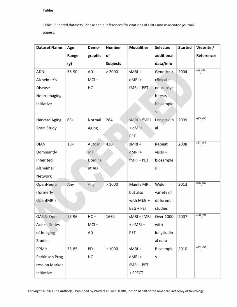