original article paclitaxel exerts anticancer bioactivity on … · 2016-08-09 · tometry,...
TRANSCRIPT

Int J Clin Exp Pathol 2016;9(2):651-661www.ijcep.com /ISSN:1936-2625/IJCEP0015702
Original ArticlePaclitaxel exerts anticancer bioactivity on SGC7901 human gastric adenocarcinoma cell line through inhibition of Akt/mTOR signaling pathway
Dong Sun1*, Ke-Shu Shan1*, Jin-Shen Wang1, Yue-Zhi Chen1, Liang Shang1, Yarmus Khan2, Bang-Zhen Ma2, Le-Ping Li1
Departments of 1Gastrointestinal Surgery, 2Hepatobiliary Surgery, Shandong Provincial Hospital Affiliated to Shan-dong University, Jinan 250021, People’s Republic of China. *Equal contributors.
Received September 6, 2015; Accepted December 9, 2015; Epub February 1, 2016; Published February 15, 2016
Abstract: Gastric cancer is one of the most prevalent malignancies and the leading cause of cancer mortality. Pa-clitaxel, a traditional medicine, is originally extracted from pacific yew tree. It has been reported that paclitaxel can treat multiple human malignancies including gastric cancer. However, the underlying mechanism has not been fully understood. In our study, SGC-7901 human gastric adenocarcinoma cell line was cultured and treated with pacli-taxel. Cell Counting Kit-8 (CCK-8) and colony formation assay were performed to examine cell viability. Cell migration and metastasis ability were measured by cell invasion assay. Moreover, cell cycle was determined by the Flow Cy-tometry, Hoechst33342/PI (propidium iodide) double staining and Flow Cytometry were both employed to test cell apoptosis. To further explore the underlying mechanisms of paclitaxel in gastric cancer, western blotting was per-formed to detect the protein expression of phospho-Akt (p-Akt) and phospho-mTOR (p-mTOR). Our results showed that paclitaxel obviously reduced SGC-7901 cell viability and cellular colony formation ratio compared with the control (P<0.01). Meanwhile, paclitaxel effectively inhibited cell migration and metastasis, the number of invaded cells reduced significantly (P<0.01). Flow Cytometry and Hoechst/PI double staining results also showed paclitaxel induced SGC-7901 cell apoptosis (P<0.01) and let cell cycle arrest at G2/M phase. Furthermore, paclitaxel strongly decreased the expression of p-Akt and p-mTOR proteins. Collectively, our study indicated that paclitaxel exerts an obvious anticancer bioactivity on SGC-7901 human gastric adenocarcinoma cell line, which is possibly, mediated through inhibition of Akt/mTOR signaling pathway.
Keywords: Paclitaxel, gastric cancer, SGC-7901, Akt/mTOR signaling pathway
Introduction
Gastric cancer, also known as stomach cancer, is the fourth most common malignancies and the second cause of cancer related death wo- rldwide [1, 2]. Among many kinds of malignan-cies, gastric cancer is the leading cause of can-cer mortality in China [3]. The majority of pa- tients diagnosed with gastric cancer at ad- vanced stage have distance organ metastasis and only 15% of them can survive beyond six months due to its resistance to radiotherapy and chemotherapy. Even diagnosed in early stage of gastric cancer, only 60% patients can survive more than 5 years [4, 5]. Although the most effective therapy method is resection operation, chemotherapy is indispensable for gastric cancer treatment, especially for advan- ced stage.
Paclitaxel, a traditional medicine, was first ex- tracted from Pacific yew tree. In 1971, research-ers obtained the pure paclitaxel and verified its anticancer activity [6, 7]. Since then, paclitaxel has been proven to treat multiple human malig-nancies and has already been used as an che-motherapy in breast cancer, ovarian cancer, non-small cell lung cancer (NSCLC), pancreatic cancer and other malignancies [8-11]. Espe- cially, paclitaxel is an effective chemotherapeu-tic drug for gastric cancer [12]. In Japan, pa- tients received weekly paclitaxel treatment has already regarded as the perfect second-line chemotherapy in unresectable gastric cancer cases [13]. And, patients adapted periodical chemotherapy with paclitaxel after resection surgery were observed with low recurrence ra- te, metastasis rate and long survival time [14].

Paclitaxel exerts anticancer effects on SGC-7901 cell
652 Int J Clin Exp Pathol 2016;9(2):651-661
However, the underlying mechanism has not been fully understood.
Although many studies have reported the role of paclitaxel on gastric cancer, most of them focused on the effects of paclitaxel on cell gr- owth, apoptosis, cytotoxicity and anti-angiogen-esis, etc [15]. Investigators have already found that PI3K (phosphoinositide-3-kinase) pathway plays a central role in cell proliferation, apop- tosis, energy metabolism, autophagy and oth- er cell functions. Moreover, PI3K pathway was reported aberrantly activated or over-expressed in gastric cancer [16]. Akt and mTOR belong to the classical PI3K/Akt signal pathway. Recently, Gou WF and his colleagues demonstrated that over-expression of ING5 in gastric cancer cells can induce cell autophagy, differentiation and inhibit proliferation and apoptosis, which is rel-ative to p-Akt [17]. A recent study also suggest-ed that inhibition of PI3K/HIF-1α pathway can enhance the therapeutic effect of paclitaxel in human hypoxic gastric cancer cells [18]. Gene- tic variants in the PI3K pathway may predict platinum-based chemotherapy response in lu- ng cancer cells [19]. Thus, we hypothesize that paclitaxel can exert anticancer bio-activity th- rough regulating Akt/mTOR signaling pathway, however, little is known about it in SGC-7901 human gastric adenocarcinoma cells.
To test our hypothesis, in this study, SGC-7901 human gastric adenocarcinoma cells were cul-tured and treated with paclitaxel. The cell viabil-ity, invasion, apoptosis and cell cycle were ex- amined in cultured cells. Furthermore, western blotting was employed to determine the expres-sion of Akt/mTOR signal pathway. Our study will be beneficial for better understanding of pacli-taxel’s anticancer bioactivity and provide pos-sibly targets for treating gastric cancer.
Materials and methods
Cell culture
The SGC-7901 cell was incubated in RPMI1640 medium (Hyclone, Logan, UT) supplemented with 10% fetal bovine serum (FBS) (Hyclone, Logan, UT) and 1% penicillin (100 U/ml) and streptomycin (100 μg/ml) at 37°C in 5% CO2 incubator. The paclitaxel powder (Yuanye Co- mpany, Shanghai, China) was dissolved with dimethylsulphoxide (DMSO) at a concentration of 20 mg/ml, and then diluted to the desired concentration by RPMI1640 medium before using. The highest concentration of DMSO in
culture medium was less than 0.1%, to make sure it did not affect cellular viability [20].
Cell proliferation analysis
Cell Counting Kit-8 (CCK-8) (Dojindo, Japan) was employed to detect cell viability according to a previous report [21]. Cells with the concen-tration of 3×10³ were seeded into the 96-well plate and incubated with normal medium or medium contains 0.25 μg/ml and 0.5 μg/ml paclitaxel. Cell viability was examined at 24 h, 48 h, 72 h and 96 h in vitro culture, respec-tively. 10 μl CCK-8 was added to each well and incubated for additional 1 h at 37°C. The cell absorbance was measured by microplate read-er (Molecular Devices, America) at 450 nm wavelength.
Colony formation analysis
SGC-7901 cells (about 200 cells) treated with or without paclitaxel were suspended in 2 ml RPMI1640 medium contains 10% FBS and then cultured at cell incubator for two weeks. After washed with Phosphate Buffered Saline (PBS), the cells were fixed by 4% formaldehyde for 15 min and stained with hematoxylin for 10 min at room temperature. The cell colony num-ber was obtained in five random sights of diff- erent groups under an inverted phase contrast microscope (Olympus, Japan). The colony for-mation rate was calculated by following formu-la: colony formation rate = (the average number of colony)/200×100%.
In vitro cell invasion assay
The cell migration and metastasis ability were determined by the non-coated transwell mem-brane (8.0 μm pore size, Corning, America) and the membrane pro-coated with Matrigel diluted by serum-free 1640 medium according to a previous report [22]. Briefly, the cultured cells were suspended in serum-free medium and planted on the upper chamber. Medium con-tains 10% FBS was added to the lower chamber as a chemo-attract. After incubated for 24 h in vitro, cells can’t invade the chamber were re- moved by cotton swabs. Subsequently, the in- sert membrane was fixed with 4% formalde-hyde for 15 min and stained with hematoxylin for 10 min at room temperature. The invaded cells were counted in five random fields under the inverted phase contrast microscope.
Paclitaxel exerts anticancer effects on SGC-7901 cell
653 Int J Clin Exp Pathol 2016;9(2):651-661
Hoechst33342/PI (propidium iodide) double staining
Hoechst33342/PI double staining was applied to test cell apoptosis treated with paclitaxel according to manufacture protocols. After cul-tured in 24-well plate, cells were washed twice with PBS, and incubated with 10 μg/ml Hoe- chst33342 and PI for 15 min at cell incubator. Then, the cells were rinsed and observed under an inverted phase-contrast microscope (Nikon, Japan). Five random fields were selected to count the mean number of dead and surviving cells. The death rate was calculated by the fol-lowing formula: Death rate = (mean number of PI positive stain cells)/Hoechst positive stain cell in the same field.
Cell apoptosis analysis
Flow Cytometry was employed to further detect cell apoptosis. SGC-7901 cells treated with or
without paclitaxel were collected after digested with 0.25% trypsin. Cells were re-suspended by pre-cold PBS for three times and then stained with Annexin-V-FITC and PE-7AAD for 15 min at room temperature in the dark according to the manufacture protocol of Becton Company. Cell apoptosis was analyzed by the Flow Cytometry (Becton Dickinson, Franklin Lakes, NJ, America).
Cell cycle analysis
Cells of different groups were digested with 0.25% trypsin and washed three times with cold PBS, then the cells were fixed with pre-cold 70% ethanol for 24 h at 4°C. The next day, 500 μl PI was added to each tube and then incu-bated at 37°C in dark for additional 30 min according to the manufacturer’s guidelines (Le- agent Company, Beijing, China). The cell cycle was analyzed by the Flow Cytometry according to the fluorescence intensity.
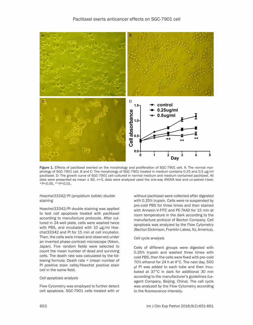
Figure 1. Effects of paclitaxel exerted on the morphology and proliferation of SGC-7901 cell. A: The normal mor-phology of SGC-7901 cell. B and C: The morphology of SGC-7901 treated in medium contains 0.25 and 0.5 μg/ml paclitaxel. D: The growth curve of SGC-7901 cell cultured in normal medium and medium contained paclitaxel. All data were presented as mean ± SD, n=3, data were analyzed used the one-way ANOVA test and un-paired t-test; *P<0.05, **P<0.01.

Paclitaxel exerts anticancer effects on SGC-7901 cell
654 Int J Clin Exp Pathol 2016;9(2):651-661
Western blot
Cell proteins were extracted using RIPA lysis buffer contained 1% PMSF (Excell, Shanghai, China) according to the manufacture instruc-tions, protein concentration was quantified us- ed BCA kit (Excell, Shanghai, China). Equal am- ount of proteins (40 μg per lane) were separat-ed on 6% SDS-PAGE gels. Then the isolation protein bands were transferred to the PVDF membrane and blocked with 5% non-fat milk for 1 h, after washed by 1×TBST for three times, all membranes were incubated overnight at 4°C with the following primary antibodies res- pectively: β-actin (Excell, Shanghai, China), p- Akt and Akt (CST, America), p-mTOR and mTOR
(Abcam, England). In the following day, the cor-responding secondary antibodies were added to specific bind the primary antibodies for 1 h at room temperature. Finally, the Amersham Ima- ger 600 imaging system (GE Company, America) was employed to visualize and analyze the pro-tein band.
Statistical analysis
The data were analyzed used the SPSS 17.0 software. All data were reported as the mean ± standard deviation. The differences between control and experiment groups were analyzed with one-way ANOVA test, differences between groups were analyzed with t-test. P<0.05 were considered as statistically significantly.
Figure 2. Effects of paclitaxel exerted on cell colony formation. Data shown are means ± SD, n=3, data were ana-lyzed by the one-way ANOVA test and un-paired t-test; *P<0.05, **P<0.01.

Paclitaxel exerts anticancer effects on SGC-7901 cell
655 Int J Clin Exp Pathol 2016;9(2):651-661

Paclitaxel exerts anticancer effects on SGC-7901 cell
656 Int J Clin Exp Pathol 2016;9(2):651-661
Results
Paclitaxel inhibits cell proliferation and viability
SGC-7901 is an adenocarcinoma cell line de- rived from human gastric cancer. After cultured 24 h in normal medium, SGC-7901 cell can be found grow fast with less dead cells (Figure 1A). However, when added 0.25 μg/ml pacli-taxel in medium for 24 h in vitro culture, cells became round shape while membrane swelled (Figure 1B). When cells were incubated in 0.5 μg/ml paclitaxel, dead cells could be easily found in the medium (Figure 1C). CCK-8 was performed to further test cellular viability and the absorbance data showed that the optical density of cells decreased significantly after treated with paclitaxel, among them, the 0.5 μg/ml paclitaxel exerted more obvious inhi- bitory effect (P<0.01) (Figure 1D). It indicates that paclitaxel can inhibit SGC-7901 growth and viability in a dose-dependent manner.
Paclitaxel decrease the colony formation of SGC-7901
Cell colony formation assay was determined to assess cellular viability exerted by the pacli-taxel. The colony number of the control group, 0.25 and 0.5 μg/ml paclitaxel groups were 109.0 ± 12.7, 56.7 ± 7.1 and 34.0 ± 8.9, res- pectively (Figure 2A-C). Further analysis indi-cated paclitaxel could significantly inhibit the colony formation of SGC-7901 cell (P<0.01) and 0.5 μg/ml paclitaxel showed more obvious inhibitory effects than the 0.25 μg/ml group (P<0.05) (Figure 2D), means that paclitaxel could inhibit the colony formation of SGC-7901 cell in a dose-dependent manner.
Paclitaxel inhibit the migration and metastasis ability of SGC-7901 cell
To investigate the influence of paclitaxel exert-ed on cellular invasion ability, we detected cel-lular migration and metastasis ability using Tr- answell assay. For migration assay, the number of cells invaded the membrane was 195.0 ± 20.0, 119.2 ± 17.6, 82.2 ± 24.5 respectively for control, 0.25 μg/ml and 0.5 μg/ml paclitax-el group (Figure 3A-C); similarly, in the metasta-
sis test, the invaded cell number was 255.6 ± 40.8, 131.2 ± 24.6, 52.2 ± 21.7 for control and experimental groups (Figure 3E-G). The statisti-cal analysis results showed paclitaxel could sig-nificantly decrease the invaded cell number both in migration and metastasis (P<0.01). Mo- reover, data also indicate high paclitaxel con-centration can inhibit cell invasion ability more significantly than lower concentration (Figure 3D and 3H). The results demonstrated pacli-taxel can restrain the invasion ability of SGC-7901 cell.
Paclitaxel induce SGC-7901 cell apoptosis
Both Hoechst33342/PI double staining and the flow cytometry were employed to test cell apoptosis. The results of double staining sh- owed after treated with paclitaxel, the dead cells (red fluorescence) was obvious increased compared to the normal SGC-7901 cell (Figure 4E), consistent with this, the flow cytometry results showed the apoptosis rate of normal SGC-7901 cell group was 7.1 ± 1.1% (Figure 4A). However, when treated with 0.25 μg/ml and 0.5 μg/ml paclitaxel, the rate raised to 12.0 ± 1.6% and 15.6 ± 1.2%, almost twice than the control group with a P<0.01 (Figure 4B, 4C). Similarly to our previous results, pacli-taxel also showed dose-effects on cell apo- ptosis.
Paclitaxel arrest SGC-7901 cell cycle
As shown in Figure 5, the flow cytometry results showed after treated with paclitaxel, the G2/M phase ratio of SGC-7901 was raised. The ratio was 10.81 ± 1.23% in control group (Figure 5A), and raised to 14.57 ± 1.78% and 15.54 ± 2.17% when treated with 0.25 μg/ml and 0.5 μg/ml paclitaxel (Figure 5B, 5C). The results showed paclitaxel can arrest cell cycle at G2/M phase (P<0.05) (Figure 5D), but different from our previous results, paclitaxel did not show obvious dose-effect on cell cycle arrest in this experiment (P>0.05).
Paclitaxel affect the Akt/mTOR signaling pathway
As shown in Figure 6, paclitaxel can regulate the Akt/mTOR signaling pathway. After treated
Figure 3. Effects of paclitaxel exerted on the invasion ability of SGC-7901 cell. A-C: The inhibition effects of paclitaxel exerted on the migration ability of SGC-790 cell. E-G: The inhibition effects of paclitaxel exerted on the metastasis ability of SGC-7901 cell. All data were presented as mean ± SD, n=5, data were analyzed used the one-way ANOVA test and un-paired t-test; *P<0.05, **P<0.01.

Paclitaxel exerts anticancer effects on SGC-7901 cell
657 Int J Clin Exp Pathol 2016;9(2):651-661
with paclitaxel, the expression level of p-Akt and p-mTOR were both down-regulated, their expression level showed a declined trend with the increase concentration of paclitaxel, the expression level was much lower in high con-centration group. While the p-Akt and p-mTOR were down-regulated, however, the expression of Akt and mTOR maintain at a constant line (Figure 6A, 6B), confirmed that it was their
phosphorylated form that involved in the pacli-taxel anti-cancer mechanism.
Discussion
In this study, we employed the gastric adeno-carcinoma cell line SGC-7901 to investigate its reaction to paclitaxel. Through the experiment, we found that paclitaxel can attenuate cell via-
Figure 4. Effects of paclitaxel exerted on SGC-7901 apoptosis. A-C: The flow cytometry results of cell apoptosis induced by paclitaxel. E: The results of Hoechst33342/PI double staining. All data were presented as mean ± SD, n=3, data were analyzed by the one-way ANOVA test and un-paired t-test; *P<0.05, **P<0.01.

Paclitaxel exerts anticancer effects on SGC-7901 cell
658 Int J Clin Exp Pathol 2016;9(2):651-661
bility, inhibit cell colony formation, arrest cell cycle at G2/M phase and induce cell apoptosis. Especially, we tested the possible molecular mechanisms, and found that paclitaxel can sig-nificantly inhibit the expression of Akt/mTOR signaling pathway. This experiment revealed the possible mechanism of paclitaxel in the treatment of gastric cancer.
Paclitaxel is a very effective and widely used natural chemotherapeutic drug in clinical. It was first permitted to cure advanced uterine cancer by the American FDA (Food and Drug Administration) in 1992, then it was used as a chemotherapeutic in clinical. Since large por-tion of gastric cancer are adenocarcinoma, through consulting literatures, we found little was done to explore the effects of paclitaxel
exerts on the SGC-7901 cell, so we choose SGC-7901 cell to do the test. Through the ex- periment, we verified that paclitaxel can inhibit cell viability, attenuate cell invasion ability and induce cell apoptosis in a dose dependent manner. However we did not observe the same effect in cell cycle arrest, results showed pacli-taxel can arrest cell cycle at G2/M phase with-out obvious dose effect (P>0.05), it was differ-ent from previous papers, the reason need to be further explored.
A growing body of report has shown that pacli-taxel can exert its anti-tumor function through various signaling pathways. Previous research-es have confirmed paclitaxel can induce cell apoptosis by regulating the BCL-2, BCL-x, p-53, and bax bypass pathways [23]. Akt/mTOR sig-
Figure 5. Effects of paclitaxel exerted on the cell cycle of SGC-7901 cell. Data shown are mean ± SD, n=3, data were analyzed used the one-way ANOVA test and un-paired t-test; *P<0.05, **P<0.01.

Paclitaxel exerts anticancer effects on SGC-7901 cell
659 Int J Clin Exp Pathol 2016;9(2):651-661
naling pathway is an important part of the PI3K pathway, lots of papers have confirmed the Akt/mTOR signaling pathway is closely related to many malignancies such as sarcomas, gas-tric and colorectal cancer [24-26]. So we decide to investigate its role in gastric cancer treated with paclitaxel. In our study, we found that paclitaxel can significantly suppress the Akt/mTOR signaling pathway and can decrease the expression of p-Akt and p-mTOR protein. In pre-vious study, it was also reported that paclitaxel can suppress this signal pathway, Boh-Ram Kim’s results showed that paclitaxel can exert anti-tumor function in ovarian cancer through inhibiting the expression of p-mTOR in the mTORC1-S6K/4EBP signaling pathway, thereby lowering the HIF-1-α and the VEGF level, which is important for tumorigenesis and metastasis [27]. Another report also confirmed that pacli-taxel can induce NSCLC cell apoptosis by sup-pressing the expression of Akt protein in lung cancer [28].
Gastric cancer, as the fourth most common and second cause of cancer mortality world-wide, has caused great danger to people’s health. Due to its resistance to chemotherapy and radiotherapy, surgery is still the most effec-tive therapeutic option in present, unfortunate-ly, many patients were diagnosed at an ad- vanced stage, some of them even lost the sur-gery opportunity, for them, there is still no good method. But, it is a feasible treatment strategy if we can specially induce cell death by target drugs. Researchers have reported the NVP-BEZ235 (an inhibitor of PI3K/mTOR signaling pathway) can down-regulate the expression of p-mTOR to enhance the anti-tumor effect of paclitaxel in gastric cancer [29], similarly, in NSCLC, the inhibitor of CK2 (casein kinase2), CX-4945, can also induced lung cancer cell apoptosis by lowering the expression of p-mTOR [30]. These evidences suggested that it is a new field that need to be further explored to treat gastric cancer.
Figure 6. The influence of paclitaxel exerted on the Akt/mTOR signal pathway. A: The expression level of Akt and p-Akt in SGC-7901 cells treated with or without paclitaxel; B: The expression level of mTOR and p-mTOR in normal and paclitaxel groups. All data were presented as mean ± SD, n=3, data were analyzed by the one-way ANOVA test and un-paired t-test; #P>0.05, *P<0.05, **P<0.01.

Paclitaxel exerts anticancer effects on SGC-7901 cell
660 Int J Clin Exp Pathol 2016;9(2):651-661
Our experiments confirmed that paclitaxel can indeed exert anti-tumor effects on SGC-7901 human gastric cancer cell line and the possib- le mechanism maybe through suppressing the Akt/mTOR signaling pathway. Gastric cancer is a common malignancy in china, its treatment is of great importance for patients, paclitaxel has already been used as an chemotherapeutics in many malignancies including gastric cancer, detailed explore its mechanism in gastric can-cer is vital for its further application in gastric cancer and can provide more effective guid-ance for the clinical treatment of gastric ca- ncer.
Disclosure of conflict of interest
None.
Address correspondence to: Dr. Leping Li, Depart- ment of Gastrointestinal Surgery, Shandong Provin- cial Hospital Affiliated to Shandong University, Jinan 250021, People’s Republic of China. Tel: +86 0531-68776388; E-mail: [email protected]
References
[1] Siegel RL, Miller KD and Jemal A. Cancer sta-tistics, 2015. CA Cancer J Clin 2015; 65: 5-29.
[2] Jemal A, Center MM, DeSantis C and Ward EM. Global patterns of cancer incidence and mor-tality rates and trends. Cancer Epidemiol Bio-markers Prev 2010; 19: 1893-1907.
[3] Yu HG, Ai YW, Yu LL, Zhou XD, Liu J, Li JH, Xu XM, Liu S, Chen J, Liu F, Qi YL, Deng Q, Cao J, Liu SQ, Luo HS and Yu JP. Phosphoinositide 3-kinase/Akt pathway plays an important ro- le in chemoresistance of gastric cancer cells against etoposide and doxorubicin induced cell death. Int J Cancer 2008; 122: 433-443.
[4] Qiu H, Yashiro M, Shinto O, Matsuzaki T and Hirakawa K. DNA methyltransferase inhibitor 5-aza-CdR enhances the radiosensitivity of gastric cancer cells. Cancer Sci 2009; 100: 181-188.
[5] Liu T, Tang H, Lang Y, Liu M and Li X. MicroRNA-27a functions as an oncogene in gastric ade-nocarcinoma by targeting prohibitin. Cancer Lett 2009; 273: 233-242.
[6] Hossein G, Keshavarz M, Ahmadi S and Naderi N. Synergistic effects of PectaSol-C modified citrus pectin an inhibitor of Galectin-3 and pa-clitaxel on apoptosis of human SKOV-3 ovarian cancer cells. Asian Pac J Cancer Prev 2013; 14: 7561-7568.
[7] Wani MC, Taylor HL, Wall ME, Coggon P and McPhail AT. Plant antitumor agents. VI. The iso-lation and structure of taxol, a novel antileuke-
mic and antitumor agent from Taxus brevifolia. J Am Chem Soc 1971; 93: 2325-2327.
[8] Volk LD, Flister MJ, Chihade D, Desai N, Trieu V and Ran S. Synergy of Nab-paclitaxel and Bev-acizumab in Eradicating Large Orthotopic Bre- ast Tumors and Preexisting Metastases. Neo-plasia 2011; 13: 327-IN314.
[9] Coleman RL, Brady WE, McMeekin DS, Rose PG, Soper JT, Lentz SS, Hoffman JS and Shahin MS. A phase II evaluation of nanoparticle, al-bumin-bound (nab) paclitaxel in the treatment of recurrent or persistent platinum-resistant ovarian, fallopian tube, or primary peritoneal cancer: a Gynecologic Oncology Group study. Gynecol Oncol 2011; 122: 111-115.
[10] Socinski MA, Bondarenko I, Karaseva NA, Ma- khson AM, Vynnychenko I, Okamoto I, Hon JK, Hirsh V, Bhar P, Zhang H, Iglesias JL and Ren-schler MF. Weekly nab-paclitaxel in combina-tion with carboplatin versus solvent-based pa-clitaxel plus carboplatin as first-line therapy in patients with advanced non-small-cell lung cancer: final results of a phase III trial. J Clin Oncol 2012; 30: 2055-2062.
[11] Von Hoff DD, Ramanathan RK, Borad MJ, La-heru DA, Smith LS, Wood TE, Korn RL, Desai N, Trieu V, Iglesias JL, Zhang H, Soon-Shiong P, Shi T, Rajeshkumar NV, Maitra A and Hidalgo M. Gemcitabine plus nab-paclitaxel is an ac-tive regimen in patients with advanced pancre-atic cancer: a phase I/II trial. J Clin Oncol 2011; 29: 4548-4554.
[12] Vallon M, Seidl C, Blechert B, Li Z, Gilbertz KP, Baumgart A, Aichler M, Feuchtinger A, Gaert-ner FC, Bruchertseifer F, Morgenstern A, Walch AK, Senekowitsch-Schmidtke R and Essler M. Enhanced efficacy of combined 213Bi-DTPA-F3 and paclitaxel therapy of peritoneal carcino-matosis is mediated by enhanced induction of apoptosis and G2/M phase arrest. Eur J Nucl Med Mol Imaging 2012; 39: 1886-1897.
[13] Fushida S, Kaji M, Oyama K, Hirono Y, Nezuka H, Takeda T, Tsukada T, Fujimoto D, Ohyama S, Fujimura T and Ohta T. Randomized Phase II trial of paclitaxel plus valproic acid vs paclitax-el alone as second-line therapy for patients with advanced gastric cancer. Onco Targets Ther 2015; 8: 939-941.
[14] Egawa T, Kemmochi T, Nishiya S, Mihara K, Ito Y and Nagashima A. [Nanoparticle albumin-bound Paclitaxel for unresectable or recurrent gastric cancer]. Gan To Kagaku Ryoho 2014; 41: 2251-2253.
[15] Wang T, Gao J, Yu J and Shen L. Synergistic in-hibitory effect of wogonin and low-dose pacli-taxel on gastric cancer cells and tumor xeno-grafts. Chin J Cancer Res 2013; 25: 505-513.
[16] Ang KL, Shi DL, Keong WW and Epstein RJ. Up-regulated Akt signaling adjacent to gastric can-

Paclitaxel exerts anticancer effects on SGC-7901 cell
661 Int J Clin Exp Pathol 2016;9(2):651-661
cers: implications for screening and chemo-prevention. Cancer Lett 2005; 225: 53-59.
[17] Gou WF, Shen DF, Yang XF, Zhao S, Liu YP, Sun HZ, Su RJ, Luo JS and Zheng HC. ING5 sup-presses proliferation, apoptosis, migration and invasion, and induces autophagy and differen-tiation of gastric cancer cells: a good marker for carcinogenesis and subsequent progres-sion. Oncotarget 2015; 6: 19552-19579.
[18] Zhang J, Guo H, Zhu JS, Yang YC, Chen WX and Chen NW. Inhibition of phosphoinositide 3-ki-nase/Akt pathway decreases hypoxia induc-ible factor-1alpha expression and increases therapeutic efficacy of paclitaxel in human hy-poxic gastric cancer cells. Oncol Lett 2014; 7: 1401-1408.
[19] Xu JL, Wang ZW, Hu LM, Yin ZQ, Huang MD, Hu ZB, Shen HB and Shu YQ. Genetic Variants in the PI3K/PTEN/AKT/mTOR Pathway Predict Platinum-based Chemotherapy Response of Advanced Non-small Cell Lung Cancers in a Chinese Population. Asian Pacific Journal of Cancer Prevention 2012; 13: 2157-2162.
[20] Xie XH, Zang N, Li SM, Wang LJ, Deng Y, He Y, Yang XQ and Liu EM. Resveratrol Inhibits respi-ratory syncytial virus-induced IL-6 production, decreases viral replication, and downregulates TRIF expression in airway epithelial cells. In-flammation 2012; 35: 1392-1401.
[21] Wang LL, Hu RC, Dai AG and Tan SX. Bevaci-zumab induces A549 cell apoptosis through the mechanism of endoplasmic reticulum stress in vitro. Int J Clin Exp Pathol 2015; 8: 5291-5299.
[22] Liang C, Yang L and Guo S. All-retinoic acid inhibits migration, invasion and proliferation, and promotes apoptosis in glioma cells. Oncol Lett 2015; 9: 2833-2838.
[23] Oltvai ZN, Milliman CL and Korsmeyer SJ. Bcl-2 heterodimerizes in vivo with a conserved ho-molog, Bax, that accelerates programmed cell death. Cell 1993; 74: 609-619.
[24] Takahashi Y, Kohashi K, Yamada Y, Endo M, Setsu N, Ishii T, Yamamoto H, Iwamoto Y and Oda Y. Activation of the Akt/mammalian target of rapamycin pathway in myxofibrosarcomas. Hum Pathol 2014; 45: 984-993.
[25] Li JC, Zhu HY, Chen TX, Zou LY, Wang XY, Zhao HC and Xu J. Roles of mTOR and p-mTOR in Gastrointestinal Stromal Tumors. Asian Pacific Journal of Cancer Prevention 2013; 14: 5925-5928.
[26] Wang XW and Zhang YJ. Targeting mTOR net-work in colorectal cancer therapy. World J Gas-troenterol 2014; 20: 4178-4188.
[27] Kim BR, Yoon K, Byun HJ, Seo SH, Lee SH and Rho SB. The anti-tumor activator sMEK1 and paclitaxel additively decrease expression of HIF-1alpha and VEGF via mTORC1-S6K/4E- BP-dependent signaling pathways. Oncotarget 2014; 5: 6540-51.
[28] Nguyen DM, Chen GA, Reddy R, Tsai W, Sch- rump WD, Cole G Jr and Schrump DS. Potentia-tion of paclitaxel cytotoxicity in lung and esoph-ageal cancer cells by pharmacologic inhibition of the phosphoinositide 3-kinase/protein ki-nase B (Akt)-mediated signaling pathway. J Thorac Cardiovasc Surg 2004; 127: 365-375.
[29] Zhang C, Awasthi N, Schwarz MA and Schwarz RE. The dual PI3K/mTOR inhibitor NVP-BEZ- 235 enhances nab-paclitaxel antitumor re-sponse in experimental gastric cancer. Int J Oncol 2013; 43: 1627-1635.
[30] So KS, Rho JK, Choi YJ, Kim SY, Choi CM, Chun YJ and Lee JC. AKT/mTOR down-regulation by CX-4945, a CK2 inhibitor, promotes apoptosis in chemorefractory non-small cell lung cancer cells. Anticancer Res 2015; 35: 1537-1542.