ovarian cancer screening mati zolti m.d. department of obstetrics & gynaecology sheba medical...
Post on 19-Dec-2015
223 views
TRANSCRIPT

Ovarian Cancer Screening
Mati Zolti M.D.
Department of Obstetrics & Gynaecology Sheba Medical Center
Tel Hashomer, Israel
Ovarian Cancer: Burden of suffering
• 4th leading cause of cancer death in women in the U.S. (after lung, breast and colon)
• Overall 5-year survival rate is 35%• The “silent killer”: asymptomatic in early stages• 75% diagnosed with advanced stage disease; 5-year
survival only 10-28%• Woman’s lifetime risk of dying from ovarian cancer is
1.1%

Ovarian Cancer in Israel
New Cases : 220
Death : 95
Median age : 51
(2XXX)
Ovarian Cancer
Symptoms of ovarian cancer :• asymptomatic• Lower abdominal pain/pressure • mass• Abdominal enlargement• Vaginal bleeding• Urinary/bowel symptoms
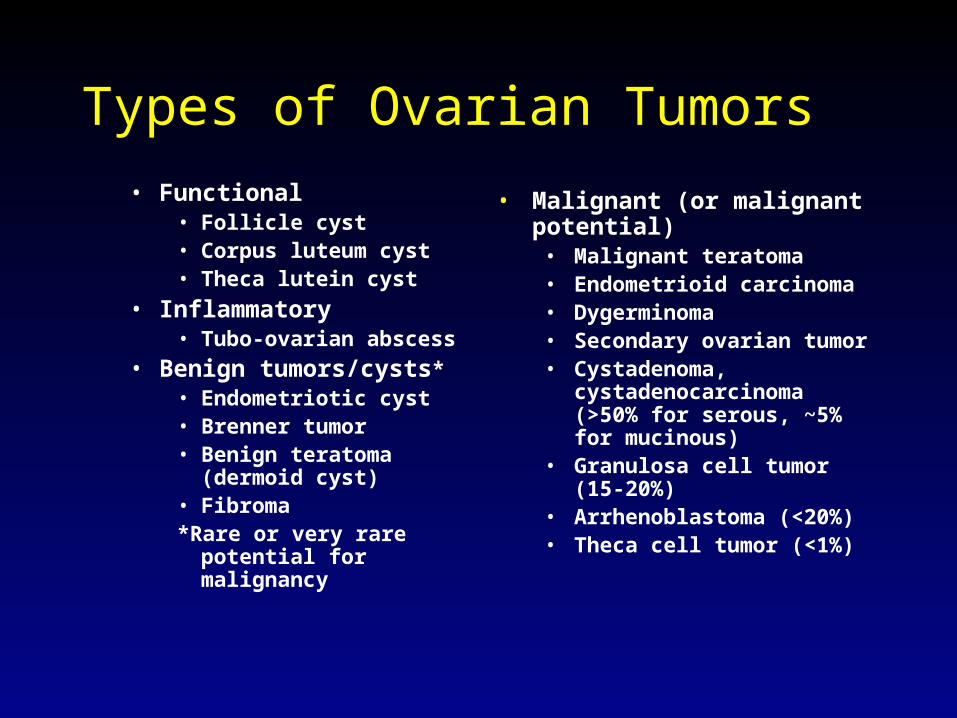
Types of Ovarian Tumors
• Functional• Follicle cyst• Corpus luteum cyst• Theca lutein cyst
• Inflammatory• Tubo-ovarian abscess
• Benign tumors/cysts*• Endometriotic cyst• Brenner tumor• Benign teratoma
(dermoid cyst)• Fibroma*Rare or very rare potential
for malignancy
• Malignant (or malignant potential)
• Malignant teratoma• Endometrioid carcinoma• Dygerminoma• Secondary ovarian tumor• Cystadenoma,
cystadenocarcinoma (>50% for serous, ~5% for mucinous)
• Granulosa cell tumor (15-20%)
• Arrhenoblastoma (<20%)• Theca cell tumor (<1%)
Epithelial Ovarian Cancer
• Overall 5-year survival rate is 75-95% if cancer confined to ovaries; decreases to 10-17% if distant metastases
• Survival improved when cancer detected in early stage
• Only 25% diagnosed in Stage I
Early Detection and Mortality
• No direct evidence that women with early stage cancer found on screening have lower mortality than women with more advanced disease
• Indirect evidence supports benefits of early detection:– Most important prognostic factor in patients with advanced
ovarian cancer is tumor burden after initial debulking– Surgical debulking and chemo more effective when cancer
detected early
The challenge
• Natural history of ovarian cancer not well understood– No well-defined precursor lesion– Length of time from localized tumor to
dissemination is unknown
• Multiple efforts underway to develop effective screening method for early detection
Risk factors
• The majority of women with ovarian cancer have no known risk factors
• Most significant risk factor is genetic predisposition
Risk factors: Heredity
• Up to 10% of epithelial ovarian cancer cases are familial
• 3 familial syndromes– familial breast-ovarian cancer syndrome– site-specific ovarian cancer– cancer family syndrome (Lynch type II)
• Familial breast-ovarian cancer and site-specific ovarian cancer syndromes both associated with mutations of the BRCA1 suppressor gene; account for 90% of familial ovarian cancers
Rollins,G. Ann Int Med 2000;133:1021-1024
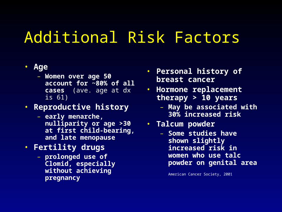
Additional Risk Factors
• Age– Women over age 50
account for ~80% of all cases (ave. age at dx is 61)
• Reproductive history– early menarche,
nulliparity or age >30 at first child-bearing, and late menopause
• Fertility drugs– prolonged use of Clomid,
especially without achieving pregnancy
• Personal history of breast cancer
• Hormone replacement therapy > 10 years– May be associated with
30% increased risk
• Talcum powder– Some studies have shown
slightly increased risk in women who use talc powder on genital area
American Cancer Society, 2001

Protective factors
• Multiparity: First pregnancy before age 30• Oral contraceptives: 5 years of use cuts risk
nearly in half• Tubal ligation• Hysterectomy• Lactation• Bilateral oopherectomy
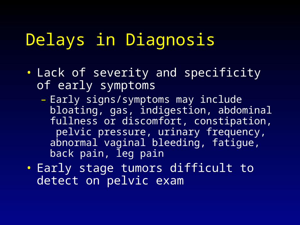
Delays in Diagnosis
• Lack of severity and specificity of early symptoms – Early signs/symptoms may include bloating, gas,
indigestion, abdominal fullness or discomfort, constipation, pelvic pressure, urinary frequency, abnormal vaginal bleeding, fatigue, back pain, leg pain
• Early stage tumors difficult to detect on pelvic exam
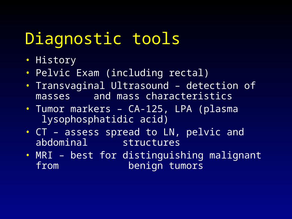
Diagnostic tools• History• Pelvic Exam (including rectal)• Transvaginal Ultrasound – detection of masses
and mass characteristics
• Tumor markers – CA-125, LPA (plasma lysophosphatidic acid)
• CT – assess spread to LN, pelvic and abdominal structures
• MRI – best for distinguishing malignant from benign tumors
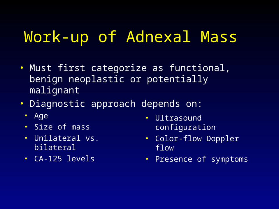
Work-up of Adnexal Mass
• Age• Size of mass• Unilateral vs. bilateral• CA-125 levels
• Ultrasound configuration• Color-flow Doppler flow• Presence of symptoms
• Must first categorize as functional, benign neoplastic or potentially malignant
• Diagnostic approach depends on:
Diagnostic approach
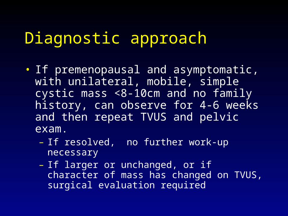
• If premenopausal and asymptomatic, with unilateral, mobile, simple cystic mass <8-10cm and no family history, can observe for 4-6 weeks and then repeat TVUS and pelvic exam.– If resolved, no further work-up necessary– If larger or unchanged, or if character of mass has
changed on TVUS, surgical evaluation required
Diagnostic Approach
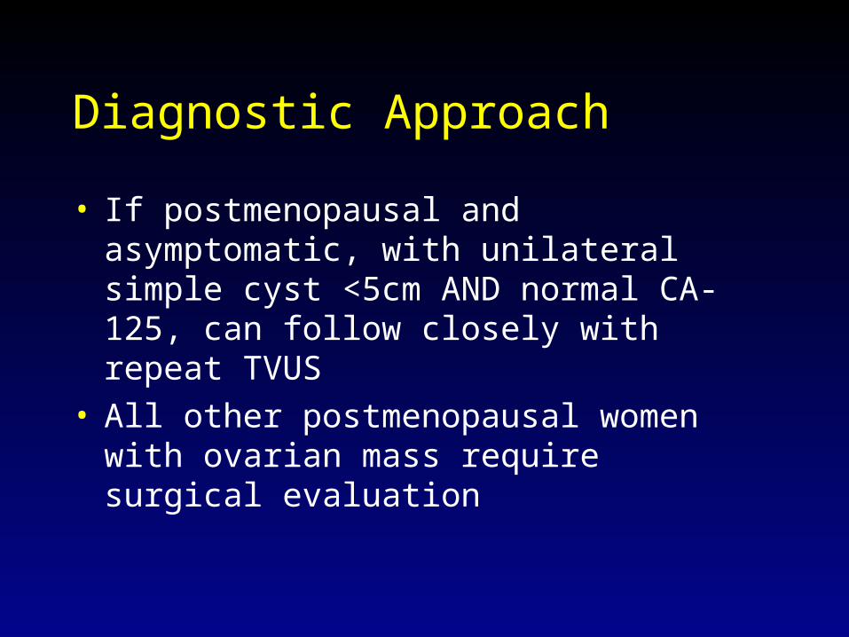
• If postmenopausal and asymptomatic, with unilateral simple cyst <5cm AND normal CA-125, can follow closely with repeat TVUS
• All other postmenopausal women with ovarian mass require surgical evaluation

Surgical Evaluation
• Refer to Gyn-Onc specialist• Exploratory laparotomy has been the gold
standard and includes:– Peritoneal washings for cytology– Evaluation of frozen section– Complete staging procedure if borderline or
malignant tumor on frozen section
Surgical Evaluation
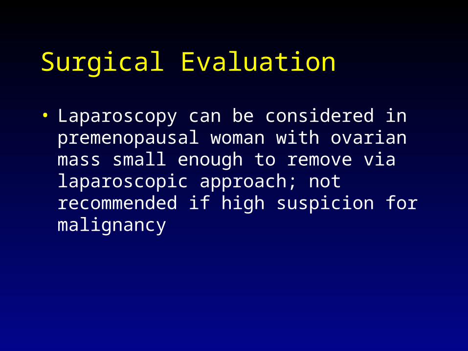
• Laparoscopy can be considered in premenopausal woman with ovarian mass small enough to remove via laparoscopic approach; not recommended if high suspicion for malignancy

Stages Ia, Ib, Ic

Stages IIa, IIb, IIc

Stages IIIa, IIIb, IIIc

Stage IV

Treatment
• Depends on staging, tumor type, age, desire for future fertility
• Can include surgery, chemotherapy and/or radiation therapy
• Clinical trials are ongoing

Surgical treatment
• Primary debulking and cytoreduction; may include:– Bilateral salpingo-oopherectomy– Hysterectomy– Lymphadenectomy (para-aortic, inguinal)– Omentectomy– “brushing” of diaphragm, examination of liver

Chemotherapy and Radiation
• Usually 6 cycles of chemotherapy• Cisplatin (or Carboplatin) plus Paclitaxel most
commonly used combination therapy• XRT
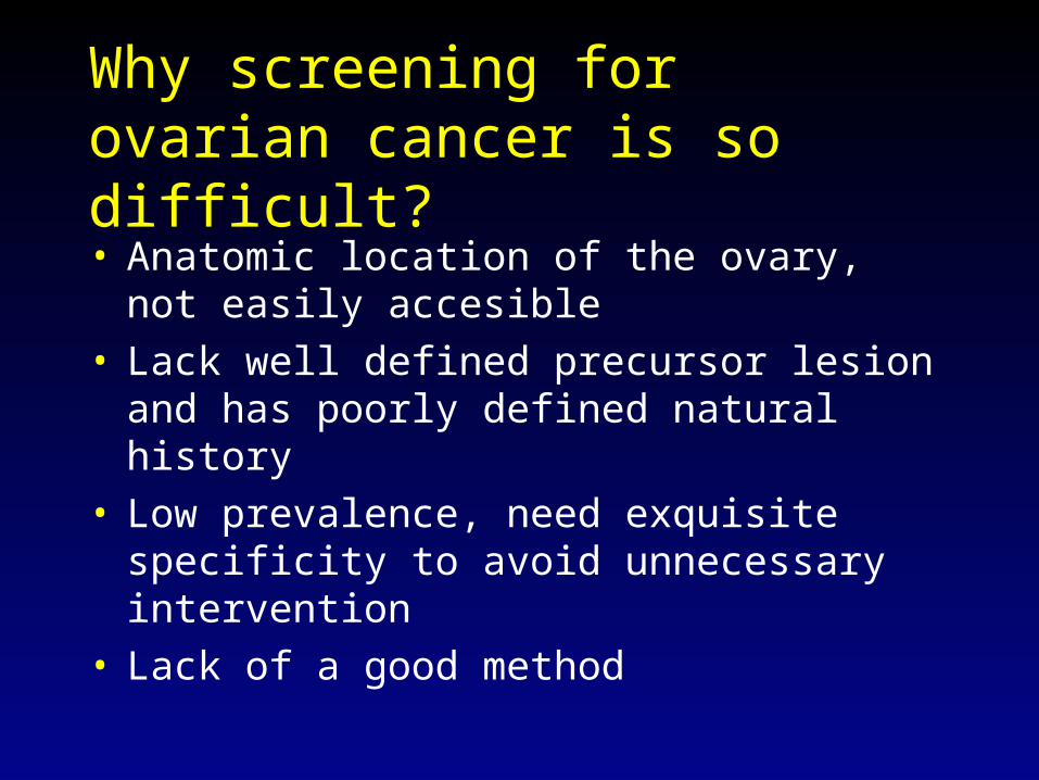
Why screening for ovarian cancer is so difficult?• Anatomic location of the ovary, not easily
accesible• Lack well defined precursor lesion and has
poorly defined natural history• Low prevalence, need exquisite specificity to
avoid unnecessary intervention• Lack of a good method
Screening Strategies
• Ultrasound (transvaginal vs transabdominal)• Color-flow doppler• CA-125• Other tumor markers
Ultrasound
• Both tranabdominal and transvaginal techniques identify enlarged ovaries or abnormal morphology; TVUS has better resolution
• One large study of TVUS underway has reported sensivity of 81% and specificity of 98.9%
• Major limitations are poor PPV in asymptomatic women and inability to detect malignances when ovaries are normal size
• Allows earlier stage detection
Color-flow Doppler
• Used in conjunction with TVUS• Measures resistance in blood vessels
supplying the ovaries• May provide additional information to help
distinguish malignant from benign masses

CA-125
• Sustained elevation in 82% of women with advanced ovarian cancer, but fewer than 1% of healthy women
• Poor sensitivity (elevated in only 50% of women with Stage I disease)
• Poor specificity (elevated in many gynecologic and non-gynecologic malignancies as well as benign conditions)
CA-125
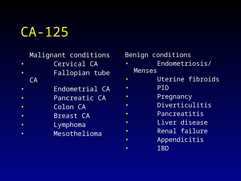
Malignant conditions• Cervical CA• Fallopian tube CA• Endometrial CA• Pancreatic CA• Colon CA• Breast CA• Lymphoma• Mesothelioma
Benign conditions• Endometriosis/
Menses• Uterine fibroids• PID• Pregnancy• Diverticulitis• Pancreatitis• Liver disease• Renal failure• Appendicitis• IBD
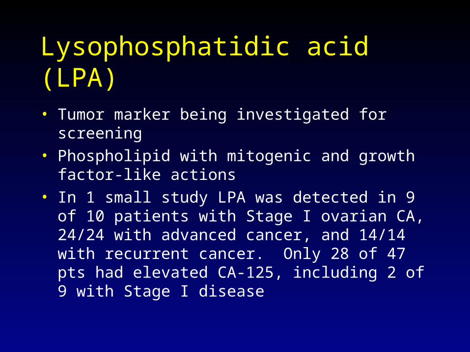
Lysophosphatidic acid (LPA)
• Tumor marker being investigated for screening• Phospholipid with mitogenic and growth factor-like
actions• In 1 small study LPA was detected in 9 of 10 patients
with Stage I ovarian CA, 24/24 with advanced cancer, and 14/14 with recurrent cancer. Only 28 of 47 pts had elevated CA-125, including 2 of 9 with Stage I disease
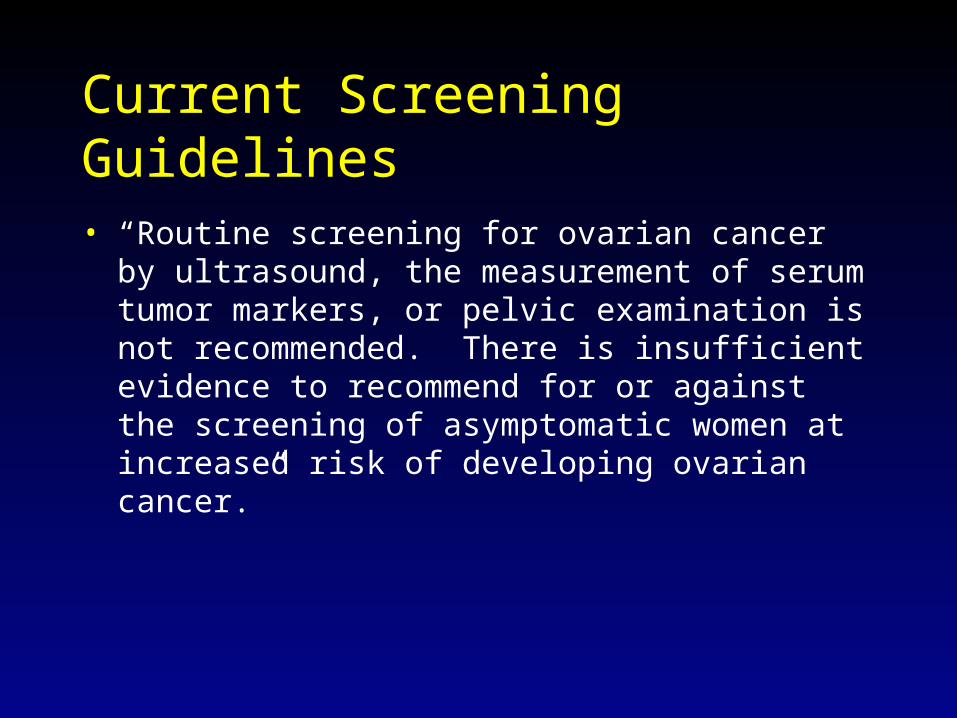
Current Screening Guidelines
• “Routine screening for ovarian cancer by ultrasound, the measurement of serum tumor markers, or pelvic examination is not recommended. There is insufficient evidence to recommend for or against the screening of asymptomatic women at increased risk of developing ovarian cancer.”

Screening Guidelines– cont’d
• NIH Consensus Conference (1994)– women with presumed hereditary cancer syndrome should
undergo annual pelvic exams, CA-125 measurements, and TVUS until childbearing is complete or at age 35, at which time prophylactic bilateral oopherectomy is recommended.
• ACP – counsel high risk women about potential harms and benefits
of screening
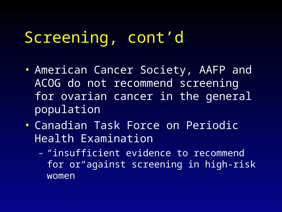
Screening, cont’d
• American Cancer Society, AAFP and ACOG do not recommend screening for ovarian cancer in the general population
• Canadian Task Force on Periodic Health Examination – “insufficient evidence to recommend for or against
screening in high-risk women”
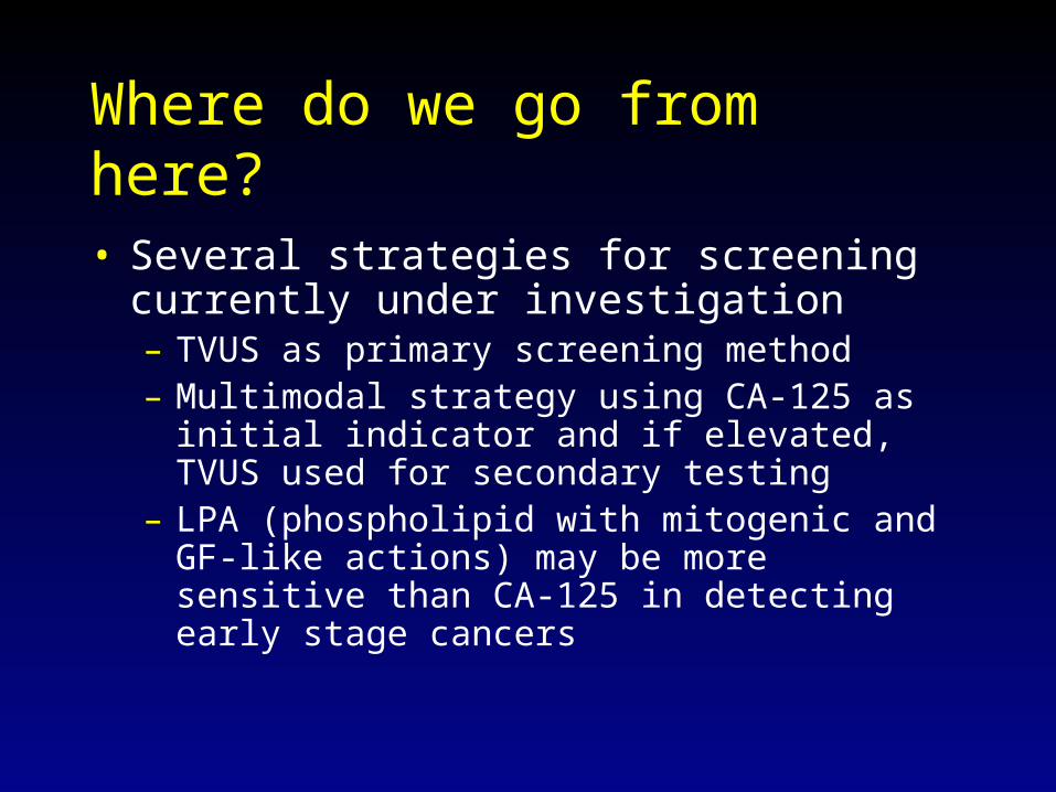
Where do we go from here?
• Several strategies for screening currently under investigation– TVUS as primary screening method– Multimodal strategy using CA-125 as initial
indicator and if elevated, TVUS used for secondary testing
– LPA (phospholipid with mitogenic and GF-like actions) may be more sensitive than CA-125 in detecting early stage cancers
Ovarian Cancer Screening Trials
1. The United Kingdom Collaborative Trial of Ovarian Cancer Screening: will compare TVUS and multimodal screening to control
2. The European Study: RCT to screen women with TVUS at 18-month or 3-year intervals
3. The NIH Prostate, Lung, Colorectal, and Ovarian Cancer Screening Trial: 10-year study using multimodal strategy
Take home points
• Screening not indicated at this time • ASK about family history of cancers• LISTEN when women present with non-
specific GI complaints; include OC in DDx• DO perform careful bimanual exam and rectal
exam as part of pelvic exam• Refer women with + Family Hx to GynOnc
Marker Updates - Ca 125
• Approved marker to test for recurrence• Remains the single most sensitive andspecific marker for ovarian cancer to date• Addition of other markers might improvesensitivity and specificity• Longitudinal assessment may also improvesensitivity and specificity

ROCA: Risk of Ovarian Cancer Algorithm
• In 2002, a paper in Lancet described an approach to ovarian cancer screening using mass spectrometry to create protein patterns from blood and computer software to find patterns associated with disease.
• In 2003, the software developer announced it would market a test called OvaCheck for screening high risk populations. The test would be offered as a “homebrew” diagnostic circumventing the need for premarket review.• In July 2004, the FDA ruled that the computer software was, in fact, a medical device and would require review.
• In 2004 and 2005, the developer announces partnerships with several hospitals to further validate the assay and patents “A process for distinguishing between biological states based upon hidden patterns from biological data.”
Marker Updates—OvaCheck
• Collaborating with several academic centers, Ciphergen has sought to combine mass spectrometry with Protein Chip arrays including some antibody-based chips
• In 2004, a paper in Cancer Research identified three biomarkers:Apoliporotein A1 and transthyretin (both downregulated), and a fragment of inter-α-trypsin (up-regulated) in ovarian cancer. The three markers plus CA 125 had a sensitivity of 74% for early stage disease and specificity of 97%. An update in 2006 CEBP, found somewhat lower sensitivity and specificity when benign disease included. Testing is underway on a panel of 7 markers.
Marker Updates—Ciphergen Panel
Marker Updates - Yale’s Panel
• In a paper in PNAS in 2004, a group led by Yale investigators used antibody microarrays to identify four proteins that distinguished ovarian cancer: leptin, prolactin, osteopontin, and insulin-like growth factor II.
• The combination had a sensitivity of 95% and specificityof 95% for distinguishing ovarian cancer, all stages.
• In 2006, Yale announced it would partner with a Chinese diagnostics company to develop this panel as a screening test for ovarian cancer.

Marker Updates - Luminex Panel• In an AACR abstract, Lokshin et al from the
Univ. of Pittsburgh used the “bead-based” Luminex system for multiplexing many antibody-based assays to distinguish ovarian cancer cases from controls.
• Eight biomarkers had the highest diagnostic power including: CA 125, CA 19-9, EGFR, G-CSF, Eotaxin, IL-2r, cVCAM, and MIF.
• For postmenopausal ovarian cancer the sensitivity was 100% at a specificity of 98.6%.
• Has partnered with Pittsburgh biostatisticians to develop an algorithm to combine the markers.