overexpression of the serpin megsin induces...
TRANSCRIPT

IntroductionMesangial cells play a central role in maintaining bothstructure and function of the glomerulus. In order toelucidate pathogenesis of glomerular diseases, werecently cloned a new human mesangium-predomi-nant gene, megsin, which is a new member of the ser-ine protease inhibitor (serpin) superfamily (1). Theamino acid sequence in the reactive loop site of megsinexhibits the characteristic features of functional ser-pins. Northern blot and RT-PCR analyses of various tis-sues and cells demonstrated that megsin was predom-inantly expressed in human mesangial cells. Thesefindings were further confirmed by in situ hybridiza-tion (1, 2) and by immunohistochemistry usingmegsin-specific antibodies (3). In IgA nephropathy anddiabetic nephropathy, megsin mRNA expression inglomeruli was upregulated (1, 2). A similar upregula-tion of megsin was observed in the experimental anti-Thy1 nephritis model of rats (4).
To further understand a role of megsin in mesangialfunction, we overexpressed the human megsin cDNAin the mouse genome. Two lines of megsin transgenicmice have been obtained. They developed progressivemesangial matrix expansion, an increase in the number
of mesangial cells, and an augmented immune complexdeposition. Our in vitro assays utilizing recombinantmegsin confirmed that megsin serves as a functionalserpin. These findings demonstrate that megsin exertsa biologically relevant influence on mesangial function.
MethodsMegsin transgenic mice. To generate the human megsintransgene construct, the entire coding sequence ofmegsin cDNA was subcloned in the sense orientationinto the pBsCAG-2 (5). The megsin transgene isolatedby digestion of pBsCAG-2 containing megsin cDNAwas microinjected into one pronucleus of fertilizedB6C3F1 × C57BL/6N hybrid eggs, followed by transferinto the oviducts of pseudopregnant mice as describedelsewhere (6). Mouse genomic DNA extracted from tailtissue was used to detect the transgene by Southernblot analysis with megsin transgene probe. Simultane-ously, transgenic mice were also identified by PCRusing specific primers for megsin or pBsCAG-2 vector.Primers for the cytomegalovirus enhancer (Pr1 in Fig-ure 1a) were CMV-F1 (5′-GTC GAC ATT GAT TAT TGACTA G-3′) and CMV-R1 (5′-CCA TAA GGT CAT GTA CTG-3′), with an amplified 250-bp fragment. Primers for the
The Journal of Clinical Investigation | March 2002 | Volume 109 | Number 5 585
Overexpression of the serpin megsin induces progressive mesangial cell proliferation and expansion
Toshio Miyata,1 Reiko Inagi,1 Masaomi Nangaku,2 Toshiyuki Imasawa,1 Masahiro Sato,1
Yuko Izuhara,1 Daisuke Suzuki,1 Atsusi Yoshino,1 Hiroshi Onogi,1 Minoru Kimura,1
Satoshi Sugiyama,3 and Kiyoshi Kurokawa1
1Molecular and Cellular Nephrology, Institute of Medical Sciences and Department of Internal Medicine, Tokai University School of Medicine, Kanagawa, Japan
2Division of Nephrology and Endocrinology, University of Tokyo School of Medicine, Tokyo, Japan3Division of Nephrology, Fujita Health University School of Medicine, Aichi, Japan
Address correspondence to: Toshio Miyata, Institute of Medical Sciences and Department of Medicine, Tokai University School of Medicine, Bohseidai, Isehara, Kanagawa 259-1193, Japan. Phone: 81-463-93-1936; Fax: 81-463-93-1938; E-mail: [email protected].
Received for publication October 3, 2001, and accepted in revised form January 28, 2002.
Mesangial cells maintain normal glomerular function by mediating ECM remodeling and immunecomplex disposal. We have recently identified megsin, a novel member of the serine protease inhibitor(serpin) superfamily predominantly expressed in the mesangium. While our previous studies sug-gested a role for megsin in the pathogenesis of human glomerular diseases, its exact biological sig-nificance remained unknown. Here we produced two lines of megsin transgenic mice. Overexpres-sion of megsin led to progressive mesangial matrix expansion and an increase in the number ofmesangial cells. These glomerular lesions were accompanied by an augmented immune complex dep-osition, together with Ig’s and complement. Binding and functional assays in vitro identified plas-min as one biological substrate of megsin and confirmed its activity as a proteinase inhibitor. Trans-genic animals exhibiting nephritis as a result of treatment with anti–glomerular basement membraneantiserum showed significantly more persistent expansion of the mesangial ECM than was seen inparental mice. Megsin therefore exerts a biologically relevant influence on mesangial function, andon the mesangial microenvironment, such that simple overexpression of this endogenous serpinengenders elementary mesangial lesions.
J. Clin. Invest. 109:585–593 (2002). DOI:10.1172/JCI200214336.

5′ junction between vector and inserted megsin gene(Pr2) were β-gl-3 (5′-CTT CTG GCG TGT GAC CGG CG-3′)and hM2-2 (5′-TCA CAA TGC TGA GAT CAT AAT CCTTGT GGG ATG C-3′), with an amplified 400-bp frag-ment. Primers for the 3′ junction between vector andinserted megsin gene (Pr3) were hM8-1 (5′-TTA TTCAGT GGC AAA GTT TCT TGC CCT TGA-3′) and β-globinR (5′-TCG AGG GAT CTT CAT AAG AGA AGA G-3′), withan amplified 563-bp fragment.
Animals were treated in accordance with the guide-lines of the Committee on Ethical Animal Care andUse of Tokai University. Urine was collected 1 daybefore sacrifice by cervical dislocation. Urinary albu-min excretion was measured by a kit (Mouse AlbuminELISA Quantitation Kit; Bethyl Laboratories, Mont-gomery, Texas, USA) according to the manufacturer’sprotocol. Blood samples were also obtained at the timeof sacrifice (6, 15, 20, and 40 weeks) for hematologicaland biochemical analyses.
ELISA for circulating immune complexes. Levels of circu-lating immune complexes were determined by the C1qELISA as previously reported by Sekine et al., with somemodifications (7). Briefly, microtiter wells coated withhuman C1q (Sigma Chemical Co., St. Louis, Missouri,USA) were incubated with serum samples diluted 1:50with PBS containing 1% BSA, 0.01 M EDTA, and 0.05%Tween-20 overnight at 4°C. After washing with PBS con-taining 0.05% Tween-20, the plate was reacted with per-oxidase-conjugated goat anti-mouse IgG (γ-chain–spe-cific; Sigma Chemical Co.) for 3 hours at roomtemperature, followed by reaction with substrate solu-tion containing o-phenylenediamine dihydrochlorideand 0.012% H2O2 for color development. Then theabsorbance at 492 nm was measured. Each sample wasmeasured in duplicate. The sera of 25-week-old MRL/lprmice (7) were used as a positive control.
Northern blot analysis. Total RNA was isolated from thequickly frozen kidney tissues using ISOGEN (WakoPure Industries Ltd., Osaka, Japan). Twenty microgramsof RNA was electrophoresed on a 1% agarose-formalde-hyde gel, followed by capillary transfer and hybridiza-tion using a human megsin cDNA fragment as a probe.
Western blot analysis. Kidney tissues (10 mg) werehomogenized in 100 µl of 0.35 M Tris-HCl (pH 6.8)containing 10% SDS, 36% glycerol, 5% β-mercap-toethanol, and 0.012% bromophenol blue and cen-trifuged at 5,000 g for 15 minutes. Megsin protein wasdetected by rabbit anti-human megsin IgG (10 µg/ml)(3) as the first antibody and alkaline phosphatase–con-jugated goat anti-rabbit IgG (ICN PharmaceuticalsInc.–Cappel Research Products, Costa Mesa, California,USA) as the secondary antibody. As a positive control,fusion megsin protein with maltose-binding proteinexpressed in Escherichia coli was used (3).
Histopathology. Kidneys were fixed in 4% neutralbuffered formaldehyde, embedded in paraffin, andsectioned at 4 µm thickness, followed by the periodicacid-Schiff (PAS) staining for light microscopic analy-sis and morphometry.
Immunohistochemistry. In order to examine ECM dep-osition, 4-µm sections of methyl Carnoy’s fixed orfrozen tissues were stained with antibodies as follows.Type IV and type I collagen was localized using an indi-rect immunoperoxidase method with a polyclonal goatanti–type IV collagen and anti–type I collagen antibody,respectively (Southern Biotechnology Associates, Birm-ingham, Alabama, USA). Fibronectin was identifiedusing the rabbit anti-fibronectin antibody (ChemiconInternational, Temecula, California, USA). Lamininwas identified using the rabbit anti-laminin antibody(Chemicon International). For detection of humanmegsin products, frozen sections were incubated withrabbit anti-human megsin IgG (10 µg/ml) as the firstantibody, followed by peroxidase-conjugated swineanti-rabbit antibody (DAKO A/S, Glostrup, Denmark)as the secondary antibody. The developed tissues werecounterstained with hematoxylin.
Immunofluorescence. Frozen sections were incubatedwith FITC-conjugated goat anti-mouse IgG, IgA, IgM, orC3 antibodies (Cappel Research Products). All tissueswere cut, stained, and analyzed in a single setting to min-imize effects due to variations in tissue processing or inthe intensity of the ultraviolet light source. In order to
586 The Journal of Clinical Investigation | March 2002 | Volume 109 | Number 5
Figure 1Generation and characterization of human megsin transgenic mice.(a) Megsin transgene construct. Full-length human megsin cDNAwas subcloned in the rabbit β-globin gene including a part of the sec-ond intron, the third exon, and the 3′ untranslated region. The posi-tions of primers for PCR analysis are indicated above the construct.(b) Identification of human megsin transgene by PCR of genomicDNA. Lane 1, a wild-type mouse DNA; lane 2, a wild-type mouseDNA with one copy of megsin transgene added; lane 3, F0 megsintransgenic DNA (line A); lane 4, F0 megsin transgenic DNA (line B).(c) Identification of human megsin transgene by genomic Southernblot analysis. Southern blot analysis after EcoRV digestion of genom-ic DNA. Lane 1, a wild-type mouse DNA; lane 2, F0 megsin transgenicDNA (line A); lane 3, F0 megsin transgenic DNA (line B). Approxi-mately 9.0 kb and 2.6 kb of fragments in line A and 10.0 kb and 1.5kb of fragments in line B, but not endogenous murine megsingenome, are detected with human megsin transgene probe.

detect leukocyte infiltration, mAb’s against mousemacrophage F4/80 antigen, neutrophil antigen, CD4antigen, and CD8a antigen were purchased from CaltagLaboratories Inc. (Burlingame, California, USA). A semi-quantitative scoring system was used for evaluation ofmouse IgG deposition as follows: grade 1, normal; grade2, 0–25% of glomerular area involved; grade 3, 25–50%;grade 4, 50–75%; and grade 5, >75%. Glomerular crosssections containing only a minor portion of theglomerular tuft (<20 discrete capillary segments percross section) were not used. The glomeruli were scoredin more than 20 glomeruli cross sections per specimenin a blinded manner by two pathologists independently.
Electron microscopy. Tissue samples were immersed for2 hours in 0.1 M sodium phosphate buffer (pH 7.4)containing 2% glutaraldehyde, postfixed in 2% osmiumtetroxide, and embedded in Epon 812 (TAAB Labora-tories Equipment Ltd., Berkshire, United Kingdom).Ultrathin sections were stained with uranyl acetate andlead aceton and analyzed by electron microscopy (JEM-1200EX; JEOL Ltd., Tokyo, Japan).
Morphometry. PAS-stained kidney sections describedabove were used for morphometric analysis. Each stain-ing picture was scanned using a 3CCD camera (Olym-pus Optical Co., Tokyo, Japan), and the glomerular tuftareas, glomerular sizes, and numbers of glomerular cellnuclei were analyzed in a blinded manner using thesoftware Image Grabber PCI (Fuji Photo Film Co.,Tokyo, Japan) and Mac Aspect (Mitani Co., Tokyo,Japan). The glomerular size was defined by encirclingthe outer region of the glomerular capillary tuft. Twen-ty consecutive glomeruli in the midcortex were meas-ured. Glomerular cross sections containing only aminor portion of the glomerular tuft (<20 discrete cap-illary segments per cross section) were not used. Inorder to avoid examiner’s bias, both the largest and thesmallest glomerulus in the 20 glomeruli were excluded.Eighteen glomeruli from each section were examined,and averages were expressed as means ± SD.
Anti–glomerular basement membrane (GBM) nephritis.The preparation of rabbit anti–glomerular basementmembrane (anti-GBM) antiserum was performed asdescribed by Hisada et al. (8) with some modifications.Anti-GBM nephritis was induced in either megsintransgenic mice (line A) or wild-type littermates at 8weeks of age. The mice were immunized intraperi-toneally with 0.025 mg/g body weight of rabbit IgG(Organon Teknika Corp., West Chester, Pennsylvania,USA) emulsified with CFA. Five days after immuniza-tion, 100 µl of anti-GBM antiserum was injected intra-venously via tail vein. In preliminary experiments, weconfirmed that 100 µl of the anti-GBM antiseruminduced glomerulonephritis with mesangial expansionwith complete recovery of the manifestations by 4months after the induction.
At 6 hours (n = 5), 3 days (n = 5), 7 days (n = 5), and 28days (n = 10) after administration of anti-GBM anti-serum to both megsin transgenic and wild-type mice,the mice were sacrificed. The obtained kidneys were
stained by periodic acid-methenamine-silver and sub-sequently assessed for the mesangial matrix expansionby scoring 1 to 4: grade 1, normal; grade 2, mild (lessthan one-third of glomerular tuft area); grade 3, mod-erate (less than two-thirds of glomerular tuft area); andgrade 4, severe (greater than two-thirds of glomerulartuft area). The glomeruli were scored in more than 20glomeruli cross sections per specimen in a blindedmanner by two pathologists independently.
Recombinant megsin. Recombinant human megsin waspurified from the cultured supernatant of megsin-transfected Chinese hamster ovary (CHO) cells (3). Inbrief, the entire coding sequence of megsin cDNA wastagged with c-myc and (His)6 at the N-terminus usingPCR-based mutagenesis. The tagged megsin was clonedinto pREP9 (Invitrogen Corporation, Carlsbad, Cali-fornia, USA), a mammalian expression vector. The sta-ble megsin transfectants of CHO cells were obtained,and the megsin recombinant protein tagged with c-myc–(His)6 (CHO-megsin) was purified from the cul-ture supernatant by using the (His)6 affinity column.
Functional assays. Serine proteases including plasmin,kallikrein, elastase, trypsin, urokinase, cathepsin G(Sigma Chemical Co.), tissue plasminogen activator (t-PA; Biopool AB, Ventura, California, USA), andthrombin (Calbiochem-Novabiochem Corp., La Jolla,California, USA) were used. To examine complex for-mation of megsin with serine proteases, the purifiedmegsin was incubated at 37°C for 30 minutes witheach serine protease at 2:1 molar ratio in PBS (pH 7.4)and analyzed by SDS-PAGE under a nonreducing con-dition, followed by Coomassie brilliant blue (CBB)staining or by immunoblot analysis (for t-PA) usingsheep anti-human t-PA antibody (Cedarlane Laborato-ries Ltd., Hornby, Ontario, Canada).
The reaction mixture was also applied onto analiphatic reverse phase chip (H4 Protein Chip; Cipher-gen Biosystems Inc., Fremont, California, USA) and air-dried. The chip was reacted with sinapinic acid, fol-lowed by mass analysis with surface-enhanced laserdesorption/ionization time-of-flight (SELDI-TOF)mass spectrometry (SELDI Protein Biology System II;Ciphergen Biosystems Inc.).
To assess the inhibitory activity of megsin with plas-min activity, the purified megsin was incubated at 37°Cfor 30 minutes with plasmin at various molar ratios in0.2 M Tris-HCl (pH 8.0). Each reaction mixture was fur-ther incubated with 1 mM synthetic fluorometric plas-min substrate, t-butyloxycarbonyl-Glu-Lys-Lys-4-methyl-coumaryl-7-amid (Boc-Glu-Lys-Lys-MCA) (PeptideInstitute Inc., Osaka, Japan), followed by fluorometricaldetermination of the cleavage of peptide-MCA to amino-4-methyl-coumarin (AMC) at the wavelength of excita-tion (λex) = 380 nm and the wavelength of emission(λem) = 460 nm. Antiplasmin (Sigma Chemical Co.) wasused as a control. In some experiments, the megsin activ-ity was inactivated by incubation of purified megsin at60°C for 15 hours in 0.25 M trisodium citrate and 10mM Tris-HCl (pH 7.4).
The Journal of Clinical Investigation | March 2002 | Volume 109 | Number 5 587
Statistical analysis. Data were expressed as means ± SD.ANOVA was used to evaluate the statistical significanceof various differences. If a significant difference wasindicated by the analysis, Scheffe’s t test was used tocompare results obtained from transgenic and non-transgenic control mice.
ResultsGeneration of megsin transgenic mice. To generate humanmegsin transgenic mice, the full-length megsincDNA was overexpressed under control of thecytomegalovirus enhancer/chicken β-actin promotersystem (Figure 1a). Of 56 putative founders, six hadmegsin transgene detected by PCR analysis of thegenomic DNA. Two of the six transgenic founderswere successfully bred to C57BL/6N mice, and twoindependent lines (lines A and B) were established.Both lines had 250-bp (Pr1 in Figure 1), 400-bp (Pr2),and 563-bp (Pr3) fragments of megsin transgene byPCR analysis (Figure 1b). By Southern blot analysisof mouse genomic DNA, each lineage had a distinctintegration pattern (Figure 1c). Southern blot andPCR analyses confirmed that both lines carried onecopy of the transgene in their genomes.
Both lines transmitted the transgene to their proge-ny, and litter sizes were comparable to controls, sug-gesting normal reproductive behavior. Hematologicaland biochemical analyses of blood samples of trans-genic (n = 12) and wild-type (n = 10) mice demonstrat-ed no statistically significant difference between thetwo groups in the number of erythrocytes, white bloodcells, or platelets. The serum levels of glucose, bloodurea nitrogen, creatinine, cholesterol, transaminases,IgG, IgA, and IgM were also identical at 40 weeks afterbirth. The amounts of albuminuria did not differ
between the wild-type and transgenic animals (21.56 ± 18.51 µg/ml vs. 23.28 ± 11.17 µg/ml). The levels of circulating immune complexes, measured by ELISA using C1q binding assay, were signifi-cantly higher (P < 0.01) in transgenic mice (OD492
0.029 ± 0.003) than in wild-type mice (OD492
0.018 ± 0.001). As compared with the levels of circu-lating immune complexes in MRL/lpr mice (OD492
0.215 ± 0.014), these values were significantly lower (P < 0.001). Urine examination revealed no statistical-ly significant difference between the two groups in thelevels of urinary protein excretion.
While transgenic mice were grossly indistinguishablefrom their normal littermates at birth, transgenic prog-eny of the two lines subsequently developed renal dis-orders as described below.
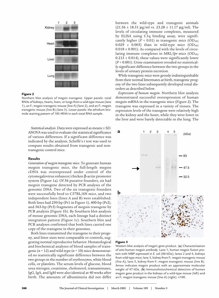
Expression of human megsin. Northern blot analysisdemonstrated successful overexpression of humanmegsin mRNA in the transgenic mice (Figure 2). Thetransgene was expressed in a variety of tissues. Theexpression levels of the transgene were relatively highin the kidney and the heart, while they were lower inthe liver and were barely detectable in the lung. The
588 The Journal of Clinical Investigation | March 2002 | Volume 109 | Number 5
Figure 2Northern blot analysis of megsin transgene. Upper panels: totalRNAs of kidneys, hearts, livers, or lungs from a wild-type mouse (lane1), an F1 megsin transgenic mouse (line A) (lane 2), and an F1 megsintransgenic mouse (line B) (lane 3). Lower panels: the ethidium bro-mide staining pattern of 18S rRNA in each total RNA sample.
Figure 3Western blot analysis of megsin gene product. (a) Characterizationof anti-human megsin antibody. Lane 1, human megsin fusion pro-tein with MBP expressed in E. coli (86 kDa); lanes 2 and 4, kidneysfrom wild-type mice; lane 3, kidney from F1 megsin transgenic mouse(line A); lane 5, kidney from F1 megsin transgenic mouse (line B).Arrow indicates megsin product with an approximate molecularweight of 47 kDa. (b) Immunohistochemical detection of humanmegsin gene product in the kidneys of a wild-type mouse (left) andan F1 megsin transgenic mouse (line A) (right). ×100.

expression of human megsin cDNA in the transgenickidney started at preimplantation stage and continuedeven after 40 weeks of age.
The protein expression of human megsin gene prod-ucts in transgenic mice was confirmed by immunoblotanalysis of kidney crude homogenates using anti-human megsin peptide antibody (Figure 3a). Immuno-histochemical studies revealed the megsin proteinexpression ubiquitously in both glomeruli and tubulesof these mice (Figure 3b). Their immunoreactivitieswere completely abolished after preincubation of theantibodies with an excess of human megsin peptide.
Transgenic mice developed mesangial matrix expansion andan increase in the number of mesangial cells. Until 15 weeksof age, megsin transgenic mice did not show any signif-icant pathological changes. At 20 weeks, some mice fromline A exhibited glomerular morphological changes,which became even more apparent at 40 weeks. Thesechanges included an increased number of cells in themesangial area and an expansion of mesangial matrixwith deposits therein (Figure 4). Although megsin trans-gene was also expressed in the other tissues as describedabove, pathological changes detected histologically bylight microscopy were observed exclusively in the kidney.
In order to quantify these glomerular changes, weperformed computer-assisted morphometrical analy-ses. We measured glomerular sizes and numbers ofmesangial cells in 25 wild-type mice and decided nor-mal limits as a range of mean + 2 SD. Using this defi-nition, 3 out of 14 heterozygous F1 transgenic micefrom line A developed either of these changes at 20weeks after birth. At 40 weeks, among 19 heterozygoustransgenic mice from line A, 11 (57.9%) developed anincrease in glomerular size and 12 (63.2%) developedan increase in glomerular cell numbers (Figure 5).Altogether, our histological analysis revealed glomeru-lar abnormalities in 14 (73.0%) of 19 transgenic mice.In line B at 40 weeks, essentially the same glomerularabnormalities as in line A were observed in 18.2% ofthe transgenic animals (4 of 22). We did not observeany sex differences in either severity or penetrance ofthe disease. We did not detect any pathologicalchanges in other organs. Except for one wild-typemouse, which developed mild expansion of mesangialmatrix at 40 weeks, no wild-type mice exhibited patho-logical changes. Detailed analyses of glomerularabnormalities were performed as below by employingline A as representative animals.
Immunohistochemical analysis revealed accumulation oftype IV collagen and laminin. In order to analyze abnor-mal glomeruli in the transgenic animals in detail, weperformed immunohistochemical analysis of variousECM components (Figure 6). We did not observe accu-mulation of type I collagen in either wild-type or trans-genic mice. Transgenic mice showed marked accumu-lation of type IV collagen and laminin in their scleroticglomeruli. The amount of fibronectin was less in thesclerotic glomeruli of the transgenic animals than inthose of wild-type mice.
Our immunohistochemical analysis and PAS stain-ing revealed no infiltration of leukocytes at any timepoint, either in the glomerulus or in the tubulointer-stitium. Therefore, it is likely that hypercellularity inthe mesangial area of the transgenic mice was due toproliferation of resident glomerular cells.
Glomerular change was associated with an augmentedimmune complex deposition. Immunofluorescent studieswere negative before 20 weeks of age. At 20 weeks wedetected Ig’s (IgA, IgG, and IgM) and complement dep-osition in mouse glomeruli for the first time. Immunecomplex deposition increased in an age-dependent
The Journal of Clinical Investigation | March 2002 | Volume 109 | Number 5 589
Figure 4Histopathological analysis of murine kid-neys. PAS staining. As compared with 40-week-old wild-type mice (left, ×200), F1
megsin transgenic mice (line A) of the sameage developed mesangial matrix expansionand an increase in the number of mesangialcells (middle, ×200; right, ×50).
Figure 5Computer-assisted morphometry of glomerular abnormalities inmegsin transgenic mice. Glomerular tuft areas (a) and numbers ofmesangial cells (b) were measured in 25 wild-type mice and 19megsin transgenic mice (line A). Normal limits are considered as arange of mean + 2 SD in wild-type mice. Filled circles represent ani-mals above the normal limits, while open circles demonstrate micewithin normal limits. The mean ± SD is indicated.

manner thereafter. Of note, immune complex deposi-tion was markedly augmented in glomeruli of thetransgenic mice when renal changes developed at 40weeks (Figure 7).
Electron microscopic analysis confirmed these find-ings. While we observed a small number of electron-dense deposits in the mesangial area of some nontransgenic mice (Figure 8), the presence of elec-tron-dense deposits was markedly augmented in thetransgenic mice; sizes of the deposits varied(100–1000 nm). Hypercellularity in the mesangialarea of the transgenic mice was due to an increase inthe number of mesangial cells, accompanied by anexpansion of matrix. Pathological findings were con-fined to the mesangial area. Capillary endotheliumgenerally maintained a normal appearance, andendothelial fenestrations remained preserved.Podocytes also remained intact, maintaining normalmorphologic features of foot processes. Corticaltubules demonstrated normal appearances. Infiltra-tion of mononuclear or polymorphonuclear leuko-cytes was undetectable.
An augmented immune complex deposition wasalready observed in transgenic mice at 20 weeks both byimmunofluorescence studies and electron microscopicanalysis. At 6 weeks, we observed no immune complexdeposition in control animals or transgenic mice.
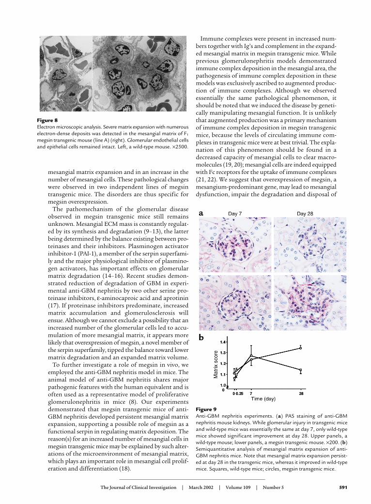
Mesangial expansion in anti-GBM nephritis persisted only inmegsin transgenic mice. In order to examine whether thestressed mice overexpressing megsin would develop anovert glomerulopathy, we induced anti-GBM nephritisin the transgenic and wild-type mice and evaluated thedegree of matrix expansion (Figure 9). Animals fromboth groups developed the same degree of mesangialmatrix expansion at day 7. At day 28, mesangial matrixexpansion persisted in transgenic mice, whereas that inwild-type animals improved. Our semiquantitativeanalysis showed that glomerular deposition of mouseIgG was essentially the same between the two groups atday 28. Serum creatinine levels of transgenic mice andwild-type animals did not differ throughout the timecourse (0.11 ± 0.01 vs. 0.10 ± 0.02 mg/dl, respectively, atday 7, and 0.09 ± 0.03 vs. 0.08 ± 0.04 mg/dl, respective-ly, at day 28).
Megsin inhibits the plasmin activity in vitro. Since theP17–P8 sequence of megsin (EGTEATAAT) was consis-tent with the consensus sequence among inhibitory ser-pins (EGTEAAAAT) (1), we evaluated the activity ofmegsin on serine proteases by in vitro binding studies.SDS-PAGE revealed appearance of a new band whenrecombinant megsin was mixed with plasmin, but notother serine proteases (t-PA and thrombin in Figure 10a).The binding of megsin to plasmin was also confirmed bySELDI-TOF mass spectrometry: in a reaction mixture ofmegsin and plasmin, a peak was identified with a molec-ular mass of 120,163 Da, which was consistent with thevalue of megsin (46,883 Da) and plasmin (73,264 Da)complexes. The complex formation was not observedwhen recombinant megsin was heat-inactivated. Allthese studies suggested that megsin bound to plasmin.
In addition, functional assays demonstrated aninhibitory effect of megsin on the enzymatic activity ofplasmin (Figure 10b). By contrast, neither ovalbumin noralbumin inhibited the plasmin activity, demonstratingthe specific activity of megsin. Inactivated megsin did notexhibit such an activity. The inhibitory activity of megsinwas also confirmed using another substrate of plasmin(Boc-Val-Leu-Lys-MCA) (data not shown).
DiscussionOur data demonstrate that megsin has a biologicallyrelevant influence on mesangial cell functions. Indeed,overexpression of this protein results in progressive
590 The Journal of Clinical Investigation | March 2002 | Volume 109 | Number 5
Figure 6Immunohistochemical analysis ofECM. Marked deposition of type IVcollagen and laminin was observed inthe glomeruli of transgenic mice,while the amount of fibronectin wasmuch less in the transgenic animalsthan in wild-type mice. Accumulationof type I collagen was not observed ineither wild-type or transgenic ani-mals. Upper row, a wild-type mouse;lower row, a megsin transgenicmouse. ×200.
Figure 7Immunofluorescent studies of Ig’s. Diffuse granular deposits of IgGand complement were augmented in the mesangial area of 40-week-old F1 transgenic mice (line A) (right) as compared with wild-typemice of the same age (left). ×50.
mesangial matrix expansion and in an increase in thenumber of mesangial cells. These pathological changeswere observed in two independent lines of megsintransgenic mice. The disorders are thus specific formegsin overexpression.
The pathomechanism of the glomerular diseaseobserved in megsin transgenic mice still remainsunknown. Mesangial ECM mass is constantly regulat-ed by its synthesis and degradation (9–13), the latterbeing determined by the balance existing between pro-teinases and their inhibitors. Plasminogen activatorinhibitor-1 (PAI-1), a member of the serpin superfami-ly and the major physiological inhibitor of plasmino-gen activators, has important effects on glomerularmatrix degradation (14–16). Recent studies demon-strated reduction of degradation of GBM in experi-mental anti-GBM nephritis by two other serine pro-teinase inhibitors, ε-aminocaproic acid and aprotinin(17). If proteinase inhibitors predominate, increasedmatrix accumulation and glomerulosclerosis willensue. Although we cannot exclude a possibility that anincreased number of the glomerular cells led to accu-mulation of more mesangial matrix, it appears morelikely that overexpression of megsin, a novel member ofthe serpin superfamily, tipped the balance toward lowermatrix degradation and an expanded matrix volume.
To further investigate a role of megsin in vivo, weemployed the anti-GBM nephritis model in mice. Theanimal model of anti-GBM nephritis shares majorpathogenic features with the human equivalent and isoften used as a representative model of proliferativeglomerulonephritis in mice (8). Our experimentsdemonstrated that megsin transgenic mice of anti-GBM nephritis developed persistent mesangial matrixexpansion, supporting a possible role of megsin as afunctional serpin in regulating matrix deposition. Thereason(s) for an increased number of mesangial cells inmegsin transgenic mice may be explained by such alter-ations of the microenvironment of mesangial matrix,which plays an important role in mesangial cell prolif-eration and differentiation (18).
Immune complexes were present in increased num-bers together with Ig’s and complement in the expand-ed mesangial matrix in megsin transgenic mice. Whileprevious glomerulonephritis models demonstratedimmune complex deposition in the mesangial area, thepathogenesis of immune complex deposition in thesemodels was exclusively ascribed to augmented produc-tion of immune complexes. Although we observedessentially the same pathological phenomenon, itshould be noted that we induced the disease by geneti-cally manipulating mesangial function. It is unlikelythat augmented production was a primary mechanismof immune complex deposition in megsin transgenicmice, because the levels of circulating immune com-plexes in transgenic mice were at best trivial. The expla-nation of this phenomenon should be found in adecreased capacity of mesangial cells to clear macro-molecules (19, 20); mesangial cells are indeed equippedwith Fc receptors for the uptake of immune complexes(21, 22). We suggest that overexpression of megsin, amesangium-predominant gene, may lead to mesangialdysfunction, impair the degradation and disposal of
The Journal of Clinical Investigation | March 2002 | Volume 109 | Number 5 591
Figure 8Electron microscopic analysis. Severe matrix expansion with numerouselectron-dense deposits was detected in the mesangial matrix of F1
megsin transgenic mouse (line A) (right). Glomerular endothelial cellsand epithelial cells remained intact. Left, a wild-type mouse. ×2500.
Figure 9Anti-GBM nephritis experiments. (a) PAS staining of anti-GBMnephritis mouse kidneys. While glomerular injury in transgenic miceand wild-type mice was essentially the same at day 7, only wild-typemice showed significant improvement at day 28. Upper panels, awild-type mouse; lower panels, a megsin transgenic mouse. ×200. (b)Semiquantitative analysis of mesangial matrix expansion of anti-GBM nephritis mice. Note that mesangial matrix expansion persist-ed at day 28 in the transgenic mice, whereas it improved in wild-typemice. Squares, wild-type mice; circles, megsin transgenic mice.

immune complexes, and alter the microenvironment ofmesangial matrix. However, it should be noted that wedid not observe any difference in mouse IgG depositionbetween anti-GBM nephritis transgenic mice and wild-type mice (12 weeks old) at day 28. Although a limitedsensitivity of our semiquantitative analysis may be thereason that we could not detect a difference, matrixexpansion of these anti-GBM nephritis transgenic micecould be attributed to imbalance of matrix degradationrather than a decreased capacity of mesangial cells toclear macromolecules.
In order to identify potential substrates of megsin, weperformed assays using recombinant megsin. Ourbinding and functional studies suggested that megsinbinds to plasmin and has an inhibitory effect on theenzymatic activity of plasmin. In the context ofmesangium matrix turnover, the plasminogen activa-tor/plasmin cascade plays an important role throughits ability to directly degrade some matrix components(23). Furthermore, plasmin also induces activation ofmatrix metalloproteinase, resulting in indirect degra-dation of extracellular matrix (24). The amount of plas-min generation is tightly regulated through the inhibi-tion by specific inhibitors that belong to the serpinfamily, such as PAI-1, PAI-2, and PAI-3 (25–27). Ourresults, together with these reports, suggest thatmegsin may act as an inhibitor of plasmin and relatedserine proteases, leading to alterations of mesangiummatrix turnover. While megsin may have some otherbiological substrates in glomeruli, our findings clearlydemonstrate that megsin serves as a functional serpin.
Although megsin is normally confined to theglomerular mesangium, the transgenic model report-ed in this paper is characterized by the expression of
megsin in all tissues. This feature is due to the ubiqui-tous promoter for the transgene. Although immuno-histochemical studies revealed the presence of megsinin a host of tissues as well as in nonmesangial areas ofthe kidney, pathogenic effects of megsin overexpressionwere restricted within glomeruli. This finding may sug-gest specific localization of megsin ligand (serine pro-tease) other than plasmin within the mesangium. Inother words, a role of megsin appears restricted to themesangium for two reasons: first, because it is normal-ly expressed only there, but also, second, because itrequires the existence of unknown megsin ligand pres-ent only in this area.
Do our observations have any relevance for theunderstanding of human pathology? The describedlesions are different from those observed in the agingkidney: the latter is characterized by a mild expansionof the mesangial matrix, resulting in glomerular obso-lescence without an increased number of mesangialcells (28–30). By contrast, the coexistence of mesan-gial proliferation, matrix expansion, and increasedimmune complex deposition is frequently observed inhuman glomerular diseases. However, when they arepresent in a renal biopsy, the pathologist cannotunravel the sequence of pathological events. Ourtransgenic mice provide a unique opportunity to fol-low sequential events in a disease model relevant tohuman kidney diseases.
In conclusion, megsin transgenic mice developed pro-gressive mesangial matrix expansion, which was asso-ciated with an increase in the number of mesangialcells. These glomerular lesions were accompanied by anaugmented immune complex deposition, demonstrat-ing a biological role of megsin as a functional serpin.
592 The Journal of Clinical Investigation | March 2002 | Volume 109 | Number 5
Figure 10Functional assays of megsin. (a) SDS-PAGE under a nonreduced condition, followed by CBB staining (left and middle panels) or immunoblot-ting using anti–t-PA antibody (right panel), revealed functional binding of megsin to plasmin, but not to other serine proteases such as t-PAand thrombin. Lane 1, plasmin; lane 2, antiplasmin; lane 3, plasmin plus antiplasmin; lane 4, megsin; lane 5, plasmin plus megsin; lane 6,thrombin; lane 7, antithrombin; lane 8, thrombin plus antithrombin; lane 9, megsin; lane 10, thrombin plus megsin; lane 11, t-PA; lane 12,PAI-1; lane 13, t-PA plus PAI-1; lane 14, megsin; lane 15, t-PA plus megsin. The complex of serine protease and serpin is indicated by aster-isks. (b) The inhibitory effect of megsin to plasmin activity. Diamonds, substrate alone; open squares, plasmin; filled squares, plasmin andantiplasmin (1:2); open circles, plasmin and megsin (1:10); filled circles, plasmin and inactivated megsin (1:10); open triangles, ovalbumin;filled triangles, albumin.

AcknowledgmentsWe thank C. van Yepersele de Strihou for helpful dis-cussions and T. Watanabe, K. Isaka, M. Asakura, T.Umezono, and H. Uemura for their excellent technicalassistance. This study was supported by grants fromthe Japan Science and Technology Cooperation (to T.Miyata), from the Japanese Ministry of Science andTechnology Agency (Millenium Project to T. Miyata),from the Japanese Ministry of Health, Labour and Wel-fare (to T. Miyata), from the Japanese Ministry of Edu-cation, Culture, Sports, Science and Technology (to R.Inagi and K. Kurokawa), from Sankyo Foundation ofLife Science (to R. Inagi), and from the Takeda ScienceFoundation (to R. Inagi).
1. Miyata, T., et al. 1998. A mesangium-predominant gene, megsin, is a newserpin up-regulated in IgA nephropathy. J. Clin. Invest. 102:828–836.
2. Suzuki, D., et al. 1999. Expression of megsin mRNA, a novel mesangium-predominant gene, in the renal tissues of various glomerular diseases. J.Am. Soc. Nephrol. 10:2606–2613.
3. Inagi, R., et al. 2001. Specific tissue distribution of megsin, a novel ser-pin, in the glomerulus and its up-regulation in IgA nephropathy.Biochem. Biophys. Res. Commun. 286:1098–1106.
4. Nangaku, M., et al. 2001. Cloning of rodent megsin revealed its up-reg-ulation in mesangioproliferative nephritis. Kidney Int. 60:641–652.
5. Kawarabayashi, T., et al. 1996. Accumulation of β-amyloid fibrils in pan-creas of transgenic mice. Neurobiol. Aging. 17:215–222.
6. Hogan, B., Costantini, F., and Lacy, E. 1986. Manipulating the mouseembryo: a laboratory manual. Cold Spring Harbor Laboratory Press. ColdSpring Harbor, New York, USA. 487 pp.
7. Sekine, H., et al. 2001. Complement component C3 is not required forfull expression of immune complex glomerulonephritis in MRL/lprmice. J. Immunol. 166:6444–6451.
8. Hisada, Y., et al. 1999. Angiotensin II plays a pathogenic role in immune-mediated renal injury in mice. J. Clin. Invest. 103:627–635.
9. Johnson, R.J., Lovett, D., Lehrer, R.I., Couser, W.G., and Klebanoff, S.J.1994. Role of oxidants and proteases in glomerular injury. Kidney Int.45:352–359.
10. Davie, M., Martin, J., Thomas, G.J., and Lovett, D.H. 1992. Proteinasesand glomerular matrix turnover. Kidney Int. 41:671–678.
11. Baricos, W.H., and Shah, S.V. 1991. Proteolytic enzymes as mediators ofglomerular injury. Kidney Int. 40:161–173.
12. McLennan, S., Fisher, E., Yue, D.K., and Turtle, J.R. 1994. High glucoseconcentration causes a decrease in mesangium degradation. A factor inthe pathogenesis of diabetic nephropathy. Diabetes. 43:1041–1045.
13. Davies, M., Coles, G.A., Thomas, G.J., Martin, J., and Lovett, D.H. 1990.Proteinases and the glomerulus: their role in glomerular diseases. Klin.Wochenschr. 68:1145–1149.
14. Fogo, A.B. 2000. The role of angiotensin II and plasminogen activatorinhibitor-1 in progressive glomerulosclerosis. Am. J. Kidney Dis.35:179–188.
15. Loskutoff, D.J. 1993. A slice of PAI. J. Clin. Invest. 92:2563.16. Nakamura, S., Nakamura, I., Ma, L., Vaughan, D.E., and Fogo, A.B. 2000.
Plasminogen activator inhibitor-1 expression is regulated by theangiotensin type 1 receptor in vivo. Kidney Int. 58:251–259.
17. Hruby, Z., et al. 2000. Mechanism of antinephritic effect of proteinaseinhibitors in experimental anti-GBM glomerulopathy. Res. Exp. Med.(Berl.) 199:295–307.
18. Turck, J., Pollock, A.S., Lee, L.K., Marti, H.P., and Lovett, D.H. 1996.Matrix metalloproteinase 2 (gelatinase A) regulates glomerular mesan-gial cell proliferation and differentiation. J. Biol. Chem. 271:15074–15083.
19. Farquhar, M.G., and Palade, G.E. 1961. Glomerular permeability. II. Fer-ritin transfer across the glomerular capillary wall in nephrotic rats. J. Exp.Med. 144:699–715.
20. Sterzel, R.B., and Rupprecht, H.D. 1997. Glomerular mesangial cells. InImmunologic renal diseases. E.G. Neilson and W.G. Couser, editors. Lip-pincott-Raven Publishers. Philadelphia, Pennsylvania, USA. 595–626.
21. Neuwirth, R., et al. 1988. Evidence for immunoglobulin Fc receptor-mediated prostaglandin 2 and platelet-activating factor formation bycultured rat mesangial cells. J. Clin. Invest. 82:936–944.
22. Santiago, A., Mori, T., Satriano, J., and Schlondorff, D. 1991. Regulationof Fc receptors for IgG on cultured rat mesangial cells. Kidney Int.39:87–94.
23. Mclennan, S.V., et al. 2000. Effects of glucose on matrix metallopro-teinase and plasmin activities in mesangial cells: possible role in diabet-ic nephropathy. Kidney Int. Suppl. 77:S81–S87.
24. Baricos, W.H., Cortez, S.L., el-Dahr, S.S., and Schnaper, H.W. 1995. ECMdegradation by cultured human mesangial cells is mediated by aPA/plasmin/MMP-2 cascade. Kidney Int. 47:1039–1047.
25. Rerolle, J.P., Hertig, A., Nguyen, G., Sraer, J.D., and Rondeau, E.P. 2000.Plasminogen activator inhibitor type 1 is a potential target in renal fibro-genesis. Kidney Int. 58:1841–1850.
26. Eitzman, D.T., and Ginsburg, D. 1997. Of mice and men. The functionof plasminogen activator inhibitors (PAIs) in vivo. Adv. Exp. Med. Biol.425:131–141.
27. Scott, F.L., et al. 1999. Human ovalbumin serpin evolution: phylogenicanalysis, gene organization, and identification of new PI8-related genessuggest that two interchromosomal and several intrachromosomalduplications generated the gene clusters at 18q21-q23 and 6p25.Genomics. 62:490–499.
28. Baylis, C., and Corman, B. 1998. The aging kidney: insights from exper-imental studies. J. Am. Soc. Nephrol. 9:699–709.
29. Couser, W.G., and Stilmant, N.M. 1975. Mesangial lesions and focalglomerulosclerosis in the aging rat. Lab. Invest. 33:491–501.
30. Goldstein, R.S., Tarloff, J.B., and Hook, J.B. 1988. Age-related nephropa-thy in laboratory rats. FASEB J. 2:2241–2251.
The Journal of Clinical Investigation | March 2002 | Volume 109 | Number 5 593