overexpressionofsmad7blocksprimarytumorgrowthand lung...
TRANSCRIPT

Cancer Therapy: Preclinical
Overexpression of Smad7 Blocks Primary TumorGrowth andLung Metastasis Development in Osteosarcoma
Audrey Lamora1,2,3,4, Julie Talbot1,2,3, Gwenola Bougras1,2,3, J�erome Amiaud1,2,3,Marion Leduc1,2,3, Julie Chesneau1,2,3, Julien Taurelle1,2,3, Verena Stresing1,2,3,Marie C�ecile Le Deley5, Marie Francoise Heymann1,2,3, Dominique Heymann1,2,3,Francoise Redini1,2,3, and Franck Verrecchia1,2,3
AbstractPurpose:Osteosarcoma is themainmalignant primary bone tumor in children and adolescents for whom
the prognosis remains poor, especially when metastasis is present at diagnosis. Because transforming growth
factor-b (TGFb) has been shown to promotemetastasis in many solid tumors, we investigated the effect of the
natural TGFb/Smad signaling inhibitor Smad7 and the TbRI inhibitor SD-208 on osteosarcoma behavior.
Experimental Design: By using amousemodel of osteosarcoma induced by paratibial injection of cells,
we assessed the impact of Smad7 overexpression or SD-208 on tumor growth, tumor microenvironment,
bone remodeling, and metastasis development.
Results: First, we demonstrated that TGFb levels are higher in serum samples from patients with
osteosarcoma compared with healthy volunteers and that TGFb/Smad3 signaling pathway is activated in
clinical samples. Second, we showed that Smad7 slows the growth of the primary tumor and increases mice
survival. We furthermore demonstrated that Smad7 expression does not affect in vitro osteosarcoma cell
proliferation but affects themicroarchitectural parameters of bone. In addition, Smad7-osteosarcoma bone
tumors expressed lower levels of osteolytic factors such as RANKL, suggesting that Smad7 overexpression
affects the "vicious cycle" established between tumor cells and bone cells by its ability to decrease osteoclast
activity. Finally, we showed that Smad7overexpression in osteosarcoma cells and the treatment ofmicewith
SD208 inhibit the development of lung metastasis.
Conclusion: Taken together, these results demonstrate that the inhibition of the TGFb/Smad signaling
pathway may be a promising therapeutic strategy against tumor progression of osteosarcoma, specifically
against the development of lung metastasis. Clin Cancer Res; 20(19); 5097–112. �2014 AACR.
IntroductionOsteosarcoma is the most common primary malignant
bone tumor in children and adolescents with a second peakof incidence in adults over the age of 65 (1). Althoughseveral predisposing environmental (e.g., ionizing radia-tion) and genetic (e.g., TP53) factors have been identified(2), the exact etiology of this disease remains unknown (3).These rare tumors, believed to originate frommesenchymalcells forming the primitive bone, preferentially grow in themetaphysis of long bone (4). Approximately 20% of
patients have lung metastasis at initial diagnosis and anadditional 40% will develop metastasis during the laterstages of disease. The presence of metastasis at diagnosisis themost important predictor of disease-free survival witha 5-year survival rate of only 20% for osteosarcoma patientswith metastasis compared with 65% for patients with local-ized disease (5). The standard treatment of osteosarcomaconsists of complete surgical resection associated withneoadjuvant and adjuvant chemotherapy composed of 4agents: doxorubicin, cisplatin, methotrexate or ifosfamide(6). These combined treatment protocols have significantlyimproved survival of the nonmetastatic patients over thepast several decades (7, 8). Unfortunately, such therapeuticstrategies have a limited efficacy in the treatment of meta-static disease, and the metastatic relapse or recurrent con-ditions have remained unchanged over the last 3 decades(8). Treating metastatic osteosarcoma thus remains a chal-lenge in bone cancer (9).
Transforming growth factor-b (TGFb) family membersare a class of cytokines that control a variety of biologicprocesses, including proliferation, differentiation, extra-cellular matrix production, and apoptosis. Three isoformsof TGFb exist in mammals: TGFb1, TGFb2, and TGFb3.
1INSERM, UMR 957, Equipe labellis�ee Ligue contre le Cancer 2012,Nantes, France. 2Universit�e de Nantes, Laboratoire de Physiopathologiede la R�esorption Osseuse et Th�erapie des Tumeurs Osseuses Primitives,Nantes, France. 3CHU Hotel Dieu, Nantes, France. 4Inserm Liliane Betten-court School, Paris, France. 5Institut Gustave Roussy, Villejuif, France.
Note: Supplementary data for this article are available at Clinical CancerResearch Online (http://clincancerres.aacrjournals.org/).
Corresponding Author: Franck Verrecchia, INSERM, UMR 957, Facult�ede M�edecine, 1 rue Gaston Veil, 44000 Nantes, France. Phone: 33-240412842; Fax: 33-240412860; E-mail: [email protected]
doi: 10.1158/1078-0432.CCR-13-3191
�2014 American Association for Cancer Research.
ClinicalCancer
Research
www.aacrjournals.org 5097
on December 26, 2019. © 2014 American Association for Cancer Research.clincancerres.aacrjournals.org Downloaded from
Published OnlineFirst August 8, 2014; DOI: 10.1158/1078-0432.CCR-13-3191

Dimers of TGFb initiate the canonical signaling cascadevia the serine/threonine kinase receptor cell surfacecomplexes (TbRI and TbRII), which phosphorylate theligand-specific receptor-activated Smads (R-Smad: Smad2or Smad3). Upon phosphorylation by type I receptors,R-Smads form a heteromeric complex with the Common-Smad, Smad4. The R-Smad/Smad4 complex then trans-locates to the nucleus to regulate gene transcription(10–13). A third group of Smad proteins, the inhibitorySmads (Smad7), inhibits the canonical Smad signaling(14, 15) by different means; Smad7 (i) binds TbRI andprevents R-Smad phosphorylation, (ii) recruits E3-typeubiquitin ligases to the receptor complexes ultimatelyleading to their degradation, and (iii) interacts withGADD34, the regulatory subunit of the protein phospha-tase PP1 to inactive TbRI (10–16).
The role of TGFb in cancer is complex. During the firststages of the development of primitive tumors fromepithelial origin, the TGFb/Smad cascade acts as a tumorsuppressor mainly through inhibition of cell proliferationand/or promotion of cell apoptosis (11, 12, 17). Con-trarily, during the later stages, the TGFb cascade promotestumor progression mainly by its ability to stimulateepithelial-to-mesenchymal transition, tumor invasion,metastatic dissemination, and/or evasion of the immunesystem (12, 13, 17). With regard to bone cancers, moststudies have focused on the role of TGFb in the develop-ment of bone metastasis. The contributions of TGFb tobreast cancer and melanoma bone metastasis have beenwell described (18–21). TGFb promotes bone metastasisby its ability to promote the metastatic process by stim-ulating MMP2 production and thus promoting cellularinvasion of melanoma (19, 20). It has been reported thatTGFb contributes to the establishment of a vicious cycle
between epithelial tumor cells and bone cells. Briefly, thetumor cells secrete osteoclast-activating factors that pro-mote bone degradation, thus stimulating the release offactors which in turn will promote bone metastasis devel-opment (22, 23). In this context, it has been shown thatoverexpression of the TGFb inhibitor Smad7 in melano-ma cells reduces the development of melanoma bonemetastasis, and that the use of chemical inhibitors target-ing TbRI reduces the development and progression ofboth melanoma and breast cancer bone metastasis (21).With regard to primary bone sarcomas, few studies havedescribed the role of TGFb on tumor development. It hasbeen shown that the production of TGFb1 is associatedwith high-grade osteosarcoma (24, 25) and that TGFbstimulates the growth of several osteosarcoma cell lines inculture (26), suggesting that TGFb could favor osteosar-coma development.
In this study, we particularly demonstrate that the over-expression of the inhibitory Smad, Smad7, in osteosarcomacells and treatment of mice with a specific inhibitor of TbRI(SD-208) block the development of osteosarcoma lungmetastasis.
Materials and MethodsMeasurement of circulating TGFb levels in serum
The levels of circulating TGFbs were measured in serumfrom healthy controls (n ¼ 20) from the EtablissementFrancais du Sang (EFS) and patients with osteosarcoma (n¼40) from the OS 2006 protocol (PAC SARCOME, Sarcome09/0603, EudraCT No. 2006-00337727) with the Bio-PlexPro Assay TGFb Standard 3-Plex system (Bio-Rad). Serumsamples were obtained with written informed consent.
Cell cultures and reagentsHOS and SaOS2 osteosarcoma cells were purchased from
ATCC (CRL-1544 andHTB-85, respectively) and cultured inDulbecco’s modified Eagle medium (DMEM; Lonza) sup-plemented with 10% fetal bovine serum (Hyclone Perbio).All cell lines were authenticated by short tandem repeat(STR) profiling. TGFb1 and BMP-6, and G418 were, respec-tively, from R&D System Inc. and Sigma. SD-208 has beensynthetized by the laboratory CEISAM UMR6230, NantesUniversity (France).
Western blot analysisWestern blot analysis were performed as previously
described (27). Membranes were immunoblotted withanti–phospho-Smad3 (Millipore), anti-Smad3 (Millipore),anti-Smad7 (Santa Cruz Biotechnology), anti–phospho-Erk (Cell Signaling Technology), anti-Erk (Cell Signaling),or anti–b-actin (Sigma) antibodies.
Proliferation assayCell growth and viability were determined by using a 2,3-
bis(2 methoxy-4 nitro-5-sulfophenyl)-2H-tetrazolium-5-carboxanilide (XTT) Reagent Assay Kit (Roche MolecularBiomedicals).
Translational RelevanceWe have demonstrated that the TGFb/Smad signal-
ing pathway plays a crucial role in osteosarcoma met-astatic progression. We have first shown that TGFblevels are higher in serum samples from patients withosteosarcoma compared with healthy volunteers andthat the TGFb/Smad3 signaling pathway is activated inclinical samples of patients. Second, using a murinemodel of osteosarcoma, we demonstrated that blockingthe TGFb/Smad signaling pathway via overexpression ofinhibitory Smad slows the growth of the primary bonetumormainly by affecting the tumormicroenvironmentby controlling the "vicious cycle" established betweentumor cells and bone cells. Third, blocking TGFb signal-ing inhibits the development of lung metastasis at leastby inhibition of cell migration and invasion. Here, weshow for the first time that blocking TGFb signalingrepresents a novel therapeutic approach for the treat-ment of lung metastasis in patients with osteosarcoma,which has a poor prognosis.
Lamora et al.
Clin Cancer Res; 20(19) October 1, 2014 Clinical Cancer Research5098
on December 26, 2019. © 2014 American Association for Cancer Research.clincancerres.aacrjournals.org Downloaded from
Published OnlineFirst August 8, 2014; DOI: 10.1158/1078-0432.CCR-13-3191
Real-time polymerase chain reactionTotal RNA from cell lines was extracted using NucleoS-
pinRNAII (Macherey Nagel). Total RNA from tumors wasextracted using the TRizol reagent (Invitrogen Life Technol-ogies) after mechanical grinding with Turrax (IKA). qRT-PCR were performed as previously described (27). Primersequences are provided in Table 1.
Transient cell transfections, reporter assays, andplasmid constructsTransient cell transfections were performed with jetPEI
(polyplus-transfection). The phRLMLP-Renilla luciferaseexpression vector was cotransfected in all experiments tomonitor transfection efficiencies. Luciferase activity wasdetermined with the dual-luciferase reporter assay system(Promega). The (CAGA)9-Luc construct was used as areporter construct specific for Smad3/4-driven signaling(28). The pcDNA-Smad7 vector is a kind gift fromDr. AlainMauviel (19).
Osteosarcoma mouse modelFour-week-old female Rj:NMRI-nudemice (Elevages Jan-
vier) were maintained under pathogen-free conditions atthe Experimental Therapy Unit (Faculty of Medicine,Nantes, France) in accordance with the institutional guide-lines of the French Ethical Committee (CEEA Pays de laLoireNo. 06; project authorizationNo.CEEA-2010-23) andunder the supervision of authorized investigators. Themicewere anesthetized by inhalation of an isoflurane/airmixture(1.5%, 1 L/min) before receiving an intramuscular injectionof 1 � 106 HOS or SaOS2 osteosarcoma cells in closeproximity to the tibia, leading to a rapidly growing tumorin soft tissuewith secondary contiguous bone invasion.Oneday after HOS cells injection, some mice received differentdoses (20or 60mg/kg) of SD-208or control vehicle bydailygavage. The tumor volume (V) was calculated from themeasurement of 2 perpendicular diameters using a caliper,according to the following formula: V ¼ 0.5 � L � (S)2, aspreviously described (29). Mice were sacrificed when thetumor volume reached 2,500 mm3 for ethical reasons.
Under these conditions, pulmonary metastasis developedwhen tumor volumes were �2,000 mm3.
Micro-CT analysisAnalysis of bone microarchitecture was performed as
previously described (27) at different tumor volumes(250, 1,000, and 2,500 mm3). All tibiae/fibulae werescanned using the same parameters (pixel size 18 mm, 50kV, 0.5-mm Al filter, and 0.8 degrees per rotation step).Three-dimensional reconstructions and analysis of boneparameters were performed using the CTvol and CTansoftware (Skyscan).
Histologic analysisAfter sacrifice, the tibiae were conserved and fixed in 10%
buffered formaldehyde, decalcified (4% EDTA, 0.2% para-formaldehyde, pH 7.4), and embedded in paraffin. Three-micrometer sections of tumor-bearing tibiae were cut andstained for tartrate-resistant acid phosphatase (TRAP) toanalyze osteoclast activity. Lungswere fixed in 10%bufferedformaldehyde and embedded in paraffin. Lung sections (3-mm-thick) were mounted on glass slides and stained withhematoxylin–eosin (HE).
Immunohistochemistry of human and mice tumorsamples
Three-micrometer sections of human tumor tissues(embedded in paraffin) were cut and stained for phospho-Smad3 using rabbit polyclonal anti–phospho-Smad3 anti-body (Abcam). Patient tumor clinical samples collected atNantes University Hospital (Nantes, France) were obtainedfollowingpatient informed consent andafter ethical approv-al by the Nantes University Hospital Ethics Committee.
Sections (3 mm) of mice tumor tissues (embedded inparaffin) were cut and stained for osterix, RANKL, osteo-calcin, caspase-3, Ki67, and CD146 using, respectively,rabbit polyclonal anti-osterix (Abcam), anti-RANKL (SantaCruz Biotechnology), anti-osteocalcin (Abcam), anti–cas-pase-3 (Cell Signaling), anti-Ki67 (Dako), anti CD146(Abcam) antibodies. Immunodetection was performed
Table 1. Primer sequence for quantitative RT-PCR
Sens Antisens
ANGPTL4 gAC CCg gCT CAC AAT gTC CCC TgA ggC Tgg ATT TCACol1A1 CTg gAC CTA Aag gTg cTg cT gCT CCA gCC TCT CCA TCT TTCTGF CTC CTg Cag gCT AgA gAA gC gAT gCA CTT TTT gCC CTT CTTCXCR4 CCg Agg AAA Tgg gCT CAg ggg A TgA Tgg AgT AgA Tgg Tgg gCA ggAGAPDH Tgg gTg TgA ACC Atg AgA AgT Atg ggT gCA ggA ggC ATT gCTID1 AAT CAT gAA AgT CgC Cag Tg ATg TCg Tag AgC AgC ACg TTTIL11 gCA gCg gAC Agg gAA ggg TTA A ACA ggC TCA gCA CgA CCA ggMMP2 AgA Agg CTg TgT TCT TTg CAg Agg CTg gTC AgT ggC TTgOPN gAg ggC TTg gTT gTC AgC CAA TTC TCA Tgg Tag TgA gTT TTC CPAI-1 Cag ACC Aag AgC CTC TCC AC ATC ACT Tgg CCC Atg AAA AgRANKL TCg TTg gAT CAC AgC ACA TCA TCg TTg gAT CAC AgC ACA TCAVEGF CTT gCC TTg CTg CTC TAC CTC C CAT CCA TgA ACT TCA CCA CTT CgT
Role of TGFb in Osteosarcoma
www.aacrjournals.org Clin Cancer Res; 20(19) October 1, 2014 5099
on December 26, 2019. © 2014 American Association for Cancer Research.clincancerres.aacrjournals.org Downloaded from
Published OnlineFirst August 8, 2014; DOI: 10.1158/1078-0432.CCR-13-3191

A
P M S7
ββ-Actin
Smad7
Smad3
P-Smad3
- +P M S7
TGFβ - + - +
0
5
10
15
20
25
30
35
40
TGFβ - +P M S7
- + - +
C
Rel
ativ
e C
OL
1A1
mR
NA
leve
l
0
5
10
15
20
25
30D
0
2
4
6
8
10
Rel
ativ
e P
AI-
1 m
RN
Ale
vel
0
1
2
3
4
5
6
7
8
Rel
ativ
e C
TG
F m
RN
Ale
vel
TGFβ - +P M S7
- + - + TGFβ - +P M S7
- + - + TGFβ - +P M S7
- + - +
B
** ***
**n.s
***n.s
***n.s
*n.s
TG
Fb1
(n
g/m
L)
TG
Fb2
(p
g/m
L)
Health
yOS
0
50000
100000
150000
Health
yOS
0
200
400
600150
100
50
0
600
400
200
0OSHealthy Healthy OS
Smad3
P-Smad3
- +TGFβ - +- SD-208
0
10
20
30
40
50
TGFβ - +- SD-208
- +
***
0
1
2
3
4
5
6
0
1
2
3
4
5
6
Rel
ativ
e C
TG
F m
RN
Ale
vel
Rel
ativ
e P
AI-
1 m
RN
Ale
vel
0
5
10
15
20
25
30
35
Rel
ativ
e C
OL
1A1
mR
NA
leve
l
TGFβ - +- SD-208
******
- + TGFβ - +- SD-208
- +
***
TGFβ - +- SD-208
- +
Rel
ativ
e (C
AG
A)9
act
ivit
y
Rel
ativ
e (C
AG
A)9
act
ivit
y
50µm 50 mm 50 mm
Lamora et al.
Clin Cancer Res; 20(19) October 1, 2014 Clinical Cancer Research5100
on December 26, 2019. © 2014 American Association for Cancer Research.clincancerres.aacrjournals.org Downloaded from
Published OnlineFirst August 8, 2014; DOI: 10.1158/1078-0432.CCR-13-3191

using DAB Substrate-Chromogen (Dako) and counter-stained with hematoxylin.
Collagen degradationThe degradation of collagen was evaluated by the mea-
sure of pyridinoline excretion in mice serum using theMicroVue Serum PYD EIA Kit.
Transwell motility and invasionOsteosarcoma cells (30,000 cells/well) were pretreated
with 5 ng/mL TGFb in the presence or absence of SD-208(10mmol/L) for 24 hours and seeded onto the upper surfaceof transwell inserts (Falcon) coated with 0.1 mg/mL ofgrowth factor–reduced Matrigel (Biocoat; BD Biosciences)for the invasion assay, or on uncoated transwells (migrationassay) and incubated at 37�C for 48 hours. At the end of theincubation period, cells on the upper surface of the insertswere wiped off, and the cells on the underside of themembrane were fixed, stained with "cristal violet" andcounted by bright-field microscopy in 5 random fields.
Gelatin zymographyCells were cultured for 48 hours without serum and their
conditioned media were analyzed by gelatin zymographyin 10% polyacrylamide gels containing 1 mg/mL gelatin(Sigma-Aldrich) as described previously (19).
Statistical analysisAll analyses were performed using GraphPad Prism 4.0
software (GraphPad Software). Results of in vitro experi-ments were analyzed with the unpaired t-test and are givenasmeans� SD. For in vivo experiments, results from groupsoverexpressing Smad7 were compared with control groups(parental andmock) and results frommice treated with SD-208were comparedwith untreatedmice using the unpairedt-test and are given as means � SEM. Results of animalsurvival were analyzed using the log-rank test. Results withP < 0.05 were considered significant.
ResultsHigh levels of TGFb1 and TGFb2 aremeasured in serumsamples from patients with osteosarcomaTGFb1, TGFb2, and TGFb3 levels were measured in the
serum samples of 40 patients with osteosarcoma and com-
pared with 20 age-matched healthy volunteers. As shownin Fig. 1A (top), TGFb1 (left) and TGFb2 (right) concentra-tionsmeasured in the serumwere significantly higher in theserum samples of patients with osteosarcoma comparedwith healthy volunteers (P < 0.01 and P < 0.0001 for TGFb1and TGFb2, respectively). Note that the TGFb3 serum levelswere very low, under the limit of detection in our experi-mental conditions (not shown). Second, immunohisto-chemical experiments were performed to analyze the levelsof phospho-Smad3 in 6 clinical samples of patients withosteosarcoma. As shown in Fig. 1A (bottom), a high level ofphospho-Smad3 was detected in the nucleus of osteosar-coma cells, demonstrating the activation of the TGFb/Smad3 cascade. Interestingly, this level of phospho-Smad3was higher in clinical samples from patients with pulmo-narymetastasis than without pulmonarymetastasis at diag-nosis (Fig. 1A, right vs. left).
Overexpression of Smad7 and a chemical inhibitor ofTbRI (SD-208) block the TGFb/Smad3 signalingpathway in osteosarcoma cells
To evaluate the effect of Smad7 overexpression onosteosarcoma growth and progression, 2 human osteo-sarcoma cell lines (HOS and SaOS2) were stably trans-fected with either empty pcDNA or pcDNA-Smad7 encod-ing Smad7. First, endogenous Smad7 was not detectablein either parental or mock-transfected HOS and SaOS2cells, whereas Smad7-transfected osteosarcoma cellsexpressed high levels of the protein (Fig. 1B, left andSupplementary Fig. S1A). Second, Smad7 expressioninhibits the ability of TGFb to induce the phosphoryla-tion of Smad3 (Fig. 1B, middle), to transactivate theSmad3/4-specific reporter construct (CAGA)9-luc (Fig.1C, left), and to stimulate the expression of CTGF, PAI-1, and COL1A1 (Fig. 1D, top). Similar results wereobtained with the SaOS2 cell line (Supplementary Fig.S1). Note that the overexpression of Smad7 partiallyinhibits a BMP-specific target gene such as ID1 (Supple-mentary Fig. S1C) but not the ability of TGFb to inducethe activation of MAPKinases such as ERK1/2 (Supple-mentary Fig. S1D). To specifically target the TGFb cascade,we secondly studied the effect of a TbRI inhibitor, thechemical compound SD-208. As expected, SD-208 (10mmol/L) effectively blocks the ability of TGFb to induce
Figure 1. Evaluation of TGFb levels in serum of patients with osteosarcoma. Overexpression of Smad7 and SD-208 in HOS cells block the TGFb/Smad3cascade. A, top, comparison of TGFb1 (left) and TGFb2 (right) levels in serum from healthy age-matched controls (n ¼ 20) or patients with osteosarcoma(n ¼ 40; median; ���, P < 0.005; ��, P < 0.01); bottom, clinical tumor samples of patients with osteosarcoma with (n ¼ 3, right) or without (n ¼ 3, left)pulmonary metastasis at diagnosis were immunostained with phospho-Smad3 antibody. One representative photomicrograph per group is shown. Arrowsindicate the localization of P-Smad3 in the nucleus of osteosarcoma cells. B, left: Smad7 production was detected by Western blot analysis in HOScells [parental (P), mock- (M), and Smad7-transfected cells (S7)]; middle: phospho-Smad3 and Smad3 were detected byWestern blot analysis in parental (P),mock- (M), and Smad7-transfected (S7) HOS cells treated or not with TGFb1 (5 ng/mL) for 15 minutes; right: parental HOS cells were treated withTGFb (5 ng/mL, 15minutes) in thepresenceor absenceof SD-208 (10mmol/L). After incubation, phospho-Smad3andSmad3 levelsweredetectedbyWesternBlot analysis of whole cell lysates. C, cells were transfected with the Smad3/4-specific construct (CAGA)9-luc. Twenty-four hours after transfection,TGFb (5 ng/mL) was added and incubation was continued for another 48 hours in the presence or absence of SD-208 (10 mmol/L) as indicated. Bars, means�SD of at least three independent experiments, each performed in duplicate (���, P < 0.005; ��, P < 0.01). D, cells were treated with TGFb1 (5 ng/mL) for6 or 24 hours in the presence or absence of SD-208 (10 mmol/L as indicated). After incubation, mRNA steady-state levels of the specific TGFb target genesCTGF (6 hours), PAI-1 (24 hours), and COL1A1 (24 hours) were determined by quantitative RT-PCR. Bars, means � SD of at least three independentexperiments, each performed in duplicate (���, P < 0.005; �, P < 0.05).
Role of TGFb in Osteosarcoma
www.aacrjournals.org Clin Cancer Res; 20(19) October 1, 2014 5101
on December 26, 2019. © 2014 American Association for Cancer Research.clincancerres.aacrjournals.org Downloaded from
Published OnlineFirst August 8, 2014; DOI: 10.1158/1078-0432.CCR-13-3191
0 10 20 30 40 500
500
1000
1500
2000
P
M
S7
A
***
n.s
00
10 20 30 40 50
500
1,000
1,500
2,000
Time (days after injection)
Tu
mo
rvo
lum
e (m
m3 )
0 10 20 30 400
500
1000
1500
P
M
S7 ***
n.s
00
10 20 30 40
500
1,000
1,500
Time (days after injection)
Tu
mo
rvo
lum
e (m
m3 )
n.s
100 30200
500
1,500
2,000
1,000
Time (days after injection)
Tu
mo
rvo
lum
e (m
m3 )
B
S7M
0
0.2
0.4
0.6
0.8
1
1.2
TGFββ - +P S7
- +M
- +
0 10 20 30 40 500
20
40
60
80
100
P
M
S7
***
00
20 40 60 80 100
20
40
60
80
100
Time (days after injection)
Su
rviv
alra
te (
%)
0 20 40 60 80 1000
20
40
60
80
100
P
M
S7
***
00
20 40 60 80 100
20
40
60
80
100
Time (days after injection)
Su
rviv
alra
te (
%)
C
250 mm
D
100 mm100 mm
S7M
250 mm
Via
bili
ty(%
of
con
tro
l)SaOS2HOS
Control
20 mg/kg/day
60 mg/kg/day
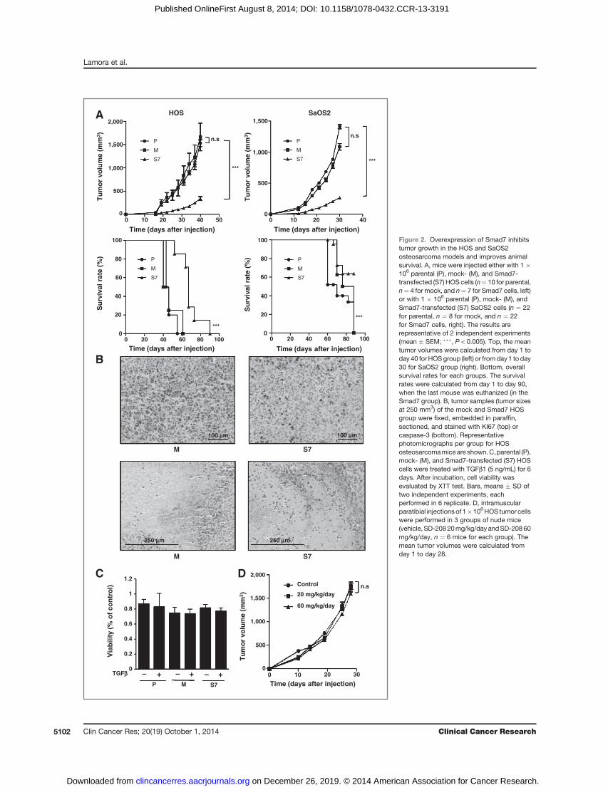
Figure 2. Overexpression of Smad7 inhibitstumor growth in the HOS and SaOS2osteosarcoma models and improves animalsurvival. A, mice were injected either with 1 �106 parental (P), mock- (M), and Smad7-transfected (S7) HOS cells (n¼ 10 for parental,n¼ 4 for mock, and n¼ 7 for Smad7 cells, left)or with 1 � 106 parental (P), mock- (M), andSmad7-transfected (S7) SaOS2 cells (n ¼ 22for parental, n ¼ 8 for mock, and n ¼ 22for Smad7 cells, right). The results arerepresentative of 2 independent experiments(mean � SEM; ���, P < 0.005). Top, the meantumor volumes were calculated from day 1 today 40 for HOSgroup (left) or fromday 1 to day30 for SaOS2 group (right). Bottom, overallsurvival rates for each groups. The survivalrates were calculated from day 1 to day 90,when the last mouse was euthanized (in theSmad7 group). B, tumor samples (tumor sizesat 250 mm3) of the mock and Smad7 HOSgroup were fixed, embedded in paraffin,sectioned, and stained with KI67 (top) orcaspase-3 (bottom). Representativephotomicrographs per group for HOSosteosarcomamice are shown.C,parental (P),mock- (M), and Smad7-transfected (S7) HOScells were treated with TGFb1 (5 ng/mL) for 6days. After incubation, cell viability wasevaluated by XTT test. Bars, means � SD oftwo independent experiments, eachperformed in 6 replicate. D, intramuscularparatibial injectionsof 1�106HOS tumor cellswere performed in 3 groups of nude mice(vehicle, SD-20820mg/kg/day andSD-20860mg/kg/day, n ¼ 6 mice for each group). Themean tumor volumes were calculated fromday 1 to day 28.
Lamora et al.
Clin Cancer Res; 20(19) October 1, 2014 Clinical Cancer Research5102
on December 26, 2019. © 2014 American Association for Cancer Research.clincancerres.aacrjournals.org Downloaded from
Published OnlineFirst August 8, 2014; DOI: 10.1158/1078-0432.CCR-13-3191

S7MPControl
A
B
****
n.s.
******
0
5
10
15 ***
n.s.
***
n.s.
0
5
10
15
0
2
4
6
To
tal b
on
evo
lum
e (m
m3 )
Ect
op
icb
on
evo
lum
e (m
m3 )
P M S7
S7MCtrl0
0.1
0.2
0.3
0
1
2
3
4
5
Tb
.th
ickn
ess
(mm
)T
b.n
um
ber
(/m
m)
P M S7
C
S7MCtrl
*
** n.s.D
***
n.s.
***
n.s.
0
5
10
15
20
0
5
10
15
To
tal b
on
e vo
lum
e (m
m3 )
P M S7
20
0
2
4
6
10
8
P M S7
Ect
op
icb
on
evo
lum
e (m
m3 )
6.0
6.5
7.0
7.5
8.0
8.5
6
7
8
9
10
11
Control 20 mg/kg 60 mg/kg Control 20 mg/kg 60 mg/kg
To
tal b
on
evo
lum
e (m
m3)
To
tal b
on
evo
lum
e (m
m3 )
HOS
SaOS2
Figure 3. Overexpression of Smad7 in tumor cells inhibits tumor-associated bone resorption. Mice were either injected with 1 � 106 parental (P), mock- (M),and Smad7-transfected (S7) HOS cells (n ¼ 10 for parental, n ¼ 4 for mock and n ¼ 7 for Smad7 cells) or with 1 � 106 parental (P), mock- (M), andSmad7-transfected (S7) SaOS2 cells (n¼ 7 for parental, n¼ 7 for mock, and n¼ 7 for Smad7). The results are representative of 2 independent experiments.A, 3D reconstructions of one representative tibia/fibula of each group (parental, mock, and Smad7 HOS cells) were performed when tumor sizeswere approximately 2,500 mm3 and compared with a healthy group bearing no tumors (control). B, left: graphs represent the total bone volume of eachindividual animal in agivengroup injectedwith eitherHOScells (top) orwithSaOS2cells (bottom; ���,P<0.005); right: graph represents themeanectopic bonevolume of each individual animal in a given group injected with either HOS cells (top) or with SaOS2 cells (bottom; ���, P < 0.005). C, histograms represent themean trabecular (Tb.) thickness (top) and the number of bone trabecular (bottom) in mock and Smad7 HOS groups compared with control groupcorresponding to mice bearing no tumors (���, P < 0.005; �, P < 0.05). D, intramuscular paratibial injections of 1 � 106 HOS tumor cells were performed in 3groups of nudemice (vehicle, SD-208 20mg/kg/day and SD-208 60mg/kg/day). The bone volumes of tibia weremeasured both at the leg having undergonethe injection of tumor cells (right) and at the counterpart legs (left) when tumor volume reached 1,000 mm3. Graphs represent the bone volume of eachindividual animal in a given group (��, P < 0.01).
Role of TGFb in Osteosarcoma
www.aacrjournals.org Clin Cancer Res; 20(19) October 1, 2014 5103
on December 26, 2019. © 2014 American Association for Cancer Research.clincancerres.aacrjournals.org Downloaded from
Published OnlineFirst August 8, 2014; DOI: 10.1158/1078-0432.CCR-13-3191

500 mm 500 mm
250 mm250 mm
TGFb
A
0
0.2
0.4
0.6
0.8
1
1.2
IL11 OPN
*
M S7
Rel
ativ
e R
AN
KL
mR
NA
leve
l
M S7
0
1
2
3
4
5
6
7
8
9
10
TGFb - +P M S7
- + - +
Rel
ativ
e R
AN
KL
mR
NA
leve
l
0
5
10
15
20
TGFb (ng/mL) 0 1 2 5
Rel
ativ
e R
AN
KL
mR
NA
leve
l
**
n.s
B
C D
M S7
0
1
2
3
4
5
6
PY
D (
nm
ol/L
)
M S7
*
0
1
2
3
4
5
6
7
8
9
IL11 OPN
- + - + - + - +
***
***
0
0.2
0.4
0.6
0.8
1
1.2
Rel
ativ
e m
RN
A le
vel
*** ***
M S7
250 mm 250 mm
250 mm 250 mm
Rel
ativ
e m
RN
A le
vel
Lamora et al.
Clin Cancer Res; 20(19) October 1, 2014 Clinical Cancer Research5104
on December 26, 2019. © 2014 American Association for Cancer Research.clincancerres.aacrjournals.org Downloaded from
Published OnlineFirst August 8, 2014; DOI: 10.1158/1078-0432.CCR-13-3191

the phosphorylation of Smad3 (Fig. 1B, right), to trans-activate the Smad3/4-specific reporter construct(CAGA)9-luc (Fig. 1C, right), and to stimulate the expres-sion of CTGF, PAI-1, and COL1A1 (Fig. 1D, bottom) inHOS cells and in SaOS2 cells (Supplementary Fig. S1).Note that in contrast to Smad7 overexpression, the SD-208 inhibitor is able to block the ability of TGFb toactivate the phosphorylation of ERK1/2 (SupplementaryFig. S1D, bottom).These results confirmed that both Smad7 overexpression
and SD-208 block the TGFb/Smad3 cascade in osteosarco-ma cells.
Overexpression of Smad7 in osteosarcoma cell linesdramatically inhibits in vivo tumor growthA preclinical experimental model of osteosarcoma
induced by paratibial injection of osteosarcoma cellswas developed. Smad7 overexpression in HOS or SaOS2cells inhibited tumor growth in both models (Fig. 2A,top). The mean tumor size at day 40 in mice injectedwith parental or mock-transfected HOS cells was 1,531.8� 73.4 mm3 and 1,663.9 � 297.0 mm3 respectively,compared with only 341.5 � 43.4 mm3 in mice injectedwith HOS-S7 cells (means � SEM, P < 0.005; Fig. 2A, topHOS panel). Similar results were obtained in the SaOS2model (Fig 2A, top SaOS2 panel). Consequently, Smad7overexpression resulted in an increased animal survivalin both models (Fig. 2A, bottom). In this context, immu-nohistochemical staining for the proliferative markerKi67 in tumor samples from mice showed that Smad7overexpression decreased cell proliferation as comparedwith the mock-transfected group (Fig. 2B, top) when thetumor sizes reached 250 mm3. By contrast, caspase-3immunostaining of the same samples showed no signif-icant difference between mice injected with Smad7-trans-fected cells and animals receiving mock-transfected cells(Fig. 2B, bottom). To better understand the mechanismsunderlying the effect of Smad7 on osteosarcoma tumorgrowth, we next carried out in vitro experiments. Inter-estingly, treatment of osteosarcoma cells with TGFb (5ng/mL) did not affect osteosarcoma cell proliferationeven after 6 days of TGFb treatment, whether the cellsexpressed Smad7 or not (Fig. 2C). In contrast to the effectof Smad7 overexpression, the treatment of mice with SD-
208 (20 or 60 mg/kg/day) does not affect the in vivotumor growth of osteosarcoma (Fig. 2D).
These results demonstrate that overexpression of Smad7reduced in vivo tumor growth and suggest that this effect isnot because of a direct effect of Smad7 on osteosarcoma cellproliferation.
Smad7 overexpression in HOS and SaOS2osteosarcoma cells inhibits tumor-associated boneresorption
Because osteosarcoma-associated alteration of boneremodeling plays a central role in the development andprogression of osteosarcomabone tumors, we evaluated theability of Smad7 andSD-208 to alter tumor-associated boneremodeling. The microarchitecture of bone in mice bearingosteosarcoma tumors was first examined when the tumorsizes reached 2,500 mm3 using a high-resolution X-raymicro-CT system.
Visual inspection of the 3D reconstructions of the tibiasuggests that Smad7 overexpression enhanced the tumor-associated bone formation in HOS osteosarcoma models(Fig. 3A). Indeed, the total bone volume in mice injectedwith parental or mock-transfected HOS cells was 9.34 �0.08 and 8.77 � 0.15 mm3 respectively, compared with11.44 � 0.28 mm3 in mice bearing HOS-S7 tumors (P <0.005; Fig. 3B, HOS left panel). Similarly, Smad7 over-expression enhanced total bone volume in mice injectedwith SaOS2 cells (Fig. 3B, SaOS2 left panel).
We next determined whether this increase in total bonevolume was due either to direct new bone formation(ectopic bone) and/or to an inhibition of bone resorption.As shown in Fig. 3B (HOS right panel), the ectopic bonevolume in mice injected with parental or mock-transfectedHOS cells was significantly lower than in mice bearingHOS-S7 tumors (1.91 � 0.11 mm3 and 1.43 � 0.26 mm3
vs. 3.77 � 0.18 mm3 respectively, P < 0.005). Similarly,Smad7 enhanced ectopic bone volume in the SaOS2model (P < 0.005; Fig. 3B, SaOS2 right panel). We thenanalyzed the ability of Smad7 to alter bone osteolysis byevaluating the trabecular number (Tb.N) and trabecularthickness (Tb.Th) when tumor sizes were around 250 mm3
, because at higher volumes (1,000 mm3 or 2,500 mm3)trabecular bone is completely destroyed by the tumor cells(data not shown). As shown in Fig. 3C (top), mice injected
Figure 4. Overexpression of Smad7 in osteosarcoma reduces osteoclast activity. A, tumor samples (tumor sizes at 1,000 mm3) of the mock and Smad7group were fixed, embedded in paraffin, sectioned, and stained for TRAP (red stained cells, top), osteocalcin (middle), and RANKL (bottom).Representative photomicrographs per group for HOS osteosarcoma mice are shown. B, RANKL mRNA steady-state levels were determined byquantitative RT-PCR in the presence or absence of TGFb as indicated. Bars, mean � SD of at least three independent experiments carried out induplicate (top). Parental (P), mock- (M), and Smad7-transfected (S7) HOS cells were treated with TGFb1 (5 ng/mL) for 24 hours. After incubation,RANKL mRNA steady-state levels were determined by quantitative RT-PCR (middle). RNA was extracted from tumor biopsies of mice injected withmock- (black) or Smad7 (gray) transfected HOS cells. RANKL mRNA steady-state levels were determined by quantitative RT-PCR (bottom). Bars,means � SD of at least 3 independent experiments, performed in duplicate (�, P < 0.05; ��, P < 0.01). C, concentrations of pyridinoline in mice serumof the mock and Smad7 groups were measured using the MicroVue Serum PYD EIA Kit. Bars, means � SEM of two independent experiments,performed in duplicate (�, P < 0.05). D, left: treatment of mock- (black) and Smad7-transfected (gray) HOS cells with TGFb1 (5 ng/mL) for 24 hours. Afterincubation, IL11 and OPNmRNA steady-state levels were determined by quantitative RT-PCR. Bars, mean � SD of at least 3 independent experimentscarried out in duplicate (���, P < 0.005); right: RNA was extracted from tumor biopsies of mice injected with mock- (black) and Smad7 (gray) transfectedHOS cells. IL11 and OPN mRNA steady-state levels were determined by quantitative RT-PCR. Bars, mean � SD of 2 independent experiments carriedout in duplicate (���, P < 0.005).
www.aacrjournals.org Clin Cancer Res; 20(19) October 1, 2014 5105
Role of TGFb in Osteosarcoma
on December 26, 2019. © 2014 American Association for Cancer Research.clincancerres.aacrjournals.org Downloaded from
Published OnlineFirst August 8, 2014; DOI: 10.1158/1078-0432.CCR-13-3191

A
B***
n.s
MP S7
HOS P
HOS M
HOSS7
0
2
4
6
8
P M S70
2
4
6
8
Nu
mb
ero
f p
ulm
on
ary
met
asta
sis
1 mm 1 mm 1 mm
MP S7
0
1
2
3
4
5
Control 20 mg/kg/d 60 mg/kg/d
Nu
mb
ero
f p
ulm
on
ary
met
asta
sis ***
***
S7P
C
60 mg/kg/day20 mg/kg/dayControl
M
100 mm 100 mm
100 mm 100 mm
100 mm
100 mm
SAOS2 P
SAOS2 M
SAOS2 S7
0
2
4
6n.s
P M S70
2
4
6
8
Nu
mb
ero
f p
ulm
on
ary
met
asta
sis **
D
Rel
ativ
e m
RN
Ale
vel
Rel
ativ
e m
RN
Ale
vel
0
0.2
0.4
0.6
0.8
1
1.2
CXCR4ANGPTL4VEGF0
0.2
0.4
0.6
0.8
1
1.2
CXCR4ANGPTL4VEGF
Figure 5. Overexpression of Smad7in osteosarcoma and treatment ofmice with SD-208 block lungmetastasis development. Micewere either injected with parental(P), mock- (M), and Smad7-transfected (S7) osteosarcomacells (HOS or SaOS2) or injectedwith parental-HOS cells andtreated with vehicle or SD-208 asindicated. Mice were sacrificedwhen tumor sizes reached 2,500mm3 and lungs were removed. A,photographs of representativelungs of P, M, and S7 HOS group.Arrows point to lung metastasis(top). H&E staining of lungs.Arrows indicate metastatic tumorcells (bottom). B, graphs indicateindividual (dots) and mean (lines)numbers of lung metastasismeasured in lungs fromeach group(���, P < 0.005; ��, P < 0.01),respectively for P, M, and S7-HOSmodel (left), P, M, and S7-SAOS2model (middle) and for micetreated or not with SD-208 (right).C, tumor samples (tumor sizes at2,500 mm3) of the P, M, and S7group (top) and from mice treatedor not with SD-208 as indicated(bottom) were fixed, embedded inparaffin, sectioned, and stainedwith CD146. Representativephotomicrographs per group forHOS osteosarcoma mice areshown. D, RNAwas extracted fromtumor biopsies of mice injectedwith P (black), M (gray), and S7(white) transfected HOS cells (left)or from tumor biopsies from micetreated with SD-208 at 20 mg/kg/day (gray), SD-208at 60mg/kg/day(white), or with vehicle (black; right).VEGF, ANGPTL4, and CXCR4mRNA steady-state levels weredetermined by quantitative RT-PCR. Bars, mean � SD of 2independent experiments carriedout in duplicate.
Clin Cancer Res; 20(19) October 1, 2014 Clinical Cancer Research5106
Lamora et al.
on December 26, 2019. © 2014 American Association for Cancer Research.clincancerres.aacrjournals.org Downloaded from
Published OnlineFirst August 8, 2014; DOI: 10.1158/1078-0432.CCR-13-3191

with mock-transfected HOS cells had a lower Tb.Th thanmice bearing HOS-S7 tumors (0.15 � 0.01 mm vs. 0.22 �0.01 mm, P < 0.005). Moreover, the Tb.N was also lower inthe mock group than in the HOS-S7 tumor-bearing group(2.92 � 0.03 mm vs. 4.18 � 0.02 mm; P < 0.005; Fig. 3C,bottom). Note that Tb.Th and Tb.N in mice bearing HOS-S7 tumors are similar to those observed in healthy controlmice. Interestingly, although the treatment of mice withSD-208 affects bone remodeling with a significant increaseof tibia bone volume in the absence of tumor (Fig. 3D, left)as previously described (30), the SD-208 does not affectsignificantly the tumor-associated bone remodeling (Fig.3D, right).These results demonstrate that in contrast with SD-208
mice treatment, overexpression of Smad7 in osteosarco-ma cells both decreased tumor-associated bone osteolysisand promoted tumor-associated bone formation.
Overexpression of Smad7 in HOS and SaOS2osteosarcoma cells reduces osteoclast activityThe activity of osteoclasts and osteoblasts, 2 cell lineages
implicated in bone remodeling, was then assessed duringthe early stages of tumor growth (tumor size �250 mm3 or�1,000 mm3). TRAP staining in sections of tumor-bearingtibia showed that HOS-S7 tumor cells reduced the numberof TRAPþ multinucleated cells at the interface betweentumor and cortical bone (Fig. 4A, top) and in the growthplate (not shown), relative to the control conditions at thesame tumor size (1,000 mm3). Similar results wereobtained when tumors had reached 250 mm3 (data notshown). By contrast, osteocalcin (Fig. 4A, middle) andosterix (data not shown) immunostaining of the samesamples showed no significant difference between miceinjectedwith Smad7-transfected cells and animals receivingmock-transfected cells.To understand the effect of Smad7 on osteoclast activ-
ity, we analyzed RANKL gene expression in HOS osteo-sarcoma cell lines. qRT-PCR analysis indicated thatthe mRNA steady-state level of RANKL was increased inresponse to TGFb and that Smad7 overexpression pre-vented such induction (Fig 4B, top and middle, respec-tively). Moreover, Smad7 overexpression decreasedRANKL production by HOS tumor cells (Fig. 4A, bottom)evaluated by immunohistochemical experiments. Inaccordance with this observation, HOS-S7 bone tumorsexpressed significantly lower mRNA levels of RANKL (Fig.4B, bottom). Finally, the degradation of collagen evalu-ated by the measure of pyridinoline excretion in miceserum is decreased when Smad7 is overexpressed in oste-osarcoma cells (Fig. 4C).In addition to RANKL, the expression of other TGFb
target genes implicated in bone remodeling (18, 20, 21)such as interleukin-11 (IL11) and osteopontin (OPN) isdecreased when Smad7 is overexpressed. As shownin Fig. 4D, q-PCR analysis indicated that the IL11 andOPN expressions were increased in response to TGFb andthat Smad7 overexpression prevented such induction(Fig 4D, left). HOS-S7 bone tumors expressed signifi-
cantly lower levels of IL11 and OPN mRNA (Fig. 4D,right).
These results demonstrate that Smad7 overexpressiondecreases osteoclast activity and thus bone osteolysis atleast in part via the modulation of osteolytic genes such asRANKL.
Overexpression of Smad7 in HOS and SaOS2osteosarcoma cells or treatment of mice with SD-208inhibits dissemination of pulmonary metastasis
To evaluate the effect of Smad7 on pulmonary metas-tasis development (Fig. 5A), the lungs of mice wereremoved when primary tumor volumes reached 2,500mm3. As shown in Fig. 5B, a high incidence of pulmo-nary metastasis was observed in mice inoculated withparental or mock-transfected HOS cells, respectively 9 of10 (90%) and 4 of 4 (100%) mice. By contrast, only 1 of7 (14%) mice bearing HOS-S7 cells developed lungmetastasis (Fig. 5B, left). As shown in Fig. 5B (middle),similar results were obtained in the SaOS2 model.Remarkably, the treatment of mice with SD-208 inhibitsthe development of lung metastasis. As shown in Fig. 5B(right), 5 of 6 (83.3%) mice treated with vehicle (controlgroup) developed lung metastasis. By contrast, only 1 of6 (16.7%) mice treated with 20 mg/kg/day developedlung metastasis, and no mice treated with 60 mg/kg/daydeveloped lung metastasis. Interestingly, immunohisto-chemical staining for the endothelial marker CD146 inmice tumor samples showed that Smad7 overexpressionor SD-208 treatment dramatically decreased the angio-genic process as compared respectively with the parentaland mock-transfected groups or with the untreatedgroup (Fig. 5C). In addition, qPCR analysis from micebiopsies indicated that the expression by tumor cells ofVEGF (implicated in the angiogenic process), andCXCR4 and ANGPTL4 (respectively identified as keyplayers to prime breast cancer cells for metastasis towardthe lungs and/or the bones) were both reduced whenSmad7 was overexpressed or when mice were treatedwith SD-208 (Fig. 5D).
The influence of Smad7 overexpression in osteosarcomacells or the treatment of tumor cells with SD-208 was thenexamined on several aspects of tumor cell behavior in vitro.As shown in Fig. 6 and in Supplementary Fig. S2, Smad7overexpression in HOS and SaOS2 cells or treatment ofHOS cells with SD-208 lead to a strongly reduced capacityof TGFb to stimulate cell migration (Fig. 6A and Supple-mentary Fig. S2A) and invasion (Fig. 6B and Supplemen-tary Fig. S2B). In addition, exogenous TGFb-inducedsecretion of the active form of matrix metalloproteinaseMMP2 was strongly diminished both in Smad7-trans-fected cells and in cells treated with SD-208 (Fig. 6C andsupplementary Fig. S2C). qRT-PCR analysis indicated areduction of TGFb-induced MMP2 mRNA levels inSmad7-transfected– or SD-208–treated cells (Fig. 6D,top). MMP2 mRNA steady-state levels were increased byapproximately 5-fold in parental and mock-transfectedHOS cells, but only by 2-fold in HOS-S7 cells after
www.aacrjournals.org Clin Cancer Res; 20(19) October 1, 2014 5107
Role of TGFb in Osteosarcoma
on December 26, 2019. © 2014 American Association for Cancer Research.clincancerres.aacrjournals.org Downloaded from
Published OnlineFirst August 8, 2014; DOI: 10.1158/1078-0432.CCR-13-3191

Cel
l nu
mb
erC
ell n
um
ber
Cel
l nu
mb
erC
ell n
um
ber
S7
P
M
Ctrl +
0
20
40
60
80
100
120
140
- +P M S7
- + - +0
10
20
30
40
50
60
70
80
90
100
A
B
D
TGFb
TGFb
TGFb
TGFb
TGFb
TGFb
TGFb
TGFb
TGFb
TGFb- +P M S7
- + - +
***n.s
***n.s
Ctrl +
S7
P
M
0
20
40
60
80
100
120
140
0
20
40
60
80
100
120
- +
- SD-208
- +
- +
- SD-208
- +
***
***
- +P M S7
- + - +
C
MMP9
pro-MMP2
- + - +- SD-208
Active MMP2
MMP9
pro-MMP2
Active MMP2
Rel
ativ
e M
MP
2 m
RN
A le
vel
- +P M S7
- + - +0
1
2
3
4
5
6
7
*
n.s
0
1
2
3
4
5
6
7
Rel
ativ
e M
MP
2 m
RN
Ale
vel
- +
- SD-208
- +
***
Rel
ativ
e M
MP
2 m
RN
A le
vel
Rel
ativ
e M
MP
2 m
RN
A le
vel
0
0.2
0.4
0.6
0.8
1
1.2
1.4
0
0.2
0.4
0.6
0.8
1
1.2
P M S7
**
Control
Clin Cancer Res; 20(19) October 1, 2014 Clinical Cancer Research5108
Lamora et al.
on December 26, 2019. © 2014 American Association for Cancer Research.clincancerres.aacrjournals.org Downloaded from
Published OnlineFirst August 8, 2014; DOI: 10.1158/1078-0432.CCR-13-3191

treatment of osteosarcoma cells with TGFb (5 ng/mL) for24 hours. Similar results were obtained when Smad7 wasoverexpressed in SaOS2 cells (Supplementary Fig. S2D) orwhen osteosarcoma cells were treated with SD-208 (Fig.6D, right and Supplementary Fig. S2D, right). In addi-tion, both HOS-S7 bone tumors and HOS cells from micetreated with SD-208 expressed significantly lower levels ofMMP2 (Fig. 6D, bottom).These results demonstrated that overexpression of Smad7
in osteosarcoma cells or treatment of mice with SD-208blocks the formation of lung metastasis.
DiscussionIncreased TGFb1 mRNA and/or protein expression has
been correlated with a wide range of cancers such ascolorectal cancer, gastric carcinoma, or prostate cancer(31–34). This increase in serum TGFb1 production and/or TGFb1 staining in tumor cells has been associated withdisease progression to metastasis in these carcinomas(31). Here, we demonstrated that high concentrations ofTGFb1 and TGFb2 measured in serum of patients isassociated with osteosarcoma disease. In addition, usingclinical samples, we demonstrated that the Smad3 cas-cade is activated in osteosarcoma cells particularly inhigh-risk patients when lung metastasis are detected atdiagnosis. Previous studies have reported that high levelsof TGFb1 mRNA in tumor cells are associated with high-grade osteosarcoma, which shows an aggressive behaviorand frequently metastasizes to lung or other sites (24).These observations together with our results suggest thathigh levels of TGFb1 in serum could be associated with apoor prognosis in osteosarcoma.Following these observations, we first inhibited the
Smad signaling cascade in osteosarcoma cells via theoverexpression of the inhibitory Smad, Smad7, and thenwe used the chemical inhibitor of TbRI, SD-208, tospecifically inhibit the signaling cascade downstream the
receptor TbRI. By using various in vitro approaches, wedemonstrated that Smad7 overexpression and SD-208efficiently inhibit the TGFb transcriptional responsemediated by Smad3/4 in 2 human osteosarcoma celllines, HOS and SaOS2.
Using a murine model of osteosarcoma induced byparatibial injection of osteosarcoma cells overexpressingSmad7, we then demonstrated that Smad7 overexpres-sion slows primary tumor growth, a process associatedwith a reduction of the immunohistochemical stainingfor the proliferative marker Ki67. Because TGFb is acytokine widely implicated in the control of cell prolif-eration (17), the effect of TGFb and overexpression ofSmad7 was studied on osteosarcoma cell proliferation invitro. In contrast to previous observations that demon-strated an effect of TGFb on cell proliferation (35, 36), noeffect of TGFb and/or Smad7 overexpression wasobserved on the in vitro proliferation rate of osteosarcomacells under our experimental conditions, suggesting thatSmad7 does not directly affect the proliferation of oste-osarcoma cells but rather affects the tumor microenvi-ronment indirectly involved in the control of tumor cellproliferation.
In this context, because osteosarcoma-associatedalterations of bone remodeling play a central role inthe development and progression of osteosarcoma, westudied the effect of Smad7 on bone remodeling. Weclearly demonstrated that Smad7 overexpression in oste-osarcoma cells slows bone destruction associated withthe tumor growth. In this context, we demonstrated thatthis process was mainly associated with a reduction oftrabecular bone destruction during the early stages oftumor growth (when tumor volumes were below 250mm3) and with an increase of ectopic bone formationduring the late stages of tumor growth (when tumorvolumes were greater than 1000 mm3). The presence of a"vicious cycle" established between tumor proliferationand paratumor osteolysis plays a crucial role in the
Figure 6. Overexpression of Smad7 in osteosarcoma cells or treatment of cells with SD-208 inhibit the ability of TGFb to induce osteosarcoma cellmigration and invasion. A and B, left: 30,000 parental, mock-, or Smad7-transfected HOS cells pretreated during 24 hours with 5 ng/mL TGFb wereseeded onto the upper surface of uncoated (A) or transwell coated with 2 mg Matrigel (B) inserts. Forty-eight hours after incubation in the presenceor absence of TGFb (5 ng/mL), the cells on the underside of the membrane were fixed, stained with "cristal violet," and counted by bright-fieldmicroscopy in 5 random fields (magnification: �200). Bars, mean � SD of at least 3 independent experiments carried out in duplicate (���, P < 0.005);middle: 30,000 parental HOS cells pretreated during 24 hours with 5 ng/mL TGFb in the presence or absence of SD-208 (as indicated) wereseeded onto the upper surface of uncoated (A) or transwell coated with 2 mg Matrigel (B) inserts. Forty-eight hours after incubation in the presence orabsence of TGFb (5 ng/mL) and SD-208 (as indicated), the cells on the underside of the membrane were fixed, stained with "cristal violet," andcounted by bright-field microscopy in 5 random fields (magnification: �200). Bars, mean � SD of at least 3 independent experiments carried out induplicate (���, P < 0.005); right: photographs of representative random fields (magnification: �200) of each group. C, top: zymography analysis ofconditioned media from 48 hours serum-free cultures of HOS-P, -M, and -S7 cells treated with 5 ng/mL TGFb or untreated. A Coomassie bluestained gel representative of 3 independent experiments is shown; bottom: zymography analysis of conditioned media from 48 hours serum-freecultures of HOS cells treated with 5 ng/mL TGFb in the presence or absence of SD-208 (10 mmol/L). A Coomassie blue stained gel representativeof 3 independent experiments is shown. D, top: HOS-P, -M, and -S7 cells were incubated with TGFb1 (5 ng/mL) for 24 hours (left). HOS cellswere incubated with TGFb1 (5 ng/mL) in the presence or absence of SD-208 (10 mmol/L) for 24 hours (right). After incubation,MMP2mRNA steady-statelevels were determined by quantitative RT-PCR. Bars, mean � SD of at least 3 independent experiments carried out in duplicate (�, P < 0.05;���, P < 0.005). Bottom, RNA was extracted from tumor biopsies of mice injected with parental (black), mock- (gray), and Smad7- (white) transfectedHOS cells (left). RNA was extracted from tumor biopsies of mice injected with HOS cells and treated with SD-208 at 20 mg/kg/day (gray),SD-208 at 60 mg/kg/day (white), or with vehicle (black; right).MMP2mRNA steady-state levels were determined by quantitative RT-PCR. Bars, mean �SD of 2 independent experiments carried out in duplicate (�, P < 0.05).
www.aacrjournals.org Clin Cancer Res; 20(19) October 1, 2014 5109
Role of TGFb in Osteosarcoma
on December 26, 2019. © 2014 American Association for Cancer Research.clincancerres.aacrjournals.org Downloaded from
Published OnlineFirst August 8, 2014; DOI: 10.1158/1078-0432.CCR-13-3191

development of primary bone tumors (37). Cancer cellsproduce soluble factors that activate directly or indirectlyvia osteoblasts, the osteoclast differentiation, and mat-uration (38, 39). In turn, during bone degradation,osteoclasts allow the release of growth factors stored inthe mineralized bone matrix that are able to stimulatetumor growth. In this context, we demonstrated that theresulting increase in bone volume observed after Smad7overexpression in osteosarcoma cells is due in large partto the inhibition of osteoclast activity. The decrease ofTRAP activity at the growth plate level, which is not indirect contact with the tumor, suggests that Smad7 over-expression affects the ability of the tumor cells to pro-duce a soluble factor able to regulate osteoclast activity.Here, we clearly demonstrated that Smad7 overexpres-sion in osteosarcoma cells inhibits their ability to pro-duce RANKL or IL11, 2 cytokines that play a central rolein bone osteolysis process (40). Together, these resultssuggest that Smad7 slows the tumor growth by acting atleast at the tumor environment level, by inhibiting thetumor associated bone osteolysis.
Surprisingly, we did not observe a significant effect ofSD-208 on tumor growth. Several hypotheses can beproposed to explain this difference between Smad7and SD-208 effects on tumor growth. First, we cannotrule out that Smad7 may also exert some of its actionindependently from its role as a TGFb signaling inhib-itor. Indeed, Smad7 is able to inhibit other signalingpathways such as the cascade of bone morphogeneticprotein family members (BMP), highly involved in boneformation (41). This hypothesis seems unlikely becausethe BMPs are known to promote bone formation. Thus,we can hypothesize that BMP inhibitors should promotebone degradation by inhibiting the bone formation incontrast to the results observed by overexpression ofSmad7. Second, Smad7 might have several distinct func-tions in cellular signaling (12). For example it has beenshown that Smad7 overexpression is able to potentiateapoptosis in prostate carcinoma and in PC-3U cells (12).In addition, Smad7 is also able to activate some signalingpathways such as the JNK cascade (42). Although wehave not observed an effect of Smad7 on the prolifera-tion and apoptosis of osteosarcoma cells, or on theability of TGFb to stimulate the MAPK pathway, wecannot rule out this hypothesis. Third, the inefficiencyof SD-208 on tumor growth can be explained by itsinability to reduce the tumor-associated bone osteolysis.Indeed, although a systemic treatment of mice with SD-208 promotes bone formation in absence of tumor aspreviously described (30), this systemic treatment doesnot seem to reduce the tumor associated bone osteolysisin contrast with the overexpression of Smad7 effect inosteosarcoma cells. In addition, the HOS mice modelused in these experiments is a high aggressive model witha fast bone degradation associated with tumor growth.The effectiveness of a local mice treatment with SD-208directly into the tumor cells using a less aggressive modelshould be tested.
Finally, we showed that both Smad7 overexpressionand SD-208 strongly affect the ability of the primary bonetumor to develop lung metastasis, demonstrating thecrucial role of TGFb/Smad signaling pathway in themetastatic process of osteosarcoma. During the last dec-ades, the role of TGFb in the metastatic process of carci-nomas has been widely described. A major step in thisprocess is the ability of TGFb to stimulate epithelial-to-mesenchymal transition and thus the ability of tumorcells to invade adjacent tissues (17). In the context ofosteosarcoma cells that are from mesenchymal origin, wespecifically demonstrated that Smad7 overexpression andSD-208 block the ability of TGFb to stimulate osteosar-coma migration and invasion. In this context, we clearlydemonstrated that Smad7 and SD-208 are able to blockTGFb-induced MMP2 activity, mainly involved in theinvasion process such as described in the context ofmelanoma bone metastasis (19, 20). Another major stepin the metastatic process is the ability of TGFb to stim-ulate tumor-associated angiogenesis and thus the dissem-ination of tumor cells into the bloodstream (17). In thiscontext, we clearly demonstrated that both Smad7 andSD-208 reduce the angiogenic process as shown byimmunohistochemical staining for the endothelial mark-er CD146. In addition, both Smad7 and SD-208 inhibitTGFb-induced VEGF expression, mainly involved in theangiogenic process. Moreover, both Smad7 and SD-208are able to inhibit the ability of TGFb to stimulate theexpression of ANGPLT4 and CXCR4 identified as keyplayers to prime breast cancer cells for metastasis respec-tively toward the lungs (43) and toward the bones or thelungs (44).
In conclusion, this report provides evidence that blockingTGFb signalingmay represent a novel therapeutic approachto treat lung metastasis in patients with osteosarcoma,which have a poor prognosis.
Disclosure of Potential Conflicts of InterestNo potential conflicts of interest were disclosed.
Authors' ContributionsConception and design: A. Lamora, F. Redini, F. VerrecchiaDevelopmentofmethodology:A. Lamora, J. Talbot, G. Bougras, J. Amiaud,M. Leduc, J. Chesneau, F. VerrecchiaAcquisitionofdata (provided animals, acquired andmanagedpatients,provided facilities, etc.): A. Lamora, M.C. Le Deley, M.F. Heymann,F. VerrecchiaAnalysis and interpretation of data (e.g., statistical analysis, biosta-tistics, computational analysis):A. Lamora, J. Talbot, J. Amiaud,M. Leduc,J. Chesneau, D. Heymann, F. Redini, F. VerrecchiaWriting, review, and/or revision of the manuscript: A. Lamora,V. Stresing, D. Heymann, F. Redini, F. VerrecchiaAdministrative, technical, or material support (i.e., reporting or orga-nizing data, constructing databases): A. Lamora, J. Talbot, J. Amiaud,M. Leduc, J. Chesneau, J. Taurelle, F. VerrecchiaStudy supervision: A. Lamora, M.C. Le Deley, M.F. Heymann, F. Verrecchia
AcknowledgmentsThe authors thank Nicolas Pelletier, Arnaud Tessier, and Jacques
Lebreton (CEISAM UMR6230, Nantes University, France) for the synthe-sis of SD-208.
Clin Cancer Res; 20(19) October 1, 2014 Clinical Cancer Research5110
Lamora et al.
on December 26, 2019. © 2014 American Association for Cancer Research.clincancerres.aacrjournals.org Downloaded from
Published OnlineFirst August 8, 2014; DOI: 10.1158/1078-0432.CCR-13-3191

Grant SupportThis work was supported by INSERM, Ligue contre le Cancer (Allocation
doctorale to A. Lamora), Ligue contre le Cancer (Equipe labellis�ee 2012), andFondation Bettencourt Schueller.
The costs of publication of this article were defrayed in part by thepayment of page charges. This article must therefore be hereby marked
advertisement in accordance with 18 U.S.C. Section 1734 solely to indicatethis fact.
ReceivedNovember 28, 2013; revised July 8, 2014; accepted July 18, 2014;published OnlineFirst August 8, 2014.
References1. Mirabello L, Troisi RJ, Savage SA. International osteosarcoma inci-
dence patterns in children and adolescents, middle ages and elderlypersons. Int J Cancer 2009;125:229–34.
2. Broadhead ML, Clark JCM, Myers DE, Dass CR, Choong PFM. Themolecular pathogenesis of osteosarcoma: a review. Sarcoma 2011;2011:1–12.
3. Yarber JL, Agulnik M. Targeted therapies in bone sarcomas: currentapproach and future directions. Expert Opin Investig Drugs 2011;20:973–9.
4. Meyers PA, Gorlick R. Osteosarcoma. Pediatr Clin North Am 1997;44:973–89.
5. Hughes DPM. Strategies for the targeted delivery of therapeutics forosteosarcoma. Expert Opin Drug Deliv 2009;6:1311–21.
6. Anninga JK, Gelderblom H, Fiocco M, Kroep JR, Taminiau AHM,Hogendoorn PCW, et al. Chemotherapeutic adjuvant treatment forosteosarcoma: where do we stand? Eur J Cancer 2011;47:2431–45.
7. Ando K, Mori K, Corradini N, Redini F, Heymann D. Mifamurtide for thetreatment of nonmetastatic osteosarcoma. Expert Opin Pharmacother2011;12:285–92.
8. PosthumaDeBoer J, Witlox MA, Kaspers GJL, van Royen BJ.Molecular alterations as target for therapy in metastatic osteo-sarcoma: a review of literature. Clin Exp Metastasis 2011;28:493–503.
9. Eccles SA, Welch DR. Metastasis: recent discoveries and novel treat-ment strategies. Lancet 2007;369:1742–57.
10. Feng X-H, Derynck R. Specificity and versatility in TGF-b signalingthrough Smads. Annu Rev Cell Dev Biol 2005;21:659–93.
11. Shi Y, Massagu�e J. Mechanisms of TGF-b signaling from cell mem-brane to the nucleus. Cell 2003;113:685–700.
12. Park SH. Fine tuning and cross-talking of TGF-b signal by inhibitorySmads. J Biochem Mol Biol 2005;38:9–16.
13. Katsuno Y, Lamouille S, Derynck R. TGF-b signaling and epithelial-mesenchymal transition in cancer progression. Curr Opin Oncol2013;25:76–84.
14. Nakao A, Afrakhte M, Mor�en A, Nakayama T, Christian JL, Heuchel R,et al. Identification of Smad7, a TGF-b-inducible antagonist of TGF-bsignalling. Nature 1997;389:631–5.
15. Hayashi H, Abdollah S, Qiu Y, Cai J, Xu YY, Grinnell BW, et al. TheMAD-related protein Smad7 associates with the TGF-b receptorand functions as an antagonist of TGF-b signaling. Cell 1997;89:1165–73.
16. Verrecchia F, Mauviel A, Farge D. Transforming growth factor-b sig-naling through the Smad proteins: role in systemic sclerosis. Auto-immun Rev 2006;5:563–9.
17. Meulmeester E, Ten Dijke P. The dynamic roles of TGF-b in cancer.J Pathol 2011;223:205–18.
18. Yin JJ, Selander K, Chirgwin JM, Dallas M, Grubbs BG,Wieser R, et al.TGF-b signaling blockade inhibits PTHrP secretion by breast cancercells and bone metastases development. J Clin Invest 1999;103:197–206.
19. Javelaud D, Delmas V, M€oller M, Sextius P, Andr�e J, Menashi S,et al. Stable overexpression of Smad7 in human melanoma cellsinhibits their tumorigenicity in vitro and in vivo. Oncogene 2005;24:7624–9.
20. Javelaud D, Mohammad KS, McKenna CR, Fournier P, Luciani F,Niewolna M, et al. Stable overexpression of Smad7 in humanmelanoma cells impairs bone metastasis. Cancer Res 2007;67:2317–24.
21. MohammadKS, JavelaudD, Fournier PGJ, NiewolnaM,McKennaCR,Peng XH, et al. TGF-b-RI kinase inhibitor SD-208 reduces the devel-
opment and progression of melanoma bone metastases. Cancer Res2011;71:175–84.
22. Mundy GR. Metastasis to bone: causes, consequences and thera-peutic opportunities. Nat Rev Cancer 2002;2:584–93.
23. Kozlow W, Guise TA. Breast cancer metastasis to bone: mechanismsof osteolysis and implications for therapy. J Mammary Gland BiolNeoplasia 2005;10:169–80.
24. Franchi A, Arganini L, Baroni G, Calzolari A, Capanna R, CampanacciD, et al. Expression of transforming growth factor-b isoforms inosteosarcoma variants: association of TGF-b1 with high-grade oste-osarcomas. J Pathol 1998;185:284–9.
25. Mohseny AB, Cai Y, Kuijjer M, XiaoW, van den Akker B, de Andrea CE,et al. The activities of Smad and Gli mediated signalling pathways inhigh-grade conventional osteosarcoma. Eur J Cancer Oxf Engl 19902012;48:3429–38.
26. Matsuyama S, Iwadate M, Kondo M, Saitoh M, Hanyu A, Shimizu K,et al. SB-431542 and Gleevec inhibit transforming growth factor-b-induced proliferation of human osteosarcoma cells. Cancer Res2003;63:7791–8.
27. Talbot J, Brion R, Picarda G, Amiaud J, Chesneau J, Bougras G, et al.Loss of connexin43 expression in Ewing's sarcoma cells favors thedevelopment of the primary tumor and the associated bone osteolysis.Biochim Biophys Acta 2013;1832:553–64.
28. Dennler S, Itoh S, Vivien D, ten Dijke P, Huet S, Gauthier JM. Directbinding of Smad3 and Smad4 to critical TGF-b-inducible elements inthe promoter of human plasminogen activator inhibitor-type 1 gene.EMBO J 1998;17:3091–100.
29. HeymannD,OryB, Blanchard F,HeymannM-F,CoipeauP,Charrier C,et al. Enhanced tumor regression and tissue repair when zoledronicacid is combined with ifosfamide in rat osteosarcoma. Bone 2005;37:74–86.
30. Mohammad KS, Chen CG, Balooch G, Stebbins E, McKenna CR,Davis H, et al. Pharmacologic inhibition of the TGF-b type I receptorkinase has anabolic and anti-catabolic effects on bone. PLoS ONE2009;4:e5275.
31. Levy L, Hill CS. Alterations in components of the TGF-b superfamilysignaling pathways in human cancer. Cytokine Growth Factor Rev2006;17:41–58.
32. Dalal BI, Keown PA, Greenberg AH. Immunocytochemical localizationof secreted transforming growth factor-b1 to the advancing edges ofprimary tumors and to lymph node metastases of human mammarycarcinoma. Am J Pathol 1993;143:381–9.
33. Tsushima H, Kawata S, Tamura S, Ito N, Shirai Y, Kiso S, et al. Highlevels of transforming growth factor-b1 in patients with colorectalcancer: association with disease progression. Gastroenterology 1996;110:375–82.
34. Wikstr€om P, Stattin P, Franck-Lissbrant I, Damber JE, Bergh A.Transforming growth factor-b1 is associated with angiogenesis,metastasis, and poor clinical outcome in prostate cancer. Prostate1998;37:19–29.
35. Pfeilschifter J, D'SouzaSM,MundyGR. Effects of transforming growthfactor-b on osteoblastic osteosarcoma cells. Endocrinology 1987;121:212–8.
36. Kloen P, Jennings CL, Gebhardt MC, Springfield DS, Mankin HJ.Expression of transforming growth factor-b (TGF-b) receptors, TGF-b1 and TGF-b2 production and autocrine growth control in osteosar-coma cells. Int J Cancer 1994;58:440–5.
37. Halvorson KG, Sevcik MA, Ghilardi JR, Rosol TJ, Mantyh PW. Sim-ilarities and differences in tumor growth, skeletal remodeling and painin an osteolytic and osteoblastic model of bone cancer. Clin J Pain2006;22:587–600.
www.aacrjournals.org Clin Cancer Res; 20(19) October 1, 2014 5111
Role of TGFb in Osteosarcoma
on December 26, 2019. © 2014 American Association for Cancer Research.clincancerres.aacrjournals.org Downloaded from
Published OnlineFirst August 8, 2014; DOI: 10.1158/1078-0432.CCR-13-3191
38. Guise TA, Yin JJ, Taylor SD, Kumagai Y, Dallas M, Boyce BF, et al.Evidence for a causal role of parathyroid hormone-relatedprotein in thepathogenesis of human breast cancer-mediated osteolysis. J ClinInvest 1996;98:1544–9.
39. Grano M, Mori G, Minielli V, Cantatore FP, Colucci S, Zallone AZ.Breast cancer cell line MDA-231 stimulates osteoclastogenesis andbone resorption in human osteoclasts. Biochem Biophys ResCommun 2000;270:1097–100.
40. Boyle WJ, Simonet WS, Lacey DL. Osteoclast differentiation andactivation. Nature 2003;423:337–42.
41. Mochizuki T, Miyazaki H, Hara T, Furuya T, Imamura T,Watabe T, et al.Roles for the MH2 domain of Smad7 in the specific inhibition of
transforming growth factor-b superfamily signaling. J Biol Chem2004;279:31568–74.
42. Mazars A, Lallemand F, Prunier C,Marais J, FerrandN, PessahM, et al.Evidence for a role of the JNK cascade in Smad7-mediated apoptosis.J Biol Chem 2001;276:36797–803.
43. Padua D, Zhang XH-F, Wang Q, Nadal C, Gerald WL, GomisRR, et al. TGFb primes breast tumors for lung meta-stasis seeding through angiopoietin-like 4. Cell 2008;133:66–77.
44. Liang Z, Wu T, Lou H, Yu X, Taichman RS, Lau SK, et al. Inhibition ofbreast cancer metastasis by selective synthetic polypeptide againstCXCR4. Cancer Res 2004;64:4302–8.
Clin Cancer Res; 20(19) October 1, 2014 Clinical Cancer Research5112
Lamora et al.
on December 26, 2019. © 2014 American Association for Cancer Research.clincancerres.aacrjournals.org Downloaded from
Published OnlineFirst August 8, 2014; DOI: 10.1158/1078-0432.CCR-13-3191

2014;20:5097-5112. Published OnlineFirst August 8, 2014.Clin Cancer Res Audrey Lamora, Julie Talbot, Gwenola Bougras, et al. Metastasis Development in OsteosarcomaOverexpression of Smad7 Blocks Primary Tumor Growth and Lung
Updated version
10.1158/1078-0432.CCR-13-3191doi:
Access the most recent version of this article at:
Material
Supplementary
http://clincancerres.aacrjournals.org/content/suppl/2014/08/16/1078-0432.CCR-13-3191.DC1
Access the most recent supplemental material at:
Cited articles
http://clincancerres.aacrjournals.org/content/20/19/5097.full#ref-list-1
This article cites 44 articles, 7 of which you can access for free at:
Citing articles
http://clincancerres.aacrjournals.org/content/20/19/5097.full#related-urls
This article has been cited by 1 HighWire-hosted articles. Access the articles at:
E-mail alerts related to this article or journal.Sign up to receive free email-alerts
Subscriptions
Reprints and
To order reprints of this article or to subscribe to the journal, contact the AACR Publications Department at
Permissions
Rightslink site. Click on "Request Permissions" which will take you to the Copyright Clearance Center's (CCC)
.http://clincancerres.aacrjournals.org/content/20/19/5097To request permission to re-use all or part of this article, use this link
on December 26, 2019. © 2014 American Association for Cancer Research.clincancerres.aacrjournals.org Downloaded from
Published OnlineFirst August 8, 2014; DOI: 10.1158/1078-0432.CCR-13-3191