p53 and braf gene mutations and progressiondownloads.hindawi.com/journals/acp/2006/465050.pdfp53...
TRANSCRIPT

Cellular Oncology 28 (2006) 161–166 161IOS Press
KRAS, p53 and BRAF gene mutations andaneuploidy in sporadic colorectal cancerprogression
Daniele Calistri a,∗, Claudia Rengucci a, Ian Seymour a, Elena Leonardi b, Mauro Truini c,Davide Malacarne c, Patrizio Castagnola c and Walter Giaretti c
a Department of Medical Oncology, Morgagni-Pierantoni Hospital, Forlì, Italyb Department of Pathology and Cytometry Laboratory, St. Chiara Hospital, Trento, Italyc National Institute for Cancer Research, Genoa, Italy
Abstract. Background: The origin and mechanisms of chromosomal instability are still widely unknown. We previously in-vestigated a limited number of human sporadic colorectal cancers (CRCs) and observed a statistically different occurrence ofKRAS and p53 mutations among predetermined subgroups of tumors with different degrees of DNA aneuploidy. The aim ofthe present study was to further verify these observations by including BRAF gene analysis and by investigating a larger seriesof cases subdivided into Dukes’ stages A to D to reconstruct some form of chronological modulation for events during CRCprogression. Methods: KRAS, p53, BRAF mutations and flow cytometric DNA Index were evaluated by established techniquesin a series of 135 human sporadic CRCs. Results: p53, KRAS and BRAF mutations were found in 39%, 34%, and 4% of tu-mors, respectively. The frequency of p53 mutations increased from 15% for stage A to 48% for stage D and was highest innear-diploid (DI < 1.4 and DI �= 1) and high-aneuploid (DI > 1.6) tumors. A similar correlation between gene mutations andDI values was observed for KRAS. The simultaneous presence of KRAS and p53 mutations was observed in only 11% of cases.Moreover, the co-occurrence of p53 and KRAS mutations was only observed in near-diploid and high-aneuploid tumors. Con-clusion: Our findings suggest that KRAS and p53 gene mutations, which are rarely simultaneous and are associated with spe-cific DI aneuploid values, do not represent a synergistic evolutionary pathway but may influence mechanisms of chromosomalinstability.
Keywords: Gene mutations, aneuploidy, colorectal cancer
1. Introduction
Colorectal cancer (CRC) is characterized by a multi-step accumulation of somatic mutations in tumor sup-pressor genes and oncogenes and, as for the largemajority of solid tumors, by genomic alterations atthe chromosomal level which are the bona fide con-sequence of chromosomal instability (CIN) [3,8,17,24].
Losses or gains of defined chromosomal regions areobserved even in colorectal adenomas of very smallsize, suggesting an important role of these alterationsin driving tumor transformation [27]. The molecular
*Corresponding author: Daniele Calistri, PhD, Department ofMedical Oncology, Morgagni-Pierantoni Hospital, Via Forlanini 34,47100 Forlì, Italy. Tel.: +39 0543 731623; Fax: +39 0543 731736;E-mail: [email protected].
mechanisms of CIN, especially in generating aneu-ploidy, seem to be associated with telomere dysfunc-tion, mitotic checkpoint impairment, defects in cytoki-nesis, or centrosome amplification [17]. A large num-ber of genes have been identified that are likely to beinvolved in chromosomal integrity and linked to CINphenotype [1,10,20,31].
Chromosomal alterations seem to be importantevents in the multistep development of CRC that coop-erate with mutations of tumor-associated master genessuch as APC, KRAS and p53 [1,6,10,12,17–20,27,31].However, the relation between CIN and these muta-tions is not yet fully understood. One possible role re-cently proposed for CIN and aneuploidy in tumor de-velopment is their association with an increase in themutation rate of these and other genes [8].
Conversely, mutations in these genes could con-tribute to the CIN phenotype, whilst also accelerating
1570-5870/06/$17.00 2006 – IOS Press and the authors. All rights reserved

162 D. Calistri et al. / Gene mutations in colorectal cancer
the multistep process of transformation and progres-sion [17]. Although still controversial, this hypothe-sis has been put forward for KRAS and CIN in severalstudies using both in vitro and in vivo models and biop-sies from colorectal adenomas [6,12]. Moreover, thevast majority of publications on p53 show that this on-cosuppressor gene, which is inactivated late in a highpercentage of CRCs, is involved in the control of manycellular processes such as apoptosis, DNA repair andDNA integrity including CIN [16,26].
Previous data indicate that the simultaneous pres-ence of KRAS and p53 gene mutations in the same tu-mor is not a frequent event, suggesting that these muta-tions do not represent a synergistic evolutionary path-way in CRC [5,28].
Additionally, a recent pilot study with a small num-ber of CRC cases showed that the frequency of KRASand p53 mutations is associated with different DI ane-uploid groups, indicating that KRAS and p53 may in-fluence CIN mechanisms [14].
Moreover, KRAS mutations in CRC have generallybeen found to be inversely correlated with BRAF mu-tations [7,11,29,33]. The BRAF oncogene is one of theRaf genes involved in the important Ras/Raf/MEK/MAP kinase intracellular signalling pathway. In spo-radic CRCs with the CIN phenotype, BRAF mutationsare rarely observed, they are much more frequent inCRCs displaying the microsatellite instability pheno-type [25,32].
In the present study we investigated potential rela-tionships between KRAS, p53 and BRAF mutations, thedegree of DNA ploidy, tumor stage and site in humansporadic CRC.
2. Methods
2.1. Case series
Samples from 135 histologically confirmed CRCswere analyzed. Patients were recruited by Forlì and Ri-mini Hospitals and by the National Institute for CancerResearch, Genoa, and the series was consecutive foreach tumor stage. Pathological stage was defined ac-cording to Dukes’ classification: 26 patients had stageA, 40 stage B, 44 had stage C and 25 had stage Ddisease. Furthermore, 42 cancers were proximal and93 were distal. Of the 135 patients, 66 were male and69 were female. Median age was 69 years (range 35–90).
2.2. Mutation analysis
p53 exons 5–8 and KRAS exons 1–2 alterations weredetected by single strand conformation polymorphism(SSCP) analysis. The primer sequences used for muta-tion analysis have been previously described [21,22].Briefly, DNA amplification was performed in a finalvolume of 25 µl containing 0.4 mM of each primer,200 µM dNTPs, 1 × reaction buffer with 3.5 mMMgCl2 and 1 unit of Taq polymerase (Qiagen, Hilden,Germany). The reaction mixture was subjected to 38PCR cycles: 60 seconds at 94◦C, 60 seconds at 58◦Cand 60 seconds at 72◦C.
All mutations were confirmed by sequencing witha 3100-Avant Genetic Analyzer (Applied Biosystems,Foster City, CA, USA) according to the supplier’s in-structions.
BRAF gene exons 11 and 15 were amplified us-ing previously published primers [4] and directly se-quenced by a 3100-Avant Genetic Analyzer (AppliedBiosystems) according to the supplier’s instructions.
2.3. DNA Index analysis
The degree of DNA ploidy (DNA Index, DI) wasevaluated by flow cytometry according to consensuscriteria [23] using suspensions of nuclei stained with4,6-diamidino-2-phenylindole-2-hydrochloride (DAPI;Sigma Chemical, St. Louis, MO, USA). Flow cytom-etry was performed to evaluate three parameters: nu-clear DAPI fluorescence, proportional to DNA content,and forward and perpendicular nuclear scatter signals,which reflect nuclear size and internal structure andare useful to separate inflammatory from epithelial nu-clei [13]. DNA aneuploidy was subdivided into fourgroups: DNA diploid (DI = 1), near-diploid (DI < 1.4and �=1), near-triploid (DI = 1.5 ± 0.1) and high ane-uploid (DI > 1.6).
DI analysis was not performed for 18 cases becauseof insufficient levels of starting material.
2.4. Statistical analysis
Pearson’s chi-square was used to test the hypothesisof equal distribution of molecular alterations in differ-ent stages of disease. A chi-square probability of 0.05or less was considered to be statistically significant. Noadjustment was made for performing multiple tests.
D. Calistri et al. / Gene mutations in colorectal cancer 163
3. Results
3.1. KRAS, p53, BRAF gene mutations andclinicopathological associations
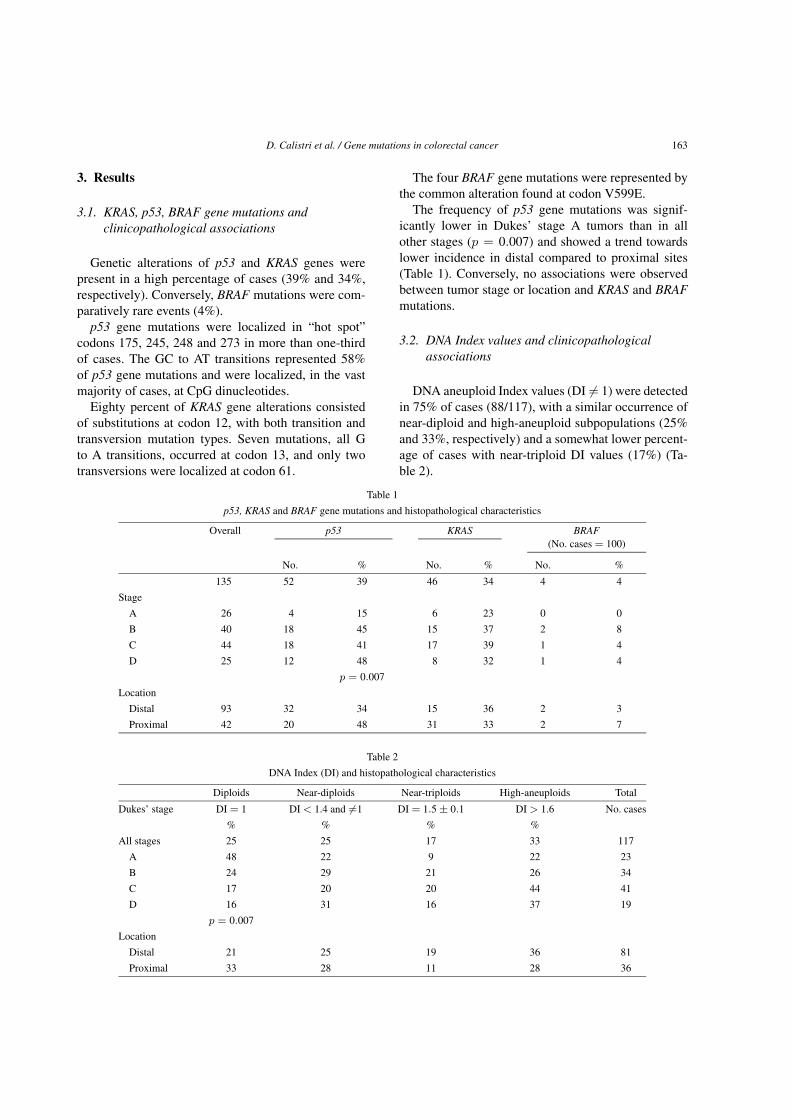
Genetic alterations of p53 and KRAS genes werepresent in a high percentage of cases (39% and 34%,respectively). Conversely, BRAF mutations were com-paratively rare events (4%).
p53 gene mutations were localized in “hot spot”codons 175, 245, 248 and 273 in more than one-thirdof cases. The GC to AT transitions represented 58%of p53 gene mutations and were localized, in the vastmajority of cases, at CpG dinucleotides.
Eighty percent of KRAS gene alterations consistedof substitutions at codon 12, with both transition andtransversion mutation types. Seven mutations, all Gto A transitions, occurred at codon 13, and only twotransversions were localized at codon 61.
The four BRAF gene mutations were represented bythe common alteration found at codon V599E.
The frequency of p53 gene mutations was signif-icantly lower in Dukes’ stage A tumors than in allother stages (p = 0.007) and showed a trend towardslower incidence in distal compared to proximal sites(Table 1). Conversely, no associations were observedbetween tumor stage or location and KRAS and BRAFmutations.
3.2. DNA Index values and clinicopathologicalassociations
DNA aneuploid Index values (DI �= 1) were detectedin 75% of cases (88/117), with a similar occurrence ofnear-diploid and high-aneuploid subpopulations (25%and 33%, respectively) and a somewhat lower percent-age of cases with near-triploid DI values (17%) (Ta-ble 2).
Table 1
p53, KRAS and BRAF gene mutations and histopathological characteristics
Overall p53 KRAS BRAF(No. cases = 100)
No. % No. % No. %
135 52 39 46 34 4 4
Stage
A 26 4 15 6 23 0 0
B 40 18 45 15 37 2 8
C 44 18 41 17 39 1 4
D 25 12 48 8 32 1 4
p = 0.007
Location
Distal 93 32 34 15 36 2 3
Proximal 42 20 48 31 33 2 7
Table 2
DNA Index (DI) and histopathological characteristics
Diploids Near-diploids Near-triploids High-aneuploids Total
Dukes’ stage DI = 1 DI < 1.4 and �=1 DI = 1.5 ± 0.1 DI > 1.6 No. cases
% % % %
All stages 25 25 17 33 117
A 48 22 9 22 23
B 24 29 21 26 34
C 17 20 20 44 41
D 16 31 16 37 19
p = 0.007
Location
Distal 21 25 19 36 81
Proximal 33 28 11 28 36

164 D. Calistri et al. / Gene mutations in colorectal cancer
Table 3
DNA Index and mutation status
DNA Index classes No. cases p53 KRAS Mutation At least one gene
(%) (%) co-presence mutation
(%) (%)
Overall 117 37 33 11 59
Diploids 29 28 21 0 48
Near-triploids 20 25 10 0 35
Near-diploids 29 45 59 28 76
High-aneuploids 39 44 36 13 67
p = 0.05 p = 0.001
Break-down analyses as a function of tumor stageand location showed a significantly higher percentageof DNA diploid tumors (DI = 1) in Dukes’ stage Atumors (48%) compared with more advanced stages(p = 0.007) (Table 2). In particular, for stage A tumorsthe lowest frequency was observed for near-triploid DIvalues, whereas the other DI values were equally rep-resented. No noticeable differences were seen in the DIdistribution values of any of the other Dukes’ stage tu-mors. Similarly, the frequency for different DI valueswas not significantly related to tumor site.
3.3. Association between gene mutations and DNAIndex values
DNA Index analysis results and gene mutation statuswere simultaneously available for 117 cases. A highfrequency of at least one gene mutation was observedin all DNA Index classes. The highest frequenciesof KRAS and p53 mutations were observed amongnear-diploid and high-aneuploid tumors, with a statis-tically significant difference with respect to the otherclasses (p53, p = 0.05; KRAS, p = 0.001) (Ta-ble 3). Moreover, we noted a higher co-occurrence ofthese two gene mutations, about one third of cases, innear-diploid tumors with respect to other subgroups inwhich mutations were rare (high-aneuploid) or absent.
Due to the low frequency of BRAF gene mutations,it was not possible to carry out any correlation analy-ses.
4. Discussion
There is increasing evidence that chromosomal in-stability (CIN), revealed by widespread cytogeneticabnormalities such as specific chromosomal gains,losses and rearrangements, plays an important role inthe development and progression of human cancer to-
gether with mutations of oncogenes and tumor sup-pressor genes [1,6,10,13,17,20,31]. CIN is the pre-dominant form of genomic instability in most humansolid tumors and in CRC in particular [15,18,19]. Un-fortunately, the relationship between gene mutationsand CIN remains poorly understood.
In the present study we investigated the hypothe-sis of an association between KRAS, p53 and BRAFgene mutations and CIN, as reflected by the degree ofDNA ploidy (DI values) in CRC patients, using tumorstage and location as covariates. Mutation frequenciesof 39%, 34%, and 4% were found for p53, KRAS andBRAF genes, respectively, which is within the rangeof those reported in other studies [2,7,11,16,25,29,30,32,33]. The low occurrence (4%) of BRAF mutationsclearly limited the power of the statistical tests. Inter-estingly, the simultaneous presence of KRAS and p53mutations was observed only in 11% of cases (13 out of117 CRCs). According to the colorectal cancer modelof Vogelstein [9], KRAS and p53 alterations are ex-pected to accumulate during the CRC progression sothat their combined occurrence should, in theory, behigher that the occurrence predicted for the two para-meters taken as independent events. This was not con-firmed in the present series of cases, suggesting that,in the presence of one mutation, there is no pressure toselect the other, as reported in previous studies [5,28].
The frequency of p53 mutations was found to in-crease from Dukes’ stage A onwards, whereas that ofDNA diploid (DI = 1) subpopulations dramatically de-creased from early through later stages. Thus, at lowerDukes’ stages CRCs are still capable of maintaininga stable genome, characterized in a high number ofcases (48%) by DNA diploidy (DI = 1) and a lowerfrequency of KRAS and p53 mutations. Interestingly,we found a statistically significant association betweenKRAS and p53 mutations and predetermined subgroupsof tumors with different DNA Index values. It was ob-served that p53 and, in particular, KRAS mutations,

D. Calistri et al. / Gene mutations in colorectal cancer 165
were significantly associated with tumors with near-diploid and high-aneuploid DNA Index values. More-over, it was seen that KRAS and p53 double-mutatedCRCs were found within these two DNA Index sub-groups, whereas none was present in the DNA diploidor near-triploid groups. This suggests that multiplepathways of CRC genesis and progression may exist.Our findings would also seem to indicate that near-diploid and high-aneuploid cell subpopulations share acommon CIN genetic mechanism which differs fromthe mechanism of near-triploidization, as previouslyhypothesized [13,14].
The exact mechanisms explaining such differenceshave yet to be defined. We believe that near-diploidand high-aneuploid tumor cell subpopulations are gen-erated by mechanisms of asymmetric chromosomalsegregation and endoreduplication that are associatedwith KRAS and p53 mutations. Aneuploidization inthe near-triploid group may, conversely, be dependenton different mechanisms that are not associated withthese gene mutations such as the formation of tripo-lar octaploid mitosis and its division into one diploidand two near-triploid cells [6]. In other words, it thusappears that these mutations may influence CIN andaneuploidy, suggesting that both genetic mutations andlarge-scale genomic aberrations could cooperativelydrive CRC tumor progression.
The association observed between KRAS and p53mutations and different subgroups of DNA index sug-gests the possible existence of multiple pathways forCRC genesis and progression, which could have po-tential clinical implications. In particular, the presenceof DNA diploid CRCs with wild type p53 or KRAS,commonly associated with Dukes’ stage A, may indi-cate a low probability for evolution into invasive andhigh-stage tumors.
Further studies focused on colorectal adenomas andin vitro and in vivo mouse models are needed to clar-ify CIN-associated molecular mechanisms and validatethese alterations as biomarkers of clinical relevance.
Acknowledgements
The Authors wish to thank Prof. Rosella Silvestrinifor her invaluable scientific contribution and GráinneTierney for editing the manuscript. Supported by Isti-tuto Oncologico Romagnolo, Forlì, Consiglio Naziona-le della Ricerca (CNR), Rome (02.00382.ST97 andCU03.00291), and by grants from Ministero dellaSalute (FSN 2004), S. Paolo Oncology Program, andRegione Sicilia, Italy.
References
[1] D.G. Albertson, C. Collins, F. McCormick and J.W. Gray,Chromosome aberrations in solid tumors, Nat. Genet. 34(2003), 369–376.
[2] H.J. Andreyev, A.R. Norman, D. Cunningham, J. Oates, B.R.Dix, B.J. Iacopetta, J. Young, T. Walsh, R. Ward, N. Hawkins,M. Beranek, P. Jandik, R. Benamouzig, E. Jullian, P. Laurent-Puig, S. Olschwang, O. Muller, I. Hoffmann, H.M. Rabes, C.Zietz, C. Troungos, C. Valavanis, S.T. Yuen, J.W. Ho, C.T.Croke, D.P. O’Donoghue, W. Giaretti, A. Rapallo, A. Russo,V. Bazan, M. Tanaka, K. Omura, T. Azuma, T. Ohkusa, T.Fujimori, Y. Ono, M. Pauly, C. Faber, R. Glaesener, A.F. deGoeij, J.W. Arends, S.N. Andersen, T. Lovig, J. Breivik, G.Gaudernack, O.P. Clausen, P.D. De Angelis, G.I. Meling, T.O.Rognum, R. Smith, H.S. Goh, A. Font, R. Rosell, X.F. Sun, H.Zhang, J. Benhattar, L. Losi, J.Q. Lee, S.T. Wang, P.A. Clarke,S. Bell, P. Quirke, V.J. Bubb, J. Piris, N.R. Cruickshank, D.Morton, J.C. Fox, F. Al-Mulla, N. Lees, C.N. Hall, D. Snary, K.Wilkinson, D. Dillon, J. Costa, V.E. Pricolo, S.D. Finkelstein,J.S. Thebo, A.J. Senagore, S.A. Halter, S. Wadler, S. Malik,K. Krtolica and N. Urosevic, Kirsten ras mutations in patientswith colorectal cancer: the ‘RASCAL II’ study, Br. J. Cancer85 (2001), 692–696.
[3] G. Auer, U. Kronenwett, U. Roblick, B. Franzén, J. Haber-mann, R. Sennerstam and T. Ried, Human breast adenocarci-noma: DNA content, chromosomes, gene expression and prog-nosis, Cell. Oncol. 26 (2004), 171–174.
[4] M.S. Brose, P. Volpe, M. Feldman, M. Kumar, I. Rishi, R. Ger-rero, E. Einhorn, M. Herlyn, J. Minna, A. Nicholson, J.A. Roth,S.M. Albelda, H. Davies, C. Cox, G. Brignell, P. Stephens, P.A.Futreal, R. Wooster, M.R. Stratton and B.L. Weber, BRAF andRAS mutations in human lung cancer and melanoma, CancerRes. 62 (2002), 6997–7000.
[5] D. Calistri, C. Rengucci, I. Seymour, A. Lattuneddu, A.M. Po-lifemo, F. Monti, L. Saragoni and D. Amadori, Mutation analy-sis of p53, K-ras, and BRAF genes in colorectal cancer pro-gression, J. Cell. Physiol. 204 (2005), 484–488.
[6] P. Castagnola and W. Giaretti, Mutant KRAS, chromosomal in-stability and prognosis in colorectal cancer, Biochim. Biophys.Acta 1756 (2005), 115–125.
[7] T.L. Chan, W. Zhao, S.Y. Leung and S.T. Yuen, CancerGenome Project: BRAF and KRAS mutations in colorectalhyperplastic polyps and serrated adenomas, Cancer Res. 63(2003), 4878–4881.
[8] P. Duesberg, R. Li, D. Rasnick, A. Fabarius and R. Hehlmann,Carcinogenesis by aneuploidization, Cell. Oncol. 26 (2004),181–186.
[9] E.R. Fearon and B. Vogelstein, A genetic model for colorectaltumorigenesis, Cell 61 (1990), 759–767.
[10] R. Fodde, R. Smits and H. Clevers, APC, signal transductionand genetic instability in colorectal cancer, Nat. Rev. Cancer 1(2001), 55–67.
[11] K. Fransen, M. Klintenas, A. Osterstrom, J. Dimberg, H.J.Monstein and P. Soderkvist, Mutation analysis of the BRAF,ARAF and RAF-1 genes in human colorectal adenocarcino-mas, Carcinogenesis 25 (2004), 527–533.
166 D. Calistri et al. / Gene mutations in colorectal cancer
[12] W. Giaretti, S. Molinu, J. Ceccarelli and C. Prevosto, Chro-mosomal instability, aneuploidy, and gene mutations in humansporadic colorectal adenomas, Cell. Oncol. 26 (2004), 301–305.
[13] W. Giaretti, N. Pujic, A. Rapallo, S. Nigro, A. Di Vinci, E.Geido and M. Risio, K-ras2 G-C and G-T transversions corre-late with DNA aneuploidy in colorectal adenomas, Gastroen-terology 108 (1995), 1040–1047.
[14] W. Giaretti, T. Venesio, A. Sciutto, C. Prevosto, E. Geido andM. Risio, Near-diploid and near-triploid human sporadic col-orectal cancer adenocarcinoma differ for KRAS2 and TP53mutational status, Genes Chrom. Cancer 37 (2003), 207–213.
[15] M. Hermsen, C. Postma, J. Baak, M. Weiss, A. Rapallo, A.Sciutto, G. Roemen, J.W. Arends, R. Williams, W. Giaretti, A.De Goeij and G. Meijer, Colorectal adenoma to carcinoma pro-gression follows multiple pathways of chromosomal instability,Gastroenterology 123 (2002), 1109–1119.
[16] B. Iacopetta, TP53 mutation in colorectal cancer, Hum. Mutat.21 (2003), 271–276.
[17] G.J.P.L. Kops, B.A.A. Weaver and D.W. Cleveland, On theroad to cancer aneuploidy and the mitotic checkpoint, NatureRev. Cancer 5 (2005), 773–785.
[18] A. Leslie, N.R. Pratt, K. Gillespie, M. Sales, N.M. Kernohan,G. Smith, C.R. Wolf, F.A. Carey and R.J. Steele, Mutations ofAPC, K-ras, and p53 are associated with specific chromoso-mal aberrations in colorectal adenocarcinomas, Cancer Res. 63(2003), 4656–4661.
[19] A. Leslie, A. Stewart, D.U. Baty, D. Mechan, L. McGreavey,G. Smith, C.R. Wolf, M. Sales, N.R. Pratt, R.J. Steele and F.A.Carey, Chromosomal changes in colorectal adenomas: relation-ship to gene mutations and potential for clinical utility, GenesChromosomes Cancer 45 (2006), 126–135.
[20] F. Michor, Y. Iwasa and M.A. Nowak, Dynamics of cancer pro-gression, Nat. Rev. Cancer 4 (2004), 197–205.
[21] T. Nedergaard, P. Guldberg, E. Ralfkler and J. Zeuthen, A one-step DGGE scanning method for detection of mutations in theK-, N- and H-ras oncogenes: mutations at codons 12, 13 and61 are rare in B-cell non-Hodgkin’s lymphoma, Int. J. Cancer71 (1997), 364–369.
[22] C.D. O’ Connell, D.H. Atha, M.C. Oldemburg, J. Tian, M.Siebert, R. Handrow, K. Grooms, L. Heisler and M. de Arruda,Detection of p53 gene mutation: analysis by single-strand con-formation polymorphism and cleavase fragment length poly-morphism, Electrophoresis 20 (1999), 1211–1223.
[23] M.G. Ormerod, B. Tribukait and W. Giaretti, Consensus reportof the task force on standardisation of DNA flow cytometry inclinical pathology, Anal. Cell. Pathol. 17 (1998), 103–110.
[24] I. Petersen, Chromosome, ploidy and genetic imbalances oflung cancer, Cell. Oncol. 26 (2004), 173–174.
[25] W.S. Samowitz, C. Sweeney, J. Herrick, H. Albertsen, T.R.Levin, M.A. Murtaugh, R.K. Wolff and M.L. Slattery, Poor sur-vival associated with the BRAF V600E mutation in microsat-ellite-stable colon cancers, Cancer Res. 65 (2005), 6063–6069.
[26] S. Sengupta and C.C. Harris, p53: traffic cop at the crossroadsof DNA repair and recombination, Nat. Rev. Mol. Cell. Biol. 6(2005), 44–55.
[27] I.M. Shih, W. Zhou, S.N. Goodman, C. Lengauer, K.W. Kinzlerand B. Vogelstein, Evidence that genetic instability occurs at anearly stage of colorectal tumorigenesis, Cancer Res. 61 (2001),818–822.
[28] G. Smith, F.A. Carey, J. Beattie, M.J. Wilkie, T.J. Lightfoot, J.Coxhead, R.C. Garner, R.J. Steele and C.R. Wolf, Mutationsin APC, Kirsten-ras, and p53-alternative genetic pathways tocolorectal cancer, Proc. Natl. Acad. Sci. USA 99 (2002), 9433–9438.
[29] F. Sommerer, M. Vieth, A. Markwarth, K. Rohrich, S. Vom-schloss, A. May, C. Ell, M. Stolte, U.R. Hengge, C. Wittekindand A. Tannapfel, Mutations of BRAF and KRAS2 in the de-velopment of Barrett’s adenocarcinoma, Oncogene 23 (2004),554–558.
[30] R. Soong, B. Powell, H. Elsaleh, G. Gnanasampanthan, D.R.Smith, H.S. Goh, D. Joseph and B. Iacopetta, Prognostic signif-icance of TP53 gene mutation in 995 cases of colorectal carci-noma. Influence of tumour site, stage, adjuvant chemotherapyand type of mutation, Eur. J. Cancer 36 (2000), 2053–2060.
[31] Z. Wang, J.M. Cummins, D. Shen, D.P. Cahill, P.V. Jallepalli,T.L. Wang, D.W. Parsons, G. Traverso, M. Awad, N. Silliman,J. Ptak, S. Steve Szabo, J.K.V. Willson, S.D. Markowitz, M.L.Goldberg, R. Karess, K.W. Kinzler, B. Vogelstein, V.E. Vel-culescu and C. Lengauer, Three classes of genes mutated incolorectal cancers with chromosomal instability, Cancer Res.64 (2004), 2998–3001.
[32] L. Wang, J.M. Cunningham, J.L. Winters, J.C. Guenther, A.J.French, L.A. Boardman, L.J. Burgart, S.K. McDonnell, D.J.Schaid and S.N. Thibodeau, BRAF mutations in colon cancerare not likely attributable to defective DNA mismatch repair,Cancer Res. 63 (2003), 5209–5212.
[33] S.T. Yuen, H. Davies, T.L. Chan, J.W. Ho, G.R. Bignell, C.Cox, P. Stephens, S. Edkins, W.W. Tsui, A.S. Chan, P.A.Futreal, M.R. Stratton, R. Wooster and S.Y. Leung, Similarityof the phenotypic patterns associated with BRAF and KRASmutations in colorectal neoplasia, Cancer Res. 62 (2002),6451–6455.

Submit your manuscripts athttp://www.hindawi.com
Stem CellsInternational
Hindawi Publishing Corporationhttp://www.hindawi.com Volume 2014
Hindawi Publishing Corporationhttp://www.hindawi.com Volume 2014
MEDIATORSINFLAMMATION
of
Hindawi Publishing Corporationhttp://www.hindawi.com Volume 2014
Behavioural Neurology
EndocrinologyInternational Journal of
Hindawi Publishing Corporationhttp://www.hindawi.com Volume 2014
Hindawi Publishing Corporationhttp://www.hindawi.com Volume 2014
Disease Markers
Hindawi Publishing Corporationhttp://www.hindawi.com Volume 2014
BioMed Research International
OncologyJournal of
Hindawi Publishing Corporationhttp://www.hindawi.com Volume 2014
Hindawi Publishing Corporationhttp://www.hindawi.com Volume 2014
Oxidative Medicine and Cellular Longevity
Hindawi Publishing Corporationhttp://www.hindawi.com Volume 2014
PPAR Research
The Scientific World JournalHindawi Publishing Corporation http://www.hindawi.com Volume 2014
Immunology ResearchHindawi Publishing Corporationhttp://www.hindawi.com Volume 2014
Journal of
ObesityJournal of
Hindawi Publishing Corporationhttp://www.hindawi.com Volume 2014
Hindawi Publishing Corporationhttp://www.hindawi.com Volume 2014
Computational and Mathematical Methods in Medicine
OphthalmologyJournal of
Hindawi Publishing Corporationhttp://www.hindawi.com Volume 2014
Diabetes ResearchJournal of
Hindawi Publishing Corporationhttp://www.hindawi.com Volume 2014
Hindawi Publishing Corporationhttp://www.hindawi.com Volume 2014
Research and TreatmentAIDS
Hindawi Publishing Corporationhttp://www.hindawi.com Volume 2014
Gastroenterology Research and Practice
Hindawi Publishing Corporationhttp://www.hindawi.com Volume 2014
Parkinson’s Disease
Evidence-Based Complementary and Alternative Medicine
Volume 2014Hindawi Publishing Corporationhttp://www.hindawi.com