paramedic resource manual - lakeridge health · this gland is situated below the larynx at the...
TRANSCRIPT

Paramedic Resource Manual
ENDOCRINE SYSTEM
SECTION NINE
2005 Update by Ontario Base Hospital Group Education Subcommittee Copyright 2005, 1985 Ministry of Health and Long Term Care
ONTARIO BASE HOSPITAL GROUP
__________________________________________________________________________ OBHG Education Subcommittee 289
OBJECTIVES: ENDOCRINE SYSTEM The objectives indicate what you should know, understand and be prepared to explain upon completion of this module. The self assessment questions and answers will enable you to judge your understanding of the material. Upon completion of this module, the student should be able to: 1. define and give an example of: a) Endocrine gland b) Exocrine gland c) Mixed gland d) Double gland. 2. name and locate the principal endocrine glands. 3. list the major functions and secretions of each principal gland. 4. describe the action of each secretion listed in 3. 5. describe the mechanism for maintenance of normal blood glucose levels. 6. define gluconeogenesis and glycogenolysis. 7. describe the pathophysiology of diabetes, differentiating between: Type I and Type II diabetes Hypoglycemia and hyperglycemia DKA and HHNK. 8. differentiate clinically between hypoglycemic and hyperglycemic emergencies. If you have studied this subject previously, you may test your ability using the self assessment questions. If you are able to obtain 90% or greater, you may choose not to do the unit and merely review the sections, or parts of sections, where weakness may exist. If you obtain less than 90%, it is recommended that the module be done in its entirety, stressing areas where more review is needed.
__________________________________________________________________________ OBHG Education Subcommittee 290
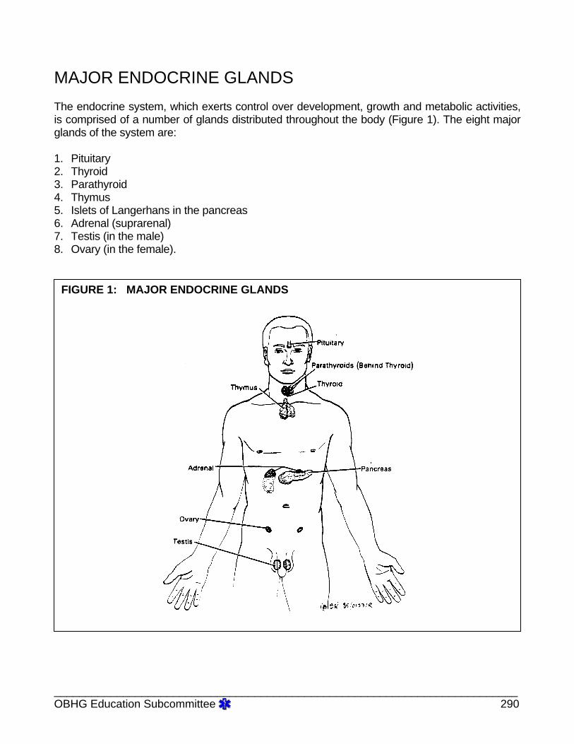
MAJOR ENDOCRINE GLANDS The endocrine system, which exerts control over development, growth and metabolic activities, is comprised of a number of glands distributed throughout the body (Figure 1). The eight major glands of the system are: 1. Pituitary 2. Thyroid 3. Parathyroid 4. Thymus 5. Islets of Langerhans in the pancreas 6. Adrenal (suprarenal) 7. Testis (in the male) 8. Ovary (in the female).
FIGURE 1: MAJOR ENDOCRINE GLANDS
__________________________________________________________________________ OBHG Education Subcommittee 291
This module confines itself primarily to a discussion of the anatomy and physiology of the major endocrine glands. There are, however, other hormonal organs, tissue or cells in the body which also have a secretory function. These include the kidney, pineal gland, placenta and cells in the gastrointestinal tract. Although dysfunction may occur in any gland, the pancreas is the one most commonly involved in pathologies seen by the Paramedic. Therefore, a more detailed discussion of the pancreas is presented in the second section of this module.
TYPES OF GLANDS ENDOCRINE GLAND Endocrine glands are ductless glands, although not all of the ductless glands in the body are endocrine glands, e.g. spleen. Their secretions are always hormones which pass directly into the bloodstream. Endocrine glands are made up of clusters of glandular epithelium. Hormones are the secretions produced by the endocrine glands, and may be a form of protein, amine or steroid (lipid or fat). The function of the hormones is to maintain homeostasis (a constant internal body environment) by changing the physiological activities of cells. A hormone may stimulate changes in the cells of an organ or groups of organs, called target organs; a hormone may also directly affect the activity of all the cells in the body. MIXED GLAND A mixed gland is a gland which has both endocrine and exocrine functions. The exocrine component has ducts, through which its secretions pass. The pancreas is a gland with both endocrine (insulin production) and exocrine (production of digestive enzymes) functions. DOUBLE GLAND A double gland consists of two main parts in which the two components differ in their embryological derivation, nature of hormones released and the mechanism of neural control. The pituitary and adrenal glands are examples of double glands.

__________________________________________________________________________ OBHG Education Subcommittee 292
PITUITARY GLAND (HYPOPHYSIS CEREBRI) The pituitary was formerly referred to as the master endocrine gland, because of its controlling influence on many of the other endocrine glands. This reddish-brown gland, the size of a garden pea, lies in the sella turcica (pituitary fossa, hypophyseal fossa), a fossa on the superior aspect of the body of the sphenoid bone in the base of the skull. The pituitary is attached to the hypothalamus at the base of the brain by a stalk called the infundibulum. The anterior lobe (adenohypophysis) of this double gland is responsible for the secretion of: 1. Growth hormone - somatotropic (STH) 2. Thyroid stimulating hormone - thyrotropic (TSH) 3. Adrenal cortex stimulating hormone - adrenocorticotropic (ACTH) 4. Sex gland stimulating hormones - gonadotropic (LH and FSE) 5. Mammary gland stimulating hormones - prolactin.
FIGURE 2: LOCATION OF THE PITUITARY GLAND

__________________________________________________________________________ OBHG Education Subcommittee 293
The posterior lobe secretes: 1. Oxytocin 2. Vasopressin (antidiuretic hormone - ADH). Both of these cause the contraction of the smooth muscle in various organs. HYPOTHALAMUS Most of the hormones secreted by the anterior pituitary stimulate the production of hormones by other endocrine glands (target glands). Control of the rate of secretions in these target glands is accomplished by the hypothalamus through cells sensitive to changes in hormone levels in the blood. Since the hypothalamus
FIGURE 3: HORMONES RELEASED BY THE PITUITARY

__________________________________________________________________________ OBHG Education Subcommittee 294
responds to an absence of pituitary hormones by producing substances (releasing factors) which enhance hormone secretion, this is referred to as a negative feedback mechanism. THYROID GLAND This gland is situated below the larynx at the upper part of the trachea. it consists of two lateral pear-shaped lobes united at the lower part by the isthmus, which lies in front of the second, third and fourth tracheal rings.
FIGURE 4: THYROID AND PARATHYROID GLAND
Thyroid cartilage of the Larynx
Thyroid Gland
Parathyroid Glands (Behind the Thyuroid)
Trachea

__________________________________________________________________________ OBHG Education Subcommittee 295
Inadvertent penetration of this very vascular gland during needle or surgical cricothyroidotomy may result in severe hemorrhage. The function of the thyroid gland is to: 1. Take up the inorganic iodine (derived from food) present in the blood. 2. Produce the hormone thyroxine. 3. Store thyroxine as thyroglobin. 4. Release thyroxine when stimulated to do so by the thyroid stimulating hormone (TSH)
secreted by the adenohypophysis (pituitary gland). THYROXINE 1. Thyroxine controls the metabolic rate of the body (the rate at which the body uses
oxygen and metabolizes food products). 2. Thyroxine is necessary for normal physical and mental development to occur.
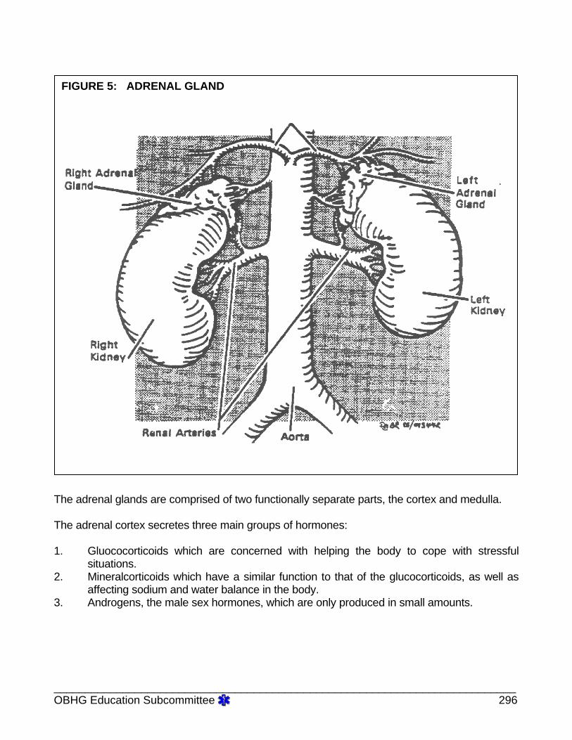
PARATHYROID GLANDS These, the smallest of the endocrine glands, may be variable in number and position. Normally there are four - two situated on the posterior surface of each of the lateral lobes of the thyroid gland. They lie beneath the capsule of the thyroid and may in fact be embedded in its tissue. The parathyroid glands secrete a hormone, parathyroid hormone (parathormone) (PTH), which is concerned with calcium metabolism. The secretion of this hormone is regulated by blood calcium levels, and is not under the control of the adenohypophysis. THYMUS The thymus lies in the anterior and superior parts of the thorax. It is a relatively large structure at birth and continues to grow until puberty. Although the thymus decreases in size during adult life its physiological importance does not. It provides the mold, or model, for other organs, such as the liver and spleen, to produce T-lymphocytes (white blood cells concerned with the defense against infection and disease). ADRENAL (SUPRARENAL) GLANDS These two asymmetrical glands are found in close proximity to the upper poles of the kidneys. The right one is pyramidal and embraces the upper pole of the right kidney; while the left is crescentic and is located on the supramedial aspect of the left kidney above the hilum. Each measures about 5 cm long and 3 cm thick. Each gland is supplied by three relatively large arterial branches, indicating its relative importance to the body.
__________________________________________________________________________ OBHG Education Subcommittee 296
FIGURE 5: ADRENAL GLAND
The adrenal glands are comprised of two functionally separate parts, the cortex and medulla. The adrenal cortex secretes three main groups of hormones: 1. Gluococorticoids which are concerned with helping the body to cope with stressful
situations. 2. Mineralcorticoids which have a similar function to that of the glucocorticoids, as well as
affecting sodium and water balance in the body. 3. Androgens, the male sex hormones, which are only produced in small amounts.

__________________________________________________________________________ OBHG Education Subcommittee 297
Clinical vignette Pheochromocytoma is a rare tumor of the adrenal gland that causes excess secretion of adrenalin. This may lead to hypoglycemia in non-diabetics. Administration of Glucagon may affect the adrenal gland/tumor resulting in even greater release of adrenalin and this may, paradoxically, worsen hypoglycemia.
The action of the hormones secreted by the adrenal medulla is to prepare the body for sudden action at a time of pain or fear (fight or flight situations). These hormones are:
Adrenalin (epinephrine) Noradrenalin (norepinephrine).
GONADS At the time of puberty, the adenohypophysis starts to secrete progressively more gonadotropic hormones. There are two in particular which affect both the ovaries and the testes. These are:
Follicle stimulating hormone (FSH) Luteinizing hormone (LH) or interstitial cell stimulating hormone (ICSH) as it is called in
the male. OVARIES The FSH and LH secreted by the adenohypophysis stimulate the development of all the female reproductive organs and initiate the menstrual cycle. The menstrual cycle is a series of rhythmic changes which occur throughout the childbearing years. Each cycle lasts about 28 days and during this time three major hormone related events occur. 1. The FSH stimulates the maturation of the follicle containing an ovum which in turn produces
the hormone estrogen. 2. The FSH and LH together bring about ovulation (the release of the ovum from the follicle and
ovary). 3. The LH causes the lining of the uterus, the endometrium, to thicken in preparation for the
implantation of a fertilized ovum. LH also causes the follicular cells left behind in the ovary to change their characteristics and colour to become the corpus luteum. The corpus luteum in turn produces large quantities of the hormone progesterone and small quantities of estrogen.
If fertilization and implantation do not take place, the corpus luteum will degenerate, the production of progesterone and estrogen will decrease and menstruation (bleeding) will occur.

__________________________________________________________________________ OBHG Education Subcommittee 298
Clinical vignette The ovaries and corpus luteum are sites where the formation of cysts is relatively common. These cysts can cause torsion of the ovary resulting in pain, or they can bleed or rupture.
(Estrogen production will also decrease after ovulation has taken place). At this time the negative feedback mechanism will be initiated.
TESTES The seminiferous tubules of the testes are stimulated by the secretion of FSH to produce spermatozoa and bring them to maturation. The secretion of the ICSH stimulates the interstitial cells, the cells of Leydig, to produce the male hormone testosterone. The action of testosterone is to stimulate development and activity of male sex glands, as well as bringing about the changes that produce the secondary sexual characteristics (growth of body hair, and voice change). Testosterone levels are controlled by a negative feedback loop. “FRINGES” OF THE ENDOCRINE SYSTEM There are body structures whose tissue have a glandular appearance and have been suspected, despite the lack of convincing evidence, of having endocrine properties. PINEAL GLAND Located in the forebrain, the pineal gland was the subject of speculation for years. In fact, as recently as 1985, when this pre-course manual was first written, understanding of the pineal gland’s function was very poorly understood. Today, a little bit more is understood about the Pineal gland, however, Physiologists and Endocrinologists still have a long way to go. The pineal gland produces Melatonin which plays a role in the body’s circadian rhythm or “biological time clock”. In the dark, the pineal gland is stimulated to produce and release melatonin and in the daylight, melatonin production is reduced. It’s recently been suggested that treatment with melatonin may prevent and/or reduce jetlag, reduce the risk of developing certain tumors and may slow the aging process. The Pineal Gland also regulates our patterns of eating (hunger), sleeping, reproduction (female reproductive cycle), and behaviour. Seasonal affective disorder (SAD) has been linked to the Pineal Gland, as there is a reduction in Melatonin during the winter months when daylight is shortened. In some occult religions, it is sometimes referred to as the "third eye" and is believed to be a dormant organ that can be awakened to give mystical powers.
__________________________________________________________________________ OBHG Education Subcommittee 299
Clinical vignette The pineal gland has a midline position within the brain and in some adults it is calcified, making it a visible landmark on skull x-rays. if it has shifted out of the midline, it may indicate the presence of a pathological process such as an expanding lesion.
INTESTINAL TRACT GLANDS Intestinal tract glands that secrete gastrin, secretin and cholecystokinin are endocrine glands because their secretions pass directly into the circulatory system. The significance of these hormones are not discussed in this precourse material. THE KIDNEY The peptide angiotensin, found in the bloodstream, is effective in causing high blood pressure and its presence is primarily the result of kidney activity. This "hormone" is not produced in the kidney, but an enzyme from this organ, renin, is responsible for initiating chemical changes in a protein in the blood stream which cause eventual production of the "hormone". Also, evidence indicates that red blood cell formation is influenced by a hormonal agent, erythropoietin and again the kidney is involved. In this section, we have primarily examined the anatomy of the organs other than the pancreas that have been conveniently classified as endocrine glands. However, we should remain aware there are certain organs, tissues or cells, whose primary role is not endocrine, but may emit secretions to the circulation and assume an endocrine function.
__________________________________________________________________________ OBHG Education Subcommittee 300
ADVANCED LIFE SUPPORT PRECOURSE
ENDOCRINE SYSTEM
SELF-ASSESSMENT: MAJOR ENDOCRINE GLANDS
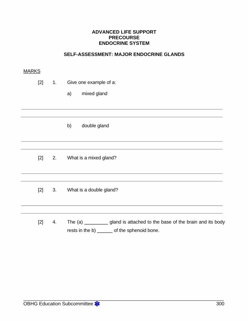
MARKS [2] 1. Give one example of a:
a) mixed gland
b) double gland
[2] 2. What is a mixed gland?
[2] 3. What is a double gland?
[2] 4. The (a) gland is attached to the base of the brain and its body
rests in the b) of the sphenoid bone.

__________________________________________________________________________ OBHG Education Subcommittee 301
[1] 5. The secretions of endocrine glands pass directly into the blood stream.
True or False.
[2] 6. Name two different endocrine glands that are located in the abdomen.
[2] 7. What is the anatomical location of the thyroid gland?
[1] 8. Where are the parathyroid glands found?
[1] 9. What chemical substances are released by the adrenal medulla?
[2] 10. Which of the following hormones are produced by the adenohypophysis? a) Oxytocin b) Estrogen c) Growth hormone d) Thyroxine e) Adrenalin
__________________________________________________________________________ OBHG Education Subcommittee 302
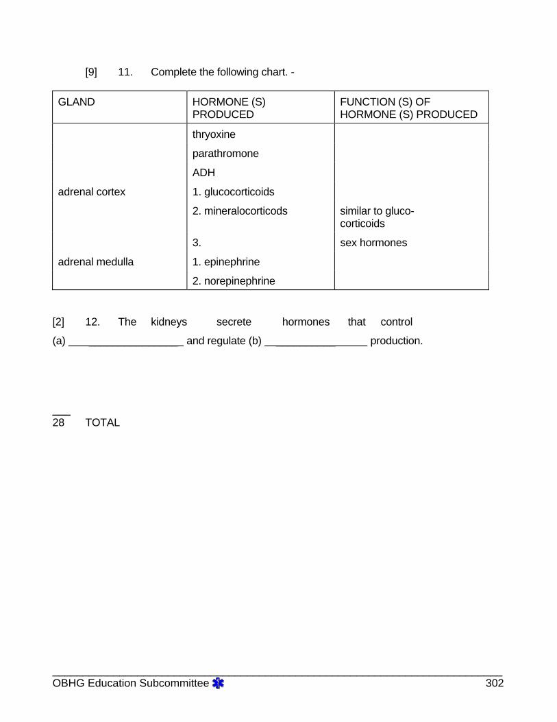
[9] 11. Complete the following chart. -
GLAND HORMONE (S) PRODUCED
FUNCTION (S) OF HORMONE (S) PRODUCED
thryoxine
parathromone
ADH
adrenal cortex 1. glucocorticoids
2. mineralocorticods similar to gluco- corticoids
3. sex hormones
adrenal medulla 1. epinephrine
2. norepinephrine
[2] 12. The kidneys secrete hormones that control
(a) _______________ and regulate (b) __________ production.
___ 28 TOTAL
__________________________________________________________________________ OBHG Education Subcommittee 303
ADVANCED LIFE SUPPORT PRECOURSE
ENDOCRINE SYSTEM
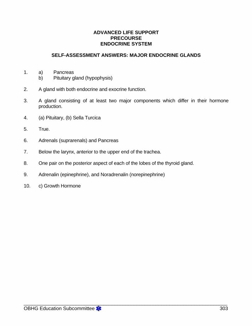
SELF-ASSESSMENT ANSWERS: MAJOR ENDOCRINE GLANDS 1. a) Pancreas b) Pituitary gland (hypophysis) 2. A gland with both endocrine and exocrine function. 3. A gland consisting of at least two major components which differ in their hormone
production. 4. (a) Pituitary, (b) Sella Turcica 5. True. 6. Adrenals (suprarenals) and Pancreas 7. Below the larynx, anterior to the upper end of the trachea. 8. One pair on the posterior aspect of each of the lobes of the thyroid gland. 9. Adrenalin (epinephrine), and Noradrenalin (norepinephrine) 10. c) Growth Hormone

__________________________________________________________________________ OBHG Education Subcommittee 304
11. GLAND HORMONE(S)
PRODUCED FUNCTION OF HORMONE(S
thyroid thyroxine control of metabolic rate of tissues
parathyroids parathormone calcium metabolism
pituitary- posterior lobe
ADH conserves water
adrenal cortex 1. glucocorticoids stress response
2. mineralocorticoids similar to glucocorticoids
3.androgens sex hormones
adrenal 1. epinephrine "fight or flight response"
medulla
2. norephinephrine
12. (a) Blood pressure; (b) Red blood cell
__________________________________________________________________________ OBHG Education Subcommittee 305
ADVANCED LIFE SUPPORT PRECOURSE
ENDOCRINE SYSTEM
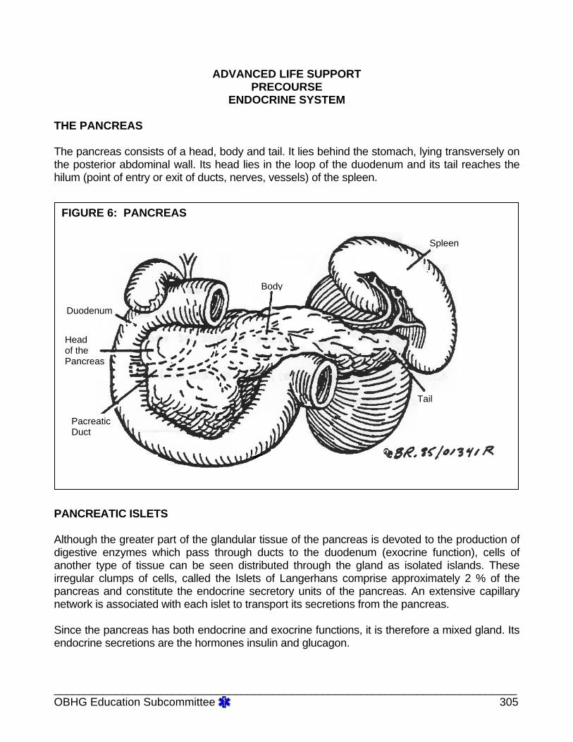
THE PANCREAS The pancreas consists of a head, body and tail. It lies behind the stomach, lying transversely on the posterior abdominal wall. Its head lies in the loop of the duodenum and its tail reaches the hilum (point of entry or exit of ducts, nerves, vessels) of the spleen. PANCREATIC ISLETS Although the greater part of the glandular tissue of the pancreas is devoted to the production of digestive enzymes which pass through ducts to the duodenum (exocrine function), cells of another type of tissue can be seen distributed through the gland as isolated islands. These irregular clumps of cells, called the Islets of Langerhans comprise approximately 2 % of the pancreas and constitute the endocrine secretory units of the pancreas. An extensive capillary network is associated with each islet to transport its secretions from the pancreas. Since the pancreas has both endocrine and exocrine functions, it is therefore a mixed gland. Its endocrine secretions are the hormones insulin and glucagon.
FIGURE 6: PANCREAS
Duodenum
Head of the Pancreas
Pacreatic Duct
Body
Spleen
Tail
__________________________________________________________________________ OBHG Education Subcommittee 306
The islet cells may be classified as alpha or beta; the beta cells being most abundant. Insulin is produced by the beta cells in the Islets of Langerhans to lower blood sugar levels. The release of insulin by the pancreas occurs when blood sugar levels are high. Glucagon, produced by the alpha cells in the Islets of Langerhans, has the opposite effect to insulin. It stimulates the conversion of glycogen (the storage form of glucose) to glucose which is released into the blood, thus raising the level of blood sugar. There is little nervous influence on insulin hormonal secretion, which appears to be mainly controlled by the level of glucose passing through the islets. The action of insulin is to facilitate the transport of glucose across the cell membrane, making it available to the cell for oxidation in the Kreb's Cycle. Insulin also facilitates storage of glucose as glycogen in depots primarily in the liver, muscle and kidney. Glucose is the primary cell fuel. Certain cells can only use glucose. The most important of these are the cells of the CNS. Ninety percent of brain cell metabolism utilizes glucose. In fact, glucose is so important to normal brain cell function that it does not require insulin to facilitate its passage across the cell membrane. To some degree this compensates for lack of a backup stockpile of stored glucose (glycogen) here. The brain is therefore dependent on a constant supply of glucose to maintain its cellular activities. Let us examine how this is achieved. REGULATION OF BLOOD GLUCOSE LEVELS About 50% of western world diets consist of carbohydrates which are readily absorbed from the gut after breakdown by digestive enzymes. The bulk of digested carbohydrate is in the form of various sugars, the most plentiful being glucose which is ready for transport by the bloodstream and immediate use by the cell. Since insulin is the key for the entry of glucose into many cells (brain and liver excluded) it must be produced and transported to the cells, along with the glucose. Circulating levels of glucose are the trigger for the beta cells in the Islets of Langerhans to release insulin. Promoting the storage of glucose as glycogen (primarily in the liver) is another important role of insulin. In doing so, it inhibits the conversion of glycogen to glucose at this time. However, most of us have long periods, i.e. overnight, where we do not have a food intake. As glucose from a meal is either used up or stored, insulin levels fall. The hormone glucagon (from the alpha cells of the islets of Langerhans) is then released along with epinephrine from the adrenal medulla. This causes mobilization of glycogen stores, again primarily from the liver. Conversion of glycogen to glucose (glycogenolysis) raises blood sugar levels, ensuring a constant supply to the cells. However, these stores are limited. In fact, the liver barely stores enough glycogen to meet the metabolic needs of brain tissue for a 24 hour period. As glycogen stores are consumed proteins and fats from other sources (mostly muscle tissue and adipose) are freed for conversion to
__________________________________________________________________________ OBHG Education Subcommittee 307
Clinical vignette Administration of Glucagon will raise the blood glucose level in a patient who has sufficient glycogen stores. A second dose of Glucagon may be indicated for patients whose blood glucose level remains < 4 mmol/L after the first dose, however, if the glycogen stores are inadequate, there will be not effect on BGL.
glucose by the liver (gluconeogenesis). This process is mediated by glucocorticoids from the adrenal cortex. In times of prolonged starvation the kidneys become involved in gluconeogenesis from fats. This process is facilitated by insulin and enhanced by epinephrine and growth hormone (from the pituitary).
It should now be apparent that our homeostatic systems are geared to provision of a constant supply of glucose to the CNS, sparing protein and fat for other duties. As with any homeostatic regulatory mechanism, there exists a potential for problems at any one of the stages. By far the most common endocrine emergency encountered by prehospital personnel will involve the diabetic patient. What follows is a discussion of the more important features of diabetes mellitus, and the implications for you as a care provider. Before proceeding, let's clarify one point ... diabetes insipidus is a different pathology involving primarily the pituitary gland. It will not be discussed in this unit. DIABETES MELLITUS (DM) Diabetes is a chronic disorder affecting the way the body uses and stores the foods eaten and is caused by a deficiency or the ineffectiveness of insulin secreted by the pancreas. Over 2 million Canadians live from diabetes (http://www.diabetes.ca/Section_about/index.asp). Insulin can be unavailable for many reasons: 1. Destruction of the beta cells in the Islets of Langerhans which is believed to involve a
genetically transmitted autoimmune problem. A viral infection either directly destroys beta cells or the resulting autoimmune reaction is
responsible. In either case - no beta cells, means no insulin. These patients are dependent upon insulin for life, and without it will develop ketosis or
ketoacidosis and die. More about this later. Since most, but not all, of these patients are young at onset, this type of diabetes was
formerly referred to as "juvenile diabetes". More appropriately, it is now called insulin dependent diabetes mellitus (IDDM).

__________________________________________________________________________ OBHG Education Subcommittee 308
2. Resistance to normal or elevated serum insulin levels. Predisposition to this process is also believed to be genetically transmitted. The defect is believed to be related to a decrease in the number of insulin receptors on the cell membrane, which reduces the ability of insulin to act at this level.
There is a correlation between overeating and the onset of this problem. These patients
tend to be over 40 and overweight at onset. This accounts for the former term of "age onset diabetes". Currently, it is referred to as non-insulin dependent diabetes (NIDDM) or Type II.
Investigators believe that between 35 and 40 etiologies of this type of disease may be
possible. Management of NIDDM may consist of weight reduction, with or without the use of oral
hypoglycemic agents. Insulin may be added temporarily to prevent hyperglycemia, but without it, these individuals can still survive.
3. Other conditions such as surgical removal of the pancreas, pancreatitis, pancreatic
injuries and rare endocrine disorders may lead to diabetes. The goals of management of the diabetic patient are to maintain as nearly normal levels of blood glucose as possible using current therapies. In the non-diabetic person this is achieved by homeostatic feedback mechanisms which constantly monitor and adjust blood glucose levels. Current therapy involving extensive patient education in the regulation of diet and exercise, the monitoring of blood glucose levels and administration of medication sometimes by continuous infusion (insulin pump) is still very inexact, compared with natural mechanisms. Unfortunately, there is a relatively narrow range of "normal" blood glucose values tolerated by human tissues. Just as glucose deficits within the cell are problematic, glucose excesses are, over time, toxic. Diabetics may develop:
Blindness Kidney failure Heart disease and stroke: diabetes is the underlying cause of 50% of all heart attacks
and strokes Peripheral neuropathies Peripheral vascular disease.
__________________________________________________________________________ OBHG Education Subcommittee 309
HYPOGLYCEMIC EMERGENCIES Decreased levels of blood glucose cause the patient to be symptomatic by two mechanisms: 1. SNS stimulation (beta adrenergic) which results in anxiety, trembling, tachycardia,
palpitations, diaphoresis, faintness, dilated pupils, pallor. 2. CNS depression (primarily at the cortical level) resulting in weakness, headache, blurred
vision, impaired judgement, confusion, lack of co-ordination, seizures and coma. The respiratory pattern is usually normal in these patients. Most often hypoglycemia is seen in the IDDMs who has missed a meal, over-exercised, altered insulin regime, or has an infection. Other causes can include oral hypoglycemic agents, and ethanol abuse over time (primarily due to inadequate glycogen stores and defective gluconeogenesis). Prolonged or recurrent hypoglycemia can result in brain damage (often in the areas affecting personality) and/or death. The damage is irreversible. The goals of therapy are directed at rapidly restoring blood glucose levels to the normal range and replacing depleted glycogen stores.
HYPERGLYCEMIC EMERGENCIES Hyperglycemic states involve increased levels of blood glucose which may be present in some diabetics. However, we will focus on those patients in whom hyperglycemia presents as a life-threatening emergency. These conditions are:
diabetic ketoacidosis (DKA) hyperglycemic, hyperosmolar, nonketotic coma (HHNK).
DIABETIC KETOACIDOSIS (DKA) Increased levels of blood glucose and ketones due to a relative lack of insulin are usually, but not always, seen in IDDM. Symptoms can be attributed to an excess of glucagon, glucocorticoids, growth hormone and catecholamines usually due to stressful events, such as illness or emotional stress. These hormones, you'll recall, are the ones associated with the body's homeostatic mechanisms in response to decreased blood glucose at the cellular level. They are the hormones of glycogenolysis, and gluconeogenesis in response to cellular starvation. Schematically, the sequence of events is represented in Figure 7.

__________________________________________________________________________ OBHG Education Subcommittee 310
It is apparent that a number of vicious cycles operate here: 1. In the absence of insulin, and the cell's inability to utilize glucose, the hyperglycemia will
tend to get worse due to the glucose production from protein and fat breakdown. 2. As the serum glucose becomes very high, the kidney is unable to reabsorb the high
concentration of glucose in the renal tubules. The glucose trapped in the renal tubules (in urine) results in a large volume of water being extracted (polyuria or osmotic diuresis). This osmotic diuresis also removes electrolytes such as Na+ and K+.
3. As fats are mobilized for glucose production, the glycerol portion is metabolized to
glucose (causing hyperglycemia). The breakdown of the free fatty acid portion results in ketonuria, again contributing to the diuresis and electrolyte depletion.
FIGURE 7:
INSULIN DEFICIENCY IN TYPE 1 DIABETES
CELLULAR STARVATION ( Available Glucose)
Glucose production in liver (from glycogen)
Protein breakdown Fat breakdown
Hyperglycemia (blurred vision, hunger, thirst,
urine “spilled” into urine)
Gluconeogenesis
Glycerol Free Fatty Acids
Keytones
Urine + Blood
Polyurea (osmotic diuresis)
Dehydration (BP, HR)
Electrolyte depletion (Na+, K+, PO4
3+)
Metabolic Acidosis (pH, HCO3
-) (Nausea, vomiting, abdominal pain, ventilatory rate& depth,
acetone breath, peripheral vasodilation, hot, dry skin,
LOC)
Plasma osmolality ( LOC)
__________________________________________________________________________ OBHG Education Subcommittee 311
Ketones in blood are acidic. Compensatory buffering systems (see acid/base unit)
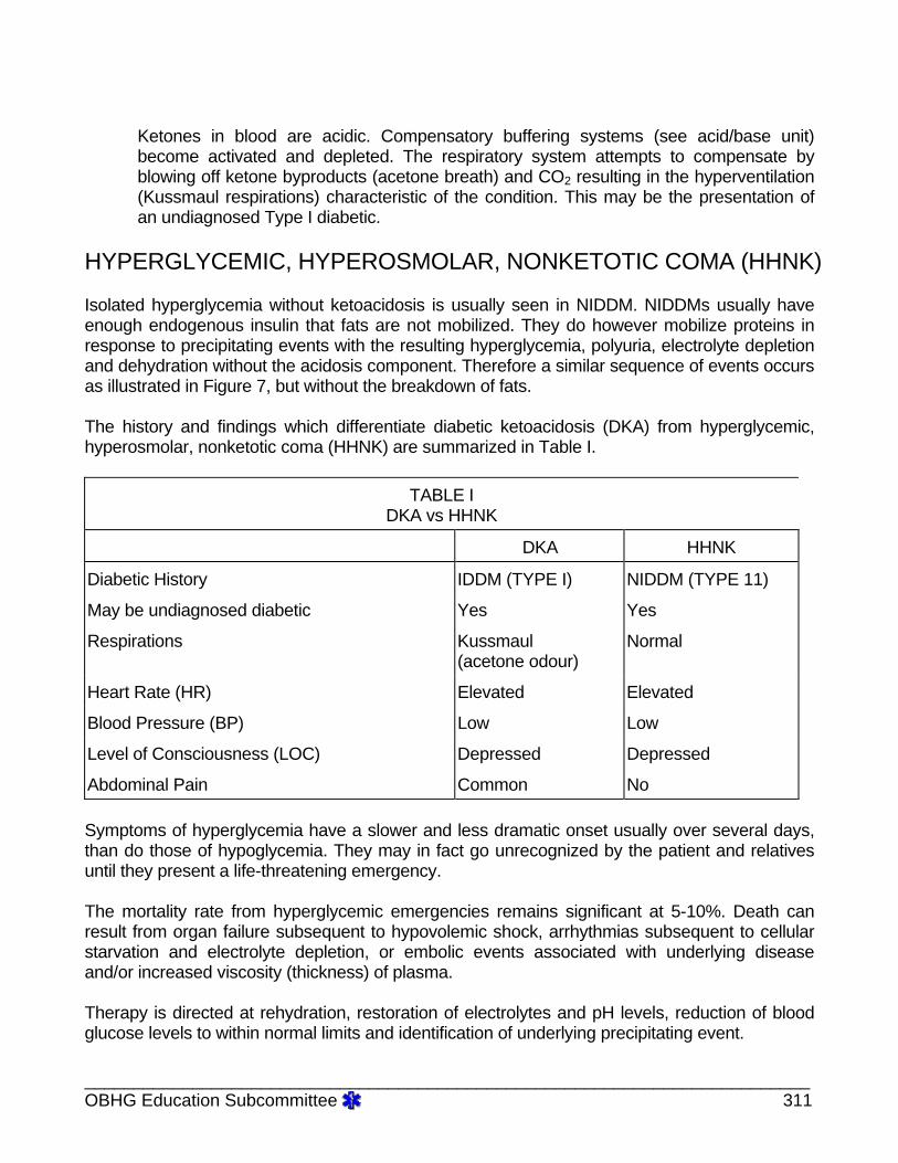
become activated and depleted. The respiratory system attempts to compensate by blowing off ketone byproducts (acetone breath) and CO2 resulting in the hyperventilation (Kussmaul respirations) characteristic of the condition. This may be the presentation of an undiagnosed Type I diabetic.
HYPERGLYCEMIC, HYPEROSMOLAR, NONKETOTIC COMA (HHNK) Isolated hyperglycemia without ketoacidosis is usually seen in NIDDM. NIDDMs usually have enough endogenous insulin that fats are not mobilized. They do however mobilize proteins in response to precipitating events with the resulting hyperglycemia, polyuria, electrolyte depletion and dehydration without the acidosis component. Therefore a similar sequence of events occurs as illustrated in Figure 7, but without the breakdown of fats. The history and findings which differentiate diabetic ketoacidosis (DKA) from hyperglycemic, hyperosmolar, nonketotic coma (HHNK) are summarized in Table I.
TABLE I DKA vs HHNK
DKA HHNK
Diabetic History IDDM (TYPE I) NIDDM (TYPE 11)
May be undiagnosed diabetic Yes Yes
Respirations Kussmaul (acetone odour)
Normal
Heart Rate (HR) Elevated Elevated
Blood Pressure (BP) Low Low
Level of Consciousness (LOC) Depressed Depressed
Abdominal Pain Common No
Symptoms of hyperglycemia have a slower and less dramatic onset usually over several days, than do those of hypoglycemia. They may in fact go unrecognized by the patient and relatives until they present a life-threatening emergency. The mortality rate from hyperglycemic emergencies remains significant at 5-10%. Death can result from organ failure subsequent to hypovolemic shock, arrhythmias subsequent to cellular starvation and electrolyte depletion, or embolic events associated with underlying disease and/or increased viscosity (thickness) of plasma. Therapy is directed at rehydration, restoration of electrolytes and pH levels, reduction of blood glucose levels to within normal limits and identification of underlying precipitating event.
__________________________________________________________________________ OBHG Education Subcommittee 312
ADVANCED LIFE SUPPORT PRECOURSE
ENDOCRINE SYSTEM
SELF-ASSESSMENT: THE PANCREAS MARKS [3] 1. Describe the location of the pancreas in relation to the stomach, spleen
and duodenum.
[2] 2. Define: Gluconeogenesis
Glycogenolysis
[4] 3. The pancreas produces two hormones; these are: (a) which (raises/lowers) blood glucose levels and (b)
which (raises/lowers) blood glucose levels.
[1] 4. Other factors which lower blood glucose are and .

__________________________________________________________________________ OBHG Education Subcommittee 313
[1] 5. Secretion of insulin is mainly controlled by .
6. Compare Type 1 and Type II diabetes by completing this chart. TYPE I TYPE II
[1] Common abbreviation ___________ _____________
[1] Usual age of onset ___________ _____________
[3.5] Controlled by 1. ____ 1. _______
2. ____ 2. ____________
3. ____ 3. ____________
4. ____________
[1] Ketoacidosis prone? ____________ ______________
[1] Non-Ketotic Hyperosmolar coma prone? ____________ ______________ [1] 7. The mechanism of action of insulin is:
[.5] 8. In the absence of insulin brain cells (are/are not) able to utilize
glucose.

__________________________________________________________________________ OBHG Education Subcommittee 314
[1] 9. Diabetes is a result of .
[1] 10. Many diabetic patients have major illnesses secondary to diabetes. Name
two of these illnesses that you are likely to encounter in the field.
[1.5] 11. In response to cellular starvation the body breaks down ________ , _______ and ___________ . [.5]. 12. The cause of ketoacidosis is related to the breakdown of _________ . [2] 13. The conditions resulting from ketoacidosis are primarily (a) _______ acidosis, ________ and (c) ________ depletion. The (d) _____ pain may serve to mislead the examiner. 14. Differentiate between the important features of hypoglycemia and
pathologic hyperglycemia below. HYPOGLYCEMIA HYPERGLYCEMIA
[1] Onset of symptoms ________________
[1] Skin condition _______ ________________
[1] Respirations ______________ _________________
[1] Onset of coma ______________ _________________
30 TOTAL

__________________________________________________________________________ OBHG Education Subcommittee 315
ADVANCED LIFE SUPPORT PRECOURSE
ENDOCRINE SYSTEM
SELF-ASSESSMENT ANSWERS: THE PANCREAS 1. Behind the stomach, resting on a loop of duodenum on the right, touching the hilum of
the spleen on the left. 2. Gluconeogenesis - production of glucose from the breakdown of proteins and fats in the
liver. Glycogenolysis - production of glucose from the breakdown of glycogen in the liver. 3. a) Insulin, lowers b) Glucagon, raises 4. Exercise, starvation 5. Blood glucose levels (passing through the Islets of Langerhans). 6. TYPE I TYPE II Common abbreviation IDDM NIDDM Usual age of onset younger older Controlled by 1. insulin 1. diet 2. diet 2. exercise 3. exercise 3. + or - oral hypoglycemics 4. +/- insulin Ketoacidosis prone? Yes not usually Non-Ketotic Hyper- Not usually Yes osmolar coma prone? 7. Facilitating glucose transport across the cell membrane. 8. Are

__________________________________________________________________________ OBHG Education Subcommittee 316
9. Lack of insulin (either relative or absolute) 10. (Arteriosclerotic) heart disease and stroke Renal disease 11. Glycogen, protein, fats 12. Fat 13. a) Metabolic b) Dehydration (osmotic diuresis) c) Electrolyte d) Abdominal 14. HYPOGLYCEMIA HYPERGLYCEMIA Onset of symptoms Rapid over Gradual over minutes to hours hours to days Skin condition Pale, wet Hot, dry Respirations Normal Rapid, deep (Kussmaul) in DKA Normal in HHNK Onset of coma Rapid Much longer,
__________________________________________________________________________ OBHG Education Subcommittee 317
ADVANCED LIFE SUPPORT PRECOURSE
THE NERVOUS SYSTEM
EVALUATION
Upon completion of this module, please fill in and return this form to your base hospital co-ordinator. Your comments will help to ensure that this unit is a useful learning module. Please indicate any problems that you may have encountered. All suggestions for improvement are welcomed. 1. How long did it take to complete this module? Please estimate.
Reading hours Self assessment hours Total time hours
2. Were the objectives of the module clearly stated?
[ ] yes [ ] no
If no, please comment. 3. Did you see any of the resource materials?
[ ] yes [ ] no
If yes, which items
Were they helpful? 4. Were the reference notes adequate?
[ ] yes [ ] no
If no, please comment.

__________________________________________________________________________ OBHG Education Subcommittee 318
5. Were the reference notes easy to follow?
[ ] yes [ ] no
If no, please comment. 6. Were the examples provided satisfactory?
[ ] yes [ ] no
If no, please comment. 7. Were any of the self-assessment questions poorly worded?
[ ] yes [ ] no
If yes, please specify. 1. Was the level of the module satisfactory for your program of study?
[ ] yes [ ] no If no, please comment.
Base Hospital 9. General comments or suggested improvements.