minimally invasive hip replacement through the … technique innovations in minimally...
TRANSCRIPT

Surgical Technique *smith&nephewINNOVATIONS IN MINIMALLYINVASIVE JOINT SURGERY
Minimally Invasive HipReplacement through theDirect Lateral Approach

Prosthetic replacement of the hip joint is universally accepted as one of the mostimportant advances in orthopaedic surgery. Results have been spectacular andcomplications few as the benefits of this procedure have been successfully extended toboth the very young and the very old. Advancements in materials and betterunderstanding of failure modes have gradually lead to improvement in the lifespan oftotal hip replacement implants.
Minimally invasive surgery represents a natural progression in total hip replacementsurgery. With minimally invasive surgery has come the prospect of less pain, a shorterhospital stay and quicker recovery. These claims are directly related to the distinctsurgical techniques associated with minimally invasive surgery. Less surgical dissection,reduced retraction of tissue and limited cutting of muscle can set the stage for fasterrehabilitation. It is important to recognize that minimally invasive surgery representsmore than "minimal incision surgery" and that adherence to minimally invasive surgicaltechniques will result in less surgical morbidity and improved patient satisfaction.
Minimally invasive total hip replacement surgery has been performed through differingsurgical approaches. Each has its unique advantages and disadvantages which are notunlike those associated with these approaches in standard non minimally invasivesurgery.
Some minimally invasive surgery approaches have the potential for even greatermorbidity than is associated with traditional total hip replacement due to insufficientvisualization of the femur. Of particular concern is the inability to properly implant astable prosthesis and the risk of fracturing the femur.
Minimally invasive total hip replacement performed through a direct lateral approachoffers all of the advantages of minimally invasive surgical technique without the addedmorbidity associated with some minimally invasive surgery approaches. Using theIncision Locator pictured in this technique, the incision can be accurately placed withoutfluoroscopy for a trans-gluteal approach to the hip joint that can permit limited softtissue dissection and yet afford excellent visualization of the femur and acetabulum.Since the posterior hip joint capsule is left intact, joint stability is improved anddislocation rates are lower than for most total hip replacement approaches. Theproximal femur is directly visualized ensuring that proper implantation of the femoralstem is not compromised. Acetabular exposure is obtained through the same incisionand from an anterior perspective which makes achieving stable component orientationless problematic.
The minimally invasive direct lateral approach is a low morbidity alternative to othersurgical approaches for total hip replacement. It is technically easy to master and offersthe advantages of less pain, faster recovery and improved joint stability.
Robert F. Kepley, MDCrystal ClinicAkron, Ohio
Introduction
1
Patient Preparation
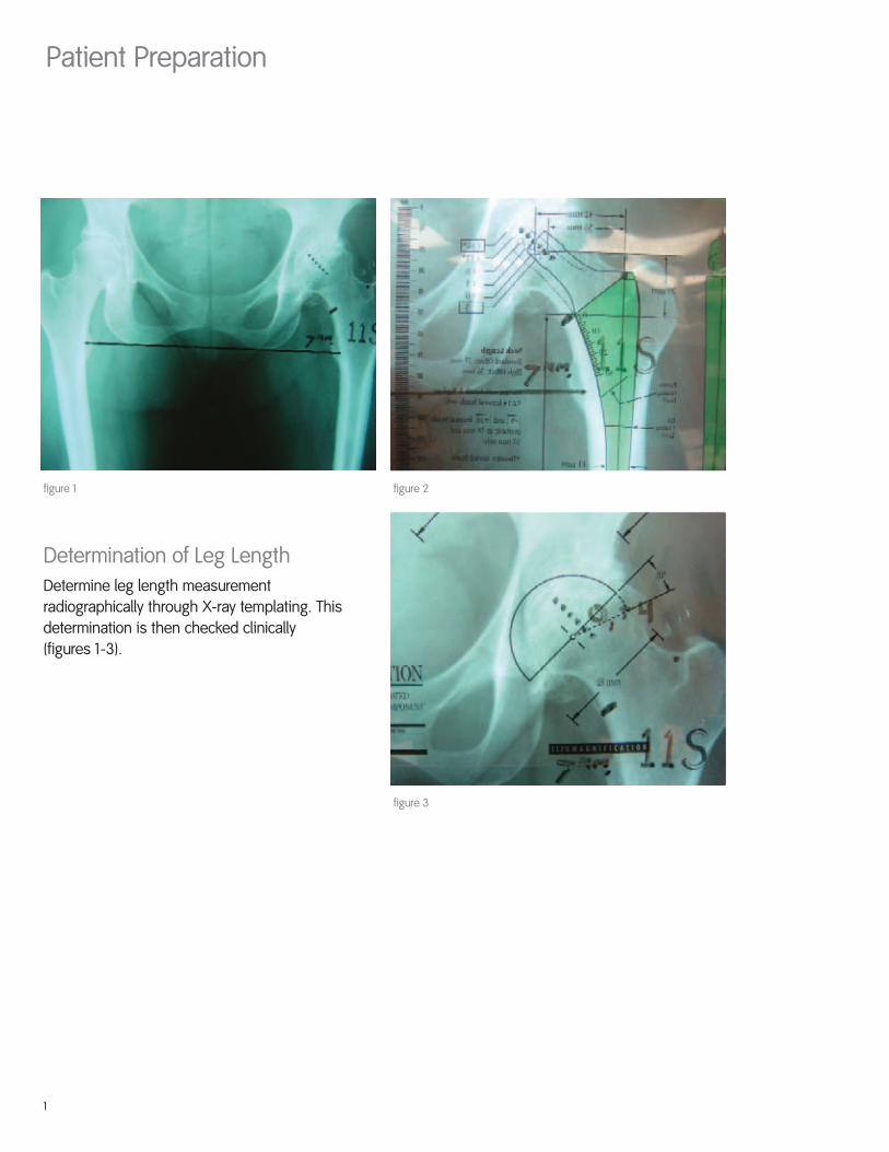
Determination of Leg LengthDetermine leg length measurementradiographically through X-ray templating. Thisdetermination is then checked clinically(figures 1-3).
figure 2figure 1
figure 3
2
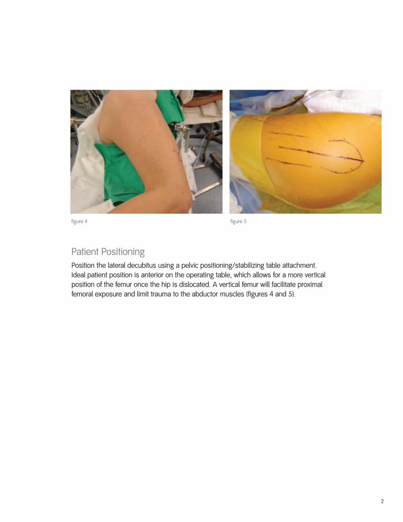
Patient PositioningPosition the lateral decubitus using a pelvic positioning/stabilizing table attachment.Ideal patient position is anterior on the operating table, which allows for a more verticalposition of the femur once the hip is dislocated. A vertical femur will facilitate proximalfemoral exposure and limit trauma to the abductor muscles (figures 4 and 5).
figure 4 figure 5
Surgical Technique
Incision Planning
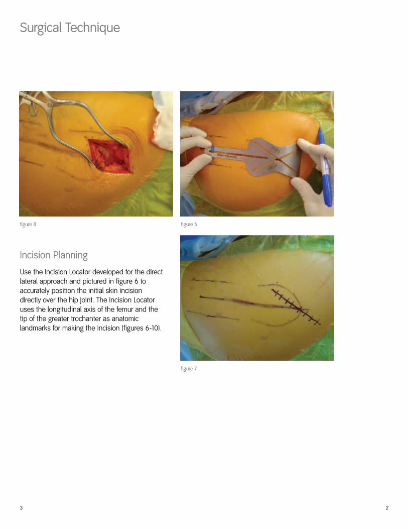
Use the Incision Locator developed for the direct lateral approach and pictured in figure 6 toaccurately position the initial skin incisiondirectly over the hip joint. The Incision Locatoruses the longitudinal axis of the femur and thetip of the greater trochanter as anatomiclandmarks for making the incision (figures 6-10).
2
figure 6figure 8
figure 7
3
4
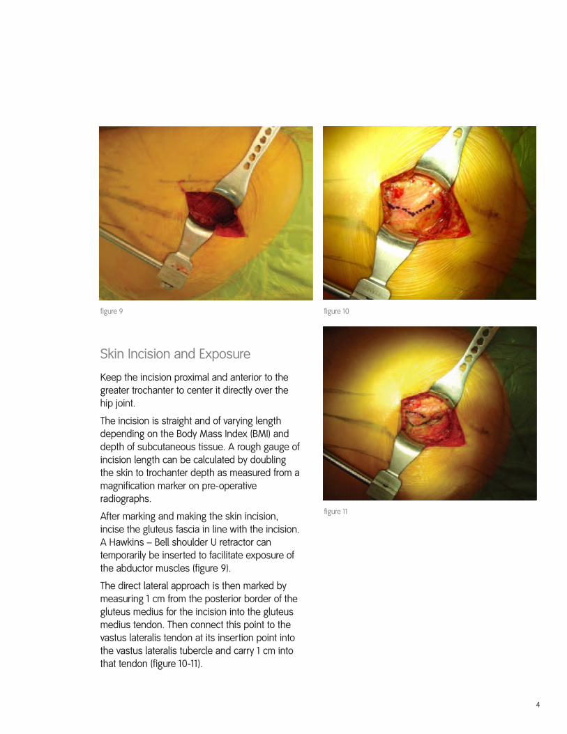
Skin Incision and Exposure
Keep the incision proximal and anterior to thegreater trochanter to center it directly over thehip joint.
The incision is straight and of varying lengthdepending on the Body Mass Index (BMI) anddepth of subcutaneous tissue. A rough gauge ofincision length can be calculated by doublingthe skin to trochanter depth as measured from amagnification marker on pre-operativeradiographs.
After marking and making the skin incision,incise the gluteus fascia in line with the incision.A Hawkins – Bell shoulder U retractor cantemporarily be inserted to facilitate exposure ofthe abductor muscles (figure 9).
The direct lateral approach is then marked bymeasuring 1 cm from the posterior border of thegluteus medius for the incision into the gluteusmedius tendon. Then connect this point to thevastus lateralis tendon at its insertion point intothe vastus lateralis tubercle and carry 1 cm intothat tendon (figure 10-11).
figure 10figure 9
figure 11
Surgical Technique
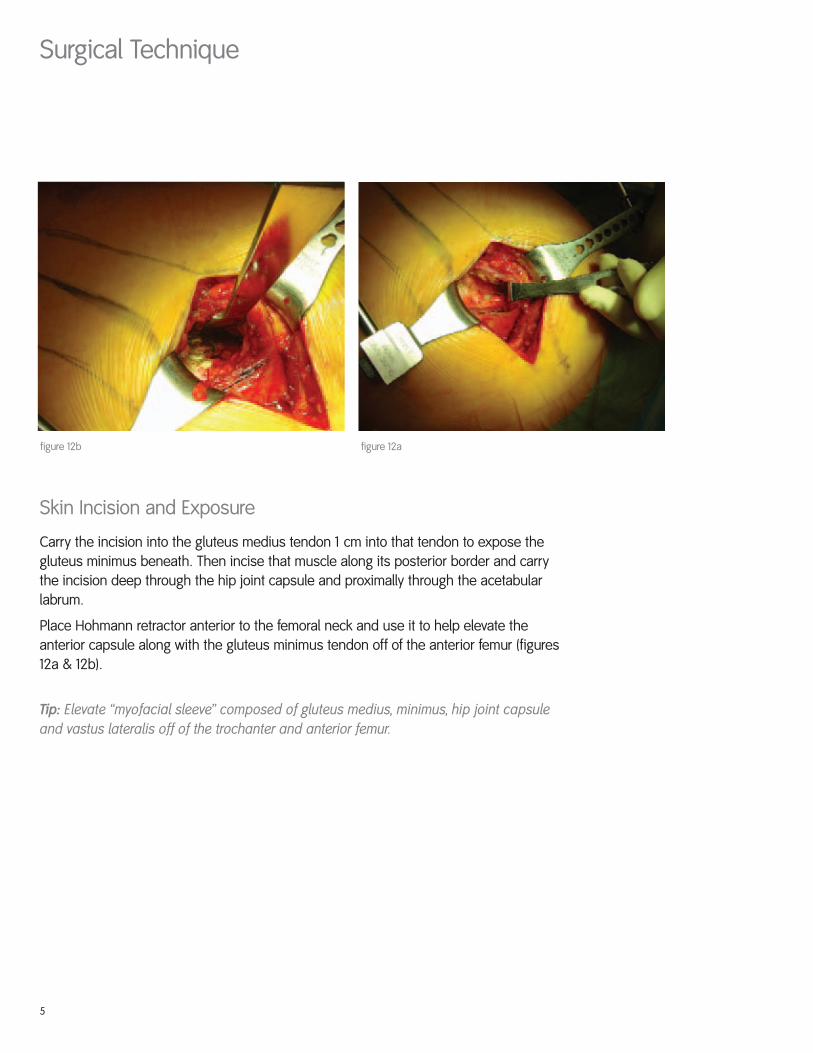
Skin Incision and Exposure
Carry the incision into the gluteus medius tendon 1 cm into that tendon to expose thegluteus minimus beneath. Then incise that muscle along its posterior border and carrythe incision deep through the hip joint capsule and proximally through the acetabularlabrum.
Place Hohmann retractor anterior to the femoral neck and use it to help elevate theanterior capsule along with the gluteus minimus tendon off of the anterior femur (figures12a & 12b).
Tip: Elevate “myofacial sleeve” composed of gluteus medius, minimus, hip joint capsuleand vastus lateralis off of the trochanter and anterior femur.
5
figure 12afigure 12b
6
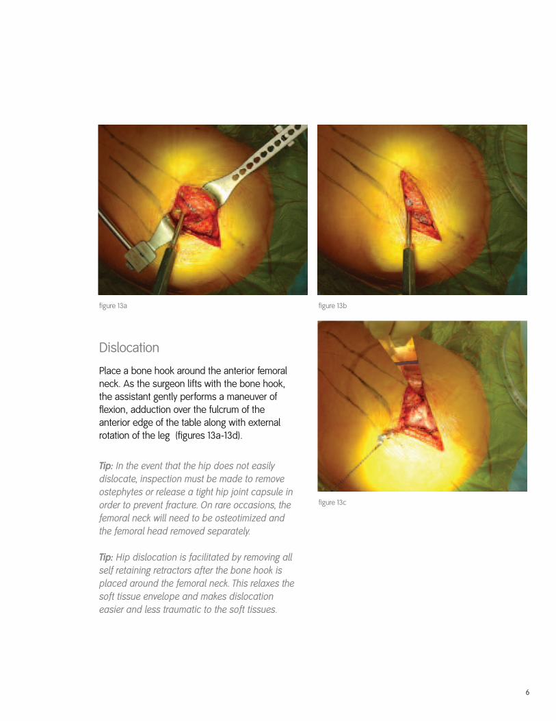
Dislocation
Place a bone hook around the anterior femoralneck. As the surgeon lifts with the bone hook,the assistant gently performs a maneuver offlexion, adduction over the fulcrum of theanterior edge of the table along with externalrotation of the leg (figures 13a-13d).
Tip: In the event that the hip does not easilydislocate, inspection must be made to removeostephytes or release a tight hip joint capsule inorder to prevent fracture. On rare occasions, thefemoral neck will need to be osteotimized andthe femoral head removed separately.
Tip: Hip dislocation is facilitated by removing allself retaining retractors after the bone hook isplaced around the femoral neck. This relaxes thesoft tissue envelope and makes dislocationeasier and less traumatic to the soft tissues.
figure 13bfigure 13a
figure 13c

7
Surgical Technique
Femoral Preparation
Place thin narrow Hohmann retractor anterior toboth the femoral neck and anterior wall of theacetabulum. Leverage on this retractor willadequately expose the anterior femoral neck forosteotomy. Place Charnley pin retractor into thetrochanter and use a right angle retractor tofurther enhance exposure and protect the skin(figures 14a-14c).
The femoral neck is osteotomized with a saggitalsaw and the femoral head is removed.
Synovial adhesions to the posterior femoralhead and neck will frequently need to bereleased as the head is removed in retrogradefashion using a bone holding forcep.
figure 14bfigure 14a
figure 14c

8
Femoral Preparation (continued)
Replace the Hohmann retractor with a mediumDeaver retractor anterior to the proximal femur.Reposition the Charnley pin retractor to facilitateexposure of the proximal femur. Use a thinmalleable retractor to protect the soft tissues asfemoral reamers and broaches are used toprepare the femur (figures 15-17).
figure 16figure 15
figure 17

Surgical Technique
9
Acetabular Preparation
Exposure for acetabular preparation is madewith a Homann retractor placed anterior andparallel to the anterior wall of the acetablum.Then place Charnley pin retractors superior intothe ileum and posterior into the ischium toexpose the acetablum (figures 18-20).
Remove the acetabular labrum and osteophytestaking care not to disturb the posterior capsule.
Tip: A fundamental principle of acetabularpreparation is to ream a hemisphere and not adirection! Reaming in the direction of theintended cup position will tend to create anelliptical acetabulum instead of a hemisphere.This is due to the vector applied to the reamingdevice by all of the tissues but principally by thefemur.
figure 19figure 18
figure 20
10
Closure
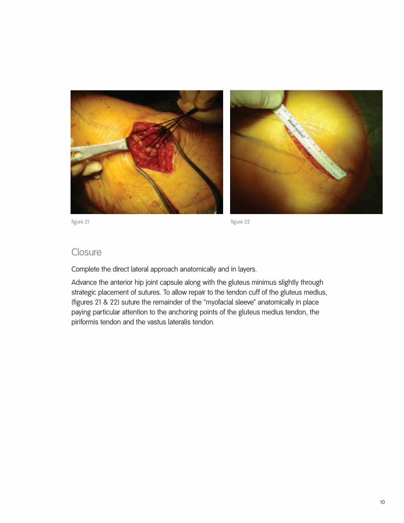
Complete the direct lateral approach anatomically and in layers.
Advance the anterior hip joint capsule along with the gluteus minimus slightly throughstrategic placement of sutures. To allow repair to the tendon cuff of the gluteus medius,(figures 21 & 22) suture the remainder of the “myofacial sleeve” anatomically in placepaying particular attention to the anchoring points of the gluteus medius tendon, thepiriformis tendon and the vastus lateralis tendon.
figure 22figure 21

11
Surgical Technique
Rehabilitation Protocol
There is generally no need for an abduction pillow following exposure through a directlateral approach. Patients are encouraged to begin transfers out of bed on the day ofsurgery and to begin formal gait training with partial weight bearing on the day of or theday after surgery. Discharge from the hospital can occur on the day of surgery inselected patients when same day physical therapy and home health care support isavailable. Other patients can be discharged 1-3 days earlier than with standard nonminimally invasive total hip replacement surgery patients.
Crutch or walker-protected ambulation continues for the first 3 weeks followed by fullweight bearing with a cane for an additional 2-3 weeks. Driving is usually permitted in3 weeks along with the transition to a cane.


OrthopaedicsSmith & Nephew, Inc.1450 Brooks RoadMemphis, TN 38116USA
Telephone: 1-901-396-2121Information: 1-800-821-5700Orders/inquiries: 1-800-238-7538
www.smith-nephew.com
45800102 7138-0933 01/05