pharmacoscan assay 24-array format manual workflow · 2018-06-13 · channel instrument, a system...
TRANSCRIPT

PharmacoScan™ Assay 24-Array Format Manual WorkflowUSER GUIDE
Catalog Numbers 903010TS and 903011TS
Publication Number 703286
Revision 4
For Research Use Only. Not for use in diagnostic procedures.
The information in this guide is subject to change without notice.
DISCLAIMER: TO THE EXTENT ALLOWED BY LAW, LIFE TECHNOLOGIES AND/OR ITS AFFILIATE(S) WILL NOT BE LIABLE FOR SPECIAL, INCIDENTAL, INDIRECT, PUNITIVE, MULTIPLE, OR CONSEQUENTIAL DAMAGES IN CONNECTION WITH OR ARISING FROM THIS DOCUMENT, INCLUDING YOUR USE OF IT.
Important Licensing Information
This product may be covered by one or more Limited Use Label Licenses. By use of this product, you accept the terms and conditions of all applicable Limited Use Label Licenses.
Corporate entity
Life Technologies | Carlsbad, CA 92008 USA | Toll Free in USA 1 800 955 6288
TRADEMARKS
All trademarks are the property of Thermo Fisher Scientific and its subsidiaries unless otherwise specified.
UV-Star is a registered trademarks of GREINER BIO-ONE. Eppendorf and Mastercycler are registered trademarks of Eppendorf AG. Bio-Rad and Hard-Shell are registered trademarks of Bio-Rad Laboratories, Inc. Microsoft, and Excel are either registered trademarks or trademarks of Microsoft Corporation in the United States and/or other countries.
©2018 Thermo Fisher Scientific Inc. All rights reserved.
Thermo Fisher Scientific Baltics UABV.A. Graiciuno 8, LT-02241Vilnius, Lithuania
Products: PharmacoScan™ Reagent Kit
Affymetrix Pte Ltd7 Gul Circle #2M-01Keppel Logistics BuildingSingapore 629563
Products: PharmacoScan™ Array Plates
Revision history: Pub. No. 703286
Revision Date Description
4 17 May 2018 Updated to include an option for a three-hour DNA precipitation step to enable faster assay turnaround time.
3 30 January 2018 Updated to the current document template, with associated updates to the warranty, trademarks, and logos. Two additional sample types have been validated and added to the document: saliva and buccal cell.
2 05 October 2016 Baseline for revision history.

PharmacoScan™ Assay 24-Array Format Manual Workflow User Guide
Contents
CHAPTER 1 About the PharmacoScan™ Solution . . . . . . . . . . . . . . . . 9
Overview . . . . . . . . . . . . . . . . . . . . . . . . . . . . . . . . . . . . . . . . . . . . . . . . . . . . . . . . . . . . . . . . . 9
Introduction to the PharmacoScan™ Assay 24-Array Format Manual Workflow . . . . . . . . . . . 9
PharmacoScan™ Assay 24-Array Format Manual Workflow . . . . . . . . . . . . . . . . . . . . . . . . . 10
What’s new . . . . . . . . . . . . . . . . . . . . . . . . . . . . . . . . . . . . . . . . . . . . . . . . . . . . . . . . . . . . . . 10
CHAPTER 2 Genomic DNA preparation and requirements. . . . . . . . . 12
Sources of genomic DNA . . . . . . . . . . . . . . . . . . . . . . . . . . . . . . . . . . . . . . . . . . . . . . . . . . . 12
General requirements . . . . . . . . . . . . . . . . . . . . . . . . . . . . . . . . . . . . . . . . . . . . . . . . . . . . . . 13
Special requirements . . . . . . . . . . . . . . . . . . . . . . . . . . . . . . . . . . . . . . . . . . . . . . . . . . . . . 13
Assess the quality of genomic DNA using 1% Agarose E-gels . . . . . . . . . . . . . . . . . . . . . 14
Genomic DNA extraction/purification methods . . . . . . . . . . . . . . . . . . . . . . . . . . . . . . . . . . . 15
Genomic DNA cleanup . . . . . . . . . . . . . . . . . . . . . . . . . . . . . . . . . . . . . . . . . . . . . . . . . . . . . 15
Genomic DNA preparation . . . . . . . . . . . . . . . . . . . . . . . . . . . . . . . . . . . . . . . . . . . . . . . . . . . 16
Duration . . . . . . . . . . . . . . . . . . . . . . . . . . . . . . . . . . . . . . . . . . . . . . . . . . . . . . . . . . . . . . . 16
Equipment, consumables, and reagents required . . . . . . . . . . . . . . . . . . . . . . . . . . . . . . . 16
1. Thaw samples and controls . . . . . . . . . . . . . . . . . . . . . . . . . . . . . . . . . . . . . . . . . . . . . . 17
2. Quantitate and dilute gDNA . . . . . . . . . . . . . . . . . . . . . . . . . . . . . . . . . . . . . . . . . . . . . . 17
3. Aliquot the diluted samples and the controls DNA 1 and DNA 2 . . . . . . . . . . . . . . . . . 18
4. Freeze or proceed . . . . . . . . . . . . . . . . . . . . . . . . . . . . . . . . . . . . . . . . . . . . . . . . . . . . . 19
5. Create a GeneTitan Array Plate Registration file . . . . . . . . . . . . . . . . . . . . . . . . . . . . . . 19
CHAPTER 3 Preparation before you start . . . . . . . . . . . . . . . . . . . . . . 21
Introduction . . . . . . . . . . . . . . . . . . . . . . . . . . . . . . . . . . . . . . . . . . . . . . . . . . . . . . . . . . . . . . 21
PharmacoScan™ Reagent Kit 4x24 Reactions, arrays, and GeneTitan™ consumables required . . . . . . . . . . . . . . . . . . . . . . . . . . . . . . . . . . . . . . . . . . . . . . . . . . . . . . . . . . . . . . . 21
Requirements and recommendations . . . . . . . . . . . . . . . . . . . . . . . . . . . . . . . . . . . . . . . . . . 21
Room temperature . . . . . . . . . . . . . . . . . . . . . . . . . . . . . . . . . . . . . . . . . . . . . . . . . . . . . . . 21
Special requirements . . . . . . . . . . . . . . . . . . . . . . . . . . . . . . . . . . . . . . . . . . . . . . . . . . . . . 22
Plate requirements and recommendations . . . . . . . . . . . . . . . . . . . . . . . . . . . . . . . . . . . . 22
Thermal cycler recommendations and protocols . . . . . . . . . . . . . . . . . . . . . . . . . . . . . . . 23
Thermal cycler consumables . . . . . . . . . . . . . . . . . . . . . . . . . . . . . . . . . . . . . . . . . . . . . . . 24
Oven recommendations . . . . . . . . . . . . . . . . . . . . . . . . . . . . . . . . . . . . . . . . . . . . . . . . . . . 24
3

Contents
Plate centrifuge . . . . . . . . . . . . . . . . . . . . . . . . . . . . . . . . . . . . . . . . . . . . . . . . . . . . . . . . . 24
Plate shakers . . . . . . . . . . . . . . . . . . . . . . . . . . . . . . . . . . . . . . . . . . . . . . . . . . . . . . . . . . . 25
Equipment care and calibration . . . . . . . . . . . . . . . . . . . . . . . . . . . . . . . . . . . . . . . . . . . . . 25
Procedures . . . . . . . . . . . . . . . . . . . . . . . . . . . . . . . . . . . . . . . . . . . . . . . . . . . . . . . . . . . . . . 25
Seal, vortex, and spin . . . . . . . . . . . . . . . . . . . . . . . . . . . . . . . . . . . . . . . . . . . . . . . . . . . . 25
Sample quantitation . . . . . . . . . . . . . . . . . . . . . . . . . . . . . . . . . . . . . . . . . . . . . . . . . . . . . . 26
About the reagents and master mix preparation . . . . . . . . . . . . . . . . . . . . . . . . . . . . . . . . 26
Pipettes and pipetting . . . . . . . . . . . . . . . . . . . . . . . . . . . . . . . . . . . . . . . . . . . . . . . . . . . . 28
Divided reservoir use . . . . . . . . . . . . . . . . . . . . . . . . . . . . . . . . . . . . . . . . . . . . . . . . . . . . . 29
Freeze-thaw instructions . . . . . . . . . . . . . . . . . . . . . . . . . . . . . . . . . . . . . . . . . . . . . . . . . . 30
Equipment, consumables, labware, and reagents required . . . . . . . . . . . . . . . . . . . . . . . . . 31
Equipment required for the PharmacoScan Assay 24-Array Format Manual Workflow . . 31
Consumables required for PharmacoScan™ Assay 24-Array Format Manual Workflow . 31
GeneTitan™ MC Instrument consumables . . . . . . . . . . . . . . . . . . . . . . . . . . . . . . . . . . . . . 34
Label GeneTitan™ hybridization and reagent trays . . . . . . . . . . . . . . . . . . . . . . . . . . . . . . 38
Reagent Kit for the PharmacoScan™ Assay 24-Array Format Manual Workflow . . . . . . . 41
CHAPTER 4 Target preparation. . . . . . . . . . . . . . . . . . . . . . . . . . . . . . 42
Introduction . . . . . . . . . . . . . . . . . . . . . . . . . . . . . . . . . . . . . . . . . . . . . . . . . . . . . . . . . . . . . . 42
Stage 1A: Multiplex PCR (mPCR) . . . . . . . . . . . . . . . . . . . . . . . . . . . . . . . . . . . . . . . . . . . . . 43
Duration . . . . . . . . . . . . . . . . . . . . . . . . . . . . . . . . . . . . . . . . . . . . . . . . . . . . . . . . . . . . . . . 43
Input required . . . . . . . . . . . . . . . . . . . . . . . . . . . . . . . . . . . . . . . . . . . . . . . . . . . . . . . . . . 43
Equipment, consumables, and reagents required . . . . . . . . . . . . . . . . . . . . . . . . . . . . . . . 44
1: Prepare for mPCR . . . . . . . . . . . . . . . . . . . . . . . . . . . . . . . . . . . . . . . . . . . . . . . . . . . . . 45
2: Prepare the mPCR Master Mix . . . . . . . . . . . . . . . . . . . . . . . . . . . . . . . . . . . . . . . . . . . 46
3: Set up the mPCR reaction plate . . . . . . . . . . . . . . . . . . . . . . . . . . . . . . . . . . . . . . . . . . 46
4: Freeze QIAGEN reagents . . . . . . . . . . . . . . . . . . . . . . . . . . . . . . . . . . . . . . . . . . . . . . . . 46
5: Run the PharmacoScan mPCR thermal cycler protocol . . . . . . . . . . . . . . . . . . . . . . . . 47
6: Freeze mPCR Reaction Plate or proceed . . . . . . . . . . . . . . . . . . . . . . . . . . . . . . . . . . . 47
Stage 1B: DNA amplification . . . . . . . . . . . . . . . . . . . . . . . . . . . . . . . . . . . . . . . . . . . . . . . . . 49
Duration . . . . . . . . . . . . . . . . . . . . . . . . . . . . . . . . . . . . . . . . . . . . . . . . . . . . . . . . . . . . . . . 49
Input required . . . . . . . . . . . . . . . . . . . . . . . . . . . . . . . . . . . . . . . . . . . . . . . . . . . . . . . . . . 49
Equipment, consumables, and reagents required . . . . . . . . . . . . . . . . . . . . . . . . . . . . . . . 50
1: Prepare for DNA amplification . . . . . . . . . . . . . . . . . . . . . . . . . . . . . . . . . . . . . . . . . . . . 51
2: Prepare the Denaturation Master Mix . . . . . . . . . . . . . . . . . . . . . . . . . . . . . . . . . . . . . . 52
3: Add Denaturation Master Mix to samples . . . . . . . . . . . . . . . . . . . . . . . . . . . . . . . . . . . 53
4: Add Neutralization Solution to samples . . . . . . . . . . . . . . . . . . . . . . . . . . . . . . . . . . . . 53
5: Prepare the Amplification Master Mix . . . . . . . . . . . . . . . . . . . . . . . . . . . . . . . . . . . . . . 54
6: Add Amplification Master Mix to samples . . . . . . . . . . . . . . . . . . . . . . . . . . . . . . . . . . . 54
7: Freeze reagents . . . . . . . . . . . . . . . . . . . . . . . . . . . . . . . . . . . . . . . . . . . . . . . . . . . . . . . 54
8: Freeze or proceed . . . . . . . . . . . . . . . . . . . . . . . . . . . . . . . . . . . . . . . . . . . . . . . . . . . . . 54
4 PharmacoScan™ Assay 24-Array Format Manual Workflow User Guide

Contents
Stage 2: Fragmentation and precipitation . . . . . . . . . . . . . . . . . . . . . . . . . . . . . . . . . . . . . . . 56
Duration . . . . . . . . . . . . . . . . . . . . . . . . . . . . . . . . . . . . . . . . . . . . . . . . . . . . . . . . . . . . . . . 56
Input required . . . . . . . . . . . . . . . . . . . . . . . . . . . . . . . . . . . . . . . . . . . . . . . . . . . . . . . . . . . 56
Equipment, consumables, and reagents required . . . . . . . . . . . . . . . . . . . . . . . . . . . . . . . 56
1: Prepare for fragmentation and precipitation . . . . . . . . . . . . . . . . . . . . . . . . . . . . . . . . . 57
2: mPCR spike-in to Amplification Plate . . . . . . . . . . . . . . . . . . . . . . . . . . . . . . . . . . . . . . 59
3. Incubate samples in pre-heated ovens . . . . . . . . . . . . . . . . . . . . . . . . . . . . . . . . . . . . . 60
4: Prepare the Fragmentation Master Mix . . . . . . . . . . . . . . . . . . . . . . . . . . . . . . . . . . . . . 60
5: Add the Fragmentation Master Mix to samples . . . . . . . . . . . . . . . . . . . . . . . . . . . . . . . 60
6: Add the Stop Solution to the samples . . . . . . . . . . . . . . . . . . . . . . . . . . . . . . . . . . . . . . 61
7: Prepare the Precipitation Master Mix . . . . . . . . . . . . . . . . . . . . . . . . . . . . . . . . . . . . . . 62
8: Prepare and add isopropanol to Precipitation Plate . . . . . . . . . . . . . . . . . . . . . . . . . . . 62
9: Freeze the Precipitation Plate . . . . . . . . . . . . . . . . . . . . . . . . . . . . . . . . . . . . . . . . . . . . 63
10: Freeze reagents . . . . . . . . . . . . . . . . . . . . . . . . . . . . . . . . . . . . . . . . . . . . . . . . . . . . . . 63
Stage 3: Centrifuge and drying, resuspension and hybridization preparation, and sample QC . . . . . . . . . . . . . . . . . . . . . . . . . . . . . . . . . . . . . . . . . . . . . . . . . . . . . . . . . . . . . . . 66
Duration . . . . . . . . . . . . . . . . . . . . . . . . . . . . . . . . . . . . . . . . . . . . . . . . . . . . . . . . . . . . . . . 66
Input required . . . . . . . . . . . . . . . . . . . . . . . . . . . . . . . . . . . . . . . . . . . . . . . . . . . . . . . . . . . 66
Equipment, consumables, and reagents required . . . . . . . . . . . . . . . . . . . . . . . . . . . . . . . 67
Stage 3A: Centrifuge Precipitation Plate and dry the DNA pellet . . . . . . . . . . . . . . . . . . . . . 69
Stage 3B: Resuspension and hybridization preparation . . . . . . . . . . . . . . . . . . . . . . . . . . . . 70
1: Prepare for Resuspension and hybridization preparation . . . . . . . . . . . . . . . . . . . . . . . 70
2: Prepare DNA pellets and warm the resuspension buffer . . . . . . . . . . . . . . . . . . . . . . . . 70
3: Thaw and prepare the reagents . . . . . . . . . . . . . . . . . . . . . . . . . . . . . . . . . . . . . . . . . . . 70
4: Label tubes and reservoirs . . . . . . . . . . . . . . . . . . . . . . . . . . . . . . . . . . . . . . . . . . . . . . . 71
5: Add Resuspension Buffer to DNA pellets . . . . . . . . . . . . . . . . . . . . . . . . . . . . . . . . . . . 71
6: Resuspension of DNA pellets . . . . . . . . . . . . . . . . . . . . . . . . . . . . . . . . . . . . . . . . . . . . 71
7: Prepare the Hybridization Master Mix . . . . . . . . . . . . . . . . . . . . . . . . . . . . . . . . . . . . . . 72
8: Prepare the Hyb Ready Sample Plate . . . . . . . . . . . . . . . . . . . . . . . . . . . . . . . . . . . . . . 72
9: Freeze or proceed . . . . . . . . . . . . . . . . . . . . . . . . . . . . . . . . . . . . . . . . . . . . . . . . . . . . . 72
Stage 3C: (Recommended) Perform quantitation and fragmentation QC checks . . . . . . . . 73
1: Prepare for sample QC . . . . . . . . . . . . . . . . . . . . . . . . . . . . . . . . . . . . . . . . . . . . . . . . . 73
2: Perform QC checks . . . . . . . . . . . . . . . . . . . . . . . . . . . . . . . . . . . . . . . . . . . . . . . . . . . . 74
3: Freeze or proceed . . . . . . . . . . . . . . . . . . . . . . . . . . . . . . . . . . . . . . . . . . . . . . . . . . . . . 74
Stage 4: Denaturation and hybridization . . . . . . . . . . . . . . . . . . . . . . . . . . . . . . . . . . . . . . . . 78
Duration . . . . . . . . . . . . . . . . . . . . . . . . . . . . . . . . . . . . . . . . . . . . . . . . . . . . . . . . . . . . . . . 78
Required input from previous stage . . . . . . . . . . . . . . . . . . . . . . . . . . . . . . . . . . . . . . . . . . 78
Equipment, consumables, and reagents required . . . . . . . . . . . . . . . . . . . . . . . . . . . . . . . 78
1: Prepare for denaturation and hybridization . . . . . . . . . . . . . . . . . . . . . . . . . . . . . . . . . . 80
2: Prepare hybridization ready samples stored at –20°C . . . . . . . . . . . . . . . . . . . . . . . . . 80
3: Prepare the GeneTitan™ MC Instrument . . . . . . . . . . . . . . . . . . . . . . . . . . . . . . . . . . . . 80
4: Denature the Hyb Ready Sample plate . . . . . . . . . . . . . . . . . . . . . . . . . . . . . . . . . . . . . 81
5: Prepare hybridization tray and load into GeneTitan™ MC Instrument . . . . . . . . . . . . . . 82
PharmacoScan™ Assay 24-Array Format Manual Workflow User Guide 5
Contents
Stage 5: GeneTitan™ reagent preparation . . . . . . . . . . . . . . . . . . . . . . . . . . . . . . . . . . . . . . . 85
Equipment, consumables, and reagents required . . . . . . . . . . . . . . . . . . . . . . . . . . . . . . . 86
1: Prepare for GeneTitan™ reagent preparation . . . . . . . . . . . . . . . . . . . . . . . . . . . . . . . . . 88
2: Prepare the stain, ligation, and stabilization master mixes . . . . . . . . . . . . . . . . . . . . . . 91
3: Aliquot Master Mixes and Hold Buffer into trays . . . . . . . . . . . . . . . . . . . . . . . . . . . . . . 93
4: Store remaining reagents . . . . . . . . . . . . . . . . . . . . . . . . . . . . . . . . . . . . . . . . . . . . . . . . 98
CHAPTER 5 Array processing with the GeneTitan™ Multi-Channel Instrument . . . . . . . . . . . . . . . . . . . . . . . . . . . . . . . . . . . . . . . . . . . . . . 102
Before using the GeneTitan™ MC Instrument . . . . . . . . . . . . . . . . . . . . . . . . . . . . . . . . . . . 102
Proper tray alignment and loading . . . . . . . . . . . . . . . . . . . . . . . . . . . . . . . . . . . . . . . . . 102
Stain trays and covers . . . . . . . . . . . . . . . . . . . . . . . . . . . . . . . . . . . . . . . . . . . . . . . . . . . 104
Label GeneTitan™ hybridization and reagent trays . . . . . . . . . . . . . . . . . . . . . . . . . . . . . 105
Email and telephone notifications from the GeneTitan™ MC Instrument . . . . . . . . . . . . . 106
GeneTitan™ MC Instrument lamp . . . . . . . . . . . . . . . . . . . . . . . . . . . . . . . . . . . . . . . . . . 107
Setup options for array plate processing . . . . . . . . . . . . . . . . . . . . . . . . . . . . . . . . . . . . 107
Abort a process . . . . . . . . . . . . . . . . . . . . . . . . . . . . . . . . . . . . . . . . . . . . . . . . . . . . . . . . 110
Stage 1: Create and upload GeneTitan Array Plate Registration file . . . . . . . . . . . . . . . . . . 111
Stage 2: Hybridization . . . . . . . . . . . . . . . . . . . . . . . . . . . . . . . . . . . . . . . . . . . . . . . . . . . . . 112
Reagents required . . . . . . . . . . . . . . . . . . . . . . . . . . . . . . . . . . . . . . . . . . . . . . . . . . . . . . 112
Set up the instrument . . . . . . . . . . . . . . . . . . . . . . . . . . . . . . . . . . . . . . . . . . . . . . . . . . . 112
Load a PharmacoScan™ array plate and hybridization tray onto the GeneTitan™ MC Instrument . . . . . . . . . . . . . . . . . . . . . . . . . . . . . . . . . . . . . . . . . . . . . . . . . . . . . . . . . . . 117
Load a second PharmacoScan™ array plate and hybridization tray onto the GeneTitan™ MC Instrument . . . . . . . . . . . . . . . . . . . . . . . . . . . . . . . . . . . . . . . . . . . . . . 123
Status window prompts and actions required . . . . . . . . . . . . . . . . . . . . . . . . . . . . . . . . . . . 125
Stage 3: Ligate, wash, stain and scan . . . . . . . . . . . . . . . . . . . . . . . . . . . . . . . . . . . . . . . . . 129
Equipment, consumables, and reagents required . . . . . . . . . . . . . . . . . . . . . . . . . . . . . . 129
Proper installation of the GeneTitan™ tray consumables . . . . . . . . . . . . . . . . . . . . . . . . 130
Load tray consumables onto the GeneTitan™ MC Instrument . . . . . . . . . . . . . . . . . . . . 131
Continue the workflow . . . . . . . . . . . . . . . . . . . . . . . . . . . . . . . . . . . . . . . . . . . . . . . . . . . . . 138
Shut down the GeneTitan™ MC Instrument . . . . . . . . . . . . . . . . . . . . . . . . . . . . . . . . . . . . . 139
CHAPTER 6 Troubleshooting . . . . . . . . . . . . . . . . . . . . . . . . . . . . . . 140
GeneTitan™ Multi-Channel Instrument . . . . . . . . . . . . . . . . . . . . . . . . . . . . . . . . . . . . . . . . 140
Miscellaneous messages . . . . . . . . . . . . . . . . . . . . . . . . . . . . . . . . . . . . . . . . . . . . . . . . . 141
Fluidic diagnostic messages . . . . . . . . . . . . . . . . . . . . . . . . . . . . . . . . . . . . . . . . . . . . . . 143
Wash/Scan Resume . . . . . . . . . . . . . . . . . . . . . . . . . . . . . . . . . . . . . . . . . . . . . . . . . . . . 147
Abort a run . . . . . . . . . . . . . . . . . . . . . . . . . . . . . . . . . . . . . . . . . . . . . . . . . . . . . . . . . . . . 147
6 PharmacoScan™ Assay 24-Array Format Manual Workflow User Guide
Contents
APPENDIX A Fragmentation quality control gel protocol . . . . . . . . . 148
Run a fragmentation quality control gel . . . . . . . . . . . . . . . . . . . . . . . . . . . . . . . . . . . . . . . . 148
Equipment required . . . . . . . . . . . . . . . . . . . . . . . . . . . . . . . . . . . . . . . . . . . . . . . . . . . . . 148
E-Gels and reagents . . . . . . . . . . . . . . . . . . . . . . . . . . . . . . . . . . . . . . . . . . . . . . . . . . . . 148
Consumables . . . . . . . . . . . . . . . . . . . . . . . . . . . . . . . . . . . . . . . . . . . . . . . . . . . . . . . . . . 148
Dilute the TrackIt™ Cyan/Orange Loading Buffer and 25 bp ladder . . . . . . . . . . . . . . . . 149
Run the fragmentation QC gel protocol . . . . . . . . . . . . . . . . . . . . . . . . . . . . . . . . . . . . . . 149
APPENDIX B Sample quantitation after resuspension . . . . . . . . . . . 151
Protocol for sample quantitation after resuspension . . . . . . . . . . . . . . . . . . . . . . . . . . . . . . 151
Equipment required . . . . . . . . . . . . . . . . . . . . . . . . . . . . . . . . . . . . . . . . . . . . . . . . . . . . . 151
Quantitate the diluted samples . . . . . . . . . . . . . . . . . . . . . . . . . . . . . . . . . . . . . . . . . . . . 151
Assess the OD readings . . . . . . . . . . . . . . . . . . . . . . . . . . . . . . . . . . . . . . . . . . . . . . . . . . 152
Suggested Protocol for OD Quantitation Using the DTX 880 . . . . . . . . . . . . . . . . . . . . . . . 153
Perform sample quantitation on a plate reader other than the DTX880 . . . . . . . . . . . . . . . 160
APPENDIX C Register samples in GeneChip™ Command Console™161
Create a GeneTitan™ Array Plate Registration file . . . . . . . . . . . . . . . . . . . . . . . . . . . . . . . . 161
APPENDIX D Deionizing procedure for GeneTitan™ trays and covers. . . . . . . . . . . . . . . . . . . . . . . . . . . . . . . . . . . . . . . . . . . . . . 164
Deionization procedure . . . . . . . . . . . . . . . . . . . . . . . . . . . . . . . . . . . . . . . . . . . . . . . . . . . . 165
Ion-indicator cap . . . . . . . . . . . . . . . . . . . . . . . . . . . . . . . . . . . . . . . . . . . . . . . . . . . . . . . . . 167
APPENDIX E GeneTitan™ Multi-Channel Instrument care . . . . . . . . 168
Introduction . . . . . . . . . . . . . . . . . . . . . . . . . . . . . . . . . . . . . . . . . . . . . . . . . . . . . . . . . . . . . 168
Cleaning and maintenance . . . . . . . . . . . . . . . . . . . . . . . . . . . . . . . . . . . . . . . . . . . . . . . . . 168
Monthly . . . . . . . . . . . . . . . . . . . . . . . . . . . . . . . . . . . . . . . . . . . . . . . . . . . . . . . . . . . . . . 168
Every six months . . . . . . . . . . . . . . . . . . . . . . . . . . . . . . . . . . . . . . . . . . . . . . . . . . . . . . . 168
Service the outer enclosure fan filters . . . . . . . . . . . . . . . . . . . . . . . . . . . . . . . . . . . . . . . 169
Replace the bottle filters . . . . . . . . . . . . . . . . . . . . . . . . . . . . . . . . . . . . . . . . . . . . . . . . . 170
Replace the xenon lamp in the GeneTitan™ MC Instrument . . . . . . . . . . . . . . . . . . . . . . 172
Troubleshooting . . . . . . . . . . . . . . . . . . . . . . . . . . . . . . . . . . . . . . . . . . . . . . . . . . . . . . . . . . 177
Log files . . . . . . . . . . . . . . . . . . . . . . . . . . . . . . . . . . . . . . . . . . . . . . . . . . . . . . . . . . . . . . 177
GCC log files for GeneTitan™ MC systems . . . . . . . . . . . . . . . . . . . . . . . . . . . . . . . . . . . 178
Problems and solutions . . . . . . . . . . . . . . . . . . . . . . . . . . . . . . . . . . . . . . . . . . . . . . . . . . 178
Insufficient disk space notice . . . . . . . . . . . . . . . . . . . . . . . . . . . . . . . . . . . . . . . . . . . . . . 179
PharmacoScan™ Assay 24-Array Format Manual Workflow User Guide 7
Contents
APPENDIX F mPCR quality control gel protocol. . . . . . . . . . . . . . . . 180
Run a mPCR quality control gel . . . . . . . . . . . . . . . . . . . . . . . . . . . . . . . . . . . . . . . . . . . . . 180
Equipment required . . . . . . . . . . . . . . . . . . . . . . . . . . . . . . . . . . . . . . . . . . . . . . . . . . . . . 180
E-Gels and reagents required . . . . . . . . . . . . . . . . . . . . . . . . . . . . . . . . . . . . . . . . . . . . . 180
Consumables . . . . . . . . . . . . . . . . . . . . . . . . . . . . . . . . . . . . . . . . . . . . . . . . . . . . . . . . . . 181
Prepare NEB 50 bp DNA Ladder . . . . . . . . . . . . . . . . . . . . . . . . . . . . . . . . . . . . . . . . . . . 181
Prepare mPCR samples for gel analysis . . . . . . . . . . . . . . . . . . . . . . . . . . . . . . . . . . . . . 181
Run mPCR QC gel protocol . . . . . . . . . . . . . . . . . . . . . . . . . . . . . . . . . . . . . . . . . . . . . . . 181
APPENDIX G Safety . . . . . . . . . . . . . . . . . . . . . . . . . . . . . . . . . . . . . 183
Precautions . . . . . . . . . . . . . . . . . . . . . . . . . . . . . . . . . . . . . . . . . . . . . . . . . . . . . . . . . . . . . 183
Chemical safety . . . . . . . . . . . . . . . . . . . . . . . . . . . . . . . . . . . . . . . . . . . . . . . . . . . . . . . . . . 184
Biological hazard safety . . . . . . . . . . . . . . . . . . . . . . . . . . . . . . . . . . . . . . . . . . . . . . . . . . . . 185
Documentation and support . . . . . . . . . . . . . . . . . . . . . . . . . . . . . . . 186
Related documentation . . . . . . . . . . . . . . . . . . . . . . . . . . . . . . . . . . . . . . . . . . . . . . . . . . . . 186
Customer and technical support . . . . . . . . . . . . . . . . . . . . . . . . . . . . . . . . . . . . . . . . . . . . . 187
Limited product warranty . . . . . . . . . . . . . . . . . . . . . . . . . . . . . . . . . . . . . . . . . . . . . . . . . . 188
8 PharmacoScan™ Assay 24-Array Format Manual Workflow User Guide

1 About the PharmacoScan™
Solution
Overview
Developed in collaboration with experts across the field of pharmacogenomics, PharmacoScan™ Solution is the industry’s broadest content genetic analysis system specifically designed to provide insight into the absorption, distribution, metabolism, and excretion (ADME) and transport of commonly prescribed medicines. By interrogating more than 4,600 markers in nearly 1,200 genes known to play a role in drug metabolism, traditional clinical researchers gain unprecedented understanding into an individual’s ability to process those drugs with high evidence for genetic association, as well as those markers where moderate, low, preliminary and unknown evidence exists. PharmacoScan Solution utilizes the proven GeneTitan™ Multi-Channel Instrument, a system that is preferred worldwide by genetic researchers requiring efficient workflow, high throughput, economic pricing and lot-to-lot consistency required to support multi-year data collection and analysis efforts.
Introduction to the PharmacoScan™ Assay 24-Array Format Manual Workflow
PharmacoScan™ Assay 24-Array Format Manual Workflow is available as a bundled kit that includes the arrays, reagents and consumables needed for processing four 24-format plates, each having 22 samples and two controls.
PharmacoScan interrogates SNPs, indels and copy number variation (CNV) in a single assay workflow. Starting with genomic DNA, the samples are processed by performing a manual target preparation protocol followed by automated processing of the array plates on the GeneTitan MC Instrument.
• Target preparation uses methods including DNA amplification, fragmentation, purification and resuspension of the target in hybridization cocktail.
• The hyb-ready targets are then transferred to the GeneTitan™ Multi-Channel (MC) Instrument for automated, hands-free processing including hybridization, staining, washing and imaging.
PharmacoScan provides pharmacogenomic variation information for more than 4,600 ADME markers in nearly 1,200 genes. This content is sourced from globally endorsed consortium databases including, but not limited to CPIC, PharmGKB, and PharmaADME. Also included on PharmacoScan are high value markers for human leukocyte antigen (HLA) imputation, markers for killer cell immunoglobulin-like receptors (KIR), markers for human ancestry identification (AIM), a marker GWAS backbone, and markers for sample ID and tracking. The combination of these high value markers, in addition to PharmacoScan’s ability to precisely call variants in critical genes on a microarray, compliments Thermo Fisher Scientific’s current solutions for pharmacogenomics using the Real-Time PCR OpenArray and Ion AmpliSeq NGS Panels for Targeted Sequencing platforms.
PharmacoScan™ Assay 24-Array Format Manual Workflow User Guide 9
Chapter 1 About the PharmacoScan™ SolutionPharmacoScan™ Assay 24-Array Format Manual Workflow1
PharmacoScan is a multiplex genotyping assay which combines the proven Axiom chemistry in a 24 sample format with the incorporation of a multiplex PCR step to overcome some of the complexities associated with genotyping highly homologous markers. PharmacoScan software and algorithm developments include an allele translation and phenotyping tool and copy number aware genotyping. Array plates are processed on a GeneTitan™ MC Instrument controlled by GeneChip Command Console™ 4.3 or higher. The resulting CEL files are analyzed by Axiom™ Analysis Suite 3.0 or higher, or by Applied Biosystems Microarray Power Tools 1.19 or newer.
PharmacoScan™ Assay 24-Array Format Manual Workflow
Running the PharmacoScan Assay 24-Array Format Manual Workflow requires the following sets of steps:
1. Genomic DNA Prep—Resulting in samples that meet requirements spelled out in Chapter 2, ʺGenomic DNA preparation and requirementsʺ on page 12.
2. A multiplex PCR step (mPCR) followed by target preparation of the samples (see Chapter 4, ʺTarget preparationʺ on page 42).
3. Array Processing, done with
• GeneTitan MC Instrument
• GeneTitan Instrument Control software
• GCC Portal software
See Chapter 5, ʺArray processing with the GeneTitan™ Multi-Channel Instrumentʺ on page 102.
A list of the required equipment and supplies for running the PharmacoScan Assay 24-Array Format Manual Workflow can be found in the PharmacoScan™ Assay 24-Array Format Manual Workflow Site Preparation Guide, Pub. No. 703287.
What’s new
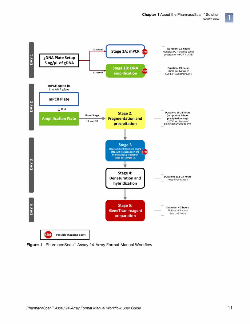
This revision of the user guide introduces the option for a three-hour DNA precipitation step to enable a faster assay turnaround time, going from sample to CEL file within 72 hours. The three-hour precipitation step shortens Stage 2: Fragmentation and precipitation to enable the operator to advance to Stage 3: Centrifuge and drying, resuspension and hybridization preparation, and sample QC followed by Stage 4: Denaturation and hybridization all on day 2 of the assay workflow. See Figure 1. Note that this workflow option will require approximately nine hours to complete these combined day 2 activities (fragmentation to initiation of hybridization on the GeneTitan MC Instrument).
The standard PharmacoScan Assay workflow, in which the DNA is precipitated overnight, provides a convenient stopping point to support single operator assay execution within an eight-hour workday.
10 PharmacoScan™ Assay 24-Array Format Manual Workflow User Guide
Chapter 1 About the PharmacoScan™ SolutionWhat’s new 1
Figure 1 PharmacoScan™ Assay 24-Array Format Manual Workflow
10 μL
10 μL/well
20 μL/well
From Stage
1A and 1B
Duration: 3.5 hours
Multiplex PCR thermal cycler program of mPCR PLATE
Stage 1A: mPCR
Stage 1B: DNA amplification
Duration: 23 hours
37°C incubation of AMPLIFICATION PLATE
DA
Y 1
D
AY
2
Stage 2: Fragmentation and
precipitation
DA
Y 3
Duration: 16-24 hours
(or optional 3-hour
precipitation step)
–20°C incubation of PRECIPITATION PLATE
Stage 3 Stage 3A: Centrifuge and drying
Stage 3B: Resuspension and hybridization preparation
Stage 3C: Sample QC
Stage 4:Denaturation and
hybridization
Duration: 23.5-24 hours
Array hybridization
DA
Y 4 Stage 5:
GeneTitan reagent preparation
Duration: ~ 7 hours
Fluidics: 4.5 hours Scan: ~2 hours
mPCR spike-in
into AMP plate
Possible stopping point
Amplification Plate
mPCR Plate
gDNA Plate Setup5 ng/μL of gDNA
PharmacoScan™ Assay 24-Array Format Manual Workflow User Guide 11

2 Genomic DNA preparation andrequirements
Sources of genomic DNA . . . . . . . . . . . . . . . . . . . . . . . . . . . . . . . . . . . . . . . . . . . . . . 12
General requirements . . . . . . . . . . . . . . . . . . . . . . . . . . . . . . . . . . . . . . . . . . . . . . . . . 13
Genomic DNA extraction/purification methods. . . . . . . . . . . . . . . . . . . . . . . . . . . 15
Genomic DNA cleanup. . . . . . . . . . . . . . . . . . . . . . . . . . . . . . . . . . . . . . . . . . . . . . . . 15
Genomic DNA preparation . . . . . . . . . . . . . . . . . . . . . . . . . . . . . . . . . . . . . . . . . . . . 16
The general requirements for genomic DNA (gDNA) sources and extraction methods are described in this chapter. The success of this assay requires uniform amplification of the genome starting with relatively intact gDNA. To achieve this, the gDNA must be of high quality, and must be free of contaminants that can affect the enzymatic reactions to be performed.
For this protocol, you use the PharmacoScan™ Reagent Kit 4x24 Reactions (Table 5 on page 21). The kit contains two Control gDNAs, Control DNA 1 and Control DNA 2. This DNA meets the requirements outlined below, and both Control DNAs must be included on every plate for data analysis purposes. The size and purity of sample gDNA can be compared with those of the control DNA to assess sample quality.
Assay performance can vary for gDNA samples that do not meet the general requirements described below. However, the reliability of any given result should be assessed in the context of overall experimental design and goals.
Sources of genomic DNA
The following sources of human gDNA have been successfully tested in the PharmacoScan Assay 24-Format Manual Assay with DNA that meets the above requirements.
• Blood
• Saliva
• Buccal cell
• Cell line
Other sample types have not been validated in this assay and are not currently supported.
Note: DNA derived from Formalin-Fixed Paraffin-Embedded (FFPE) blocks should not be used with this assay.
12 PharmacoScan™ Assay 24-Array Format Manual Workflow User Guide

Chapter 2 Genomic DNA preparation and requirementsGeneral requirements 2
General requirements
• Starting DNA must be double-stranded for the purpose of accurate concentration determination.
• DNA must be of high purity.DNA should be free of DNA polymerase inhibitors. Examples of inhibitors include high concentrations of heme (from blood) and high concentrations of chelating agents (i.e., EDTA). The gDNA extraction/ purification method should render DNA that is generally salt-free because high concentrations of particular salts can also inhibit enzyme reactions. DNA purity is indicated by OD260/OD280
and OD260/OD230 ratios. The OD260/OD280 ratio should be between 1.8 and 2.0 and the OD260/OD230 ratio should be greater than 1.5. We recommend that DNA samples that do not meet these criteria be cleaned up as described under ʺGenomic DNA cleanupʺ on page 15.
• DNA must not be degraded.The approximate average size of gDNA can be assessed on a 1% agarose gel using an appropriate size standard control. Approximately 90% of the DNA must be greater than 10 Kb in size. Control DNA can be run on the same gel for side-by-side comparison.
Note: DNA size integrity is important for successful assay performance. It is strongly advised to assess gDNA by gel electrophoresis as described below. This is of particular importance for DNA extracted from saliva and buccal cells, sample types prone to DNA degradation.
Special requirements
Preamplification areaPrecautions are required when manipulating genomic DNA to avoid contamination with foreign DNA amplified in other reactions and procedures. It is recommended that genomic DNA manipulations are performed in a dedicated preamplification room or area separate from the main laboratory.
This preamplification area should have a dedicated set of pipettes and plasticware. If no dedicated area is available, use of a dedicated bench or a dedicated biosafety hood and dedicated pipettes is suggested. If no dedicated bench or biosafety hood is available, a set of dedicated pipettes is recommended.
Ideally, this preamplification area would be separate from the amplification staging area described in Chapter 3, on page 22, however these areas can be combined due to space and equipment limitations.
PharmacoScan™ Assay 24-Array Format Manual Workflow User Guide 13

Chapter 2 Genomic DNA preparation and requirementsGeneral requirements2
Assess the quality of genomic DNA using 1% Agarose E-gels
We strongly recommend this quality control step to asses the quality of the gDNA prior to starting the assay.
Guidelines for preparing the genomic DNA plate for gel analysis• Loading a DNA mass of 10 ng to 20 ng per well is recommended. If lower
amounts are loaded, omission of the loading dye is recommended in order to improve visualization. Loading 25 ng gDNA per well can improve the image.
• Add 3 µL of 0.1X of RediLoad dye to each sample.
• Bring each sample to a total volume of 20 µL using H2O (for example, if the volume of genomic DNA is 5 µL, add 3 µL of RediLoad, and bring to 20 µL total by adding 12 µL of H2O).
• Seal, vortex, and spin.
Run a 48-lane 1% Agarose E-Gel
1. Power on for E-Base (red light).
2. Push the Power/Prg button to ensure the program is at EG mode (not EP).
3. Adjust the run time to ~27 minutes.
4. Insert the 48 well 1% Agarose E-Gels into the slot.
5. Remove the combs.
6. Load 20 µL from the above plate onto two 48 well 1% agarose E-Gels.
7. Load 15 µL of diluted High Range DNA Marker (1:3 dilution or ~0.34 X from stock) into all marker wells (as needed).
8. Fill all empty wells with water.
9. Push the Power/Prg button again (it changes from red to green).
When run time is reached (the ladder band reaches the end of the lane), the system automatically powers off. The gel is then ready for imaging.
Figure 2 shows gel images of intact gDNA (that is suitable for use in the PharmacoScan™ Assay 24-Array Format Manual Workflow) and degraded gDNA samples. Customers whose gDNA is degraded (similar to the image in Figure 2) should perform a test experiment to investigate the performance of their samples in the PharmacoScan Assay 24-Array Format Manual Workflow prior to beginning any large
Table 1 E-Gel® and reagents required
Item Supplier Cat. No.
Mother E-Base Device
Thermo Fisher Scientific
EB-M03
Daughter E-Base Device EB-D03
E-Gel® 48 1% agarose gels G8008-01
RediLoad™ 750026
E-Gel® 96 High Range DNA Marker 12352-019
14 PharmacoScan™ Assay 24-Array Format Manual Workflow User Guide

Chapter 2 Genomic DNA preparation and requirementsGenomic DNA extraction/purification methods 2
scale genotyping projects.
Genomic DNA extraction/purification methods
Genomic DNA extraction and purification methods that meet the general requirements outlined above should yield successful results. Methods that include boiling or strong denaturants are not acceptable because the DNA would be rendered single-stranded and can no longer be accurately quantitated using a PicoGreen-based assay.
Genomic DNA cleanup
If a gDNA preparation is suspected to contain inhibitors, the following cleanup procedure can be used:
1. Add 0.5 volumes of 7.5 M NH4OAc, 2.5 volumes of absolute ethanol (stored at –20°C), to gDNA.
2. Vortex and incubate at –20°C for 60 minutes.
3. Centrifuge at 12,000 x g in a microcentrifuge at room temperature for 20 minutes.
4. Remove supernatant and wash pellet with 80% ethanol.
5. Centrifuge at 12,000 x g at room temperature for 5 minutes.
6. Remove the 80% ethanol and repeat the 80% ethanol wash one more time.
7. Resuspend the pellet in reduced EDTA TE Buffer (10 mM Tris-HCl pH 8.0, 0.1 mM EDTA).
Figure 2 Gel images showing intact gDNA and degraded gDNA
Intact samples Degraded samples
10 kb —
4 kb —
2 kb —
0.8 kb —
0.4 kb —
PharmacoScan™ Assay 24-Array Format Manual Workflow User Guide 15
Chapter 2 Genomic DNA preparation and requirementsGenomic DNA preparation2
Genomic DNA preparation
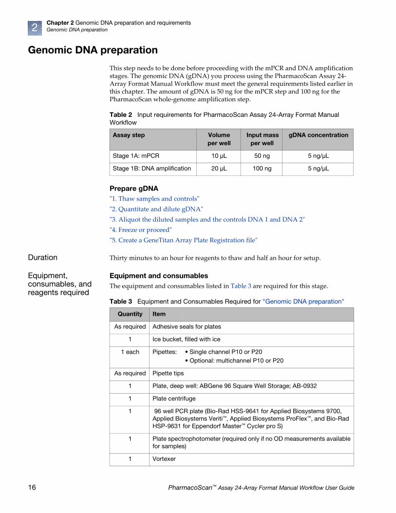
This step needs to be done before proceeding with the mPCR and DNA amplification stages. The genomic DNA (gDNA) you process using the PharmacoScan Assay 24-Array Format Manual Workflow must meet the general requirements listed earlier in this chapter. The amount of gDNA is 50 ng for the mPCR step and 100 ng for the PharmacoScan whole-genome amplification step.
Prepare gDNAʺ1. Thaw samples and controlsʺ
ʺ2. Quantitate and dilute gDNAʺ
ʺ3. Aliquot the diluted samples and the controls DNA 1 and DNA 2ʺ
ʺ4. Freeze or proceedʺ
ʺ5. Create a GeneTitan Array Plate Registration fileʺ
Duration Thirty minutes to an hour for reagents to thaw and half an hour for setup.
Equipment, consumables, and reagents required
Equipment and consumablesThe equipment and consumables listed in Table 3 are required for this stage.
Table 2 Input requirements for PharmacoScan Assay 24-Array Format Manual Workflow
Assay step Volume per well
Input mass per well
gDNA concentration
Stage 1A: mPCR 10 μL 50 ng 5 ng/μL
Stage 1B: DNA amplification 20 μL 100 ng 5 ng/μL
Table 3 Equipment and Consumables Required for "Genomic DNA preparation"
Quantity Item
As required Adhesive seals for plates
1 Ice bucket, filled with ice
1 each Pipettes: • Single channel P10 or P20• Optional: multichannel P10 or P20
As required Pipette tips
1 Plate, deep well: ABGene 96 Square Well Storage; AB-0932
1 Plate centrifuge
1 96 well PCR plate (Bio-Rad HSS-9641 for Applied Biosystems 9700, Applied Biosystems Veriti™, Applied Biosystems ProFlex™, and Bio-Rad HSP-9631 for Eppendorf Master™ Cycler pro S)
1 Plate spectrophotometer (required only if no OD measurements available for samples)
1 Vortexer
16 PharmacoScan™ Assay 24-Array Format Manual Workflow User Guide
Chapter 2 Genomic DNA preparation and requirementsGenomic DNA preparation 2
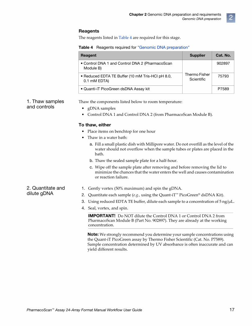
ReagentsThe reagents listed in Table 4 are required for this stage.
1. Thaw samples and controls
Thaw the components listed below to room temperature:
• gDNA samples
• Control DNA 1 and Control DNA 2 (from PharmacoScan Module B).
To thaw, either• Place items on benchtop for one hour
• Thaw in a water bath:
a. Fill a small plastic dish with Millipore water. Do not overfill as the level of the water should not overflow when the sample tubes or plates are placed in the bath.
b. Thaw the sealed sample plate for a half-hour.
c. Wipe off the sample plate after removing and before removing the lid to minimize the chances that the water enters the well and causes contamination or reaction failure.
2. Quantitate and dilute gDNA
1. Gently vortex (50% maximum) and spin the gDNA.
2. Quantitate each sample (e.g., using the Quant-iT™ PicoGreen® dsDNA Kit).
3. Using reduced EDTA TE buffer, dilute each sample to a concentration of 5 ng/µL.
4. Seal, vortex, and spin.
Note: We strongly recommend you determine your sample concentrations using the Quant-iT PicoGreen assay by Thermo Fisher Scientific (Cat. No. P7589). Sample concentration determined by UV absorbance is often inaccurate and can yield different results.
Table 4 Reagents required for "Genomic DNA preparation"
Reagent Supplier Cat. No.
• Control DNA 1 and Control DNA 2 (PharmacoScan Module B)
Thermo Fisher Scientific
902897
• Reduced EDTA TE Buffer (10 mM Tris-HCl pH 8.0, 0.1 mM EDTA)
75793
• Quanti-iT PicoGreen dsDNA Assay kit P7589
IMPORTANT! Do NOT dilute the Control DNA 1 or Control DNA 2 from PharmacoScan Module B (Part No. 902897). They are already at the working concentration.
PharmacoScan™ Assay 24-Array Format Manual Workflow User Guide 17

Chapter 2 Genomic DNA preparation and requirementsGenomic DNA preparation2
3. Aliquot the diluted samples and the controls DNA 1 and DNA 2
Next, the samples and controls are placed in a deep well plate for amplification and into a 96-well PCR plate for mPCR:
Note: Ensure gDNA is well mixed before plating.
Amplification sample plate• ABgene 96 Square Well Storage; AB-0932.
Aliquot diluted samples and controls to columns 5, 7, and 9 of the deep well plate
1. Aliquot 20 µL of each diluted gDNA sample to columns 5, 7, and 9 of the ABgene deep well plate as shown in Figure 3.
2. Pipet 20 µL of Control DNA 1 to well G09 and 20 µL of Control DNA 2 to well H09.
3. Seal and spin.
mPCR sample plate• Bio-Rad 96-well plate; HSS-9641 for Applied Biosystems 9700, Applied
Biosystems Veriti, Applied Biosystems ProFlex, Bio-Rad 96 well plate; HSP-9631 for Eppendorf Mastercycler pro S
Aliquot diluted samples and controls to columns 5, 7, and 9 of the mPCR sample plate
1. Aliquot 10 µL of each diluted gDNA sample to columns 5, 7, and 9 of 96-well PCR plate as shown in Figure 3.
2. Pipet 10 µL of Control DNA 1 to well G09 and 10 µL of Control DNA 2 to well H09.
3. Seal and spin.
IMPORTANT! Control DNA 1 and Control DNA 2 are required for assay performance. Both controls must be included on mPCR and Amplification Sample Plates and placed in indicated wells. Controls need to be run every time assay is performed.
18 PharmacoScan™ Assay 24-Array Format Manual Workflow User Guide

Chapter 2 Genomic DNA preparation and requirementsGenomic DNA preparation 2
4. Freeze or proceed
At this point you can:
• Store the sample plate at –20°C, or
• Proceed to DNA Amplification for Manual Target Prep. See Chapter 4, ʺTarget preparationʺ on page 42.
Note: You can leave the gDNA sample plates at room temperature if proceeding immediately to mPCR and DNA Amplification
5. Create a GeneTitan Array Plate Registration file
GeneTitan Array Plate Registration files contain information that is critical for:
• Data file generation during imaging.
• Tracking the experimental results for each sample loaded onto an array plate.
Detailed instructions for creating this file are located in Appendix C, ̋ Register samples in GeneChip™ Command Console™ʺ on page 161. See also Figure 4 for a screen shot showing an example of a GeneTitan Array Plate Registration file.
1. Open GCC Portal Samples, and select:
a. GeneTitan Array Plate Registration.
b. The array plate format.
c. Click Download.
Figure 3 Aliquoting to columns 5, 7, and 9
A
B
C
D
E
F
G
H
1 2 3 4 5 6 7 8 9 10 11 12
C1
C2
1 2 3 4 5 6 7 8 9 10 11 12
A
B
C
D
E
F
G
H
C1C2
Aliquot diluted gDNA samples to columns 5, 7, and 9 only
Amplification Sample Plate
Aliquot diluted gDNA samples to columns 5, 7, and 9 only
mPCR Sample Plate
ABgene 96 Square Well Plate20 μL/well
96 Well PCR Plate10 μL/well
C1 = Control DNA 1C2 = Control DNA 2
IMPORTANT! It is very important to create and upload a GeneTitan Array Plate Registration file with your sample information prior to loading the array plate and hybridization tray in the GeneTitan Instrument. We recommend that you create (but not upload) this file at the same time you prepare your plate of genomic DNA. When your samples are ready for hybridization, you scan the array plate barcode and upload the file to GeneChip Command Console (GCC).
PharmacoScan™ Assay 24-Array Format Manual Workflow User Guide 19

Chapter 2 Genomic DNA preparation and requirementsGenomic DNA preparation2
2. Enter a unique name for each sample and any additional information.
3. Save the file.
The array plate barcode is not scanned until you are ready to load the array plate and samples onto the GeneTitan MC Instrument for processing.
Figure 4 Example of a GeneTitan Array Plate Registration file
Your specific information is populated here.
20 PharmacoScan™ Assay 24-Array Format Manual Workflow User Guide

3 Preparation before you start
Introduction
This manual assay format allows the user to run the PharmacoScan™ Assay for 24 Samples four (4) times using one PharmacoScan™ Reagent Kit 4x24 Reactions and one QIAGEN Multiplex PCR Plus Kit (Cat. No. 206152), which must be purchased separately. This section provides information on procedures that are performed multiple times during manual target preparation and on steps that are critical to the success of the manual target preparation. It is essential that you familiarize yourself with the information in this section prior to running the PharmacoScan Assay.
One key item this manual assay format requires is the use of disposable divided reservoirs with a “trough within a trough” design, which maximizes the amount of liquid accessible to pipette tips when using small amounts of reagent.
A list of all equipment and resources required for the PharmacoScan Assay is in the PharmacoScan™ Assay 24-Array Format Manual Workflow Site Preparation Guide, Pub. No. 703287.
PharmacoScan™ Reagent Kit 4x24 Reactions, arrays, and GeneTitan™ consumables required
The table below lists the PharmacoScan reagents and GeneTitan consumables required to process four PharmacoScan 24F Array Plates. The table also lists the QIAGEN Multiplex PCR kit required for the PharmacoScan assay. See the PharmacoScan™ Assay 24-Array Format Manual Workflow Site Preparation Guide, Pub. No. 703287 for detailed information regarding the necessary materials required to run the PharmacoScan Assay.
Requirements and recommendations
This section describes requirements and recommendations for facilities and equipment needed to perform the PharmacoScan Assay 24-Array Format Manual Workflow.
Room temperature When referred to in the PharmacoScan Assay 24-Array Format Manual Workflow, room temperature is 18°C to 25°C.
Table 5 Arrays, reagents, and GeneTitan consumables required
Cat. No. Description Quantity
902994 PharmacoScan™ 24F Array Plate 4
901606 Axiom™ GeneTitan™ Consumables Kit 4
902908TS PharmacoScan™ Reagent Kit 4x24 Reactions 1
206152 QIAGEN Multiplex PCR Plus Kit, 100 Reactions 1
PharmacoScan™ Assay 24-Array Format Manual Workflow User Guide 21

Chapter 3 Preparation before you startRequirements and recommendations3
Special requirements
Amplification staging areaPrecautions are required when setting up amplification reactions to avoid contamination with foreign DNA amplified in other reactions and procedures. It is recommended that amplification reaction set up is performed in a dedicated amplification staging area separate from the main laboratory.
This amplification staging area should have a dedicated set of pipettes and plasticware. If no dedicated amplification staging area is available, use of a dedicated bench or a dedicated biosafety hood and dedicated pipettes is suggested. If no dedicated bench or biosafety hood is available, a set of dedicated pipettes is recommended.
Fume hoodAt certain steps in the protocol we recommend the use of adequate local or general ventilation to keep airborne concentrations low.
A fume hood is suggested as a way to achieve the desired concentration. Thus, a fume hood is strongly recommended for several steps of this assay.
Control requirementsA negative control is not required for this assay.
Two controls are required for proper data analysis. These controls, Control DNA 1 and Control DNA 2, are included in the PharmacoScan Assay Reagent Kit 4x24 Reactions.
Plate requirements and recommendations
The following types of plates are required for performing manual target preparation. See PharmacoScan™ Assay 24-Array Format Manual Workflow Site Preparation Guide, Pub. No. 703287, for vendor information.
• ABgene 96 Square Well Storage Plate, 2.2 mL
• Bio-Rad Hard Shell Semi-skirted 96-well plate, Cat. No. HSS-9641 for the Applied Biosystems 9700, Applied Biosystems Veriti, and Applied Biosystems ProFlex thermal cyclers. Use the Bio-Rad Hard Shell Low-profile 96-well plate, Cat. No. HSP-9631 for the Eppendorf Mastercycler pro S. See the PharmacoScan™ Assay 24-Array Format Manual Workflow Site Preparation Guide, Pub. No. 703287, for vendor information.
• 96-well UV Star Plates, 370 µL/well
22 PharmacoScan™ Assay 24-Array Format Manual Workflow User Guide

Chapter 3 Preparation before you startRequirements and recommendations 3
Thermal cycler recommendations and protocols
The following thermal cyclers are recommended for the PharmacoScan Assay 24-Array Format Manual Workflow:
• Applied Biosystems 9700 (with gold-plated or silver block)
• Applied Biosystems Veriti
• Applied Biosystems ProFlex
• Eppendorf Mastercycler pro S
PharmacoScan Assay 24-Array Format Manual Workflow has been validated with the Applied Biosystems 9700 (with gold-plated or silver block) Applied Biosystems Veriti, Applied Biosystems ProFlex, and Eppendorf Mastercycler pro S. Use of other thermal cyclers can result in assay failure and may violate the array and reagent replacement policy.
IMPORTANT! Always use the heated lid option when programming protocols. The PharmacoScan mPCR protocol was validated using the “9600 Mode” on the Applied Biosystems 9700, Applied Biosystems Veriti, and Applied Biosystems ProFlex thermal cyclers. The “Safe” mode was used for the Eppendorf Mastercycler pro S. See the manufacturer’s instructions for instrument programming.
Figure 5 PharmacoScan mPCR Thermal Cycler Protocol (Stage 1A)
Figure 6 PharmacoScan Denature Thermal Cycler Protocol (Stage 4)
WARNING! Evaporation during denaturation can negatively impact assay performance. Use the recommended thermal cycler consumables and sealing film to eliminate condensation and evaporation.
PharmacoScan™ Assay 24-Array Format Manual Workflow User Guide 23

Chapter 3 Preparation before you startRequirements and recommendations3
Thermal cycler consumables
Table 6 provides details into the consumables to be used with the Applied Biosystems 9700 thermal cycler.
Oven recommendations
The following ovens are recommended:
• ED 56 drying oven by BINDER (replaces BINDER Model ED 53)See the PharmacoScan Assay 24-Array Format Manual Workflow Site Preparation Guide, Pub. No. 703287, for vendor information.
• GeneChip Hybridization Oven 645
Note: The GeneChip™ Hybridization Oven 640 is currently not supported with the PharmacoScan Assay 24-Array Format Manual Workflow; however, if you want to utilize it in the workflow contact your field service engineer (FSE) or Thermo Fisher Scientific Technical Support regarding the compatibility of this oven with the PharmacoScan Assay.
– If using a GeneChip hybridization oven, set the rotation speed to 15 rpm to aid in even heat distribution.
– For either GeneChip hybridization oven, plates are placed in the bottom of the oven. To avoid interfering with the rotation apparatus, do not stack plates in the oven.
– Up to four plates can fit into a GeneChip Hybridization Oven 645
Plate centrifuge One plate centrifuge is required for the PharmacoScan Assay 24-Array Format Manual Workflow. See the PharmacoScan Assay 24-Array Format Manual Workflow Site Preparation Guide, Pub. No. 703287, for an appropriate plate centrifuge that can be used. When centrifuging and drying pellets as instructed under ʺStage 3A: Centrifuge Precipitation Plate and dry the DNA pelletʺ on page 69, the centrifuge must be able to spin down plates at:
• Rcf: 3200 x g (4,000 rpm for the Eppendorf 5810R with the rotor configuration described in the PharmacoScan Assay 24-Array Format Manual Workflow Site Preparation Guide, Pub. No. 703287).
• Temperature: 4°C and room temperature.
In addition, the bottom of the rotor buckets should be soft rubber to ensure that the deep-well plates do not crack. Do not spin plates in metal or hard plastic buckets.
Table 6 Thermal Cycler Consumables for the PharmacoScan Assay 24-Array Format Manual Workflow
Thermal Cycler Model
PCR Plate Type Seal
Applied Biosystems 9700
• BioRad Hard-Shell Full-Height 96-Well Semi-Skirted PCR Plate (Cat. No. HSS-9641)
MicroAmp Clear Adhesive Film, Thermo Fisher Scientific (Cat. No. 4306311)
Applied Biosystems Veriti
• BioRad Hard-Shell Full Height 96-well Semi-Skirted PCR Plate (Cat. No. HSS-9641)
MicroAmp Clear Adhesive Film, Thermo Fisher Scientific (Cat. No. 4306311)
Applied Biosystems ProFlex
• BioRad Hard-Shell Full Height 96-well Semi-Skirted PCR Plate (Cat. No. HSS-9641)
MicroAmp Clear Adhesive Film, Thermo Fisher Scientific (Cat. No. 4306311)
Eppendorf Mastercycler pro S
• BioRad Hard-Shell Low Profile 96-well Full-Skirt PCR Plate (Cat. No. HSP-9631)
MicroAmp Clear Adhesive Film, Thermo Fisher Scientific (Cat. No. 4306311)
24 PharmacoScan™ Assay 24-Array Format Manual Workflow User Guide
Chapter 3 Preparation before you startProcedures 3
Plate shakers We recommend using one of the following shakers listed in Table 7.
Equipment care and calibration
Lab instrumentation plays an important role in the successful completion of this assay. To aid in maintaining consistency across samples and operators, all equipment must be regularly calibrated and well maintained, including:
• All pipettes, thermal cyclers, and ovens
• Plate spectrophotometer
Procedures
This section covers procedures you may need to do repeatedly during the workflow, or which are critical to the performance of the assay.
Seal, vortex, and spin
Unless otherwise noted, when the protocol instructs you to seal, vortex, and spin:
• Seal plates—we recommend using MicroAmp Clear Adhesive Films to seal your plates.
Blot-dry—Prior to sealing plates, we recommend checking the top of the plate to ensure that there are no droplets. If droplets are present, blot-dry the top of the plate before sealing to ensure a tight seal.
d. To remove droplets prior to sealing overlay a sheet of Kimwipe laboratory tissue across the top of the plate and gently pat down to dry.
e. Lift the sheet off the plate and discard. Confirm the top of the plate is dry and seal the plate as usual.
• Vortex:
– Plates:
• For deep well plates (such as ABgene 2.2 mL square well storage plates), vortex five seconds in each sector for a total of five sectors (Figure 7).
• For PCR plates vortex two seconds in each sector for a total of five sectors (Figure 7).
– Reagent Vials: three times, one second each time.
Table 7 Shakers
Shaker Supplier Cat. No.
Thermo Scientific™ Compact Digital Microplate Shaker
Thermo Scientific 88880023
Jitterbug™ Boekel Scientific Model 130 000
IMPORTANT! Always ensure that your plates are tightly sealed. A tight seal prevents sample loss and cross-well contamination, particularly when plates are being vortexed.
PharmacoScan™ Assay 24-Array Format Manual Workflow User Guide 25

Chapter 3 Preparation before you startProcedures3
• Spin—when instructed to spin plates or reagent vials, follow these guidelines unless otherwise instructed (for example, when centrifuging and drying pellets, see Step 2 in the section ʺStage 3A: Centrifuge Precipitation Plate and dry the DNA pelletʺ on page 69).
– Plates:
• Spin at 1,000 rpm for 30 seconds at room temperature.
• Do not spin for more than one minute.
– Reagent Vials: three seconds.
Note: In the procedures, “vortex twice” means to repeat the vortexing step.
Sample quantitation This protocol has been optimized using a PicoGreen assay to determine genomic DNA concentrations. Other quantitation methods such as UV Absorbance can give different readings. Therefore, you should correlate readings from other methods to the equivalent PicoGreen-determined concentration.
See Chapter 2, ʺGenomic DNA preparation and requirementsʺ on page 12 for more information.
About the reagents and master mix preparation
PharmacoScan Reagent Kit 4x24 Reactions components
• Caps on the vials are color-coded by assay stage.
• Properly store all enzyme reagents, especially enzyme-containing vials. Improper storage methods can profoundly impact activity.
Figure 7 Vortexing plates
IMPORTANT! This kit includes Module 5.
• Module 5: Pouch 1 of 2, –25°C to –15°C: Part No. 902796
Contains an extra tube of Axiom Ligate Enzyme for back-up purposes in the event that it is needed.
• Module 5: Pouch 2 of 2, 2°C to 8°C: Part No. 902797
Contains three Axiom Hold Buffer bottles that should be used to prepare the scan tray for second, third, and fourth plate.
IMPORTANT! The PharmacoScan Assay 24-Array Format Manual Workflow is compatible only with reagents from a PharmacoScan Reagent Kit 4x24 Reactions. These reagents are not interchangeable with reagents from other Applied Biosystems reagent kits, such as SNP 6.0, DMET Plus, etc.
26 PharmacoScan™ Assay 24-Array Format Manual Workflow User Guide

Chapter 3 Preparation before you startProcedures 3
QIAGEN reagentsQIAGEN Multiplex PCR Plus Kit
QIAGEN Multiplex PCR Plus Kit (Cat. No. 206152) is used with PharmacoScan Reagent Kit 4x24 Reactions to process 24 samples four times. The QIAGEN kit configuration is as follows:
• Three tubes of 0.85 mL of Multiplex PCR Master Mix, 2X
• One tube of 2 mL of Q-Solution, 5X
• Two tubes of 1.9 mL of RNase-free Water
• One tube of 1.2 mL of CoralLoad Dye, 10X
Note: The CoralLoad Dye is not needed for PharmacoScan Assay 24-Array Format Manual Workflow and can be discarded
QIAGEN kit mPCR reagents can be freeze-thawed multiple times without affecting assay performance; however, it is convenient to use one tube of 2X Master Mix for one 24-format assay plate. After each use, the tube with remaining 2X QIAGEN Master Mix should be returned to the kit and stored at –20°C. The fourth assay plate can be processed by thawing and pooling the Master Mix remaining in these three tubes. It is recommended to use one tube of Water and Q-Solution to process all four PharmacoScan 24F Array Plates. Freeze unused reagents after each use.
Reagents from other suppliers• Use only fresh reagents from the recommended vendors to help eliminate
changes in pH or the salt concentration of buffers.
• Consult the appropriate MSDS for reagent storage and handling requirements.
Master mix preparation• Carefully follow each master mix recipe. Use pipettes that have been calibrated to
± 5%.
• If you run out of master mix during any of these procedures, a volume error has been made or the pipettes are not accurate. We recommend that you stop and repeat the experiment.
Note: The volumes of Master Mixes prepared are designed to provide consistent handling of reagents and consistent assay results. The percent overage of different master mixes can differ, depending upon the reagent volumes involved.
When using reagents at the lab bench• Properly chill essential equipment such as reagent coolers before use.
• Ensure that enzymes are kept at –20°C until needed. When removed from the freezer, immediately place in a cooler that has been chilled to –20°C.
PharmacoScan™ Assay 24-Array Format Manual Workflow User Guide 27

Chapter 3 Preparation before you startProcedures3
Pipettes and pipetting
To efficiently process samples:
• Use a pipette of appropriate size for the volume of liquid being transferred (Table 8).
• We recommend the use of Rainin pipettes and tips. Thermo Fisher Scientific has only verified the use of Rainin multichannel pipettes in this assay. The use of other pipettes can impact the timing of the protocol and can adversely impact the assay. Pipette substitution may violate the terms of the PharmacoScan Assay 24-Array Format Manual Workflow and array replacement policy.
• Always use pipettes that have been calibrated.
• It is essential that you be proficient with the use of single and multichannel pipettes. To familiarize yourself with the use of multichannel pipettes, we strongly recommend practicing several times before processing actual samples. Use water and reagent reservoirs to get a feel for aspirating and dispensing solutions to multiple wells simultaneously.
Single-channel pipettes and serological pipettesUse single-channel pipettes for preparing master mixes and for puncturing bubbles in GeneTitan trays. The single-channel pipettes is not used for working with the plates or trays otherwise.
• Use single channel pipettes for volumes less than or equal to 2 mL. For volumes between 1 and 2 mL, add the reagent in two portions with a fresh tip for each portion.
• Use serological pipette for volumes 2 mL.
Multichannel pipettesUse 8 or 12-channel pipettes when working to add master mix or to transfer samples to plates and GeneTitan trays.
• Use a pipette of appropriate size for the volume of liquid being transferred.
• Change pipette tips after each transfer or addition.
Table 8 Recommended pipette sizes
Pipette size Recommended volume range
Single channel P20 / 8-channel P20 1-20 μL
Single channel P200 / 8 and 12-channel P200 20-200 μL
Single channel P1000 / 8-channel P1200 200-1000 μL
28 PharmacoScan™ Assay 24-Array Format Manual Workflow User Guide
Chapter 3 Preparation before you startProcedures 3
Divided reservoir use
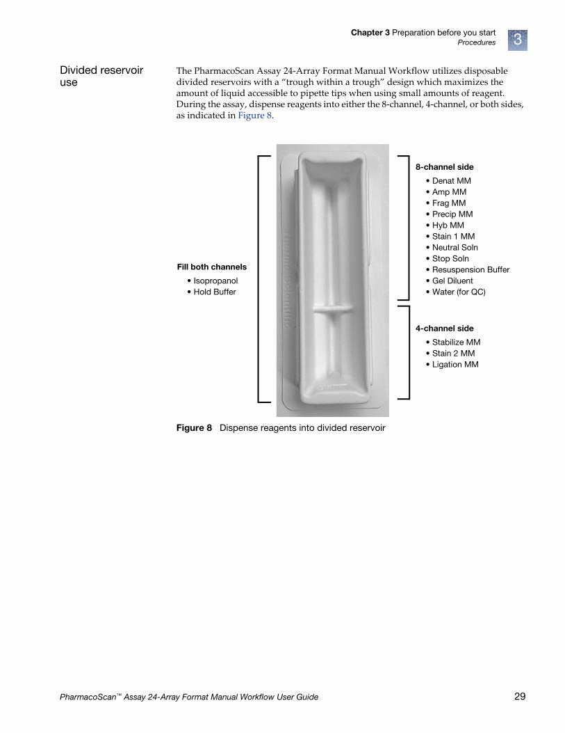
The PharmacoScan Assay 24-Array Format Manual Workflow utilizes disposable divided reservoirs with a “trough within a trough” design which maximizes the amount of liquid accessible to pipette tips when using small amounts of reagent. During the assay, dispense reagents into either the 8-channel, 4-channel, or both sides, as indicated in Figure 8.
Figure 8 Dispense reagents into divided reservoir
Fill both channels
• Isopropanol• Hold Buffer
8-channel side
• Denat MM• Amp MM• Frag MM• Precip MM• Hyb MM• Stain 1 MM• Neutral Soln• Stop Soln• Resuspension Buffer• Gel Diluent• Water (for QC)
4-channel side
• Stabilize MM• Stain 2 MM• Ligation MM
PharmacoScan™ Assay 24-Array Format Manual Workflow User Guide 29

Chapter 3 Preparation before you startProcedures3
Freeze-thaw instructions
Excess volume of the PharmacoScan Reagent Kit can be stored in a freezer at –25°C to –15°C or a refrigerator at 2°C to 8°C to be used in subsequent experiments for up to 60 days after initial use (Table 9). Thermo Fisher Scientific recommends that reagents not exceed three freeze-thaw cycles. Monitor the freeze-thaw cycles of the reagents by following the guidelines below.
Mark reagent pouches, tubes and bottles to track useTo keep track of usage, we recommend that users mark the pouch while the reagents are thawing.
• Using a permanent marker, label the module pouch with “Thaw #1: XX/XX/XX” and any other useful information (i.e., experiment name, user name, etc.).
• Using a permanent marker, make a tally mark on each reagent tube or bottle to indicate how many times the reagent has been thawed.
• After the experiment, gather all PharmacoScan reagents.
• Place all tubes and bottles back in the appropriate pouch and place in proper storage temperature. See Table 9.
Note: QIAGEN Multiplex PCR Plus Kit reagents are stored at –20°C. See page 27 for recommendations on freeze-thawing components.
IMPORTANT! PharmacoScan Module A and PharmacoScan Module B reagents are packaged for single-use only and any remaining reagent should be discarded.
Figure 9 Example of Labeling a Reagent Pouch
Figure 10 Example of a properly marked reagent bottle that has been thawed 3 times.
Table 9 Reagent storage temperature
Storage temperature
Module 1 Module 2-1
Module 2-2
Module 4-1
Module 4-2
2°C to 8°C
–25°C to –15°C
Thaw tally marks
30 PharmacoScan™ Assay 24-Array Format Manual Workflow User Guide
Chapter 3 Preparation before you startEquipment, consumables, labware, and reagents required 3
Equipment, consumables, labware, and reagents required
Equipment required for the PharmacoScan Assay 24-Array Format Manual Workflow
Thermal cyclerSee ʺThermal cycler recommendations and protocolsʺ on page 23.
Oven See ʺOven recommendationsʺ on page 24.
Plate centrifugeSee ʺPlate centrifugeʺ on page 24.
Plate shakerSee ʺPlate shakersʺ on page 25.
Consumables required for PharmacoScan™ Assay 24-Array Format Manual Workflow
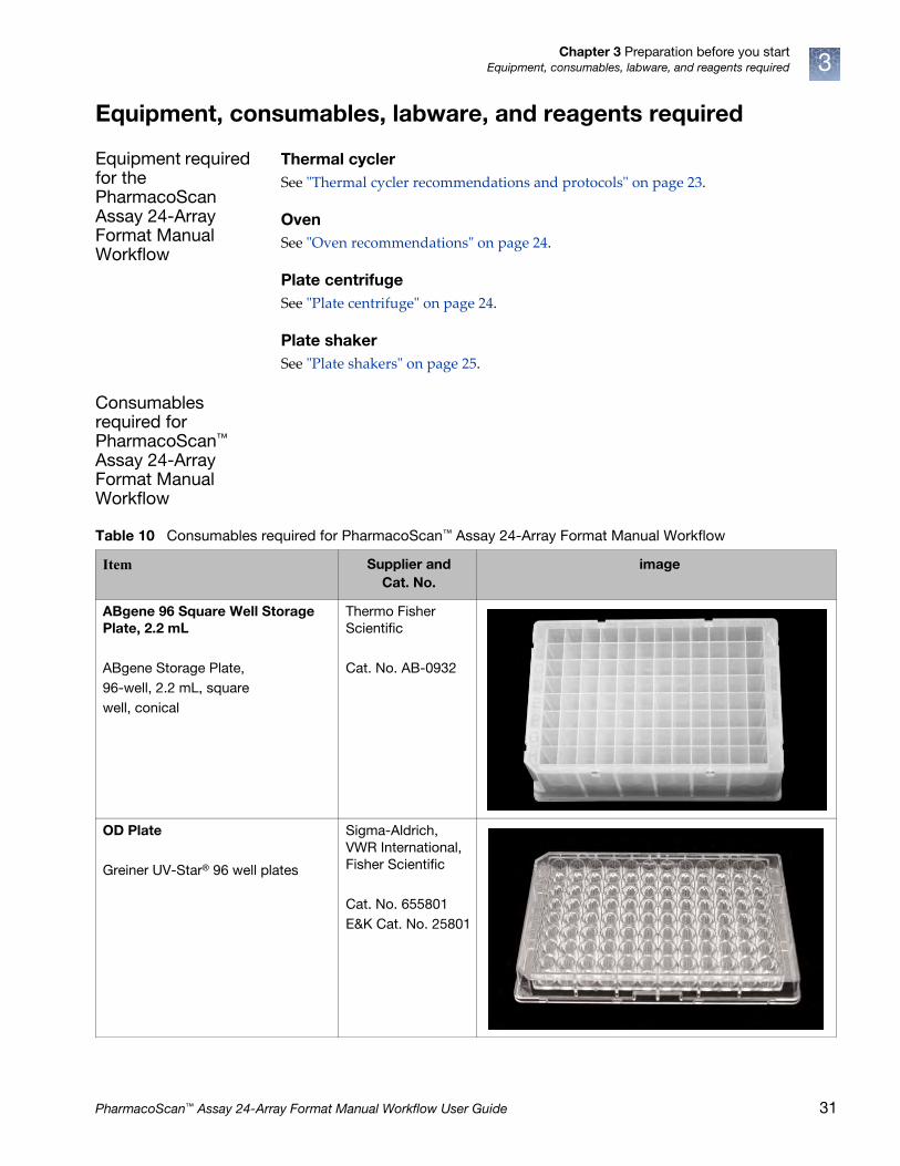
Table 10 Consumables required for PharmacoScan™ Assay 24-Array Format Manual Workflow
Item Supplier and Cat. No.
image
ABgene 96 Square Well Storage Plate, 2.2 mL
ABgene Storage Plate,96-well, 2.2 mL, squarewell, conical
Thermo Fisher Scientific
Cat. No. AB-0932
OD Plate
Greiner UV-Star® 96 well plates
Sigma-Aldrich, VWR International, Fisher Scientific
Cat. No. 655801 E&K Cat. No. 25801
PharmacoScan™ Assay 24-Array Format Manual Workflow User Guide 31

Chapter 3 Preparation before you startEquipment, consumables, labware, and reagents required3
Bio-Rad Hard Shell 96-well plate
Bio-Rad Hard-Shell® Low-Profile 96-Well Skirted PCR Plates
Note: See Table 6 for the PCR plate type recommended for your specific thermal cycler.
Bio-Rad
Cat. No. HSP-9631
96 Half-Skirt Plate
Bio-Rad Hard-Shell® High-Profile 96-Well Semi-Skirted PCR Plates
Note: See Table 6 for the PCR plate type recommended for your specific thermal cycler.
BioRad
Cat. No. HSS-9641
1.7 mL Microcentrifuge Tubes, DNAse and RNAse-free
Common labware - order through your preferred labware supplier
8-well strip tubes with caps,DNAse and RNAse-free
Common labware - order through your preferred labware supplier
Table 10 Consumables required for PharmacoScan™ Assay 24-Array Format Manual Workflow (Continued)
Item Supplier and Cat. No.
image
32 PharmacoScan™ Assay 24-Array Format Manual Workflow User Guide

Chapter 3 Preparation before you startEquipment, consumables, labware, and reagents required 3
50 mL and 15 mL Conical-bottom Centrifuge Tubes, polypropylene
Various
Zerostat Anti-static Gun and Ion-Indicator Cap
Milty Zerostat,
Thermo Fisher Scientific Cat. No. 74-0014
96-well Block
Cooling Chamber for 0.2 mL tubes, 96 holes (4 for 1.5 mL & 6 for 0.5 mL tubes), Dim.: 6 1/8”L x 3 1/8”W x 1” H
Diversified Biotech
Cat. No. CHAM-1000
25 mL Reagent Reservoir with Divider
Thermo Fisher Scientific
Cat. No. 8095
Table 10 Consumables required for PharmacoScan™ Assay 24-Array Format Manual Workflow (Continued)
Item Supplier and Cat. No.
image
PharmacoScan™ Assay 24-Array Format Manual Workflow User Guide 33
Chapter 3 Preparation before you startEquipment, consumables, labware, and reagents required3
GeneTitan™ MC Instrument consumables
All consumables for the GeneTitan MC Instrument are provided by Thermo Fisher Scientific. Table 11 provides guidance on the consumables that are shipped with the array plate.
IMPORTANT! All GeneTitan trays and tray covers must have barcodes. Discard any consumable tray or tray cover without a barcode.
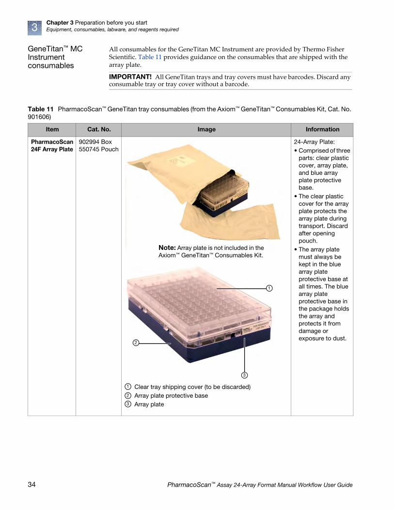
Table 11 PharmacoScan™ GeneTitan tray consumables (from the Axiom™ GeneTitan™ Consumables Kit, Cat. No. 901606)
Item Cat. No. Image Information
PharmacoScan 24F Array Plate
902994 Box 550745 Pouch
24-Array Plate:• Comprised of three
parts: clear plastic cover, array plate, and blue array plate protective base.
• The clear plastic cover for the array plate protects the array plate during transport. Discard after opening pouch.
• The array plate must always be kept in the blue array plate protective base at all times. The blue array plate protective base in the package holds the array and protects it from damage or exposure to dust.
Note: Array plate is not included in the Axiom™ GeneTitan™ Consumables Kit.
Clear tray shipping cover (to be discarded)Array plate protective baseArray plate
1
2
3
1
2
3
34 PharmacoScan™ Assay 24-Array Format Manual Workflow User Guide
Chapter 3 Preparation before you startEquipment, consumables, labware, and reagents required 3
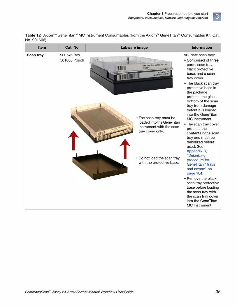
Table 12 Axiom™ GeneTitan™ MC Instrument Consumables (from the Axiom™ GeneTitan™ Consumables Kit, Cat. No. 901606)
Item Cat. No. Labware image Information
Scan tray 900746 Box501006 Pouch
96-Plate scan tray:• Comprised of three
parts: scan tray, black protective base, and a scan tray cover.
• The black scan tray protective base in the package protects the glass bottom of the scan tray from damage before it is loaded into the GeneTitan MC Instrument.
• The scan tray cover protects the contents in the scan tray and must be deionized before used. See Appendix D, "Deionizing procedure for GeneTitan™ trays and covers" on page 164.
• Remove the black scan tray protective base before loading the scan tray with the scan tray cover into the GeneTitan MC Instrument.
• The scan tray must be loaded into the GeneTitan Instrument with the scan tray cover only.
• Do not load the scan tray with the protective base.
PharmacoScan™ Assay 24-Array Format Manual Workflow User Guide 35
Chapter 3 Preparation before you startEquipment, consumables, labware, and reagents required3
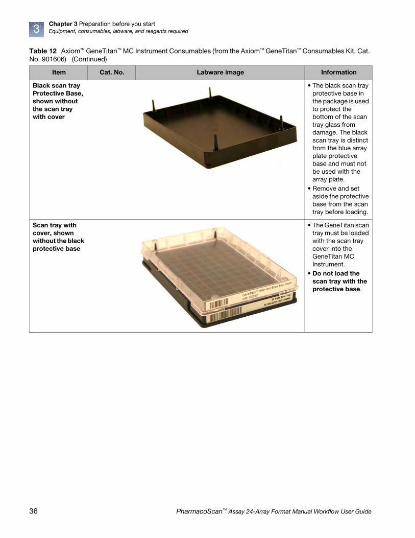
Black scan tray Protective Base, shown without the scan tray with cover
• The black scan tray protective base in the package is used to protect the bottom of the scan tray glass from damage. The black scan tray is distinct from the blue array plate protective base and must not be used with the array plate.
• Remove and set aside the protective base from the scan tray before loading.
Scan tray with cover, shown without the black protective base
• The GeneTitan scan tray must be loaded with the scan tray cover into the GeneTitan MC Instrument.
• Do not load the scan tray with the protective base.
Table 12 Axiom™ GeneTitan™ MC Instrument Consumables (from the Axiom™ GeneTitan™ Consumables Kit, Cat. No. 901606) (Continued)
Item Cat. No. Labware image Information
36 PharmacoScan™ Assay 24-Array Format Manual Workflow User Guide

Chapter 3 Preparation before you startEquipment, consumables, labware, and reagents required 3
GeneTitan 5 Stain Trays Kit
4249910 Kit501025 Tray
• The GeneTitan Stain Tray Kit comes with 5 stain trays packaged in zip-top bags to keep them free of dust.
• The GeneTitan stain trays are barcoded and the trays have separator walls that are flush with the frame of the stain tray, as shown by the yellow line and the yellow oval in the lower photo.
Table 12 Axiom™ GeneTitan™ MC Instrument Consumables (from the Axiom™ GeneTitan™ Consumables Kit, Cat. No. 901606) (Continued)
Item Cat. No. Labware image Information
PharmacoScan™ Assay 24-Array Format Manual Workflow User Guide 37

Chapter 3 Preparation before you startEquipment, consumables, labware, and reagents required3
GeneTitan™ stain and scan tray cover
202757 • The GeneTitan stain and scan tray covers prevent evaporation of the stains in stain trays and the array holding buffer in the scan tray.
• All stain and scan trays must be placed in the GeneTitan MC Instrument with the GeneTitan stain tray cover.
• All tray covers must be deionized to remove static electricity prior to placing the cover on the tray.
• See the section Appendix D, "Deionizing procedure for GeneTitan™ trays and covers" on page 164 for the anti-static procedure.
Table 12 Axiom™ GeneTitan™ MC Instrument Consumables (from the Axiom™ GeneTitan™ Consumables Kit, Cat. No. 901606) (Continued)
Item Cat. No. Labware image Information
38 PharmacoScan™ Assay 24-Array Format Manual Workflow User Guide

Chapter 3 Preparation before you startEquipment, consumables, labware, and reagents required 3
Label GeneTitan™ hybridization and reagent trays
When preparing the hybridization and reagent trays to be loaded onto the GeneTitan MC Instrument, you need to mark each tray in a way that identifies its contents.
Proper labeling for hybridization trays and reagent trays is described in:
• ʺLabel hybridization traysʺ, below
• ʺLabel stain traysʺ on page 40
Label hybridization traysYou can label the hybridization tray on the front part of the short side of the tray, next to the notch at the left, as shown in Figure 11. The proper section for labeling is closest to the notched corner, corresponding to the A1 and B1 wells.
GeneTitan stain tray shown with the stain tray cover
Tray 501025Cover 202757
Hybridization Tray
900747 • After aliquoting the denatured hybridization ready samples into the hybridization tray, the tray should be immediately loaded into the GeneTitan MC Instrument with the barcode facing away from the operator, i.e., Barcode should be on the back side.
Table 12 Axiom™ GeneTitan™ MC Instrument Consumables (from the Axiom™ GeneTitan™ Consumables Kit, Cat. No. 901606) (Continued)
Item Cat. No. Labware image Information
IMPORTANT! It is critical that you write only on the proper locations of the proper sides of hybridization and stain trays. Do not write in any other location, as this can interfere with sensors inside the GeneTitan MC Instrument and result in experiment failure. To ensure proper placement of covers onto stain trays, and trays onto the GeneTitan MC Instrument, you can also mark the notched corner of the trays and covers.
PharmacoScan™ Assay 24-Array Format Manual Workflow User Guide 39
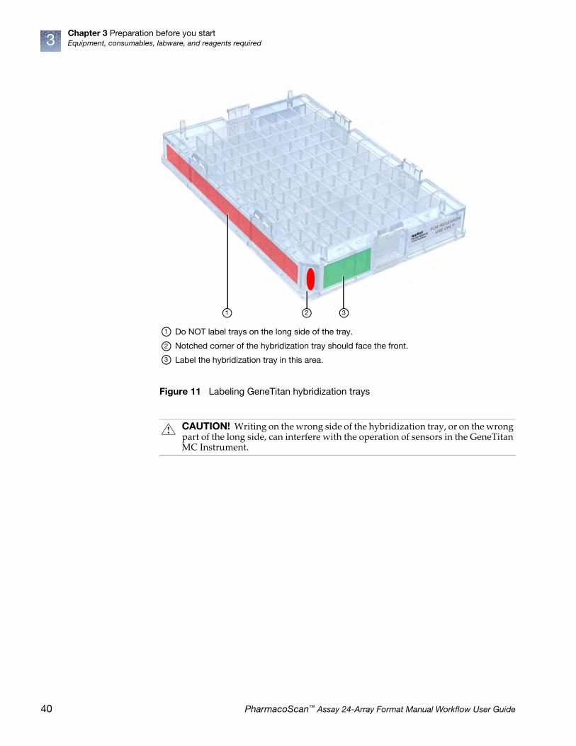
Chapter 3 Preparation before you startEquipment, consumables, labware, and reagents required3
Figure 11 Labeling GeneTitan hybridization trays
CAUTION! Writing on the wrong side of the hybridization tray, or on the wrong part of the long side, can interfere with the operation of sensors in the GeneTitan MC Instrument.
Do NOT label trays on the long side of the tray.
Notched corner of the hybridization tray should face the front.
Label the hybridization tray in this area.
1
2
3
1 2 3
40 PharmacoScan™ Assay 24-Array Format Manual Workflow User Guide

Chapter 3 Preparation before you startEquipment, consumables, labware, and reagents required 3
Label stain traysYou can label the stain trays on the left side of the front of the tray as shown in Figure 12. The correct side is closest to the notched corner, corresponding to the A1 through C1 wells.
(see ʺStage 5: GeneTitan™ reagent preparationʺ on page 85 for detailed information).
Figure 12 Label GeneTitan stain tray (stain tray shown with cover)
Do NOT label trays on the long side of the tray.
Notched corner of the stain tray should face the front.
Label the stain tray here.
1 2 3
1
2
3
PharmacoScan™ Assay 24-Array Format Manual Workflow User Guide 41

Chapter 3 Preparation before you startEquipment, consumables, labware, and reagents required3
Reagent Kit for the PharmacoScan™ Assay 24-Array Format Manual Workflow
The PharmacoScan Assay 24-Array Format Manual Workflow uses the PharmacoScan Reagent Kit 4x24 Reactions (Cat. No. 902908TS). One PharmacoScan Reagent Kit 4x24 Reactions was developed to process four 24 Format samples. Kits consist of 7 modules for different stages of the assay with some modules having both 4°C and –20°C pouches and is sufficient for four 24-array format plates. There are specific instructions for which reagents are needed and how to treat them within each stage.
Table 13 PharmacoScan™ Reagent Kit 4x24 Reactions, Cat. No. 902908TS
Component Storage
Module 1: Part No. 901711• Axiom Denat Soln 10X• Axiom Neutral Soln • Axiom Water
• Axiom Amp Soln• Axiom Amp Enzyme
–25°C to –15°C
Module 2: Pouch 1 of 2: Part No. 901528• Axiom Frag Enzyme• Axiom 10X Frag Buffer• Axiom Precip Soln 2
• Axiom Hyb Buffer• Axiom Hyb Soln 1
–25°C to –15°C
Module 2: Pouch 2 of 2: Part No. 901529• Axiom Frag Diluent• Axiom Frag Rxn Stop• Axiom Precip Soln 1
• Axiom Resusp Buffer• Axiom Hyb Soln 2
2°C to 8°C
Module 3• Axiom Wash Buffer A: PN 901446 (eight bottles per kit)• Axiom Wash Buffer B: PN 901447 (four bottles per kit)
• Axiom Water: PN 901578 (4 bottles per kit)
room temperature
Module 4: Pouch 1 of 2: Part No. 901278• Axiom Ligate Buffer• Axiom Ligate Enzyme• Axiom Ligate Soln 1
• Axiom Probe Mix 1• Axiom Stain Buffer• Axiom Stabilize Soln
–25°C to –15°C
Module 4: Pouch 2 of 2: Part No. 901276• Axiom Ligate Soln 2• Axiom Probe Mix 2• Axiom Wash A• Axiom Stain 1-A• Axiom Stain 1-B
• Axiom Stain 2-A• Axiom Stain 2-B• Axiom Stabilize Diluent• Axiom Water• Axiom Hold Buffer
2°C to 8°C
Module 5: Pouch 1 of 2: Part No. 902796• Axiom Ligate Enzyme
–25°C to –15°C
Module 5: Pouch 2 of 2: Part No. 902797• Axiom Hold Buffer (three bottles)
2°C to 8°C
PharmacoScan Module A: Part No. 902896• 10X mPCR primers (four tubes, use one per array)
–25°C to –15°C
PharmacoScan Module B: Part No. 902897• Control DNA 1 (four tubes, use one per array)• Control DNA 2 (four tubes, use one per array)
–25°C to –15°C
42 PharmacoScan™ Assay 24-Array Format Manual Workflow User Guide
4 Target preparation
Introduction
Target preparation for the PharmacoScan™ Assay 24-Array Format Manual Workflow enables you to perform target preparation to process 24 samples at a time. The protocol is performed in two parts:
• Part 1: Target preparation as described in this chapter
• Part 2: Array processing is performed on the GeneTitan™ Multi-Channel (MC) Instrument
Array handling and processing protocols require the use of a GeneTitan MC Instrument, as described in Chapter 5, ʺArray processing with the GeneTitan™ Multi-Channel Instrumentʺ on page 102.
A list of all equipment and resources required for the PharmacoScan Assay 24-Array Format Manual Workflow is in the PharmacoScan Assay 24-Array Format Manual Workflow Site Preparation Guide, Pub. No. 703287.
The protocol for target preparation is presented in the following sections:
• ʺStage 1A: Multiplex PCR (mPCR)ʺ on page 43
• ʺStage 1B: DNA amplificationʺ on page 49
• ʺStage 2: Fragmentation and precipitationʺ on page 56
• ʺStage 3: Centrifuge and drying, resuspension and hybridization preparation, and sample QCʺ on page 66
• ʺStage 3A: Centrifuge Precipitation Plate and dry the DNA pelletʺ on page 69
• ʺStage 3B: Resuspension and hybridization preparationʺ on page 70
• ʺStage 4: Denaturation and hybridizationʺ on page 78
• ʺStage 5: GeneTitan™ reagent preparationʺ on page 85
For the PharmacoScan Assay 24-Array Format Manual Workflow, target preparation as well as GeneTitan Reagent Preparation use only columns 5, 7, and 9 of the plates and trays consumables.
IMPORTANT! Read all the instructions in Chapter 3, ʺPreparation before you startʺ on page 21, before performing manual target preparation.
42 PharmacoScan™ Assay 24-Array Format Manual Workflow User Guide
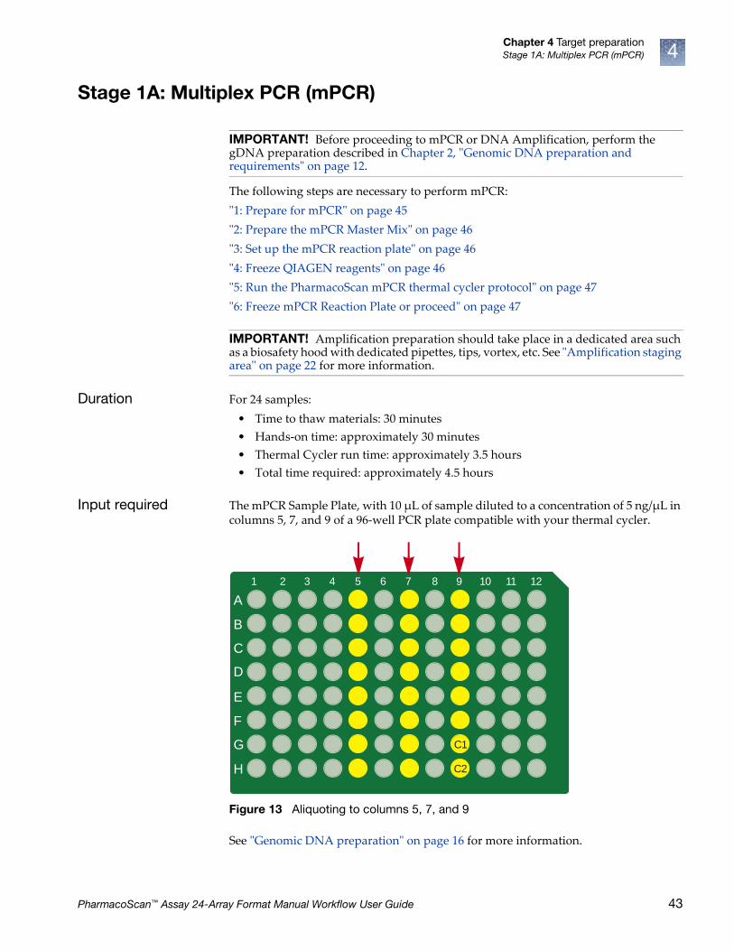
Chapter 4 Target preparationStage 1A: Multiplex PCR (mPCR) 4
Stage 1A: Multiplex PCR (mPCR)
The following steps are necessary to perform mPCR:
ʺ1: Prepare for mPCRʺ on page 45
ʺ2: Prepare the mPCR Master Mixʺ on page 46
ʺ3: Set up the mPCR reaction plateʺ on page 46
ʺ4: Freeze QIAGEN reagentsʺ on page 46
ʺ5: Run the PharmacoScan mPCR thermal cycler protocolʺ on page 47
ʺ6: Freeze mPCR Reaction Plate or proceedʺ on page 47
Duration For 24 samples:
• Time to thaw materials: 30 minutes
• Hands-on time: approximately 30 minutes
• Thermal Cycler run time: approximately 3.5 hours
• Total time required: approximately 4.5 hours
Input required The mPCR Sample Plate, with 10 µL of sample diluted to a concentration of 5 ng/µL in columns 5, 7, and 9 of a 96-well PCR plate compatible with your thermal cycler.
See ʺGenomic DNA preparationʺ on page 16 for more information.
IMPORTANT! Before proceeding to mPCR or DNA Amplification, perform the gDNA preparation described in Chapter 2, ʺGenomic DNA preparation and requirementsʺ on page 12.
IMPORTANT! Amplification preparation should take place in a dedicated area such as a biosafety hood with dedicated pipettes, tips, vortex, etc. See ̋ Amplification staging areaʺ on page 22 for more information.
Figure 13 Aliquoting to columns 5, 7, and 9
A
B
C
D
E
F
G
H
1 2 3 4 5 6 7 8 9 10 11 12
C1
C2
PharmacoScan™ Assay 24-Array Format Manual Workflow User Guide 43
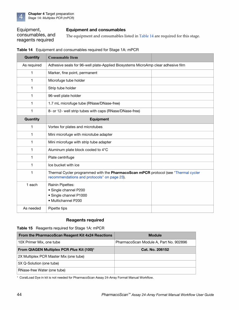
Chapter 4 Target preparationStage 1A: Multiplex PCR (mPCR)4
Equipment, consumables, and reagents required
Equipment and consumablesThe equipment and consumables listed in Table 14 are required for this stage.
Reagents required
Table 14 Equipment and consumables required for Stage 1A: mPCR
Quantity Consumable Item
As required Adhesive seals for 96-well plate-Applied Biosystems MicroAmp clear adhesive film
1 Marker, fine point, permanent
1 Microfuge tube holder
1 Strip tube holder
1 96-well plate holder
1 1.7 mL microfuge tube (RNase/DNase-free)
1 8- or 12- well strip tubes with caps (RNase/DNase-free)
Quantity Equipment
1 Vortex for plates and microtubes
1 Mini microfuge with microtube adapter
1 Mini microfuge with strip tube adapter
1 Aluminum plate block cooled to 4°C
1 Plate centrifuge
1 Ice bucket with ice
1 Thermal Cycler programmed with the PharmacoScan mPCR protocol (see "Thermal cycler recommendations and protocols" on page 23).
1 each Rainin Pipettes: • Single channel P200• Single channel P1000• Multichannel P200
As needed Pipette tips
Table 15 Reagents required for Stage 1A: mPCR
From the PharmacoScan Reagent Kit 4x24 Reactions Module
10X Primer Mix, one tube PharmacoScan Module A, Part No. 902896
From QIAGEN Multiplex PCR Plus Kit (100)1 Cat. No. 206152
2X Multiplex PCR Master Mix (one tube)
5X Q-Solution (one tube)
RNase-free Water (one tube)
1 CoralLoad Dye in kit is not needed for PharmacoScan Assay 24-Array Format Manual Workflow.
44 PharmacoScan™ Assay 24-Array Format Manual Workflow User Guide

Chapter 4 Target preparationStage 1A: Multiplex PCR (mPCR) 4
1: Prepare for mPCR
To Prepare for mPCR
1. Power on the thermal cycler programmed with PharmacoScan mPCR protocol (see Figure 5 on page 23). Ensure the heated lid option has been selected.
2. Thaw the mPCR Reagents and prepare the mPCR Reaction Plate.
To Thaw and Prepare the Reagents:
1. If the gDNA mPCR Sample Plate was frozen, thaw at room temperature on the benchtop, vortex, and pulse-spin.
2. Place mPCR Sample Plate on ice or a cold aluminum block after thawed.
3. Thaw the following reagents on the benchtop at room temperature and place on ice after thawed:
• From –20°C stored PharmacoScan™ Reagent Kit 4x24 Reactions (Cat. No. 902908TS), PharmacoScan Module A (Part No. 902896):
• 1 tube of 10X mPCR primers
• From –20°C stored QIAGEN Multiplex PCR Plus Kit (Cat. No. 206152):
• 1 vial of QIAGEN Multiplex PCR Master Mix, 2X
• 1 vial Q-Solution, 5X
• 1 vial RNase-free Water
IMPORTANT! gDNA samples must be 10 µL volume at a concentration of 5 ng/µL in columns 5, 7, and 9 of 96-well PCR plate (see Genomic DNA preparation, Figure 3 on page 19).
IMPORTANT! • Ensure reagents are thoroughly mixed prior to use.
• Vortex water and Q-Solution.
• Master Mix should be thoroughly mixed by inverting tube ten times. DO NOT VORTEX.
PharmacoScan™ Assay 24-Array Format Manual Workflow User Guide 45
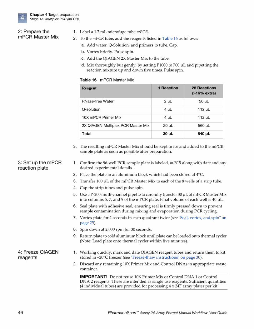
Chapter 4 Target preparationStage 1A: Multiplex PCR (mPCR)4
2: Prepare the mPCR Master Mix
1. Label a 1.7 mL microfuge tube mPCR.
2. To the mPCR tube, add the reagents listed in Table 16 as follows:
a. Add water, Q-Solution, and primers to tube. Cap.
b. Vortex briefly. Pulse spin.
c. Add the QIAGEN 2X Master Mix to the tube.
d. Mix thoroughly but gently, by setting P1000 to 700 µL and pipetting the reaction mixture up and down five times. Pulse spin.
3. The resulting mPCR Master Mix should be kept in ice and added to the mPCR sample plate as soon as possible after preparation.
3: Set up the mPCR reaction plate
1. Confirm the 96-well PCR sample plate is labeled, mPCR along with date and any desired experimental details.
2. Place the plate in an aluminum block which had been stored at 4°C.
3. Transfer 100 µL of the mPCR Master Mix to each of the 8 wells of a strip tube.
4. Cap the strip tubes and pulse spin.
5. Use a P-200 mutli-channel pipette to carefully transfer 30 µL of mPCR Master Mix into columns 5, 7, and 9 of the mPCR plate. Final volume of each well is 40 µL.
6. Seal plate with adhesive seal, ensuring seal is firmly pressed down to prevent sample contamination during mixing and evaporation during PCR cycling.
7. Vortex plate for 2 seconds in each quadrant twice (see ʺSeal, vortex, and spinʺ on page 25).
8. Spin down at 2,000 rpm for 30 seconds.
9. Return plate to cold aluminum block until plate can be loaded onto thermal cycler (Note: Load plate onto thermal cycler within five minutes).
4: Freeze QIAGEN reagents
1. Working quickly, mark and date QIAGEN reagent tubes and return them to kit stored in –20°C freezer (see ʺFreeze-thaw instructionsʺ on page 30).
2. Discard any remaining 10X Primer Mix and Control DNAs in appropriate waste container.
Table 16 mPCR Master Mix
Reagent 1 Reaction 28 Reactions (>16% extra)
RNase-free Water 2 μL 56 μL
Q-solution 4 μL 112 μL
10X mPCR Primer Mix 4 μL 112 μL
2X QIAGEN Multiplex PCR Master Mix 20 μL 560 μL
Total 30 μL 840 μL
IMPORTANT! Do not reuse 10X Primer Mix or Control DNA 1 or Control DNA 2 reagents. These are intended as single use reagents. Sufficient quantities (4 individual tubes) are provided for processing 4 x 24F array plates per kit.
46 PharmacoScan™ Assay 24-Array Format Manual Workflow User Guide

Chapter 4 Target preparationStage 1A: Multiplex PCR (mPCR) 4
5: Run the PharmacoScan mPCR thermal cycler protocol
1. Place mPCR on thermal cycler and run PharmacoScan mPCR protocol (see Figure 5 on page 23).
Note: The mPCR Sample Plate is now referred to as the mPCR Reaction Plate from this point forward.
6: Freeze mPCR Reaction Plate or proceed
1. After the PharmacoScan mPCR thermal cycler protocol is complete, remove the plate from thermal cycler, pulse spin, and store plate at –20°C. Ensure plate is well sealed to prevent evaporation during storage.
2. The PharmacoScan suggested workflow is to proceed to ʺStage 1B: DNA amplificationʺ on page 49 after the mPCR Reaction Plate has been placed on the thermal cycler.
3. If the Amplification Step has already been completed, and it is desired to proceed to ̋ Stage 2: Fragmentation and precipitationʺ on page 56, then the mPCR Reaction Plate can be left at 4°C for several hours until needed for mPCR Spike-In (on page 59).
PharmacoScan™ Assay 24-Array Format Manual Workflow User Guide 47

Chapter 4 Target preparationStage 1A: Multiplex PCR (mPCR)4
Figure 14 Stage 1A: mPCR preparation workflow diagram
mPCR preparation
mPCR preparation – page 1 of 1
Final Volume: 40 μL/well
Q-solution
Prepare mPCR Master Mix – Part 1
112 μL56 μL
Vortex briefly, pulse-spin.
Labware and reagents needed
QTY 1 (QIAGEN Multiplex
PCR Plus Kit, Cat. No. 206152)
QTY 1
QTY 1QTY 1 (mPCR Sample Plate)
112 μLRNase-free Water
Run Multiplex PCR
Thermal Cycler Protocol
(Duration: ~3.5 hours)
30 μL/well
100 μL/well
Prepare mPCR Master Mix – Part 22X QIAGEN Multiplex
PCR Master Mix
mPCR Plate
Vortex, seal tight, pulse-spin
Pipet up and down 5 times to mix, pulse-spin
Cap tubes, pulse-spin
560 μL
10X mPCR Primer Mix
48 PharmacoScan™ Assay 24-Array Format Manual Workflow User Guide

Chapter 4 Target preparationStage 1B: DNA amplification 4
Stage 1B: DNA amplification
The following sets of steps are necessary to perform DNA amplification:
ʺ1: Prepare for DNA amplificationʺ on page 51
ʺ2: Prepare the Denaturation Master Mixʺ on page 52
ʺ3: Add Denaturation Master Mix to samplesʺ on page 53
ʺ4: Add Neutralization Solution to samplesʺ on page 53
ʺ5: Prepare the Amplification Master Mixʺ on page 54
ʺ6: Add Amplification Master Mix to samplesʺ on page 54
ʺ7: Freeze reagentsʺ on page 54
ʺ8: Freeze or proceedʺ on page 54
Duration For 24 samples:
• Time to thaw materials: 1 hr
• Hands-on time: approximately 0.5 hr
• Incubation at 37°C: 23 ± 1 hr
• Total time required: approximately 24.5 hr
Input required The gDNA Sample Plate, with 20 µL of each gDNA diluted to a concentration of 5 ng/µL in columns 5, 7, and 9 of an ABgene 96 square well storage plate, 2.2 mL.
See ʺGenomic DNA preparationʺ on page 16 for more information.
IMPORTANT! Before proceeding to DNA Amplification, perform the gDNA preparation described in Chapter 2, ̋ Genomic DNA preparation and requirementsʺ on page 12.
IMPORTANT! Amplification preparation should take place in a dedicated area such as a biosafety hood with dedicated pipettes, tips, vortex, etc. See ʺAmplification staging areaʺ on page 22 for more information.
Figure 15 Aliquoting to columns 5, 7, and 9
1 2 3 4 5 6 7 8 9 10 11 12
A
B
C
D
E
F
G
H
C1C2
PharmacoScan™ Assay 24-Array Format Manual Workflow User Guide 49
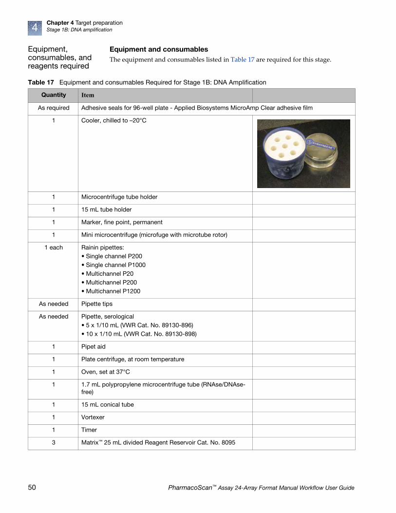
Chapter 4 Target preparationStage 1B: DNA amplification4
Equipment, consumables, and reagents required
Equipment and consumablesThe equipment and consumables listed in Table 17 are required for this stage.
Table 17 Equipment and consumables Required for Stage 1B: DNA Amplification
Quantity Item
As required Adhesive seals for 96-well plate - Applied Biosystems MicroAmp Clear adhesive film
1 Cooler, chilled to –20°C
1 Microcentrifuge tube holder
1 15 mL tube holder
1 Marker, fine point, permanent
1 Mini microcentrifuge (microfuge with microtube rotor)
1 each Rainin pipettes:• Single channel P200• Single channel P1000• Multichannel P20• Multichannel P200• Multichannel P1200
As needed Pipette tips
As needed Pipette, serological• 5 x 1/10 mL (VWR Cat. No. 89130-896)• 10 x 1/10 mL (VWR Cat. No. 89130-898)
1 Pipet aid
1 Plate centrifuge, at room temperature
1 Oven, set at 37°C
1 1.7 mL polypropylene microcentrifuge tube (RNAse/DNAse-free)
1 15 mL conical tube
1 Vortexer
1 Timer
3 Matrix™ 25 mL divided Reagent Reservoir Cat. No. 8095
50 PharmacoScan™ Assay 24-Array Format Manual Workflow User Guide

Chapter 4 Target preparationStage 1B: DNA amplification 4
Reagents required
1: Prepare for DNA amplification
1. Set an incubator/oven temperature at 37°C.
We recommend using one of these ovens:
• Binder ED 56
• GeneChip™ 645 Hybridization Oven (turn rotation on to 15 rpm)
2. Set the centrifuge temperature to room temperature.
3. Thaw and Prepare the Reagents and Sample Plate.
Thaw and prepare the reagents
1. Thaw the Sample Plate on the benchtop at room temperature and pulse-spin.
2. Thaw the following reagents in a small water bath on the benchtop at room temperature (small water bath: small tray or container, such as a pipet tip box, filled with fresh filtered water):
• Axiom Denat Soln 10X
• Axiom Neutral Soln
• Axiom Amp Soln
• Axiom Water
• Leave the Axiom Amp Enzyme in the cooler in the freezer until ready to use.
3. Vortex all reagents (except Axiom Amp Enzyme), then place at room temperature.
• Axiom Amp Soln: Vortex for 30 seconds to thoroughly mix.
• Axiom Neutral Soln: Vortex for 30 seconds to thoroughly mix.
• Axiom Denat Soln 10X: Vortex and pulse-spin before use.
• Axiom Amp Enzyme: Gently invert and flick the tube three times to mix and pulse-spin just before use.
Table 18 Reagents required for Stage 1B: DNA amplification
From the PharmacoScan™ Reagent Kit 4x24 Reactions Module
Axiom Denat Soln 10X
Module 1, –20°CPart No. 901711
Axiom Neutral Soln
Axiom Amp Soln
Axiom Water
Axiom Amp Enzyme
IMPORTANT! • gDNA samples must be brought to room temperature before proceeding
with denaturation.
• gDNA samples must be 20 µL volume of each gDNA at a concentration of 5 ng/µL in columns 5, 7, and 9 of an ABgene 96 square well storage plate, 2.2 mL (see ʺGenomic DNA preparationʺ on page 16).
PharmacoScan™ Assay 24-Array Format Manual Workflow User Guide 51

Chapter 4 Target preparationStage 1B: DNA amplification4
Note: Allow ~60 minutes for Axiom Amp Soln to thaw on the benchtop at room temperature. If the solution is not completely thawed after 60 minutes, vortex briefly and return to the benchtop to complete thawing. The bottles can also be thawed in a dish with Millipore water. The Axiom Amp Soln must be thoroughly mixed before use.
4. Label the 1.7 mL microcentrifuge tube and the 15 mL conical tube as indicated in the table below:
5. Label three Matrix 25 mL divided Reagent Reservoirs (Cat. No. 8095) as indicated in the table below:
2: Prepare the Denaturation Master Mix
Carry out the following steps at room temperature.
1. Per Table 21, dilute the appropriate volume of Axiom Denat Soln 10X using the Axiom Water.
2. Vortex, pulse spin, and leave at room temperature.
Table 19 Labeling tubes
Label Tube size Temperature Contents
• D MM 1.7 mL leave tube at room temperature
Denaturation Master Mix
• Amp MM 15 mL leave tube at room temperature
Amplification Master Mix
Table 20 Labeling reagent reservoirs for DNA amplification
Label Temperature Contents Reservoir side
• D MM Leave reservoir at room temperature
Denaturation Master Mix 8-channel
• N Soln Leave reservoir at room temperature
Neutralization Solution 8-channel
• Amp MM Leave reservoir at room temperature
Amplification Master Mix 8-channel
Table 21 Preparing Denaturation Master Mix (D MM)
Reagent per sample Master Mix 24+
To the 1.7 mL tube marked D MM, add:
Axiom Denat Soln 10X 2 μL 116 μL
Axiom Water 18 μL 1044 μL
Total volume 20 μL 1160 μL
52 PharmacoScan™ Assay 24-Array Format Manual Workflow User Guide

Chapter 4 Target preparationStage 1B: DNA amplification 4
3: Add Denaturation Master Mix to samples
Carry out the following steps at room temperature.
1. Pulse-spin the Sample Plate.
Remember: Samples must be at room temperature for this step.
2. Using a P1000, gently pipet the Denaturation Master Mix into the 8-channel side of the reagent reservoir marked D MM.
3. Carefully remove the seal from the Sample Plate and discard the seal.
4. Using a P20 8-channel pipette, add 20 µL of Denaturation Master Mix to each sample in columns 5, 7, and 9 (Total volume: 40 µL/well).
• Pipet directly into the liquid of each well. Do not mix by pipetting up and down.
• Change tips between each addition.
• This plate is now known as the Denaturation Plate.
5. Seal and vortex the Denaturation Plate. Start the timer for a 10 minute incubation after vortexing.
6. Pulse-spin the Denaturation Plate at room temperature.
Note: The quick spin time is included in the 10 minute incubation.
7. Visually examine the volume in each well.
a. Keep a record of any wells that visually appear to have a particularly low or high volume; these samples may need to be repeated.
b. Do NOT stop to measure volumes; proceed without delay.
8. Complete the 10 minute incubation on the benchtop at room temperature.
While completing the incubation at room temperature, prepare the Neutralization Soln as described in Step 1 on page 53.
9. After incubation immediately add the Neutralization Soln as described in ʺ4: Add Neutralization Solution to samplesʺ on page 53.
4: Add Neutralization Solution to samples
Carry out the following steps at room temperature.
1. Measure 3.64 mL of Axiom Neutral Soln and slowly pipet the reagent into the 8-channel side of the reagent reservoir marked N Soln.
2. Carefully remove the seal from the Denaturation Plate and discard the seal.
3. Using a P200 8-channel pipette, add 130 µL of Axiom Neutral Soln to each sample (Total volume: 170 µL/well).
• Pipet down the wall of each well. Change tips between each addition.
• The plate is now known as the Neutralization Plate.
4. Seal, vortex, and pulse-spin the Neutralization Plate.
5. Visually examine the volume in each well (should be ~170 µL/well) and:
a. Keep a record of any wells that visually appear to have a particularly low or high volume; these samples may need to be repeated.
b. Do NOT stop to measure volumes.
6. Proceed immediately to ʺ5: Prepare the Amplification Master Mixʺ on page 54.
PharmacoScan™ Assay 24-Array Format Manual Workflow User Guide 53
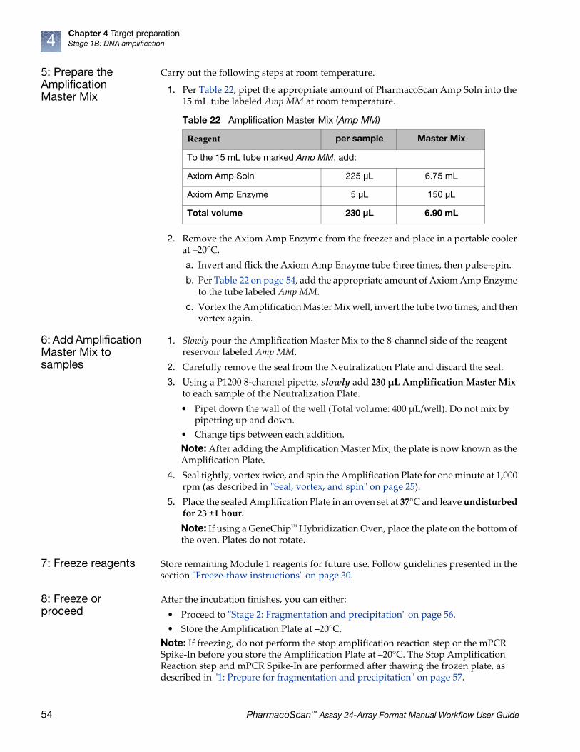
Chapter 4 Target preparationStage 1B: DNA amplification4
5: Prepare the Amplification Master Mix
Carry out the following steps at room temperature.
1. Per Table 22, pipet the appropriate amount of PharmacoScan Amp Soln into the 15 mL tube labeled Amp MM at room temperature.
2. Remove the Axiom Amp Enzyme from the freezer and place in a portable cooler at –20°C.
a. Invert and flick the Axiom Amp Enzyme tube three times, then pulse-spin.
b. Per Table 22 on page 54, add the appropriate amount of Axiom Amp Enzyme to the tube labeled Amp MM.
c. Vortex the Amplification Master Mix well, invert the tube two times, and then vortex again.
6: Add Amplification Master Mix to samples
1. Slowly pour the Amplification Master Mix to the 8-channel side of the reagent reservoir labeled Amp MM.
2. Carefully remove the seal from the Neutralization Plate and discard the seal.
3. Using a P1200 8-channel pipette, slowly add 230 µL Amplification Master Mix to each sample of the Neutralization Plate.
• Pipet down the wall of the well (Total volume: 400 µL/well). Do not mix by pipetting up and down.
• Change tips between each addition.
Note: After adding the Amplification Master Mix, the plate is now known as the Amplification Plate.
4. Seal tightly, vortex twice, and spin the Amplification Plate for one minute at 1,000 rpm (as described in ʺSeal, vortex, and spinʺ on page 25).
5. Place the sealed Amplification Plate in an oven set at 37°C and leave undisturbed for 23 ±1 hour.
Note: If using a GeneChip™ Hybridization Oven, place the plate on the bottom of the oven. Plates do not rotate.
7: Freeze reagents Store remaining Module 1 reagents for future use. Follow guidelines presented in the section ʺFreeze-thaw instructionsʺ on page 30.
8: Freeze or proceed
After the incubation finishes, you can either:
• Proceed to ʺStage 2: Fragmentation and precipitationʺ on page 56.
• Store the Amplification Plate at –20°C.
Note: If freezing, do not perform the stop amplification reaction step or the mPCR Spike-In before you store the Amplification Plate at –20°C. The Stop Amplification Reaction step and mPCR Spike-In are performed after thawing the frozen plate, as described in ʺ1: Prepare for fragmentation and precipitationʺ on page 57.
Table 22 Amplification Master Mix (Amp MM)
Reagent per sample Master Mix
To the 15 mL tube marked Amp MM, add:
Axiom Amp Soln 225 μL 6.75 mL
Axiom Amp Enzyme 5 μL 150 μL
Total volume 230 μL 6.90 mL
54 PharmacoScan™ Assay 24-Array Format Manual Workflow User Guide

Chapter 4 Target preparationStage 1B: DNA amplification 4
Figure 16 Stage 1B: Amplification workflow diagram
DNA amplification
DNA amplification – page 1 of 1
Incubate Sample Plate @ 37˚C for 23 ±1 hours
Final volume: 400 μL/well
20 μL/well
Axiom Water
Denaturation step
Denat Soln 10X
116 μL1,044 μL
Denaturation Plate
Vortex, pulse-spin.Incubate at room
temperature for 10 minutes
Vortex, pulse-spin
Use 8-channel side
130 μL/well
Neutral Soln
Neutralization step
Neutralization Plate
Vortex, pulse-spin
Use 8-channel side
3.64 mL
230 μL/well
Prepare for DNA Amplification
Amp Enzyme
150 μL6.75 mL
Amplification Plate
Vortex for 30 secondspulse-spin
Vortex
Use 8-channel side
Amp Soln
Labware and reagents needed
Axiom™ 2.0 Module 1
QTY 1 (with gDNA)QTY 1
QTY 3QTY 1
PharmacoScan™ Assay 24-Array Format Manual Workflow User Guide 55

Chapter 4 Target preparationStage 2: Fragmentation and precipitation4
Stage 2: Fragmentation and precipitation
The following sets of steps are necessary to perform fragmentation and precipitation:
ʺ1: Prepare for fragmentation and precipitationʺ on page 57
ʺ2: mPCR spike-in to Amplification Plateʺ on page 59
ʺ3. Incubate samples in pre-heated ovensʺ on page 60
ʺ4: Prepare the Fragmentation Master Mixʺ on page 60
ʺ5: Add the Fragmentation Master Mix to samplesʺ on page 60
ʺ6: Add the Stop Solution to the samplesʺ on page 61
ʺ7: Prepare the Precipitation Master Mixʺ on page 62
ʺ8: Prepare and add isopropanol to Precipitation Plateʺ on page 62
ʺ9: Freeze the Precipitation Plateʺ on page 63
ʺ10: Freeze reagentsʺ on page 63
Duration Total time: approximately two hours (plus time for precipitation incubation at –20°C, three hours or overnight).
Input required • mPCR Reaction Plate from ʺStage 1A: Multiplex PCR (mPCR)ʺ on page 43.
• Amplification Plate from ʺStage 1B: DNA amplificationʺ on page 49.
Equipment, consumables, and reagents required
Equipment and consumablesThe equipment and consumables listed in Table 23 are required for this stage.
Table 23 Equipment and consumables required for Stage 2: Fragmentation and precipitation
Quantity Item
As required Adhesive seals for 96-well plates
1 Freezer set to –20°C (Designate a shelf where the precipitation plates can be left undisturbed)
1 Cooler, chilled to –20°C
1 Ice bucket, filled with ice
1 Marker, fine point, permanent
1 each Rainin pipettes: • Single channel P1000• Single channel P200• Multichannel P20• Multichannel P200• Multichannel P1200
As needed Pipette tips for pipettes listed above
1 Pipet-aid
As needed Pipette, serological
• 5 x 1/10 mL • 10 x 1/10 mL
1 Plate centrifuge set at room temperature
56 PharmacoScan™ Assay 24-Array Format Manual Workflow User Guide

Chapter 4 Target preparationStage 2: Fragmentation and precipitation 4
Reagents required
1: Prepare for fragmentation and precipitation
Set ovens and centrifuge
1. Set two incubators/ovens as follows, preferably the night before:
• One oven set at 37°C. Use an oven that can sustain a constant temperature of 37°C and has a temperature accuracy of 1°C.
• One oven set at 65°C.
Note: If your are running the three-hour precipitation workflow, three ovens are recommended. Set one oven at 37°C, another at 65°C, and the final oven at 48°C in preparation for ʺStage 4: Denaturation and hybridizationʺ.
2. Set the centrifuge temperature to room temperature.
Tip: Keep a set of balance plates ready to minimize any time delays before spinning the Fragmentation Plate in-between steps.
1 Mini microcentrifuge (microfuge with microtube rotor)
2-3 Ovens (see "Oven recommendations" on page 24):• One oven set at 37°C• One oven set to 65°C
2 15 mL conical tube
4 Matrix™ 25-mL divided Reagent Reservoir Cat. No. 8095
1 Vortexer
Table 23 Equipment and consumables required for Stage 2: Fragmentation and precipitation
Quantity Item
Table 24 Reagents required for Stage 2: Fragmentation and precipitation
Reagent Module
From the PharmacoScan Reagent Kit 4x24 Reactions
Axiom Frag Enzyme (leave at –20°C until ready to use)Module 2-1, –20°C
Part No. 901528Axiom 10X Frag Buffer
Axiom Precip Soln 2
Axiom Frag DiluentModule 2-2, 2–8°C
Part No. 901529Axiom Frag Rxn Stop
Axiom Precip Soln 1
User-supplied - See the PharmacoScan Assay 24-Array Format Manual Workflow Site Preparation Guide, Pub. No. 703287
Isopropanol (2-Propanol), 99.5% 24 samples: 20 mL per 24-array format plate
PharmacoScan™ Assay 24-Array Format Manual Workflow User Guide 57

Chapter 4 Target preparationStage 2: Fragmentation and precipitation4
Thaw and prepare the amplified DNA samples, mPCR Reaction Plate, and reagents
If the plate of Amplified DNA Samples is frozen (skip this step if the Amplified Sample Plate was not frozen at the end of the previous stage):
1. Place the deep-well plate in a small water bath.For example, pour fresh filtered water into a small tray. Place the frozen plate on the water in the tray.
2. Leave the plate in the water bath for ~50 minutes until all wells have thawed.
3. Spin down the plate at 1,000 rpm for 30 seconds.
4. To avoid cross-contamination of wells during vortexing:
a. Remove the seal and blot the top of the plate with a Kimwipe laboratory tissue.
b. Tightly re-seal the plate using a fresh seal.
5. Vortex the plate for 30 seconds to thoroughly mix.
6. Spin at 1,000 rpm for 30 seconds.
Thaw and prepare the mPCR Reaction Plate
If the mPCR Reaction Plate is frozen: (skip this step if the mPCR Reaction Plate was not frozen at the end of Stage 1A):
1. Thaw the plate at room temperature for about 30 minutes or until all samples are thawed.
2. Spin down the plate at 1,000 rpm for 30 seconds.
3. To avoid cross-contamination of wells during vortexing, remove the seal and tightly re-seal the plate using a fresh seal.
4. Vortex the plate for 10 seconds to thoroughly mix.
5. Spin at 1,000 rpm for 30 seconds.
Thaw and Prepare the Reagents
To Thaw and Prepare the Fragmentation Reagents:
1. Axiom 10X Frag Buffer:
• Thaw on the benchtop at room temperature then place on ice.
• Vortex before use.
2. Axiom Frag Diluent:
• Place on ice.
• Vortex and pulse-spin before use.
3. Frag Enzyme: Leave at –20°C until ready to use. Just before use, gently flick the tube three times to mix and pulse-spin.
58 PharmacoScan™ Assay 24-Array Format Manual Workflow User Guide

Chapter 4 Target preparationStage 2: Fragmentation and precipitation 4
To thaw and prepare the precipitation reagents:
1. Axiom Precip Soln 1
• Place on benchtop to warm to room temperature.
• Vortex before use.
2. Axiom Precip Soln 2:
• Thaw on the bench top at room temperature and leave at room temperature.
• Vortex and pulse-spin before use.
3. Isopropanol (user-supplied)
• Keep at room temperature.
Label tubes and reagent reservoirs
1. Label the 15 mL conical tubes as indicated in the table below:
2. Label four Matrix 25 mL divided Reagent Reservoirs (Cat. No. 8095) as indicated in the table below.
2: mPCR spike-in to Amplification Plate
• If proceeding directly from the end of ʺStage 1B: DNA amplificationʺ on page 49, remove the Amplification Plate from the 37°C oven.
• If working with a thawed Amplification Plate, change the seal, vortex, and pulse-spin the Amplification Plate.
1. Carefully transfer 10 µL of the mPCR reaction into the corresponding well of the Amplification Plate. Pipette should be submerged in sample and mixing performed by pipetting up and down a few times. Ensure complete liquid transfer from pipette tip.
2. Seal plate well. Ensure the seal is securely attached to the plate to minimize evaporation during next steps.
3. Thoroughly mix by vortexing plate for 30 seconds and pulse spin.
4. Immediately proceed to next step, incubate samples in pre-heated ovens.
Label Tube size
Temperature Contents
• Frg MM 15 mL Place tube on ice Fragmentation Master Mix
• Precip MM 15 mL Place tube at room temperature
Precipitation Master Mix
Label Temperature Contents
• Frg MM Leave reservoir at room temperature Fragmentation Master Mix
• Stop Leave reservoir at room temperature Frag Rxn Stop
• Precip MM Leave reservoir at room temperature Precipitation Master Mix
• ISO Leave reservoir at room temperature Isopropanol
IMPORTANT! Ensure that the mPCR Reaction Plate has been thoroughly mixed before adding to Amplification Plate.
PharmacoScan™ Assay 24-Array Format Manual Workflow User Guide 59

Chapter 4 Target preparationStage 2: Fragmentation and precipitation4
3. Incubate samples in pre-heated ovens
Stop the DNA amplification reaction
1. Place the Amplification Plate in the 65°C oven.
2. Incubate for 20 minutes.
Prepare for Fragmentation:
1. Place the Amplification Plate in the 37°C oven:
• Remove the Amplification Plate from the 65°C oven and check the seal. Press on the seal, if needed, then transfer the plate to the 37°C oven.
2. Incubate for 45 minutes.
4: Prepare the Fragmentation Master Mix
Prepare the Fragmentation Master Mix:
1. Start making the Fragmentation Master Mix when there is still five minutes to the finish of the 37°C incubation, using the values in the table below.Transfer the Axiom Frag Enzyme to a –20°C portable cooler until ready to use.
• Add the reagents from Table 25 to the Frg MM tube in the order shown, using appropriate single channel pipettes.
• Just before the end of the 45 minute 37°C incubation, flick the Axiom Frag Enzyme tube 2 to 3 times, and spin.
• Add the Frag Enzyme to the Fragmentation Master Mix at the end of the 45 minute 37°C incubation.
Note: Leave the Axiom Frag Enzyme at –20°C until ready to use.
2. Vortex twice and place on ice.
3. Using a P1000 pipet, slowly transfer the Fragmentation Master Mix in the 8-channel side of the reagent reservoir labeled Frg MM placed at room temperature.
5: Add the Fragmentation Master Mix to samples
1. Carefully remove the Amplification Plate from the 37°C oven and place on the bench top at room temperature.
Do not place the Amplification Plate on ice.
2. Carefully remove the seal from the Amplification Plate and discard the seal.
Table 25 Fragmentation Master Mix
Reagent per sample Master Mix 24+
To the 15 mL tube marked Frg MM, add:
Axiom 10X Frag Buffer 45.7 μL 1.69 mL
Axiom Frag Diluent 10.3 μL 381 μL
Axiom Frag Enzyme 1.0 μL 37 μL
Total volume 57 μL 2.11 mL
IMPORTANT! Work quickly to perform this set of steps to minimize the time that the Fragmentation Plate is out of the 37°C oven.
60 PharmacoScan™ Assay 24-Array Format Manual Workflow User Guide
Chapter 4 Target preparationStage 2: Fragmentation and precipitation 4
3. Pipetting directly into the liquid of each well, use a P200 8-channel pipette to add 57 µL of Fragmentation Master Mix to each reaction. Do not mix by pipetting up and down.
• Change tips after each addition.
• After adding the Fragmentation Master Mix to the plate, the plate is now known as the Fragmentation Plate.
4. Seal the Fragmentation Plate and vortex twice.
5. Start the timer for 30 minutes.
6. Pulse-spin the Fragmentation Plate in the plate centrifuge at room temperature.
7. Quickly transfer plate to 37°C oven and incubate for 30 minutes.
Prepare the Stop solution a few minutes before the end of the 30 minute incubation period, as described in ʺ6: Add the Stop Solution to the samplesʺ, below.
6: Add the Stop Solution to the samples
Carry out the following steps at room temperature.
1. A few minutes before the end of the 30 minute incubation period, measure 988 µL of the Axiom Frag Rxn Stop solution and transfer into the 8-channel side of the reagent reservoir labeled Stop.
2. Remove the Fragmentation Plate from the oven and place on the bench top at room temperature.
3. At the end of the 30 minute fragmentation incubation period, carefully remove the seal from the Fragmentation Plate and discard the seal.
4. Using a P20 8-channel pipette, end the fragmentation reaction by adding 19 µL of Stop Solution to each reaction. Do not mix by pipetting up and down.
• Pipet directly into the liquid of each well.
• Change tips after each addition.
• Proceed immediately to the next step.
5. Seal and vortex and do a quick spin at 1,000 rpm.
6. Leave the Fragmentation Plate on the benchtop while you prepare the Precipitation Master Mix.
CAUTION! Be watchful for the end of the thirty minute incubation period. Fragmentation is an exact 30 minute incubation step. Longer and shorter incubation times may lead to poor performance of the assay.
PharmacoScan™ Assay 24-Array Format Manual Workflow User Guide 61

Chapter 4 Target preparationStage 2: Fragmentation and precipitation4
7: Prepare the Precipitation Master Mix
Carry out the following steps at room temperature.
1. Prepare Precipitation Master Mix in the 15 mL conical tube labeled Precip MM.
Note: Use a 5 mL serological pipette to pipet PharmacoScan Precip Soln 1.
2. Vortex the Precip MM tube and place on benchtop at room temperature.
3. Pour the Precipitation Master Mix into the 8-channel side of the reagent reservoir labeled Precip MM.
4. Carefully remove the seal from the Fragmentation Plate and discard the seal.
5. Using a P1200 8-channel pipette, add 240 µL Precipitation Master Mix to each sample. Rest each pipette tip against the wall of each well while delivering. You do not need to mix up and down. Change tips after each addition.
Note: After adding the Precipitation Master Mix, the plate is now known as the Precipitation Plate.
6. Seal, vortex, and pulse-spin the Precipitation Plate.
8: Prepare and add isopropanol to Precipitation Plate
1. Remove the Precipitation Plate from the centrifuge and place on the benchtop at room temperature.
2. Pour 20 mL of isopropanol into both sides of the reagent reservoir labeled ISO.
3. Carefully remove the seal from the Precipitation Plate and discard the seal.
4. Using a P1200 8-channel pipette, add 600 µL isopropanol to each sample and mix well by pipetting up and down 6-7 times within the solution. Observe the solution while it is within the tips - it should look homogeneous after pipetting 5-7 times. If not, repeat mixing a few more times until the solution looks homogeneous.
Do not vortex the plate after isopropanol addition to avoid cross-contamination of the samples.
Change the tips after each addition.
5. Blot the top of the plate with a Kimwipe laboratory tissue and seal tightly with a MicroAmp Adhesive Seal.
Table 26 Precipitation Master Mix
Reagent per sample Master Mix 24+
To the 15 mL tube marked Precip MM, add:
Axiom Precip Soln 1 238 μL 6.19 mL
Axiom Precip Soln 2 2 μL 52 μL
Total volume 240 μL 6.24 mL
62 PharmacoScan™ Assay 24-Array Format Manual Workflow User Guide

Chapter 4 Target preparationStage 2: Fragmentation and precipitation 4
9: Freeze the Precipitation Plate
Carefully transfer the Precipitation Plate into the –20°C freezer and incubate overnight (16-24 hrs).
A new option for DNA target precipitation is to incubate the plate in the –20°C freezer for three hours, instead of overnight. This shortened precipitation allows you to proceed to ʺStage 3: Centrifuge and drying, resuspension and hybridization preparation, and sample QCʺ followed by ̋ Stage 4: Denaturation and hybridizationʺ in on day 2 of the assay workflow.
Note: It is recommended to designate a shelf in a –20°C freezer where the plates can be left undisturbed. In addition, the freezer should not be subjected to frequent temperature excursions.
10: Freeze reagents Store remaining Module 2-1 and Module 2-2 reagents for future use. Follow guidelines presented in the section ʺFreeze-thaw instructionsʺ on page 30.
Store the mPCR Plate at –20°C for optional QC purposes if desired. See Appendix F, ʺmPCR quality control gel protocolʺ on page 180.
IMPORTANT! The three-hour DNA precipitation workflow extends the day 2 assay schedule. Approximately nine hours are required to complete Stage 2 through Stage 4 (Figure 1 on page 11). Review Chapter 3 and Chapter 4 for timing details.
PharmacoScan™ Assay 24-Array Format Manual Workflow User Guide 63

Chapter 4 Target preparationStage 2: Fragmentation and precipitation4
Figure 17 mPCR spike-in flowchart
mPCR spike-in
mPCR Spike-In – page 1 of 1
Final volume: 410 μL/well
Proceed to Fragmentation
Addition of mPCR products
mPCR + Amp Plate
Vortex, pulse-spin
10 μL/well
Labware and reagents needed
QTY 1 Amp Plate
QTY 1 mPCR Plate
64 PharmacoScan™ Assay 24-Array Format Manual Workflow User Guide

Chapter 4 Target preparationStage 2: Fragmentation and precipitation 4
Figure 18 Stage 2: Fragmentation and precipitation
Fragmentation
Incubate samples in pre-heated ovens1) 20 minutes @ 65°C2) 45 minutes @ 37°C
Final volume: 1,327 μL/well
Incubate Precipitation Plate @ –20˚C overnight
(or optional 3-hour precipitation step)
19 μL/well
Stop Soln
Stop fragmentation reaction
Fragmentation Plate
Vortex, pulse-spin
Use 8-channel side
988 μL
Addition of 2-Propanol
Precipitation Plate
Pipet up and down to mixDo not vortex
600 μL/well
Pour 20 mL 2-propanol
into both sides
240 μL/well
Preparation for precipitationPrecip Soln 2
52 μL6.19 mL
Precipitation Plate
Vortex for 30 secondspulse-spin
Vortex
Use 8-channel side
Precip Soln 1
57 μL/well
10X Frag Buffer
Fragmentation stepFrag
Enzyme
37 μL1.69 mL
Fragmentation Plate
Vortex, pulse-spinIncubate at 37°C
for 30 minutes
Invert tube and vortex to mix
Use 8-channel side
381 μL
Frag Diluent
Labware and reagents needed
QTY 1 (Samples)
QTY 2QTY 4 Axiom™ 2.0:
Module 2-1Module 2-2
20 mL 2-Propanol
PharmacoScan™ Assay 24-Array Format Manual Workflow User Guide 65

Chapter 4 Target preparationStage 3: Centrifuge and drying, resuspension and hybridization preparation, and sample QC4
Stage 3: Centrifuge and drying, resuspension and hybridization preparation, and sample QC
This stage requires the following sets of steps:
ʺStage 3A: Centrifuge Precipitation Plate and dry the DNA pelletʺ on page 69
ʺStage 3B: Resuspension and hybridization preparationʺ on page 70
ʺ1: Prepare for Resuspension and hybridization preparationʺ on page 70
ʺ2: Prepare DNA pellets and warm the resuspension bufferʺ on page 70
ʺ3: Thaw and prepare the reagentsʺ on page 70
ʺ4: Label tubes and reservoirsʺ on page 71
ʺ5: Add Resuspension Buffer to DNA pelletsʺ on page 71
ʺ6: Resuspension of DNA pelletsʺ on page 71
ʺ7: Prepare the Hybridization Master Mixʺ on page 72
ʺ8: Prepare the Hyb Ready Sample Plateʺ on page 72
ʺ9: Freeze or proceedʺ on page 72
ʺStage 3C: (Recommended) Perform quantitation and fragmentation QC checksʺ on page 73
ʺ1: Prepare for sample QCʺ on page 73
ʺ2: Perform QC checksʺ on page 74
ʺ3: Freeze or proceedʺ on page 74
Duration • Centrifuge and dry plates: 80 minutes
• Resuspension and hybridization mix preparation: 25 minutes
• Gel QC and OD: 45 minutes
Total: 2.5 hours
Input required Precipitation Plate from ʺStage 2: Fragmentation and precipitationʺ on page 56.
CAUTION! Some of the steps in this stage should be performed under a fume hood.
IMPORTANT! For troubleshooting and support purposes, we strongly recommend that you perform the gel QC and OD quantitation process controls after Resuspension.
66 PharmacoScan™ Assay 24-Array Format Manual Workflow User Guide

Chapter 4 Target preparationStage 3: Centrifuge and drying, resuspension and hybridization preparation, and sample QC 4
Equipment, consumables, and reagents required
The equipment and consumables listed in Table 27 are required for this stage.
Table 27 Equipment and consumables Required for Stage 3: Drying, resuspension, and QC
Quantity Item
As required Adhesive seals for 96-well plates
1 Marker, fine point, permanent
1 each Rainin Pipettes:• Single channel P20• Single channel P-100• Multichannel P20• Multichannel P-200
As needed Pipette tips for pipettes listed above
2 Any 96-well PCR plate for making the dilutions:• QC Dilution Plate• Gel Samples Plate
1 Bio-Rad Hard-Shell Semi-Skirted PCR Cat. No. HSS-9641 (for Applied Biosystems 9700 thermal cycler) • Hyb Ready Plate
1 OD plate: 96-well UV Star, 370 μL/well
1 Oven set at 37°C
1 Mini microcentrifuge (microfuge with microtube rotor)
1 Fume Hood
1 Plate centrifuge set at 4°C
1 15 mL conical tube
1 5 mL Serological Pipette
1 Pipet aid
1 Shaker, either:• Thermo Scientific™ Compact Digital Microplate Shaker • Jitterbug
1 Vortexer
4 Matrix™ 25 mL divided Reagent Reservoir Cat. No. 8095
PharmacoScan™ Assay 24-Array Format Manual Workflow User Guide 67

Chapter 4 Target preparationStage 3: Centrifuge and drying, resuspension and hybridization preparation, and sample QC4
Reagents required
Gels and related materials requiredAt the end of this stage, verifying the fragmentation reaction is highly recommended. See Appendix A, ʺFragmentation quality control gel protocolʺ on page 148 for the required gel and related materials.
Examining the mPCR Reaction Plate by agarose gel electrophoresis is optional and recommended only if assay troubleshooting is required.
Table 28 Reagents required for Stage 3: Drying, resuspension, and QC
Reagent Module
From the PharmacoScan Reagent Kit 4x24 Reactions
Axiom Hyb Buffer Module 2-1, –20°CPart No. 901528
Axiom Hyb Soln 1
Axiom Resusp Buffer Module 2-2, 2–8°CPart No. 901529
Axiom Hyb Soln 2
Other Reagents required for QC steps (optional)
Gel Diluent, 5 mL of 1,000-fold dilution of TrackIt™ Cyan/Orange Loading Buffer (see Appendix A, "Fragmentation quality control gel protocol" on page 148 for dilution instructions.)
Gel Sample Plate
15 fold dilution of TrackIt™ 25 bp DNA Ladder (Cat. No. 10488-022)
Nuclease-free water, ultrapure MB Grade, 5 mL (Cat. No. 71786; for OD and Dilution Plate preparation)
68 PharmacoScan™ Assay 24-Array Format Manual Workflow User Guide

Chapter 4 Target preparationStage 3A: Centrifuge Precipitation Plate and dry the DNA pellet 4
Stage 3A: Centrifuge Precipitation Plate and dry the DNA pellet
Centrifuge and dry the DNA pellets
1. Turn the oven on and preheat to 37°C.
Use an oven that can sustain a constant temperature of 37°C and has a temperature accuracy of 1°C (we recommend the Binder ED 56). If using a GeneChip Hybridization Oven, set the rotation speed to 15 rpm to distribute heat.
2. Transfer the Precipitation Plate from the –20°C freezer to a pre-chilled centrifuge. Centrifuge the plate for 40 minutes at 4°C at 3,200 x g (4,000 rpm for the Eppendorf 5810R centrifuge with the rotor configuration described in the PharmacoScan Assay 24-Array Format Manual Workflow Site Preparation Guide, Pub. No. 703287).
.
3. Immediately after the 40 minutes centrifugation period, empty the liquid from each plate as follows:
a. Carefully remove the seal from the Precipitation Plate and discard the seal.
b. Invert the plate over a waste container and allow the liquid to drain.
c. While still inverted, gently press the plates on a pile of Kimwipe laboratory tissues on a bench and allow them to drain for 5 minutes. Transfer the plate to a new pile of tissues twice during the 5 minute period.
4. Turn the plate right side up and place in an oven for 20 minutes at 37°C to dry.
• Tightly seal the plate upon completion
5. Do one of the following:
• Proceed directly to ̋ Stage 3B: Resuspension and hybridization preparationʺ on page 70, even if some droplets of liquid remain. Leave the sample plate at room temperature. It is helpful to begin preparing reagents for Stage 3B while centrifuging and drying pellets.
• Store the plate for resuspension later in the same day:
• Tightly seal the plate.
• If resuspension is be carried within 4 hours, keep the plate at room temperature.
• Store the plate for resuspension on another day:
• Tightly seal the plate.
• Store the plate at –20°C.
CAUTION! During this step, handle the Precipitation Plate gently to avoid disturbing the pellets. Do not bump or bang the plate against another object.
WARNING! Use rotor buckets with a soft rubber bottom to ensure that the deep well plates do not crack. Do not use buckets where the plates sit directly on a metal or hard plastic bottom, such as the A-4-62 rotor with a WO-15 plate carrier (hard bottom) for the Eppendorf 5810R centrifuge. Use of hard bottom plate carriers may result in cracked plates, loss of sample, unbalanced centrifugation, damage to the instrument and possible physical injury.
Note: If using a GeneChip™ Hybridization Oven 645 oven:
• Place the plate on the bottom of the oven. Plates do not rotate.
• Turn off the rotor during the 20 minutes drying time.
PharmacoScan™ Assay 24-Array Format Manual Workflow User Guide 69

Chapter 4 Target preparationStage 3B: Resuspension and hybridization preparation4
Stage 3B: Resuspension and hybridization preparation
1: Prepare for Resuspension and hybridization preparation
1. Set the centrifuge to room temperature.
2: Prepare DNA pellets and warm the resuspension buffer
The equilibration of the plate of pelleted DNA and resuspension buffer to room temperature (18-25°C) is very critical for the success of the PharmacoScan Target Prep. When any of these are cooler than room temperature, pellets may not resuspend completely. This may result in compromised assay performance. Note following guidelines on how to work with plates with fresh, cold, or frozen pellets:
DNA pellet types• Fresh Pellets: A plate with fresh pellets can be kept at room temperature if
proceeding with the resuspension and hybridization preparation protocol within 4 hours.
• Cold Pellets: A plate with fresh pellets that are not processed within 4 hours can be transferred to a refrigerator (2-8°C) if processed during the same day. However, it is critical to equilibrate the plate to room temperature for at least 30 minutes before proceeding with the Resuspension and Hybridization Preparation protocol.
• Frozen Pellets: A plate with frozen pellets must be pre-equilibrated at room temperature for at least 1.5 hour before proceeding with the Resuspension and Hybridization Preparation protocol.
Warm the Resuspension Buffer• The Resuspension buffer needs at least 60 minutes to equilibrate to room
temperature.
3: Thaw and prepare the reagents
1. Thaw Axiom Hyb Soln 1 on the benchtop at room temperature.
2. Warm Axiom Resusp Buffer, Axiom Hyb Buffer, and Axiom Hyb Soln 2 on the benchtop at room temperature for at least one hour.
3. Vortex the Axiom Resusp Buffer and the Axiom Hyb Buffer. Keep at room temperature.
4. Vortex and pulse-spin Axiom Hyb Soln 1 and Axiom Hyb Soln 2 before use.
IMPORTANT! The plate of pelleted DNA and resuspension reagent must be at room temperature before proceeding with this step.
70 PharmacoScan™ Assay 24-Array Format Manual Workflow User Guide
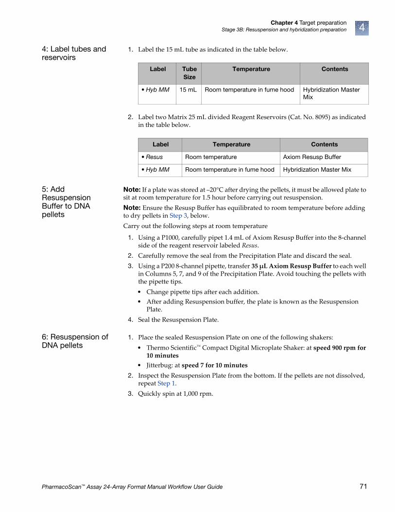
Chapter 4 Target preparationStage 3B: Resuspension and hybridization preparation 4
4: Label tubes and reservoirs
1. Label the 15 mL tube as indicated in the table below.
2. Label two Matrix 25 mL divided Reagent Reservoirs (Cat. No. 8095) as indicated in the table below.
5: Add Resuspension Buffer to DNA pellets
Note: If a plate was stored at –20°C after drying the pellets, it must be allowed plate to sit at room temperature for 1.5 hour before carrying out resuspension.
Note: Ensure the Resusp Buffer has equilibrated to room temperature before adding to dry pellets in Step 3, below.
Carry out the following steps at room temperature
1. Using a P1000, carefully pipet 1.4 mL of Axiom Resusp Buffer into the 8-channel side of the reagent reservoir labeled Resus.
2. Carefully remove the seal from the Precipitation Plate and discard the seal.
3. Using a P200 8-channel pipette, transfer 35 µL Axiom Resusp Buffer to each well in Columns 5, 7, and 9 of the Precipitation Plate. Avoid touching the pellets with the pipette tips.
• Change pipette tips after each addition.
• After adding Resuspension buffer, the plate is known as the Resuspension Plate.
4. Seal the Resuspension Plate.
6: Resuspension of DNA pellets
1. Place the sealed Resuspension Plate on one of the following shakers:
• Thermo Scientific™ Compact Digital Microplate Shaker: at speed 900 rpm for 10 minutes
• Jitterbug: at speed 7 for 10 minutes
2. Inspect the Resuspension Plate from the bottom. If the pellets are not dissolved, repeat Step 1.
3. Quickly spin at 1,000 rpm.
Label Tube Size
Temperature Contents
• Hyb MM 15 mL Room temperature in fume hood Hybridization Master Mix
Label Temperature Contents
• Resus Room temperature Axiom Resusp Buffer
• Hyb MM Room temperature in fume hood Hybridization Master Mix
PharmacoScan™ Assay 24-Array Format Manual Workflow User Guide 71

Chapter 4 Target preparationStage 3B: Resuspension and hybridization preparation4
7: Prepare the Hybridization Master Mix
1. While the Resuspension Plate is shaking, prepare the Hybridization Master Mix in the Hyb MM 15 mL tube.
a. Add the reagents in Table 29 to the Hyb MM tube in the order shown.
b. Vortex twice to mix.
8: Prepare the Hyb Ready Sample Plate
1. Choose a 96-well plate that is compatible with the thermal cycler model that is used for sample denaturation. See Table 6 on page 24 for information on thermal cycler consumables.
2. Label the 96-well PCR plate as Hyb Ready [Sample ID].
3. Set a P200 8-channel pipette to 45 µL (this is slightly higher than the volume of the sample in each well of the Resuspension Plate).
4. Using the P200 pipette, transfer the entire contents of each well in columns 5, 7, and 9 of the Resuspension Plate to the corresponding columns of the labeled Hyb Ready Plate.
• Change pipette tips after each transfer.
5. Pour the Hyb Master Mix into the 8-channel side of the reagent reservoir labeled Hyb MM.
6. Using a P200 8-channel pipette, add 80 µL of the Hyb Master Mix to each well of columns 5, 7, and 9 the Hyb Ready Plate.
• Change pipette tips after each addition.
7. Seal, vortex twice, and pulse-spin.
9: Freeze or proceed
At this point you can:
• Proceed to ʺStage 3C: (Recommended) Perform quantitation and fragmentation QC checksʺ (highly recommended), below; or
• Proceed to ʺStage 4: Denaturation and hybridizationʺ; or
• Store the hybridization ready samples at –20°C.
• Store remaining Module 2-1 and Module 2-2 reagents for future use. Follow the guidelines presented in the section ʺFreeze-thaw instructionsʺ on page 30.
CAUTION! It is recommended that the remainder of the steps in this stage be preformed under a fume hood.
Table 29 Hybridization Master Mix
Reagent per sample Master Mix 24+
To the 15 mL tube labeled Hyb MM, add:
Axiom Hyb Buffer 70.5 μL 2.26 mL
Axiom Hyb Soln 1 0.5 μL 16 μL
Axiom Hyb Soln 2 9 μL 288 μL
Total volume 80 μL 2.56 mL
72 PharmacoScan™ Assay 24-Array Format Manual Workflow User Guide

Chapter 4 Target preparationStage 3C: (Recommended) Perform quantitation and fragmentation QC checks 4
Stage 3C: (Recommended) Perform quantitation and fragmentation QC checks
Before proceeding to ʺStage 4: Denaturation and hybridizationʺ, we highly recommend that you perform quantitation and fragmentation quality control checks.
1: Prepare for sample QC
Prepare the reagentsObtain the Reagents for Sample QC:
1. 5 mL of nuclease-free water for the water reservoir.
2. 5 mL of Gel Diluent
The Gel Diluent is a 1,000-fold dilution of the TrackIt Cyan/Orange Loading Buffer (Invitrogen) as described in ʺDilute the TrackIt™ Cyan/Orange Loading Buffer and 25 bp ladderʺ on page 149.
3. 90 µL of a 15-fold dilution of TrackIt™ 25 bp DNA Ladder, Cat. No. 10488-022, Thermo Fisher Scientific
4. One E-Gel 48 Agarose Gel, 4% Agarose, Cat. No. G8008-04, Thermo Fisher Scientific
Label reservoirsLabel two Matrix 25 mL divided Reagent Reservoirs (Cat. No. 8095) as indicated below:
• Label one reservoir as H2O
• Label the second reservoir as Loading Dye i
Prepare sample QC plates
1. Label two 96-well PCR plates for making the dilutions:
• Label one plate as Dil QC
• Label the second plate as Gel QC
2. Obtain one 96-well UV Star, 370 µL/well plate
Note: Change tips while transferring samples from the Hyb Ready Plate and the QC Dilution Plate to avoid cross-contamination.
Table 30 Label Reagent Reservoirs for QC
Label Temperature Contents
• NF H2O Leave reservoir at room temperature Nuclease-free Water
• Loading Dye Leave reservoir at room temperature Diluted Loading dye
PharmacoScan™ Assay 24-Array Format Manual Workflow User Guide 73

Chapter 4 Target preparationStage 3C: (Recommended) Perform quantitation and fragmentation QC checks4
2: Perform QC checks
Carry out the following steps at room temperature.
1. Prepare Dilution QC Plate and OD Plate:Pour 5 mL nuclease-free water into the 8-channel side of the reagent reservoir labeled NF H20. The water is used to make the QC Dilution Plate and the OD Plate.
a. Add 33 µL nuclease-free water to each well in column 5, 7 and 9 of the Dil QC Plate.
b. Add 90 µL nuclease-free water to each well in column 5, 7 and 9 of the OD Plate.
2. Prepare the Dilution QC Plate:
a. Transfer 3 µL of the Hyb Ready sample from each well of the Hyb Ready Plate to the corresponding well of the Dil QC Plate. Change pipette tips after each transfer.
b. Seal, vortex, and pulse-spin.
3. Prepare OD Plate:
a. Carefully remove the seal from the QC Dilution Plate and discard the seal.
b. Transfer 10 µL of each Dil QC sample to the to the corresponding wells of the 96-well UV Star plate. Change pipette tips after each transfer.
c. Mix by pipetting up and down.
• Change pipette tips after each addition.
• Final sample mass dilution is 120-fold.
See Appendix B, ʺSample quantitation after resuspensionʺ on page 151 for more information on performing the Sample Quantitation.
4. Prepare Gel QC Plate:
a. Pour 5 mL Gel Diluent into the 8-channel side of the reagent reservoir labeled Loading Dye.
b. Add 120 µL gel diluent to each well in column 5, 7 and 9 of the Gel QC Plate.
c. Transfer 3 µL of each Dil QC sample to the corresponding wells of the Gel QC Plate.Change pipette tips after each transfer.
d. Seal, vortex, and pulse-spin the plate.
5. Run gel as described in Appendix A, ̋ Fragmentation quality control gel protocolʺ on page 148.
After the QC checks, the Dilution QC Plate, OD Plate, and remaining Gel QC samples can be discarded after satisfactory results from the gel and OD 260 readings have been obtained.
3: Freeze or proceed
At this point you can:
• Proceed to ʺStage 4: Denaturation and hybridizationʺ, below; or
• Store the hybridization ready samples at –20°C.
74 PharmacoScan™ Assay 24-Array Format Manual Workflow User Guide

Chapter 4 Target preparationStage 3C: (Recommended) Perform quantitation and fragmentation QC checks 4
Figure 19 Stage 3: Resuspension, #1 of 2
Resuspension and hybridization preparation
Resuspension and hybridization preparation – page 1 of 2
Centrifuge Precipitation Plate to pellet DNASpeed: 3,200 x g
Duration: 40 minutesTemperature: 4°C
Dry DNA Pellets�� Decant liquid by inverting plate�� Blot-dry inverted plate for 5 minutes�� Incubate @ 37�C for 20 minutes right-
side up
Resuspension of DNA pelletDuration: 10 minutes
Jitterbug: Speed 7Thermo Scientific Titer Plate Shaker: 900 rpm
Continue to Resuspension and hybridization preparation,
page 2
Add Resuspension Buffer
35 μL/well
1.4 mL
Resusp Buffer
Resuspension Plate
Resuspend pellets onplate shaker for 10 minutes.
Pulse-spin.
Use 8-channel side
Labware and reagents needed
Hyb Ready Plate: QTY 1(depending on the thermal cycler used)
QTY 1 Precip Plate(Samples)
QTY 1QTY 2
Axiom™ 2.0: Module 2-1Module 2-2
HSS-9641for Applied Biosystems 9700
PharmacoScan™ Assay 24-Array Format Manual Workflow User Guide 75
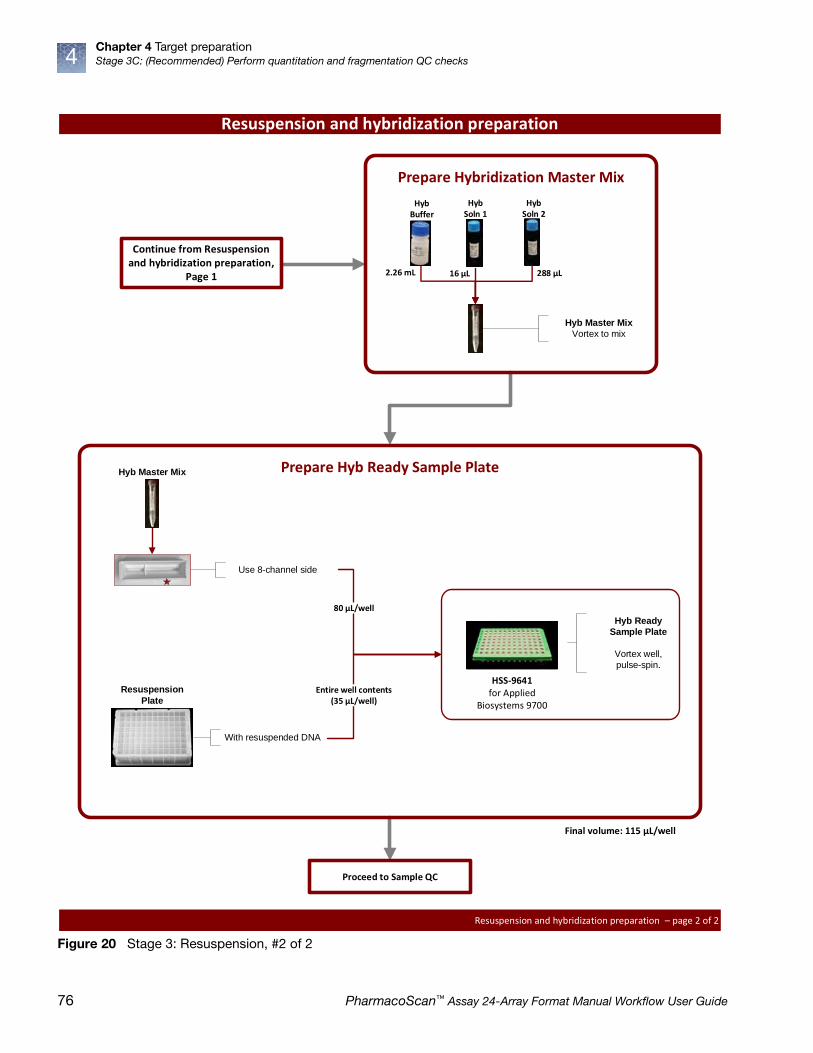
Chapter 4 Target preparationStage 3C: (Recommended) Perform quantitation and fragmentation QC checks4
Figure 20 Stage 3: Resuspension, #2 of 2
Resuspension and hybridization preparation
Resuspension and hybridization preparation – page 2 of 2
Proceed to Sample QC
Final volume: 115 μL/well
Continue from Resuspension and hybridization preparation,
Page 1
80 μL/well
Use 8-channel side
Prepare Hyb Ready Sample Plate
With resuspended DNA
Hyb Ready
Sample Plate
Vortex well,pulse-spin.
HSS-9641for Applied
Biosystems 9700
Hyb Master Mix
Resuspension
Plate
Prepare Hybridization Master MixHyb
BufferHyb
Soln 2
288 μL2.26 mL
Hyb Master Mix
Vortex to mix
16 μL
Hyb Soln 1
Entire well contents(35 μL/well)
76 PharmacoScan™ Assay 24-Array Format Manual Workflow User Guide

Chapter 4 Target preparationStage 3C: (Recommended) Perform quantitation and fragmentation QC checks 4
Figure 21 Stage 3: QC
Sample QC
QC – page 1 of 1
Proceed to Stage 4 or Store Hyb Ready Samples at –20�C
33 μL/well
Aliquot water to Dilution QC and OD Plates
Use 8-channel side
5 mL Water
90 μL/well
OD QC PlateDilution QC Plate
120 μL/well
Use 8-channel side
5 mL Gel Diluent
Gel QC Plate
Aliquot gel diluent to Gel QC Plate
3 μL/well
Prepare Dilution QC Plate
Dilution QC Plate
Hyb Ready Plate:
Vortex, pulse-spin
Vortex, pulse-spin
3 μL/well
Prepare Gel QC Plate
Dilution QC Plate
Vortex, Pulse-spin.
Load 20 µL into a4% agarose E-Gel for
analysisGel QC Plate
10 μL/well
Prepare OD Plate
Dilution QC Plate
Read Abs�260, 280, and 320 nm
OD QC Plate
Labware and reagents needed
QTY 1 (Hyb Ready Sample Plate)
QTY 2QTY 2
QTY 1
5 mL water
5 mL gel diluent
PharmacoScan™ Assay 24-Array Format Manual Workflow User Guide 77
Chapter 4 Target preparationStage 4: Denaturation and hybridization4
Stage 4: Denaturation and hybridization
You proceed to Stage 4 in one of two ways:
• Directly from Stage 3 without interruption.
• With hybridization ready samples that were stored at –20°C after Stage 3.
This stage requires the following sets of steps:
ʺ1: Prepare for denaturation and hybridizationʺ on page 80
ʺ2: Prepare hybridization ready samples stored at –20°Cʺ on page 80
ʺ3: Prepare the GeneTitan™ MC Instrumentʺ on page 80
ʺ4: Denature the Hyb Ready Sample plateʺ on page 81
ʺ5: Prepare hybridization tray and load into GeneTitan™ MC Instrumentʺ on page 82
ʺ4: Store remaining reagentsʺ on page 98
Perform Stage 4• If the Hyb Ready Plate was stored at –20°C, go to ʺ2: Prepare hybridization ready
samples stored at –20°Cʺ on page 80.
• If you are proceeding directly from the end of Stage 3 on page 74, go to ̋ 3: Prepare the GeneTitan™ MC Instrumentʺ on page 80.
Duration • Hands-on: 45 minutes including denaturation time
• in GeneTitan MC Instrument: 23.5 to 24 hours hybridization time
Required input from previous stage
• Hyb Ready Sample Plate
Equipment, consumables, and reagents required
The following thermal cycler is recommended:
• Applied Biosystems 9700
The thermal cycler needs to be programmed with the PharmacoScan Denature protocol (see ʺThermal cycler recommendations and protocolsʺ on page 23).
CAUTION! Parts of this stage should be performed under a fume hood.
IMPORTANT! Always use the heated lid option when programming protocols.
78 PharmacoScan™ Assay 24-Array Format Manual Workflow User Guide

Chapter 4 Target preparationStage 4: Denaturation and hybridization 4
Table 31 Equipment Required for Stage 4: Denaturation and hybridization
Quantity Equipment
1 GeneTitan MC Instrument
1 Rainin P200 8-channel Pipette
As needed Pipette tips
1 Thermal cycler Appropriate thermal cycler, programmed with the PharmacoScan Denature protocol (see "Thermal cycler recommendations and protocols" on page 23).
1 96 well metal chamber warmed in a 48°C oven1
1 The metal chamber coming out of a 48°C oven is warm to the touch. Gloves and mitts can be used ifit feels too hot.
Table 32 Consumables required for Stage 4: Denaturation and hybridization
Quantity Consumable Cat. No.
1 • One PharmacoScan™ 24F Array Plate in a protective base
902994
1 • Hybridization tray1
1 The Consumables for the GeneTitan MC Instrument are packaged separately from thePharmacoScan array plates. The consumables are available in the Axiom™ GeneTitan™ ConsumablesKit (Cat. No. 901606). The hybridization tray is available in the Axiom™ GeneTitan™ Consumables Kit.
Part No. 500867
Table 33 Reagents required from the PharmacoScan Reagent Kit 4x24 Reactions
Reagent Module
Axiom Wash Buffer A (both bottles; 1 L)Module 3,
Room TemperaturePart No. 901472
Axiom Wash Buffer B
Axiom Water
PharmacoScan™ Assay 24-Array Format Manual Workflow User Guide 79

Chapter 4 Target preparationStage 4: Denaturation and hybridization4
1: Prepare for denaturation and hybridization
1. Preheat the 96-well metal chamber in a 48°C oven.
2. Allow array plate to equilibrate to room temperature for a minimum of 25 minutes.
a. Leave the array plate in the pouch at room temperature, for a minimum of 25 minutes, before opening and loading on the GeneTitan MC Instrument to allow the plate to come to room temperature.
b. At the end of the array warm up time, open the pouch and scan the array plate barcode into the GeneTitan Array Plate Registration file as described in Appendix C, ʺRegister samples in GeneChip™ Command Console™ʺ on page 161.
3. Power up the thermal cycler and prepare for the PharmacoScan Denature protocol to run with the heated lid option selected.
2: Prepare hybridization ready samples stored at –20°C
1. Warm up the Hyb Ready Plate at room temperature for 5 minutes. It is not necessary to equilibrate the plate for longer duration.
2. Ensure the Hyb Ready Plate is sealed well.
If the plate is not sealed well:
a. Spin the plate and carefully remove the old seal.
b. If there is condensation on the top of the plate, blot dry gently with a Kimwipe laboratory tissue.
c. Use a fresh seal and tightly reseal the plate.
3. Vortex the Hyb Ready Plate briefly, then spin at 1,000 rpm for 30 seconds.
4. Place the Hyb Ready Plate at room temperature.
3: Prepare the GeneTitan™ MC Instrument
Before you denature your hybridization ready samples, ensure that the GeneTitan MC Instrument is ready for use by following the instructions given in Chapter 5, ʺStage 2: Hybridizationʺ on page 112 and Appendix C, ʺRegister samples in GeneChip™ Command Console™ʺ on page 161.
A brief summary of the steps which may need to be performed is:
1. Prepare the reagents from Module 3 as described in Table 34:
WARNING! Do not remove the array plate from the protective base or touch the surface of any arrays.
Table 34 Reagents from Module 3
Reagent Temperature out of module1
1 Temperature out of module: the temperature the reagent is held at immediately after removalfrom module.
Treatment
Axiom Wash Buffer A (Part No. 901446)
Room temperature Invert 2-3X for mixing before filling GT bottle
Axiom Wash Buffer B (Part No. 901447)
Room temperature Invert 2-3X for mixing before filling GT bottle
Axiom Water (Part No. 901578)
Room temperature N/A
80 PharmacoScan™ Assay 24-Array Format Manual Workflow User Guide

Chapter 4 Target preparationStage 4: Denaturation and hybridization 4
2. Launch GCC and select GCC GeneTitan Control.
3. Upload your sample registration file now.If you do not upload your samples before scanning the array plate barcode, the software assigns names to your sample.
4. Select the System Setup tab.
5. Configure the software as follows:
a. Setup Option: Hyb-Wash-Scan.
b. Click Next.
c. Plate information:
• Barcode: Scan or manually enter the PharmacoScan array plate barcode and click Next.
• Protocol Name: Select the protocol name and click Next.
6. Fill the Wash A, Wash B and Rinse bottles.
7. Empty the Waste bottle.
4: Denature the Hyb Ready Sample plate
1. Ensure the thermal cycler is powered on and the PharmacoScan Denature protocol with the heated lid option has been selected.
2. Open the lid of the thermal cycler and place the sealed Hyb Ready Plate on the thermal cycler. Check the integrity of the seal as evaporation during denaturation can negatively impact assay performance.
3. Close the lid.
4. Start the PharmacoScan Denature protocol, described on ʺThermal cycler recommendations and protocolsʺ on page 23).
PharmacoScan™ Assay 24-Array Format Manual Workflow User Guide 81

Chapter 4 Target preparationStage 4: Denaturation and hybridization4
5: Prepare hybridization tray and load into GeneTitan™ MC Instrument 1. After the PharmacoScan Denature protocol has completed, remove the Hyb
Ready Plate from the thermal cycler and place into a 96-well metal chamber that has been pre-warmed in an oven at 48°C.
2. Move the metal chamber containing the denatured Hyb Ready Plate to a fume hood.
3. Remove seal from Hyb Ready Plate and discard.
4. Remove the hybridization tray (from Axiom GeneTitan Consumables Kit) from packaging.
5. Label the hybridization tray. See the note below and Figure 11 on page 40 for more information.
6. Place the hybridization tray under the fume hood.
7. Using a P200 8-channel pipette set at 105 µL, slowly transfer the denatured samples in columns 5, 7, and 9 of the Hyb Ready Plate into the corresponding wells of the hybridization tray. Dispense to the first stop to avoid creating bubbles.
• Change pipette tips after each transfer; discard the tip even if it shows some volume left.
• Ensure that there are no air bubbles present in the hybridization tray. Puncture any air bubbles that you see using a clean pipette tip.
• There is no need to spread the sample around the bottom of the hybridization tray wells. Sample distribution across the well occurs when the array plate is stacked together with the hybridization tray by the GeneTitan MC Instrument.
8. Load the array plate and hybridization tray into the GeneTitan MC Instrument (see ʺLoad a PharmacoScan™ array plate and hybridization tray onto the GeneTitan™ MC Instrumentʺ on page 117).
CAUTION! It is recommended to perform the next set of steps under a fume hood.
IMPORTANT! It is critical that you write only on the proper location of the hybridization tray (on the edge in front of wells A1 and B1) as illustrated in Figure 11 on page 40. Do not write on any other side, as this can interfere with sensors inside of the GeneTitan MC Instrument and result in experiment failure.
IMPORTANT! Do not confuse hybridization trays with stain trays.
82 PharmacoScan™ Assay 24-Array Format Manual Workflow User Guide

Chapter 4 Target preparationStage 4: Denaturation and hybridization 4
Hybridization continues on the GeneTitan MC Instrument for 23.5-24 hours before you can load the Ligation/Staining/Stabilization reagent trays into the GeneTitan MC Instrument.
Near the end of the 23.5 to 24 hour hybridization period in the GeneTitan MC, proceed to ʺStage 5: GeneTitan™ reagent preparationʺ on page 85.
IMPORTANT! The array plate must be loaded on the left side on its protective blue base, as shown in the figure below. The clear plastic cover on top of the array plate SHOULD NOT be loaded in the GeneTitan MC Instrument.
Load the hybridization tray on the right side without any covering. The hybridization tray should not have any bubbles.
IMPORTANT! After the GeneTitan MC Instrument has stacked the array plate and hybridization tray, the instrument extends the drawer. Manually check the stacking by gently pressing the six latching points to confirm that the two parts are clamped properly, and check underneath the arrays to ensure there are no bubbles. If bubbles are found, gently tap the plate on top and the bubbles should disappear. Do NOT tip/tilt the array plate/hybridization tray sandwich while inspecting the bottom for bubbles.
1
2
3
Clear tray shipping cover (to be discarded)Array plate protective baseArray plate
1
2
3
PharmacoScan™ Assay 24-Array Format Manual Workflow User Guide 83

Chapter 4 Target preparationStage 4: Denaturation and hybridization4
Figure 22 Stage 4: Denaturation and hybridization
Denaturation and hybridization
Denaturation and hybridization – page 1 of 1
Process hybridization tray on the GeneTitan Instrument
Denature hyb ready samples in thermal cycler1) 10 minutes @ 95°C2) 3 minutes @ 48°C
Labware and reagents needed
QTY 1QTY 1 (Hyb Ready Sample Plate)
105 μL/well
Transfer denatured hyb ready samples to hybridization tray
Denatured Hyb
Ready Samples
Keep samples on a 48�C heat block
Pierce bubbles,if need be
Hybridization tray with Samples
84 PharmacoScan™ Assay 24-Array Format Manual Workflow User Guide
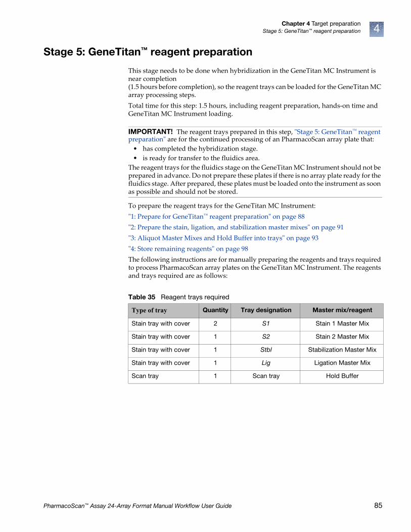
Chapter 4 Target preparationStage 5: GeneTitan™ reagent preparation 4
Stage 5: GeneTitan™ reagent preparation
This stage needs to be done when hybridization in the GeneTitan MC Instrument is near completion (1.5 hours before completion), so the reagent trays can be loaded for the GeneTitan MC array processing steps.
Total time for this step: 1.5 hours, including reagent preparation, hands-on time and GeneTitan MC Instrument loading.
To prepare the reagent trays for the GeneTitan MC Instrument:
ʺ1: Prepare for GeneTitan™ reagent preparationʺ on page 88
ʺ2: Prepare the stain, ligation, and stabilization master mixesʺ on page 91
ʺ3: Aliquot Master Mixes and Hold Buffer into traysʺ on page 93
ʺ4: Store remaining reagentsʺ on page 98
The following instructions are for manually preparing the reagents and trays required to process PharmacoScan array plates on the GeneTitan MC Instrument. The reagents and trays required are as follows:
IMPORTANT! The reagent trays prepared in this step, ʺStage 5: GeneTitan™ reagent preparationʺ are for the continued processing of an PharmacoScan array plate that:
• has completed the hybridization stage.
• is ready for transfer to the fluidics area.
The reagent trays for the fluidics stage on the GeneTitan MC Instrument should not be prepared in advance. Do not prepare these plates if there is no array plate ready for the fluidics stage. After prepared, these plates must be loaded onto the instrument as soon as possible and should not be stored.
Table 35 Reagent trays required
Type of tray Quantity Tray designation Master mix/reagent
Stain tray with cover 2 S1 Stain 1 Master Mix
Stain tray with cover 1 S2 Stain 2 Master Mix
Stain tray with cover 1 Stbl Stabilization Master Mix
Stain tray with cover 1 Lig Ligation Master Mix
Scan tray 1 Scan tray Hold Buffer
PharmacoScan™ Assay 24-Array Format Manual Workflow User Guide 85
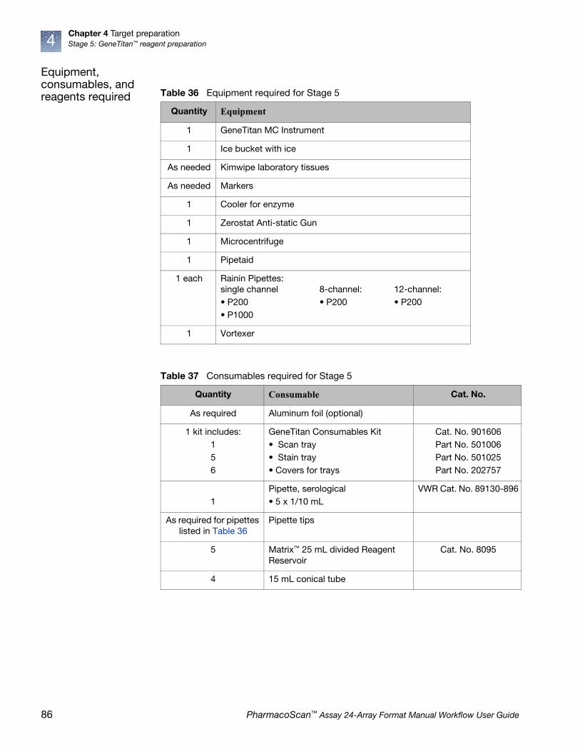
Chapter 4 Target preparationStage 5: GeneTitan™ reagent preparation4
Equipment, consumables, and reagents required Table 36 Equipment required for Stage 5
Quantity Equipment
1 GeneTitan MC Instrument
1 Ice bucket with ice
As needed Kimwipe laboratory tissues
As needed Markers
1 Cooler for enzyme
1 Zerostat Anti-static Gun
1 Microcentrifuge
1 Pipetaid
1 each Rainin Pipettes:single channel• P200• P1000
8-channel:• P200
12-channel:• P200
1 Vortexer
Table 37 Consumables required for Stage 5
Quantity Consumable Cat. No.
As required Aluminum foil (optional)
1 kit includes:156
GeneTitan Consumables Kit• Scan tray • Stain tray• Covers for trays
Cat. No. 901606Part No. 501006Part No. 501025Part No. 202757
1Pipette, serological• 5 x 1/10 mL
VWR Cat. No. 89130-896
As required for pipettes listed in Table 36
Pipette tips
5 Matrix™ 25 mL divided Reagent Reservoir
Cat. No. 8095
4 15 mL conical tube
86 PharmacoScan™ Assay 24-Array Format Manual Workflow User Guide

Chapter 4 Target preparationStage 5: GeneTitan™ reagent preparation 4
Table 38 PharmacoScan reagents required for stain and ligation stage
Reagent Module
Axiom Ligate Buffer
Module 4-1, –20°C Part No. 901278
Axiom Ligate Enzyme
Axiom Ligate Soln 1
Axiom Probe Mix 1
Axiom Stain Buffer
Axiom Stabilize Soln
Axiom Ligate Soln 2
Module 4-2, 2-8°CPart No. 901276
Axiom Probe Mix 21
1 These solutions are light sensitive. Keep tubes out of direct light for a prolonged period of time.
Axiom Wash A
Axiom Stain 1-A1
Axiom Stain 1-B1
Axiom Stain 2-A1
Axiom Stain 2-B1
Axiom Stabilize Diluent
Axiom Water
Axiom Hold Buffer1
Axiom Ligate Enzyme (extra tube for back-up purposes) Module 5-1, –25°C to –15°CPart No. 902796
Axiom Hold Buffer (three bottles) Module 5-2, 2°C to 8°CPart No. 902797
PharmacoScan™ Assay 24-Array Format Manual Workflow User Guide 87

Chapter 4 Target preparationStage 5: GeneTitan™ reagent preparation4
1: Prepare for GeneTitan™ reagent preparation
Thaw and prepare the reagentsNote: Ligation Buffer and Ligation Solution 2 require approximately 30 to 40 minutes to thaw on the benchtop at room temperature.
Table 39 Reagents required for GeneTitan™ MC Instrument reagent tray preparation
Module Reagent Thaw on benchtop, then place on ice
Place on ice Place on benchtop at room temperature
Module 3Room
Temperature
Axiom Wash Buffer A
Axiom Wash Buffer B
Axiom Water (1 bottle required)
Module 4-1–20°C
Axiom Ligate Buffer - for 30 minutes
Axiom Ligate Enzyme1 Keep at –20°C until ready to use
Axiom Ligate Soln 1
Axiom Probe Mix 1
Axiom Stain Buffer
Axiom Stabilize Soln
Module 4-22°C to 8°C
Axiom Ligate Soln 2 - for 30 to 40 minutes
Axiom Probe Mix 22
Axiom Wash A - for 30 minutes
Axiom Stain 1-A2
Axiom Stain 1-B2
Axiom Stain 2-A2
Axiom Stain 2-B2
Axiom Stabilize Diluent
Axiom Water
Axiom Hold Buffer2 (1 bottle required)3
1 An extra tube of Ligate Enzyme is in Module 5-1 in the event that it is needed.2 These solutions are light sensitive. Keep tubes out of direct light for a prolonged period of time.3 Axiom Hold Buffer for preparing the scan tray for the 2nd, 3rd, and 4th plate are provided in Module 5-2.
88 PharmacoScan™ Assay 24-Array Format Manual Workflow User Guide
Chapter 4 Target preparationStage 5: GeneTitan™ reagent preparation 4
Axiom Wash A and Axiom Stabilize Diluent preparationDuring storage of the Axiom Wash A and Axiom Stabilize Diluent (in Module 4-2 stored at 4°C), precipitation in the form of clear crystals can sometimes occur. Therefore, follow the procedure below to ensure that any precipitate is returned to solution prior to use.
Note: The presence of some precipitate is okay and does not adversely impact assay performance. Follow the instructions below to resuspend any precipitate before use.
Prepare the Axiom Wash A:
1. Vortex the bottle for 30 seconds.
2. Place on the benchtop at room temperature for 30 minutes.
3. Examine the reagent for precipitate (look into the top of the bottle).
4. If precipitate is still present, vortex again for 30 seconds.
Prepare the Stabilize DiluentIf crystals are observed in the Axiom Stabilize Diluent:
1. Vortex and spin.
2. Look for precipitate.
If any:
• Warm tube to room temperature and vortex again.
Axiom Ligate Buffer preparationWhite precipitate is sometimes observed when the Axiom Ligate Buffer is thawed.
Note: The presence of some precipitate is okay and does not adversely impact assay performance. Follow the instructions below to attempt to resuspend a majority of precipitate before use.
Prepare the Axiom Ligate Buffer
1. Place on the benchtop at room temperature for 30 minutes. This bottle can also be thawed in a dish with room temperature Millipore water.
2. Examine the buffer for precipitate (look into the top of the bottle).
3. If precipitate is present, vortex the bottle for 30 seconds.
4. Re-examine the buffer for precipitate.
5. If precipitate is still present, warm the bottle with your hands and vortex again for 30 seconds.
6. If precipitate is still present after hand warming proceed with the protocol below.
PharmacoScan™ Assay 24-Array Format Manual Workflow User Guide 89

Chapter 4 Target preparationStage 5: GeneTitan™ reagent preparation4
Prepare the remaining reagents
1. Leave the Axiom Ligate Enzyme at –20°C until ready to use.
2. Thaw the following reagents from Module 4-1 at –20°C on the benchtop at room temperature, then vortex, spin and place on ice:
• Axiom Ligate Soln 1
• Axiom Probe Mix 1
• Axiom Stabilize Soln
• Axiom Stain Buffer
3. Prepare the remaining reagents from Module 4-2 as follows:
a. Gently flick each tube to to three times to mix, then spin.
b. Place reagents on ice, except for the Hold Buffer, Ligate Soln 2 and Water— leave these reagents on the benchtop at room temperature.
Label the master mix tubes
1. Mark the side of each tube with one of designations shown in Table 40.
Label the reagent reservoirs
1. Label five Matrix 25 mL divided Reagent Reservoirs (Cat. No. 8095) as indicated in the table below.
Table 40 Labeling master mix tubes for stain, ligation, and stabilization reagents
Conical tube
Number of tubes
Designation Contents Place
15 mL 1 S1 • Stain 1 Master Mix On ice
15 mL 1 S2 • Stain 2 Master Mix On ice
15 mL 1 Stbl • Stabilization Master Mix
On ice
15 mL 1 Lig • Ligation Master Mix On ice
Table 41 Labeling reagent reservoirs
Reservoir designation Contents
S1 Stain 1 Master Mix
S2 Stain 2 Master Mix
Stbl Stabilization Master Mix
Lig Ligation Master Mix
Hold Hold Buffer
90 PharmacoScan™ Assay 24-Array Format Manual Workflow User Guide

Chapter 4 Target preparationStage 5: GeneTitan™ reagent preparation 4
2: Prepare the stain, ligation, and stabilization master mixes
Prepare the Stain 1 Master Mix
1. Use appropriate serological and single-channel pipettes to add reagents to the 15 mL tube labeled S1 in the order shown in Table 42. This recipe provides enough for both S1 reagent trays.
2. Gently invert the tube 10 times to mix. Do not vortex.
3. Place on ice and protect from direct light (e.g., cover with aluminum foil or ice bucket lid).
Prepare the Stain 2 Master Mix
1. Use appropriate single-channel pipettes to add reagents to the 15 mL tube labeled S2 in the order shown in Table 43.
2. Gently invert the S2 MM tube 10 times to mix. Do not vortex.
3. Place on ice and protect from direct light (e.g., cover with aluminum foil or ice bucket lid).
Table 42 Stain 1 Master Mix
Reagent per array Master Mix 24+
To the tube marked S1, add:
• Axiom Wash A 201.6 μL 5.24 mL
• Axiom Stain Buffer 4.2 μL 109.2 μL
• Axiom Stain 1-A 2.1 μL 54.6 μL
• Axiom Stain 1-B 2.1 μL 54.6 μL
Total 210 μL(105 μL x 2)
5.46 mL
Table 43 Stain 2 Master Mix
Reagent per array Master Mix 24+
To the tube marked S2, add:
• Axiom Wash A 100.8 μL 2.62 mL
• Axiom Stain Buffer 2.1 μL 54.6 μL
• Axiom Stain 2-A 1.05 μL 27.3 μL
• Axiom Stain 2-B 1.05 μL 27.3 μL
Total 105 μL 2.73 mL
PharmacoScan™ Assay 24-Array Format Manual Workflow User Guide 91
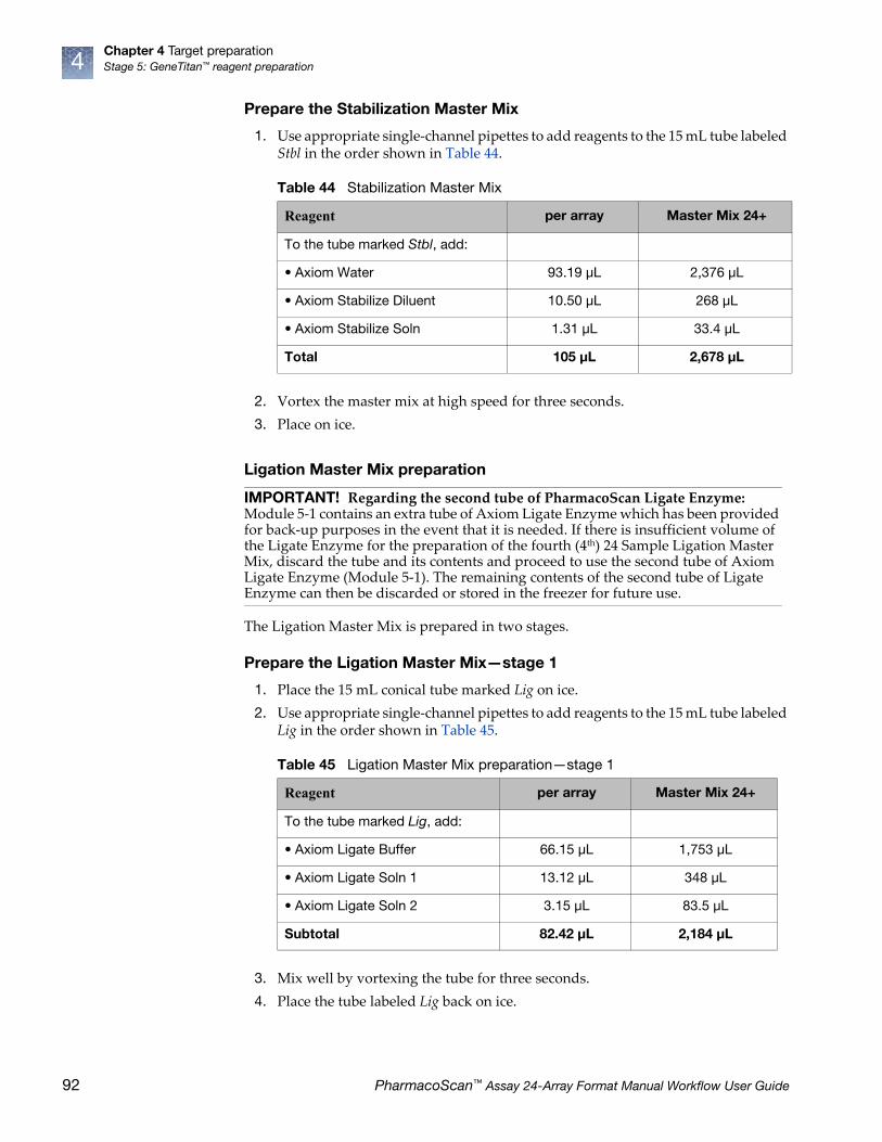
Chapter 4 Target preparationStage 5: GeneTitan™ reagent preparation4
Prepare the Stabilization Master Mix
1. Use appropriate single-channel pipettes to add reagents to the 15 mL tube labeled Stbl in the order shown in Table 44.
2. Vortex the master mix at high speed for three seconds.
3. Place on ice.
Ligation Master Mix preparation
The Ligation Master Mix is prepared in two stages.
Prepare the Ligation Master Mix—stage 1
1. Place the 15 mL conical tube marked Lig on ice.
2. Use appropriate single-channel pipettes to add reagents to the 15 mL tube labeled Lig in the order shown in Table 45.
3. Mix well by vortexing the tube for three seconds.
4. Place the tube labeled Lig back on ice.
Table 44 Stabilization Master Mix
Reagent per array Master Mix 24+
To the tube marked Stbl, add:
• Axiom Water 93.19 μL 2,376 μL
• Axiom Stabilize Diluent 10.50 μL 268 μL
• Axiom Stabilize Soln 1.31 μL 33.4 μL
Total 105 μL 2,678 μL
IMPORTANT! Regarding the second tube of PharmacoScan Ligate Enzyme:Module 5-1 contains an extra tube of Axiom Ligate Enzyme which has been provided for back-up purposes in the event that it is needed. If there is insufficient volume of the Ligate Enzyme for the preparation of the fourth (4th) 24 Sample Ligation Master Mix, discard the tube and its contents and proceed to use the second tube of Axiom Ligate Enzyme (Module 5-1). The remaining contents of the second tube of Ligate Enzyme can then be discarded or stored in the freezer for future use.
Table 45 Ligation Master Mix preparation—stage 1
Reagent per array Master Mix 24+
To the tube marked Lig, add:
• Axiom Ligate Buffer 66.15 μL 1,753 μL
• Axiom Ligate Soln 1 13.12 μL 348 μL
• Axiom Ligate Soln 2 3.15 μL 83.5 μL
Subtotal 82.42 μL 2,184 μL
92 PharmacoScan™ Assay 24-Array Format Manual Workflow User Guide

Chapter 4 Target preparationStage 5: GeneTitan™ reagent preparation 4
Prepare the Ligation Master Mix—stage 2
1. Remove the Axiom Ligate Enzyme from the –20°C freezer and place in a cooler chilled to –20°C.
2. Use appropriate serological and single-channel pipettes to add reagents to the 15 mL tube labeled Lig in the order shown in Table 46.
Gently flick the PharmacoScan Ligate Enzyme tube 2-3 times, then perform a quick spin immediately prior to adding the enzyme to the Master Mix.
3. Gently invert 10 times to mix. Do not vortex.
4. Place on ice and protect from direct light (e.g., cover with aluminum foil or ice bucket lid).
3: Aliquot Master Mixes and Hold Buffer into trays
Label the trays
1. Gather the scan tray and the stain trays and covers from the Axiom™ GeneTitan™ Consumables Kit.
2. Label two stain trays S1
3. Label the remaining stain trays:
• S2
• Stbl
• Lig
When preparing the hybridization and reagent trays to be loaded onto the GeneTitan MC Instrument, you need to mark the front of each tray in a way that identifies its contents.
Table 46 Ligation Master Mix Preparation—Stage 2
Reagent per array Master Mix 24+
• Ligation Master Mix from Stage 1 82.42 μL 2,184 μL
• Axiom Probe Mix 1 10.5 μL 278 μL
• Axiom Probe Mix 2 10.5 μL 278 μL
• Axiom Ligate Enzyme 1.58 μL 42 μL
Total 105 μL 2,783 μL
IMPORTANT! It is critical that you write only on the proper side of the front edge of stain trays, as illustrated in Figure 12 on page 41. The front edge of the tray is the short side with the lettering A through H. Do not write on any other side, as this can interfere with sensors inside of the GeneTitan MC Instrument and result in experiment failure. To ensure proper placement of lids onto stain trays, and trays onto the GeneTitan MC Instrument, you can also mark the notched corner of the trays and lids.
IMPORTANT! Do not confuse hybridization trays with stain trays.
PharmacoScan™ Assay 24-Array Format Manual Workflow User Guide 93

Chapter 4 Target preparationStage 5: GeneTitan™ reagent preparation4
Deionize trays and coversDeionize the inside of each tray and cover now. Return the trays and covers to the bench top after deionizing.
See Appendix D, ̋ Deionizing procedure for GeneTitan™ trays and coversʺ on page 164 for the recommended technique.
About aliquoting reagents to traysStain trays: Only fill columns 5, 7, and 9 with ligation, staining, and stabilization reagents.
Scan tray: It is important to fill all 96 wells with Hold Buffer. The scan tray has an open-bottom design, so it is very important that all 96 wells of the scan tray receive 150 µL of Hold Buffer.
For all trays, pipet into trays on the bench top. If the trays are not being used immediately, protect them from light by covering with foil or placing in a cabinet.
When aliquoting ligation, staining, and stabilization reagents to the trays, it is not necessary to spread the reagent to each corner of the well. The reagent spreads evenly when the array plate is inserted into the reagent tray during processing with the GeneTitan MC Instrument.
IMPORTANT! Always aliquot reagents to the bottom of the tray. Avoid touching the sides or the top of the wells with the pipette tips. Droplets close to or on the top of the well dividers may cause the lid to stick to the tray during GeneTitan MC Instrument processing.
IMPORTANT! Remember to deionize the stain trays and the covers before aliquoting master-mixes
94 PharmacoScan™ Assay 24-Array Format Manual Workflow User Guide

Chapter 4 Target preparationStage 5: GeneTitan™ reagent preparation 4
Aliquot the Stain 1 Master Mix
1. Pour the S1 Master Mix into the 8-channel side of the reagent reservoir marked S1, placed on the bench top at room temperature.
2. Load a P200 8-channel pipette with 8 new pipette tips and aliquot 105 µL per well to columns 5, 7, and 9 of both S1 trays. Dispense to the first stop only to avoid creating bubbles. You do not need to change pipette tips between additions of the Stain 1 Master Mix.
3. If:
• bubbles are present, puncture them with a pipette tip.
• droplets of liquid splashed onto the well dividers, place a Kimwipe laboratory tissue on top of the tray to blot and remove.
4. Place covers on the S1 trays. Orient cover correctly on the tray with the notched corners together (Figure 23).
5. Protect the trays from light if not immediately loading onto the GeneTitan MC Instrument.
IMPORTANT! Leaving liquid on the top of the dividers may cause excessive evaporation or may form a seal that restricts the removal of the GeneTitan tray cover.
Figure 23 Placing cover on stain tray. Notched corners should face front.
Example of a droplet of liquid that has splashed onto the well divider of a stain tray during reagent aliquoting.
Ensure no droplets of liquid are on top of the wells dividers. Blot with a Kimwipe laboratory tissue to remove.
Notched cornersof stain tray and lid
PharmacoScan™ Assay 24-Array Format Manual Workflow User Guide 95

Chapter 4 Target preparationStage 5: GeneTitan™ reagent preparation4
Aliquot the Stain 2 Master Mix
1. Carefully pipet or pour the Stain 2 Master Mix into the 4-channel side of the reagent reservoir marked S2, placed on the bench top at room temperature.
2. Load a P200 8-channel pipette with 4 new pipette tips and aliquot 105 µL per well to columns 5, 7, and 9 of the S2 tray. Dispense to the first stop only to avoid creating bubbles.
You do not need to change pipette tips between additions of the Stain 2 Master Mix.
3. If:
• bubbles are present, puncture them with a pipette tip.
• droplets of liquid splashed onto the well dividers, place a Kimwipe laboratory tissue on top of the tray to blot and remove.
4. Place a cover on the S2 tray. Orient the cover correctly on the tray with the notched corners together (Figure 23).
5. Protect the tray from light if not immediately loading onto the GeneTitan MC.
Aliquot the Stabilization Master Mix
1. Carefully pipet or pour the Stabilization Master Mix into the 4-channel side of the reagent reservoir marked Stbl, placed on the bench top at room temperature.
2. Load a P200 8-channel pipette with 4 new pipette tips and aliquot 105 µL per well to columns 5, 7, and 9 of the Stbl tray. Dispense to the first stop only to avoid creating bubbles.
You do not need to change pipette tips between additions of the Stabilization Master Mix.
3. If:
• bubbles are present, puncture them with a pipette tip.
• droplets of liquid splashed onto the well dividers, blot the top of the tray with a Kimwipe laboratory tissue.
4. Place a cover on the tray. Orient cover correctly on the tray with the notched corners together (Figure 23).
Aliquot the Ligation Master Mix
1. Carefully pipet or pour the Ligation Master Mix into the 4-channel side of the reagent reservoir marked Lig, placed on the bench top at room temperature.
2. Load a P200 8-channel pipette with 4 new pipette tips and aliquot 105 µL per well to columns 5, 7, and 9 of the Lig tray. Dispense to the first stop only to avoid creating bubbles. You do not need to change pipette tips between additions of the Ligation Master Mix.
3. If:
• bubbles are present, puncture them with a pipette tip.
• droplets of liquid splashed onto the well dividers, place a Kimwipe laboratory tissue on top of the tray to blot and remove.
4. Place a cover on the tray. Orient cover correctly on the tray with the notched corners together (Figure 23).
5. Protect the tray from light if not immediately loading onto the GeneTitan MC.
96 PharmacoScan™ Assay 24-Array Format Manual Workflow User Guide

Chapter 4 Target preparationStage 5: GeneTitan™ reagent preparation 4
Aliquot the Axiom Hold Buffer to the scan trayNote: Module 5-2 has three Axiom Hold Buffer bottles that should be used to prepare the scan tray for second, third, and fourth plate.
1. Ensure that the Axiom Hold Buffer has equilibrated to room temperature. Vortex and then pour the Hold Buffer into both the 4-channel and 8-channel side of the reagent reservoir labeled Hold, placed on the bench top at room temperature.
2. Remove the scan tray from its pouch.
3. Remove the scan tray cover, but leave the scan tray on its protective black base.
4. Prepare the barcoded scan tray cover (Part No. 202757) that came with the scan tray by completing the deionization procedure described in Appendix D, ʺDeionizing procedure for GeneTitan™ trays and coversʺ on page 164. Place the cover as shown in Figure 25 on page 98 to prevent dust or static from accumulating on the bottom of the cover.
• Use a 12-channel P200 pipette with new pipette tips to aliquot 150 µL to EACH of the 96 wells of the 96 plate scan tray. Dispense to the first stop and avoid touching the bottom of the tray.
• You do not need to change pipette tips between additions of the Hold buffer.
5. If droplets of liquid splashed onto the well dividers, place a Kimwipe laboratory tissue on top of the tray to blot and remove.
6. Cover the tray by orienting the notched corner of the scan tray cover over the notched edge of the tray and the flat side of the cover against the scan tray (Figure 24).
See ʺStage 3: Ligate, wash, stain and scanʺ on page 129 for instructions on loading the reagent trays.
IMPORTANT! The scan tray has an open-bottom design, so it is very important that all 96 wells of the scan tray receive 150 µL of Axiom Hold Buffer.
CAUTION! Do not remove the scan tray from its protective black base until loading onto the GeneTitan MC instrument. To avoid scratching, do not touch the bottom of the tray with pipette tips. Dispense hold buffer to the first stop only.
PharmacoScan™ Assay 24-Array Format Manual Workflow User Guide 97

Chapter 4 Target preparationStage 5: GeneTitan™ reagent preparation4
4: Store remaining reagents
Store remaining Module 4-1 and Module 4-2 reagents for future use. Follow the guidelines presented in the section ʺFreeze-thaw instructionsʺ on page 30.
Figure 24 Scan tray with the clear cover and protective base
Figure 25 Loading the scan tray with Hold Buffer
Barcoded scan tray cover, Part No. 202757
GeneTitan scan tray, Part No. 501006 or Part No. 500860
Notched corners of cover, tray, and base line up
Scan tray protective base
1
1
2
3
4
2
3
4
Protective Black Base
Leave the scan tray in its protective black base while loading with Hold Buffer.
Cover
98 PharmacoScan™ Assay 24-Array Format Manual Workflow User Guide

Chapter 4 Target preparationStage 5: GeneTitan™ reagent preparation 4
Figure 26 Stage 5: GeneTitan reagent preparation, #1 of 3
GeneTitan reagent preparation
GeneTitan reagent preparation – page 1 of 3
Continue to GeneTitan reagent preparation
page 2
105 μL/well on Cols 5, 7, 9
Prepare Stain 2 tray
Tray Name: S2Confirm there are no
droplets on well dividers
Invert tube 10X to mix.Do not vortex.
Use 4-channel side
Wash A Stain 2A
27.3 μL2.62 mL
Stain Buffer
27.3 μL
Stain 2B
54.6 μL
105 μL/well on Cols 5, 7, 9 For 2 trays
Prepare Stain 1 trays
Tray Names: S1Confirm there are no
droplets on well dividers
Invert tube 10X to mix.Do not vortex.
Use 8-channel side
Wash A Stain 1A
54.6 μL5.24 mL 109.2 μL
Stain Buffer
54.6 μL
Stain 1B
2 Trays
Labware and reagents needed
QTY 5 QTY 1
Axiom™ 2.0:Module 4-1Module 4-2
QTY 4
QTY 5
PharmacoScan™ Assay 24-Array Format Manual Workflow User Guide 99

Chapter 4 Target preparationStage 5: GeneTitan™ reagent preparation4
Figure 27 Stage 5: GeneTitan reagent preparation, #2 of 3
GeneTitan reagent preparation
GeneTitan reagent preparation – page 2 of 3
Continue to GeneTitan reagent preparation
page 3
Continue from GeneTitan reagent preparation
page 1
105 μL/well on Cols 5, 7, 9
Prepare Stabilization Tray
Tray Name: StblConfirm there are no
droplets on well dividers
Vortex
Use 4-channel side
Axiom Water
Stabilize Soln
33.4 μL2376 μL 268 μL
Stabilize Diluent
100 PharmacoScan™ Assay 24-Array Format Manual Workflow User Guide

Chapter 4 Target preparationStage 5: GeneTitan™ reagent preparation 4
Figure 28 Stage 5: GeneTitan reagent preparation, #3 of 3
GeneTitan reagent preparation
GeneTitan reagent preparation – page 3 of 3
Continue from GeneTitan reagent preparation
page 2
Process plates on the GeneTitan Instrument
Prepare Ligation Master Mixpart 1
Ligation Master Mix:
Vortex to mix
Ligate Buffer
Ligate Soln 2
83.5 μL1,753 μL 348 μL
Ligate Soln 1
105 μL/well on Cols 5, 7, 9
Prepare Ligation Master Mix part 2 and prepare Ligation Tray
Tray Name: LigConfirm there are no
droplets on well dividers
Add reagents to
Ligation Master Mix tube
Invert tube 10X to mix.Do not vortex.
Use 4-channel side
Probe Mix 1
Ligate Enzyme
42 μL278 μL 278 μL
Probe Mix 2
150 μL/well
Hold Buffer
Aliquot Hold Buffer to scan tray
Confirm there are no droplets on well dividers
Use both sides
PharmacoScan™ Assay 24-Array Format Manual Workflow User Guide 101

5 Array processing with the GeneTitan™
Multi-Channel Instrument
The PharmacoScan™ Assay 24-Array Format Manual Workflow is designed for processing 24 samples at a time. The protocol is performed in two sets of steps:
• Target Preparation, performed on the lab bench without advanced automation. See Chapter 4, ʺTarget preparationʺ on page 42.
• Array processing, performed on the GeneTitan™ Multi-Channel (MC) Instrument.
This chapter includes instructions for Part 2: Array Processing. These instructions are presented as follows:
• ʺBefore using the GeneTitan™ MC Instrumentʺ on page 102
– ʺProper tray alignment and loadingʺ on page 102
– ʺStain trays and coversʺ on page 104
– ʺLabel GeneTitan™ hybridization and reagent traysʺ on page 105
– ʺLoad tray consumables onto the GeneTitan™ MC Instrumentʺ on page 131
• ʺStage 1: Create and upload GeneTitan Array Plate Registration fileʺ on page 111
• ʺStage 2: Hybridizationʺ on page 112
• ʺStage 3: Ligate, wash, stain and scanʺ on page 129
Before using the GeneTitan™ MC Instrument
Proper tray alignment and loading
Proper alignment and loading of plates, covers and trays is critical when using the GeneTitan MC Instrument. Each plate, cover and tray has one notched corner. The notched corner of plates, trays, covers and bases must be in vertical alignment with each other, and placed in position A1 per the Tray Alignment guide inside each GeneTitan MC drawer (Figure 29 on page 103 and Figure 30 on page 104).
Note: Mark the notched corner of each plate, cover and tray with permanent marker to help ensure proper alignment and loading onto the GeneTitan MC Instrument.
Note: The instrument control software displays a warning if it detects a problem during the fluid dispense operations. The filters in the GeneTitan Wash A, Wash B and DI Water bottles should be replaced if the software displays such a warning. See Appendix E, ʺGeneTitan™ Multi-Channel Instrument careʺ on page 168 for the message displayed to the user and the procedure for replacing the filters.
IMPORTANT! When running a multi-plate workflow, you must pay careful attention to the software prompts instructing which side of the drawer to place or remove a plate/tray.
CAUTION! Take care not to damage the consumables or bend the blue cover posts or scan tray posts.
102 PharmacoScan™ Assay 24-Array Format Manual Workflow User Guide

Chapter 5 Array processing with the GeneTitan™ Multi-Channel InstrumentBefore using the GeneTitan™ MC Instrument 5
Figure 29 Proper alignment and loading of plates, covers and trays in the GeneTitan™ MC Instrument
Plates and trays must be seated in this rectangular recess.
The notched corner of all plates, bases, and covers and must be seated in this corner of the drawer per the Tray Alignment guide.
Tip: Mark the notched corner of each plate, cover and tray with permanent marker to help ensure proper alignment and loading.
Notched corner of array plate aligned with notched corner of blue base.
IMPORTANT! Remove the plastic protective shipping tray cover.
1
2
3
Clear tray shipping cover (to be discarded)Array plate protective baseArray plate
1
2
344
5
5
6
7
7
6
PharmacoScan™ Assay 24-Array Format Manual Workflow User Guide 103

Chapter 5 Array processing with the GeneTitan™ Multi-Channel InstrumentBefore using the GeneTitan™ MC Instrument5
Stain trays and covers
Figure 30 Array plate with protective blue base and the hybridization tray aligned and properly loaded into drawer 6
IMPORTANT! When you install the consumables, ensure that the fingers are retracted. Do not lay the consumables on top of the drawer fingers - this indicates that the instrument is not functioning correctly. Notify your field service engineer if the fingers do not retract automatically. You should place the trays into the instrument drawers when a drawer is fully extended by the instrument. The fingers are retracted when the drawer is open and are extended when the drawer is closed in order to restrain the consumable.
Array plate with protective blue base
Hybridization tray
1
2
1 2
IMPORTANT! Always place the flat side of the cover against the stain tray.
Figure 31 Placement of covers on trays
Correct placement of cover on stain tray. Incorrect placement of cover on stain tray.
Notched corners of tray and lid should be aligned and facing front
104 PharmacoScan™ Assay 24-Array Format Manual Workflow User Guide

Chapter 5 Array processing with the GeneTitan™ Multi-Channel InstrumentBefore using the GeneTitan™ MC Instrument 5
Label GeneTitan™ hybridization and reagent trays
When preparing the hybridization and reagent trays to be loaded onto the GeneTitan MC Instrument, you need to mark each tray in a way that identifies its contents.
Proper labeling for hybridization trays and reagent trays is described in:
• ʺLabel hybridization traysʺ, below
• ʺLabel stain traysʺ on page 106
Label hybridization traysYou can label the hybridization tray on the front part of the short side of the tray, next to the notch at the left, as shown in Figure 32. The proper section for labeling is closest to the notched corner, corresponding to the A1 and B1 wells.
IMPORTANT! It is critical that you write only on the proper locations of the proper sides of hyb and stain trays. Do NOT write in any other location, as this can interfere with sensors inside the GeneTitan MC Instrument and result in experiment failure. To ensure proper placement of lids onto stain trays, and trays onto the GeneTitan MC Instrument, you can also mark the notched corner of the trays and lids.
IMPORTANT! Do not confuse hybridization trays with stain trays.
Figure 32 Labeling GeneTitan hybridization trays
CAUTION! Writing on the wrong side of the hybridization tray, or on the wrong part of the long side, can interfere with the operation of sensors in the GeneTitan MC Instrument.
Do NOT label trays on the long side of the tray.
Notched corner of the hybridization tray should face the front.
Label the hybridization tray in this area.
1
2
3
1 2 3
PharmacoScan™ Assay 24-Array Format Manual Workflow User Guide 105

Chapter 5 Array processing with the GeneTitan™ Multi-Channel InstrumentBefore using the GeneTitan™ MC Instrument5
Label stain traysYou can label the stain trays on the left side of the front of the tray as shown in Figure 33. The correct side is closest to the notched corner, corresponding to the A1 through C1 wells.
Email and telephone notifications from the GeneTitan™ MC Instrument
We strongly recommend that you configure the Applied Biosystems GeneChip™ Command Console (GCC) software to send you GeneTitan MC notifications. It is critical that you know when the instrument requires your attention—either for sample handling or troubleshooting. Rapid notification can lessen the risk of sample loss.
Notifications can be sent to e-mail addresses and telephones. See the GCC user guide for instructions.
The types of notifications available let you know when a process:
• Starts
• Completes
• Aborts
• Encounters an error
Figure 33 Label GeneTitan stain tray (stain Tray shown with lid)
Do NOT label trays on the long side of the tray.
Notched corner of the stain tray should face the front.
Label the stain tray here.
1 2 3
1
2
3
106 PharmacoScan™ Assay 24-Array Format Manual Workflow User Guide

Chapter 5 Array processing with the GeneTitan™ Multi-Channel InstrumentBefore using the GeneTitan™ MC Instrument 5
GeneTitan™ MC Instrument lamp
The GeneTitan MC Instrument uses a xenon arc lamp system that is warranted for 500 hours to provide illumination for imaging the array at two wavelengths. The xenon lamp has a limited lifetime and needs to be replaced at regular intervals.
The GeneTitan Instrument Control software provides a timer that indicates the remaining useful life of the bulb and notifies you when it requires replacement. It is important to adhere to the warnings specified in the GeneTitan MC Instrument user guide.
See the GeneTitan™ MC Instrument User Guide, Pub. No. 08-0308, or Appendix E, ʺGeneTitan™ Multi-Channel Instrument careʺ on page 168 of this user guide for details on replacing the lamp.
See the GeneTitan™ MC Instrument User Guide, Pub. No. 08-0308, for the Lambda LS and Smart controller system. The Lamp and the controller should NEVER be switched ON or OFF manually. The GeneTitan MC Instrument control software manages the lamp activity and switches the lamp ON and OFF as required. It takes 10 minutes to warm-up the lamp. In idle mode the lamp remains ON for two hours before it is automatically switched OFF and if there are no more plates being transferred from the fluidics to the imaging station. This is by design and intended behavior. Do not try to save the lamp life by turning OFF the switch on the lamp.
Note: The power switch on the shutter box should be ON at all times. The OPEN/CLOSE switch on the shutter box should be at AUTO position at all times.
Setup options for array plate processing
The processes (setup options) available for processing array plates are shown in Figure 34. A brief description of each option is given below.
Figure 34 Setup options for processing array plates
System Setup tab
Setup options
1
2
1
2
PharmacoScan™ Assay 24-Array Format Manual Workflow User Guide 107
Chapter 5 Array processing with the GeneTitan™ Multi-Channel InstrumentBefore using the GeneTitan™ MC Instrument5
Hyb-Wash-ScanThis setup option enables you to hybridize, wash-ligate-stain-fix, and scan an array plate on the GeneTitan MC Instrument.
• Hyb: the array plate is moved to the hybridization oven inside the instrument. Each denatured sample in the hybridization tray is hybridized to an array on the array plate.
– Duration for 24 samples = 23.5 hours
• Wash: samples on arrays are ligated, washed, stained and fixed.
– Duration for 24 samples = ~5 hours
Note: The instrument control software displays a warning if it detects a problem during the fluid dispense operations. The filters in the GeneTitan Wash A, Wash B and DI Water bottles should be replaced if the software displays such a warning. See Appendix E, ʺGeneTitan™ Multi-Channel Instrument careʺ on page 168 for the message displayed to the user and the procedure for replacing the filters.
• Scan: The array plate is moved to the imaging device in the GeneTitan MC Instrument and each array is scanned.
– Duration for 24 samples = 2 hours
Hyb-WashIf this setup option is selected, array plate processing stops after the array has gone through fluidics processing. Use this option if an array plate cannot be scanned on the same GeneTitan MC Instrument as the one used for hybridization and fluidics processing.
If the array plate cannot be scanned immediately after the Hyb-Wash Process is complete:
1. Wrap the array plate (in the scan tray with black protective base) in aluminum foil to protect from light.
No lid is required. Do not invert the plate stack. If inverted, the Hold Buffer spills out of the tray. To prevent liquid spillage, try to keep the plate level when handling the plates. Do not touch the bottom optical surface of the scan tray.
2. Store at 4°C.
3. Scan the array plate within three days or less.
When ready to scan the array plate:
1. Keeping the plate protected from light, bring the plate to room temperature for ~50 minutes.
2. Remove the aluminum foil and load onto the GeneTitan MC Instrument.
IMPORTANT! When running a multi-plate workflow, you must pay careful attention to the software prompts instructing which side of the drawer to place or remove a plate/tray.
108 PharmacoScan™ Assay 24-Array Format Manual Workflow User Guide

Chapter 5 Array processing with the GeneTitan™ Multi-Channel InstrumentBefore using the GeneTitan™ MC Instrument 5
Wash-ScanUse this option if:
• It was necessary to hybridize the array plate in an oven separate from the GeneTitan MC Instrument.
• You wish to bypass the Hybridization step and perform only the Wash/Stain and Scan steps.
Note: It usually takes 25-30 minutes to warm up Wash B if this option is selected.
Wash-Scan-ResumeUse this option if:
• Fluidics processing has been interrupted (e.g., a power failure occurs at your facility).
ScanUse this option:
• To rescan an entire array plate or specific arrays on a plate that failed to scan for reasons such as bubbles or gridding failure.
• If you have hybridized and performed the fluidics processes off the GeneTitan MC Instrument.
Unload Plates Use this option to unload plates and trays from the instrument when:
• Array plate processing is complete.
• Array plate processing has been aborted.
PharmacoScan™ Assay 24-Array Format Manual Workflow User Guide 109

Chapter 5 Array processing with the GeneTitan™ Multi-Channel InstrumentBefore using the GeneTitan™ MC Instrument5
Abort a process If necessary, you can abort the processing of one or more array plates. Instructions and an example are shown below in Figure 35.
If the instrument aborts a process, you can retrieve the array plate and related consumables as described in Figure 35. An instrument-initiated abort can occur:
• Due to improper placement of plates
• If the UPS detects a long power interruption, draining the UPS to 75% power.
Figure 35 Manually abort an array plate.
To abort array plate processing:
1. Click the Stop button.2. Select the array plate that you want to abort.3. Click Abort.4. Click Yes.5. Wait until the status of the array plate in the
WorkFlow window changes from AbortRequest… to Aborted.
6. After aborted, retrieve the array plate and other related consumables by:• Using Setup Option: Unload Plates• Loading a new array plate.
Exception: If reagents are loading, abort the plate using the Cancel button displayed in the reagent load step.
Note: If the gripper is required to complete the Abort process, the plate remains in the “AbortRequest” state until the gripper becomes available.
1
2
3
4
5
110 PharmacoScan™ Assay 24-Array Format Manual Workflow User Guide

Chapter 5 Array processing with the GeneTitan™ Multi-Channel InstrumentStage 1: Create and upload GeneTitan Array Plate Registration file 5
Stage 1: Create and upload GeneTitan Array Plate Registration file
In the GCC software, you must create and upload a GeneTitan Array Plate Registration file before you begin ʺStage 2: Hybridizationʺ on page 112 (example shown in Figure 36). This file contains information critical for:
• Data file generation during scanning
• Tracking the experimental results for each sample loaded onto an array plate
1. If you have not already created a GeneTitan Array Plate Registration file, create one now. (See Appendix C, ʺRegister samples in GeneChip™ Command Console™ʺ on page 161 for detailed instructions.)
2. In GCC, select the array plate format (24 samples) and open a GeneTitan Array Plate Registration file template.
3. Scan the array plate barcode into the yellow barcode field.
4. Enter a unique name for each sample and any additional information.
5. Save the file.
6. Upload the file.
IMPORTANT! It is very important to create and upload a GeneTitan Array Plate Registration file with your sample information prior to starting ʺStage 2: Hybridizationʺ on page 112.
Figure 36 Example of a GeneTitan Array Plate Registration file for an array plate
PharmacoScan™ Assay 24-Array Format Manual Workflow User Guide 111
Chapter 5 Array processing with the GeneTitan™ Multi-Channel InstrumentStage 2: Hybridization5
Stage 2: Hybridization
Reagents required
• PharmacoScan 24-array plate is required for this step. Prior to inserting this plate into the GeneTitan MC Instrument for hybridization, the array plate should be brought to room temperature as described on Step a on page 80.
• A hybridization tray containing denatured samples (from Step 8 on page 82 in Chapter 4) is also required for this step. The denatured samples should be transferred to the hybridization tray only after the GeneTitan MC Instrument is ready for loading the hybridization tray in the ʺLoad tray consumables onto the GeneTitan™ MC Instrumentʺ on page 131.
Set up the instrument
1. Launch GCC Launcher and select GCC GeneTitan Control (Figure 37).
The system initializes. After initialization, the System Status tab is selected and the status of the Hybridization Oven is displayed at the bottom of the Log window. The status should read: <Time of day> System Ready
Reagent Module
Axiom Wash Buffer A (both bottles; 1 L), Part No. 901446
Module 3,room temperature
Axiom Wash Buffer B, Part No. 901447
Axiom Water, Part No. 901578
Note: The instrument control software displays a warning if it detects a problem during the fluid dispense operations. The filters in the GeneTitan Wash A, Wash B, and DI Water bottles should be replaced if the software displays such a warning. See Appendix E, ʺGeneTitan™ Multi-Channel Instrument careʺ on page 168 for the message displayed to the user and the procedure for replacing the filters.
IMPORTANT! Do not close the scanner application by right-clicking on it and choosing the “Close” option. This causes the scanner application to exit abnormally and cause undue delay in processing the next plate. The correct way to close the application is described in ʺShut down the GeneTitan™ MC Instrumentʺ on page 139.
112 PharmacoScan™ Assay 24-Array Format Manual Workflow User Guide
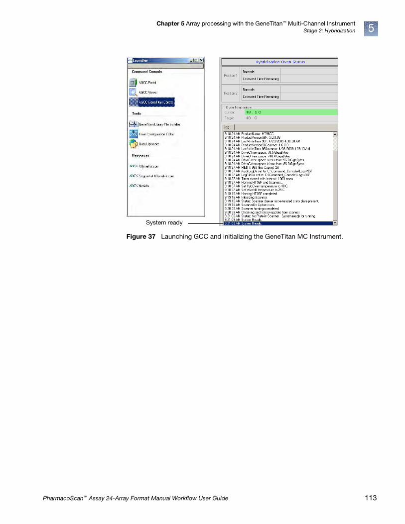
Chapter 5 Array processing with the GeneTitan™ Multi-Channel InstrumentStage 2: Hybridization 5
Figure 37 Launching GCC and initializing the GeneTitan MC Instrument.
System ready
PharmacoScan™ Assay 24-Array Format Manual Workflow User Guide 113

Chapter 5 Array processing with the GeneTitan™ Multi-Channel InstrumentStage 2: Hybridization5
2. Select the System Setup tab (Figure 38).
3. Configure the software as follows:
c. Setup Option: Hyb-Wash-Scan
Other options available are described under ʺSetup options for array plate processingʺ on page 107.
d. Click Next.
Figure 38 System Setup tab and the information displayed in this pane.
Status: This field displays the actions that must be performed to prepare or unload the GeneTitan MC Instrument for the setup option that has been selected.
After each action you are instructed to click the Next button or to press the blinking blue Confirmation button located on the GeneTitan MC Instrument.
System Setup tab
Setup Option: The various options you can choose for processing Axiom array plates.
Workflow Steps: This field displays an overview of the user actions required to process an array plate based on the setup option selected.
Barcode: The array plate barcode. Can be scanned or entered manually.
Protocol Name: The protocol that GeneTitan MC Instrument runs. The list of protocols displayed is based on the first 6 digits of the array plate barcode. Only the protocols that are valid for the type of array plate loaded are displayed.
114 PharmacoScan™ Assay 24-Array Format Manual Workflow User Guide
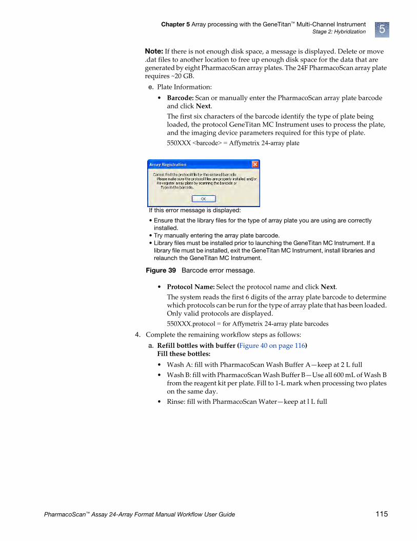
Chapter 5 Array processing with the GeneTitan™ Multi-Channel InstrumentStage 2: Hybridization 5
Note: If there is not enough disk space, a message is displayed. Delete or move .dat files to another location to free up enough disk space for the data that are generated by eight PharmacoScan array plates. The 24F PharmacoScan array plate requires ~20 GB.
e. Plate Information:
• Barcode: Scan or manually enter the PharmacoScan array plate barcode and click Next.
The first six characters of the barcode identify the type of plate being loaded, the protocol GeneTitan MC Instrument uses to process the plate, and the imaging device parameters required for this type of plate.
550XXX <barcode> = Affymetrix 24-array plate
• Protocol Name: Select the protocol name and click Next.
The system reads the first 6 digits of the array plate barcode to determine which protocols can be run for the type of array plate that has been loaded. Only valid protocols are displayed.
550XXX.protocol = for Affymetrix 24-array plate barcodes
4. Complete the remaining workflow steps as follows:
a. Refill bottles with buffer (Figure 40 on page 116)Fill these bottles:
• Wash A: fill with PharmacoScan Wash Buffer A—keep at 2 L full
• Wash B: fill with PharmacoScan Wash Buffer B—Use all 600 mL of Wash B from the reagent kit per plate. Fill to 1-L mark when processing two plates on the same day.
• Rinse: fill with PharmacoScan Water—keep at l L full
Figure 39 Barcode error message.
If this error message is displayed:
• Ensure that the library files for the type of array plate you are using are correctly installed.
• Try manually entering the array plate barcode.• Library files must be installed prior to launching the GeneTitan MC Instrument. If a
library file must be installed, exit the GeneTitan MC Instrument, install libraries and relaunch the GeneTitan MC Instrument.
PharmacoScan™ Assay 24-Array Format Manual Workflow User Guide 115

Chapter 5 Array processing with the GeneTitan™ Multi-Channel InstrumentStage 2: Hybridization5
f
b. Empty the waste bottle.
c. Press the Confirmation button on the GeneTitan MC Instrument to continue. A fluidics check is run (~1 minute).
IMPORTANT! • Always ensure that the GeneTitan bottles containing Wash A and Rinse
are above the 50% mark when setting up the system to process an array plate. All 600 mL of the Wash buffer B from the PharmacoScan Reagent Kit 4x24 Reactions should be emptied into the GeneTitan Wash B bottle when setting up the system to process a plate. This ensures that the GeneTitan Wash B bottle is filled to more than the requisite 35% of Wash B bottle volume. Also, do not overfill the bottles. Fill Wash Buffer B and Water bottles to the 1-L mark only. Wash A keep at 2 L. We strongly recommend refilling these bottles every time you are prompted to do so.
If the volume in any of these bottles becomes too low during a run, a message is displayed (see Chapter 6, ʺTroubleshootingʺ on page 140). However, even if you fill the bottle at this time, the instrument may not be able to successfully complete the step that was in progress.
• Wash B: if you intend to load two array plates on the same day, fill the Wash B bottle to the 1-L mark (use both bottles from the PharmacoScan Reagent Kit).
Figure 40 Example of the remaining workflow steps
Workflow step
Specific instructions for each workflow step
116 PharmacoScan™ Assay 24-Array Format Manual Workflow User Guide
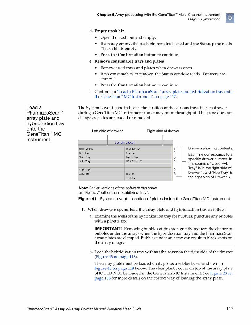
Chapter 5 Array processing with the GeneTitan™ Multi-Channel InstrumentStage 2: Hybridization 5
d. Empty trash bin
• Open the trash bin and empty.
• If already empty, the trash bin remains locked and the Status pane reads “Trash bin is empty.”
• Press the Confirmation button to continue.
e. Remove consumable trays and plates
• Remove used trays and plates when drawers open.
• If no consumables to remove, the Status window reads “Drawers are empty.”
• Press the Confirmation button to continue.
f. Continue to ʺLoad a PharmacoScan™ array plate and hybridization tray onto the GeneTitan™ MC Instrumentʺ on page 117.
Load a PharmacoScan™ array plate and hybridization tray onto the GeneTitan™ MC Instrument
The System Layout pane indicates the position of the various trays in each drawer during a GeneTitan MC Instrument run at maximum throughput. This pane does not change as plates are loaded or removed.
1. When drawer 6 opens, load the array plate and hybridization tray as follows:
a. Examine the wells of the hybridization tray for bubbles; puncture any bubbles with a pipette tip.
b. Load the hybridization tray without the cover on the right side of the drawer (Figure 43 on page 118).
The array plate must be loaded on its protective blue base, as shown in Figure 43 on page 118 below. The clear plastic cover on top of the array plate SHOULD NOT be loaded in the GeneTitan MC Instrument. See Figure 29 on page 103 for more details on the correct way of loading the array plate.
Figure 41 System Layout—location of plates inside the GeneTitan MC Instrument
IMPORTANT! Removing bubbles at this step greatly reduces the chance of bubbles under the arrays when the hybridization tray and the PharmacoScan array plates are clamped. Bubbles under an array can result in black spots on the array image.
Drawers showing contents.
Each line corresponds to a specific drawer number. In this example “Used Hyb Tray” is in the right side of Drawer 1, and “Hyb Tray” is the right side of Drawer 6.
Note: Earlier versions of the software can show as “Fix Tray” rather than “Stabilizing Tray”.
Right side of drawerLeft side of drawer
1234
56
PharmacoScan™ Assay 24-Array Format Manual Workflow User Guide 117
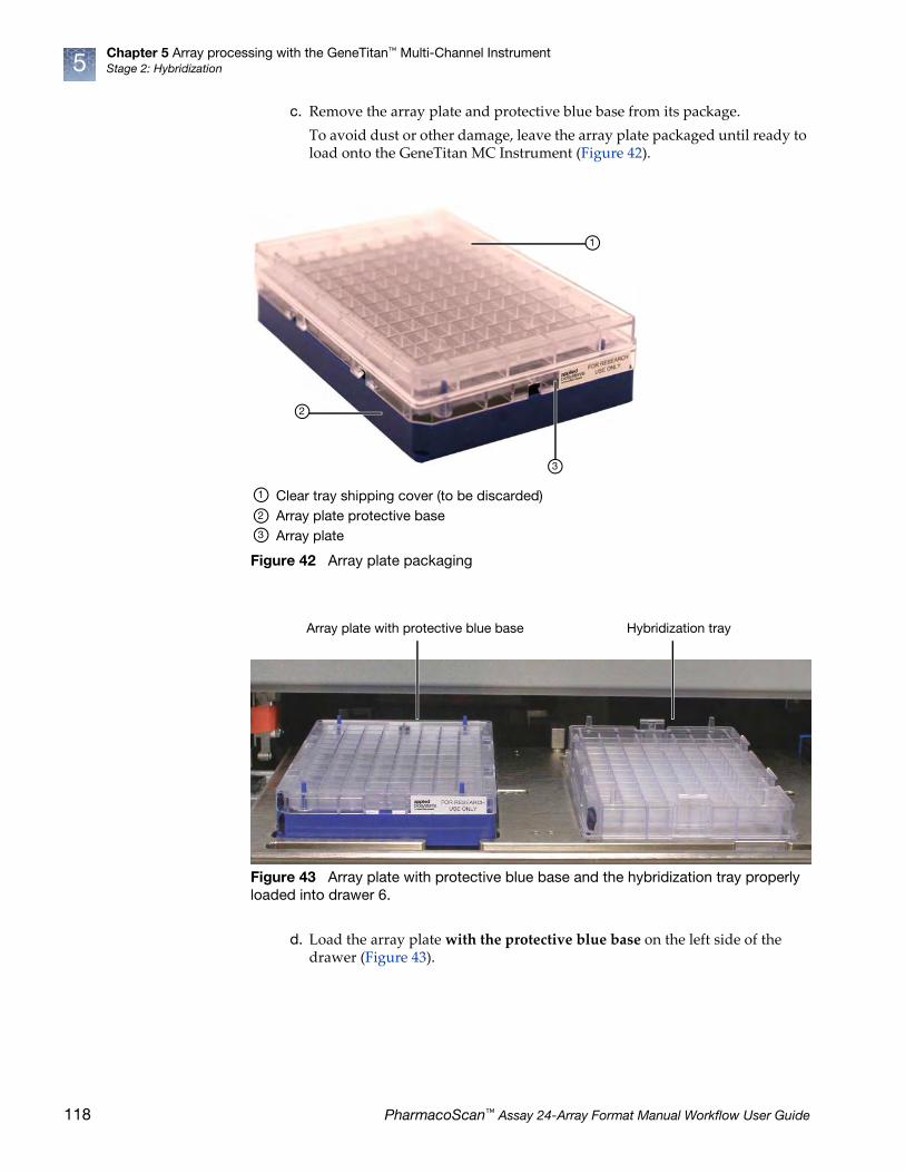
Chapter 5 Array processing with the GeneTitan™ Multi-Channel InstrumentStage 2: Hybridization5
c. Remove the array plate and protective blue base from its package.
To avoid dust or other damage, leave the array plate packaged until ready to load onto the GeneTitan MC Instrument (Figure 42).
d. Load the array plate with the protective blue base on the left side of the drawer (Figure 43).
Figure 42 Array plate packaging
Figure 43 Array plate with protective blue base and the hybridization tray properly loaded into drawer 6.
1
2
3
Clear tray shipping cover (to be discarded)Array plate protective baseArray plate
1
2
3
Array plate with protective blue base Hybridization tray
118 PharmacoScan™ Assay 24-Array Format Manual Workflow User Guide

Chapter 5 Array processing with the GeneTitan™ Multi-Channel InstrumentStage 2: Hybridization 5
e. Press the Confirmation button.
When you load the array plate on the left side of the drawer: The internal bar code reader reads the barcode of the array plate and compares it with the barcode and the plate type specified in the Barcode field and Plate Type field on the Setup page. If the information is correct, the application allows you to proceed to the next step. If the instrument is unable to read the barcode, it pushes the tray out and a message window (Figure 44) instructs you to load the correct plate with the proper orientation into the instrument (Figure 43).
• Click OK to retry and check the loading of the array plate; or
• Click Skip if the instrument has problems reading the barcode and after verifying that the trays have been placed in the proper orientation.
CAUTION! The notched corner of each plate, cover and tray must be aligned. When loading onto the GeneTitan MC Instrument, the notched edge plates, covers and trays must be aligned as indicated by the Tray Alignment guide in the drawer (Figure 43). The error message shown may be displayed. Plate barcodes must face the internal barcode reader (back of the drawer). Improper tray positioning can cause the GeneTitan MC Instrument to crash, and can result in substantial damage to the instrument and loss of samples.
Figure 44 Barcode error message
IMPORTANT! Do not install a three-plate stack of trays. Confirm that you have removed the clear plastic shipping cover as shown in Figure 29 on page 103.
PharmacoScan™ Assay 24-Array Format Manual Workflow User Guide 119

Chapter 5 Array processing with the GeneTitan™ Multi-Channel InstrumentStage 2: Hybridization5
f. Select the arrays to scan (instructions in Figure 45).
By default, all arrays are selected.
2. Click Next, then click OK to begin processing the samples.
The array plate is placed on top of the hybridization tray and clamped (now referred to as the plate stack).
The software starts the process for clamping the array plate to the hybridization tray. Press OK on the dialog shown in Figure 46 and wait for the drawer to open before retrieving the array plate and hybridization tray combo for inspection. The sandwich of the array plate and hybridization tray needs to be manually inspected before the array processing can begin. After clamping is complete the dialog shown in Figure 47 on page 121 is displayed. If you do not press OK in Figure 46 the dialog box goes away without intervention and Figure 47 on page 121 is displayed.
Figure 45 Selecting which arrays to scan on an array plate
Figure 46
Message in Status window.
Default – all arrays are selected
Single array - click one box
Multiple arrays:
– Click one box– Hold down the Ctrl key– Click another box in the same column
Group of arrays:
– Click one box– Hold down the Shift key– Click and drag the mouse
120 PharmacoScan™ Assay 24-Array Format Manual Workflow User Guide

Chapter 5 Array processing with the GeneTitan™ Multi-Channel InstrumentStage 2: Hybridization 5
3. When drawer 6 opens and the prompt in Figure 47 is displayed:
a. Remove the plate stack and gently press the two plates together at each clamping point.
b. Listen for a clicking sound which indicates that the plates are now clamped. No clicking sound indicates the plates are already clamped (See Figure 48 for an example of a array plate hybridization tray sandwich).
Figure 47 Location of camping points on the array plate and hybridization tray.
Clamping points on an PharmacoScan array plate and hybridization tray
Array plate
Hybridization tray
Notched corners
1 1
1 1
1 1
1
2
3
4
2
3
4
PharmacoScan™ Assay 24-Array Format Manual Workflow User Guide 121

Chapter 5 Array processing with the GeneTitan™ Multi-Channel InstrumentStage 2: Hybridization5
c. Inspect the bottom of the plate stack for bubbles under the arrays—do NOT invert the plates.
d. If bubbles are present, gently tap the plate until the bubbles move out from under the arrays—do NOT unclamp the plate stack.
e. Return the plate stack to the drawer, and press the Confirmation button to proceed.
The message in Figure 49 may be displayed again if plate orientation is incorrect or if the hybridization tray barcode cannot be read. Click OK to proceed.
Figure 48 Array plate hyb sandwich
Figure 49 Verification message
122 PharmacoScan™ Assay 24-Array Format Manual Workflow User Guide

Chapter 5 Array processing with the GeneTitan™ Multi-Channel InstrumentStage 2: Hybridization 5
Load a second PharmacoScan™ array plate and hybridization tray onto the GeneTitan™ MC Instrument
When you can load a second array plate and hybridization trayAfter processing begins, you have a specific period of time during which you can load another PharmacoScan array plate and hybridization tray. This period of time is displayed above the Hyb Oven Status pane (Figure 50). You cannot load another hybridization tray before or after this period of time.
Note: While the first plate is in the oven, you can load another plate if the time spacing requirement is met. This is to ensure that the second plate does not have to wait for system resources in its workflow. The time spacing is roughly equal to the longer of the wash-stain or scan time of the first plate.
IMPORTANT! You must load the next array plate and hybridization tray during the period of time displayed above the Hyb Oven Status. You cannot load another hybridization tray before or after this period of time. You must wait until the current process is finished which results in disruption of the eight plate workflow and fewer than eight plates processed per week.
Figure 50 Loading a second hybridization tray and Hybridization Oven Status information
This pane displays the period of time during which another array plate and hybridization tray can be loaded.
Additional plates cannot be loaded before or after this period of time while the instrument is operating.
In this example the system is currently available.
Position of plate stack in the hybridization oven. Only one plate being processed in this figure. As such, position 2 is blank.
Position 1 - left side of oven
Position 2 - right side of oven
Oven Temperature.
• Green indicates the current oven temperature is within the target temperature range.
• Yellow indicates oven temperature outside of target temperature range.
1
2
3
1
2
3
PharmacoScan™ Assay 24-Array Format Manual Workflow User Guide 123
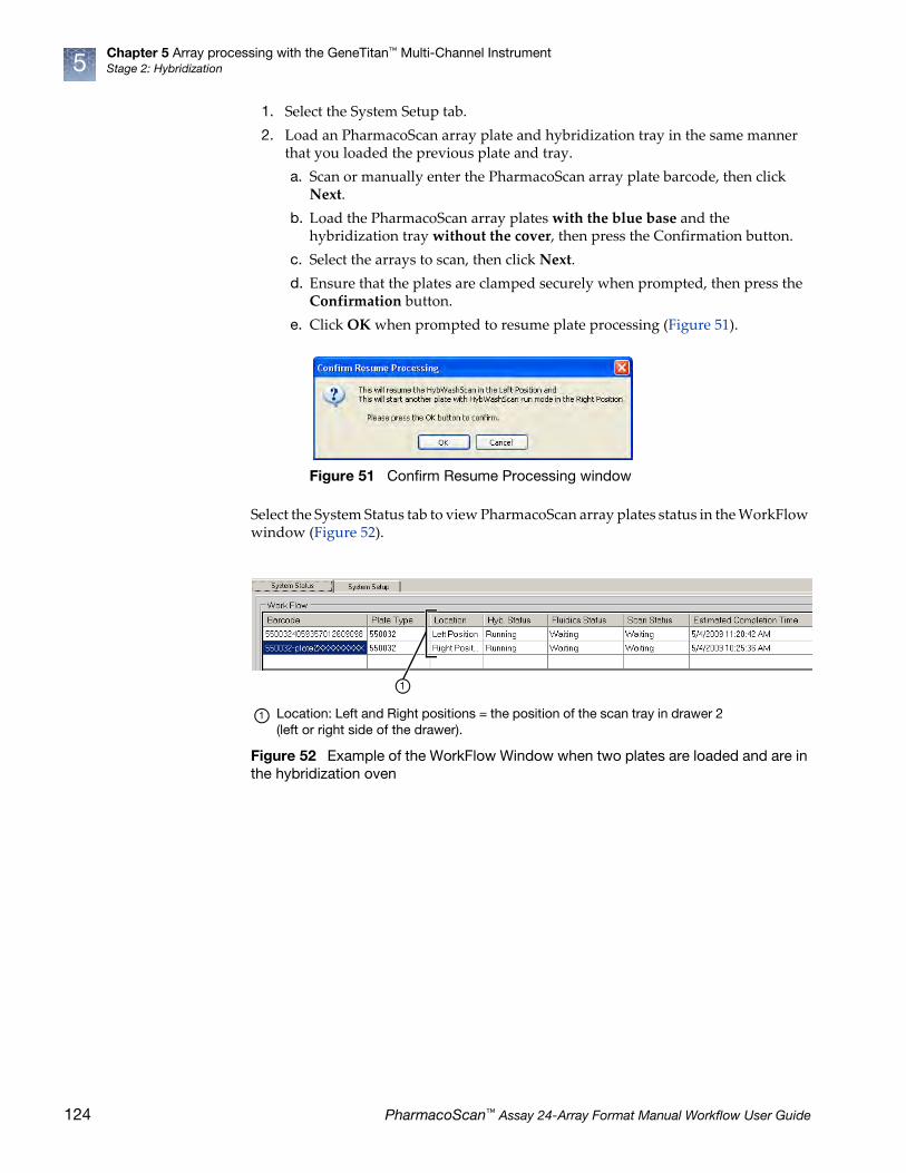
Chapter 5 Array processing with the GeneTitan™ Multi-Channel InstrumentStage 2: Hybridization5
1. Select the System Setup tab.
2. Load an PharmacoScan array plate and hybridization tray in the same manner that you loaded the previous plate and tray.
a. Scan or manually enter the PharmacoScan array plate barcode, then click Next.
b. Load the PharmacoScan array plates with the blue base and the hybridization tray without the cover, then press the Confirmation button.
c. Select the arrays to scan, then click Next.
d. Ensure that the plates are clamped securely when prompted, then press the Confirmation button.
e. Click OK when prompted to resume plate processing (Figure 51).
Select the System Status tab to view PharmacoScan array plates status in the WorkFlow window (Figure 52).
Figure 51 Confirm Resume Processing window
Figure 52 Example of the WorkFlow Window when two plates are loaded and are in the hybridization oven
Location: Left and Right positions = the position of the scan tray in drawer 2 (left or right side of the drawer).
1
1
124 PharmacoScan™ Assay 24-Array Format Manual Workflow User Guide

Chapter 5 Array processing with the GeneTitan™ Multi-Channel InstrumentStatus window prompts and actions required 5
Status window prompts and actions required
As a part of normal GeneTitan MC Instrument operations you may see the following status prompts. Table 47, Table 48 Table 49 and explains the necessary actions required. Table 50 and Table 51 explain possible barcode error messages and the necessary action required.
Table 47 Refilling buffer bottles and emptying the waste bottle
Status window prompt Action required Receptacle – reagent
Buffer bottles have been depressurized. Refill buffer into the bottles. Empty the waste bottle.
• Replenish the fluid in Wash Bottles A and B, and the Rinse bottle1.
• Empty the Waste Bottle.• Press the Confirmation button to
continue.
• Wash Bottle A: fill with Axiom Wash Buffer A up to 2 L.
• Wash Bottle B: fill with Axiom Wash Buffer B to the 1-L mark.
• Rinse: fill with Axiom Water to the 1-L mark.
Do not overfill these bottles.
1 Every time you are prompted to refill the buffer bottles, the system runs a fluidics check (duration ~1 minute).
Table 48 Emptying the trash bin
Status window prompt Action required Receptacle – Reagent
Empty trash bin • Open and empty the trash bin.• Press the Confirmation button to
continue.
Note: If the trash bin is empty, you are not be able to open it. Continue the process by pressing the blue confirmation button.
—
Table 49 Selecting which arrays to scan
Status window prompt Action required Reagent and receptacle
Select arrays to scan • Accept the default (all arrays selected) if appropriate. Otherwise, select the arrays to be scanned.
• Click Next, then click OK to start processing.
—
PharmacoScan™ Assay 24-Array Format Manual Workflow User Guide 125
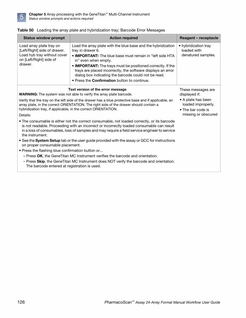
Chapter 5 Array processing with the GeneTitan™ Multi-Channel InstrumentStatus window prompts and actions required5
Table 50 Loading the array plate and hybridization tray; Barcode Error Messages
Status window prompt Action required Reagent – receptacle
Load array plate tray on [Left/Right] side of drawer. Load hyb tray without cover on [Left/Right] side of drawer.
Load the array plate with the blue base and the hybridization tray in drawer 6.• IMPORTANT: The blue base must remain in “left side HTA
in” even when empty.• IMPORTANT: The trays must be positioned correctly. If the
trays are placed incorrectly, the software displays an error dialog box indicating the barcode could not be read.
• Press the Confirmation button to continue.
• hybridization tray loaded with denatured samples.
These messages are displayed if:• A plate has been
loaded improperly.• The bar code is
missing or obscured
Text version of the error messageWARNING: The system was not able to verify the array plate barcode.
Verify that the tray on the left side of the drawer has a blue protective base and if applicable, an array plate, in the correct ORIENTATION. The right side of the drawer should contain a hybridization tray, if applicable, in the correct ORIENTATION.
Details:
• The consumable is either not the correct consumable, not loaded correctly, or its barcode is not readable. Proceeding with an incorrect or incorrectly loaded consumable can result in a loss of consumables, loss of samples and may require a field service engineer to service the instrument.
• See the System Setup tab or the user guide provided with the assay or GCC for instructions on proper consumable placement.
• Press the flashing blue confirmation button or...– Press OK, the GeneTitan MC Instrument verifies the barcode and orientation.– Press Skip, the GeneTitan MC Instrument does NOT verify the barcode and orientation.
The barcode entered at registration is used.
126 PharmacoScan™ Assay 24-Array Format Manual Workflow User Guide
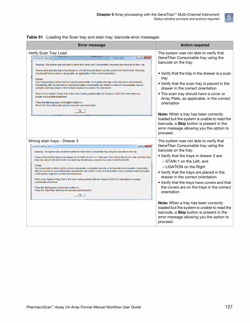
Chapter 5 Array processing with the GeneTitan™ Multi-Channel InstrumentStatus window prompts and actions required 5
Table 51 Loading the Scan tray and stain tray; barcode error messages
Error message Action required
Verify Scan Tray Load The system was not able to verify that GeneTitan Consumable tray using the barcode on the tray.
• Verify that the tray in the drawer is a scan tray
• Verify that the scan tray is placed in the drawer in the correct orientation
• The scan tray should have a cover or Array Plate, as applicable, in the correct orientation
Note: When a tray has been correctly loaded but the system is unable to read the barcode, a Skip button is present in the error message allowing you the option to proceed.
Wrong stain trays - Drawer 3 The system was not able to verify that GeneTitan Consumable tray using the barcode on the tray. • Verify that the trays in drawer 3 are:
– STAIN 1 on the Left, and – LIGATION on the Right
• Verify that the trays are placed in the drawer in the correct orientation
• Verify that the trays have covers and that the covers are on the trays in the correct orientation
Note: When a tray has been correctly loaded but the system is unable to read the barcode, a Skip button is present in the error message allowing you the option to proceed.
PharmacoScan™ Assay 24-Array Format Manual Workflow User Guide 127

Chapter 5 Array processing with the GeneTitan™ Multi-Channel InstrumentStatus window prompts and actions required5
Wrong stain trays - Drawer 4 The system detected the wrong GeneTitan Consumable Tray using the barcode on the tray.
• Verify that the trays in drawer 4 are: – STAIN 2 on the Left, and – STABILIZING on the Right
• Verify that the trays are placed in the drawer in the correct orientation
Note: When a tray has been correctly loaded but the system is unable to read the barcode, a Skip button is present in the error message allowing you the option to proceed.
Wrong stain tray - Drawer 5 The system detected the wrong GeneTitan Consumable Tray using the barcode on the tray.
• Verify that the tray in drawer 5 has: – STAIN1 on the Left
• Verify that the tray is placed in the drawer in the correct orientation
Note: When a tray has been correctly loaded but the system is unable to read the barcode, a Skip button is present in the error message allowing you the option to proceed.
Table 51 Loading the Scan tray and stain tray; barcode error messages
Error message Action required
128 PharmacoScan™ Assay 24-Array Format Manual Workflow User Guide

Chapter 5 Array processing with the GeneTitan™ Multi-Channel InstrumentStage 3: Ligate, wash, stain and scan 5
Stage 3: Ligate, wash, stain and scan
Equipment, consumables, and reagents required
Scan tray with Axiom Hold Buffer• Cover the tray by orienting the notched corner of the cover over the notched edge
of the tray and leave on the bench top (no need to protect from light; Figure 53).
CAUTION! Do not remove the scan tray from its protective black base. Leave the scan tray in the base until loaded onto the GeneTitan MC Instrument. When handling the scan tray, the bottom glass surface of the tray should not be touched.
Figure 53 The scan tray with cover on the black base
Notched corner of the cover is aligned with the notched corner of the scan tray.
Always leave the scan tray in its protective black base.
1
1
2
2
PharmacoScan™ Assay 24-Array Format Manual Workflow User Guide 129

Chapter 5 Array processing with the GeneTitan™ Multi-Channel InstrumentStage 3: Ligate, wash, stain and scan5
Proper installation of the GeneTitan™ tray consumables
It is very important that you install the GeneTitan tray consumables in the proper orientation. The barcode faces into the instrument.
Note: The instrument control software displays a warning if it detects a problem during the fluid dispense operations. The filters in the GeneTitan Wash A, Wash B and DI Water bottles should be replaced if the software displays such a warning. See Appendix E, ʺGeneTitan™ Multi-Channel Instrument careʺ on page 168 for the message displayed to the user and the procedure for replacing the filters.
Figure 54 You must rotate and install the trays so that the barcode faces into the instrument.
Figure 55 The proper installation of the GeneTitan tray consumables (the image shows the stain tray and the stain tray cover as an example)
Notch(This faces out and left)
Barcode(This faces BACK TO THE REAR of the instrument)
Turn the tray and cover combo so that the barcode faces BACK AND INTO the instrument and the notch faces OUT AND TO THE LEFT.
FRONT(OF INSTRUMENT FACING YOU)
Notch faces out and left.“For Research Use Only” faces out.
Barcode faces in and back.
FOR RESEARCHUSE ONLY
FOR RESEARCHUSE ONLY
130 PharmacoScan™ Assay 24-Array Format Manual Workflow User Guide

Chapter 5 Array processing with the GeneTitan™ Multi-Channel InstrumentStage 3: Ligate, wash, stain and scan 5
Load tray consumables onto the GeneTitan™ MC Instrument
When hybridization of an PharmacoScan array plate has finished, a message (Figure 56) alerts you to resume the workflow setup. Press OK and the software takes you directly back to the System Setup tab.
This prompt to continue into reagent load (Figure 56) occurs when the hyb is complete. “Estimated Time Remaining” displayed under “Hybridization Oven Status” may display a time remaining of 0 to 30 minutes when the prompt occurs.
The GeneTitan MC Instrument allows reagent load to take place after either:
• the estimated time counts down to zero or
• the actual real world hyb time (as indicated by the computer clock) indicates the hyb is complete.
Note: The time estimate displayed on some systems may lag due to high CPU utilization. The GeneTitan MC Instrument allows the workflow to synchronize with the system clock to compensate for this situation during the final half hour of the hyb time estimate. When this prompt to resume reagent loading is displayed to the user there is no need to wait for the estimated time to count down to zero.
Follow the prompts displayed to continue with staining, ligation, fixing and scanning.
1. Follow the prompts in the Status window.
a. Wash Bottles A and B, and the Rinse Bottle—refill as necessary (the system primes itself again); Waste bottle—empty if necessary.
Wash bottle A—2 L. Wash Bottle B and Rinse Bottle—fill to 1-L mark only.
b. Empty the trash bin.
c. Remove consumable trays and plates as instructed, except for the blue base.
Leave the blue array plate base in drawer 6 even though the base is empty.
2. Load consumable trays and plates as follows:
a. Follow the prompts in the Status window (load sequence and prompts in Table 52).
b. After loaded, examine each cover for droplets of liquid.
c. If any liquid is present, remove the tray, clean the cover and top of the tray with Kimwipes laboratory tissue, and reload the tray.
Figure 56 The Resume Workflow Setup message
CAUTION! • Orient trays as indicated by the guide inside the drawer. Improper
orientation may cause the run to fail.
• Remove the protective black base from the scan tray immediately prior to loading Figure 57 on page 133).
• Examine each cover for droplets of liquid after loading. Liquid on the cover can result in capillary phenomenon. As a result, the tray may stick to the cover and be lifted out of place inside the instrument.
PharmacoScan™ Assay 24-Array Format Manual Workflow User Guide 131

Chapter 5 Array processing with the GeneTitan™ Multi-Channel InstrumentStage 3: Ligate, wash, stain and scan5
Table 52 Sequence for Loading the Trays with Reagents
Loading sequence by drawer number
Left Right
Note: If the software is unable to verify the barcode on the scan tray and the scan tray cover, the software displays the following error message
2 Scan tray with cover—do not load the protective black base(left side of drawer as indicated in Status window)
Figure 57 on page 133
3 Stain Tray with Stain 1 Ligation Tray
Figure 58 on page 134
4 Stain Tray with Stain 2 Stbl Tray
Figure 59 on page 135
132 PharmacoScan™ Assay 24-Array Format Manual Workflow User Guide

Chapter 5 Array processing with the GeneTitan™ Multi-Channel InstrumentStage 3: Ligate, wash, stain and scan 5
5 Stain Tray with Stain 1 Empty
Table 52 Sequence for Loading the Trays with Reagents (Continued)
Loading sequence by drawer number
Left Right
Figure 57 Scan tray loaded in drawer 2.
Do NOT load the protective black base packaged with the scan tray.
PharmacoScan™ Assay 24-Array Format Manual Workflow User Guide 133

Chapter 5 Array processing with the GeneTitan™ Multi-Channel InstrumentStage 3: Ligate, wash, stain and scan5
IMPORTANT! When you load the plates, or trays, insert them under the tabs, or fingers, that may protrude into the stage. Confirm that the tray is not resting on these fingers.
1
1
1 Tab or “finger” in GeneTitan drawer.
Figure 58 Stain 1 tray (left) and ligation tray (right) loaded in drawer 3.
134 PharmacoScan™ Assay 24-Array Format Manual Workflow User Guide

Chapter 5 Array processing with the GeneTitan™ Multi-Channel InstrumentStage 3: Ligate, wash, stain and scan 5
3. In the window shown in Figure 61, click Yes to load another PharmacoScan array plate and hybridization tray.
Figure 59 Stain 2 tray (left) and Stbl tray (right) loaded in drawer 4.
Figure 60 Stain 1 tray loaded in drawer 5
Figure 61 Prompt asking to load another plate. Right or left position determined by the position of PharmacoScan array plates already in the GeneTitan MC Instrument.
PharmacoScan™ Assay 24-Array Format Manual Workflow User Guide 135

Chapter 5 Array processing with the GeneTitan™ Multi-Channel InstrumentStage 3: Ligate, wash, stain and scan5
4. Follow the prompts and:
a. Setup Option: select Setup Another Run, then click Next.
b. Scan or manually enter the PharmacoScan array plate barcode, then click Next.
c. Select a protocol, then click Next.
d. When drawer 6 opens:
• Remove the blue cover from the previous PharmacoScan array plate.
• Load a new PharmacoScan array plate and new blue base on the left; load a new hybridization tray on the right.
• Press the Confirmation button.
e. Click OK when prompted (Figure 62).
f. When drawer 6 opens, confirm that the plate stack is securely clamped, then press the Confirmation button.
When processing resumes:
1. The plate stack which has finished hybridization is moved from the Hyb oven to drawer 1 temporarily and then moved to the unclamp station after step 2 (it remains clamped).
2. The plate stack in drawer 6 is moved to the Hyb oven.
3. The plate is moved to the unclamped station.
4. The plate stack in the unclamp area is unclamped and moved into the fluidics area.
Note: At the end of a Hyb-Wash-Scan run, all plate and tray covers and the fixing tray cover should be in the trash.
Figure 63 is an example of how the System Status Workflow window appears when three PharmacoScan array plates are being processed.
Figure 62 Confirm Resume Processing message.
136 PharmacoScan™ Assay 24-Array Format Manual Workflow User Guide
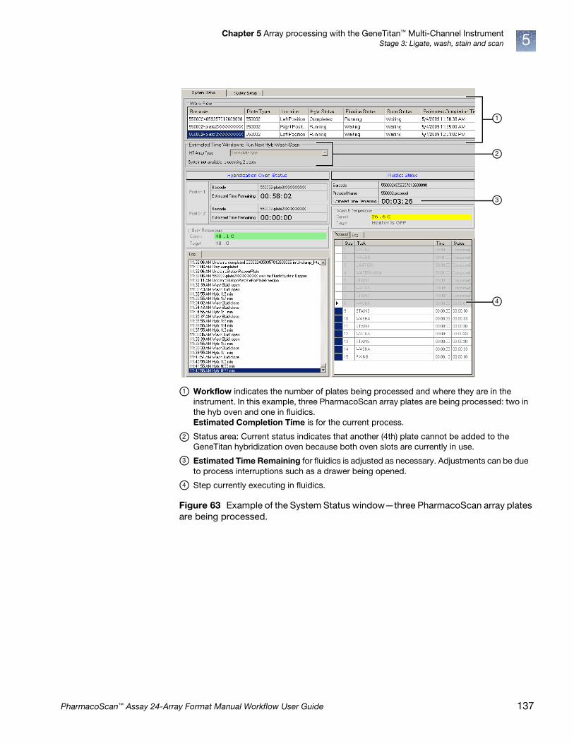
Chapter 5 Array processing with the GeneTitan™ Multi-Channel InstrumentStage 3: Ligate, wash, stain and scan 5
Figure 63 Example of the System Status window—three PharmacoScan array plates are being processed.
Workflow indicates the number of plates being processed and where they are in the instrument. In this example, three PharmacoScan array plates are being processed: two in the hyb oven and one in fluidics.Estimated Completion Time is for the current process.
Status area: Current status indicates that another (4th) plate cannot be added to the GeneTitan hybridization oven because both oven slots are currently in use.
Estimated Time Remaining for fluidics is adjusted as necessary. Adjustments can be due to process interruptions such as a drawer being opened.
Step currently executing in fluidics.
1
1
2
2
3
4
3
4
PharmacoScan™ Assay 24-Array Format Manual Workflow User Guide 137

Chapter 5 Array processing with the GeneTitan™ Multi-Channel InstrumentContinue the workflow5
Continue the workflow
After a plate has gone through the fluidics stage of the process, it is moved to the imaging device.
When the scanning process begins, the window shown in Figure 64 is displayed. This window must remain open while PharmacoScan array plates are being scanned.
CAUTION!
• The Scan Control window must remain open while PharmacoScan array plates are being scanned. Closing this window halts the scanning process. You can minimize this window if necessary without creating any interference to the imaging.
• Do not manually, or through the GCC transfer utility, move any data associated with the current plate that is being processed/scanned. Transferring data dramatically slows scanning and can cause the computer to freeze.
Figure 64 Scan Control window
138 PharmacoScan™ Assay 24-Array Format Manual Workflow User Guide
Chapter 5 Array processing with the GeneTitan™ Multi-Channel InstrumentShut down the GeneTitan™ MC Instrument 5
Shut down the GeneTitan™ MC Instrument
This procedure assumes that all of the PharmacoScan array plates loaded onto the GeneTitan MC Instrument have been processed.
1. On the System Setup page, open the Setup Options drop-down menu and select Unload Plates.
2. Unload all of the consumables as prompted.
3. Power off the GeneTitan MC Instrument by opening Tools Shutdown from the menu.
4. Exit the GCC software if it does not close automatically.
Note: If the instrument is processing an array plate, the software does not allow you to shut down the system.
WARNING! Do not attempt to shut down the GeneTitan MC Instrument while array plates are being processed.
PharmacoScan™ Assay 24-Array Format Manual Workflow User Guide 139
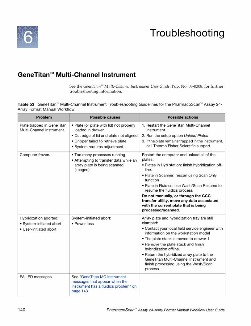
6 Troubleshooting
GeneTitan™ Multi-Channel Instrument
See the GeneTitan™ Multi-Channel Instrument User Guide, Pub. No. 08-0308, for further troubleshooting information.
Table 53 GeneTitan™ Multi-Channel Instrument Troubleshooting Guidelines for the PharmacoScan™ Assay 24-Array Format Manual Workflow
Problem Possible causes Possible actions
Plate trapped in GeneTitan Multi-Channel Instrument.
• Plate (or plate with lid) not properly loaded in drawer.
• Cut edge of lid and plate not aligned.• Gripper failed to retrieve plate.• System requires adjustment.
1. Restart the GeneTitan Multi-Channel Instrument.
2. Run the setup option Unload Plates3. If the plate remains trapped in the instrument,
call Thermo Fisher Scientific support.
Computer frozen. • Too many processes running• Attempting to transfer data while an
array plate is being scanned (imaged).
Restart the computer and unload all of the plates.• Plates in Hyb station: finish hybridization off-
line.• Plate in Scanner: rescan using Scan Only
function• Plate in Fluidics: use Wash/Scan Resume to
resume the fluidics processDo not manually, or through the GCC transfer utility, move any data associated with the current plate that is being processed/scanned.
Hybridization aborted:• System-initiated abort• User-initiated abort
System-initiated abort:• Power loss
Array plate and hybridization tray are still clamped:• Contact your local field service engineer with
information on the workstation model• The plate stack is moved to drawer 1.• Remove the plate stack and finish
hybridization offline.• Return the hybridized array plate to the
GeneTitan Multi-Channel Instrument and finish processing using the Wash/Scan process.
FAILED messages See "GeneTitan MC Instrument messages that appear when the instrument has a fluidics problem" on page 143
140 PharmacoScan™ Assay 24-Array Format Manual Workflow User Guide

Chapter 6 TroubleshootingGeneTitan™ Multi-Channel Instrument 6
Miscellaneous messages
FLUIDIC DIAGNOSTIC messages
See "Fluidic diagnostic messages" on page 143.
Fluidics aborted:• System-initiated abort• User-initiated abort
System-initiated abort:• Power loss
User-initiated abort:• Incorrect protocol selected
Follow the recommendations and instructions under "Wash/Scan Resume" on page 147.
Table 53 GeneTitan™ Multi-Channel Instrument Troubleshooting Guidelines for the PharmacoScan™ Assay 24-Array Format Manual Workflow
Problem Possible causes Possible actions
Table 54 Miscellaneous messages and recommended actions
Message and recommended action
Indicates that an item is in the gripper, and normal startup of the GeneTitan Multi-Channel Instrument is not possible. The item must be removed from the instrument before you can begin processing array plates.
Recommendation: click Yes.If you click No, nothing occurs. Homing does not complete and you are not able to use the system.The item held by the gripper is moved to either:• Drawer 2—plates and trays• Trash Bin—coversThe drawer names reflect the location (left or Right) and the drawer number (1 through 6).Examples: Drawer2L_Hta_DOWN = Scan tray on left side of drawer 2HtaHyb = Clamped hybridization tray and array plateDrawer(n)L/R_Hta_DOWN where n is the drawer number and L or R to indicate the left or right side.The _Hta_ (second term) indicates the item held. An example is drawer1R_HtaHyb_DOWN indicating it is an array plate with a hybridization tray or Drawer2L_ScanHta_Pk_DOWN indicating it is an array plate with a scan tray
PharmacoScan™ Assay 24-Array Format Manual Workflow User Guide 141

Chapter 6 TroubleshootingGeneTitan™ Multi-Channel Instrument6
The drawer listed in the message is not fully closed. Manually push the drawer back into the instrument until it is fully closed. There are two stop positions with audible clicks; push until you hear the second click and the drawer is fully seated.
• Check that the array plate barcode has been entered correctly.
• Ensure that the library files required for the type of array plate you are using have been installed, and are installed in the correct directory.
• Restart the GeneTitan MC instrument control software after library files have been installed.
Table 54 Miscellaneous messages and recommended actions
Message and recommended action
142 PharmacoScan™ Assay 24-Array Format Manual Workflow User Guide
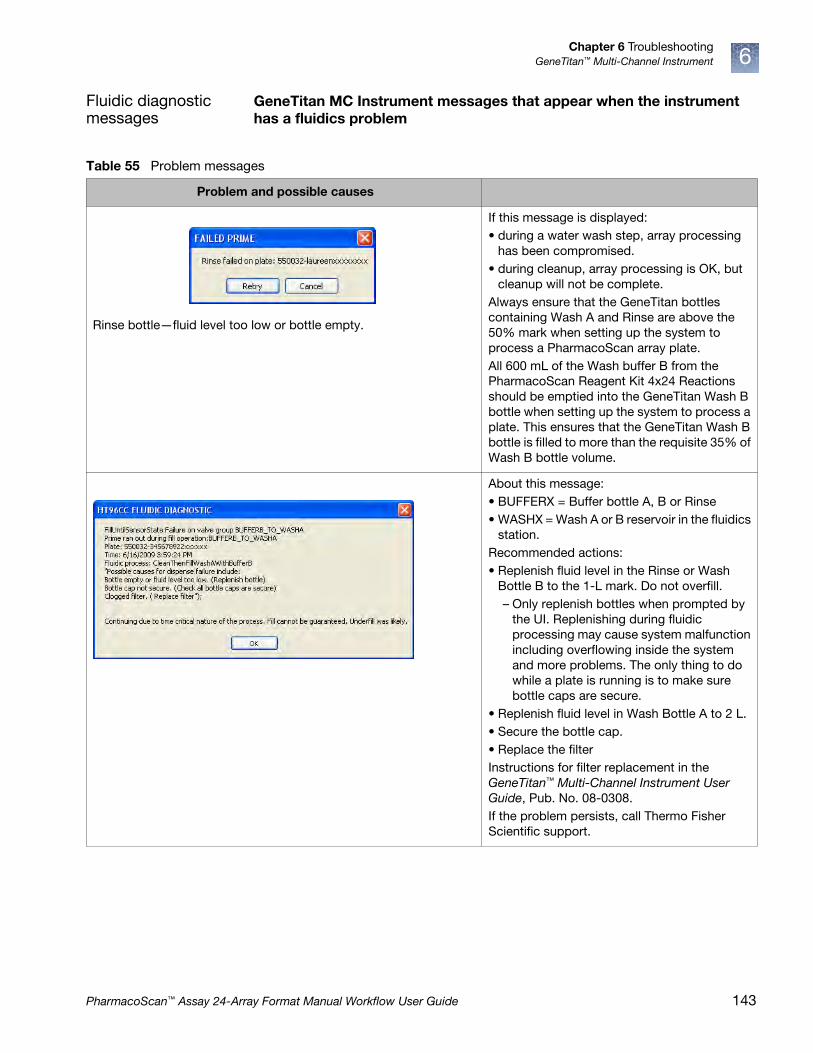
Chapter 6 TroubleshootingGeneTitan™ Multi-Channel Instrument 6
Fluidic diagnostic messages
GeneTitan MC Instrument messages that appear when the instrument has a fluidics problem
Table 55 Problem messages
Problem and possible causes
Rinse bottle—fluid level too low or bottle empty.
If this message is displayed:• during a water wash step, array processing
has been compromised.• during cleanup, array processing is OK, but
cleanup will not be complete.Always ensure that the GeneTitan bottles containing Wash A and Rinse are above the 50% mark when setting up the system to process a PharmacoScan array plate.All 600 mL of the Wash buffer B from the PharmacoScan Reagent Kit 4x24 Reactions should be emptied into the GeneTitan Wash B bottle when setting up the system to process a plate. This ensures that the GeneTitan Wash B bottle is filled to more than the requisite 35% of Wash B bottle volume.
About this message:• BUFFERX = Buffer bottle A, B or Rinse• WASHX = Wash A or B reservoir in the fluidics
station.Recommended actions:• Replenish fluid level in the Rinse or Wash
Bottle B to the 1-L mark. Do not overfill.– Only replenish bottles when prompted by
the UI. Replenishing during fluidic processing may cause system malfunction including overflowing inside the system and more problems. The only thing to do while a plate is running is to make sure bottle caps are secure.
• Replenish fluid level in Wash Bottle A to 2 L.• Secure the bottle cap.• Replace the filterInstructions for filter replacement in the GeneTitan™ Multi-Channel Instrument User Guide, Pub. No. 08-0308.If the problem persists, call Thermo Fisher Scientific support.
PharmacoScan™ Assay 24-Array Format Manual Workflow User Guide 143

Chapter 6 TroubleshootingGeneTitan™ Multi-Channel Instrument6
The typical cause is an unsecure bottle cap.If the failure is detected during priming, the instrument pauses and waits for the problem to be corrected.If the failure is detected during another process, and if the cause is a clogged filter, wait until the end of the run to replace the filter.Instructions for filter replacement in the GeneTitan™ Multi-Channel Instrument User Guide, Pub. No. 08-0308.
When the instrument experiences a loss in Clean Dry Air (CDA) pressure, the software displays the warning message.
When the pressure is detected again, a dialog message confirming the availability of CDA pressure is displayed.
Possible causesVerify that the facility CDA or the portable CDA compressor is in working condition. See the GeneTitan MC Site Preparation Guide for the portable compressor model that has been validated with the GeneTitan MC instrument.Contact your local field application specialist and notify the engineer about the error message.
Table 55 Problem messages (Continued)
Problem and possible causes
144 PharmacoScan™ Assay 24-Array Format Manual Workflow User Guide

Chapter 6 TroubleshootingGeneTitan™ Multi-Channel Instrument 6
Leak DetectedLeak checks are performed at application startup and any time a fluidic process (priming filling draining etc.) is performed. The leak detection is a hard-wired sensor which shuts off fluid flow without software control. Leaks are normally confined to the drip pan located inside the system.
Causes:• System malfunction• User killing the application using task
manager during a fill operation resulting in application exit without stopping flow.
Solution: Contact Thermo Fisher Scientific field support. The system cannot be used for any fluidic processing until this is resolved.
Leak Resolved This message is displayed when the leak is resolved (meaning the sensor LED is again lit up). If the original leak detected message was not acknowledged it is automatically removed from the GUI and replaced by the following message. It remains displayed until another leak is detected or the user acknowledges it by pressing OK. To resolve this issue complete the following tasks• Verify all internal and external tubing is
connected and clean• Verify wash reservoirs are clean• Verify all bottle caps are secure and that no
bottle cap is crimping a supply line.• Verify vacuum is working properly• Do not refill bottles or empty waste except
when prompted to by the GeneTitan application.
• Contact your facility group to ensure CDA is supplied to your GeneTitan system.
Contact an authorized Field Service Engineer to have the sensor adjusted or replaced if the problem persists even after correcting for the usual causes outlined above.
Table 55 Problem messages (Continued)
Problem and possible causes
PharmacoScan™ Assay 24-Array Format Manual Workflow User Guide 145

Chapter 6 TroubleshootingGeneTitan™ Multi-Channel Instrument6
Filter Error Messages The filters in the GeneTitan fluidics bottles (Wash A, Wash B and DI Water) need to be replaced when the filters are worn out. The software displays warning message boxes for the filter in each reagent bottle when it detects a problem or shows a trend of increased fill times during fluid fill operations.
If an error is detected as described above, then a message box titled “Filter Change Required” is displayed along with the information on the specific dispense operation. You should change all three filters when a warning is displayed for any one of the three filters.See the section "Replace the bottle filters" on page 170 in Appendix E.
Table 55 Problem messages (Continued)
Problem and possible causes
146 PharmacoScan™ Assay 24-Array Format Manual Workflow User Guide

Chapter 6 TroubleshootingGeneTitan™ Multi-Channel Instrument 6
Wash/Scan Resume
If a run is aborted during fluidics processing, the instrument places the aborted array plate into the scan tray. To restart this process, remove the PharmacoScan array plate from the scan tray and place it in its protective blue base.
The step at which the run was aborted can be identified by:
• Viewing the System Status window if you are aborting the last plate through the fluidics system.
• Initiating the resume process.
1. System Setup tab: Select Wash/Scan Resume
2. Follow the prompts to unload and reload all drawers.
The trays are loaded. It is up to you to determine whether or not to load fresh reagents or reuse the trays already in the GeneTitan Multi-Channel Instrument. Base your decision upon the step where the problem occurred.
To help ensure that the samples are processed correctly, we recommend that you:
1. Load new stain trays with fresh reagents.
2. Load a new scan tray.
We do not recommend the use of trays without reagents or holding buffers for steps that appear to have already executed.
Resume stepYou must select the step at which you wish to resume plate processing. You can select any step that has not yet been started.
For certain steps, you can enter a duration in seconds (even if the step requires 1 hour to run, you must enter the duration in seconds). You can set a step for less time than normal, but not for longer than the normal duration.
Abort a run • Abort can take up to three minutes if a plate is in the Fluidics station. Status window Abort Requested changes to Abort Completed.
• Clamped Array-Plate-Hybridization Tray sandwich aborted from the oven or from a drawer are moved to drawer 1.
• Proceed as follows:
– Use the Unload Plates option to remove the aborted plate(s).
– Start another run which forces an unload of the aborted plate(s)
System-initiated• Power interruption
• Plate loaded incorrectly
• Equipment malfunction
The system aborts the processing. Follow the instructions displayed in the user interface.
User-initiatedCan abort processing of individual array plates.
If multiple plates are being processed, the gripper may continue to process the remaining array plates.
PharmacoScan™ Assay 24-Array Format Manual Workflow User Guide 147

A Fragmentation quality control gelprotocol
Run a fragmentation quality control gel
Equipment required
E-Gels and reagents
Consumables
Table 56 Equipment required
Item Supplier Cat. No.
Gel imager Various —
Pipettes, single and multichannel P20, single channel P200
Various —
Plate centrifuge Various —
Vortex Various —
Table 57 E-Gel and reagents required
Item Supplier Cat. No.
Mother E-Base Device
Thermo Fisher Scientific
EB-M03
Daughter E-Base Device EB-D03
E-Gel® 48 4% agarose gels G8008-04
TrackIt™ 25 bp DNA Ladder 10488-022
TrackIt™ Cyan/Orange Loading Buffer 10482-028
Nuclease-free water 71786
Table 58 Consumables required
Item Supplier Cat. No.
Adhesive film – use one of the following:• MicroAmp Clear Adhesive Film• Microseal 'B' Film
Thermo Fisher Scientific Bio-Rad
4306311MSB1001
50 mL Conical Tube Various —
Pipette Tips Same brand as pipette —
148 PharmacoScan™ Assay 24-Array Format Manual Workflow User Guide

Appendix A Fragmentation quality control gel protocolRun a fragmentation quality control gel A
Dilute the TrackIt™ Cyan/Orange Loading Buffer and 25 bp ladder
The following recipe is for preparing a large batch of the Gel Diluent, a 1,000-fold dilution of the TrackIt Cyan-Orange Loading Buffer:
To dilute the TrackIt Cyan/Orange Loading Buffer (Cat. No. 10482-028, Thermo Fisher Scientific):
1. Add 50 µL of TrackIt Cyan/Orange Loading Buffer to 49.95 mL nuclease-free water.
Total volume 50 mL.
2. Vortex to mix well.
3. Store at room temperature.
To dilute the TrackIt 25bp Ladder (Cat. No. 10488-022, Thermo Fisher Scientific):
The following recipe is for preparing a 15-fold dilution of the Invitrogen TrackIt 25 bp DNA Ladder:
1. In a 1.7 mL microcentrifuge tube, add 6 µL of TrackIt 25 bp DNA Ladder to 84 µL nuclease-free water. Total volume: 90 µL.
2. Vortex tube to mix well. Pulse-spin to get droplets down.
Note: The recipe has enough volume to fill 4 marker wells of one E-Gel® 48 4% agarose gel. Scale up as needed if running multiple gels.
Run the fragmentation QC gel protocol
This protocol is based on running QC gels for 24 samples:
1. Tightly seal the Gel QC plate.
2. Vortex the plate for three seconds. Pulse-spin to get droplets down.
3. Connect an E-Base™ device(s) to an electrical outlet.
4. Push the Power/Prg button on each to ensure the program is in EG mode (not EP mode).
5. Take the gel out of the pouch and remove the combs.
6. Place the E-Gel® 48 gel into an E-Base unit.
7. Load 20 µL from each well of the Gel QC plate onto the gels.
8. Load 15 µL of diluted TrackIt 25 bp ladder into the marker wells (M).
9. Load 20 µL nuclease-free water into any unused wells.
10. Run the gels for 22 minutes.
11. Image the gel.
PharmacoScan™ Assay 24-Array Format Manual Workflow User Guide 149

Appendix A Fragmentation quality control gel protocolRun a fragmentation quality control gelA
Fragmentation QC gel images should look similar to the gel shown in Figure 65.
Figure 65 Example of a typical fragmentation QC E-gel
Fragments should fall between 25 bp and 125 bp.
25 bp ladder 25 bp ladder
125 bp
125 bp
25 bp
25 bp
150 PharmacoScan™ Assay 24-Array Format Manual Workflow User Guide

B Sample quantitation afterresuspension
Protocol for sample quantitation after resuspension
Equipment required The following equipment is required for this protocol.
Quantitate the diluted samples
During target prep, two plates of diluted samples are prepared: one for OD quantitation and one for a QC gel to check the fragmentation reaction.
For OD quantitation, readings should be taken at wavelengths of 260, 280, and 320 nm. See ̋ Suggested Protocol for OD Quantitation Using the DTX 880ʺ on page 153 for more information.
1. Launch the Multimode Analysis Software.
2. When the Protocol Selection List is displayed, select the appropriate protocol.
3. Right click the protocol and select Run the selected protocol.
4. In the Result Name field, enter your experiment name.
5. Click the Eject Plate Carrier icon.
6. Load the OD plate onto the DTX 880.
7. Click the Close Plate Carrier icon.
8. Click the Run the Selected Protocol icon at the bottom of the window.
When the protocol is finished running, a list of results is displayed. If you used the formula provided in this appendix, two XML files are generated. Open the ResultData file with Microsoft® Excel® to view and assess the OD readings. RawData file information is included in the ResultData file.
Table 59 Equipment required for sample quantitation after resuspension
Quantity Item
1 DTX 880 Multimode Detector with Genomic Filter Slide
PharmacoScan™ Assay 24-Array Format Manual Workflow User Guide 151

Appendix B Sample quantitation after resuspensionProtocol for sample quantitation after resuspensionB
Assess the OD readings
If using the formula provided in this appendix, the raw data is included in the final Result Data file. Figure 66 is an example of a Result Data file. Your OD readings should be similar to those displayed below.
OD Yield Assessment GuidelinesThe measurement of the yield of DNA after resuspension of the pellets is an important QC checkpoint in the PharmacoScan Assay 24-Array Format Manual Workflow. If the median yield for the plate is 1,000 µg DNA per sample:
• Pause the protocol.
• Assess each of the steps performed to that point to determine the possible source of the low yields.
This DNA yield corresponds to an A260 value of approximately 0.49 and an A260-A320 value of approximately 0.42.
Figure 66 Example of Result Data file with acceptable OD readings
152 PharmacoScan™ Assay 24-Array Format Manual Workflow User Guide

Appendix B Sample quantitation after resuspensionSuggested Protocol for OD Quantitation Using the DTX 880 B
Suggested Protocol for OD Quantitation Using the DTX 880
The formula suggested below requires six passes. The settings and formula are shown below.
Protocol Type—Analysis
General Settings—enter a name for the protocol
Figure 67 Protocol Type
Figure 68 General Settings
PharmacoScan™ Assay 24-Array Format Manual Workflow User Guide 153

Appendix B Sample quantitation after resuspensionSuggested Protocol for OD Quantitation Using the DTX 880B
Technique Type—select Absorbance
Labware—x_Abs_Greiner 96 UV clear std (96 microplate format)
Figure 69 Technique Type
Figure 70 Labware
154 PharmacoScan™ Assay 24-Array Format Manual Workflow User Guide

Appendix B Sample quantitation after resuspensionSuggested Protocol for OD Quantitation Using the DTX 880 B
Layout Settings—as appropriate for 96-array plates
Method Selection—add (+) the three formulas created on the Data Reduction Page to the Group 1 box
Figure 71 Layout Settings
Figure 72 Method Selection
PharmacoScan™ Assay 24-Array Format Manual Workflow User Guide 155

Appendix B Sample quantitation after resuspensionSuggested Protocol for OD Quantitation Using the DTX 880B
Data Reduction Page—create the formulas required for scans at 260, 280 and 320This protocol consists of six passes. Click Add new Pass to create passes two through six, shown in these figures below.
Figure 73 Data Reduction Page—First Pass
Figure 74 Data Reduction Page—Second Pass
156 PharmacoScan™ Assay 24-Array Format Manual Workflow User Guide

Appendix B Sample quantitation after resuspensionSuggested Protocol for OD Quantitation Using the DTX 880 B
1
k
Figure 75 Data Reduction Page—Third Pass
Figure 76 Data Reduction Page—Fourth Pass
PharmacoScan™ Assay 24-Array Format Manual Workflow User Guide 157
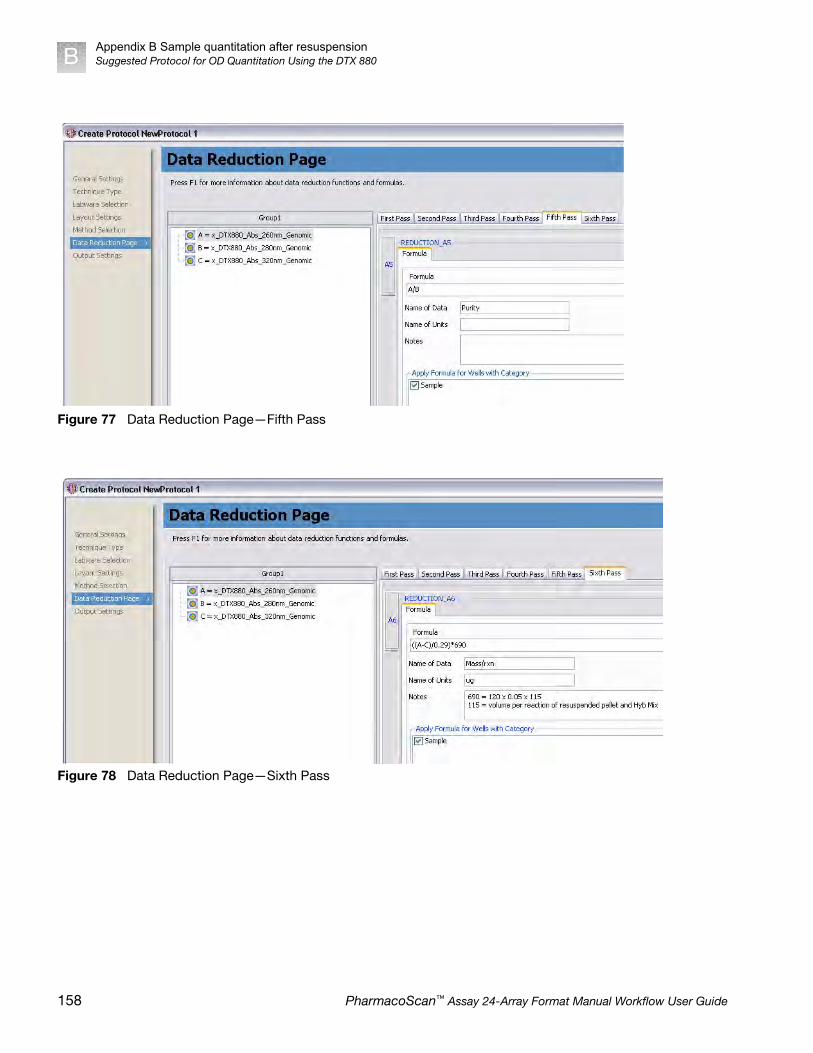
Appendix B Sample quantitation after resuspensionSuggested Protocol for OD Quantitation Using the DTX 880B
Figure 77 Data Reduction Page—Fifth Pass
Figure 78 Data Reduction Page—Sixth Pass
158 PharmacoScan™ Assay 24-Array Format Manual Workflow User Guide
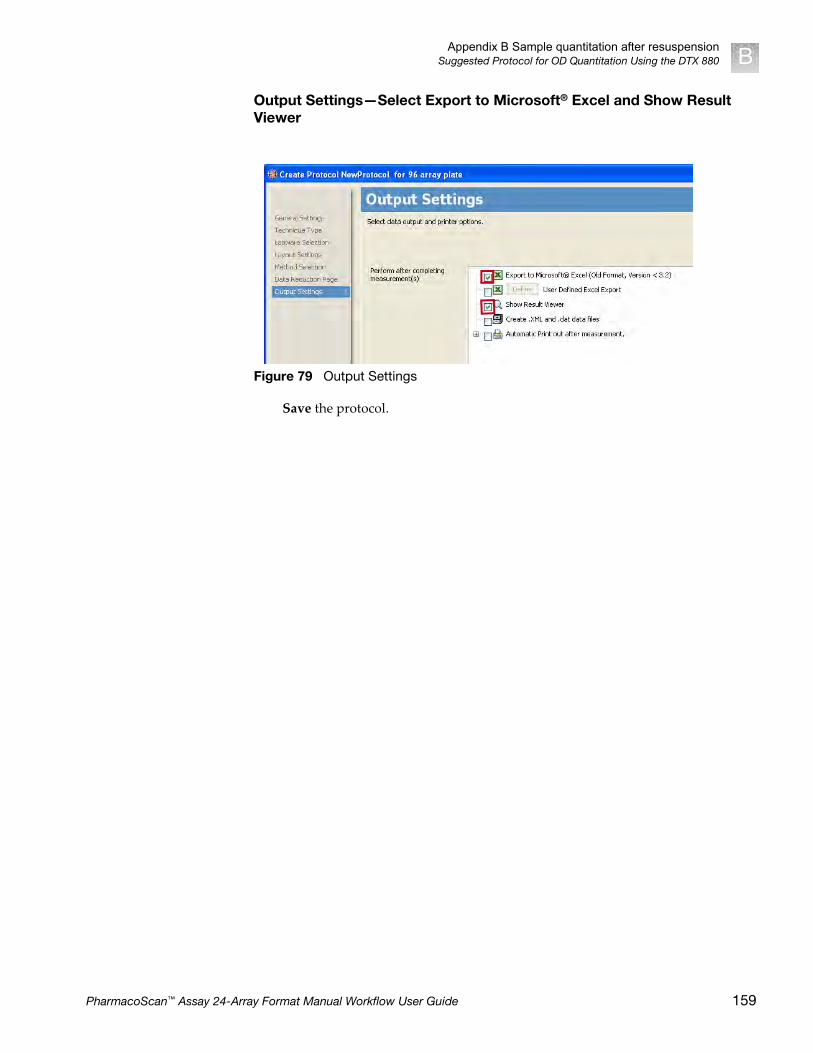
Appendix B Sample quantitation after resuspensionSuggested Protocol for OD Quantitation Using the DTX 880 B
Output Settings—Select Export to Microsoft® Excel and Show Result Viewer
Save the protocol.
Figure 79 Output Settings
PharmacoScan™ Assay 24-Array Format Manual Workflow User Guide 159

Appendix B Sample quantitation after resuspensionPerform sample quantitation on a plate reader other than the DTX880B
Perform sample quantitation on a plate reader other than the DTX880
Your plate reader should be calibrated to ensure accurate readings.
The total yield in µg per well can be calculated as:
(A - C)*D*V*E/P
Where:
A = the observed OD260
C = the observed OD320 (an estimate of a blank reading)
D = 120 (the net dilution factor when preparing the OD Sample plate as described in the automated or manual workflow)
V = 115 (the volume of the sample in µL after the resuspension step)
E = 0.05 (the extinction coefficient of duplex DNA at 260 nm)
P = the optical path length (measured in cm) for the plate type and plate reader used.
If your plate reader does not record the OD320, the OD260 of a blank solution of water only should be used for the parameter “B” above.
The optical path length is dependent on the type of plate and spectrophotometer used. Check your manufacturer’s recommendations for the path length for your instrument and plate type or for recommendations on how to measure this quantity. The SpectraMax Plus384, described as an alternative spectrophotometer in the PharmacoScan Assay 24-Array Format Manual Workflow Site Preparation Guide, Pub. No. 703287, can employ an automated path length detection system. Consult this instrument’s manual for more information.
The resulting yield calculations can be compared against the typical yields shown in column H of Figure 66 on page 152 and against ʺOD Yield Assessment Guidelinesʺ on page 152.
160 PharmacoScan™ Assay 24-Array Format Manual Workflow User Guide

C Register samples in GeneChip™
Command Console™
Create a GeneTitan™ Array Plate Registration file
A GeneTitan Array Plate Registration file is a Microsoft Excel spreadsheet that includes information on the samples you are processing on a single array plate. This information includes the array plate format, the array plate barcode, and sample file names so that you can track the samples that are loaded onto a particular array plate.
The version of Microsoft Excel must be 1997-2000 (file extension is .xls; not .xlsx).
1. In GCC Portal, open the Samples menu and select GeneTitan Array Plate Registration.
2. Step 1—Figure 81 on page 162:
a. Select the array plate type.
b. Click Download.
Figure 80 Selecting GeneTitan Array Plate Registration
PharmacoScan™ Assay 24-Array Format Manual Workflow User Guide 161

Appendix C Register samples in GeneChip™ Command Console™
Create a GeneTitan™ Array Plate Registration fileC
3. Step 2—complete the registration file as follows:
a. Click the Microsoft Excel box on the bottom bar of the monitor to open the Excel spreadsheet.
b. Enter a unique name for each sample (Sample File Name) and any additional information you would like to include.
c. Do one of the following:
• If you are ready to load the array plate onto the GeneTitan MC Instrument, scan the array plate barcode and proceed to the next step.
• If you are not ready to load the array plate onto the GeneTitan MC Instrument, proceed directly to the next step.
4. Save the file as follows:
a. Open File Save As.
b. Enter a name for the array plate registration file.
c. Click Save.
By default, the file is saved in the Affymetrix_Downloads folder.
5. Step 3—when ready to load the array plate onto the GeneTitan MC Instrument:
a. Click the Browse button, navigate to the file, and click Open.
b. Scan the array plate barcode if not already scanned.
Figure 81 Selecting the type of array plate to be processed
Figure 82 Entering sample information into a GeneTitan Array Plate Registration file.
162 PharmacoScan™ Assay 24-Array Format Manual Workflow User Guide

Appendix C Register samples in GeneChip™ Command Console™
Create a GeneTitan™ Array Plate Registration file C
c. Click the Upload button (Figure 83), wait for the information to load, then click the Save button located at the bottom of the next page that is displayed.
If the samples are successfully registered, the message in Figure 84 is displayed.
Figure 83 Uploading the array plate registration file to GCC.
Figure 84 Array plate samples successfully registered.
PharmacoScan™ Assay 24-Array Format Manual Workflow User Guide 163

D Deionizing procedure for GeneTitan™
trays and covers
We recommend the use of the Zerostat 3 Anti-Static Gun (Cat. No. 74-0014) to deionize GeneTitan™ MC Instrument stain tray trays and lids.
Deionize the inner surface of each tray and cover:
• The surface of the tray with the wells that holds reagents.
• The surface of the cover that faces the reagents.
IMPORTANT! Except for the Axiom™ array plates, scan tray and the hybridizationtray, you must deionize all GeneTitan stain trays, stain tray covers and scan tray coverusing an anti-static gun. You must do this before you fill the trays with reagents andbefore you place the covers on the trays. Deionization removes the static electricity. Thepresence of static electricity on the underside of the cover can cause the gripper to liftthe tray along with the tray cover and can result in an aborted run. See Figure 85,Figure 86 and Figure 87.
CAUTION! Do not deionize the scan tray or hybridization tray.
Figure 85 Scan tray with cover. Deionize only the cover.
164 PharmacoScan™ Assay 24-Array Format Manual Workflow User Guide
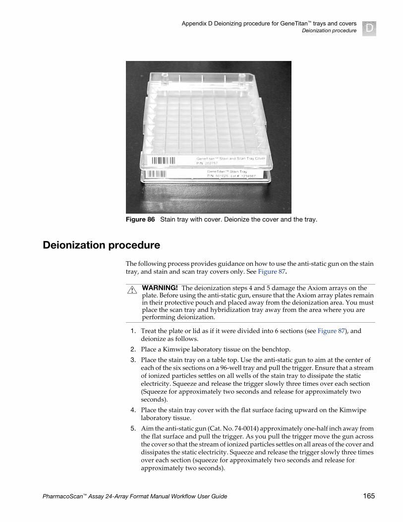
Appendix D Deionizing procedure for GeneTitan™ trays and coversDeionization procedure D
Deionization procedure
The following process provides guidance on how to use the anti-static gun on the stain tray, and stain and scan tray covers only. See Figure 87.
1. Treat the plate or lid as if it were divided into 6 sections (see Figure 87), and deionize as follows.
2. Place a Kimwipe laboratory tissue on the benchtop.
3. Place the stain tray on a table top. Use the anti-static gun to aim at the center of each of the six sections on a 96-well tray and pull the trigger. Ensure that a stream of ionized particles settles on all wells of the stain tray to dissipate the static electricity. Squeeze and release the trigger slowly three times over each section (Squeeze for approximately two seconds and release for approximately two seconds).
4. Place the stain tray cover with the flat surface facing upward on the Kimwipe laboratory tissue.
5. Aim the anti-static gun (Cat. No. 74-0014) approximately one-half inch away from the flat surface and pull the trigger. As you pull the trigger move the gun across the cover so that the stream of ionized particles settles on all areas of the cover and dissipates the static electricity. Squeeze and release the trigger slowly three times over each section (squeeze for approximately two seconds and release for approximately two seconds).
Figure 86 Stain tray with cover. Deionize the cover and the tray.
WARNING! The deionization steps 4 and 5 damage the Axiom arrays on the plate. Before using the anti-static gun, ensure that the Axiom array plates remain in their protective pouch and placed away from the deionization area. You must place the scan tray and hybridization tray away from the area where you are performing deionization.
PharmacoScan™ Assay 24-Array Format Manual Workflow User Guide 165

Appendix D Deionizing procedure for GeneTitan™ trays and coversDeionization procedureD
6. Place the treated cover or tray on the Kimwipe laboratory tissue and lift it up (see Figure 87).
7. Do one of the following:
• If the tissue does not cling to the plastic, proceed with the protocol.
• If the tissue still clings to the plastic, then perform steps 3 and 4 again. If it continues to cling to the plastic, test the device using the ion-indicator cap to confirm that the unit is still releasing ions. Otherwise, it may be time to replace the unit.
Figure 87 Removing the static charge from stain trays and lids.
Treat the inside surface of stain trays (right) and cover (left).
• If a Kimwipe laboratory tissue clings to treated surface, try the deionization procedure again. • If the Kimwipe laboratory tissue still clings, it may be time to replace the anti-static gun.
1 2
3 4
5 6
166 PharmacoScan™ Assay 24-Array Format Manual Workflow User Guide

Appendix D Deionizing procedure for GeneTitan™ trays and coversIon-indicator cap D
Ion-indicator cap
The ion-indicator cap is a testing device used to verify the release of ions when the anti-static gun is in use (Cat. No. 74-0014, Figure 87).
Testing the Zerostat 3 with the ion-indicator cap
1. Insert the ion-indicator cap into the nose of the Zerostat and then slowly squeeze the release trigger (see Figure 88).
2. Observe the discharge through the viewing slot on the ion-indicator cap of the anti-static gun. A visible light is observed in the viewing window on the cap when charged ions are discharged.
3. If you cannot see the light, the gun may be unusable and you should replace it.
4. Each Zerostat anti-static gun is capable of 50,000 trigger operations, which is sufficient for approximately 200-250 runs on the GeneTitan MC Instrument.
Figure 88 Zerostat 3 Anti-Static Gun (Cat. No. 74-0014) with Ion-Indicator Cap to Test Functionality
Ion-Indicator Cap
The Ion-Indicator Cap is attached to the Zerostat to test the functionality of the anti-static gun.
IMPORTANT: Do not use the Ion-Indicator Cap on the Zerostat gun when deionizing trays and lids.
IMPORTANT! Make sure to remove the cap from the gun before deionizing a tray orcover.
PharmacoScan™ Assay 24-Array Format Manual Workflow User Guide 167
E GeneTitan™ Multi-ChannelInstrument care
Introduction
This chapter provides instructions on caring for and maintaining the instrument and on troubleshooting if problems arise.
• Always run a Shutdown protocol when the instrument will be off or unused overnight or longer. This prevents salt crystals from forming within the fluidics system.
• Always use deionized water to prevent contamination of the lines. Change buffers with freshly prepared buffer at each system startup.
The GeneTitan™ Multi-Channel (MC) Instrument should be positioned on a sturdy level bench away from extremes in temperature and away from moving air.
Cleaning and maintenance
The GeneTitan family of instruments require little in the way of customer maintenance. The instruments must be kept clean and free of dust. Dust buildup can degrade performance. Wipe the exterior surfaces clean using a mild dish detergent solution in water. Do not use ammonia based cleaners or organic solvents such as alcohol or acetone to clean the system because they may damage the exterior surfaces.
The following tasks should be performed regularly to ensure the Imaging Device remains in working order.
Monthly Wipe down the outer surface of the Imaging Device with a dry cloth.
Every six months Replace the cooling fan air filters at the rear of the instrument.
Replace the Micropore filters in the Wash A, Wash B, and Rinse bottles. If you run 4-8 plates/week then the micro-pore filters need to be replaced more frequently.
IMPORTANT! Before performing maintenance turn off power to the instrument toavoid injury in case of an electrical malfunction.
168 PharmacoScan™ Assay 24-Array Format Manual Workflow User Guide

Appendix E GeneTitan™ Multi-Channel Instrument careCleaning and maintenance E
Service the outer enclosure fan filters
ScheduleThe GeneTitan fan filter cartridge (Figure 89) should be cleaned at least every 90 days of service. Note that in some service locations, the presence of excessive dust or particulate matter may necessitate cleaning the cartridge more often than 90 days.
A plugged filter cartridge can cause excessive temperatures within the machine that can cause unwanted evaporation on test media.
• Number of filters required per GeneTitan instrument: 3
Clean the GeneTitan filter cartridge
1. Slide the filter cartridge from the fan filter cartridge at the rear of the GeneTitan Instrument.
2. Submerse in clean DI water. Rinse and agitate gently to dislodge material.
3. Remove from water and dry with clean compressed air or towels.
4. When the filter cartridge is completely dry to the touch, re-install the cartridge.
Figure 89 The GeneTitan filter cartridge
PharmacoScan™ Assay 24-Array Format Manual Workflow User Guide 169

Appendix E GeneTitan™ Multi-Channel Instrument careCleaning and maintenanceE
Replace the bottle filters
The bottles used in GeneTitan Instrument contain a filter to remove particulates that may exist in the buffers and DI water. The filters in the GeneTitan fluidics bottles (Wash A, Wash B and DI Water) need to be replaced when the filters are clogged.
The software displays warning message boxes for the filter in each reagent bottle when it detects a problem or shows a trend of increased fill times during fluid fill operations.
If an error is detected as described above, then a message box titled “Filter Change Required” is displayed (Figure 90) along with the information on the specific dispense operation. You should change all three filters when a warning is displayed for any one of the three filters.
The message boxes displayed in Figure 90 provide information on fluid dispense errors that were detected by the instrument for any of the bottles or when the instrument detects an increase in the amount of time that is required to perform the fill operations.
Note: The reagent bottles are depressurized when this warning message is displayed. It is safe to change the filters in all three fluidic bottles when this message is displayed.
After changing the filters in all three bottles using the procedure described below, press the Yes button to continue. If you choose to ignore the error message, press the No button. This warning message is displayed each time GCC instrument control software is launched. You may also experience data quality issues if particulate matter
Figure 90 Filter Change Required Messages
170 PharmacoScan™ Assay 24-Array Format Manual Workflow User Guide

Appendix E GeneTitan™ Multi-Channel Instrument careCleaning and maintenance E
cannot be trapped by the filters if they are clogged.
We recommend that your site keep three spare filters in the event they need to be replaced. The procedure for replacing the filters is simple.
• Part details: Thermo Fisher Scientific, Cat. No. 01-0671
Remove and inspect the filter
1. Loosen and remove the cap on the bottle.
2. Carefully remove the filter from the end of the filter body.
3. Visually inspect the filter. If one of the filters appears to have a concentration of dirt or contaminate in it, discard it and replace the filter with a new one.
Replace the filter
1. Insert the filter into the end of the filter body.
2. Replace the cap onto the bottle and tighten it.
3. Repeat for each bottle.
Figure 91 Replacing the filter
IMPORTANT! Replace one filter at a time to ensure the correct connection of thebuffer supply tube to its respective bottle. The color of the buffer supply tubingmatches the bottle color code.
Buffer supply line
Filter holder
Filter
1
2
3
1 2 3
PharmacoScan™ Assay 24-Array Format Manual Workflow User Guide 171
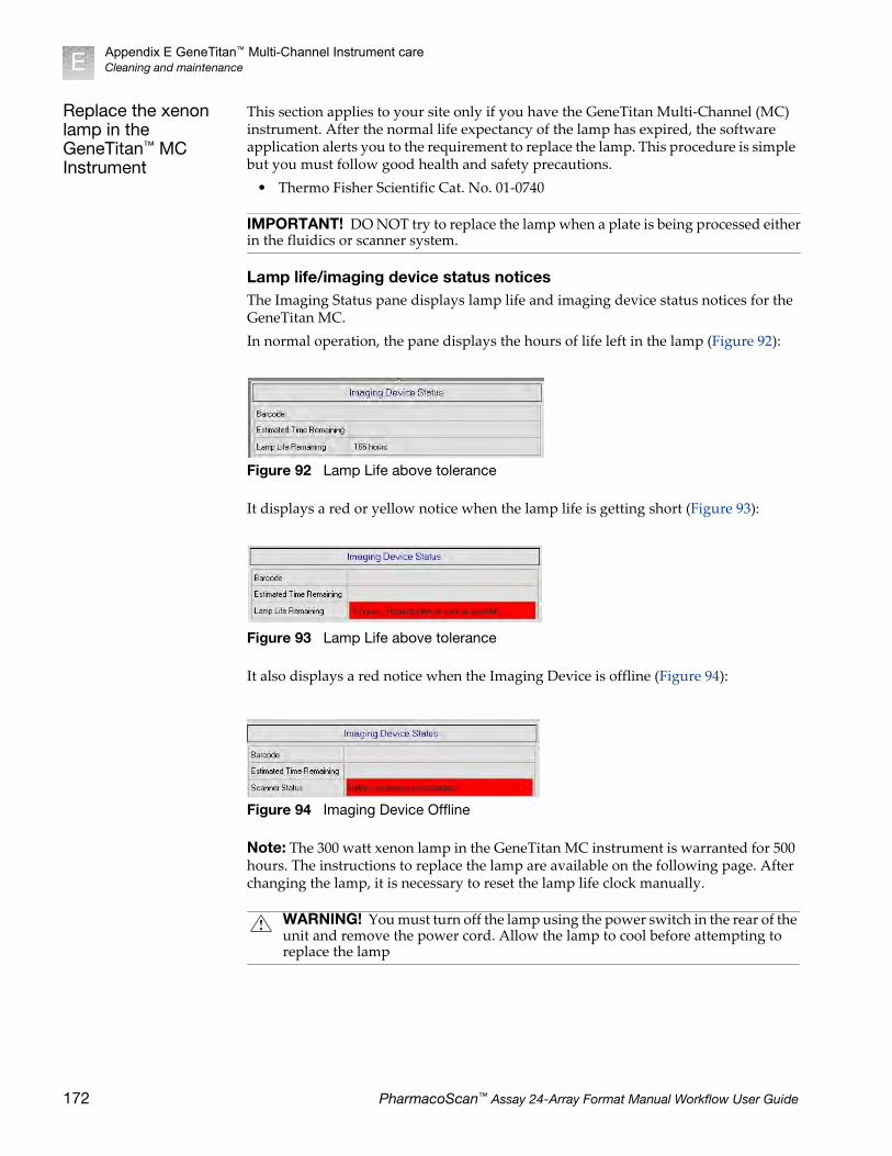
Appendix E GeneTitan™ Multi-Channel Instrument careCleaning and maintenanceE
Replace the xenon lamp in the GeneTitan™ MC Instrument
This section applies to your site only if you have the GeneTitan Multi-Channel (MC) instrument. After the normal life expectancy of the lamp has expired, the software application alerts you to the requirement to replace the lamp. This procedure is simple but you must follow good health and safety precautions.
• Thermo Fisher Scientific Cat. No. 01-0740
Lamp life/imaging device status noticesThe Imaging Status pane displays lamp life and imaging device status notices for the GeneTitan MC.
In normal operation, the pane displays the hours of life left in the lamp (Figure 92):
It displays a red or yellow notice when the lamp life is getting short (Figure 93):
It also displays a red notice when the Imaging Device is offline (Figure 94):
Note: The 300 watt xenon lamp in the GeneTitan MC instrument is warranted for 500 hours. The instructions to replace the lamp are available on the following page. After changing the lamp, it is necessary to reset the lamp life clock manually.
IMPORTANT! DO NOT try to replace the lamp when a plate is being processed eitherin the fluidics or scanner system.
Figure 92 Lamp Life above tolerance
Figure 93 Lamp Life above tolerance
Figure 94 Imaging Device Offline
WARNING! You must turn off the lamp using the power switch in the rear of the unit and remove the power cord. Allow the lamp to cool before attempting to replace the lamp
172 PharmacoScan™ Assay 24-Array Format Manual Workflow User Guide

Appendix E GeneTitan™ Multi-Channel Instrument careCleaning and maintenance E
Removing the xenon lamp
1. Unscrew the four retaining bolts. They should be finger tight (Figure 95).
2. Place each hand on each side of the blue plastic flange and lift out the lamp in a vertical motion (Figure 96). You must use both hands to remove the lamp successfully. Apply equal pressure on each side of the lamp and gently lift.
Figure 95 Unscrew the bolts
Unscrew these four bolts.
PharmacoScan™ Assay 24-Array Format Manual Workflow User Guide 173

Appendix E GeneTitan™ Multi-Channel Instrument careCleaning and maintenanceE
Replacing the lamp
1. Hold the lamp by the blue plastic flanges. Ensure that the lamp bulb faces inward toward the reflecting mirror (Figure 97) and vertically insert the lamp (Figure 98).
2. Replace the warning cover and hand tighten the bolts (Figure 95).
Figure 96 Lift out the lamp
CAUTION! Ensure that you install the lamp in the correct orientation.
174 PharmacoScan™ Assay 24-Array Format Manual Workflow User Guide
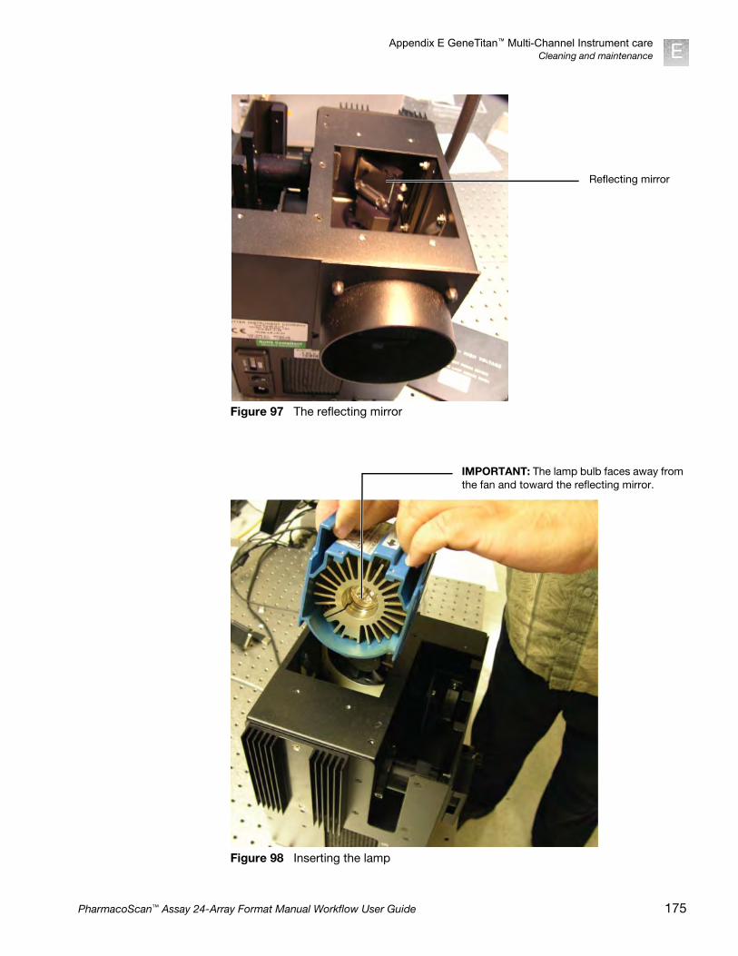
Appendix E GeneTitan™ Multi-Channel Instrument careCleaning and maintenance E
Figure 97 The reflecting mirror
Figure 98 Inserting the lamp
Reflecting mirror
IMPORTANT: The lamp bulb faces away from the fan and toward the reflecting mirror.
PharmacoScan™ Assay 24-Array Format Manual Workflow User Guide 175

Appendix E GeneTitan™ Multi-Channel Instrument careCleaning and maintenanceE
Reset the lamp counterYou must alert the software application that you have replaced the lamp so that the hours of the lamp counter are reset to zero. This menu option is only available when the system is not processing any plates.
1. On the software application click Tools Reset Counter for Life Remaining (Figure 99).
2. The software displays a message that allows you to change your mind.
Figure 99 Inserting the Lamp
176 PharmacoScan™ Assay 24-Array Format Manual Workflow User Guide

Appendix E GeneTitan™ Multi-Channel Instrument careTroubleshooting E
3. Click Yes if you want to reset the counter. The software displays a message that confirms that the software has reset the counter (Figure 101).
Troubleshooting
This section provides instructions on how to identify and solve simple problems with the GeneTitan MC Instrument. If a problem or error occurs that is not listed in this chapter contact Thermo Fisher Scientific technical support for assistance.
For software errors that do not involve hardware crashes the most common solution is to shut down the application and then restart it. If the same error occurs shut down both the application and the computer and then restart. If it still occurs shut down the GeneTitan MC Instrument and then restart.
Log files The log files are produced by different GCC components. The logs provide a record of the tasks performed by different components, such as the migration tools and installer. These log files provide useful information for troubleshooting problems. These files may be requested by your Field Application Scientist (FAS), Field Service Engineer (FSE), or when contacting Technical Support.
GCC log filesThe following files apply to the GeneTitan Instruments. All the GCC log files from C:\Command_Console\Logs The different log files include:
Figure 100 Are you sure?
Figure 101 The counter is reset
Systemlog.XML XML file with system information.
DEC.log Text file with information on the use of the Data Exchange Console (DEC).
DECError.log Text file with information on errors created while using DEC.
AGCC_LibFileImporter. log (with date and time code)
Text file with info on use of the Library File Importer.
PharmacoScan™ Assay 24-Array Format Manual Workflow User Guide 177

Appendix E GeneTitan™ Multi-Channel Instrument careTroubleshootingE
Other GCC filesYour FAS and/or FSE may request you to send the following files for troubleshooting:
1. Library files (*.PARAMS, *.MASTER, *.WORKFLOW, *.SMD, *.MEDIA) located in C:\Command_Console\Library, excluding the large analysis library files (CDF, PSI, GRC).
2. Provide a list of all sub folders and their contents under the library files folder located in C:\Command_Console\Library. Ensure there are no duplicate library files, as these can cause problems.
3. GCC system configuration file located at C:\Command_Console\Configuration\Calvin.System.config
4. Pending job order files located in C:\Command_Console\Jobs
5. Other GCC related information, such as:
a. The number of files under C:\Command_Console\Data, including sub directory.
b. If the system is a networked system or a standalone system.
c. Other applications installed on the system, such as antivirus application, MS Office, Internet Explorer versions.
GCC log files for GeneTitan™ MC systems
Log files for the GeneTitan MC Instrument control processes are placed in subdirectories of the Command Console\Logs\ folder. Thermo Fisher Scientific may need the following files for troubleshooting:
GeneTitan MC fluidics
1. C:\Command_Console\Logs\96F\
a. subdirectories named by date (e.g., Log7-29-2009)
• Collect all dated directories and contents since the GeneTitan application was started, not just the date of the event (some logging goes into files from the date the application started so this can be critical for us).
• Absolutely required are all the log directories from the date the run was started to the date of the event.
2. C:\Command_Console\Logs\96F\FluidicErrorLog - all files in this directory
GeneTitan MC imaging device
1. C:\Affymetrix\GeneChipHTScanControlMC\Log - collect all dated directories and contents since the GeneTitan application was started
2. C:\Affymetrix\GeneChipHTScanControlMC\RunLog - collect all dated directories and contents since the GeneTitan application was started
Problems and solutions
This section provides instructions on how to identify and solve problems with the unit.
If problems arise with the instruments use the following tables to locate the description that matches the problem. If you cannot find a solution call Thermo Fisher Scientific technical support for assistance.
For software errors that do not involve hardware crashes the most common solution is to shut down the application and then restart it. If the same error occurs shut down both the application and the computer and then restart. If it still occurs shut down the entire unit and then restart.
178 PharmacoScan™ Assay 24-Array Format Manual Workflow User Guide

Appendix E GeneTitan™ Multi-Channel Instrument careTroubleshooting E
Insufficient disk space notice
If there is not enough memory on the computer’s drives to save the data from an array plate, a notice appears (Figure 102) when:
• You first initialize the software and instrument
• You select arrays for imaging.
If you see this notice, you need to free up sufficient disk space before imaging starts.
Figure 102 Insufficient Disk Space notice
PharmacoScan™ Assay 24-Array Format Manual Workflow User Guide 179

F mPCR quality control gel protocol
Run a mPCR quality control gel
Note: This is an optional procedure. It is meant only as a QUALITATIVE examination of the mPCR reaction to confirm that amplification has occurred. Gene copy number differences result in differences in DNA band patterns and amplicon intensities, and therefore sample to sample variation might be observed.
Equipment required
E-Gels and reagents required
Item Supplier Cat. No.
Gel imager Various —
Pipette, single and multichannel Various —
Plate centrifuge Various —
Vortex Various —
Item Supplier Cat. No.
Mother E-Base Device
Thermo Fisher Scientific
EB-M03
Daughter E-Base Device EB-D03
E-Gel® 48 2% agarose gels G8008-02
TrackIt™ Cyan/Orange Loading Buffer 10482-028
NEB 50 bp DNA Ladder New England BioLabs N3236S
Reduced EDTA TE Buffer (10 mM Tris-HCL PH 8.0, 0.1 mM EDTA)
Thermo Fisher Scientific 75793
Diluted TrackIt Cyan/Orange Loading Buffer See "Dilute the TrackIt™ Cyan/Orange Loading Buffer and 25 bp ladder" on page 149
180 PharmacoScan™ Assay 24-Array Format Manual Workflow User Guide

Appendix F mPCR quality control gel protocolRun a mPCR quality control gel F
Consumables
Prepare NEB 50 bp DNA Ladder
The following recipe is for preparing a 250-fold dilution of the NEB 50 bp DNA Ladder (4 ng/µL final concentration).
1. In a 1.7 mL microcentrifuge tube, add 1 µL of 50 bp DNA Ladder to 249 µL of 1,000-fold diluted TrackIt dye.
2. Vortex tube to mix well. Pulse-spin to get droplets down.
Prepare mPCR samples for gel analysis
1. Thaw mPCR Reaction plate on benchtop at room temperature.
2. Ensure plate seal is secure, vortex plate, and pulse spin.
3. Dilute mPCR reaction samples 120-fold by:
a. First dilute mPCR samples 12-fold in buffer.Transfer 2 µL of the mPCR reaction into 22 µL of low EDTA TE. Seal plate. Vortex. Pulse-spin.
b. Then dilute samples another 10-fold in loading dye to prepare “mPCR Gel QC plate”. Transfer 2 µL of the 12-fold diluted mPCR reactions into 18 µL of 1,000-fold diluted TrackIt dye.
Run mPCR QC gel protocol
This protocol is based on running QC gels for 24 samples.
1. Tightly seal the mPCR Gel QC plate.
2. Vortex the plate for three seconds. Pulse-spin to get droplets down.
3. Connect an E-Base™ device(s) to an electrical outlet.
4. Push the Power/Prg button on each to ensure the program is in EG mode (not EP mode).
5. Take the gel out of the pouch and remove the combs.
6. Place the E-Gel® 48 gel into an E-Base unit.
7. Load 15 µL from each well of the mPCR Gel QC plate onto the gels.
8. Load 15 µL of diluted 50 bp ladder into the marker wells (M).
9. Load 15 µL nuclease-free water into any unused wells.
10. Run the gels for 25 minutes.
11. Image the gel.
mPCR QC Gel images should look similar to the gel shown in Figure 103.
Item Supplier Cat. No.
Adhesive film – use one of the following:• MicroAmp Clear Adhesive Film• Microseal 'B' Film
Thermo Fisher Scientific )Bio-Rad
4306311MSB1001
Pipette Tips Same brand as pipette —
96-well PCR plate Various various
PharmacoScan™ Assay 24-Array Format Manual Workflow User Guide 181

Appendix F mPCR quality control gel protocolRun a mPCR quality control gelF
Note: Variation in DNA band patterns and intensities can be observed from sample to sample due to gene copy number differences. The mPCR QC Gel is meant to be a qualitative examination of the mPCR reaction to confirm that amplification has occurred for each sample.
All samples were amplified and show DNA bands that fall between 150 bp and 700 bp.
Figure 103 Example of a Typical mPCR QC E-Gel
50 bp ladder
700 bp
50 bp ladder
150 bp
182 PharmacoScan™ Assay 24-Array Format Manual Workflow User Guide

G Safety
For research use only. Not recommended or intended for diagnosis of disease in humans or animals. Do not use internally or externally in humans or animals.
Precautions 1. PHARMACOSCAN ARRAYS AND PLATES ARE FOR RESEARCH USE ONLY; NOT FOR DIAGNOSTIC PROCEDURES.
2. Avoid microbial contamination, which can cause erroneous results.
3. WARNING: All biological specimens and materials with which they come into contact should be handled as if capable of transmitting infection and disposed of with proper precautions in accordance with federal, state, and local regulations. This includes adherence to the OSHA Bloodborne Pathogens Standard (29 CFR 1910.1030) for blood-derived and other samples governed by this act. Never pipet by mouth. Avoid specimen contact with skin and mucous membranes.
4. CAUTION: Exercise standard precautions when obtaining, handling, and disposing of potentially carcinogenic reagents.
5. Exercise care to avoid cross-contamination of samples during all steps of this procedure, as this can lead to erroneous results.
6. Use powder-free gloves whenever possible to minimize introduction of powder particles into sample or array plates.
7. CAUTION: Use care when handling the scan tray as it has protruding guiding posts that may be sharp and can stick out of the pouch if not handled carefully.
WARNING! GENERAL SAFETY. Using this product in a manner not specified in the user documentation can result in personal injury or damage to the instrument or device. Ensure that anyone using this product has received instructions in general safety practices for laboratories and the safety information provided in this document.
• Before using an instrument or device, read and understand the safety information provided in the user documentation provided by the manufacturer of the instrument or device.
• Before handling chemicals, read and understand all applicable Safety Data Sheets (SDSs) and use appropriate personal protective equipment (gloves, gowns, eye protection, etc). To obtain SDSs, see the ʺDocumentation and supportʺ section in this document.
WARNING! The following components contain harmful or toxic ingredients:
• Axiom Stabilize Soln: 8% Gluteraldehyde
• Axiom HybSoln 2: 100% Formamide
• Axiom Hyb Buffer: <55% Tetramethylammonium Chloride
In all cases customers should use adequate local and general ventilation in order to minimize airborne concentrations.
PharmacoScan™ Assay 24-Array Format Manual Workflow User Guide 183
Appendix G SafetyChemical safetyG
Chemical safety
WARNING! GENERAL CHEMICAL HANDLING. To minimize hazards, ensure laboratory personnel read and practice the general safety guidelines for chemical usage, storage, and waste provided below, and consult the relevant SDS for specific precautions and instructions:
• Read and understand the Safety Data Sheets (SDSs) provided by the chemical manufacturer before you store, handle, or work with any chemicals or hazardous materials. To obtain SDSs, see the ʺDocumentation and supportʺ section in this document.
• Minimize contact with chemicals. Wear appropriate personal protective equipment when handling chemicals (for example, safety glasses, gloves, or protective clothing).
• Minimize the inhalation of chemicals. Do not leave chemical containers open. Use only with adequate ventilation (for example, fume hood).
• Check regularly for chemical leaks or spills. If a leak or spill occurs, follow the manufacturerʹs cleanup procedures as recommended in the SDS.
• Handle chemical wastes in a fume hood.
• Ensure use of primary and secondary waste containers. (A primary waste container holds the immediate waste. A secondary container contains spills or leaks from the primary container. Both containers must be compatible with the waste material and meet federal, state, and local requirements for container storage.)
• After emptying a waste container, seal it with the cap provided.
• Characterize (by analysis if necessary) the waste generated by the particular applications, reagents, and substrates used in your laboratory.
• Ensure that the waste is stored, transferred, transported, and disposed of according to all local, state/provincial, and/or national regulations.
• IMPORTANT! Radioactive or biohazardous materials may require special handling, and disposal limitations may apply.
184 PharmacoScan™ Assay 24-Array Format Manual Workflow User Guide

Appendix G SafetyBiological hazard safety G
Biological hazard safety
WARNING! BIOHAZARD. Biological samples such as tissues, body fluids, infectious agents, and blood of humans and other animals have the potential to transmit infectious diseases. All work should be conducted in properly equipped facilities using the appropriate safety equipment (for example, physical containment devices). Safety equipment also may include items for personal protection, such as gloves, coats, gowns, shoe covers, boots, respirators, face shields, safety glasses, or goggles. Individuals should be trained according to applicable regulatory and company/ institution requirements before working with potentially biohazardous materials. Follow all applicable local, state/provincial, and/or national regulations. The following references provide general guidelines when handling biological samples in laboratory environment.
• U.S. Department of Health and Human Services, Biosafety in Microbiological and Biomedical Laboratories (BMBL), 5th Edition, HHS Publication No. (CDC) 21-1112, Revised December 2009; found at:www.cdc.gov/biosafety/publications/bmbl5/BMBL.pdf
• World Health Organization, Laboratory Biosafety Manual, 3rd Edition, WHO/CDS/CSR/LYO/2004.11; found at:www.who.int/csr/resources/publications/biosafety/Biosafety7.pdf
PharmacoScan™ Assay 24-Array Format Manual Workflow User Guide 185

Documentation and support
Related documentation
Document Publication number Description
PharmacoScan™ Assay 24-Array Format Manual Workflow User Guide
703286 This user guide provides comprehensive instructions on running the PharmacoScan Assay 24-Array Format Manual Workflow.
PharmacoScan™ Assay 24-Array Format Manual Workflow Site Preparation Guide
703287 Provides guidance on reagents, instruments, and supplies required to run the PharmacoScan Assay 24-Array Format Manual Workflow.
PharmacoScan™ Assay 24-Array Format Manual Workflow Quick Reference
703288 An abbreviated reference for the target preparation step of the PharmacoScan Assay 24-Array Format Manual Workflow. This quick reference document is intended for experienced users.
PharmacoScan™ Assay 96-Array Format Manual Workflow User Guide
703459 This user guide provides comprehensive instructions on running the PharmacoScan Assay 96-Array Format Manual Workflow.
PharmacoScan™ Assay 96-Array Format Manual Workflow Site Preparation Guide
703460 Provides guidance on reagents, instruments, and supplies required to run the PharmacoScan Assay 96-Array Format Manual Workflow.
PharmacoScan™ Assay 96-Array Format Manual Workflow Quick Reference
703461 An abbreviated reference for the target preparation step of the PharmacoScan Assay 96-Array Format Manual Workflow. This quick reference document is intended for experienced users.
GeneTitan™ MC Protocol for Axiom 2.0 Array Plate Processing Quick Reference
702988 A quick reference document for experienced GeneTitan users processing Axiom array plates.
GeneTitan™ Multi-Channel Instrument User Guide
08-0308 The GeneTitan Multi-Channel (MC) Instrument automates array processing from target hybridization to data generation by combining a hybridization oven, fluidics processing, and state-of-the art imaging device into a single bench-top instrument. This document detailing the use, care, and maintenance for the GeneTitan MC Instrument.
GeneTitan™ Multi-Channel Instrument Site Preparation Guide
08-0305 Provides guidance on creating and maintaining the proper environment required for the GeneTitan Multi-Channel Instrument.
186 PharmacoScan™ Assay 24-Array Format Manual Workflow User Guide

Documentation and supportCustomer and technical support
Customer and technical support
Visit thermofisher.com/support for the latest in services and support, including:
• Worldwide contact telephone numbers
• Product support, including:
– Product FAQs
– Software, patches, and updates
• Order and web support
• Product documentation, including:
– User guides, manuals, and protocols
– Certificates of Analysis
– Safety Data Sheets (SDSs; also known as MSDSs)
Note: For SDSs for reagents and chemicals from other manufacturers, contact the manufacturer.
Software and analysis
Applied Biosystems ™ GeneChip™ Command Console™ Software User Guide
702569 This user guide provides instructions on using Applied Biosystems GeneChip Command Console Software (GCC) used to control GeneChip instrument systems. Command Console Software provides an intuitive set of tools for instrument control and data management used in the processing of GeneChip Arrays.
Axiom™ Analysis Suite 3.0 User Guide 703307 This user guide provides instructions on using Axiom™ Analysis Suite—a single-source software package to enable complete genotyping analysis of all Axiom arrays.
Axiom™ Genotyping Solution Data Analysis Guide
702961 This guide provides information and instructions for analyzing Axiom genotyping array data. It includes the use of Axiom™ Analysis Suite, Power Tools (APT) and SNPolisher R package to perform quality control analysis (QC) for samples and plates, SNP filtering prior to downstream analysis, and advanced genotyping methods.
Document Publication number Description
PharmacoScan™ Assay 24-Array Format Manual Workflow User Guide 187

Documentation and supportLimited product warranty
Limited product warranty
Life Technologies Corporation and/or its affiliate(s) warrant their products as set forth in the Life Technologies’ General Terms and Conditions of Sale found on Life Technologies’ website at www.thermofisher.com/us/en/home/global/terms-and-conditions.html. If you have any questions, please contact Life Technologies at thermofisher.com/support.
188 PharmacoScan™ Assay 24-Array Format Manual Workflow User Guide