phosphorylation-mediated 14-3-3 protein binding regulates the function of the rho-specific guanine...
TRANSCRIPT
Phosphorylation-mediated 14-3-3 Protein Binding Regulatesthe Function of the Rho-specific Guanine NucleotideExchange Factor (RhoGEF) Syx*□S
Received for publication, October 31, 2012, and in revised form, January 8, 2013 Published, JBC Papers in Press, January 18, 2013, DOI 10.1074/jbc.M112.432682
Siu P. Ngok, Rory Geyer, Antonis Kourtidis, Peter Storz, and Panos Z. Anastasiadis1
From the Department of Cancer Biology, Mayo Clinic Comprehensive Cancer Center, Jacksonville, Florida 32224
Background: The junctional localization and RhoGEF activity of Syx mediate cell junction integrity.Results: PKD-mediated phosphorylation and 14-3-3 binding prevent the junctional localization and suppress the GEF activityof Syx.Conclusion: 14-3-3 proteins modulate the function of Syx in the context of cell-cell adhesion.Significance: Understanding mechanisms that regulate junction integrity is crucial in vascular and tumor biology.
Syx is a Rho-specific guanine nucleotide exchange factor(GEF) that localizes at cell-cell junctions and promotes junctionstability by activating RhoA and the downstream effectorDiaphanous homolog 1 (Dia1). Previously, we identified severalmolecules, including 14-3-3 proteins, as Syx-interacting part-ners. In the present study, we show that 14-3-3 isoforms interactwith Syx at both its N- and C-terminal regions in a phosphory-lation-dependent manner. We identify the protein kinaseD-mediated phosphorylation of serine 92 on Syx, and additionalphosphorylation at serine 938, as critical sites for 14-3-3 associ-ation. Our data indicate that the binding of 14-3-3 proteinsinhibits the GEF activity of Syx. Furthermore, we show thatphosphorylation-deficient, 14-3-3-uncoupled Syx exhibitsincreased junctional targeting and increased GEF activity,resulting in the strengthening of the circumferential junctionalactin ring in Madin-Darby canine kidney cells. These findingsreveal a novel means of regulating junctional Syx localizationand function by phosphorylation-induced 14-3-3 binding andfurther support the importance of Syx function in maintainingstable cell-cell contacts.
The Rho GTPases function as molecular switches that cyclebetween a GTP-bound active state and a GDP-bound inactivestate and are involved in a range of signaling pathways thatcontrol the actin cytoskeleton and fundamental cellular pro-cesses, like cell adhesion and cell migration (1). Guanine nucle-otide exchange factors (GEFs)2 catalyze the exchange of GDPfor GTP. Coordinated signaling by RhoA, Rac1, and Cdc42
GTPases regulates cell junction formation and cell polarizationby modulating junctional components and the actin cytoskele-ton (2). Precise spatiotemporal regulation of Rho GTPases byGEFs is thought to regulate the cross-talk between adhesionand polarity complexes (3, 4). Despite extensive characteriza-tion of the Rho GTPases, their activation by junction-associ-atedGEFs, and especially themechanisms by which these GEFsare regulated, remain poorly understood.We recently identified Syx, as a junctional RhoGEF that asso-
ciates with multiple members of the Crumbs polarity complex,promotes junction stability by signaling through RhoA andDiaphanous homolog 1 (Dia1), and mediates the opposingeffects of VEGF and angiopoietin 1 on endothelial junctionintegrity (5). Depletion of Syx results in endothelial cell junc-tion defects in vitro and in vivo. Syx belongs to the Dbl family ofGEFs (6). Besides its Dbl/pleckstrin homology domains, Syxcontains a PDZ binding motif (PBM) that is required for itsrecruitment to the junctions (5). Syx is proposed to play a role inendothelial cell migration and is important for angiogenesisand vascular barrier function in vivo (5, 7, 8). Both Syx activityand localization to junctions are critical for these effects, sug-gesting that misregulation of Syx function results in vasculardefects. The mechanisms that regulate Syx localization andfunction are largely unclear.In addition to its interaction with the myosin VI adaptor
protein synectin (9), the scaffold protein multiple PDZ domainprotein 1 (Mupp1) (7, 10), the protein associated with Lin7(PALS1), and Lin7, we identified several 14-3-3 isoforms asnovel Syx-binding partners (5). 14-3-3 family members associ-ate with a diverse number of proteins, including many withoncogenic or tumor suppressor properties (11, 12). Homo- orheterodimers of 14-3-3 proteins bind to select phosphoserine/threonine residues, induce conformational change, and alterthe localization, stability, and/or function of the bound protein(13). The localization and dimerization of 14-3-3 proteins are inturn regulated by post-translational modifications such asphosphorylation and acetylation (13). 14-3-3� and 14-3-3�have been suggested to play a role in cell polarization by asso-ciating with Par3 (14, 15). However, the role of 14-3-3 proteinson junction stability remains unknown.
* This work was supported, in whole or in part, by National Institutes of HealthGrants R01 NS069753 and R21 NS070117 (to P. Z. A.) and R01 GM086435and 10BG11 (to P. S.). This work was also supported by the Mayo GraduateSchool (to S. P. N.).
□S This article contains supplemental Fig. 1.1 To whom correspondence should be addressed: Dept. of Cancer Biology,
Griffin Cancer Research Bldg., Rm. 307, 4500 San Pablo Rd., Jacksonville, FL32224. Tel.: 904-953-6005; Fax: 904-953-0277; E-mail: [email protected].
2 The abbreviations used are: GEF, guanine nucleotide exchange factor; Dia1,Diaphanous homolog 1; DMSO, dimethyl sulfoxide; MDCK, Madin-Darbycanine kidney; PBM, PDZ binding motif; PMA, phorbol 12-myristate 13-ac-etate; RBD, Rho binding domain.
THE JOURNAL OF BIOLOGICAL CHEMISTRY VOL. 288, NO. 9, pp. 6640 –6650, March 1, 2013© 2013 by The American Society for Biochemistry and Molecular Biology, Inc. Published in the U.S.A.
6640 JOURNAL OF BIOLOGICAL CHEMISTRY VOLUME 288 • NUMBER 9 • MARCH 1, 2013
at UN
IVE
RS
ITY
OF
CA
LGA
RY
, on March 8, 2013
ww
w.jbc.org
Dow
nloaded from
http://www.jbc.org/content/suppl/2013/01/18/M112.432682.DC1.html Supplemental Material can be found at:

In this study, we explored the functional significance of theinteraction between Syx and 14-3-3 proteins. Our data suggestthat PKD phosphorylation regulates 14-3-3 binding to Syx.More importantly, a phospho-deficient, 14-3-3-uncoupled Syxmutant S92A/S938A displays elevated GEF activity andenhanced localization to areas of cell-cell contact. Altogether,these findings provide a mechanistic insight into how 14-3-3proteins can modulate junction stability by altering the local-ization and GEF activity of Syx.
EXPERIMENTAL PROCEDURES
Cell Culture and Transfection—HeLa and MDCK cells werecultured in DMEM (Cellgro) with 10% fetal bovine serum(Invitrogen). HeLa and MDCK cells were transfected withTransIT-HeLaMonster (Mirus) and Lipofectamine 2000(Invitrogen) according to the manufacturers’ instructions,respectively.Antibodies and Reagents—The following antibodies were
used: mouse anti-Syx (KIAA0720, 5A9; Abnova); mouse andrabbit anti-HA (Cell Signaling); mouse anti-GFP/YFP 3E6,mouse anti-ZO1, monoclonal rabbit anti-GFP/YFP (Invitro-gen); rabbit pan anti-14-3-3 (K-19), mouse anti-RhoA (26C4)(Santa Cruz Biotechnology); rabbit anti-GST, rabbit anti-actin(Sigma). Phalloidin 594 (Molecular Probes) was used to stainfor actin filaments in immunofluorescence experiments.Phorbol 12-myristate 13-acetate (PMA; Sigma) was dis-
solved in DMSO to a stock concentration of 100 �M. Proteaseand phosphatase inhibitor mixtures (Pierce) were used in allbuffers (refer to immunoprecipitation section) for the genera-tion of cell lysates.DNA Constructs and Recombinant Protein—Full-length
YFP-tagged murine Syx and HA-tagged PKD WT, kinase-ac-tive, and kinase-dead have been described previously (9, 16).Murine Syx truncation mutants were PCR-amplified frompEYFP-mSyx and then subcloned into pEYFP-C1 usingHindIIIand BamHI restriction sites. Point mutations were introducedin the respective Syx constructs (YFP-Syx, YFP-Syx(1–630),and YFP-Syx(791–1073)) to encode alanine substitutions atSer92, Ser167, Ser294, Ser806, Ser936, Ser938, and Ser964 using theQuikChange Multisite-directed Mutagenesis kit (Stratagene).GST-tagged 14-3-3 epsilon (�), HA-tagged 14-3-3 beta (�),epsilon (�), gamma (�), sigma (�), and zeta (�) were purchasedfrom Addgene. All DNA constructs generated were verified byDNA sequencing. pSuper-PKD1-RNAi and pSuper-PKD2-RNAi vectors were used as described previously to knock downPKD1 and PKD2 (17).Recombinant GST-14-3-3� was produced in Escherichia coli
BL21 DE3 (Invitrogen). Briefly, overnight culture of BL21 cellstransformed with pGEX-4T1-14-3-3� was induced with 1 mM
isopropyl 1-thio-�-D-galactopyranoside (Sigma) at room tem-perature for 3 h and harvested by centrifugation; harvested bac-terial pellet was lysedwith extraction buffer (0.5%Nonidet P-40in 1� PBS, pH 7.4, plus protease inhibitor mixture), sonicated,and clarified by centrifugation. The supernatant was incubatedwith glutathione-agarose beads (Sigma) at 4 °C for 1 h. Thebeads were then washed five times with extraction buffer, andboundproteinswere elutedwith elution buffer (50mMTris, 100mMNaCl, 1mMDTT, 20mM glutathione, pH 8.4). The concen-
tration and purity of the eluted protein were evaluated by SDS-PAGE and Coomassie Blue staining (Pierce).Immunofluorescence, Immunoprecipitation, and Immuno-
blotting—MDCK cells were seeded on coverslips in 35-mm6-well tissue culture dishes and transfected with Lipofectamine2000; cells were fixed with methanol (10 min, �20 °C) or 3%paraformaldehyde (30 min, followed by 5-min permeabiliza-tion with 0.2% Triton X-100 containing 1� PBS) the followingday as reported previously (18) and probed with primary anti-bodies followed by incubation with Alexa Fluor secondary anti-bodies (Invitrogen). Images were acquired with a Zeiss LSM510 META confocal laser-scanning microscope.For immunoprecipitation, protein G beads (Invitrogen) were
conjugated with either mouse monoclonal anti-Syx (KIAA0720;Abnova) or mouse anti-GFP 3E6 (Invitrogen) overnight at 4 °Cin 1� PBS. HeLa cells transfected with the respective con-structs were lysed with Triton X-100 lysis buffer (50 mM Tris,150 mM NaCl, 1 mM EDTA, 1% Triton X-100, pH 7.4). Celllysates were cleared by centrifugation, and the supernatant wasincubated with the antibody-coated protein G beads at 4 °C for1 h; beads were subsequently washed four times with TritonX-100 lysis buffer, and bound proteins were eluted by boiling inloading buffer.Total and immunoprecipitated protein samples were ana-
lyzed using SDS-PAGE, transferred to nitrocellulose, andprobed with the respective primary antibodies. Peroxidase-conjugated secondary antibodies (Jackson ImmunoResearch)were detected using ECL (GE Healthcare).Active Rho and RhoGEF Pulldown Assay—The level of acti-
vated Rho GTP was determined in HeLa cells using Rhotekinpulldown assays as described previously (19). Briefly, HeLa cellswere transfected with the corresponding constructs and lysedwith Rho activity lysis buffer (20mMHEPES, 100mMNaCl, 10%glycerol, 0.5% Nonidet P-40, 0.2% deoxycholic acid, 100 mM
MgCl2, pH 7.5); cell lysates were collected and centrifuged, andthe cleared supernatants were incubated with reconstitutedGST-fused Rhotekin-Rho binding domain (RBD) protein beads(Cytoskeleton) at 4 °C for 1 h. Beads were washed four timeswith the lysis buffer, and bound proteins were eluted by boilingin loading buffer. Active RhoGEF pulldown assay was per-formed using Triton X-100 lysis buffer (50 mM Tris, 150 mM
NaCl, 1 mM EDTA, 1% Triton X-100, pH 7.4) and RhoAG17A-conjugated glutathione beads (Cell Biolabs); the procedure wasidentical as described above.
RESULTS
Association of 14-3-3 with Syx Requires Ser92 and Ser938—Previously, we identified 14-3-3 proteins as novel Syx-bindingpartners in our proteomics analysis (5). To verify the interac-tion, we initially used a pan 14-3-3 antibody to show the pres-ence of endogenous 14-3-3 proteins in Syx immunoprecipitatesfromHeLa cells (Fig. 1A).We further demonstrated that ectop-ically expressed Syx associates withmultiple 14-3-3 isoforms byco-immunoprecipitating HA-tagged 14-3-3�, �, �, �, or � withYFP-Syx in HeLa cells (Fig. 1B). To provide biochemical evi-dence, we performed pulldown experiments where extracts ofHeLa cells transiently expressing YFP or YFP-Syx were incu-bated with GST or purified recombinant GST-tagged 14-3-3�
Regulation of RhoGEF Syx Function by 14-3-3 Binding
MARCH 1, 2013 • VOLUME 288 • NUMBER 9 JOURNAL OF BIOLOGICAL CHEMISTRY 6641
at UN
IVE
RS
ITY
OF
CA
LGA
RY
, on March 8, 2013
ww
w.jbc.org
Dow
nloaded from

Regulation of RhoGEF Syx Function by 14-3-3 Binding
6642 JOURNAL OF BIOLOGICAL CHEMISTRY VOLUME 288 • NUMBER 9 • MARCH 1, 2013
at UN
IVE
RS
ITY
OF
CA
LGA
RY
, on March 8, 2013
ww
w.jbc.org
Dow
nloaded from
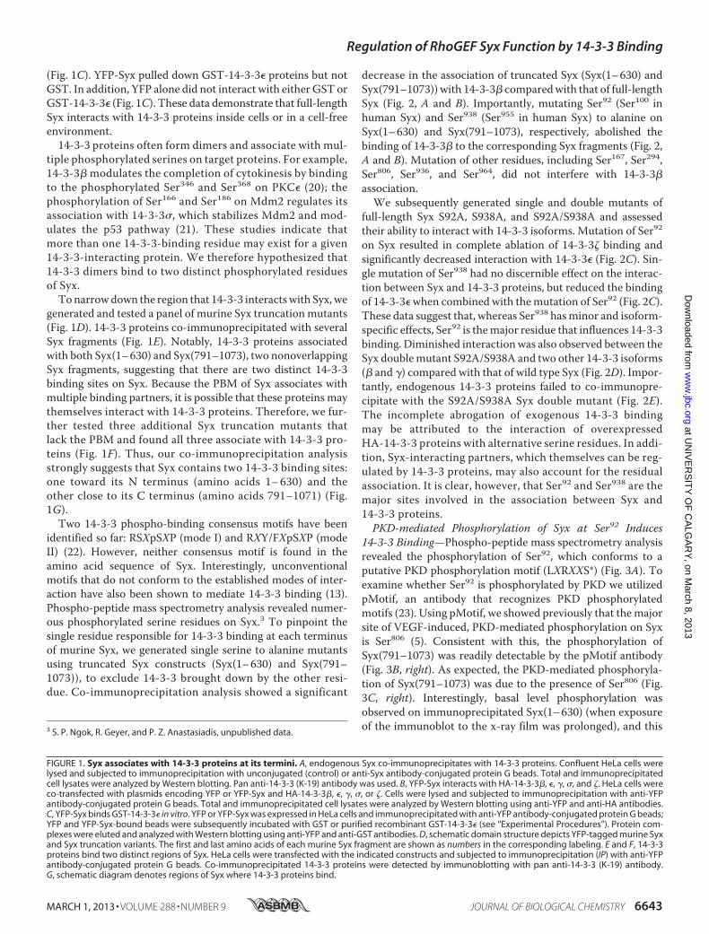
(Fig. 1C). YFP-Syx pulled down GST-14-3-3� proteins but notGST. In addition, YFP alone did not interact with either GST orGST-14-3-3� (Fig. 1C). These data demonstrate that full-lengthSyx interacts with 14-3-3 proteins inside cells or in a cell-freeenvironment.14-3-3 proteins often form dimers and associate with mul-
tiple phosphorylated serines on target proteins. For example,14-3-3� modulates the completion of cytokinesis by bindingto the phosphorylated Ser346 and Ser368 on PKC� (20); thephosphorylation of Ser166 and Ser186 on Mdm2 regulates itsassociation with 14-3-3�, which stabilizes Mdm2 and mod-ulates the p53 pathway (21). These studies indicate thatmore than one 14-3-3-binding residue may exist for a given14-3-3-interacting protein. We therefore hypothesized that14-3-3 dimers bind to two distinct phosphorylated residuesof Syx.Tonarrowdown the region that 14-3-3 interactswith Syx,we
generated and tested a panel of murine Syx truncationmutants(Fig. 1D). 14-3-3 proteins co-immunoprecipitated with severalSyx fragments (Fig. 1E). Notably, 14-3-3 proteins associatedwith both Syx(1–630) and Syx(791–1073), two nonoverlappingSyx fragments, suggesting that there are two distinct 14-3-3binding sites on Syx. Because the PBM of Syx associates withmultiple binding partners, it is possible that these proteins maythemselves interact with 14-3-3 proteins. Therefore, we fur-ther tested three additional Syx truncation mutants thatlack the PBM and found all three associate with 14-3-3 pro-teins (Fig. 1F). Thus, our co-immunoprecipitation analysisstrongly suggests that Syx contains two 14-3-3 binding sites:one toward its N terminus (amino acids 1–630) and theother close to its C terminus (amino acids 791–1071) (Fig.1G).Two 14-3-3 phospho-binding consensus motifs have been
identified so far: RSXpSXP (mode I) and RXY/FXpSXP (modeII) (22). However, neither consensus motif is found in theamino acid sequence of Syx. Interestingly, unconventionalmotifs that do not conform to the established modes of inter-action have also been shown to mediate 14-3-3 binding (13).Phospho-peptide mass spectrometry analysis revealed numer-ous phosphorylated serine residues on Syx.3 To pinpoint thesingle residue responsible for 14-3-3 binding at each terminusof murine Syx, we generated single serine to alanine mutantsusing truncated Syx constructs (Syx(1–630) and Syx(791–1073)), to exclude 14-3-3 brought down by the other resi-due. Co-immunoprecipitation analysis showed a significant
decrease in the association of truncated Syx (Syx(1–630) andSyx(791–1073)) with 14-3-3� comparedwith that of full-lengthSyx (Fig. 2, A and B). Importantly, mutating Ser92 (Ser100 inhuman Syx) and Ser938 (Ser955 in human Syx) to alanine onSyx(1–630) and Syx(791–1073), respectively, abolished thebinding of 14-3-3� to the corresponding Syx fragments (Fig. 2,A and B). Mutation of other residues, including Ser167, Ser294,Ser806, Ser936, and Ser964, did not interfere with 14-3-3�association.We subsequently generated single and double mutants of
full-length Syx S92A, S938A, and S92A/S938A and assessedtheir ability to interact with 14-3-3 isoforms. Mutation of Ser92
on Syx resulted in complete ablation of 14-3-3� binding andsignificantly decreased interaction with 14-3-3� (Fig. 2C). Sin-gle mutation of Ser938 had no discernible effect on the interac-tion between Syx and 14-3-3 proteins, but reduced the bindingof 14-3-3�when combined with themutation of Ser92 (Fig. 2C).These data suggest that, whereas Ser938 hasminor and isoform-specific effects, Ser92 is themajor residue that influences 14-3-3binding. Diminished interactionwas also observed between theSyx doublemutant S92A/S938A and two other 14-3-3 isoforms(� and �) compared with that of wild type Syx (Fig. 2D). Impor-tantly, endogenous 14-3-3 proteins failed to co-immunopre-cipitate with the S92A/S938A Syx double mutant (Fig. 2E).The incomplete abrogation of exogenous 14-3-3 bindingmay be attributed to the interaction of overexpressedHA-14-3-3 proteins with alternative serine residues. In addi-tion, Syx-interacting partners, which themselves can be reg-ulated by 14-3-3 proteins, may also account for the residualassociation. It is clear, however, that Ser92 and Ser938 are themajor sites involved in the association between Syx and14-3-3 proteins.PKD-mediated Phosphorylation of Syx at Ser92 Induces
14-3-3 Binding—Phospho-peptide mass spectrometry analysisrevealed the phosphorylation of Ser92, which conforms to aputative PKD phosphorylation motif (LXRXXS*) (Fig. 3A). Toexamine whether Ser92 is phosphorylated by PKD we utilizedpMotif, an antibody that recognizes PKD phosphorylatedmotifs (23). Using pMotif, we showed previously that themajorsite of VEGF-induced, PKD-mediated phosphorylation on Syxis Ser806 (5). Consistent with this, the phosphorylation ofSyx(791–1073) was readily detectable by the pMotif antibody(Fig. 3B, right). As expected, the PKD-mediated phosphoryla-tion of Syx(791–1073) was due to the presence of Ser806 (Fig.3C, right). Interestingly, basal level phosphorylation wasobserved on immunoprecipitated Syx(1–630) (when exposureof the immunoblot to the x-ray film was prolonged), and this3 S. P. Ngok, R. Geyer, and P. Z. Anastasiadis, unpublished data.
FIGURE 1. Syx associates with 14-3-3 proteins at its termini. A, endogenous Syx co-immunoprecipitates with 14-3-3 proteins. Confluent HeLa cells werelysed and subjected to immunoprecipitation with unconjugated (control) or anti-Syx antibody-conjugated protein G beads. Total and immunoprecipitatedcell lysates were analyzed by Western blotting. Pan anti-14-3-3 (K-19) antibody was used. B, YFP-Syx interacts with HA-14-3-3�, �, �, �, and �. HeLa cells wereco-transfected with plasmids encoding YFP or YFP-Syx and HA-14-3-3�, �, �, �, or �. Cells were lysed and subjected to immunoprecipitation with anti-YFPantibody-conjugated protein G beads. Total and immunoprecipitated cell lysates were analyzed by Western blotting using anti-YFP and anti-HA antibodies.C, YFP-Syx binds GST-14-3-3� in vitro. YFP or YFP-Syx was expressed in HeLa cells and immunoprecipitated with anti-YFP antibody-conjugated protein G beads;YFP and YFP-Syx-bound beads were subsequently incubated with GST or purified recombinant GST-14-3-3� (see “Experimental Procedures”). Protein com-plexes were eluted and analyzed with Western blotting using anti-YFP and anti-GST antibodies. D, schematic domain structure depicts YFP-tagged murine Syxand Syx truncation variants. The first and last amino acids of each murine Syx fragment are shown as numbers in the corresponding labeling. E and F, 14-3-3proteins bind two distinct regions of Syx. HeLa cells were transfected with the indicated constructs and subjected to immunoprecipitation (IP) with anti-YFPantibody-conjugated protein G beads. Co-immunoprecipitated 14-3-3 proteins were detected by immunoblotting with pan anti-14-3-3 (K-19) antibody.G, schematic diagram denotes regions of Syx where 14-3-3 proteins bind.
Regulation of RhoGEF Syx Function by 14-3-3 Binding
MARCH 1, 2013 • VOLUME 288 • NUMBER 9 JOURNAL OF BIOLOGICAL CHEMISTRY 6643
at UN
IVE
RS
ITY
OF
CA
LGA
RY
, on March 8, 2013
ww
w.jbc.org
Dow
nloaded from

FIGURE 2. Ser92 and Ser938 mediate 14-3-3 binding to Syx. A, Ser92 is a putative 14-3-3-binding residue. HeLa cells were co-transfected with plasmidsencoding HA-14-3-3� and YFP, YFP-Syx, YFP-Syx(1– 630), or one of three YFP-Syx(1– 630) mutants in which a single serine was mutated to alanine. Cells weresubjected to immunoprecipitation, and protein samples were analyzed by Western blotting. Only the replacement of Ser92 with alanine abolished theinteraction between Syx(1– 630) and 14-3-3�. B, Ser938 is a putative 14-3-3-binding residue. As in A, HeLa cells were co-transfected with plasmids encodingHA-14-3-3� and YFP, YFP-Syx, YFP-Syx(791–1073), or one of four YFP-Syx(791–1073) in which a single serine was mutated to alanine. Only the replacement ofSer938 with alanine abolished the interaction between Syx(791–1073) and 14-3-3�. Note that Ser806 was previously shown as a PKD-mediated phosphorylationtarget; mutating Ser806 to alanine, however, has no effect on the binding of 14-3-3�. C and D, Ser92 and Ser938 are essential residues for 14-3-3 interaction. HeLacells were co-transfected with the indicated constructs and subjected to immunoprecipitation. Protein samples were analyzed by Western blot analysis.Mutating Ser92 and Ser938 on Syx either abolished or diminished its interaction with 14-3-3 isoforms. Note that mutation of Ser938 alone in C has no clear effecton 14-3-3 binding, but resulted in a decrease or absence of 14-3-3 co-immunoprecipitates when combined with S92A mutation. E, endogenous 14-3-3 proteinsfail to co-immunoprecipitate with Syx S92A/S938A. HeLa cells were transfected with the indicated constructs and subjected to immunoprecipitation. Proteinsamples were analyzed by Western blotting. Mutating Ser92 and Ser938 on Syx significantly diminished its interaction with endogenous 14-3-3 proteins.
Regulation of RhoGEF Syx Function by 14-3-3 Binding
6644 JOURNAL OF BIOLOGICAL CHEMISTRY VOLUME 288 • NUMBER 9 • MARCH 1, 2013
at UN
IVE
RS
ITY
OF
CA
LGA
RY
, on March 8, 2013
ww
w.jbc.org
Dow
nloaded from

phosphorylation was increased upon PMA stimulation, a con-dition that induces PKD activation (Fig. 3B, left). Notably,mutating Ser92 to alanine abolished the PMA-induced increaseof phosphorylation on Syx(1–630), further suggesting thatSer92 is a PKD phosphorylation target. Furthermore, binding ofHA-14-3-3� positively correlated with increased phosphoryla-tion of Syx(1–630) at Ser92 (Fig. 3, B andC, left). In contrast, nochange inHA-14-3-3� bindingwas detected for Syx(791–1073)or Syx(791–1073) S806A upon PMA treatment (Fig. 3C, right),
indicating that PKD-mediated phosphorylation of Ser806 doesnot affect 14-3-3 binding.To confirm this observation in full-length Syx, we repeated
the same immunoprecipitation experiments using YFP-SyxS806A, which binds 14-3-3 equally well as wild type Syx(supplemental Fig. 1). An identical pattern was observed,where increased Syx phosphorylation and 14-3-3 (�, �, and �)binding were detected upon PMA stimulation (Fig. 3D).Therefore, the data suggest that PKD is an important mod-
FIGURE 3. PMA/PKD mediated phosphorylation of Syx at Ser92. A, Ser92 is a putative PKD phosphorylation site. The pMotif antibody recognizes a consensusmotif that contains a leucine at the �5 position and an arginine at the �3 position. Ser92 and Ser806 are the only residues within Syx that conform to theconsensus motif. B, Syx(1– 630) is a PKD phosphorylation target. HeLa cells were co-transfected with the indicated constructs and subjected to DMSO (control)or PMA (100 nM, 10 min) treatment prior to immunoprecipitation. Protein samples were analyzed by Western blot. X-ray film was exposed to the same blot fordifferent lengths of time to visualize the phosphorylation (by pMotif) of Syx(1– 630) and Syx(791–1073). C, both control and PMA-induced 14-3-3 associationsrequire Syx Ser92. As in B, transfected cells were subjected to immunoprecipitation after treatment with DMSO (control) or PMA (100 nM, 10 min). Proteinsamples were analyzed by Western blotting. Note that the mutation of Ser92 to alanine abrogated the PMA-induced increase in phosphorylation seen withpMotif in Syx(1– 630). D, Ser92 is phosphorylated by PKD in addition to Ser806. A PMA-induced increase in pMotif phosphorylation and 14-3-3 binding wasobserved upon transfecting cells with full-length Syx mutant YFP-Syx S806A. As in B, transfected cells were subjected to immunoprecipitation, and proteinsamples were analyzed by Western blotting.
Regulation of RhoGEF Syx Function by 14-3-3 Binding
MARCH 1, 2013 • VOLUME 288 • NUMBER 9 JOURNAL OF BIOLOGICAL CHEMISTRY 6645
at UN
IVE
RS
ITY
OF
CA
LGA
RY
, on March 8, 2013
ww
w.jbc.org
Dow
nloaded from

FIGURE 4. PKD1 induces binding of 14-3-3 to Syx. A, interaction between Syx and 14-3-3 increases in the presence of PKD1. HeLa cells were co-transfectedwith the indicated constructs and subjected to immunoprecipitation. Expression of wild type or kinase-active (KA) PKD1, but not kinase-dead (KD) PKD1,increases the binding of endogenous 14-3-3 proteins to YFP-Syx. B and C, PMA induces 14-3-3 binding to Syx. HeLa cells were transfected with YFP-Syx andsubjected to DMSO (control) or PMA treatment (10 versus 100 nM PMA in B, 1, 5, or 10 min of 100 nM PMA in C prior to lysing and immunoprecipitation. Increased14-3-3 binding to Syx was observed in a concentration- and time-dependent fashion. D, shRNA mediated down-regulation of PKD1 and PKD2. Western blotanalysis shows the protein levels of PKD1/2 in nontarget versus PKD1 and PKD2 shRNA-expressing HeLa cells. E, association of YFP-Syx with 14-3-3 isoforms isenhanced by PKD1 activation. PKD1/2-depleted HeLa cells were transfected with YFP-Syx, pcDNA (empty vector (EV)) or PKD1, and HA-14-3-3� or � andsubjected to DMSO (control) or PMA (100 nM) treatment prior to immunoprecipitation. Total and immunoprecipitated protein samples were analyzed byWestern blotting. F, Down-regulation of endogenous PKD1/2 suppresses Ser92 phosphorylation and 14-3-3 binding. PKD1/2-depleted HeLa cells were trans-fected with YFP-Syx S806A and subjected to immunoprecipitation. A decrease in pMotif signal and 14-3-3 binding was observed in YFP-Syx S806A uponPKD1/2 depletion. G, Ser92 phosphorylation and 14-3-3 binding are increased by PMA stimulation and PKD1 expression. PKD1/2-depleted HeLa cells weretransfected with YFP-Syx S806A and subjected to DMSO (control) or PMA treatment (100 nM, 10 min) prior to lysing and immunoprecipitation. Stepwiseincrease of pMotif staining and co-immunoprecipitated 14-3-3 was observed with PMA stimulation or PKD1 expression, or both. H, increase of Ser92 phosphor-ylation and 14-3-3 binding is dependent on PKD1 expression. PKD1/2-depleted HeLa cells were co-transfected with the indicated constructs and subjected toimmunoprecipitation. Expression of wild type or kinase-active PKD1, but not kinase-dead PKD1, increased the pMotif staining of YFP-Syx S806A and thebinding of endogenous 14-3-3 proteins to YFP-Syx S806A.
Regulation of RhoGEF Syx Function by 14-3-3 Binding
6646 JOURNAL OF BIOLOGICAL CHEMISTRY VOLUME 288 • NUMBER 9 • MARCH 1, 2013
at UN
IVE
RS
ITY
OF
CA
LGA
RY
, on March 8, 2013
ww
w.jbc.org
Dow
nloaded from

ulator of Syx phosphorylation and association with 14-3-3proteins.To test further this hypothesis, we co-expressed YFP-Syx
with PKD1 variants and observed a higher amount of co-immu-noprecipitated endogenous 14-3-3 proteins in HeLa cells thatexpressed either wild type or kinase-active (but not kinase-dead) PKD1 (Fig. 4A). Increased endogenous 14-3-3 binding toYFP-Syx also correlated strongly with PMA-induced PKD acti-vation in a concentration- and time-dependent manner (Fig. 4,B and C). We validated these observations by expressing YFP-Syx, PKD1, and HA-14-3-3� or � in PKD1/2-depleted HeLacells (Fig. 4D) and performing co-immunoprecipitation analy-ses. Binding of 14-3-3� and � to Syx positively correlated with
the expression of PKD1 (Fig. 4E). To determine whether thePKD-mediated phosphorylation of Ser92 is physiological andcorrelates with binding of endogenous 14-3-3 proteins, weexpressed and immunoprecipitated YFP-Syx S806A fromPKD1/2-depleted cells. Down-regulation of endogenousPKD1/2 decreased both Ser92 phosphorylation and 14-3-3binding (Fig. 4F). Furthermore, increased Ser92 phosphoryla-tion and 14-3-3 binding positively correlated with the expres-sion of wild type PKD1 and PMA stimulation (Fig. 4G) and theexpression of wild type and kinase-active PKD1 (Fig. 4H) inPKD1/2-depleted cells. Combined, the data indicate that theinteraction between Syx and 14-3-3 proteins is regulated byPKD-mediated phosphorylation at Ser92.
FIGURE 5. Binding of 14-3-3 proteins inhibits the nucleotide exchange activity of Syx. A, 14-3-3-uncoupled Syx has high GEF activity. HeLa cells weretransfected with plasmids encoding YFP, YFP-Syx, or YFP-Syx S92A/S938A. Cells were lysed 24 h after transfection, and the supernatants were incubated withGST-fused Rhotekin-RBD beads to bind active RhoA. Total and pulled-down active RhoA were determined by SDS-PAGE and immunoblotting. B, Syx S92A/S938A is highly active. As in A, cells were transfected with the indicated constructs and lysed 24 h after transfection. The supernatants were incubated withRhoA G17A-conjugated glutathione beads to pull down active RhoGEFs. Total and pulled-down active Syx were analyzed by SDS-PAGE and immunoblotting.C, expression of 14-3-3� and � inhibits Syx-induced RhoA activation. HeLa cells were co-transfected with YFP-Syx and an increasing amount (0, 1, or 2 �g,respectively) of HA-14-3-3� or �. As in A, active RhoA pulldown assay was performed, and protein samples were analyzed by Western blotting. D, SyxS92A/S938A-induced RhoA activation is unaffected by the expression of 14-3-3� or �. As in C, active RhoA was pulled down from lysates of HeLa cellsco-expressing YFP-Syx S92A/S938A and an increasing amount (0, 1, or 2 �g, respectively) of HA-14-3-3� or �. Total and active RhoA were determined bySDS-PAGE and immunoblotting.
Regulation of RhoGEF Syx Function by 14-3-3 Binding
MARCH 1, 2013 • VOLUME 288 • NUMBER 9 JOURNAL OF BIOLOGICAL CHEMISTRY 6647
at UN
IVE
RS
ITY
OF
CA
LGA
RY
, on March 8, 2013
ww
w.jbc.org
Dow
nloaded from

Binding of 14-3-3 Proteins Modulates the Function of Syx—To determine the physiological significance of 14-3-3 binding,we sought to evaluate the guanine nucleotide exchange activityof 14-3-3-uncoupled Syx. Because Syx was classified as a RhoA-specific GEF (6), experiments utilizing GST-tagged Rhotekin-RBD protein beads were performed to pull down GTP-boundRhoA from lysates of HeLa cells expressing YFP, YFP-Syx, orYFP-Syx S92A/S938A. Expression of YFP-Syx increased theglobal RhoA activation in cells compared with YFP alone;meanwhile, Syx S92A/S938A-expressing cells exhibited signif-icantly higher RhoA activity than Syx-expressing cells (Fig. 5A).To demonstrate that the observed increase in RhoA activationwas a direct result of increasedGEF activity of Syx S92A/S938A,we performed active RhoGEF pulldown assays. By couplingrecombinant RhoAG17A to glutathione beads, the nucleotide-
free RhoA mutant acts as a trap and binds activated RhoGEFswith high affinity (24). In agreement with our active RhoAassay, a substantially higher amount of Syx S92A/S938A wasbrought down in the active RhoGEF pulldown assay (Fig. 5B).The observation that Syx S92A/S938A exhibits elevated
nucleotide exchange activity suggested that 14-3-3 bindingsuppresses the GEF activity of Syx. To test this hypothesis, weperformed active RhoA pulldown assays using lysates fromHeLa cells that co-expressed YFP-Syx and HA-tagged 14-3-3�or �. A high level of RhoA activation was observed when cellswere transfected with Syx. The Syx-induced RhoA activation,however, was counteracted by the increased expression of14-3-3� or � (Fig. 5C). In contrast, this suppression of RhoAactivation did not occur when 14-3-3-uncoupled Syx S92A/S938A was co-expressed with 14-3-3 proteins (Fig. 5D), indi-
FIGURE 6. 14-3-3-uncoupled Syx is strongly targeted to the cell border and enhances circumferential actin accumulation. A, Syx S92A/S938A localizesstrongly at areas of cell-cell contact. MDCK cells were transfected with plasmids encoding YFP-Syx or YFP-Syx S92A/S938A. Cells were fixed and immunostainedfor YFP, ZO1, and DAPI. B, Syx S92A/S938A induces the accumulation of circumferential actin at cell contacts. MDCK cells were transfected with the indicatedconstructs, fixed 24 h after transfection, and immunostained for YFP, phalloidin (F-actin), and DAPI. C, A quantitative analysis of enhanced circumferential actinstaining in transfected cells from B was performed. The intensity of circumferential actin at the junctions of transfected cells was compared with that ofimmediate neighboring nontransfected cells using ImageJ; transfected cells that exhibited intensified circumferential actin staining are expressed as percent-age of total cells counted (mean � S.E. (error bars), n � 3, 50 cells/coverslip analyzed). Scale bars, 10 �m.
Regulation of RhoGEF Syx Function by 14-3-3 Binding
6648 JOURNAL OF BIOLOGICAL CHEMISTRY VOLUME 288 • NUMBER 9 • MARCH 1, 2013
at UN
IVE
RS
ITY
OF
CA
LGA
RY
, on March 8, 2013
ww
w.jbc.org
Dow
nloaded from

cating that this effect of 14-3-3 proteins was not due to off-target effects but specific to Syx binding. Therefore, the datastrongly argue that 14-3-3 binding modulates the GEF activityof Syx.Previously, we demonstrated that the targeting of Syx to
areas of cell-cell contact is critical for junction integrity (5).Therefore, in addition to the effects on RhoA activity, wesought to determine whether 14-3-3 binding also affects Syxlocalization. When expressed in MDCK cells, 14-3-3-uncou-pled Syx showed a notably stronger targeting to the cell junc-tions compared with its wild type counterpart (Fig. 6A, boxes).This observation implied that the interaction with 14-3-3 pro-teins could modify the intracellular localization of Syx.We showed previously that Syxmediates junction stability by
activating RhoA and its effector Dia1 specifically at cell-cellcontacts (5). To further evaluate the function of Syx S92A/
S938A, we assessed its ability to induce changes to the circum-ferential actin cytoskeleton, which correlates with the maturityand stability of adherens junctions. Cells expressing Syx S92A/S938A, but not wild type Syx, displayed enriched circumferen-tial actin staining compared with neighboring cells (Fig. 6B).When the intensity of cortical actin staining was quantified, weobserved significantly increased junctional actin staining incells expressing Syx S92A/S938A (Fig. 6C). The strengtheningof the cortical actin ring is likely the result of elevated GEFactivity of Syx S92A/S938A, which increases RhoA activation atareas of cell-cell contact. Our data support the importance ofjunctional Syx in regulating cell-cell adhesion and further delin-eate the molecular mechanisms that regulate Syx function.
DISCUSSION
The ubiquitously expressed 14-3-3 isoforms bind to a largenumber of proteins and mediate a remarkable range of cellularactivities. However, regulation of cell-cell adhesion by 14-3-3proteins is largely unknown. Here, our data strongly suggestthat Syx-regulated junction stability is in part modulated byPKD-mediated 14-3-3 binding.There are two aspects that define the functional relationship
between Syx and 14-3-3 proteins: the effects of 14-3-3 bindingon the localization and the guanine nucleotide exchange activ-ity of Syx. The 14-3-3-uncoupled mutant Syx S92A/S938Aexhibits strong junctional localization, suggesting that thebinding of 14-3-3 proteins has a negative impact on Syx local-ization and therefore its function tomaintain junction stability.Our previous study identified Syx as a target of VEGF-induced,PKD1-mediated phosphorylation at Ser806, an event thatinduced the mislocalization of Syx away from cell junctions.Mutating Ser806 to alanine did not interfere with the ability ofSyx to bind 14-3-3 proteins, but it did induce Mupp1 associa-tion and promoted the junctional localization of Syx (5). It alsoallowed us to detect the unexpected phosphorylation of Ser92by PKD1. From a structural perspective, phosphorylation ofSer806may induce conformational changes in Syx that promotenot the association per se, but the dimerization of 14-3-3 pro-teins associated with the N- and C-terminal regions of Syx andthe subsequent dissociation from cell junctions (Fig. 7). Impor-tantly, prior data suggest that the N-terminal domain (Syx(1–300)) can suppress Syx exchange activity (5). Indeed, severalRhoGEFs are subjected to inhibitory intramolecular interac-tions between their N-terminal domains and their catalytic Dblhomology domains (25). Assuming a similar mode of action,14-3-3 binding at the N and C termini and subsequentdimerization could promote an inactive Syx conformation, thusexplaining the negative effect of 14-3-3 binding on Syx GEFactivity (Fig. 7). An intramolecular interaction between Syx(1–300) with the Dbl homology domain may also explain theobserved lack of 14-3-3 binding to Syx(1–300) (Fig. 1E), despitethe presence of Ser92 within this fragment. We postulate thatthe intramolecular interaction results in three-dimensionalfolding changes that allow Ser92 phosphorylation and associa-tion with 14-3-3 proteins. Whatever the mechanism, it is clearthat PKD is a key regulator of junctional Syx function. Adhesionis a dynamic process, and as such, the function of Syx is highlyregulated by a multitude of events, including its autoinhibitory
FIGURE 7. Proposed mechanism of PKD-mediated 14-3-3 binding to Syx.A, membrane-localized Syx activates RhoA locally. B, PKD-mediated phosphor-ylation of Ser92 upon proper folding of Syx N terminus induces binding of14-3-3 protein. C, phosphorylation of Ser938 promotes 14-3-3 binding at the Cterminus. Additional phosphorylation of Ser806 (data not shown) by PKDinduces conformational changes to the C terminus and brings the two 14-3-3proteins into close proximity. As a consequence, dimerization of terminallybound 14-3-3 proteins locks Syx in an inactive state (suppresses GEF activity),and Syx is displaced from areas of cell-cell contact. PH, pleckstrin homologydomain; DH, Dbl homology domain.
Regulation of RhoGEF Syx Function by 14-3-3 Binding
MARCH 1, 2013 • VOLUME 288 • NUMBER 9 JOURNAL OF BIOLOGICAL CHEMISTRY 6649
at UN
IVE
RS
ITY
OF
CA
LGA
RY
, on March 8, 2013
ww
w.jbc.org
Dow
nloaded from

N terminus, the phosphorylation of Ser92, Ser806, and Ser938 (ofwhich the upstreamkinase is currently unknown), and the asso-ciation of 14-3-3 proteins.RhoA is thought to regulate signaling events that contribute
to cell junction formation and preservation (4, 26). Dia1, aRhoA effector that is proposed to regulate cell junction stability(27, 28), belongs to the formin family and can remodel the actincytoskeleton (29). We previously identified Dia1 as the keydownstream effector of Syx in regulating junction integrity (5).Here, we observed increased RhoA activation and intensifiedactin staining at areas of cell-cell contact in cells expressing SyxS92A/S938A. We postulate that the increased junctional tar-geting and elevated GEF activity of Syx S92A/S938A results inthe subsequent Rho-mediated activation ofDia1, the expansionof the circumferential actin bundle, and the stabilization of cell-cell contacts.In conclusion, our findings indicate a role for 14-3-3 proteins
in the regulation of cell-cell junctions by modulating the func-tion of Syx. Based on studies conducted on Syx thus far, it islikely that Syx is regulated by the integration of signals frommultiple upstream pathways. Uncovering additional mecha-nisms will provide further insights as to how Syx functions.Interestingly, our data suggest that the junction disruptiveeffects of tumor promoting phorbol esters may be mediated bySyx dysfunction. Finally, despite its function in junction integ-rity, the role of Syx in human cancer is currently unknown.
Acknowledgments—We thank Drs. Alan Fields, Aubrey Thompson,NicoleMurray, and Larry Karnitz (Mayo Clinic) for critical comments.
REFERENCES1. Etienne-Manneville, S., and Hall, A. (2002) Rho GTPases in cell biology.
Nature 420, 629–6352. Jaffe, A. B., and Hall, A. (2005) Rho GTPases: biochemistry and biology.
Annu. Rev. Cell Dev. Biol. 21, 247–2693. Iden, S., and Collard, J. G. (2008) Crosstalk between small GTPases and
polarity proteins in cell polarization. Nat. Rev. Mol. Cell Biol. 9, 846–8594. Nelson, W. J. (2008) Regulation of cell-cell adhesion by the cadherin-
catenin complex. Biochem. Soc. Trans. 36, 149–1555. Ngok, S. P., Geyer, R., Liu, M., Kourtidis, A., Agrawal, S., Wu, C., Seerapu,
H. R., Lewis-Tuffin, L. J., Moodie, K. L., Huveldt, D., Marx, R., Baraban,J. M., Storz, P., Horowitz, A., and Anastasiadis, P. Z. (2012) VEGF andangiopoietin-1 exert opposing effects on cell junctions by regulating theRhoGEF Syx. J. Cell Biol. 199, 1103–1115
6. De Toledo,M., Coulon, V., Schmidt, S., Fort, P., and Blangy, A. (2001) Thegene for a new brain specific RhoA exchange factor maps to the highlyunstable chromosomal region 1p36.2–1p36.3. Oncogene 20, 7307–7317
7. Ernkvist, M., Luna Persson, N., Audebert, S., Lecine, P., Sinha, I., Liu, M.,Schlueter, M., Horowitz, A., Aase, K.,Weide, T., Borg, J. P., Majumdar, A.,and Holmgren, L. (2009) The Amot/Patj/Syx signaling complex spatiallycontrols RhoA GTPase activity in migrating endothelial cells. Blood 113,244–253
8. Garnaas, M. K., Moodie, K. L., Liu, M. L., Samant, G. V., Li, K., Marx, R.,Baraban, J. M., Horowitz, A., and Ramchandran, R. (2008) Syx, a RhoAguanine exchange factor, is essential for angiogenesis in vivo. Circ. Res.103, 710–716
9. Liu, M., and Horowitz, A. (2006) A PDZ-binding motif as a critical deter-
minant of Rho guanine exchange factor function and cell phenotype.Mol.Biol. Cell 17, 1880–1887
10. Estévez, M. A., Henderson, J. A., Ahn, D., Zhu, X. R., Poschmann, G.,Lübbert, H., Marx, R., and Baraban, J. M. (2008) The neuronal RhoAGEF,Tech, interacts with the synaptic multi-PDZ-domain-containing protein,MUPP1. J. Neurochem. 106, 1287–1297
11. Morrison, D. K. (2009) The 14-3-3 proteins: integrators of diverse signal-ing cues that impact cell fate and cancer development. Trends Cell Biol.19, 16–23
12. Tzivion, G., Gupta, V. S., Kaplun, L., and Balan, V. (2006) 14-3-3 proteinsas potential oncogenes. Semin. Cancer Biol. 16, 203–213
13. Aitken, A. (2006) 14-3-3 proteins: a historic overview. Semin. Cancer Biol.16, 162–172
14. Ling, C., Zuo, D., Xue, B., Muthuswamy, S., and Muller, W. J. (2010) Anovel role for 14-3-3� in regulating epithelial cell polarity. Genes Dev. 24,947–956
15. Hurd, T.W., Fan, S., Liu, C. J., Kweon, H. K., Hakansson, K., andMargolis,B. (2003) Phosphorylation-dependent binding of 14-3-3 to the polarityprotein Par3 regulates cell polarity inmammalian epithelia.Curr. Biol. 13,2082–2090
16. Eiseler, T., Döppler, H., Yan, I. K., Kitatani, K., Mizuno, K., and Storz, P.(2009) Protein kinase D1 regulates cofilin-mediated F-actin reorganiza-tion and cell motility through slingshot. Nat. Cell Biol. 11, 545–556
17. Storz, P., Döppler, H., and Toker, A. (2004) Protein kinase C� selectivelyregulates protein kinase D-dependent activation of NF-�B in oxidativestress signaling.Mol. Cell. Biol. 24, 2614–2626
18. Yanagisawa,M., andAnastasiadis, P. Z. (2006) p120 catenin is essential formesenchymal cadherin-mediated regulation of cell motility and invasive-ness. J. Cell Biol. 174, 1087–1096
19. Yanagisawa, M., Huveldt, D., Kreinest, P., Lohse, C. M., Cheville, J. C.,Parker, A. S., Copland, J. A., and Anastasiadis, P. Z. (2008) A p120 cateninisoform switch affects Rho activity, induces tumor cell invasion, and pre-dicts metastatic disease. J. Biol. Chem. 283, 18344–18354
20. Saurin, A. T., Durgan, J., Cameron, A. J., Faisal, A., Marber, M. S., andParker, P. J. (2008)The regulated assembly of a PKC� complex controls thecompletion of cytokinesis. Nat. Cell Biol. 10, 891–901
21. Wood, N. T., Meek, D. W., and Mackintosh, C. (2009) 14-3-3 binding toPim-phosphorylated Ser-166 and Ser-186 of human Mdm2: potential in-terplay with the PKB/Akt pathway and p14(ARF). FEBS Lett. 583,615–620
22. Yaffe, M. B., Rittinger, K., Volinia, S., Caron, P. R., Aitken, A., Leffers, H.,Gamblin, S. J., Smerdon, S. J., andCantley, L. C. (1997) The structural basisfor 14-3-3:phosphopeptide binding specificity. Cell 91, 961–971
23. Döppler, H., Storz, P., Li, J., Comb,M. J., and Toker, A. (2005) A phosphor-ylation state-specific antibody recognizes Hsp27, a novel substrate of pro-tein kinase D. J. Biol. Chem. 280, 15013–15019
24. García-Mata, R.,Wennerberg, K., Arthur,W. T., Noren, N. K., Ellerbroek,S. M., and Burridge, K. (2006) Analysis of activated GAPs and GEFs in celllysates.Methods Enzymol. 406, 425–437
25. Rossman, K. L., Der, C. J., and Sondek, J. (2005) GEFmeans go: turning onRho GTPases with guanine nucleotide-exchange factors. Nat. Rev. Mol.Cell Biol. 6, 167–180
26. Braga, V.M. (2002) Cell-cell adhesion and signalling.Curr. Opin. Cell Biol.14, 546–556
27. Carramusa, L., Ballestrem, C., Zilberman, Y., and Bershadsky, A. D. (2007)Mammalian diaphanous-related formin Dia1 controls the organization ofE-cadherin-mediated cell-cell junctions. J. Cell Sci. 120, 3870–3882
28. Ryu, J. R., Echarri, A., Li, R., and Pendergast, A. M. (2009) Regulation ofcell-cell adhesion by Abi/Diaphanous complexes. Mol. Cell. Biol. 29,1735–1748
29. Chesarone, M. A., DuPage, A. G., and Goode, B. L. (2010) Unleashingformins to remodel the actin and microtubule cytoskeletons. Nat. Rev.Mol. Cell Biol. 11, 62–74
Regulation of RhoGEF Syx Function by 14-3-3 Binding
6650 JOURNAL OF BIOLOGICAL CHEMISTRY VOLUME 288 • NUMBER 9 • MARCH 1, 2013
at UN
IVE
RS
ITY
OF
CA
LGA
RY
, on March 8, 2013
ww
w.jbc.org
Dow
nloaded from