porphyromonas gingivalis lipids inhibit osteoblastic ... · lps from p . gingivalis is ... quattro...
TRANSCRIPT

1
Porphyromonas gingivalis Lipids Inhibit Osteoblastic Differentiation and 2
Function 3
Revised manuscript 4
5
Yu-Hsiung Wang a, Jin Jiang
b, Qiang Zhu
b, Amer Z. AlAnezi
b, Robert B. Clark
c, Xi Jiang
d, 6
David W. Rowe d and Frank C. Nichols
e 7
8
a Department of Craniofacial Sciences, Division of Pediatric Dentistry, University of 9
Connecticut, School of Dental Medicine, Farmington, CT 06030, USA 10
b Department of Oral Health and Diagnostic Sciences, Division of Endodontology, University of 11
Connecticut, School of Dental Medicine, Farmington, CT 06030, USA 12
c Departments of Immunology and Medicine, University of Connecticut, School of Medicine, 13
Farmington, CT 06030, USA 14
d Department of Reconstructive Sciences, University of Connecticut, School of Dental Medicine, 15
Farmington, CT 06030, USA 16
e Corresponding Author: Department of Oral Health and Diagnostic Sciences, Division of 17
Periodontology, University of Connecticut, School of Dental Medicine, Farmington, CT 06030, 18
USA. 19
Corresponding author: E-mail: [email protected] 20
Phone: (860) 679-3725 21
FAX: (860) 679-1027 22
23
Copyright © 2010, American Society for Microbiology and/or the Listed Authors/Institutions. All Rights Reserved.Infect. Immun. doi:10.1128/IAI.00225-10 IAI Accepts, published online ahead of print on 28 June 2010
on Novem
ber 25, 2018 by guesthttp://iai.asm
.org/D
ownloaded from

2
ABSTRACT 1
Porphyromonas gingivalis (P. gingivalis) produces unusual sphingolipids that are known to 2
promote inflammatory reactions in gingival fibroblasts and Toll-like receptor (TLR)2-dependent 3
secretion of IL-1 from dendritic cells. The aim of the present study was to examine whether P. 4
gingivalis lipids inhibit osteoblastic function. Total lipids from P. gingivalis and two fractions, 5
phosphoglycerol dihydroceramides and phosphoethanolamine dihydroceramides, were prepared 6
free of lipid A. Primary calvarial osteoblast cultures derived from 5-7 day old CD-1 mice were 7
used to examine the effects of P. gingivalis lipids on mineralized nodule formation, cell viability, 8
apoptosis, cell proliferation, and gene expression. P. gingivalis lipids inhibited osteoblast 9
differentiation and fluorescence expression of pOBCol2.3GFP in a concentration-dependent 10
manner. However, P. gingivalis lipids did not significantly alter osteoblast proliferation, 11
viability, or apoptosis. When administered during specific intervals of osteoblast growth, P. 12
gingivalis total lipids demonstrated inhibitory effects on osteoblast differentiation only after the 13
proliferation stage of culture. RTPCR confirmed down-regulation of osteoblast marker genes 14
including Runx2, ALP, OC, BSP, OPG and DMP-1 with concurrent up-regulation of RANKL, 15
TNF-α and MMP-3 genes. P. gingivalis total lipids and lipid fractions inhibited calvarial 16
osteoblast gene expression and function in vivo as determined by loss of expression of another 17
osteoblast differentiation reporter, pOBCol3.6GFPcyan, and reduced uptake of Alizurin 18
complexone stain. Finally, lipid inhibition of mineral nodule formation in vitro was dependent 19
on TLR2 expression. Our results indicate that inhibition of osteoblast function and gene 20
expression by P. gingivalis lipids represents a novel mechanism for altering alveolar bone 21
homeostasis at periodontal disease sites. 22
23
on Novem
ber 25, 2018 by guesthttp://iai.asm
.org/D
ownloaded from

3
INTRODUCTION 1
Bone loss under the influence of bacterial virulence factors is thought to occur through 2
engagement of receptors for pathogen associated molecular pattern molecules (PAMPs) resulting 3
in stimulation of osteoclasts and/or inhibition of osteoblasts. A frequently cited example is the 4
engagement of Toll-like receptor 4 (TLR4) by bacterial lipopolysaccharide (LPS) that is reported 5
to mediate bone loss in destructive periodontal diseases through activation of osteoclasts and 6
inhibition of osteoblasts (9, 21, 27, 40, 44, 45). A subgingival organism strongly associated with 7
destructive periodontal disease, Porphyromonas gingivalis, is a Gram-negative anaerobe that 8
produces LPS but this LPS is unique in that it has been shown to engage both TLR4 and TLR2 9
(4, 10, 20, 24, 37) although these reports are sometimes conflicting. Though inflammatory bone 10
loss in experimental periodontal diseases can be produced by engagement of TLR4 by LPS (28), 11
recent evidence indicates that periodontal bone loss in experimental animals mediated by live P. 12
gingivalis requires engagement of TLR2 (16). Furthermore, previous reports have shown that 13
LPS from P. gingivalis is present only to a negligible extent in diseased periodontal tissues in 14
humans (30, 33) and the lipid A of P. gingivalis cannot be detected on periodontally diseased 15
teeth or in diseased periodontal tissues (35). In contrast, unusual complex sphingolipids of P. 16
gingivalis, called phosphoethanolamine dihydroceramides (PE DHC) or phosphoglycerol 17
dihydroceramide (PG DHC) lipids, are prominent on periodontally diseased teeth and are 18
recovered in diseased periodontal tissues (35). We recently reported that the total lipids of P. 19
gingivalis and specifically the PE DHC lipids, will stimulate IL-6 secretion from dendritic cells 20
in vitro and this processs is mediated through TLR2 (31). The present investigation sought to 21
determine whether the lipids of P. gingivalis could potentially interfere with bone formation in 22
on Novem
ber 25, 2018 by guesthttp://iai.asm
.org/D
ownloaded from

4
periodontal disease sites through inhibition of osteoblast function or its phenotype and whether 1
this process requires engagement of TLR2. 2
on Novem
ber 25, 2018 by guesthttp://iai.asm
.org/D
ownloaded from

5
MATERIALS AND METHODS 1
Preparation and purification of P. gingivalis lipids 2
P. gingivalis (ATCC #33277, type strain) was inoculated into basal (peptone, trypticase 3
and yeast extract) medium supplemented with hemin and menadione (Sigma, St. Louis, MO) and 4
brain heart infusion (34). Culture purity was verified by Gram stain, lack of growth in aerobic 5
culture and formation of black colonies when inoculated on blood agar plates and grown under 6
anaerobic conditions. The suspension cultures were incubated in an anaerobic chamber flushed 7
with N2 (80%), CO2 (10%) and H2 (10%) at 37°C for five days and the bacteria were harvested 8
by centrifugation (3000 g for 20 min). Following lyophilization, approximately 4g of P. 9
gingivalis pellet was extracted for five days using a modification of the phospholipid extraction 10
procedure of Bligh and Dyer (7) and Garbus et al. (12). Specifically, 4 ml of H2O + 16 ml of 11
MeOH:CHCl3 (2:1 v/v) were added to the bacterial sample and vortexed. After 12 hours, 3 ml of 12
2 N KCl + 0.5 M K2HPO4 and 3 ml CHCl3 were added and the sample vortexed. The lower 13
organic phase was carefully removed and CHCl3 (3 ml) was added to each sample and vortexed. 14
The CHCl3 phase was removed and combined with the previous organic solvent extract. The 15
lipid extract from P. gingivalis was dried under nitrogen and stored frozen. This lipid 16
preparation was used as the total lipid extract for the experiments described below. 17
Fractionation of bacterial lipids by high-performance liquid chromatography (HPLC) was 18
accomplished using a semipreparative HPLC column (1 by 25 cm silica gel, 5 mm, Supelco Inc, 19
Bellefonte, PA) and eluted isocratically with hexane-isopropanol-water (6:8:0.75, vol/vol/vol: 20
Solvent A) (13). P. gingivalis lipid samples were dissolved in solvent A and centrifuged to 21
remove insoluble material. For each chromatographic separation, 20 mg of lipid was applied and 22
fractions were pooled from 20 column fractionations. Samples were eluted at 2.0 ml/min 23
on Novem
ber 25, 2018 by guesthttp://iai.asm
.org/D
ownloaded from
6
monitored at 205 nm with 1 min fractions collected. Fractions were dried under nitrogen and 1
resuspended in 2 ml of CHCl3. Lipid recovery in each HPLC fraction were determined by 2
drying 5 ml from each fraction onto a microbalance tray and weighing the tray using a Cahn 3
Electrobalance. Fractions shown to be enriched for PE DHC or PG DHC lipids by electrospray-4
MS (see below) were pooled, dried and refractionated. The refractionated lipid fractions were 5
then verified by electrospray-MS to be highly enriched as shown in Figure 1. 6
The lipid fractions were surveyed using electrospray (ESI)-MS analysis on a Micromass 7
Quattro II mass spectrometer system with a low flow HPLC introduction system as previously 8
described (34). Dihydroceramide lipid fractions were dissolved in hexane:isopropanol (6:8, v/v, 9
elution solvent) and were applied at a flow rate of 80 µl/min. For electrospray negative ion 10
analyses, the desolvation and inlet block temperatures were 80°C and 100°C, respectively, and 11
the transcapillary potential was 3000 volts. The cone voltage was usually held at 30 volts and 12
the mass acquisition range was 100-2000 amu for initial electrospray MS analyses. The lipid 13
preparations used for the present study were shown to be devoid of lipid A of P. gingivalis by 14
negative ion electrospray-MS (Fig. 1). Negative ions characteristic for lipid A species of P. 15
gingivalis were not observed in any of the lipid preparations shown in Figure 1 (10). 16
Primary calvarial osteoblast cultures 17
Primary cell cultures were established from calvarial cells isolated from 5- to 7-day-old 18
neonatal CD-1 mice or Toll-like receptor (TLR)2-/-
mice (a generous gift from Dr. S. Akira, 19
Osaka, Japan). Mice were maintained and bred in accordance with University of Connecticut 20
Center for Laboratory Animal Care regulations. Briefly, the calvariae were isolated from skulls 21
without sutures and adherent mesenchymal tissues and osteoblasts were released by subjecting 22
these calvariae to four sequential 15-min enzyme digestions at 37°C in a solution containing 23
on Novem
ber 25, 2018 by guesthttp://iai.asm
.org/D
ownloaded from
7
0.05% trypsin-EDTA and 0.1% collagenase P (Roche Diagnostics, Indianapolis, IN). Cells 1
released from the 2nd to 4th digestions were collected, centrifuged, resuspended, and plated at 2
1.5 x 10⁴ cells/cm
2 (i.e., 1.5 x 10
5 cells/well) in 35 mm 6-well culture plates in DMEM 3
(Invitrogen, Carlsbad, CA) containing 10% fetal calf serum, penicillin (100 units/ml), 4
streptomycin (100 µg/ml), and non-essential amino acids (100 µM). The day of plating was 5
designated day 0. Plated cells became confluent around day 5-6 at which point the culture 6
medium was changed to differentiation medium, which was α-MEM (Invitrogen) containing 7
10% fetal calf serum, penicillin (100 units/ml), streptomycin (100 µg/ml), ascorbic acid (50 8
µg/ml), and β-glycerophosphate (4 mM). Medium was changed every other day for the entire 9
duration (21 days) of culture. For the evaluation of osteoblast differentiation using fluorescence 10
microscopy, calvarial osteoblast cultures were derived from mice transgenic for pOBCol2.3GFP 11
(22) in which the green fluorescence protein (GFP) reporter was driven by a rat 2.3-kb type I 12
collagen promoter. For the evaluation of osteoblast engagement of TLR2 by P. gingivalis lipids, 13
calvarial osteoblast cultures were derived from TLR2 knockout mice that were backcrossed 9 14
times and verified by genotyping to be homozygous for TLR2-/-
. TLR2-/-
osteoblast cultures 15
were otherwise handled using the same culture protocol as described for CD-1 osteoblasts. 16
Assessment of mineralized nodule formation 17
Mineralization nodules were assessed using the modified von Kossa’s silver nitrate 18
staining method. Briefly, cultures were fixed in cold methanol for 15-20 min. After rinsing, the 19
fixed plates were incubated with a 5% silver nitrate solution under UV light using 2 cycles of 20
auto-crosslink (1200 mjoules x 100) in a UV Stratalinker (Strategene, La Jolla, CA). Mineralized 21
nodules were seen as dark brown to black spots. Culture plates stained with von Kossa were 22
scanned and the areas of mineralized nodules were quantitated using a computation program 23
on Novem
ber 25, 2018 by guesthttp://iai.asm
.org/D
ownloaded from

8
developed from Openlab (Improvision, Lexington, MA). The area of mineralized nodule 1
formation was quantified in pixels. 2
Examination of cell viability and apoptosis 3
Ethidium homodimer-1 (EthD-1) (Molecular Probes, Eugene, OR) was used to determine 4
cell viability. EthD-1, with a high affinity for DNA and low membrane permeability, permits the 5
use of a very low fluorescent dye concentration to demonstrate cell death. Osteoblast cultures 6
were incubated with EthD-1 at a final concentration of 2 µM for 30 minutes and were examined 7
for cell viability by fluorescence microscopy using a TRITC filter. The fluorescence image of 8
EthD-1 staining (red color) was photographed and cell death was quantified by calculating the 9
total number of cells with red stain divided by the total number of cells in the field. To examine 10
apoptosis, APC-conjugated annexin V was used with flow cytometry to detect apoptosis. 11
Annexin V has a high affinity for phosphatidylserine that is translocated to the outer surface of 12
the cell membrane during apoptosis. Harvested cultured cells were resuspended in annexin-13
binding buffer which consisted of 10 mM HEPES, 140 mM NaCl and 2.5 mM CaCl2 at pH 7.4. 14
APC-conjugated annexin V (Molecular Probes) was added to the cell suspension at 1:20 dilution 15
to label the apoptotic cells for 15 minutes. Cells were analyzed by flow cytometry using channel 16
FL4 to detect the apoptotic cells labeled with APC-conjugated annexin V. 17
RT-PCR analysis of gene expression 18
Total RNA was extracted from cultures using TRIzol reagent (Invitrogen) and 19
phenol/chloroform according to the manufacturer’s instructions. RNA was dissolved in Tris-20
EDTA pH 7.4 and the concentration of RNA was determined by absorbance at 260 nm. RNA 21
was treated with DNase I (Invitrogen) to remove genomic DNA contaminants. Reverse 22
transcription was carried out in a 20 µl volume containing about 3 µg of RNA, 1µl of 50 ng/µl 23
on Novem
ber 25, 2018 by guesthttp://iai.asm
.org/D
ownloaded from

9
random hexamers and 1 µl annealing buffer, 10 µl 2X first-strand reaction mix, and 2 µl 1
Superscript III/RNase OUT enzyme mix (Invitrogen) at 25°C for 10 minutes followed by 50°C 2
for 50 minutes. TaqMan real-time PCR was performed from 1 µl of cDNA using TaqMan 3
Universal PCR Master Mix (Applied Biosystems, Foster City, CA) with 100-nM primers and a 4
50-nM probe. The TaqMan real-time PCR was performed on a TaqMan ABI 7500 system 5
(Applied Biosystems). Unlabeled specific primers and the TaqMan MGB probes (6-FAM dye-6
labeled) for gene detection were: mouse Runx2 (assay ID: Mm00501580_m1), type I collagen 7
(Col1a1) (Mm00483888_ml), bone alkaline phosphatase (ALP) (Mm01187117_m1), bone 8
sialoprotein (BSP) (Mm00492555_m1), osteocalcin (OC) (Mm00649782_9H), dentin matrix 9
protein-1 (DMP-1) (Mm01208365_m1), matrix metalloproteinase-3 (MMP-3) 10
(Mm01168406_g1), osteoprotegerin (OPG) (Mm00435454-m1), tumor necrosis factor-α (TNF-11
α) (Mm00443258_m1), and receptor activator of NF-κB ligand (RANKL) (Mm01313944_g1). 12
GAPDH was used as internal control (Mm99999915_g1). Cycling conditions were as follows: 13
after an initial hold of 2 minutes at 50°C and 10 minutes at 95°C, the samples were cycled 40 14
times at 95°C for 15 seconds and 60°C for 1 minute. Each sample was assayed in triplicate. The 15
comparative Ct method was applied to determine comparative expression levels between 16
samples relative to control gene expression. To examine regulation by lipids, the amplification 17
threshold cycle value (Ct) from the lipid treated samples were subtracted from the untreated 18
sample cycle values (∆Ct = Ct untreated - Ct treated). The ratio was obtained by calculating the 19
values of each gene of interest against the house-keeping gene GAPDH. The fold change of the 20
test gene was determined as 2(∆Ct gene - ∆Ct GAPDH)
. 21
on Novem
ber 25, 2018 by guesthttp://iai.asm
.org/D
ownloaded from

10
In vivo inhibition of osteoblasts 1
Mice were bred to express the osteoblastic FP reporter, pOBCol3.6GFPcyan, on a CD-1 2
background as previously described (6). Lipid preparations, including total lipids, PE DHC 3
lipids and PG DHC lipids, were sonicated (2 min, 3 watts) in phosphate buffered saline to 4
achieve a concentration of 0.1 µg/ml. Mice were lightly sedated with isoflurane and lipid 5
preparations were administered subcutaneously to the calvaria midway between the ear and eye. 6
The right calvaria of each mouse received a subcutaneous injection of 5 µg of lipid. Control 7
mice calvaria received only phosphate buffered saline. Mice were administered Alizarin 8
complexone for labeling of newly deposited mineralized tissue during day 6 and were sacrificed 9
on day 7. Calvaria were decalcified, frozen sectioned and photographed for osteoblast reporter 10
(blue) and Alizarin complexone (red) by fluorescence microscopy. Next, sections were stained 11
with hematoxylin and eosin for histology. 12
Statistical analysis 13
All cell culture experiments were analyzed using either Student t test or one factor 14
ANOVA followed by post-hoc testing for differences between treatment categories (Scheffe 15
contrasts among pairs of means). Values were presented as mean ± SE unless noted otherwise. 16
Differences were considered statistically significant at p < 0.05. 17
RESULTS 18
Effects of P. gingivalis lipids on mineralized nodule formation in cultures 19
Calvarial osteoblast cultures derived from 5-7 day old mice were used to examine the 20
effect of P. gingivalis total lipids on mineralized nodule formation. The concentration of P. 21
gingivalis total lipids in culture medium was varied from 2500 ng/ml to 156 ng/ml. Lipids were 22
suspended in culture medium at the highest concentration by sonication and subsequently diluted 23
on Novem
ber 25, 2018 by guesthttp://iai.asm
.org/D
ownloaded from
11
in fresh medium to achieve two-fold serial dilutions. Mineralized nodule formation revealed by 1
von Kossa staining in day 21 cultures showed that P. gingivalis total lipids inhibited mineral 2
deposition in a concentration dependent manner (Fig. 2A). While the lowest concentration of 156 3
ng/ml had no significant effect, the highest concentration of 2500 ng/ml completely inhibited the 4
mineralized nodule formation. 5
To further examine the inhibitory effect of P. gingivalis total lipids on osteoblastic 6
differentiation in cultures, transgenic mice expressing the osteoblast-specific pOBCol2.3GFP 7
marker, which is driven by a 2.3-kb fragment of the rat Col1a1 promoter, were used to establish 8
calvarial osteoblast cultures. Previous studies have demonstrated that the expression of 9
pOBCol2.3GFP is observed in differentiated osteoblasts and osteocytes at the time when bone 10
sialoprotein and osteocalcin are strongly expressed in cultures (22). Consistent with mineral 11
nodule deposition in Fig. 2A, the highest concentration of lipids (2500 ng/ml) had a strong 12
inhibitory effect on the expression of pOBCol2.3GFP in day 21 cultures (Fig. 2B). When 13
quantified using replicate cultures, the dose responses were observed to be highly significant for 14
both von Kossa staining or GFP fluorescence (Fig. 2C). These results show that P. gingivalis 15
total lipids inhibit mineralized nodule formation and osteoblast differentiation in a concentration-16
dependent manner. For the remaining experiments of the present study, calvarial osteoblasts 17
cultures were treated with 1250 ng/ml of P. gingivalis total lipids or lipid fractions. 18
Effects of P. gingivalis lipids on cell viability, apoptosis, and proliferation 19
To test whether P. gingivalis total lipids affect cell viability, EthD-1, a red-fluorescent 20
indicator of cell death, was added to culture medium and EthD-1 positive cells were quantified 21
by fluorescence microscopy. Lipid-treated and control cultures (126.5±15.6 vs. 107.6±12.0, 22
respectively; p=0.35) (Fig. 3A) showed no significant difference in the percentage of cells 23
on Novem
ber 25, 2018 by guesthttp://iai.asm
.org/D
ownloaded from

12
staining for EthD-1. Next, annexin V staining was used to examine for apoptosis in cultures. 1
Apoptotic cells labeled with fluorochrome-conjugated annexin V were quantitated by 2
fluorescence activated cytometric analysis. Cells harvested from cultures treated with or without 3
P. gingivalis total lipids were similar in apoptotic rates (5.9±0.5% vs. 6.3±0.5%, respectively; 4
p=0.59) (Fig. 3B). 5
The effect of P. gingivalis total lipids on cell proliferation was evaluated by quantifying 6
cell number and DNA. At day 7 of culture, no difference in cell counts was observed between 7
lipid-treated and the control cultures (306.6±15.8 x10³ vs. 263.0±25.3 x10
3 per well, 8
respectively; p=0.18) (Fig. 3C). Moreover, at both day 7 and 21 of culture, total cell DNA 9
showed no significant difference between lipid-treated and the control cultures (Fig. 3D). In 10
summary, P. gingivalis total lipids did not affect cell proliferation, necrosis or apoptosis in 11
calvarial osteoblast cultures despite the strong inhibitory effects on osteoblastic differentiation 12
and mineralized nodule formation. Given that P. gingivalis total lipids have no significant effect 13
on cell proliferation or apoptosis, P. gingivalis lipid inhibition of osteoblast differentiation and 14
mineral nodule formation in culture cannot be explained by selective cell death or apoptosis or 15
cell proliferation. 16
Effects of P. gingivalis lipids on osteoblast differentiation in vitro 17
The 3-week period of calvarial osteoblasts cultures used in the present study can be 18
generally divided into three major in vitro developmental stages: proliferation (first week), 19
differentiation (second week), and mineralization (third week) (Fig. 4A). To examine the effects 20
on different stages of growth in cultures, P. gingivalis total lipids were added during the 1st week 21
only, 2nd week only, 3rd week only, both 1st and 2nd weeks, both 2nd and 3rd weeks, or 22
continuously for all three weeks (Fig. 4B). Results of von Kossa staining at day 21 showed that 23
on Novem
ber 25, 2018 by guesthttp://iai.asm
.org/D
ownloaded from

13
the exposure to total lipids during only the 1st week did not affect mineralized nodule formation 1
by three weeks of culture when compared with controls. However, administration of lipids 2
during either the 2nd week only or the 3rd week significantly inhibited the mineralized nodule 3
formation (Fig. 4B). Interestingly, cultures exposed to lipids for the 1st through 2nd week but not 4
the 2nd through 3rd week of culture, showed intermediate mineral nodule formation similar to 5
cultures exposed to lipids either during the 2nd week or 3rd week of culture. Cultures exposed to 6
lipids during the 2nd and 3rd weeks as well as cultures continuously exposed to lipids for 3 7
weeks showed the least mineralized nodule formation (Fig. 4B). 8
Effects of P. gingivalis lipids on gene expression 9
To examine gene expression effects of P. gingivalis total lipids, total RNA was isolated 10
from day 21 osteoblast cultures after continuous exposure to lipids (1250 ng/ml) and analyzed by 11
real-time RT-PCR. Genes selected for RT-PCR assessment were those involved with osteoblast 12
differentiation/secretion/mineralization including Runx2, ALP, BSP, OC, Col1a1, and DMP-1, 13
and those involved with matrix remodeling (MMP-3) or indirect activation of osteoclastogenesis 14
including OPG, TNF-α, and RANKL. 15
RT-PCR results showed that the expression of ALP, OC, Col1a1, and Runx2 were 16
generally down-regulated by P. gingivalis lipids when compared with the control cultures (Fig. 17
5). In particular, BSP and DMP-1 showed the strongest down-regulation (Fig. 5). Decreased 18
expression of osteoblast genes directly correlated with the observed effects of P. gingivalis lipids 19
on osteoblast differentiation as shown above by decreased pOBCol2.3GFP expression and 20
reduced mineralized nodule formation revealed by von Kossa staining. 21
Because bone formation is coupled to bone resorption through the release of soluble 22
mediators from osteoblasts, we also examined the expression of osteoblast genes capable of 23
on Novem
ber 25, 2018 by guesthttp://iai.asm
.org/D
ownloaded from

14
activating osteoclasts through soluble mediator secretion. Our results showed that exposure of 1
calvaria-derived osteoblasts to P. gingivalis total lipids increased the expression of genes for 2
soluble factors known to activate osteoclasts including TNF-α and RANKL (Fig. 5). Expression 3
of MMP-3 was also up-regulated consistent with increased bone matrix remodeling (Fig. 5). 4
Together, these in vitro results indicate that P. gingivalis total lipids can promote bone loss by 5
directly inhibiting osteoblast differentiation and suggest indirect activation of osteoclast-6
mediated bone resorption through elevated expression of genes responsible for soluble osteoclast 7
activating factors. 8
Effects of P. gingivalis total lipids and lipid fractions on calvarial osteoblasts in vivo 9
In Figure 2, a dose dependent reduction in mineral nodule deposition was observed to 10
coincide with diminished expression of the osteoblastic GFP reporter, pOBCol2.3GFP. The 11
pOBCol2.3GFP transgene is reported to be a late osteoblast differentiation marker. We next 12
sought to examine bacterial lipid inhibition of osteoblasts in vivo using the pOBCol3.6cyan 13
(blue) transgene model that reveals osteoblasts in both the early and late differentiation states. 14
The marker pOBCol3.6GFPcyan is driven by a 3.6-kb fragment of the rat Col1a1 promoter (6). 15
Histological analysis has shown that this transgene is active in periosteal fibroblasts and bone 16
lining cells thus marking both pre-osteoblasts and differentiated osteoblasts. In primary 17
osteoblast cultures derived from neonatal calvarial or bone marrow stromal fibroblasts, 18
pOBCol3.6GFPcyan expression parallels the expression of alkaline phosphatase and Col1a1 19
mRNA during the first week in culture. However, the pOBCol3.6GFPcyan signal intensifies with 20
the initiation of mineral nodule deposition, indicating osteoblast maturation. 21
For this evaluation, P. gingivalis lipid preparations were sonicated in PBS and 22
administered subcutaneously to the calvaria of mice. As shown in Figure 6, administration of 23
on Novem
ber 25, 2018 by guesthttp://iai.asm
.org/D
ownloaded from

15
bacterial lipid preparations to mice calvaria in vivo substantially reduced blue fluorescence after 1
one week and only along the external surface of the calvaria that was exposed to lipids. In 2
addition, an Alizarin complexone fluorescent stain for bone mineral deposition was diminished 3
in the area of inhibited osteoblast function. These results indicate that total lipids of P. gingivalis 4
as well as the PE DHC and PG DHC fractions significantly inhibit functional osteoblast activity 5
in vivo as determined by essentially complete loss of differentiated osteoblasts on the surface of 6
the calvaria as measured by pOBCol3.6GFPcyan fluoresence and substantial inhibition of bone 7
mineral deposition as measured by decreased Alizarin complexone staining. Whether this effect 8
is due to de-differentiation of osteoblasts in vivo or other effects remains to be established. 9
P. gingivalis lipids mediate effects on calvarial osteoblasts through TLR2 10
We next examined the role of TLR2 in lipid-induced inhibition of osteoblast 11
differentiation. The effects of P. gingivalis total lipids and lipid fractions were examined in 12
cultures derived from either TLR2 knockout (TLR2-/-
) mice or wild-type (WT) CD-1 mice. 13
Calvarial osteoblast cultures derived from both WT and TLR2-/-
mice reached similar levels of 14
mineralized nodule formation by day 21 (Fig. 7, Controls). Mineralized nodule formation was 15
substantially inhibited by P. gingivalis total lipids in the WT cultures. In contrast, P. gingivalis 16
total lipids did not inhibit mineral nodule formation in TLR2-/-
osteoblasts (Fig. 7). For 17
comparison, we also examined the effect of a TLR4 ligand, S. typhimurium LPS (1 µg/ml, Difco 18
Laboratories, Detroit, MI), on mineral nodule formation in osteoblasts derived from either WT or 19
TLR2-/-
mice. LPS effectively suppressed mineralized nodule formation in osteoblasts derived 20
from both WT and TLR2-/-
mice (Fig. 7). Finally, we evaluated the two major fractions of P. 21
gingivalis lipids, PG DHC and PE DHC lipids, for inhibition of mineralized nodule formation. 22
PG DHC lipids substantially reduced mineralized nodule formation in WT osteoblasts whereas 23
on Novem
ber 25, 2018 by guesthttp://iai.asm
.org/D
ownloaded from

16
PE DHC lipids did not have an inhibitory effect (Fig. 7, PG DHC and PE DHC). Furthermore, 1
PG DHC treatment did not inhibit mineralized nodule formation in osteoblasts derived from 2
TLR2-/-
mice when compared with control osteoblasts, indicating that PG DHC lipids act through 3
TLR2. Our results indicate that the inhibitory effect of P. gingivalis lipids on osteoblast 4
differentiation is mediated by TLR2 and the highly enriched PG DHC lipid fraction, but not PE 5
DHC lipid fraction, is a primary lipid fraction responsible for this inhibitory effect. 6
DISCUSSION 7
The present study demonstrates that P. gingivalis total lipids and two previously reported, 8
biologically active lipid fractions, specifically the PG DHC and PE DHC lipid fractions (34), 9
affect osteoblast differentiation and/or gene expression. We have previously demonstrated that 10
lipid extracts from teeth with gross amounts of subgingival calculus contain essentially all lipid 11
ions present in the total lipid extract of P. gingivalis (35). Because diseased gingival tissues are 12
exposed to total lipids of P. gingivalis through contact with subgingival calculus and previous 13
reports suggest that P. gingivalis can invade gingival tissues (25, 41, 42), the total lipid extract of 14
P. gingivalis should be the principal lipid preparation for evaluating effects on bone cells. 15
Regarding specific lipid classes of P. gingivalis, PG DHC lipids were considerably more 16
abundant on periodontally diseased teeth than the PE DHC lipids and neither of these lipid 17
classes were detected in lipids extracted from an impacted third molar (35). Of note, the 18
common lipid A species (1, 10) of P. gingivalis were not detected on the diseased root surfaces 19
(35). Lipid extracts of diseased gingival tissue samples also contained PE DHC and PG DHC 20
lipids (35) and these lipids are known to produce strong proinflammatory responses in fibroblasts 21
(34) or dendritic cells in vitro (31). Recently, lipid extracts of six diseased gingival tissue 22
samples were individually analyzed for P. gingivalis lipids using Multiple Reaction Monitoring 23
on Novem
ber 25, 2018 by guesthttp://iai.asm
.org/D
ownloaded from

17
MS/MS (unpublished data) and this analysis confirmed the previously reported recovery of PG 1
DHC and PE DHC lipids in diseased gingival tissues. At this point, the PE DHC and PG DHC 2
lipids are the only bacterial lipid classes that we have observed in lipid extracts of diseased 3
gingival tissues. For this reason and the reasons cited above, our investigation was limited to the 4
evaluation of the total lipid extract of P. gingivalis and the PE DHC and PG DHC lipids 5
fractions. 6
Though others have suggested that P. gingivalis can mediate its effects on alveolar bone 7
destruction through engagement of other Toll-like receptors (5, 28), several virulence factors of 8
P. gingivalis have been implicated as TLR2 ligands including lipoprotein (2), fimbriae (3, 17, 18, 9
38) and LPS (4, 10). One or more of these factors derived from P. gingivalis could account for 10
TLR2-dependent stimulation of alveolar bone loss (16, 43) or other pathogenic processes 11
including experimental atherosclerosis (15, 26) observed following P. gingivalis infection. 12
However, aside from lipoprotein (39), most evidence shows that LPS (24) or fimbriae (11, 19, 13
38) from P. gingivalis have limited capacity to engage TLR2 or only partially engage TLR2. We 14
previously reported that PE DHC lipids of P. gingivalis lipids stimulate IL-6 release from 15
dendritic cells in a TLR2 dependent manner (31). Live P. gingivalis is reported to stimulate 16
release of TNF-α, but not RANKL, from peritoneal macrophages and this macrophage activation 17
requires engagement of TLR2 (43). The TLR2 dependent release of TNF–α mediated by live P. 18
gingivalis was also shown to be critical for macrophage stimulation of osteoclastogenesis (43). 19
The results of our study indicate that complex lipids of P. gingivalis could mediate TLR2-20
dependent alveolar bone loss at periodontal disease sites by directly inhibiting osteoblast activity. 21
Furthermore, our study suggests that P. gingivalis lipids could indirectly stimulate osteoclast 22
activity through elevated expression of osteoblast genes capable of stimulating osteoclasts or 23
on Novem
ber 25, 2018 by guesthttp://iai.asm
.org/D
ownloaded from

18
osteoclastogenesis including increased TNF-α and RANKL expression and decreased expression 1
of OPG. Whether P. gingivalis lipids directly promote osteoclast activation will be the subject of 2
future investigation. 3
Osteoblast differentiation in vitro represents a series of proliferation and differentiation 4
stages over the course of 21 days in culture and is dependent on specific media supplements to 5
induce osteoblast differentiation. Inhibition of osteoblast differentiation by P. gingivalis lipids 6
becomes apparent in the second week of cell culture and inhibition increases into the third week 7
of osteoblast differentiation. Using similar culture conditions, a previous report demonstrated 8
inhibition of osteoblasts in vitro by P. gingivalis LPS at levels slightly lower than the doses of P. 9
gingivalis total lipids used in the present study. According to Kadono et al. (21), 100 ng/ml of P. 10
gingivalis LPS reduced mineral nodule formation by 71% whereas our study showed that 156 11
ng/ml of P. gingivalis total lipids reduced mineral nodule formation by 48% (Fig. 2). Of note, we 12
have demonstrated using electrospray-MS that our lipid fractions are not contaminated with LPS 13
or lipid A constituents. However, LPS preparations of P. gingivalis used by other investigators 14
are not established by mass spectrometric methods to be free of contaminating lipids. The 15
possibility of lipid contamination is now a concern for the evaluation of P. gingivalis LPS or 16
lipid A in bone cell assays. 17
In contrast to the inhibition of osteoblast differentiation in vitro, inhibition of osteoblast 18
function in vivo requires only the presence of the indicated P. gingivalis lipid preparations. Both 19
the differentiated osteoblast phenotype, as determined by pOBCol3.6GFPcyan transgene 20
fluorescence, and active bone mineral deposition, as measured by Alizarin complexone uptake 21
on the surface of bone, are markedly inhibited by the total lipids of P. gingivalis as well as the 22
PE DHC and PG DHC lipid classes. Though the osteoblast function and gene expression are 23
on Novem
ber 25, 2018 by guesthttp://iai.asm
.org/D
ownloaded from
19
affected by the various lipid preparations, higher magnification imaging of the hematoxylin and 1
eosin stained sections shows that the lipid-treated bone surfaces are largely covered with lining 2
cells (images not shown). In contrast to either the total lipid extract or PG DHC lipid 3
preparations, PE DHC lipids only partially Alizarin complexone uptake. This suggests that 4
active bone mineral deposition was not inhibited as strongly by the PE DHC lipids though the 5
pOBCol3.6GFPcyan transgene expression was essentially completely inhibited. Perhaps other 6
osteoblast differentiation genes, including osteocalcin and alkaline phosphatase genes, are not 7
affected by PE DHC lipids and the sustained expression of these differentiation genes could 8
partially account for the apparent continued bone mineral deposition in the presence of PE DHC 9
lipids. Also possible is that PE DHC lipids affect osteoblasts through different signaling 10
pathways than the PG DHC lipids. These issues will be the subject of future investigation. 11
The effects of the P. gingivalis lipid preparations in vivo contrast with reports of LPS 12
effects on calvarial bone after subcutaneous administration of LPS in vivo. We observed 13
impressive inhibition of pOBCol3.6GFPcyan expression after only one week of exposure to 5 µg 14
of P. gingivalis lipid preparations. Subcutaneous administration of LPS, either as a single 15
injection of 500 µg (8) or replicate injections of 250 µg (29), was required to significantly 16
increase osteoclast activity in mouse calvaria in vivo. Though lipid effects on osteoblast function 17
in vivo are not directly comparable to LPS effects in vivo, our results clearly show that the effect 18
of P. gingivalis lipids on calvarial osteoblasts occurs at relatively low doses when compared with 19
LPS levels required to promote osteoclast-activated bone resorption in vivo. Therefore, 20
inhibition of osteoblast function and gene expression promoted by P. gingivalis lipids will likely 21
result in net bone loss even without stimulation of bone resorption. 22
on Novem
ber 25, 2018 by guesthttp://iai.asm
.org/D
ownloaded from

20
TLR2-dependent inhibition of osteoblast activity in vitro by PG DHC but not PE DHC 1
lipids is likely related to the structural differences between these lipid classes (see Figure 1). The 2
most significant structural difference is the ethanolamine versus the glycerol head group (34). 3
However, the number of aliphatic chains may be equally if not more important than the head 4
group in promoting the TLR2-dependent inhibition of osteoblast activity. The PG DHC lipids 5
include three aliphatic chains, two of which are fatty acids with terminal branched aliphatic 6
chains (isobranched), and the third aliphatic chain is part of the dihydroceramide long chain base 7
structure (Fig. 1) (34). As with all dihydroceramide lipids produced by P. gingivalis, the PG 8
DHC lipid class includes three long chain base moieties of 17, 18 and 19 carbons in length and 9
the 17 or 19 carbon long chain bases have terminal branches (34). The PE DHC lipids have the 10
same core dihydroceramide structures as the PG DHC lipids but lack the second isobranched 11
fatty acid chain. This structural difference could be critical in producing the differential effects 12
of PG DHC and PE DHC lipid classes. Of note, a recent report has shown that PE DHC lipids 13
stimulate IL-6 secretion from dendritic cells through engagement of TLR2 (31). Though PE 14
DHC lipids promote impressive activation of dendritic cells in vitro, the lack of PE DHC effects 15
on either wild type or TLR2-/-
osteoblasts in vitro suggests that cell activation by PE DHC lipids 16
is in part dependent on the target cell type and culture conditions in addition to the dependence 17
on TLR2 engagement. This differential cell activation of osteoblasts by PE DHC lipids in vitro 18
versus in vivo will be the subject of future investigations. 19
A recent report demonstrated how various microbial ligands interact with TLR2 20
heterodimerized with either TLR1 or TLR6 and the dependence of this interaction on the number 21
of fatty acid aliphatic chains within the TLR2 ligand (23). Microbial lipoprotein ligands that 22
contain two aliphatic chains engage TLR2 heterodimerized with TLR6 whereas lipoprotein 23
on Novem
ber 25, 2018 by guesthttp://iai.asm
.org/D
ownloaded from

21
ligands with three fatty acids including an amide linked fatty acid, engage TLR2 with TLR1 1
(23). Our evidence suggests that the three aliphatic chains of the PG DHC lipids are required to 2
demonstrate TLR2-dependent inhibition of osteoblast function in vitro which may involve co-3
engagement with TLR1. By analogy, PE DHC lipids with two aliphatic lipid chains may co-4
engage TLR2 with TLR6 in dendritic cells. If this is the case, the observed inhibition of 5
osteoblast activity by PG DHC lipids could be related to selective expression of TLR1 over 6
TLR6 by osteoblasts. At this point, there are no reports describing such a selective expression of 7
TLR1 versus TLR6 receptors in osteoblasts or any other cell type. Also possible is that PE DHC 8
lipids may interact with a soluble co-receptor that engages TLR2 and this soluble co-receptor 9
may be present in vivo but not in vitro. Additional TLR2 heterodimer receptors have been 10
reported (36) and one or more of these additional co-receptors could also participate in PG DHC 11
lipid effects on osteobasts. This question will be the subject of future research. 12
In summary, the results of the present study indicate a potentially important mechanism 13
for promotion of alveolar bone loss in humans infected orally with P. gingivalis. Though 14
previous reports suggest that P. gingivalis LPS may be important in promoting periodontal bone 15
loss, two reports have shown that LPS of P. gingivalis is recovered at negligible levels in 16
diseased periodontal tissues in humans (30, 33) whereas the novel complex lipids of P. gingivalis 17
are recovered on periodontally diseased teeth at levels capable of promoting inflammatory 18
responses (32). The recent report demonstrating PG DHC and PE DHC lipid contamination of 19
diseased gingival tissues (35) supports the conclusion of the present investigation that PG DHC 20
and perhaps PE DHC lipids of P. gingivalis are likely to be important in alveolar bone loss in 21
periodontal disease sites. That PG DHC lipids inhibit osteoblasts through engagement of TLR2 22
is also consistent with TLR2 dependent alveolar bone loss reported using animals infected orally 23
on Novem
ber 25, 2018 by guesthttp://iai.asm
.org/D
ownloaded from

22
with P. gingivalis (14, 16). The results of the present study provide a new paradigm for bacterial 1
factor alteration of bone cell function with the potential to promote alveolar bone loss in 2
periodontal diseases and perhaps other bone destructive infectious diseases. Furthermore, this 3
study demonstrates the role of the another pathogen associated molecular pattern (PAMP) that is 4
recognized by TLR2 in modifying osteoblast function. 5
6
ACKNOWLEDGEMENT: This research was supported in part by a Research Grant from the 7
American Association of Endodontists Foundation. 8
9
on Novem
ber 25, 2018 by guesthttp://iai.asm
.org/D
ownloaded from

23
REFERENCES 1
1. Al-Qutub, M. N., P. H. Braham, L. M. Karimi-Naser, X. Liu, C. A. Genco, and R. P. 2
Darveau. 2006. Hemin-dependent modulation of the lipid A structure of Porphyromonas 3
gingivalis lipopolysaccharide. Infect Immun 74:4474-4485. 4
2. Asai, Y., Y. Makimura, and T. Ogawa. 2007. Toll-like receptor 2-mediated dendritic 5
cell activation by a Porphyromonas gingivalis synthetic lipopeptide. J Med Microbiol 6
56:459-465. 7
3. Asai, Y., Y. Ohyama, K. Gen, and T. Ogawa. 2001. Bacterial fimbriae and their 8
peptides activate human gingival epithelial cells through Toll-like receptor 2. Infect 9
Immun 69:7387-7395. 10
4. Bainbridge, B. W., S. R. Coats, and R. P. Darveau. 2002. Porphyromonas gingivalis 11
lipopolysaccharide displays functionally diverse interactions with the innate host defense 12
system. Ann Periodontol 7:29-37. 13
5. Beklen, A., M. Hukkanen, R. Richardson, and Y. T. Konttinen. 2008. 14
Immunohistochemical localization of Toll-like receptors 1-10 in periodontitis. Oral 15
Microbiol Immunol 23:425-431. 16
6. Bilic-Curcic, I., M. Kronenberg, X. Jiang, J. Bellizzi, M. Mina, I. Marijanovic, E. M. 17
Gardiner, and D. W. Rowe. 2005. Visualizing levels of osteoblast differentiation by a 18
two-color promoter-GFP strategy: Type I collagen-GFPcyan and osteocalcin-GFPtpz. 19
Genesis 43:87-98. 20
7. Bligh, E. G., and W. J. Dyer. 1959. A rapid method of total lipid extraction and 21
purification. Can. J. Biochem. Physiol. 37:911-917. 22
on Novem
ber 25, 2018 by guesthttp://iai.asm
.org/D
ownloaded from
24
8. Chiang, C. Y., G. Kyritsis, D. T. Graves, and S. Amar. 1999. Interleukin-1 and tumor 1
necrosis factor activities partially account for calvarial bone resorption induced by local 2
injection of lipopolysaccharide. Infect Immun 67:4231-4236. 3
9. Darveau, R. P., M. D. Cunningham, T. Bailey, C. Seachord, K. Ratcliffe, B. 4
Bainbridge, M. Dietsch, R. C. Page, and A. Aruffo. 1995. Ability of bacteria 5
associated with chronic inflammatory disease to stimulate E-selectin expression and 6
promote neutrophil adhesion. Infect Immun 63:1311-1317. 7
10. Darveau, R. P., T. T. Pham, K. Lemley, R. A. Reife, B. W. Bainbridge, S. R. Coats, 8
W. N. Howald, S. S. Way, and A. M. Hajjar. 2004. Porphyromonas gingivalis 9
lipopolysaccharide contains multiple lipid A species that functionally interact with both 10
toll-like receptors 2 and 4. Infect Immun 72:5041-5051. 11
11. Eskan, M. A., G. Hajishengallis, and D. F. Kinane. 2007. Differential activation of 12
human gingival epithelial cells and monocytes by Porphyromonas gingivalis fimbriae. 13
Infect Immun 75:892-898. 14
12. Garbus, J., H. F. DeLuca, M. E. Loomas, and F. M. Strong. 1968. Rapid 15
incorporation of phosphate into mitochondrial lipids. J. Biol. Chem. 238:59-63. 16
13. Geurts Van Kessel, W. S. M., W. M. A. Hax, R. A. Demel, and J. De Gier. 1977. 17
High performance liquid chromatographic separation and direct ultraviolet detection of 18
phospholipids. Biochim. Biophys. Acta 486:524-530. 19
14. Gibson, F. C., 3rd, and C. A. Genco. 2007. Porphyromonas gingivalis mediated 20
periodontal disease and atherosclerosis: disparate diseases with commonalities in 21
pathogenesis through TLRs. Curr Pharm Des 13:3665-3675. 22
on Novem
ber 25, 2018 by guesthttp://iai.asm
.org/D
ownloaded from

25
15. Gibson, F. C., 3rd, C. Hong, H. H. Chou, H. Yumoto, J. Chen, E. Lien, J. Wong, and 1
C. A. Genco. 2004. Innate immune recognition of invasive bacteria accelerates 2
atherosclerosis in apolipoprotein E-deficient mice. Circulation 109:2801-2806. 3
16. Gibson, F. C., 3rd, T. Ukai, and C. A. Genco. 2008. Engagement of specific innate 4
immune signaling pathways during Porphyromonas gingivalis induced chronic 5
inflammation and atherosclerosis. Front Biosci 13:2041-2059. 6
17. Hajishengallis, G., R. I. Tapping, E. Harokopakis, S. Nishiyama, P. Ratti, R. E. 7
Schifferle, E. A. Lyle, M. Triantafilou, K. Triantafilou, and F. Yoshimura. 2006. 8
Differential interactions of fimbriae and lipopolysaccharide from Porphyromonas 9
gingivalis with the Toll-like receptor 2-centred pattern recognition apparatus. Cell 10
Microbiol 8:1557-1570. 11
18. Hajishengallis, G., M. Wang, and S. Liang. 2009. Induction of distinct TLR2-mediated 12
proinflammatory and proadhesive signaling pathways in response to Porphyromonas 13
gingivalis fimbriae. J Immunol 182:6690-6696. 14
19. Harokopakis, E., M. H. Albzreh, M. H. Martin, and G. Hajishengallis. 2006. TLR2 15
transmodulates monocyte adhesion and transmigration via Rac1- and PI3K-mediated 16
inside-out signaling in response to Porphyromonas gingivalis fimbriae. J Immunol 17
176:7645-7656. 18
20. Hirschfeld, M., J. J. Weis, V. Toshchakov, C. A. Salkowski, M. J. Cody, D. C. Ward, 19
N. Qureshi, S. M. Michalek, and S. N. Vogel. 2001. Signaling by toll-like receptor 2 20
and 4 agonists results in differential gene expression in murine macrophages. Infect 21
Immun 69:1477-1482. 22
on Novem
ber 25, 2018 by guesthttp://iai.asm
.org/D
ownloaded from

26
21. Kadono, H., J. Kido, M. Kataoka, N. Yamauchi, and T. Nagata. 1999. Inhibition of 1
osteoblastic cell differentiation by lipopolysaccharide extract from Porphyromonas 2
gingivalis. Infect Immun 67:2841-2846. 3
22. Kalajzic, I., Z. Kalajzic, M. Kaliterna, G. Gronowicz, S. H. Clark, A. C. Lichtler, 4
and D. Rowe. 2002. Use of type I collagen green fluorescent protein transgenes to 5
identify subpopulations of cells at different stages of the osteoblast lineage. J Bone Miner 6
Res 17:15-25. 7
23. Kang, J. Y., X. Nan, M. S. Jin, S. J. Youn, Y. H. Ryu, S. Mah, S. H. Han, H. Lee, S. 8
G. Paik, and J. O. Lee. 2009. Recognition of lipopeptide patterns by Toll-like receptor 9
2-Toll-like receptor 6 heterodimer. Immunity 31:873-884. 10
24. Kumada, H., Y. Haishima, K. Watanabe, C. Hasegawa, T. Tsuchiya, K. Tanamoto, 11
and T. Umemoto. 2008. Biological properties of the native and synthetic lipid A of 12
Porphyromonas gingivalis lipopolysaccharide. Oral Microbiol Immunol 23:60-69. 13
25. Lamont, R. J., A. Chan, C. M. Belton, K. T. Izutsu, D. Vasel, and A. Weinberg. 14
1995. Porphyromonas gingivalis invasion of gingival epithelial cells. Infect. Immun. 15
63:3878-3885. 16
26. Madan, M., B. Bishayi, M. Hoge, and S. Amar. 2008. Atheroprotective role of 17
interleukin-6 in diet- and/or pathogen-associated atherosclerosis using an ApoE 18
heterozygote murine model. Atherosclerosis 197:504-514. 19
27. Miyata, Y., H. Takeda, S. Kitano, and S. Hanazawa. 1997. Porphyromonas gingivalis 20
lipopolysaccharide-stimulated bone resorption via CD14 is inhibited by broad-spectrum 21
antibiotics. Infect Immun 65:3513-3519. 22
on Novem
ber 25, 2018 by guesthttp://iai.asm
.org/D
ownloaded from

27
28. Nakamura, H., Y. Fukusaki, A. Yoshimura, C. Shiraishi, M. Kishimoto, T. Kaneko, 1
and Y. Hara. 2008. Lack of Toll-like receptor 4 decreases lipopolysaccharide-induced 2
bone resorption in C3H/HeJ mice in vivo. Oral Microbiol Immunol 23:190-195. 3
29. Nason, R., D. H. Lee, J. Y. Jung, and R. A. Chole. 2009. Radiographic and micro-4
computed tomographic imaging of lipopolysaccharide-mediated bone resorption. Ann 5
Otol Rhinol Laryngol 118:391-396. 6
30. Nichols, F. C. 1994. Distribution of 3-hydroxy iC17:0 in subgingival plaque and gingival 7
tissue samples: Relationship to adult periodontitis. Infect. Immun. 62:3753-3760. 8
31. Nichols, F. C., W. J. Housley, C. A. O'Conor, T. Manning, S. Wu, and R. B. Clark. 9
2009. Unique lipids from a common human bacterium represent a new class of Toll-like 10
receptor 2 ligands capable of enhancing autoimmunity. Am J Pathol 175:2430-2438. 11
32. Nichols, F. C., H. Levinbook, M. Shnaydman, and J. Goldschmidt. 2001. 12
Prostaglandin E2 secretion from gingival fibroblasts treated with interleukin-1beta: 13
effects of lipid extracts from Porphyromonas gingivalis or calculus. J Periodontal Res 14
36:142-152. 15
33. Nichols, F. C., and B. Maraj. 1998. Relationship between hydroxy fatty acids and 16
prostaglandin E2 in gingival tissue. Infect. Immun. 66:5805-5811. 17
34. Nichols, F. C., B. Riep, J. Mun, M. D. Morton, M. T. Bojarski, F. E. Dewhirst, and 18
M. B. Smith. 2004. Structures and biological activity of phosphorylated 19
dihydroceramides of Porphyromonas gingivalis. J Lipid Res 45:2317-2330. 20
35. Nichols, F. C., and K. Rojanasomsith. 2006. Porphyromonas gingivalis lipids and 21
diseased dental tissues. Oral Microbiol Immunol 21:84-92. 22
on Novem
ber 25, 2018 by guesthttp://iai.asm
.org/D
ownloaded from

28
36. Nilsen, N. J., S. Deininger, U. Nonstad, F. Skjeldal, H. Husebye, D. Rodionov, S. von 1
Aulock, T. Hartung, E. Lien, O. Bakke, and T. Espevik. 2008. Cellular trafficking of 2
lipoteichoic acid and Toll-like receptor 2 in relation to signaling: role of CD14 and 3
CD36. J Leukoc Biol 84:280-291. 4
37. Ogawa, T., Y. Asai, M. Hashimoto, O. Takeuchi, T. Kurita, Y. Yoshikai, K. Miyake, 5
and S. Akira. 2002. Cell activation by Porphyromonas gingivalis lipid A molecule 6
through Toll-like receptor 4- and myeloid differentiation factor 88-dependent signaling 7
pathway. Int Immunol 14:1325-1332. 8
38. Ogawa, T., Y. Asai, M. Hashimoto, and H. Uchida. 2002. Bacterial fimbriae activate 9
human peripheral blood monocytes utilizing TLR2, CD14 and CD11a/CD18 as cellular 10
receptors. Eur J Immunol 32:2543-2550. 11
39. Ogawa, T., Y. Asai, Y. Makimura, and R. Tamai. 2007. Chemical structure and 12
immunobiological activity of Porphyromonas gingivalis lipid A. Front Biosci 12:3795-13
3812. 14
40. Saglie, F. R., K. Simon, J. Merrill, and H. P. Koeffler. 1990. Lipopolysaccharide from 15
Actinobacillus actinomycetemcomitans stimulates macrophages to produce interleukin-1 16
and tumor necrosis factor mRNA and protein. Oral Microbiol Immunol 5:256-262. 17
41. Sandros, J., P. Papapanou, and G. Dahlen. 1993. Porphyromonas gingivalis invades 18
oral epithelial cells in vitro. J Periodontal Res 28:219-226. 19
42. Sandros, J., P. N. Papapanou, U. Nannmark, and G. Dahlen. 1994. Porphyromonas 20
gingivalis invades human pocket epithelium in vitro. J Periodontal Res 29:62-69. 21
on Novem
ber 25, 2018 by guesthttp://iai.asm
.org/D
ownloaded from

29
43. Ukai, T., H. Yumoto, F. C. Gibson, 3rd, and C. A. Genco. 2008. Macrophage-elicited 1
osteoclastogenesis in response to bacterial stimulation requires Toll-like receptor 2-2
dependent tumor necrosis factor-alpha production. Infect Immun 76:812-819. 3
44. Wang, P. L., M. Oido-Mori, T. Fujii, Y. Kowashi, M. Kikuchi, Y. Suetsugu, J. 4
Tanaka, Y. Azuma, M. Shinohara, and K. Ohura. 2002. Effect of anti-CD14 antibody 5
on experimental periodontitis induced by Porphyromonas gingivalis lipopolysaccharide. 6
Jpn J Pharmacol 89:176-183. 7
45. Yoshimura, A., T. Kaneko, Y. Kato, D. T. Golenbock, and Y. Hara. 2002. 8
Lipopolysaccharides from periodontopathic bacteria Porphyromonas gingivalis and 9
Capnocytophaga ochracea are antagonists for human toll-like receptor 4. Infect Immun 10
70:218-225. 11
12
13 on Novem
ber 25, 2018 by guesthttp://iai.asm
.org/D
ownloaded from

30
Figure 1. 1
2
3
Electrospray-MS characterization of total lipids of P. gingivalis and the highly enriched 4
phosphoethanolamine dihydroceramide (PE DHC) or phosphoglycerol dihydroceramide (PG 5
DHC) lipid fractions. Lipids were prepared and characterized by electrospray-MS as described 6
in the Materials and Methods section. The upper frame shows the negative ions recovered in the 7
total lipid extract of P. gingivalis lipids, the middle frame shows the ions recovered in the PE 8
DHC lipid fraction and the bottom frame shows the ions recovered in the PG DHC lipid fraction. 9
A description of the mass spectrometric interpretation of these ions and the structural deduction 10
can be found in Nichols et al. 2004 (34). The PG DHC lipids include three component lipids that 11
produce negative ions of 960, 946 and 932 m/z and by analogy, the PE DHC lipids produce 12
negative ions of 705, 691 and 677 m/z (34). Note that the dominant negative ions previously 13
on Novem
ber 25, 2018 by guesthttp://iai.asm
.org/D
ownloaded from

31
reported for lipid A species of P. gingivalis (1450, 1690 and 1770 m/z) (10) are not recovered in 1
these lipid isolates. 2
on Novem
ber 25, 2018 by guesthttp://iai.asm
.org/D
ownloaded from

32
Figure 2. 1
2
3
Con
trol
156 n
g/m
l
312 n
g/m
l
625 n
g/m
l
1250 n
g/m
l
2500 n
g/m
l0
10000
20000
0
10000
20000
30000von Kossa
GFP
Culture Treatment
von Kossa Stained Mineral Nodules
Expression of pOBCol2.3GFP
One factor ANOVA showed significant differences between treatment categories (von Kossa staining, p=0.00181 and GFP fluorecence, p=0.00007)
(Pixels)(Pixels)
C.
4
5
Effects of P. gingivalis total lipids on the mineralized nodule formation and osteoblastic 6
differentiation at day 21 calvarial osteoblast cultures. Cells were cultured with increasing 7
concentrations of P. gingivalis total lipids for the 21 day culture period. Control cultures were 8
not exposed to bacterial lipids. (A) Mineralized nodule formation revealed by von Kossa staining 9
showed that the inhibitory effect of P. gingivalis total lipids was concentration-dependent. (B) 10
The inhibition of osteoblastic differentiation revealed by fluorescence of pOBCol2.3GFP showed 11
on Novem
ber 25, 2018 by guesthttp://iai.asm
.org/D
ownloaded from
33
that the inhibitory effect of P. gingivalis total lipids was also concentration-dependent. Scale bar 1
= 10 mm. (C) Quantitation of mineralized nodule formation or fluorescence expression of 2
pOBCol2.3GFP in control osteoblast cultures or cultures exposed to increasing levels of P. 3
gingivalis total lipids. Both von Kossa staining and GFP expression were quantified by 4
summing pixels indicating either von Kossa staining or GFP. The results in Figure 2C represent 5
the mean + standard deviation for n=2 trials. 6
7
on Novem
ber 25, 2018 by guesthttp://iai.asm
.org/D
ownloaded from

34
Figure 3. 1
2
3
4
Effects of P. gingivalis total lipids on cell viability, apoptosis, and proliferation. The 5
concentration of P. gingivalis total lipids was 1250 ng/ml in all cultures. (A) Cell viability 6
indicated by EthD-1 staining at day 7 showed no significant difference in cell death between 7
lipid-treated and the control osteoblast cultures (126.5±15.6 vs. 107.6±12.0; P=0.35). (B) 8
Apoptosis observed at day 7 with annexin V staining followed by flow cytometry showed no 9
significant difference between lipid-treated and the control osteoblast cultures (5.9±0.5% vs. 10
6.3±0.5%; P=0.59). (C) Total cell counts per well at day 7 showed no significant difference in 11
total cell recovery between lipid-treated and the control osteoblast cultures (306.6±15.8 x10³ vs. 12
263.0±25.3 x10³; P=0.18). (D) DNA quantitation showed no significant differences in total 13
DNA recovered from control and lipid treated osteoblasts for day 7 cultures (4.3±0.2 µg/ml vs. 14
4.5±0.5 µg/ml; P=0.79) as well as day 21 cultures (8.5±0.3 µg/ml vs. 8.3±0.4 µg/ml; P=0.71). 15
on Novem
ber 25, 2018 by guesthttp://iai.asm
.org/D
ownloaded from

35
Figure 4. 1
2
3
4
Inhibition of the mineralized nodule formation by P. gingivalis total lipids depending on the 5
week of lipid treatment in culture. (A) Cells were exposed to P. gingivalis total lipids (1250 6
ng/ml) only during the specified culture intervals including the 1st week only, 2nd week only, 7
3rd week only, 1st and 2nd week, 2nd and 3rd week, or all three weeks. Control cultures were 8
not exposed to P. gingivalis lipids. (B) Quantitation of mineralized nodule formation stained by 9
von Kossa at day 21 cultures. The magnitude of inhibitory action of P. gingivalis total lipids was 10
dependent on the stage of growth in cultures. Control cultures and cultures treated for the 1st 11
week with lipids (&) were not significantly different from each other but both were significantly 12
different from all other treatment groups as determined by Scheffe contrasts among pairs of 13
means. Cultures treated with lipids for the second week, third week or the combined first and 14
second weeks (*) were not significantly different from each other but were significantly different 15
on Novem
ber 25, 2018 by guesthttp://iai.asm
.org/D
ownloaded from

36
from the remaining cell culture categories. Cultures treated for the second and third weeks or for 1
all weeks (#) were not significantly different from each other but were significantly different 2
from the remaining cell culture categories. 3
on Novem
ber 25, 2018 by guesthttp://iai.asm
.org/D
ownloaded from

37
Figure 5. 1
2
3
4
Effects of P. gingivalis total lipids on gene expression determined by real time RT-PCR. Cells 5
were cultured with P. gingivalis total lipids (1250 ng/ml) or control medium for the entire 21 day 6
culture period. Real time RT-PCR was performed on total RNA isolated from day 21 cultures. 7
Changes in gene expression, either as increased or decreased gene expression, in lipid-treated 8
cultures are expressed as the fold change versus control cultures. Significant up or down 9
expression of each gene was evaluated against parallel control cultures using the Student t test 10
and p values are depicted opposite each histogram bar. Only the expression of the Col1a1 and 11
RANKL genes were not significantly affected by lipid treatment of osteoblast cultures. 12
13
on Novem
ber 25, 2018 by guesthttp://iai.asm
.org/D
ownloaded from

38
Figure 6. 1
2
3
Effects of P. gingivalis lipid preparations on osteoblast function and gene expression in vivo. 4
Mice that express the osteoblastic GFP reporter, pOBCol3.6GFPcyan were lightly sedated and 5
the indicated lipid preparations (5 µg in 50 µl of PBS) were administered as a subcutaneous 6
injection to the surface of calvaria (two mice were treated with each lipid preparation including 7
the total lipid extract, PG DHC lipids and PE DHC lipids of P. gingivalis). Control mice calvaria 8
received only phosphate buffered saline. Mice were administered Alizarin complexone on day 6 9
and were sacrificed on day 7. Calvaria were decalcified, frozen sectioned and evaluated by 10
fluorescence microscopy for osteoblast reporter (blue) and alizarin complexone (red) 11
fluorescence (Upper row). The right side of each calvaria section represents the cerebral cavity 12
and the left side represents the surface treated with the indicated lipid preparation. The inserts 13
within each frame of the upper row depict magnified images of lipid treated (left) and untreated 14
(right) surfaces of each calvaria section. The exact same sections depicted in the upper row 15
were then stained with hematoxylin and eosin (H&E) and photomicrographs obtained (Lower 16
on Novem
ber 25, 2018 by guesthttp://iai.asm
.org/D
ownloaded from

39
row). Multiple sections of each calvaria specimen were evaluated for fluorescence changes and 1
the images depicted above are representative of those observed in the replicate sections. 2
on Novem
ber 25, 2018 by guesthttp://iai.asm
.org/D
ownloaded from
40
Figure 7. 1
2
3
4
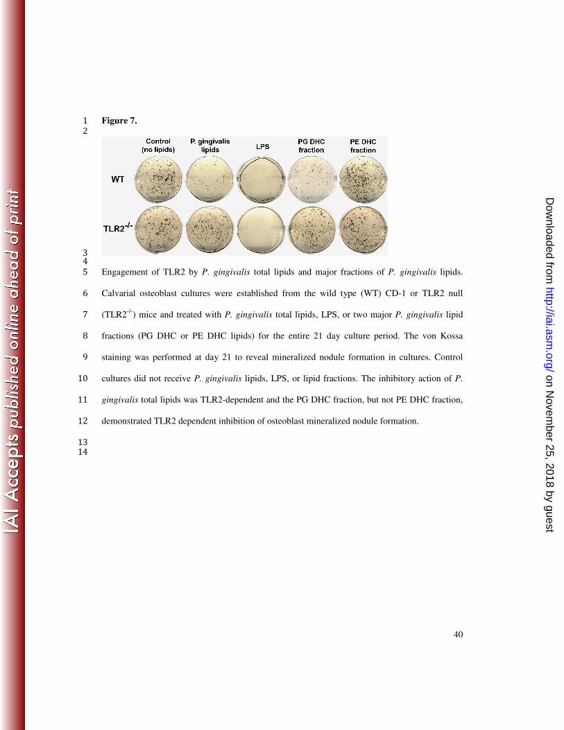
Engagement of TLR2 by P. gingivalis total lipids and major fractions of P. gingivalis lipids. 5
Calvarial osteoblast cultures were established from the wild type (WT) CD-1 or TLR2 null 6
(TLR2-/-
) mice and treated with P. gingivalis total lipids, LPS, or two major P. gingivalis lipid 7
fractions (PG DHC or PE DHC lipids) for the entire 21 day culture period. The von Kossa 8
staining was performed at day 21 to reveal mineralized nodule formation in cultures. Control 9
cultures did not receive P. gingivalis lipids, LPS, or lipid fractions. The inhibitory action of P. 10
gingivalis total lipids was TLR2-dependent and the PG DHC fraction, but not PE DHC fraction, 11
demonstrated TLR2 dependent inhibition of osteoblast mineralized nodule formation. 12
13
14
on Novem
ber 25, 2018 by guesthttp://iai.asm
.org/D
ownloaded from