postintubational laryngeal granuloma
TRANSCRIPT
8/10/2019 Postintubational Laryngeal Granuloma
http://slidepdf.com/reader/full/postintubational-laryngeal-granuloma 1/7116
BRAZILIAN JOURNAL OF OTORHINOLARYNGOLOGY 75 (1) J ANUARY /FEBRUARY 2009
http://www.rborl.org.br / e-mail: [email protected]
Clinical, histological and
electron microscopic aspects of
vocal fold granulomas
Summary
Regina Helena Garcia Martins 1 , Norimar
Hernandes Dias 2 , Daniela Carvalho dos Santos 3 ,
Alexandre Todorovic Fabro 4 , José Reinaldo
Cerqueira Braz 5
1 Livre docente certification professor in the Otorhinolaryngology Discipline, Faculdade de Medicina de Botucatu - Unesp. Faculty member of the OtorhinolaryngologyDiscipline.
2 Otorhinolaryngologist, Otorhinolaryngology Discipline, Faculdade de Medicina de Botucatu, Unesp. Master’s degree in surgery. Graduate student, Otorhinolaryngolo-gy Discipline, Faculdade de Medicina de Botucatu-Unesp.
3 Assistant professor, doctor, Morphology Department, Instituto de Biociências - UNESP - Botucatu. Faculty member of the Morphology Department. 4 Medical resident, Pathology Department, Faculdade de Medicina de Botucatu, UNESP.
5 Full professor of Anesthesiology, Faculdade de Medicina de Botucatu-Unesp. Faculty member of the Anesthesiology Department.Universidade Estadual Paulista Júlio de Mesquita Filho, Faculdade de Medicina de Botucatu-Unesp.
Address for correspondence: Regina Helena Garcia Martins - Disciplina de Otorrinolaringologia, Departamento de Oftalmologia, Otorrinolaringologia e Cirurgia deCabeça e Pescoço da Faculdade de Medicina de Botucatu, Distrito de Rubião Junior Botucatu SP 18618-970. Fone/Fax: (00xx14) 3811-6256.
This paper was submitted to the RBORL-SGP (Publishing Manager System) on 21 September 2007. Code 4810.The article was accepted on 2 January 2008.
Granulomas are bilateral and pediculated lesions of
the vocal apophysis. Etiologies: intubation, reflux, trauma, vocal abuse, idiopathic origin. Aim: To analyze the clinicaland morphological aspects of post intubation granulomas.Methods: retrospective study of patients submitted tomicrosurgery for post intubation laryngeal granulomasseen at our Medical School starting in 2002. We analyzed:age, gender, indication and time of intubation, symptoms, videolaryngoscopic diagnosis and biopsy findings. Lightmicroscopy was performed on all specimens, and electronmicroscopy on three of them. Results: ten patients (7 femalesand 3 males), between the ages of 2 and 72 years, intubationtime between 4h and 21 days. Hoarseness was a frequentsymptom, starting in the first week following extubation.
Histology shows mild epithelial hyperplasia, severeinflammation and vessel proliferation in the corion. UnderSEM, the epithelium presented mild superficial desquamation.Under TEM, intracellular junctions showed widening withstructural changes in the desmosomes. In the corion there were vessel proliferations, inflammation and fibroblasts with structural alterations. Conclusions: post intubationgranulomas appear in any age and hoarseness is a frequentsymptom. Morphological alterations occur in the corion as vessel proliferations, inflammation, and intracytoplasmaticalterations in fibroblasts suggesting cellular dysfunction anddamage.
Keywords: granuloma, intubation, larynx, morphology.
ORIGINAL ARTICLE
Braz J Otorhinolaryngol2009;75(1):116-22.
8/10/2019 Postintubational Laryngeal Granuloma
http://slidepdf.com/reader/full/postintubational-laryngeal-granuloma 2/7117
BRAZILIAN JOURNAL OF OTORHINOLARYNGOLOGY 75 (1) J ANUARY /FEBRUARY 2009
http://www.rborl.org.br / e-mail: [email protected]
INTRODUCTION
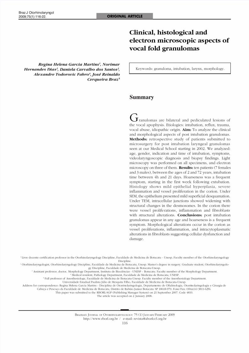
Granulomas are unilateral or bilateral rounded le-sions of various colors (rose, white or wine-color), witha pedicle most of the times, and a smooth or irregularsurface. The implantation pedicles are inserted on the
posterior area of the glottis, especially on the vocal apo-physis (Figure 1).
folds against the tube and compressing or traumatizing thelining epithelium of the vocal apophyses.4,5.
Some authors have found a higher prevalence oflaryngeal granulomas in males. This ratio is inverted whenintubation granulomas are studied specifically; these aremore common in the female larynx. This fact has been
explained by the anatomical specificities of the femalelarynx, which is smaller compared to the male larynx,thus allowing more contact between the cannula and theairway mucosa.1,6 Furthermore, the fragile perichondriumthat covers the vocal apophysis of the arytenoid cartilagesand the poor circulation of the local mucosa are additionalfactors that make the area more vulnerable to intubationtrauma. The prevalence of intubation granulomas infemales was confirmed by Pontes et al.7 in an analysisof 66 patients with laryngeal granulomas, 15 of whichdeveloped after intubation; six of these were male andnine were female.
In many cases voice is not affected, since the gra-nulomas are located on the posterior glottic commissure,and glottic coaptation is not affected. However, therehave been descriptions of large granulomas that not onlyaffect voice quality considerably, but also cause dyspnea.8 Additional symptoms include a foreign body sensation,coughing and phlegm.
Intubation granulomas occur more often after pro-longed intubation, although they have been diagnosed inpatients intubated for short periods. Kaneda et al.9 reporteda vocal fold granuloma in a female patient that was intuba-ted with a small diameter tracheal tube (6.5 mm) for only4.5 hours. Shimokojin et al.10 described shorter intubation
times resulting in laryngeal granulomas in three patients(2 hours and 20 minutes to 5 hours and 40 minutes.
Few authors have described the morphology oflaryngeal granulomas. The rare descriptions are restrictedto non-ultrastructural histological studies.1,11 Luzar et al.11 undertook a retrospective, clinical and histomorphologi-cal study of the epithelial features of the mucosal liningon 149 laryngeal granulomas. These authors applied theLjubljana12 classification in the histological analysis of epi-thelial alterations, which describes epithelium as normal,simple and abnormal hyperplasia, atypical hyperplasia,and in situ carcinoma. An analysis of epithelial atrophy
completed the study. The authors underlined the benignfeatures of laryngeal granulomas based on the finding ofsimple epithelial hyperplasia in 65.8% of cases, epithelialatrophy in 16.1% of cases, a normal epithelium in 13.4%of cases, and abnormal hyperplasia in only 4.7% of cases;they found no in situ carcinomas. The authors also foundmany pools of blood permeated with inflammatory cellsin the lamina propria.
More detailed morphological descriptions of intu-bation granulomas were not found in the literature, whichmotivated the present paper. Its purpose is to present the
Figure 1. Granulomas in both vocal apophyses
The etiological factors causing the laryngeal granu-lomas include endotracheal intubation, gastroesophageal
reflux, external laryngeal trauma, phonotrauma, and idio-pathic origin, when the cause cannot be found. Amongthese etiological factors, endotracheal intubation, voiceabuse and acid laryngitis are the most relevant, whichunderlines the need for investigating these possibilities inthe clinical history.1,2 Specific laryngeal granulomas, se-condary to specific granulomatous diseases of the larynx,such as tuberculosis, blastomycosis, leprosy, leishmaniasis,syphilis, and other, should also be taken into account.1 Thisstudy will focus mainly the intubation granulomas.
In 1932, Clawsen first described laryngeal granu-lomas as a result of endotracheal intubation, raising the
attention of other authors to the study of its predisposingfactors.3 Among them, we have: prolonged and traumaticintubation, use of larger diameter tracheal tubes, highpressure inside the tracheal tube balloons especially inanesthesia using nitrous oxide, and inadequate sedationlevels. In the latter, movement of the patient’s neck causesthe tube to move within the airway, causing friction be-tween the tube and the lining epithelium in the laryngealand tracheal mucosa. Intubated patients that are inade-quately sedated or not sedated at all perform involuntaryswallowing and phonatory movements, pushing the vocal
8/10/2019 Postintubational Laryngeal Granuloma
http://slidepdf.com/reader/full/postintubational-laryngeal-granuloma 3/7118
BRAZILIAN JOURNAL OF OTORHINOLARYNGOLOGY 75 (1) J ANUARY /FEBRUARY 2009
http://www.rborl.org.br / e-mail: [email protected]
clinical and morphological features of intubation granu-lomas, done by histological studies, and transmission andscanning electron microscopy, to show ultra-structuraldetails that might increase our understanding of the patho-physiology of granulomas and guide the treatment.
SERIES AND METHOD
A retrospective study was undertaken of patients with laryngeal granulomas due to endotracheal intuba-tion, seen at the outpatient otorhinolaryngology units ofthe Botucatu Medical School (Faculdade de Medicina deBotucatu, Unesp) from 2002 onwards, and that had un-dergone laryngeal microsurgery for removing the lesions.The Research Ethics Committee of the institution abovein which research was undertaken approved the study(protocol number 472/2007).
The following information was taken from medi-cal files: age, sex, indication for intubation, duration of
intubation, voice or respiratory symptoms manifestedafter extubation, videolaryngoscopy reports, and biopsynumber.
A qualified professional from the Pathology Depart-ment of the aforesaid institution reviewed the histologicalslides, and recorded the most relevant epithelial changes(hyperplasia, acanthosis, hyperkeratosis) and those inthe lamina propria (increased number of vessels, edema,inflammatory cell infiltrate). Histological parameters werequantified based on a semi-quantitative score: 0 (unalte-red), 1 (mildly altered), 2 (moderately altered) and 3 (in-tensely altered). A light microscope (Axiostar plus model,
Zeiss, Carl Zeiss do Brasil Ltda) was used for examinationand photography at different magnifications; a digital ca-mera was used for recording the images.
An additional three fragments of laryngeal granulo-mas had been fixated in 2.5% glutaraldehyde at the timeof surgery and sent to the Morphology Department of thesame institution for processing and electron microscopy;the procedures are described next. For scanning electronmicroscopy, the surgical specimens were fixated in 2.5%glutaraldehyde during 12 hours, the washed in 0.1 Mbuffered phosphate at pH 7.3, fixated in a 1% osmiumtetroxide solution during 1 hour, washed in bufferedphosphate, dehydrated in series in alcohol solutions (75%to 100%), then dried in a critical point dryer device (Bal-zers CPD-020) with liquid carbon dioxide. The specimens were then mounted on a metal base using silver glue andgold-covered (15 nm of gold) in a Balzers MED-010 minideposition device. A scanning electron microscope (SEM515, Philips, Netherlands) at 15 KV was used for exami-ning and photographing the specimens. For transmissionelectron microscopy, the specimens were fixated in 2.5%glutaraldehyde, washed in 0.1 M buffered phosphate atpH 7.3, sectioned into 3 mm x 1 mm specimens over pinkdental wax humidified with a 2.5% glutaraldehyde fixating
solution, then fixated in a 1% osmium tetroxide solutionand 0.1 M buffered phosphate at pH 7.3, dehydrated inseries in acetone solutions (50%, 70%, 90% and 100%),and placed in a mixture of acetone and araldite resin(Polysciences, Inc.). The fragments were then removedfrom this mixture and included in a pure araldite resin
block in an oven at 37o
C; semifine 0.5 µm sections weremade, placed on slides and stained with a 1:1 mixture of1% methylene blue and 1% Azur II. These semifine sections were examined in a light microscope and then again sec-tioned into ultrafine specimens (500), and examined andphotographed in a transmission electron microscope (EM301 model, Philips AG, Netherlands), using an Eastman5302 (Kodak Co, US) film and Kodabromide (EastmanKodak Co., US) photographic paper.
We had already defined the normalcy standardsfor the laryngeal mucosa for both histology and electronmicroscopy in previous studies. These were based on amorphological analysis of the vocal fold mucosa removedfrom three autopsy laryngeal specimens with no historyof intubation or laryngeal trauma.
The chi-square test was used for comparing theproportions. Friedman’s non-parametric test was used forcomparing the scores. The significance level was 5 %.
RESULTS
Within the study period, intubation granulomas were removed from 10 patients; Table 1 shows their clini-cal features. The age of patients ranged from 2 years and3 months to 72 years; there were mostly female patients
(7 cases). The intubation time ranged from 4 hours to 21days. The main symptom was hoarseness, which in mostcases started during the first week after extubation; in somecases, hoarseness was present immediately after removalof the cannula. The most frequent endoscopic finding wasbilateral granulomas in the vocal apophysis. Recurrenceof the lesions was seen in one patient only.
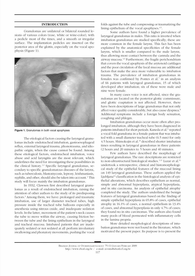
Table 2 summarizes the histological results; theepithelium was mildly altered, mostly mild hyperplasiarecorded in seven slides. The lamina propria was moredeeply altered; there was an inflammatory infiltrate ofpolymorphonuclear cells, lymphocyte and occasional his-tiocytes (Figure 2). Intense proliferation of vessels was aconstant finding in the slides (Figure 2). Table 2 highlights(*) the histological changes that were statistically significant(p<0.05) in the comparison of scores.
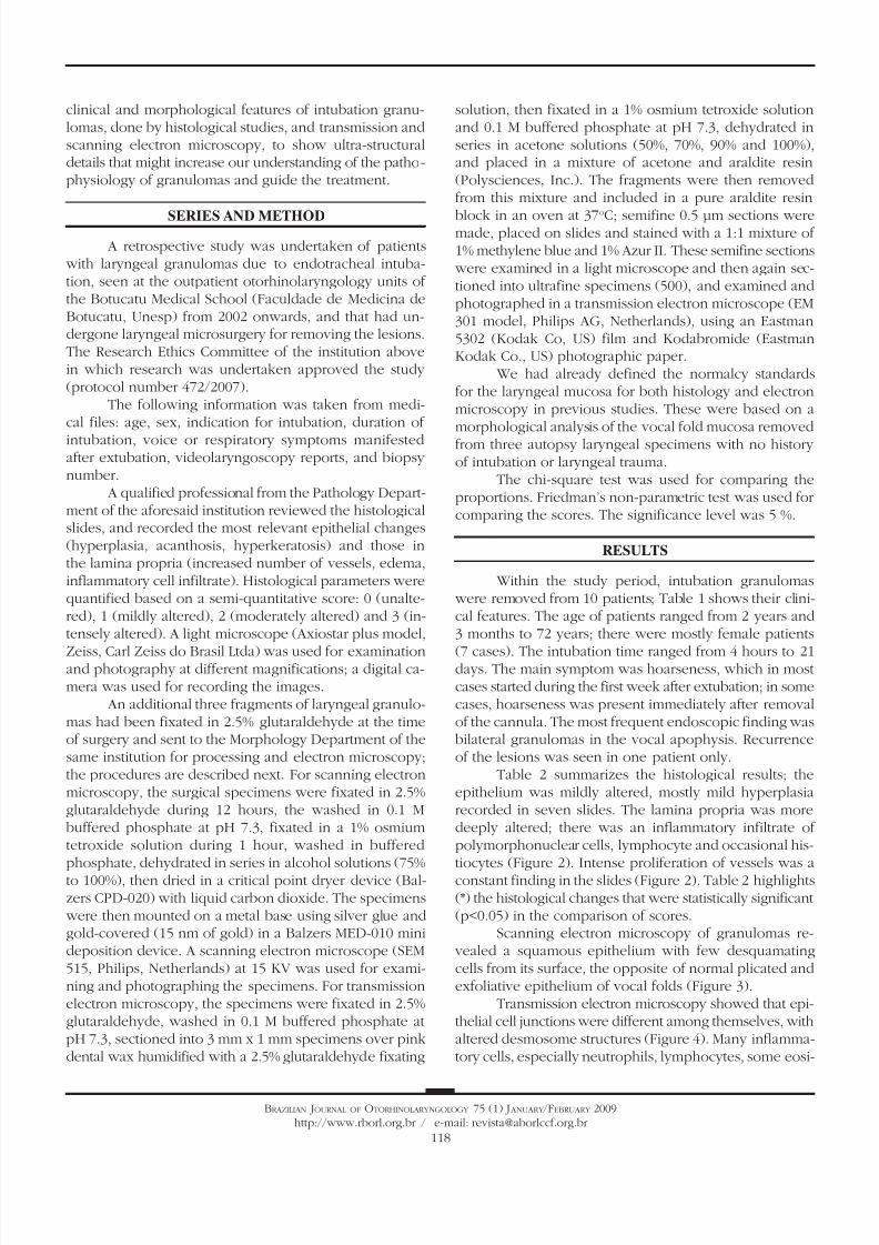
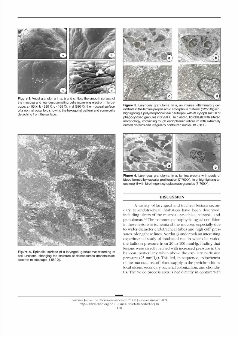
Scanning electron microscopy of granulomas re- vealed a squamous epithelium with few desquamatingcells from its surface, the opposite of normal plicated andexfoliative epithelium of vocal folds (Figure 3).
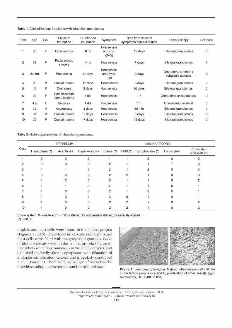
Transmission electron microscopy showed that epi-thelial cell junctions were different among themselves, withaltered desmosome structures (Figure 4). Many inflamma-tory cells, especially neutrophils, lymphocytes, some eosi-
8/10/2019 Postintubational Laryngeal Granuloma
http://slidepdf.com/reader/full/postintubational-laryngeal-granuloma 4/7119
BRAZILIAN JOURNAL OF OTORHINOLARYNGOLOGY 75 (1) J ANUARY /FEBRUARY 2009
http://www.rborl.org.br / e-mail: [email protected]
Table 1. Clinical findings inpatients with intubation granulomas
Case Age SexCause of
intubation
Duration of
intubationSymptoms
Time from onset of
symptoms and extubationLaryngoscopy Relapses
1 23 F Laparoscopy 6 hs
Hoarseness
and cou-
ghing
12 days Bilateral granulomas 3
2 53 FFacial plastic
surgery4 hs Hoarseness 7 days Bilateral granulomas 0
3 2a 3m F Pneumonia 21 days
Hoarseness
and dysp-
nea
2 daysGranuloma anterior +
subglottic stenosis0
4 23 M Cranial trauma 12 days Hoarseness 3 days Bilateral granulomas 0
5 16 F Over dose 3 days Hoarseness 30 days Bilateral granulomas 0
6 20 FPost-cesarean
complications1 dia Hoarseness 1 h Granuloma unilateral post 0
7 4 a F Seizures 1 dia Hoarseness 1 h Granuloma unilateral 0
8 72 M Angioplasty 3 days Hoarseness 60 min Bilateral granulomas 0
9 37 M Cranial trauma 2 days Hoarseness 5 days Bilateral granulomas 010 29 F Cranial trauma 7 days Hoarseness 15 days Bilateral granulomas 0
Table 2. Histological analysis of intubation granulomas
Case
EPITHELIUM LAMINA PROPRIA
Hyperplasia (*) Acanthosis Hyperkeratosis Edema (*) PMN (*) Lymphocytes (*) HistiocytesProliferation
of vessels (*)
1 0 0 0 1 1 2 0 3
2 0 0 0 2 1 1 1 3
3 1 1 0 2 1 2 0 3
4 0 0 0 2 2 1 0 3
5 1 0 0 3 1 1 0 2
6 1 1 0 2 1 1 0 1
7 1 0 0 2 1 3 0 1
8 1 0 1 3 0 1 0 1
9 1 0 0 2 2 1 0 2
10 1 0 0 2 2 1 0 2
Score system: 0 - unaltered; 1 - mildly altered; 2 - moderately altered; 3 - severely altered.
(*) p<0,05
Figure 2. Laryngeal granuloma. Marked inflammatory cell infiltrate
in the lamina propria in a and b; proliferation of small vessels (light
microscopy, HE- a-40X; b-80X).
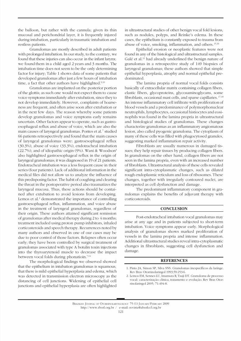
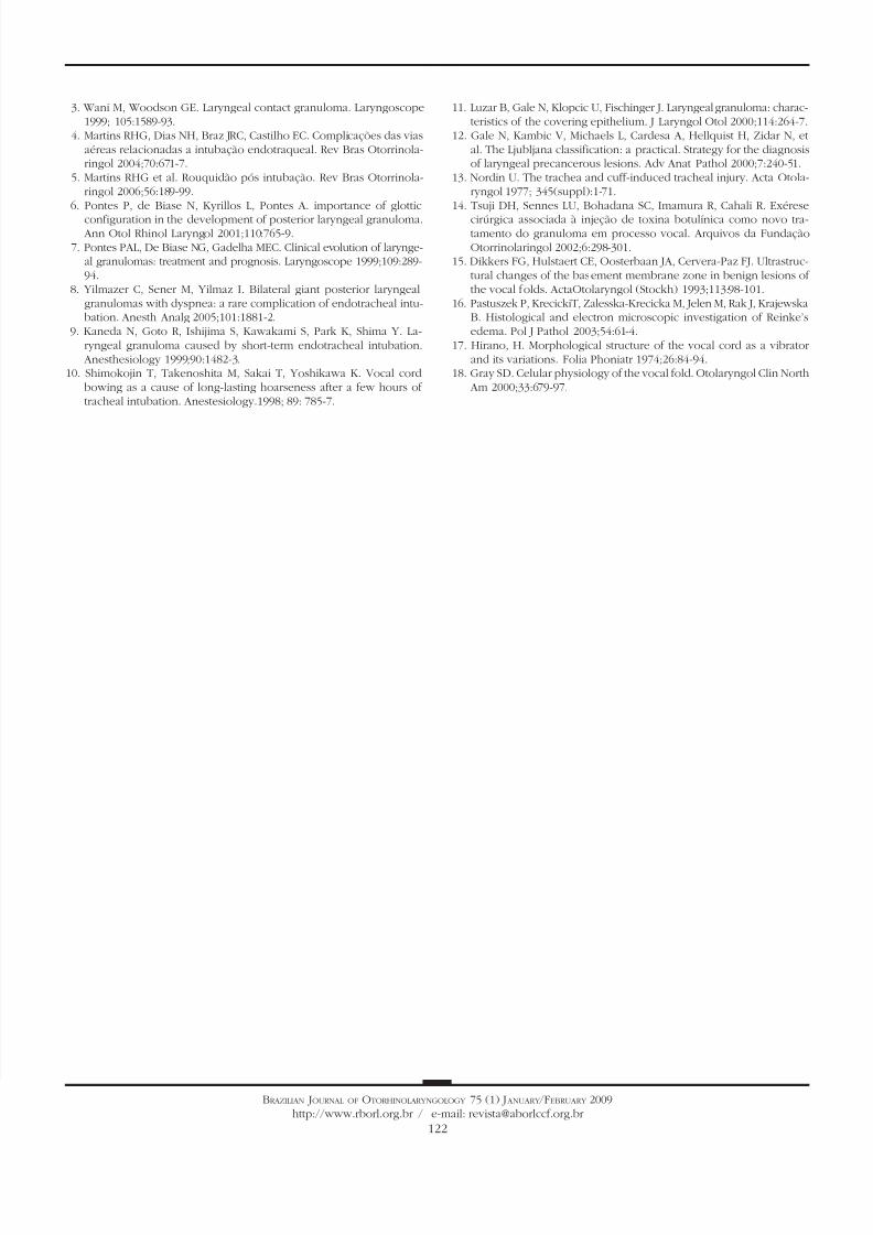
nophils and mast cells were found in the lamina propria(Figures 5 and 6). The cytoplasm of some neutrophils andmast cells were filled with phagocytosed granules. Poolsof blood were also seen in the lamina propria (Figure 6).Fibroblasts were more numerous in the lamina propria, andexhibited markedly altered cytoplasm, with dilatation ofendoplasmic reticulum cisterns and irregularly contourednuclei (Figure 5). There were no collagen fiber networks,notwithstanding the increased number of fibroblasts.
8/10/2019 Postintubational Laryngeal Granuloma
http://slidepdf.com/reader/full/postintubational-laryngeal-granuloma 5/7120
BRAZILIAN JOURNAL OF OTORHINOLARYNGOLOGY 75 (1) J ANUARY /FEBRUARY 2009
http://www.rborl.org.br / e-mail: [email protected]
DISCUSSION
A variety of laryngeal and tracheal lesions secon-dary to endotracheal intubation have been described,including ulcers of the mucosa, synechiae, stenosis, andgranulomas.4,5 The common pathophysiological conditionin these lesions is ischemia of the mucosa, especially dueto wider diameter endotracheal tubes and high cuff pres-sures. Along these lines, Nordin13 undertook an interestingexperimental study of intubated rats in which he variedthe balloon pressure from 20 to 100 mmHg, finding thatlesions were directly related with increased pressure in theballoon, particularly when above the capillary perfusionpressure (25 mmHg). This led, in sequence, to ischemiaof the mucosa, loss of blood supply to the perichondrium,local ulcers, secondary bacterial colonization, and chondri-tis. The voice process area is not directly in contact with
Figure 3. Vocal granuloma in a, b and c. Note the smooth surface of
the mucosa and few desquamating cells (scanning electron micros-
cope: a - 55 X; b - 330 X; c - 165 X). In d (890 X), the mucosal surface
of a normal vocal fold showing the hexagonal pattern and some cells
detaching from the surface.
Figure 4. Epithelial surface of a laryngeal granuloma; widening of
cell junctions, changing the structure of desmosomes (transmission
electron microscope, 1 550 X).
Figure 5. Laryngeal granuloma. In a, an intense inflammatory cell
infiltrate in the lamina propria amid amorphous material (3 250 X); in b,
highlighting a polymorphonuclear neutrophil with its cytoplasm full of
phagocytosed granules (13 250 X). In c and d, fibroblasts with alteredmorphology, containing rough endoplasmic reticulum with extremely
dilated cisterns and irregularly contoured nuclei (13 250 X).
Figure 6. Laryngeal granuloma. In a, lamina propria with pools ofblood formed by vascular proliferation (7 700 X). In b, highlighting an
eosinophil with birefringent cytoplasmatic granules (7 700 X).
8/10/2019 Postintubational Laryngeal Granuloma
http://slidepdf.com/reader/full/postintubational-laryngeal-granuloma 6/7121
BRAZILIAN JOURNAL OF OTORHINOLARYNGOLOGY 75 (1) J ANUARY /FEBRUARY 2009
http://www.rborl.org.br / e-mail: [email protected]
the balloon, but rather with the cannula; given its thinmucosal and perichondrial layer, it is frequently injuredduring intubation, particularly in traumatic intubation andrestless patients.
Granulomas are mostly described in adult patients with prolonged intubation. In our study, to the contrary, we
found that these injuries can also occur in the infant larynx; we found them in a child aged 2 years and 3 months. Theintubation time does not seem to be the only determiningfactor for injury; Table 1 shows data of some patients thatdeveloped granulomas after just a few hours of intubationtime, a fact that other authors have highlighted.9,10
Granulomas are implanted on the posterior portionof the glottis; as such one would not expect them to cause voice symptoms immediately after extubation, since they tonot develop immediately. However, complaints of hoarse-ness are frequent, and often arise soon after extubation orin the next few days. The reason whereby some patientsdevelop granulomas and voice symptoms early remainsuncertain. Other factors appear to operate, such as gastro-esophageal reflux and abuse of voice, which are also themain causes of laryngeal granulomas. Pontes et al.7 studied66 patients retrospectively and found that the main causesof laryngeal granulomas were: gastroesophageal reflux(30.3%), abuse of voice (33.3%), endotracheal intubation(22.7%), and of idiopathic origin (9%). Wani & Woodson3
also highlighted gastroesophageal reflux in the origin oflaryngeal granulomas; it was diagnosed in 19 of 21 patients.Endotracheal intubation was a less frequent cause in theirseries (four patients). Lack of additional information in themedical files did not allow us to analyze the influence of
this predisposing factor. The habit of coughing and clearingthe throat in the postoperative period also traumatizes thelaryngeal mucosa. Thus, these actions should be contai-ned after extubation to avoid lesions from developing.Lemos et al.2 demonstrated the importance of controllinggastroesophageal reflux, inflammation, and voice abusein the treatment of laryngeal granulomas, regardless oftheir origin. These authors attained significant remissionof granulomas after medical therapy during 2 to 4 months;treatment included using proton pump inhibitors, inhaledcorticosteroids and speech therapy. Recurrences noted bymany authors and observed in one of our cases may be
due to poor control of those factors. Relapses often occurearly; they have been controlled by surgical treatment ofgranulomas associated with type A botulin toxin injectionsinto the thyroarytenoid muscle to decrease the impactbetween vocal folds during phonatioin.7,14
The morphological findings we observed showedthat the epithelium in intubation granulomas is squamous,that there is mild epithelial hyperplasia and edema, which was detected in transmission electron microscopy as thedistancing of cell junctions. Widening of epithelial celljunctions and epithelial hyperplasia are often highlighted
in ultrastructural studies of other benign vocal fold lesions,such as nodules, polyps, and Reinke’s edema. In thesecases, the epithelium is constantly exposed to trauma fromabuse of voice, smoking, inflammation, and others.15,16
Epithelial erosion or neoplastic features were notfound in any of the histological and ultrastructural samples.
Galé et al.12
had already underlined the benign nature ofgranulomas in a retrospective study of 149 biopsies oflaryngeal granulomas; these authors showed that simpleepithelial hyperplasia, atrophy and normal epithelial pre-dominated.
The lamina propria of normal vocal folds consistsbasically of extracellular matrix containing collagen fibers,elastic fibers, glycoproteins, glycosaminoglycans, somefibroblasts, occasional mast cells, and sparse capillaries.17,18
An intense inflammatory cell infiltrate with proliferation ofblood vessels and a predominance of polymorphonuclearneutrophils, lymphocytes, occasional histiocytes and eosi-nophils was found in the lamina propria in ultrastructuraland histological studies of granulomas. These changescharacterize granulomas as an inflammatory angiomatouslesion, also called pyogenic granuloma. The cytoplasm ofmany of these cells was filled with phagocytosed granules,suggesting marked inflammation repair activity.
Fibroblasts are usually numerous in damaged tis-sues; they help repair tissues by producing collagen fibers.In granulomas on the other hand, collagen fibers are notseen in the lamina propria, even with an increased numberof fibroblasts. Ultrastructural analysis of these cells revealedsignificant intra-cytoplasmatic changes, such as dilatedrough endoplasmic reticulum and loss of ribosomes. These
changes, together with irregularly contoured nuclei, areinterpreted as cell dysfunction and damage.
The predominant inflammatory component in gra-nulomas explains the benefits of adjuvant therapy withcorticosteroids.
CONCLUSION
Post-endotracheal intubation vocal granulomas mayarise at any age and in patients subjected to short-termintubation. Voice symptoms appear early. Morphologicalanalysis of granulomas shows marked proliferation of vessels in the lamina propria and intense inflammation. Additional ultrastructural studies reveal intra-cytoplasmaticchanges in fibroblasts, suggesting cell dysfunction anddamage.
REFERENCES
1. Pinto JA, Simon SP, Silva SNS. Granulomas inespecíficos da laringe.Rev Bras Otorrinolaringol 1993;59:253-6.
2. Lemos EM, Sennes LU, Imamura R, Tsuji DT. Granuloma de processo vocal: caracterização clínica, tratamento e evolução. Rev Bras Otor-rinolaringol 2005; 71:494-8.
8/10/2019 Postintubational Laryngeal Granuloma
http://slidepdf.com/reader/full/postintubational-laryngeal-granuloma 7/7122
BRAZILIAN JOURNAL OF OTORHINOLARYNGOLOGY 75 (1) J ANUARY /FEBRUARY 2009
http://www.rborl.org.br / e-mail: [email protected]
3. Wani M, Woodson GE. Laryngeal contact granuloma. Laryngoscope1999; 105:1589-93.
4. Martins RHG, Dias NH, Braz JRC, Castilho EC. Complicações das viasaéreas relacionadas a intubação endotraqueal. Rev Bras Otorrinola-ringol 2004;70:671-7.
5. Martins RHG et al. Rouquidão pós intubação. Rev Bras Otorrinola-ringol 2006;56:189-99.
6. Pontes P, de Biase N, Kyrillos L, Pontes A. importance of glottic
configuration in the development of posterior laryngeal granuloma. Ann Otol Rhinol Laryngol 2001;110:765-9.
7. Pontes PAL, De Biase NG, Gadelha MEC. Clinical evolution of larynge-al granulomas: treatment and prognosis. Laryngoscope 1999;109:289-94.
8. Yilmazer C, Sener M, Yilmaz I. Bilateral giant posterior laryngealgranulomas with dyspnea: a rare complication of endotracheal intu-bation. Anesth Analg 2005;101:1881-2.
9. Kaneda N, Goto R, Ishijima S, Kawakami S, Park K, Shima Y. La-ryngeal granuloma caused by short-term endotracheal intubation.
Anesthesiology 1999;90:1482-3.10. Shimokojin T, Takenoshita M, Sakai T, Yoshikawa K. Vocal cord
bowing as a cause of long-lasting hoarseness after a few hours oftracheal intubation. Anestesiology.1998; 89: 785-7.
11. Luzar B, Gale N, Klopcic U, Fischinger J. Laryngeal granuloma: charac-teristics of the covering epithelium. J Laryngol Otol 2000;114:264-7.
12. Gale N, Kambic V, Michaels L, Cardesa A, Hellquist H, Zidar N, etal. The Ljubljana classification: a practical. Strategy for the diagnosisof laryngeal precancerous lesions. Adv Anat Pathol 2000;7:240-51.
13. Nordin U. The trachea and cuff-induced tracheal injury. Acta Otola-ryngol 1977; 345(suppl):1-71.
14. Tsuji DH, Sennes LU, Bohadana SC, Imamura R, Cahali R. Exérese
cirúrgica associada à injeção de toxina botulínica como novo tra-tamento do granuloma em processo vocal. Arquivos da FundaçãoOtorrinolaringol 2002;6:298-301.
15. Dikkers FG, Hulstaert CE, Oosterbaan JA, Cervera-Paz FJ. Ultrastruc-tural changes of the basement membrane zone in benign lesions ofthe vocal folds. ActaOtolaryngol (Stockh) 1993;113:98-101.
16. Pastuszek P, KrecickiT, Zalesska-Krecicka M, Jelen M, Rak J, KrajewskaB. Histological and electron microscopic investigation of Reinke’sedema. Pol J Pathol 2003;54:61-4.
17. Hirano, H. Morphological structure of the vocal cord as a vibratorand its variations. Folia Phoniatr 1974;26:84-94.
18. Gray SD. Celular physiology of the vocal fold. Otolaryngol Clin North Am 2000;33:679-97.