potassium drophila
TRANSCRIPT
Proc. Natl. Acad. Sci. USAVol. 91, pp. 3438-3442, April 1994Neurobiology
A family of potassium channel genes related to eag in Drophilaand mammalsJEFFREY W. WARMKE* AND BARRY GANETZKYLaboratory of Genetics, University of Wisconsin, Madison, WI 53706
Communicated by Julius Adler, November 29, 1993
ABSTRACT We have identified a conserved family ofgenes related to Drosophila eag, which encodes a distinct typeof voltage-activated K channel. Three related genes wererecovered in screens of cDNA libraries from Drosophila,mouse, and human tissues. One gene is the mouse counterpartof eag; the other two represent additional subfamilles. Thehuman gene maps to chromosome 7. Family members share atleast 47% amino acid identity in their hydrophobic cores andall contain a segment homologous to a cyclic nucleotide-bindigdomain. Sequence comparisons indicate that members of thisfamily are most closely related to vertebrate cyclic nudleotide-gated cation channels and plant inward-rectifying K+ chan-nels. The existence of another family of K+ channel structuralgenes further extends the known diversity of Ki channels andhas important implications for the structure, function, andevolution of the superfamily of voltage-sensitive ion channels.
Voltage-activated ion channels are members of evolution-arily conserved multigene families (1, 2). For example, theShaker (Sh) family of K+ channels comprises four relatedgenes in Drosophila, each of which has one or more mam-malian homologs (3). Together these genes define at least foursubfamilies of voltage-activated K+ channels within the Shfamily. Most of our present understanding of the structureand function of K+ channels is based on studies of thepolypeptides encoded by these genes (4).
Analysis ofotherK+ channels that are not members ofthe Shfamily will expand our understanding of these channels. Genesencoding additional types of K+ channels can be identified inDrosophila via molecular analysis of mutations affecting mem-brane excitability (5). Mutations of eag, identified by theirleg-shaking phenotype, cause repetitive firing and enhancedtransmitter release in motor neurons, suggesting a possibledefect in K+ channels (6,7). Molecular studies revealed that eagencodes a polypeptide structurally related both to K+ channelsin the Sh family and to cyclic nucleotide-gated cation channels(8-10). Expression in Xenopus oocytes confirms that the eagpolypeptide assembles into channels conducting a voltage-activated K+-selective outward current (11, 12).These results raise the question of whether eag is the proto-
type of a distinct K+ channel gene family analogous to the Shfamily. Here we use low-stringency screens and degeneratePCRs to isolate relatives of eag from Drosophila, mouse, andhuman tissues. Sequence alignments indicate the existence ofaconserved multigene family of eag-related loci in Drosophilaand mammals. The eag family has important implications forunderstanding the structure, function, and evolution ofvoltage-activated K+ channels and cyclic nucleotide-gated cation chan-nels.t
MATERIALS AND METHODSIsolation of cDNA Clones Encoding a Drosophila eag Ho-
molog. A 1682-bp Acc I/Cla I fragment of the eag cDNA
CH20 (encoding the hydrophobic core) was used in a low-stringency screen [30%o (vol/vol) formamide/5x SSCP (1xSSCP = 120mM NaCl/15 mM sodium citrate/20mM sodiumphosphate, pH 6.8)/0.1% SDS/lOx Denhardt's solution/salmon sperm DNA (250 ,g/ml) at 420C; wash, 0.2x SSC/0.1% SDS at 420C] of a Drosophila head cDNA library (13)(provided by B. Hamilton and E. Meyerowitz, CaliforniaInstitute of Technology, Pasedena, CA). Of three positiveclones, one (elk) that hybridized to two nonoverlappingfiagments from the eag hydrophobic core was isolated. ThiscDNA was used to screen a second head library (provided byT. Schwarz, Stanford University, Stanford, CA), at highstringency [2x SSCP/0.1% SDS/lOx Denhardt's solution/salmon sperm DNA (250 ,ug/ml) at 650C; wash, 0.2x SSC/0.1% SDS at 650C] to obtain additional elk cDNAs.PCR Amplification of a Mouse eag Homolog. Degenerate
primers were eag oligonucleotide 1 [eag peptide sequenceACIWY-5'-CA(TC)TGGCTAGC(GATC)TG(TC)AT(ATC)-TGGTA-3'J, oligonucleotide 2 [TYCDL-5'-GATGAATTC-(GATC)A(GA)(GA)TC(GA)CA(GA)TA(GATC)GT-3'], andoligonucleotide 3 [ILGKGD-5'-GCCGAATTC(GA)TC-(GATC)CC(TC)TT(GATC)CC(GATC)A(GA)(GAT)AT-3'].PCR (35 cycles) conditions were 1 min at 950C, 1 min at 420C,and 1 min at 720C with oligonucleotides 1 and 2 and random-primed mouse skeletal muscle cDNA (provided by P. Pow-ers, University of Wisconsin, Madison) as template. Analiquot ofthe reaction mixture was separated on a2% agarosegel and transferred to Zeta-Probe (Bio-Rad) membranes, andproducts corresponding to putative eag homologs were iden-tified by cross hybridization to an elk probe at low stringency(low-stringency conditions as above except for 25% forma-mide). To enrich for a cross-hybridizing product of =850 bp,the remaining reaction mixture was separated on a 2%agarose gel and fragments of 750-950 bp were recovered.This product was reamplified by PCR under the conditionsabove but with oligonucleotides 1 and 3. The reamplifiedproducts were subcloned into pBluescriptII(KS+) (Strata-gene) using the Nhe I and EcoRI sites in the primers.Subclones that hybridized at low stringency with an elkcDNA were partially sequenced (Sequenase version 2 kit,United States Biochemical).
Isolation and Sequence Analysis of Mammalan eag-LikecDNA Clones. Mouse eag (m-eag)cDNA clones were isolatedfrom a mouse brain cDNA library (AZAPMB, provided by J.Boulter and S. Heineman, Salk Institute, San Diego). Humanerg (h-erg) cDNA clones were isolated from a human hip-pocampus cDNA library (catalogue no. 936205; Stratagene).Both libraries were screened at high stringency (see above).
Abbreviations: cNBD, cyclic nucleotide binding domain; ORF, openreading frame; m-eag, mouse eag; h-erg, human erg.*Present address: Department of Genetics and Molecular Biology,RY 80Y-255, Merck Research Laboratories, P.O. Box 2000, Rah-way, NJ 07065.tThe sequences reported in this paper have been deposited in theGenBank data base (accession nos. elk, U04246; m-eag, U04294;h-erg, U04270).
3438
The publication costs of this article were defrayed in part by page chargepayment. This article must therefore be hereby marked "advertisement"in accordance with 18 U.S.C. §1734 solely to indicate this fact.
Dow
nloa
ded
by g
uest
on
Feb
ruar
y 18
, 202
2

Proc. Natl. Acad. Sci. USA 91 (1994) 3439
After subcloning into pBluescript vectors (Stratagene), se-lected cDNAs were sequenced on both strands as above.Chromosome Mapping. The chromosomal location ofh-erg
was determined by PCR using h-erg-specific primers and aseries of hamster-human hybrid cell lines. The primers(GACGTCGTCGTGGCCATCCT, derived from an h-ergexon, and CAACTATGGTCGAAAGAGCT, derived from adownstream intron present in an incompletely spliced cDNA)give a 600-bp product from human but not hamster genomicDNA templates. PCR was performed on DNA from 25hamster-human hybrid cell lines of known karyotype (Bios,New Haven, CT); lines giving the 600-bp fragment wereidentified by gel electrophoresis of the amplification prod-ucts.
RESULTSAn eag Homolog in Drosophila. An initial low-stringency
screen ofaDrosophila head cDNA library with an eag cDNAprobe identified a cDNA with an incomplete open readingframe (ORF) that was clearly related to the eag protein. ThiscDNA, designated elk (eag-like K+ channel; Table 1), wasthen used to screen a second head cDNA library at highstringency. Several overlapping cDNAs were obtained yield-ing a composite elk sequence that contained a long ORFencoding a polypeptide of 1284 amino acids with a predictedmolecular mass of 141 kDa (Fig. 1). Although the proposedinitiator Met is not a good match to the Drosophila translationstart site consensus [TCCCAW compared to CAA(A/C)AI , where the Met codon is underlined; ref. 21], thestrong similarity of the elk and eag polypeptides in thisN-terminal region suggests that this is the true translationstart site (Fig. 1).The elk locus was mapped by in situ hybridization to
polytene chromosome region 54F3-55A2 on the right arm ofthe second chromosome (data not shown). Flies heterozy-gous for deletions of this region do not exhibit any overtlocomotor defects.
Conservation of the eag Sequence in Mouse. The existenceof an eag gene family in Drosophila raised the question ofwhether counterpart genes were present in mammalian ge-nomes (see ref. 3). Because low-stringency screens of amouse brain cDNA library with the same eag probe used toisolate elk were unsuccessful, we used a PCR screen as analternative strategy. Three highly conserved regions between
Table 1. Amino acid identity between ion channel families
the eag and elk polypeptides were used to design degenerateoligonucleotide primers for a series of nested PCRs withmouse skeletal muscle cDNA. Among the products from thereaction, an 4850-bp fragment hybridized to an elk probe atlow stringency. Sequence analysis revealed that the sub-cloned fragment encodes a peptide segment with significantsimilarity to the corresponding region in eag and elk (=56%and =38% identity, respectively, over a stretch of 157 aminoacids).The cloned PCR fragment was used to screen a mouse
brain library at high stringency to obtain more completecDNAs, two of which overlapped to yield the compositesequence of the complete ORF (Fig. 1). Two closely spacedMet codons are present at theN terminus. Comparison ofthesequences flanking the first Met (GTCGGGAGGATQG) andsecond Met (AGGATGACCATGA) codons (underlined)with the vertebrate translation start site consensus sequence[GCCGCC(A/G)CCAJ~fG, refs. 21 and 22] indicates thattranslation of the mouse cDNA could begin at either one tospecify a polypeptide of either 989 or 987 amino acids with apredicted molecular mass of =111 kDa (Fig. 1). However,because the second Met codon aligns with the initiating Metcodon of eag and elk, we believe it is more likely thattranslation starts there. The very strong similarity of thepolypeptide encoded by this cDNA to that specified by eag(Table 1) suggests that the mouse gene is the counterpart ofeag, and therefore, we name it mouse-eag (m-eag).
Isolation and Mapping of a Human eqg Homolog. Becauseion-channel structural genes are potential targets for neuro-logical disorders that have a hereditary component (see ref.23), it was of interest to identify human homologs of eag. Ahuman hippocampus cDNA library was screened at highstringency with the m-eag PCR fragment. The single cDNArecovered was used to isolate several overlapping cDNAs ina second screen of the library. Although sequence analysisshowed none of the cDNAs was complete, each sharedsubstantial similarity to members of the eag family. Thecomposite ORF assembled from these cDNAs was clearlyincomplete because it began in-frame within a conserveddomain at the N terminus without a Met codon. Missingsequence was obtained by screening a genomic cosmidlibrary with a human cDNA fragment. A positively hybrid-izing cosmid clone was digested with Sau3A and probed witha fragment from the 5' end of the cDNA. An exon wasidentified that overlapped the composite cDNA by 47 bp and
eag m-eag h-erg elk KAT1 AKT1 cAMP cGMP slo Shab ShB Shal Shaw100 65 49 49 25 26 24 20 15 17 12 13 10
100 49 47 26 27 27 25 13 16 13 12 12100 49 24 27 24 23 16 15 15 17 13
100 24 26 25 20 17 14 11 13 12100 62 23 24 19 11 11 12 13
100 25 24 18 11 14 14 14100 68 15 11 13 14 11
100 14 11 14 13 11100 19 21 19 18
100 46 39 42100 43 47
100 41100
Multiple sequence alignment of the hydrophobic cores (S1-S6) of the following ion channel polypeptides was generated using the PILEUPmultiple sequence analysis program from the GCG sequence analysis software package version 7.0 (14): eag family members eag, m-eag, h-erg,and elk; the bovine retinal cGMP-gated cation channel (15); the rat olfactory cAMP-gated cation channel (16); the inward-rectifying potassiumchannels AKT1 and KAT1 from plants (17-19); the Drosophila slo calcium-activated potassium channel (20); and the Sh voltage-activatedpotassium channel family members Kv4, Shaw, RKS, Shal, Rckl, ShB, Drkl, Shab, 1k8, and K13 (2). This table was derived from the multiplesequence alignment using the DISTANCES multiple sequence analysis program from the GCG package (14). The percentage ofamino acid identitybetween each pairwise combination was determined by dividing the number ofidentical amino acid residues by the length ofthe shorter sequencewithout gaps. The percent identity for each ion channel family is shown in boldface type.
eagm-eagh-ergelkKAT1AKT1cAMPcGMPsloShabShBShalShaw
Neurobiology: Warrnke and Ganetzky
Dow
nloa
ded
by g
uest
on
Feb
ruar
y 18
, 202
2

3440 Neurobiology: Warmke and Ganetzky Proc. Nadl. Acad. Sci. USA 91 (1994)
lag ROLVAPQNTF Z"ININ8QQPDSSF6 AQVDLTV D 1IS AIVNQN aYVx(Q1aN8i ELMrDKITVGRLEYTgEfMQQQDQ IILL YTI MNLMN-ag ROLVAPQNTF N N DTN a.VD a YA LrDIDTVIXVRQTFIMYINNS LN YXINRH-Irg NP. APQNTF I IF XPFOQSR. X I ycVICTYCDACIY8AIVNQIISC L P aAQI QA LoF Y
Elk t I aPDTHSNVzSSSDIN X L IF
lag QCCALSQOC AQTQLLQVADLSQFTRXTQAL......................................... QPIDSZDTSJV.N-lag.T FFVXI.P. D V L JC S F ........................................... QPIEDDSCKH-lrg.SCFLCiEVDV GAI@AIV FLN VVVVXNV0DSPHDTNHROPPTNSWLAP0RAITFRLKLPALLALTARESSVRSGAG0AIRAP0AVVVD
Ilk 0AfiFiC.lJDI RD PTSHTL LZTNVNZECD. SVFALTAALLGARFRAGOSAWN
lag.........MUS EVFA KR~ARSVT......................... ..................
-lag. OFA RJT ALT.SR......................
H-lrg VDLTPAAPSS ZSDILJLD VTANDNHVAOLOPAZERRALVOPOSPPRSAPOQLPSPRAHSLN PDASGSSCSLARTRSRP SCASVRRASSADDI EAMRAGVLPPPPRHASTOAIlk G.1j LPGQuoaPAADODT AGINNLDVPAGCNNGRRRR.
lag ..FSAHLPTLKD .PTNQS.HHMA RNQE TPPHMLMCA I fCN-lag LAPSVQXEN VHKJS.LAdYA L SEIPPPH IILHYCV 11,I4IH-Erg NHPLRSOLLNLSTSDSDLVRYRTISXIP|QITLNFVDLXODPFLASPTSDREIIAPKIXERTHAVTEIVTQVL A L IHRWTIILHYISPIFIA4ND9LIILIL
Elk .AVLYLSGHYXPEP....VI...L....N.N.H.S. EAPFITKtLEJTQFQHARS R LWLP4L"OV. IIWDqIVDV82 S3-
lag i nIKNiVK1IIT SEDV H T P OOEV P IR IFIFDN-lag IL.T f.YYSAII SVII |
PV
.T...V ANS LI RID LJS AFID|VDFvNpFRagg |L ::::A. a fa.QaAgdilDI |I|V |LNFHSS VOPaXVspP~lA"YLt|FVIDS EIY~ NFe D v*^
H-Erg TLYAPVFSP915.AI LL OPPATECOYACQPL VINVI|VDIILJ NII A .JIAVLL IIlk AlL~JJN i AKADRQTS..I............RDTIL SR aIVVS AIRAI aR ALp L Lla A VQH L YIJS D LY.
lag..RD.E....... SALEIVV LLR ViR 4iY OAAfI IL L FYN ANLAC I w8Y0SI. RSMADNHGIQYSfIWNWKA;NVTQSRi1SYIWN-lag pgkigfadqippplegrl s IIAs V LLR R KLDTIIYO AAI L h WACIWYSI YQI FDEp| XT IRV N NSWL|YQLN D I T QT ..
H-Erg.0...............a a ?LI A LLR R FL A HNLACIN QPHN R. I10 NH DQIO jUN...Elk.D...DI...HLV...LTL L LQ l.J yLX QNTI I TJTIupNFKSlHIT ENFPES0N I.. G0,U QL" IRK INASVAILT
P S*lag SNDTOPILVNS I:S IV jL SVOFON AAID11 oWCN I Y4A KiTXN LNN EVNV I S VS
-Elag OSS wE I sLSVFGN PS| I SI N VTIIQ N N R LN S RFLLQV S VYIIVSH-lrg SS L R VS T V P LI I I AR LI FQN
Elk TAET LYFS|FT S NYSAN LSIV M RRSL SRWRDLIDFVAhNQ3JIFQT
MONDlag T E Z C C D: VFFDE T C S I D IVEVaILa ODI DQFW.
N-lag TN6N S Rla IZEVLIQICxDN IADI C|VH ALNR|KVFR EeAIR OGCLRALANE V PO D IHOPSVDSIL |VC V EVQ>D|EVVAILa KDOVIFOID VFN
H-lrg ENwt V|OI tLpt CL AD CLIHL RIS LLQHC LRDGCLRALLIVVA I OIjDIIt!|E PLN LY
Elk S SLSHOI IY Er ELT L.QLoII ltN I LNYIYYLCN J ILGRG SDINVHL
IDSAVQ(SAA |YRALSA.YCDLHXF_NS HRLIFRRVADVKFEfILAER.IATLAQ|8ICAt|VRALTYCDLHVIXD pVLYAFISHS SRN RIVFRKISDVXFIEERNK. RINEA
60DIVYALTYCDL K JSDH WSS TFNLD..NMIPCSPGSTELEGGFSRQEQ ANDIQ D CE YENQ DSDIS PSFPLPSISEDD IEAEE~A 0
QLPQNQDHLVF* IFSRFRRTPQVQAOSKELVOOSGQSDVIEODOEVERff1EV ..LPXRnPKL .QAjj3QARLARQDTIDEG. EVDSSPPSRD
PLILPPDHV_ LFQRFRQQKEARLAAER00RDLDDLDVEIRNALTDHtJSANHSLVIWSVVTVRESPATPVSF..QAA TSITINSDHAKLLHAPOriECLGPKAVSCDPAKRRPOIVSALGIPIIEAAPSSRGRPG0PW9ESPSS0PSSPESSlDEOPGRS.iSPLRLVPFLSPRPPOEPP0lPLlDCEP|SISS a CNPLSGAFSOLGA.V.S...IF SFWGDGPPSGAS LHNISNSPLHATRSPLLONOSPRNQRLHQRGRSLITLR ENKRHRTL OACSLDROSFEEPEPLEEIIQS GORPSLERLDSQVISJTLHQDVAQLSAEVRNA
SRVV IEQAA VSSjSVGPSPP.VATTSSAAiGAOVS00P0S0GTVVIEIVTSADRNLALERERQIENASSRATTIIDTYDTGLRETPPT.OWAR. ...F EDACORGIDWNKVSEIIJ SNETLP.ERTEAPG *I &PaWAR . FKDACO~ED"NtVSKEMSMESP.£R~wA~a4PwKtTDSCDS(; ............................ISKSDLRLD .......NVGETRSPQDRSPIL.AEVKHSFYPSRORQYQELPRCPAPTPSLLHIPLSSPORRPRGDVESRLDALQRQLNRLETRLSAD NjTVLQLLQRQNTLVPPAYSAVTTPGPGPTSTSPLLPVSPLPTLTLDSLSQVSQISALQENTFTSNANTSHSSLXFPPPRSIPNISOVAGTRSGVIVEHGLNOOVLAAAELJAJANQRSSSHPPEVWGRDVQLPT.NTASLIJAPSPVEPKKTNTSRSSQTDFYRID
LAQRDL AT NLVDVRLELQRNQQRLVP.QASSLG.HAPDHQ IGRIEDLfaELVXRbP..AS NAPDN SGQTTPODEII..Q.IPEQ ATVL|JSVEYELKEDIKAP AR HSIEXQLJSEILRILI SRGSAQSPQ ETGEIMRPQSPESDRD
FMACEELPPGALPPQEPTRRLSLPGQLGA|LISQPLHRHOSDPGSFPTFERFVLAPP iVLGLQGlEPAIXENEDLSQQKQTLQISPLNTIDECVSPSDHNjASSKEIRjITSISAVPTPGRIYPPLDDENSNDFRWTNKHSAS0HH CCKSTDALLS
CASCGAGOOTPTTQAPPTSAVTSPVDTVITIssPaASasasOGAGAGSAVAGAGGAGLLDPOATVVSSAGGNGLGPLMLKKRRSKSGRAPAPPEQTLASTAGTATAAPIFGASPEEQPPISILPVDATPAPSVQEVRSSKRSIRKSTSGSNSSLSsSSSSSNSCLVSQSTGNLsTTNASVHCSNSSQSVASVATTRRASWRLQHSRSGEYRRLSEATAEYSPP
108104102102
168146197169
165162307210
245226416276
333324513363
422432596451
532542703546
639649912657
717727901767
602635
1003977
887916
1113997
953964
11591097
1063969
1207
AGVA0S0NTSSAPASADQQQQHQSAADQSPTTPOAELLHLRLLEEDFTAAQLPSTSSGGOAOOGGSOSGATPTTPPPTI AG0SGS0TPTSTTATTTPTOSOTATR0KLDFL 1174A. KTPLPVAGVSYGODEIESVELLOPRRNSRPILLGVSQNQGQQANNFRPSAGDADKLEKGLR0LPSTRSLRDPSSK 1264
FIG. 1. Amino acid sequence alignment ofthe eag family ofK+ channel polypeptides. The alignment was generated using the PILEUP multiplesequence analysis program from the GCG sequence analysis software package version 7.0 (14). Positions with identical amino acids in at leastthree ofthe four sequences are boxed. Gaps in the alignment are indicated by periods. Amino acid residues are numbered on the right. Residues1-13 ofh-erg were determined from genomic DNA; the remaining sequence was determined from overlapping cDNA clones (see text). The seven
hydrophobic domains and the putative cNBD are overlined. Amino acid residues in lowercase type in the m-eag sequence denote an alternativelyspliced exon identified by comparison of different cDNAs. ADl four polypeptides share at least two potential postranslational modifications: a
consensus site for N-glycosylation (Asn-424 in eag, Asn-433 in m-eag, Asn-444 in elk, and Asn-598 in h-erg) and a consensus site for potentialprotein kinase C phosphorylation (Thr-499 in eag, Thr-509 in m-eag, Ser-513 in elk, and Thr-670 in h-erg).
that extended the ORF up to and including a putativeinitiating Met codon. We believe this is the initiator Metbecause the additional 13 amino acids, including the positionofthe initiating Met, align well with the other three eag familymembers and also because the sequence preceding this codon(GGGCTCAGGAfiC) is a good match to the vertebratetranslation start site consensus sequence. Starting from thisMet codon, the human gene encodes a polypeptide of 1159amino acids with a predicted molecular mass of 127 kDa (Fig.1). Because the human sequence is equidistant from the otherthree eag family members (47-49%o identity within the hy-drophobic core; see Table 1), it is probably not the humancounterpart of either eag or elk but represents a third branchof the eag family. Therefore, we designate it human-eag-related gene (h-erg).A set of oligonucleotide primers that specifically amplified
an m600-bp genomic fragment ofthe human gene was used todetermine its presence or absence in each of 25 hamster-human hybrid cell lines carrying a known subset of humanchromosomes. Only human chromosome 7 showed perfectconcordance for the h-erg PCR product, indicating that h-erg
is located on this chromosome (data not shown).Strutural Features of the eag Family of K+ Channel Sub-
units. The additional eag family members isolated here areshown in a protein sequence alignment with Drosophila eag
in Fig. 1. Table 1 summarizes the degree of amino acididentity both within this family and with a large group ofotherion channel proteins. It is clear from Fig. 1 that all eag familymembers share a similar overall structure. This includes a
central hydrophobic core that contains seven hydrophobicdomains corresponding to the six putative transmembranea-helical domains (S1-S6) and the pore region (P) analogousto those found in the Sh family of voltage-activated K+channels (4). The hydrophobic core is flanked by long pre-
sumptive cytoplasmic domains at the N and C termini.Although the overall length of the four polypeptides iscomparable, ranging between 989 and 1284 amino acids, thelength of the flanking cytoplasmic domains is variable. h-erghas the longest N-terminal domain but the shortest C-termi-nal region. The C terminus of the m-eag polypeptide, thesmallest family member, is also much shorter than that ofeither eag or elk. All of the hydrophobic segments are wellconserved among the family members, although some seg-
ments show an especially high degree of similarity. Thepositively charged amino acids in the S4 segment, the puta-tive voltage sensor, are identical among the four polypeptideswith only two exceptions: an Arg -+ Gln substitution at
position 386 in the elk polypeptide and an Arg -* His
substitution at position 370 in the m-eag polypeptide. Thepore region is also highly conserved, including a stretch of 8
EagN-lagH-Erg
Elk
lagM-EagH-Erg
Elk
EagN-lagH-Erg
Elk
EagN-EagH-Erg
Elk
EagN-Eag
Elk
EagElk
Dow
nloa
ded
by g
uest
on
Feb
ruar
y 18
, 202
2
Proc. NatL. Acad. Sci. USA 91 (1994) 3441
EagM-EagH-ErgElkKAT1AKT1cAMPcGMPSloShabShBShalShaw
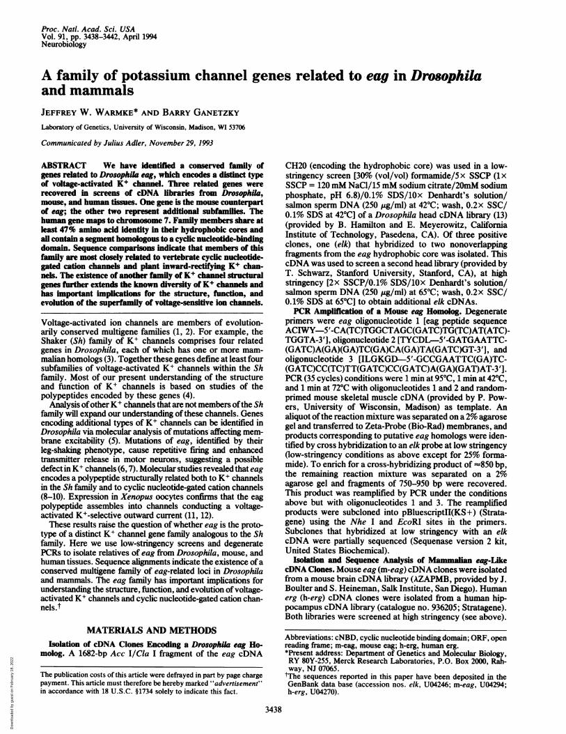
Y V T A L Y F T M T C M T S V G F G N V A A E T D N EY II S S L Y FT TS V G F G NAP T EY V T A L Y F T FS S L S V G F G N VS P N NSEY E TA L Y F T FT S L T S V G F G N V S rAKNT T A EY V T A L Y I T L T G DFHA E N P R EY VT Y W S I T L T T G Y GD L H P V N T K EY C LY W S T L T L T T IG E T P P P V K D E EY Y S L Y W S T L T LT T I G - E T P P P V R D SLEWC VYLIVTMS TVGY DMgYCY TIVLG
I P E F W W A G I T M T T V G Y D I C PTLA L G
I P DAF W W A V V T M TT V G Y GD M T P V G V W GIPA FWYEIVTMTTVGY GDMVP[ETIAGIPL GEWWALVTEMTT.VGY DM[DP1EJYIG
FIG. 2. Amino acid sequence of the pore forming region ofvarious members of the superfamily of voltage-sensitive K+ chan-nels. Amino acid residues identical to the eag sequence are boxed(see Table 1 for description of channel genes included here).
amino acids (residues 452-459 of eag) in which there is onlya single conservative substitution in m-eag (Figs. 1 and 2).Several ofthe amino acids within this region, such as Ser-453,Phe-456, and Asn-458, uniquely distinguish the pore of theeag family from all known members ofthe Sh family. Anothervery highly conserved hydrophobic segment in the eag familyoccurs in S5 (residues 410-421 of eag). In this region of 12amino acids, all family members have at least 10 amino acididentities and no more than one nonconservative substitu-tion. Again, this region is distinctive for the eag familybecause it has no similarity to the corresponding region oftheSh family. The presumed extracellular segments linking S1 toS2 and S3 to S4 vary in length and sequence within the family.In contrast, the linkers between S2 and S3 and between S4and S5, which are presumed to be cytoplasmic, have con-siderable sequence similarity and are identical in size.The similarity among members of the eag family also
extends beyond the hydrophobic core of the polypeptides.Although the length of the N-terminal sequence is variable,the first t150 amino acids have been very well conserved.Also, in the cytoplasmic region downstream from S6, all fourpolypeptides contain a segment that shares substantial sim-ilarity to the cyclic nucleotide-binding domain (cNBD) pres-ent in cyclic nucleotide-gated cation channels from verte-brate photoreceptors and olfactory epithelia (24, 25). Thissegment represents one ofthe most conserved regions withinthe eag family. The putative cNBD of the elk, m-eag, andh-erg polypeptides share 45%, 84%, and 51% identity to thatof eag, respectively. In the C-terminal region downstreamfrom the putative cNBD, there is little sequence similarityamong family members.
All eag family members have numerous potential sites forposttranslational modification. At least two ofthese potentialposttranslational modifications are shared among all fourpolypeptides (Fig. 1): a consensus site for N-glycosylationlocated 12-17 amino acids upstream of the pore region and aconsensus site for protein kinase C phosphorylation located2-4 amino acids downstream of S6.A Multigene Family ofK+ Channel Subunits. A dendrogram
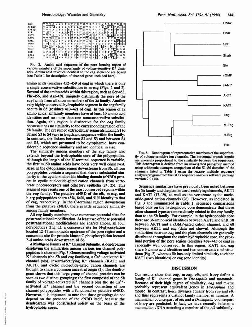
displaying the similarities among various ion channel poly-peptides is shown in Fig. 3. Genes encoding voltage-activatedK+ channels (the Sh and eag families), a Ca2+-activated K+channel (slo), inward-rectifying K+ channels (KAT1 andAKT1), and cyclic nucleotide-gated cation channels arethought to share a common ancestral origin (2). The dendro-gram shows that this large group of channel proteins can beseen as two distinct groupings: the first composed of the Shfamily of voltage-activated K+ channels plus the slo Ca2+-activated K+ channel and the second consisting of ionchannel polypeptides with a functional or putative cNBD.However, it is important to note that these groupings do notdepend on the presence of the cNBD itself, because thedendrogram was constructed solely on the basis of thehydrophobic cores.
Shaw
Shal
ShB
Shab
Slo
cGMP
cAMP
AKT1
KAT1
Eag
M-Eag
H-Erg
Elk
FIG. 3. Dendrogram of representative members of the superfam-ily of voltage-sensitive ion channels. The horizontal branch lengthsare inversely proportional to the similarity between the sequences.This dendrogram is derived from an unweighted pair-group methodusing arithmetic averages comparison of the S1-S6 domains of thechannels listed in Table 1 using the PILEUP multiple sequenceanalysis program from the GCG sequence analysis software packageversion 7.0 (14).
Sequence similarities have previously been noted betweenthe Sh family and the plant inward-rectifying channels, AKT1and KAT1 (17-19), as well as the vertebrate cyclic nucle-otide-gated cation channels (26). However, as indicated inFig. 3 and summarized in Table 1, sequence comparisonsbased only on the hydrophobic core demonstrate that theseother channel types are more closely related to the eag familythan to the Sh family. For example, in the hydrophobic corethere are 36 amino acid identities between AKT1 and ShB, 58between AKT1 and a cGMP-gated cation channel, and 68between AKT1 and eag (data not shown). Although thesimilarities between eag and the plant channels are generallydistributed throughout the entire hydrophobic core, the prox-imal portion of the pore region (residues 438-445 of eag) isespecially well conserved. In this region, KAT1 and eagshare 6 amino acid identities and two conservative substitu-tions (Fig. 2), whereas Sh has only limited similarity to eitherKAT1 (two identities) or eag (one identity).
DISCUSSIONOur results show that eag, m-eag, elk, and h-erg define afamily of K+ channel genes in Drosophila and mammals.Because of their high degree of similarity, eag and m-eagprobably represent equivalent genes in Drosophila andmouse. In contrast, h-erg diverges equally from eag and elkand thus represents a distinct family member in humans. Amammalian counterpart of elk and a Drosophila counterpartof h-erg are predicted. In fact, we have recently isolated amammalian cDNA encoding a member of the elk subfamily.
Neurobiology: Warmke and Ganetzky
Dow
nloa
ded
by g
uest
on
Feb
ruar
y 18
, 202
2

3442 Neurobiology: Warmke and Ganetzky
Thus, the eag family seems to contain at least three subfam-ilies analogous to those of the Sh family. Genes definingadditional eag subfamilies may be identified by more exhaus-tive screens.The striking hyperexcitability ofeag mutants demonstrates
the importance of eag channels in maintaining normal neu-ronal excitability in Drosophila (6, 7). The strong conserva-tion of eag polypeptide sequences from Drosophila to mam-mals suggests preservation of in vivo functions as well.Mapping h-erg more precisely on chromosome 7 is ofinterestto determine whether it is linked to any heritable neurologicaldisorders, such as those associated with seizures.The existence of an eag family of K+ channel genes
indicates that K+ channel diversity in vivo is even greaterthan has previously been inferred. If coassembly of polypep-tides occurs within the eag family or between the eag and Shfamilies, the potential number of distinct K+ channels be-comes still larger. Reduction in the amplitude of four distinctK+ currents by eag mutations, including the A-current abol-ished by Sh mutations, may indicate that eag encodes ashared component of different K+ channels (27, 28). How-ever, further experiments are necessary to determinewhether this interpretation is correct.
Inspection of the amino acid similarities and differencesbetween the eag and Sh families provides some interestinginsights into structure-function relationships. For example,in the pore region, all eag family proteins have Asn at theposition corresponding to residue 458 of eag (Fig. 2). Incontrast, all Sh family proteins contain Asp at this site (2).Substituting Asn for Asp at this position in the Sh familyproteins abolishes channel activity (29). Nonetheless, theactivity and K+ selectivity of the eag channel demonstratethat Asn at this position is not necessarily incompatible withK+ channel function (11, 12). Elucidating the basis of thisdifference between the pore region properties should proveinformative. Conversely, the invariant amino acids betweenthe eag and Sh family proteins could identify critical residuesunder severe evolutionary constraints.A distinctive feature of the eag family is the homology to
cNBDs of cyclic nucleotide-gated cation channels and cyclicnucleotide-activated protein kinases (24, 25). A 10-15%increase in macroscopic current in the presence of 2 ,uMcAMP but not cGMP has been reported for eag channels ininside-out macropatches and it is suggested that this modu-lation may involve direct binding to the channel (12). How-ever, unlike the vertebrate cyclic nucleotide-gated cationchannels, which are relatively voltage-insensitive, activationof eag channels shows a very steep voltage dependence (11,12). In addition, whereas cyclic nucleotide-activated cationchannels show little selectivity among monovalent and diva-lent cations, eag is strongly selective for K+ over Na+. Theeag family may thus be an evolutionary link between voltage-activated K+ channels and cyclic nucleotide-gated cationchannels with intermediate structural and functional proper-ties.The phylogeny of these ion channel families as well as the
inward-rectifying K+ channels in plants is still uncertain.However, conservation of a putative cNBD and a portion ofthe pore among the eag family, the plant channels, and cyclicnucleotide-gated channels indicates that these features areancient, predating the evolutionary separation of plants and
animals. Additional studies of the eag family and its rela-tionship to other channel families should, therefore, providefurther insights into the structure, function, and evolution ofK+ channels and their immediate relatives.
We thank Steven Titus for technical assistance, Pat Powers forisolation of h-erg cosmid clones, and Justin Thackeray for helpfulcomments on the manuscript. This research was supported by grantsfrom the National Institutes of Health (NS15390), a postdoctoralfellowship from the Muscular Dystrophy Association, and a Mc-Knight Neuroscience Development Award. This is paper number3390 from the Laboratory of Genetics, University of Wisconsin,Madison.
1. Hille, B. (1992) Ionic Channels ofExcitable Membranes (Sin-auer, Sunderland, MA), 2nd Ed.
2. Strong, M., Chandy, G. & Gutman, G. (1993) Mol. Biol. Evol.10, 221-242.
3. Salkoff, L., Baker, K., Butler, A., Covarrubias, M., Pak, M. D.& Wei, A. (1992) Trends Neurosci. 15, 161-166.
4. Jan, L. Y. & Jan, Y. N. (1992) Annu. Rev. Physiol. 54,537-555.5. Wu, C.-F. & Ganetzky, B. (1992) in Ion Channels, ed. Nara-
hashi, T. (Plenum, New York), Vol. 3, pp. 261-314.6. Ganetzky, B. & Wu, C.-F. (1983) J. Neurogenet. 1, 17-28.7. Ganetzky, B. & Wu, C.-F. (1985) Trends Neurosci. 8, 322-326.8. Drysdale, R. A., Warmke, J. W., Kreber, R. & Ganetzky, B.
(1991) Genetics 127, 497-505.9. Warmke, J., Drysdale, R. & Ganetzky, B. (1991) Science 252,
1560-1562.10. Guy, H. R., Durell, S. R., Warmke, J., Drysdale, R. & Gan-
etzky, B. (1991) Science 254, 730.11. Robertson, G., Warmke, J. & Ganetzky, B. (1993) Biophys. J.
64, 340 (Abstr.).12. Bruggerman, A., Pardo, L. A., Stuhmer, W. & Pongs, 0. (1993)
Nature (London) 365, 445-448.13. Palazzolo, M. J., Hamilton, B. A., Ding, D., Martin, C. H.,
Mead, D. A., Mieredorf, R. C., VuayRaghavan, K., Meyerow-itz, E. M. & Lipshitz, H. D. (1990) Gene 88, 25-36.
14. Devereaux, J., Haeberli, P. & Smithies, 0. (1984) NucleicAcids Res. 12, 387-395.
15. Kaupp, U. B., Niidome, T., Tanabe, T., Terada, S., Bonigk,W., Stuhmer, W., Cook, N. J., Kangawa, K., Matsuo, H. &Hirose, T. (1989) Nature (London) 342, 762-766.
16. Dhallan, R. S., Yau, K. W., Schrader, K. A. & Reed, R. R.(1990) Nature (London) 347, 184-187.
17. Sentenec, H., Bonneaud, N., Minet, M., Lacroute, F., Salmon,J., Gaymard, F. & Grignon, C. (1992) Science 256, 663-665.
18. Anderson, J. A., Huprikar, S. S., Kochian, L. V., Lucas,W. J. & Gaber, R. F. (1992) Proc. Natl. Acad. Sci. USA 89,3736-3740.
19. Schachtman, D. P., Schroeder, J. I., Lucas, W. J., Anderson,J. A. & Gaber, R. F. (1992) Science 258, 1654-1658.
20. Atkinson, N., Robertson, G. & Ganetzky, B. (1991) Science253, 551-555.
21. Cavener, D. (1991) Nucleic Acids Res. 19, 3185-3192.22. Kozak, M. (1989) J. Cell Biol. 108, 229-241.23. Ptacek, L. J., George, A. L., Jr., Griggs, R. C., Tawil, R.,
Kallen, R. G., Barchi, R. L., Robertson, M. & Leppert, M. F.(1991) Cell 67, 1021-1027.
24. Kaupp, U. B. (1991) Trends Neurosci. 14, 150-157.25. Shabb, J. B. & Corbin, J. (1992)J. Biol. Chem. 267, 5723-5726.26. Jan, L. Y. & Jan, Y. N. (1990) Nature (London) 345, 672.27. Zhong, Y. & Wu, C.-F. (1991) Science 252, 1562-1564.28. Zhong, Y. & Wu, C.-F. (1993) J. Neurosci. 13, 4669-4679.29. Heginbotham, L., Abramson, T. & MacKinnon, R. (1992)
Science 258, 1152-1155.
Proc. Nad. Acad Sci. USA 91 (1994)
Dow
nloa
ded
by g
uest
on
Feb
ruar
y 18
, 202
2