powerpoint presentation · pre procedure echo check list •confirm trileaflet av morphology...
TRANSCRIPT

01/21/2017
1
TAVR
VICTOR FARAH, MD, FACC, FASE, FACP
NO DISCLOSURES
• Dynamic field
• Rapidly evolving
• New valves
• New sizes
• New delivery systems
• New techniques
• NO guidelines
• General concepts
• Continuous learning
• AV anatomy
01/21/2017
2
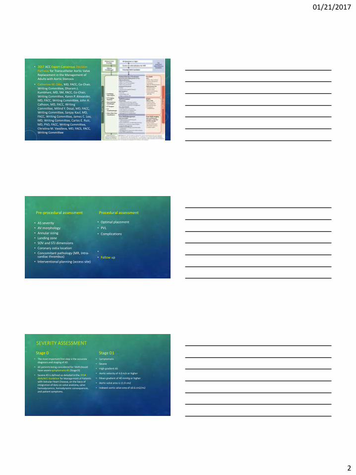
• 2017 ACC Expert Consensus Decision Pathway for Transcatheter Aortic Valve Replacement in the Management of Adults with Aortic Stenosis
• Catherine M. Otto, MD, FACC, Co-Chair, Writing Committee, Dharam J. Kumbhani, MD, SM, FACC, Co-Chair, Writing Committee, Karen P. Alexander, MD, FACC, Writing Committee, John H. Calhoon, MD, FACC, Writing Committee, Milind Y. Desai, MD, FACC, Writing Committee, Sanjay Kaul, MD, FACC, Writing Committee, James C. Lee, MD, Writing Committee, Carlos E. Ruiz, MD, PhD, FACC, Writing Committee, Christina M. Vassileva, MD, FACS, FACC, Writing Committee
Pre-procedural assessment
• AS severity
• AV morphology
• Annular sizing
• Landing zone
• SOV and STJ dimensions
• Coronary ostia location
• Concomitant pathology (MR, Intra-cardiac thrombus)
• Interventional planning (access site)
Procedural assessment
• Optimal placement
• PVL
• Complications
•
• Follow up
SEVERITY ASSESSMENT
Stage D
• The most important first step is the accurate diagnosis and staging of AS.
• All patients being considered for TAVR should have severe symptomatic AS (Stage D)
• Severe AS is defined as detailed in the 2014 AHA/ACC Guideline for Management of Patients with Valvular Heart Disease, on the basis of integration of data on valve anatomy, valve hemodynamics, hemodynamic consequences, and patient symptoms.
Stage D1
• Symptomatic
• Severe
• High-gradient AS
• Aortic velocity of 4.0 m/s or higher
• Mean gradient of 40 mmHg or higher
• Aortic valve area is ≤1.0 cm2
• Indexed aortic valve area of ≤0.6 cm2/m2

01/21/2017
3
SEVERITY ASSESSMENT
Stage D2
• Symptomatic
• Low-flow (Stroke volume index <35 ml/m2)
• Low- gradient (mean gradient <40 mm Hg)
• Low LVEF < 50%
• Severe AS
• Aortic valve area ≤1.0 cm2
• Aortic velocity is <4.0 m/s at rest
• AV velocity increases to at least 4.0 m/s on low-dose dobutamine stress echocardiography
Stage D3
• Symptomatic
• Low-flow (Stroke volume index <35 ml/m2)
• Low-gradient (mean gradient <40 mm Hg)
• Normal LVEF
• Severe AS
• Aortic valve area ≤1.0 cm2
• Aortic velocity <4.0 m/s
• Diagnosis of Stage D3 is challenging, with key features including an indexed aortic valve area of ≤0.6 cm2/m2, a stroke volume index <35 ml/m2, confirmation of hemodynamics when the patient is normotensive
BASICS OF AV EVALUATION
• V1 X A1 = V2 X A2
• Continuity equation
• AV CW
• LVOT PW
• LVOT diameter
Peak velocity
• CW Doppler
• Parallel to jet
• Multiple windows
• Strongest predictor of clinical outcome
• Flow dependent
01/21/2017
4
Mean velocity
• CW Doppler
• Multiple windows
• Parallel to jet
• Average of instantaneous gradients ever ejection period
• Most comparable to invasive measurement
• Flow dependent
STROKE VOLUME
LVOT TVI LVOT Diameter

01/21/2017
5
SOURCES OF ERROR
• Malalignment of the US beam
• LVOT diameter greatest source of error
• PW sample volume position
• Variable R-R
LVOT DIAMETER WHERE TO MEASURE?????
01/21/2017
6
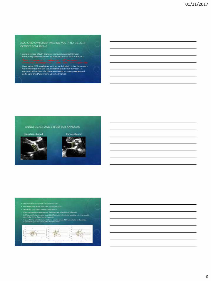
JACC: CARDIOVASCULAR IMAGING, VOL. 7, NO. 10, 2014 OCTOBER 2014:1062–8
• Annulus Instead of LVOT Diameter Improves Agreement Between Echocardiography Effective Orifice Area and Invasive Aortic Valve Area
• There is no consensus on the optimal site for LVOT measurement on transthoracic echocardiography (TTE), with guidelines permitting flexibility
• Given varied LVOT morphology and increased ellipticity below the annulus, we hypothesized that EOA calculated from the annulus diameter—as compared with sub-annular diameters—would improve agreement with aortic valve area (AVA) by invasive hemodynamics.
ANNULUS, 0.5 AND 1.0 CM SUB ANNULAR
Hourglass- shaped Funnel-shaped
• 114 consecutive adult patients with symptomatic AS
• Referred for transcatheter aortic valve implantation (TAVI)
• Two blinded, independent readers interpreted TTEs
• EOA was compared using diameters at the annulus and 0.5 and 1.0 cm subannular
• LVOT was stratified as hourglass- shaped (LVOT diameter 0.5 cm below annulus greater than annulus diameter) or funnel-shaped (remaining cases).
• Invasive AVA was calculated using the Gorlin equation (mean of 3 thermodilution cardiac output measurements) and was corrected for the catheter size.

01/21/2017
7
• This study demonstrates that:
• EOA calculated from the annular diameter—rather than the LVOT diameter 0.5 or 1.0 cm below the annulus—results in the best agreement with the AVA determined by invasive hemodynamics in AS patients referred for TAVI
• Mean EOA using the annular diameter was similar to AVA regardless of LVOT morphology, whereas use of an LVOT diameter below the annulus resulted in significant and meaningful overestimation of EOA in patients with funnel-shaped LVOTs and underestimation of EOA in those with hourglass-shaped LVOTs

01/21/2017
8
AV MORPHOLOGY
• TAVR is considered calcific valvular AS.
• TAVR in not considered for:
• Congenital AS (Bicuspid)
• Rheumatic valve disease
• Isolated aortic regurgitation
• Not been studied in clinical trials
• Make sure it is not bicuspid valve

01/21/2017
9
ANNULAR SIZING
THE CROWN

01/21/2017
10
• The scalloped configuration of the leaflets leave fibrous inter-leaflet triangles or trigones between the sinuses
• The annulus is measured at the cusps HINGE points
Kaseletal. JACC: CARDIOVASCULAR IMAGING, VOL. 6, NO. 2, 2013

01/21/2017
11
JACC: CARDIOVASCULAR INTERVENTIONS VOL. 8, NO. 1, JANUARY 2015:119–27

01/21/2017
12
SIZING CT VS. TEE
• Aortic Annular Sizing Using a Novel 3-Dimensional Echocardiographic Method Use and Comparison With Cardiac Computed Tomography
• Omar K. Khalique, MD; Susheel K. Kodali, MD; Jean-Michel Paradis, MD; Tamim M. Nazif, MD; Mathew R. Williams, MD; Andrew J. Einstein, MD, PhD; Gregory D. Pearson, MD, PhD; Kishore Harjai, MD; Kendra Grubb, MD; Isaac George, MD; Martin B. Leon, MD; Rebecca T. Hahn, MD
• Conclusions—Annulus measurements using a new method for analyzing 3D-TEE images closely approximate those of MDCT. Annulus measurements from both modalities predict mild or greater paravalvular regurgitation with equivalent accuracy. (Circ Cardiovasc Imaging. 2014;7:155-163.)
01/21/2017
13
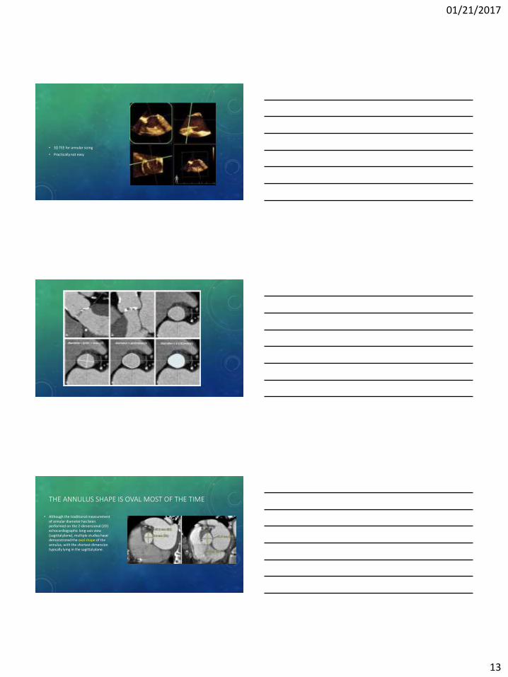
• 3D TEE for annular sizing
• Practically not easy
THE ANNULUS SHAPE IS OVAL MOST OF THE TIME
• Although the traditional measurement of annular diameter has been performed on the 2-dimensional (2D) echocardiographic long-axis view (sagittal plane), multiple studies have demonstrated the oval shape of the annulus, with the shortest dimension typically lying in the sagittal plane.

01/21/2017
14
MEASUREMENT OF THE AORTIC ANNULUS IS CRITICAL
Too large
• Annular rupture
• Central regurgitation (valvular)
• Coronary ostial obstruction
Too small
• Paravalvular AR
• Device migration
• Prosthesis-patient mismatch
LANDING ZONE
• Contrast-enhanced retrospectively ECG-gated data sets in two TAVR candidates. Transverse oblique maximum intensity projections of the aortic valve for evaluation of calcification burden show,
• A, asymmetric
• B, symmetric calcifications of all three cusps.
• C, Sagittal oblique view shows that calcifications extend into the left ventricular outflow tract.
• Blanke et al. CT in Transcatheter Aortic Valve Replacement, radiology.rsna.org n Radiology: Volume 269: Number 3—December 2013
01/21/2017
15
• The impact of calcium volume and distribution in aortic root injury related to balloon-expandable transcatheter aortic valve replacement
• Conclusion
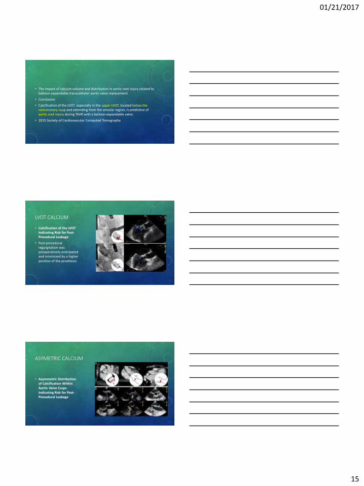
• Calcification of the LVOT, especially in the upper LVOT, located below the noncoronary cusp and extending from the annular region, is predictive of aortic root injury during TAVR with a balloon-expandable valve.
• 2015 Society of Cardiovascular Computed Tomography
LVOT CALCIUM
• Calcification of the LVOT Indicating Risk for Post-Procedural Leakage
• Post-procedural regurgitation was preoperatively anticipated and minimized by a higher position of the prosthesis
ASYMETRIC CALCIUM
• Asymmetric Distribution of Calcification Within Aortic Valve Cusps Indicating Risk for Post-Procedural Leakage

01/21/2017
16
• Schematic Drawings Indicating Morphological Risk Factors for Post-Procedural Regurgitation
• The anticipated occurrence of leakage is marked (arrow). Nonfused commissures in the neighborhood of bulky masses (left), an asymmetric distribution of calcified masses (red) within the cusps (middle), and calcified structures in the left ventricular outflow tract (LVOT) (right) are anatomical regurgitation substrates.
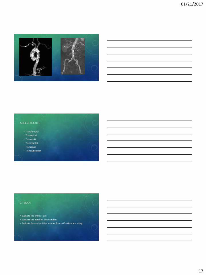
ACCESS SITE
01/21/2017
17
ACCESS ROUTES
• Transfemoral
• Transapical
• Transaortic
• Transcarotid
• Transcaval
• Transsubclavian
CT SCAN
• Evaluate the annular size
• Evaluate the aorta for calcifications
• Evaluate femoral and iliac arteries for calcifications and sizing

01/21/2017
18
PVL IS BAD!!!
• Kodali et al, NEJM 2012
• N Engl J Med 2012; 366:1686-1695
• Two-Year Outcomes after Transcatheter or Surgical Aortic-Valve Replacemen
HOW TO ASSESS
• Very difficult to assess
• Especially during or right after the procedure
• Look from the deep trans-gastric views
• PHT
• Descending aortic PW
• Color circumferential
• Invasive
PVL risk factors
• Large aortic annulus
• Undersized prosthesis
• Asymmetric cusp calcification
• LVOT calcification
• Prominent septal bulge
• Low valve deployment
• The improvement in decreasing the PVL is due to the improvement in imaging and optimizing sizing and devices
• Balance of para-valvular regurgitation vs annular rupture
• Undersized prosthesis vs oversized
01/21/2017
19
COMPLICATION EVALUATION
• Embolization
• Thrombosis
• LV dysfunction
• Coronary obstruction
• Effusion/tamponade
• Rupture of aortic root or annulus
• Cardiac perforation
• Traumatic VSD
• Septal hematoma
• Avulsion of Asc ao intima
• New or worsening MR
• Severe AR
HYPOTENSION TROUBLESHOOTING
• Severe AR
• Annular rupture
• Coronary obstruction (Aortic cusps or bulky calcium)
• Tamponade
• Hypovolemia due to bleed
FOLLOW UP
01/21/2017
20
01/21/2017
21
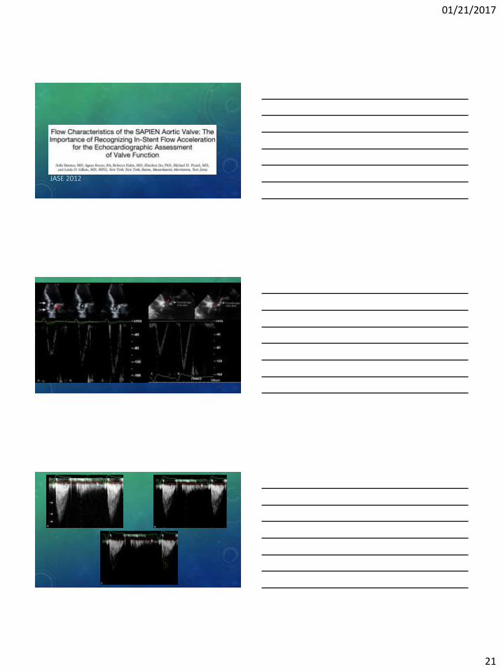
JASE 2012

01/21/2017
22
METHODS:
• 40 patients who underwent successful implantation with the SAPIEN valve.
• PW Doppler was recorded with sample volumes:
• Immediately proximal to the stent (prestent)
• Within the stent but proximal to the cusps (in-stent precusp)
• Distal to the cusps (in-stent postcusp)
• DVI and EOA were calculated using both prestent and in-stent precusp velocities to represent ‘‘subvalvular’’ flow and CW recordings of the left ventricular outflow tract and aortic valve to represent postvalvular flow.
RESULTS:
• The mean in-stent precusp peak velocities were significantly higher than the prestent values (1.5 +- 0.2 vs 1.0 +- 0.2 m/sec, P < .0001).
• ERO and DVI calculated using the prestent versus in-stent precusp velocities were significantly different (1.79 +- 0.34 vs 2.54 +- 0.46 cm2, P < .0001, and 0.48 +- 0.12 vs 0.73 6 +-.13, P < .0001, respectively).
CONCLUSIONS:
• The SAPIEN valve demonstrates flow acceleration at two levels, representing contributions of both the stent and valve cusps to the total valve gradient.
• Failure to recognize this phenomenon may result in inappropriate selection of the in-stent precusp pulsed Doppler spectrum to represent ‘‘subvalvular’’ flow, thereby overestimating the effective orifice area and Doppler velocity index.

01/21/2017
23
TAVR PROCESS
TAVR VS SAVR PARTNER TRIALS CONCLUSION
• TAVR superior to MEDICAL therapy in INOPERABLE patients (PARTNER 1)
• TAVR equivalent to SAVR in HIGH RISK patients (PARTNER 1)
• TAVR equivalent to SAVR in INTERMEDIATE RISK patients (PARTNER 2)
• LOW RISK patients (PARTNER 3)
• The less invasive the better
01/21/2017
24
PRE TAVR TESTING
• Evaluation by 2 surgeons
• Evaluation of surgical risk score and frailty assessment
• TTE or TEE confirm the severity of the stenosis (If not before)
• DSE in patient’s with LF/LG Low EF AS
• Cardiac catheterization (if not done recently, this is the interventionalist/surgeon decision)
• Annular sizing (By CT unless CT is contraindicated)
• If sizing is not clear by CT (calcification, or non contrast CT) then we do 3D TEE
PRE PROCEDURE ECHO CHECK LIST
• Confirm trileaflet AV morphology
• Presence and degree of AS, MR, TR
• LV/RV function
• Presence of basal septal hypertrophy/LVOT obstruction
• Presence of pericardial effusion
• Exclude LA or LV thrombus
• Annular sizing
• Landing zone
INTRA/POST PROCEDURAL OBJECTIVES
• Ensure proper prosthesis placement
• Assess prosthesis position and function after deployment
• Assess ventricular function
• Identify immediate post deployment complications

01/21/2017
25
AGH
TA TF All
2012 0 17 17
2013 32 26 58
2014 38 53 91
2015 46 131 177
2016 26 185 211
AGH EXPERIENCE
• First minimal approach (conscious sedation) was November 7, 2016
• Since then 95% of cases are under conscious sedation, and 5 % general anesthesia
• Length of stay for TF is 2.8 days with conscious sedation
• Surgical AVR length of stay 6.5 days
AGH
Procedure duration
• 90 minutes approximately (Including: anesthesia prep time (lines, put to sleep), Clipping, prep and draping, procedure, then perclose, wake up patient, and leave the room)
• 35 minutes procedure itself
Post-procedure
• 55% go to PACU
• 45% to ICU or CCU

01/21/2017
26
AGH
• Dr. Stephen Bailey
• Dr. Robert Moraca
• Dr. Ramzi Khalil
• Dr. David Lasorda
• Dr. Rachel Hughes-Doichev
• Dr. Moneal Shah
• Dr. Jeffrey Mueller
• Dr. Timothy Mickus
• Dr Victor Farah
• Diane Berger RN
• Laurie Weatherby RN
• Eileen Martini RN
• Linda Hamilton
• Susan Bartley
• Rachel Myers
• Alan Mathews
• Erin Hochendoner
COMPLICATION RATE
PPM 3.4%
Major vascular complication 2.3%
Renal failure 2.3%
Stroke 1.8%
Respiratory failure (prolonged intubation) 6.98 %
• Thank you

01/21/2017
27

01/21/2017
28
• 1 out of every 4 patients >65 years old have aortic sclerosis
• 1 out of every 6 patients with aortic sclerosis advance to AS within 7 years
• 4% of north America population older than 75 have AS
• About 50% of patients with mild to moderate AS progress to severe
• 45-50 years Sclerosis
• 60-75 years Asymptomatic AS
• 65-85 years Symptomatic AS

01/21/2017
29
• The maximum diameter of the annulus bisects a trigone on one side and a cusp on the other side
• When equal cusps are imaged in LAX view, the LVOT and annular diameter may be underestimated
• Measure parallel to the aortic valve plane
• Measure inner edge to inner edge in mid systole
• Zoom
• Image PW and LVOT in the same region
• The trigon between L and N coronary cusps is imaged
• Do not measure too high into the aortic root
• The true annular plane is perpendicular to the long axis of the aorta
01/21/2017
30
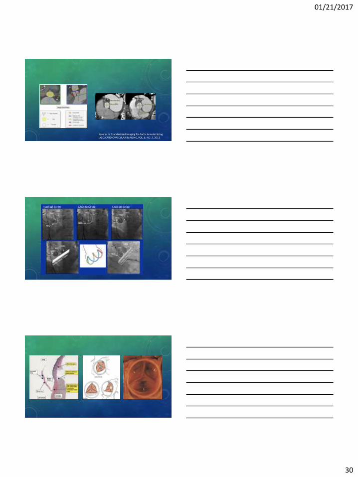
Kasel et al. Standardized Imaging for Aortic Annular Sizing JACC: CARDIOVASCULAR IMAGING, VOL. 6, NO. 2, 2013

01/21/2017
31
01/21/2017
32
01/21/2017
33
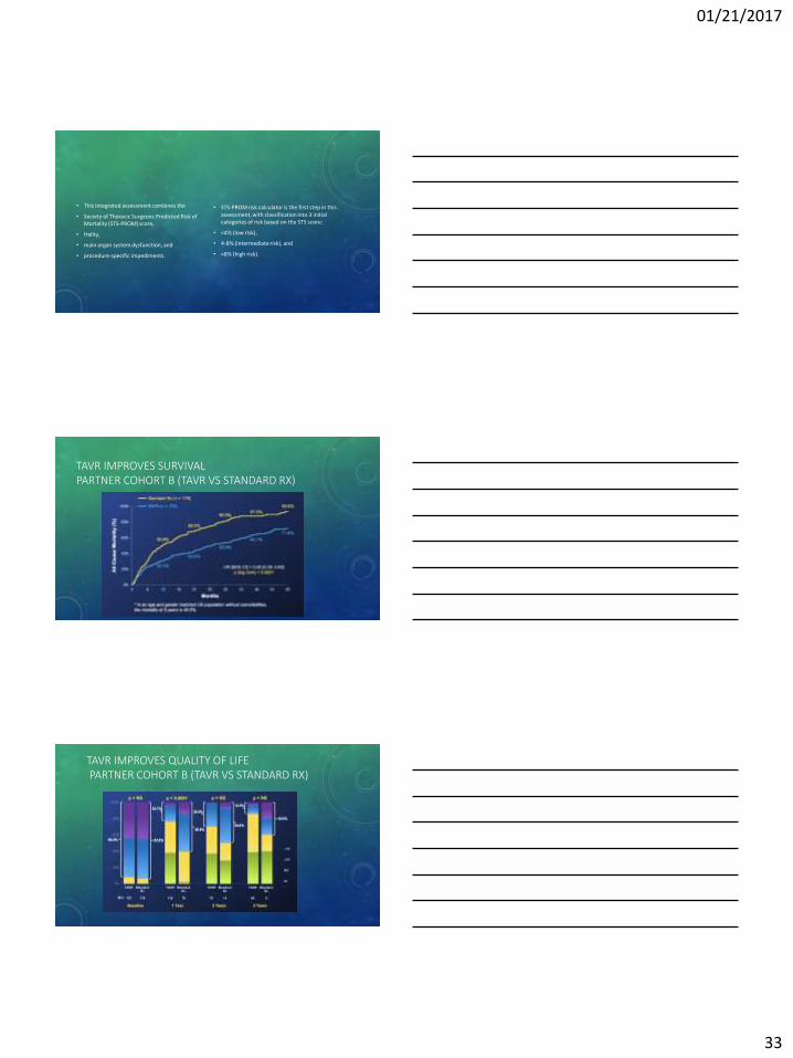
• This integrated assessment combines the
• Society of Thoracic Surgeons Predicted Risk of Mortality (STS-PROM) score,
• frailty,
• main organ system dysfunction, and
• procedure-specific impediments.
• STS-PROM risk calculator is the first step in this assessment, with classification into 3 initial categories of risk based on the STS score:
• <4% (low risk),
• 4-8% (intermediate risk), and
• >8% (high risk).
TAVR IMPROVES SURVIVAL PARTNER COHORT B (TAVR VS STANDARD RX)
TAVR IMPROVES QUALITY OF LIFE PARTNER COHORT B (TAVR VS STANDARD RX)
01/21/2017
34
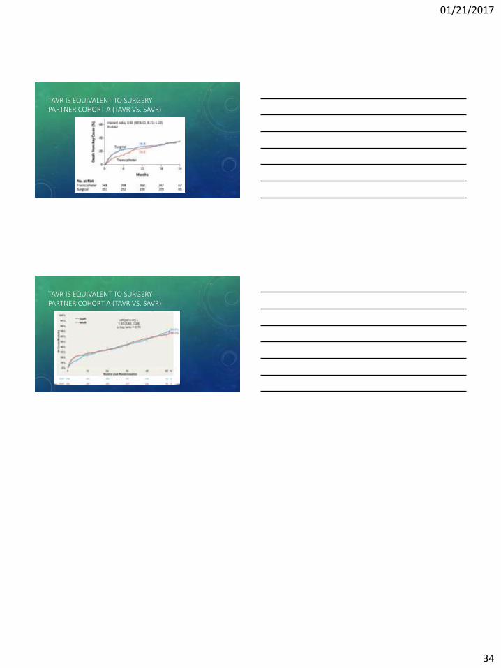
TAVR IS EQUIVALENT TO SURGERY PARTNER COHORT A (TAVR VS. SAVR)
TAVR IS EQUIVALENT TO SURGERY PARTNER COHORT A (TAVR VS. SAVR)