powerpoint presentation -...
TRANSCRIPT
8/18/2017
1
Neurologic Alterations
Heather A Martin MSN, RN, CNRN, SCRN
Neuroscience Clinical Nurse Specialist
Swedish Medical Center
Objectives
1. Describe common CNS tumors
2. Describe signs and symptoms of
neurologic alterations
3. Review key assessment skills and
nursing interventions
Disclosures
• none
3
8/18/2017
2
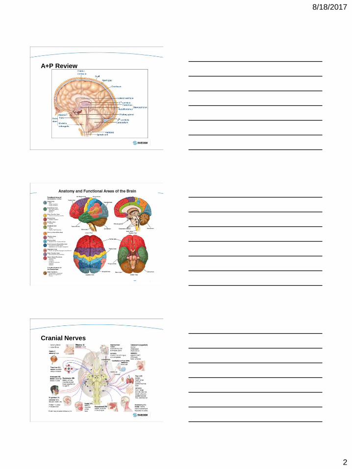
A+P Review
Cranial Nerves

8/18/2017
3
Dermatome Distribution
Neuro Assessment
• Baseline assessment is essential and needs
to be documented
• LOCATION and type of injury/insult
• Basic Assessment
– Glascow Coma Scale
– Motor/Sensory Function
– Cranial nerve dysfunction
– Pupil
Glascow Coma Scale

8/18/2017
4
Motor Strength
Pupil Assessment
Nystagmus- Rhythmic, oscillating motions of the eyes are
called nystagmus
Anascoria- unequal pupils
Focal-impairments of nerve, spinal cord, or brain function
that affects a specific region of the body
Common Terms in Neuro Assessment
8/18/2017
5
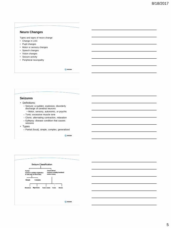
Neuro Changes
Types and signs of neuro change
• Change in LOC
• Pupil changes
• Motor or sensory changes
• Speech changes
• Vision changes
• Seizure activity
• Peripheral neuropathy
Seizures
• Definitions:
– Seizure: a sudden, explosive, disorderly discharge of cerebral neurons
• Motor, sensory, autonomic, or psychic
– Tonic: excessive muscle tone
– Clonic: alternating contraction, relaxation
– Epilepsy: disease condition that causes seizures
• Types
– Partial (focal), simple, complex, generalized
8/18/2017
6
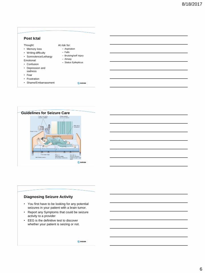
Post Ictal
Thought:
• Memory loss
• Writing difficulty
• Somnolence/Lethargy
Emotional:
• Confusion
• Depression and
sadness
• Fear
• Frustration
• Shame/Embarrassment
At risk for:
– Aspiration
– Falls
– Bruising/self injury
– Airway
– Status Epilepticus
Guidelines for Seizure Care
Diagnosing Seizure Activity
• You first have to be looking for any potential
seizures in your patient with a brain tumor.
• Report any Symptoms that could be seizure
activity to a provider
• EEG is the definitive test to discover
whether your patient is seizing or not.
8/18/2017
7
Seizure Med Management
• Benzo’s for immediate control
– Ativan/valium/versed
• Maintenance meds(may require a load)
– Phenytoin/Fosphenytoin
– Keppra
– Depakote
– -lacosamide
Common Types of CNS tumors
Breakdown of Brain Tumors
• There are more than 120 types of brain and
central nervous system (CNS) tumors
• Brain metastases are about 10 times more
common than primary tumors
• classify brain tumors by cell origin and how the
cells behave, from the least aggressive (benign) to
the most aggressive (malignant)
8/18/2017
8
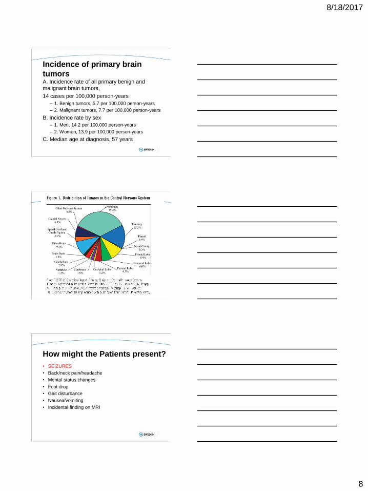
Incidence of primary brain
tumors A. Incidence rate of all primary benign and
malignant brain tumors,
14 cases per 100,000 person-years
– 1. Benign tumors, 5.7 per 100,000 person-years
– 2. Malignant tumors, 7.7 per 100,000 person-years
B. Incidence rate by sex
– 1. Men, 14.2 per 100,000 person-years
– 2. Women, 13.9 per 100,000 person-years
C. Median age at diagnosis, 57 years
23
How might the Patients present?
• SEIZURES
• Back/neck pain/headache
• Mental status changes
• Foot drop
• Gait disturbance
• Nausea/vomiting
• Incidental finding on MRI
8/18/2017
9
Primary Brain tumors – Meningioma
• Benign
• Atypical
• Malignant
– Primitive neuroectodermal tumors
(PNET) • Medulloblastoma
• Ependymoblastoma
• Pineoblastoma
– Pituitary tumors
• Pituitary adenoma
• Pituitary carcinoma
• Cranipharyngioma
• Rathke’s cleft cyst
– Pineal Tumors
• Pineal cyst
• Pineocytoma
• Pineoblastoma
• Germinoma
• Mixed germ cell tumor
• Pineal gliomas
• Pineal teratoma
– Choroid plexus tumors • Choroid plexus papilloma
• Choroid plexus carcinoma
– Other, more benign
primary tumors • Neurocytoma
• Dysembroplastic
neuroepithelial tumor
• Lipoma
• Hemangioblastoma
• Hamartoma
• Teratoma
– Tumors of nerves and/or nerve sheaths
• Neuroma
• Schwannoma
• Neurofibroma
25
Cysts
Colloid cyst Arachnoid cyst Colloid cyst
Arachnoid cysts Dermoid Epidermoid
Rathke's cleft cyst Pineal cyst
Other primary tumors,
including skull base Chondroma
Chordoma
Sarcomas
Gliosarcoma
Chondrosarcoma
Rhabdomyosarcoma
Primary Central Nervous System Lymphoma
(PCNSL)
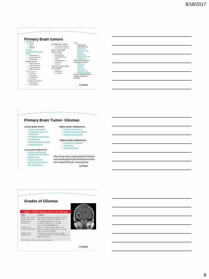
Primary Brain Tumor- Gliomas
Lowest grade tumors
– Pilocytic astrocytoma
– Subependymal giant cell
astrocytoma
– Protoplasmic astrocytoma
– Ganglioglioma
– Xanthomatous astrocytoma
– Subependymoma
Lower grade malignancies
– Fibrillary (gemistocytic,
protoplasmic) astrocytoma
– Ependymoma
– Oligodendroglioma
– Mixed oligo-astrocytoma
– Optic nerve glioma
Higher-grade malignancies
– Anaplastic astrocytoma
– Anaplastic oligodendroglioma
– Anaplastic mixed glioma
Highest-grade malignancies
– Glioblastoma multiforme
– Gliosarcoma
– Gliomatosis cerebri
http://www.aans.org/media/fact%20she
ets/classification%20of%20brain%20tu
mors.aspx#Pilocytic astrocytoma
Grades of Gliomas
8/18/2017
10
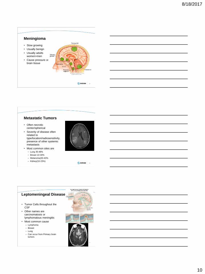
Meningioma
• Slow growing
• Usually benign
• Usually adults
women>men
• Cause pressure on
brain tissue
28
Metastatic Tumors
• Often necrotic
center/spherical
• Severity of disease often
related to
type/location/radiosensitivity,
presence of other systemic
metastasis
• Most common sites are
– Lung 35-48%
– Breast 10-30%
– Melanoma(30-40%
– Kidney(10-15%)
29
Leptomeningeal Disease
• Tumor Cells throughout the
CSF
• Other names are
carcinomatosis or
lymphomatous meningitis
• Most common cause
– Lymphoma
– Breast
– Lung
– Can occur from Primary brain
tumors
30
8/18/2017
11
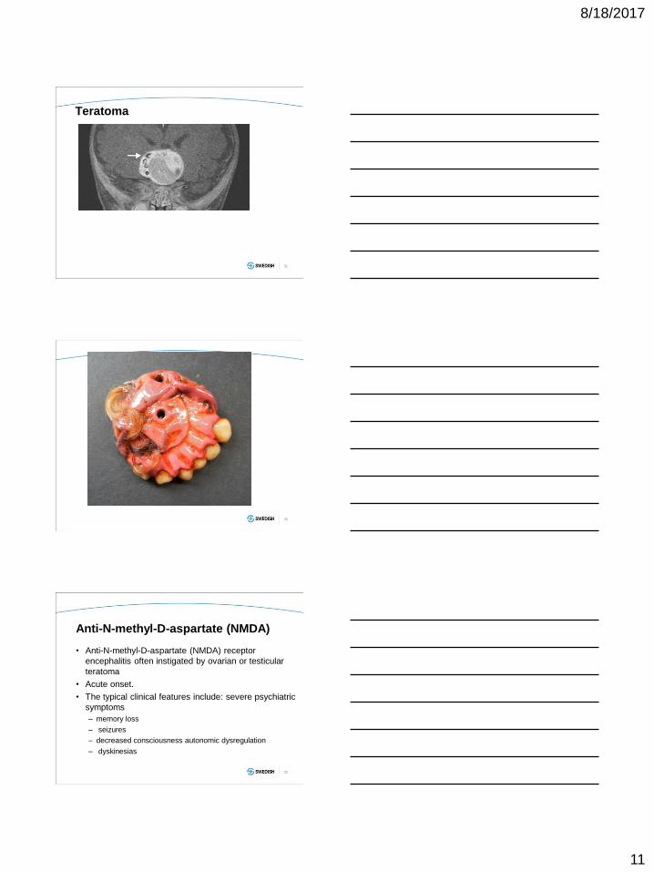
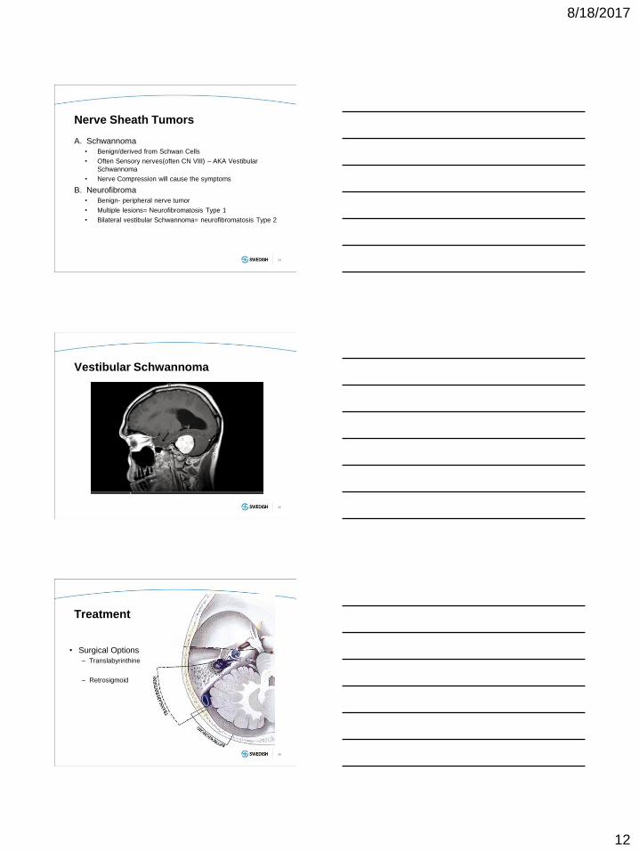
Teratoma
31
32
Anti-N-methyl-D-aspartate (NMDA)
• Anti-N-methyl-D-aspartate (NMDA) receptor
encephalitis often instigated by ovarian or testicular
teratoma
• Acute onset.
• The typical clinical features include: severe psychiatric
symptoms
– memory loss
– seizures
– decreased consciousness autonomic dysregulation
– dyskinesias
33
8/18/2017
12
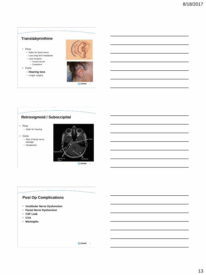
Nerve Sheath Tumors
A. Schwannoma
• Benign/derived from Schwan Cells
• Often Sensory nerves(often CN VIII) – AKA Vestibular
Schwannoma
• Nerve Compression will cause the symptoms
B. Neurofibroma
• Benign- peripheral nerve tumor
• Multiple lesions= Neurofibromatosis Type 1
• Bilateral vestibular Schwannoma= neurofibromatosis Type 2
34
Vestibular Schwannoma
35
Treatment
• Surgical Options
– Translabyrinthine
– Retrosigmoid
36
8/18/2017
13
Translabyrinthine
• Pros:
– Safer for facial nerve
– Less long term headache
– Less invasive
• Cranial nerves
• Cerebellum
• Cons
– Hearing loss
– Longer surgery
37
Retrosigmoid / Suboccipital
• Pros
– Safer for hearing
• Cons
– Risk of facial nerve
damage
– Headaches
38
Post Op Complications
• Vestibular Nerve Dysfunction
• Facial Nerve Dysfunction
• CSF Leak
• CVA
• Meningitis
39
8/18/2017
14
Hematopoietic Tumors
40
CNS Lymphoma
• Primary central nervous
system lymphoma
(PCNSL) is a high-grade
non-Hodgkin B-cell
neoplasm, usually large cell
or immunoblastic type.
• Secondary CNS:
Lymphomas can sometimes
migrate to the central
nervous system. This
secondary form of CNS
lymphoma is not common
41
CNS Lymphoma
• Systemic chemotherapy with or without stem cell rescue:
High dose methotrexate, high dose Cytarabine
• Intra-thecal chemotherapy: methotrexate (e.g. 12 mg)
• Rituxan for CD 20+ lymphoma – role in treatment unclear
• Corticosteroids –for edema, ICP, and its role in
chemotherapy (potentiates action)
• Anticonvulsants – seizures prophylaxis
• Radiation therapy – may have a role (whole brain for
primary, or to specific sites for secondary CNS lymphoma)
8/18/2017
15
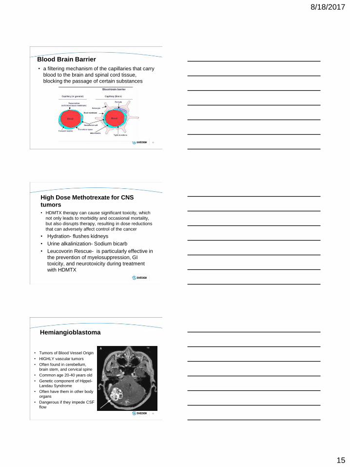
Blood Brain Barrier
• a filtering mechanism of the capillaries that carry
blood to the brain and spinal cord tissue,
blocking the passage of certain substances
43
High Dose Methotrexate for CNS
tumors
• HDMTX therapy can cause significant toxicity, which
not only leads to morbidity and occasional mortality,
but also disrupts therapy, resulting in dose reductions
that can adversely affect control of the cancer
• Hydration- flushes kidneys
• Urine alkalinization- Sodium bicarb
• Leucovorin Rescue- is particularly effective in
the prevention of myelosuppression, GI
toxicity, and neurotoxicity during treatment
with HDMTX
Hemiangioblastoma
• Tumors of Blood Vessel Origin
• HIGHLY vascular tumors
• Often found in cerebellum,
brain stem, and cervical spine
• Common age 20-40 years old
• Genetic component of Hippel-
Landau Syndrome
• Often have them in other body
organs
• Dangerous if they impede CSF
flow
45
8/18/2017
16
Neuroendocrine Tumors
46
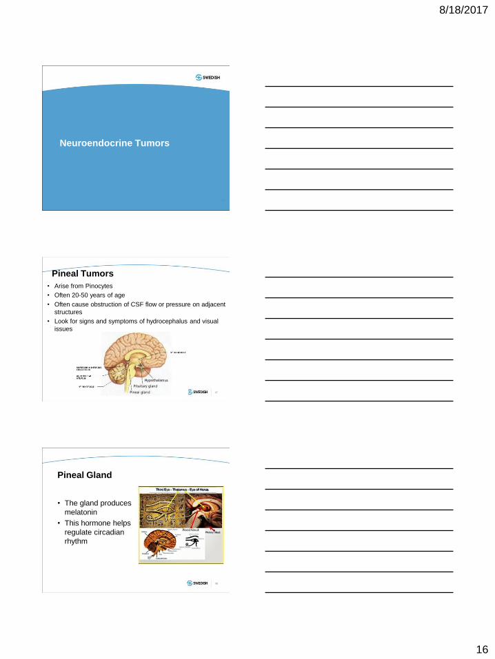
Pineal Tumors
• Arise from Pinocytes
• Often 20-50 years of age
• Often cause obstruction of CSF flow or pressure on adjacent
structures
• Look for signs and symptoms of hydrocephalus and visual
issues
47
Pineal Gland
• The gland produces
melatonin
• This hormone helps
regulate circadian
rhythm
48
8/18/2017
17
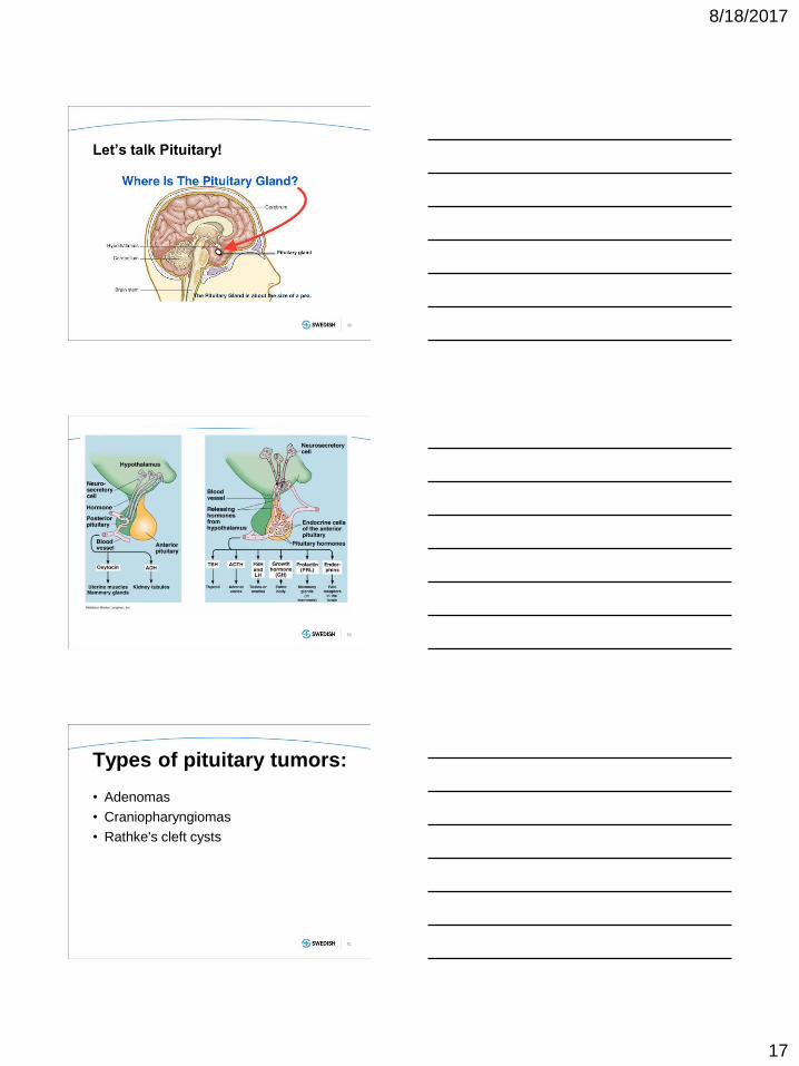
Let’s talk Pituitary!
49
50
Types of pituitary tumors: • Adenomas
• Craniopharyngiomas
• Rathke's cleft cysts
51
8/18/2017
18
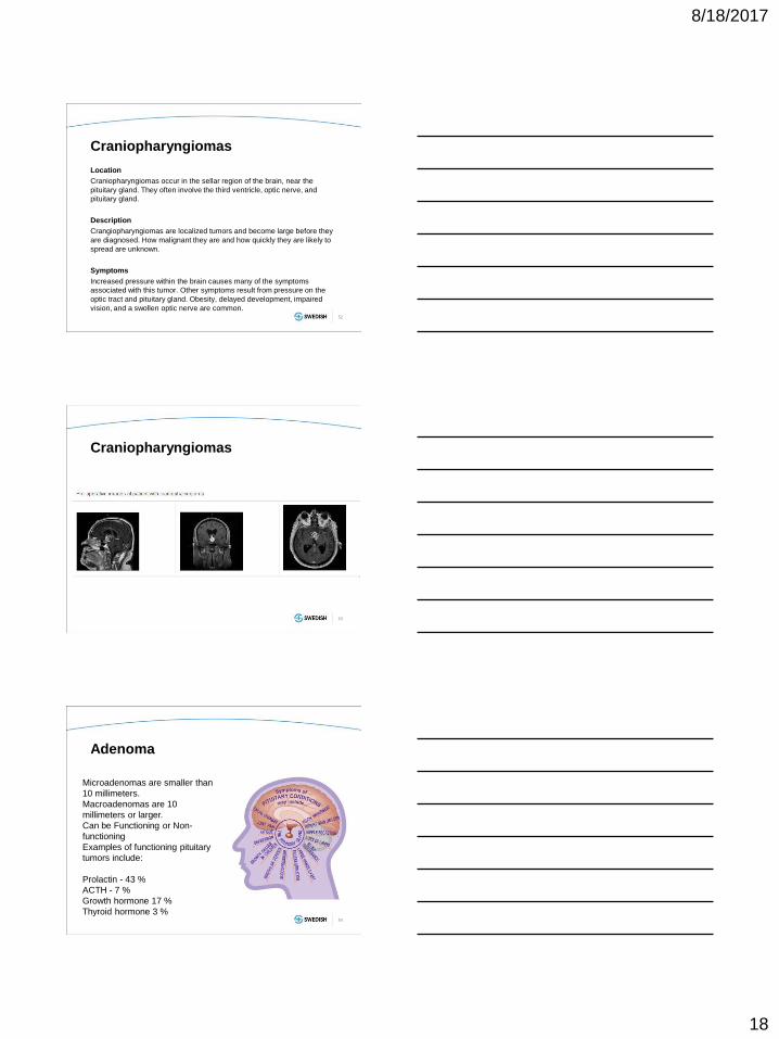
Craniopharyngiomas
Location
Craniopharyngiomas occur in the sellar region of the brain, near the
pituitary gland. They often involve the third ventricle, optic nerve, and
pituitary gland.
Description
Crangiopharyngiomas are localized tumors and become large before they
are diagnosed. How malignant they are and how quickly they are likely to
spread are unknown.
Symptoms
Increased pressure within the brain causes many of the symptoms
associated with this tumor. Other symptoms result from pressure on the
optic tract and pituitary gland. Obesity, delayed development, impaired
vision, and a swollen optic nerve are common.
52
Craniopharyngiomas
53
Adenoma
54
Microadenomas are smaller than
10 millimeters.
Macroadenomas are 10
millimeters or larger.
Can be Functioning or Non-
functioning
Examples of functioning pituitary
tumors include:
Prolactin - 43 %
ACTH - 7 %
Growth hormone 17 %
Thyroid hormone 3 %

8/18/2017
19
NAME YOUR HORMONE DYSFUNCTION
55
Rathke's cleft cysts
• Rathke cleft cysts (RCCs) are
benign (non-cancerous) fluid-
filled growths that develop
between the parts of the pituitary
gland at the base of the brain.
• Congenital deformities,
• RCC develops from a piece of
the fetus’ developing Rathke
pouch, which ultimately becomes
part of the pituitary gland
• Rarely cause problems during
childhood. Show up in adults
56
Transphenoidal Resection
57

8/18/2017
20
58
Brain Tumor TX
• Stereotactic biopsy
• Surgical Debulking
• Radiosurgery
• Chemo/Radiation
– Temozolomide –
oral/IV agent that
crosses BBB
• Gliadel wafers
• Novel treatments
AANS - Classification of Brain
Tumors
Stereotactic Biopsy
60

8/18/2017
21
Radiosurgery
61
62
www.mayoclinic.org
Surgical Resection
63

8/18/2017
22
Complications of Radiation to the Brain
• Can be direct damage at time or occur
months later
– Increased ICP(cerebral edema)
– Disruption of BBB
– Cognitive deficits
– Seizures
– Headaches
Intrathecal Chemo
Nurse Role Post lumbar
puncture
• Monitor for S/S of CSF leak
– Nausea
– Vomiting
– Headache
– fluid leaking
• Monitor for S/S of CNS
irritation
• Pain
• Hypotension
• Infection
Ommaya Resevoir
8/18/2017
23
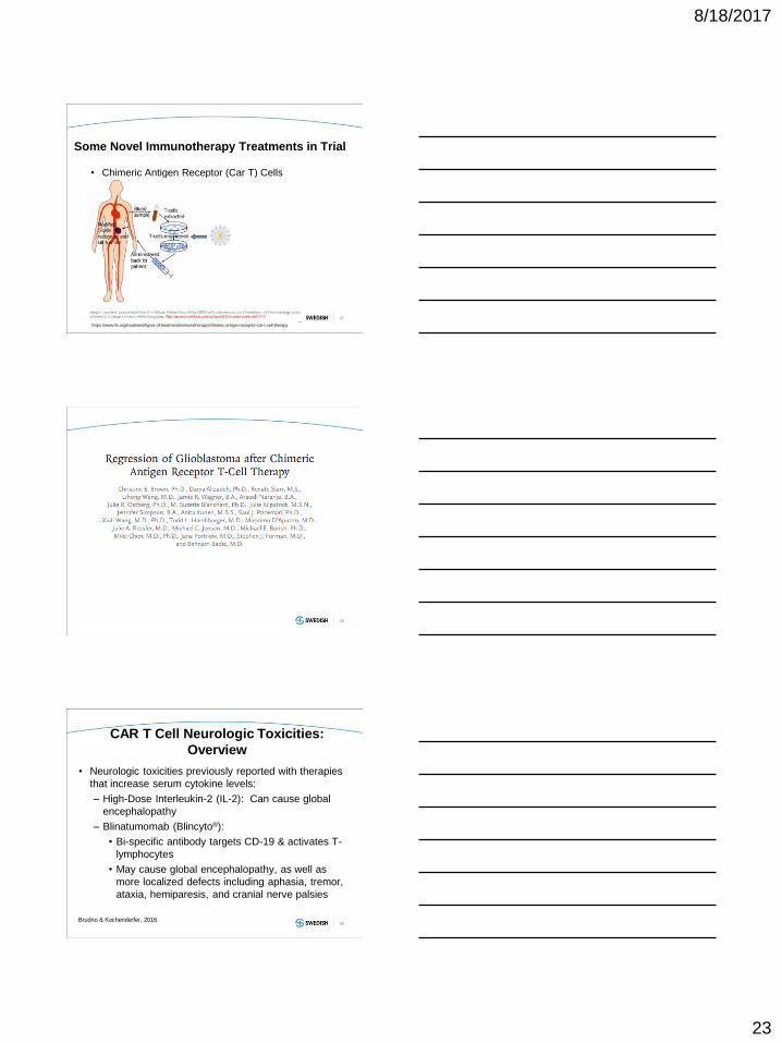
Some Novel Immunotherapy Treatments in Trial
• Chimeric Antigen Receptor (Car T) Cells
67
https://www.lls.org/treatment/types-of-treatment/immunotherapy/chimeric-antigen-receptor-car-t-cell-therapy
68
CAR T Cell Neurologic Toxicities:
Overview
• Neurologic toxicities previously reported with therapies
that increase serum cytokine levels:
– High-Dose Interleukin-2 (IL-2): Can cause global
encephalopathy
– Blinatumomab (Blincyto®):
• Bi-specific antibody targets CD-19 & activates T-
lymphocytes
• May cause global encephalopathy, as well as
more localized defects including aphasia, tremor,
ataxia, hemiparesis, and cranial nerve palsies
69 Brudno & Kochenderfer, 2016.
8/18/2017
24
CAR T Cell Neurologic Toxicities:
Overview
• Neurologic toxicities associated with anti-CD 19 CAR
T cells are similar to neurologic toxicities of
blinatumomab
• Can be diverse
• Do not localize to one specific area of neuroanatomy
70 Brudno & Kochenderfer, 2016.
Pathophysiology
• Not well understood
• May occur at different times than Cytokine Release
Syndrome(CRS) or in absence of CRS (suggests
different mechanism)
• Central nervous system (CNS) involvement of
leukemia shown NOT to be associated with CAR T
cell neurologic toxicity
• Modified T-cells have been found in CSF of
patients with neurologic toxicities, but also in
patients without neurologic toxicities (Maude, et al,
2014).
71 Brudno & Kochenderfer, 2016.
Onset & Duration
• Published studies: (Brudno & Kochenderfer,
2016)
– May occur concurrently with Cytokine
Release Syndrome(CRS), following
resolution of CRS, or in absence of CRS
toxicities
72

8/18/2017
25
Clinical Manifestations
• Can be diverse, do not localize to one specific area of
neuroanatomy
– Aphasia/dysphagia
– Confusion
– Motor neuropathy
– Somnolence
73
INCREASED ICP
74
Monroe Kellie Hypothesis
The Monro Kellie Doctrine describes the interrelation of
the various volume compartments of the CNS:
• Ventricles w/CSF
• Brain (white and gray matter)
• Subarachnoid space (SAS) w/CSF
• Volume of the blood in vessels
• The Monro Kellie Doctrine suggests that when the
volume of one compartment increases, there must be
a corresponding and compensatory decrease in the
volume of the other spaces.
8/18/2017
26
76
Increased ICP
1. altered levels of consciousness
2. changes in sensory and motor function
3. changes in pupil size, equality, and reaction
to light, and extraocular movements
4. changes in vital signs and patterns of
respiration.
Types of Herniation
a) Subfalcial herniation
b) uncal herniation
c) central transtentorial
herniation
d) external herniation
e) tonsillar herniation

8/18/2017
27
Treatment of ICP
• Medication
– Corticosteroids
– Hypertonic saline-23.4%
– Mannitol
• Surgery
• Nursing interventions
– Increase HOB(30-45 degrees)
– Keep body in alignment
– Head/neck straight
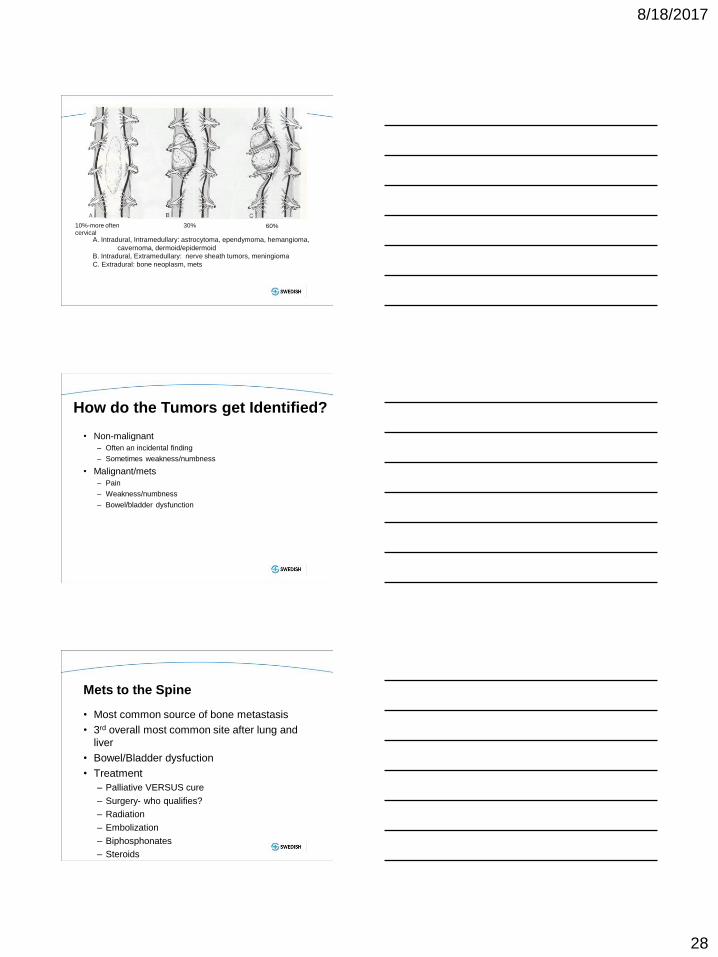
Spinal Tumors
Spinal tumors
• Primary Spinal tumors are relatively rare and affect
only a minority of the population.
• Cause significant morbidity in terms of pain and limb
dysfunction
• Associated with mortality as well
• Early diagnosis and prompt treatment is important.
• MR imaging
• tumors to be classified as – Extradural
– intradural–extramedullary
– Intradural- intramedullary
8/18/2017
28
A. Intradural, Intramedullary: astrocytoma, ependymoma, hemangioma,
cavernoma, dermoid/epidermoid
B. Intradural, Extramedullary: nerve sheath tumors, meningioma
C. Extradural: bone neoplasm, mets
60% 10%-more often
cervical
30%
How do the Tumors get Identified?
• Non-malignant
– Often an incidental finding
– Sometimes weakness/numbness
• Malignant/mets
– Pain
– Weakness/numbness
– Bowel/bladder dysfunction
Mets to the Spine
• Most common source of bone metastasis
• 3rd overall most common site after lung and
liver
• Bowel/Bladder dysfuction
• Treatment
– Palliative VERSUS cure
– Surgery- who qualifies?
– Radiation
– Embolization
– Biphosphonates
– Steroids
8/18/2017
29
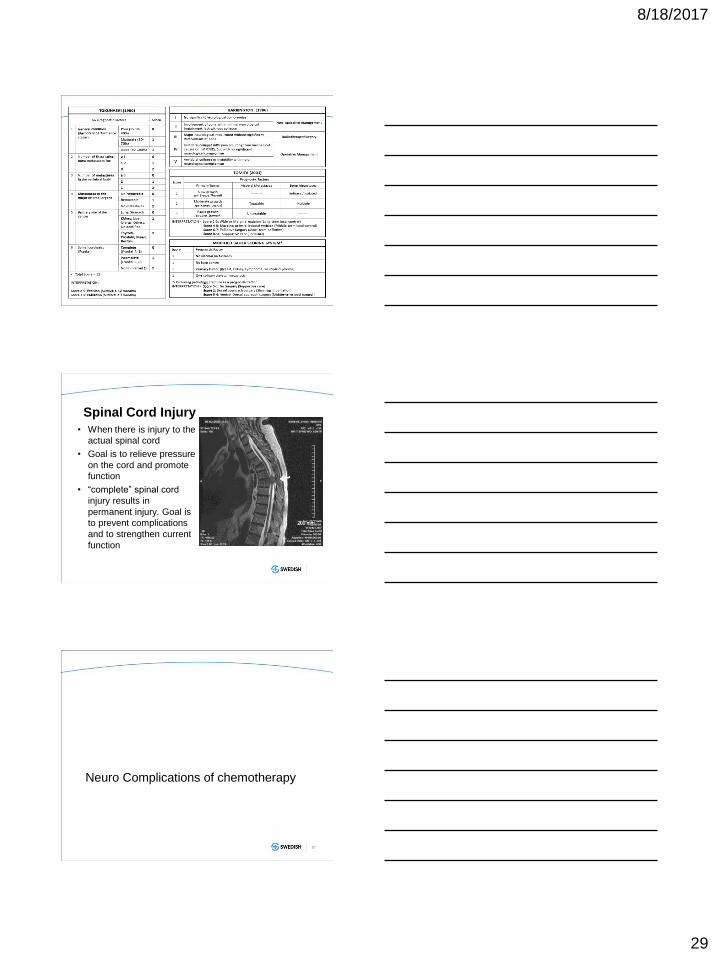
Spinal Cord Injury
• When there is injury to the
actual spinal cord
• Goal is to relieve pressure
on the cord and promote
function
• “complete” spinal cord
injury results in
permanent injury. Goal is
to prevent complications
and to strengthen current
function
Neuro Complications of chemotherapy
87
8/18/2017
30
Neuro Complications of Chemo Therapy
Peripheral Neuropathy
• Vincristine
• Cisplatin
• Taxanes
– Pacitaxel
– docetaxel
Cyclosporin/tacrolimus – Confusion
– Cortical blindness
– Brain hemorrhage
– Peripheral neuropathy
– Aphasia
– Cerebellar changes
89 Sorokin J, Saboury B, Ahn JA, Moghbel M, Basu S, Alavi A. Adverse functional effects of chemotherapy on whole-brain metabolism: a PET/CT quantitative analysis of FDG metabolic pattern of the ‘chemo-brain’. Clin Nucl Med 2014; 39:e35–e39
Peripheral Neuropathy
Peripheral neuropathy describes damage to the
peripheral nervous system
– numbness
– tingling
– pricking sensations (paresthesia)
– sensitivity to touch
– muscle weakness
– burning pain (especially at night)
– muscle wasting
– paralysis
– organ or gland dysfunction
8/18/2017
31
“Chemo or Radiation” Brain
- “Chemo brain is a common term used by cancer
survivors to describe thinking and memory
problems that can occur after cancer treatment.
Chemo brain can also be called chemo fog,
chemotherapy-related cognitive impairment or
cognitive dysfunction” - Mayo Clinic
91
92
Nursing Interventions for Neuro
Patients
8/18/2017
32
General Neuro Patient Care needs
• Neuro changes/seizure identification
• Respiratory/Airway Protection
• Cardiovascular
• GI/GU
• Delirium
• SAFETY
• Pain Assessment
• Communication
Normothermia
• Goal of Normothermia
– varies in the literature but typically try for 36-37.5
• Patients neuro exam will worsen if they are warm
• Hyperthemia in neuro = worse outcomes
• Rule out infectious origin(culture blood/any drains or
tubes, chest x-ray)
• Strategies
– PRN or scheduled tylenol
– Ibuprofen in some cases(must have NS approval as can
extend bleeding time)
– Ice Packs to Groin/axilla
Respiratory Care
Lungs/Vitals
• Monitor RR/ O2 Sats
• Pay close attention to the respiratory rhythm and any abnormal
pauses or cycling of breathes
Airway
• What kind of airway does your patient have?
• Do they have control of their airway?
• Can they manage their secretions?
• Do they have a cough/gag reflex?
• Are they aware enough that they could turn over if they vomited?
HOOK UP SUCTION IN ALL NEURO PATIENTS
ROOMS!

8/18/2017
33
Altered Breathing Patterns
Airway Management
• Side lying in patients
without airway control
• HOB >30 degrees
• Position pillow under
shoulders/neck to
prevent airway
obstruction from tongue
• Suction set up in the
room and active
• Frequent Mouth care
BEFORE YOU PUT THE HEAD
OF BED DOWN TO
REPOSITION THE PATIENT,
YOU MUST SUCTION OUT
THEIR MOUTH!

8/18/2017
34
GI
• Evaluate their ability to swallow prior to med and
food intake
• Spinal cord Mets- may need a bowel program to
facilitate bowel movement.
GU
Voiding
-need for PVR and bladder ultrasounds
-complex, requires intact nerves and control.
-requires uninjured muscles
>350 cc of urine shown to cause damage in neuro patients
bladders
NEVER TRUST A NEURO
PATIENT!
• Almost all neuro patients are
at risk for injury
• Identify patients at risk to fall
• Bed Alarms on all at risk
patients- make sure they are
on and working
8/18/2017
35
103
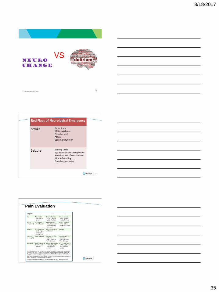
Red Flags of Neurological Emergency
Stroke Facial droop
Motor weakness Pronator drift Ataxia
Speech dysfunction
Seizure Starring spells Eye deviation and unresponsive
Periods of loss of consciousness Muscle Twitching
Periods of stuttering
104
Pain Evaluation

8/18/2017
36
I am here to speak for the neuro
patients, who often can not speak
for themselves……
Questions?
206-320-2821