ppr-smr protein sot1 has rna endonuclease activityppr-smr protein sot1 has rna endonuclease activity...
TRANSCRIPT

PPR-SMR protein SOT1 has RNA endonuclease activityWen Zhoua,b,1, Qingtao Lua,1, Qingwei Lia,b, Lei Wanga,b, Shunhua Dinga, Aihong Zhanga, Xiaogang Wena, Lixin Zhanga,and Congming Lua,c,2
aPhotosynthesis Research Center, Key Laboratory of Photobiology, Institute of Botany, Chinese Academy of Sciences, Beijing 100093, China; bCollege of LifeSciences, University of Chinese Academy of Sciences, Beijing 100049, China; and cNational Center for Plant Gene Research, Beijing 100093, China
Edited by Robert Haselkorn, University of Chicago, Chicago, IL, and approved January 13, 2017 (received for review July 28, 2016)
Numerous attempts have been made to identify and engineersequence-specific RNA endonucleases, as these would allow forefficient RNA manipulation. However, no natural RNA endonucleasethat recognizes RNA in a sequence-specific manner has been de-scribed to date. Here, we report that SUPPRESSOR OF THYLAKOIDFORMATION 1 (SOT1), an Arabidopsis pentatricopeptide repeat (PPR)protein with a small MutS-related (SMR) domain, has RNA endonu-clease activity. We show that the SMR moiety of SOT1 performs theendonucleolytic maturation of 23S and 4.5S rRNA through the PPRdomain, specifically recognizing a 13-nucleotide RNA sequence in the5′ end of the chloroplast 23S–4.5S rRNA precursor. In addition, wesuccessfully engineered the SOT1 protein with altered PPR motifs torecognize and cleave a predicted RNA substrate. Our findings pointto SOT1 as an exciting tool for RNA manipulation.
photosynthesis | PPR-SMR protein | RNA endonuclease | rRNA biogenesis
Sequence-specific RNA endonucleases are crucial to establishingRNA manipulation technology (1). Compared with DNA
editing, RNA manipulation could be more useful and reversiblebecause it does not result in permanent changes to the genome. Inaddition, sequence-specific RNA endonucleases could potentiallybe used as an RNA silencing tool to complement RNAi, becauseRNAi is sometimes ineffective in certain organisms and RNAimachinery is not present in cellular compartments such as chlo-roplasts and mitochondria. Despite extensive investigations, anatural RNA endonuclease that recognizes RNA in an intrinsicsequence-specific manner has not yet been identified.Pentatricopeptide repeat (PPR) proteins exist in eukaryotes,
have greatly expanded in terrestrial plants, and take part in mostRNA metabolic processes in organelles (2–5). The PPR domaincan specifically recognize RNAs in an intrinsic sequence-specificmanner (5–8). The 2nd, 5th, and 35th (or 1st, 4th, and 34th or 3rd,6th, and 1st in other numbering systems) residues at each repeatare considered to be RNA selection “codes” (9–11). Based onthese codes, several PPR proteins have been successfully modifiedto recognize predictable RNA targets (9, 12–17).The small MutS-related (SMR) domain was originally identified at
the C terminus of MutS2 in the cyanobacterium Synechocystis (18).SMR proteins are widely distributed in almost all organisms (19).Recent studies demonstrated the SMR domain exhibits DNA nickingnuclease activity in vitro (20–25). Furthermore, a C-terminal SMRdomain in Leishmania donovani S-phase mRNA cycling sequencebinding protein (CSBP) has RNA cleavage activity in vitro (26).These findings suggest that the SMR domain has nuclease activity.Interestingly, a small protein family containing both PPR and
SMR domains was recently described (27). PPR-SMR proteins arefound mainly in land plants. Arabidopsis thaliana contains eightPPR-SMR proteins localized to the organelles, including mito-chondria and chloroplasts (27). Currently, four PPR-SMR proteinshave been characterized. They play an essential role in plastidretrograde signaling, plastid transcription, and RNA biogenesis(28–34). Thus, PPR-SMR proteins clearly play important roles inorganelle biogenesis. However, the molecular mechanisms under-lying the functions of PPR-SMR proteins are largely unclear. Inparticular, the functions of the enigmatic SMR domain of thesePPR-SMRs are unknown.
Considering the sequence-specific RNA binding capacity ofthe PPR domain and the potential nuclease activity of the SMRdomain, it has been suggested that PPR-SMR proteins mayrepresent natural sequence-specific RNA endonucleases (27). Ifthe endonuclease activity of the SMR domain of PPR-SMRs canbe confirmed, the PPR-SMR proteins may serve as sequence-specific RNA endonucleases in nature and could be potentiallyused as tools for RNA manipulation (27).Here, we show that SUPPRESSOR OF THYLAKOID
FORMATION 1 (SOT1) has endonuclease activity and performsthe endonucleolytic maturation of 23S and 4.5S rRNA through thePPR domain, specifically recognizing a 13-nucleotide RNA se-quence in the 5′ end of the chloroplast 23S–4.5S rRNA precursor.We also show that SOT1 can be modified to recognize and cleave apredicted RNA substrate. Our findings suggest that SOT1 could beused as a tool for RNA manipulation in the future.
ResultsDisruption of SOT1 Impairs Translation in Chloroplasts. To identifyfactors required for chloroplast development, we screened anArabidopsis mutant library and isolated a mutant line (ultimatelynamed sot1-3, as described below) with a high chlorophyll fluo-rescence phenotype (SI Appendix, Fig. S1A). The mutant displayedretarded growth and a virescent-leaf phenotype compared with thewild type (WT) (SI Appendix, Figs. S1 A and B and S2A). Chlo-roplasts in the mutant displayed a vesicular shape and few thyla-koids in contrast to the crescent-shaped chloroplasts andwell-formed thylakoid structure of WT (SI Appendix, Fig. S1C).Analyses of chlorophyll fluorescence induction curves and P700 re-dox kinetics revealed a defect in the functions of photosystem II(PSII) and photosystem I (PSI) in the mutant (SI Appendix, Fig. S3).Because photosynthetic function was clearly defective in this
mutant, we investigated changes in the core subunits of key pho-tosynthetic complexes, including PSII, PSI, the cytochromeb6f complex, ATP synthase, the NADH dehydrogenase-like complex
Significance
Our results demonstrate that SUPPRESSOR OF THYLAKOIDFORMATION 1 (SOT1), anArabidopsis pentatricopeptide repeat (PPR)protein with a small MutS-related (SMR) domain, has endonucleaseactivity. The SMR moiety of SOT1 performs the endonucleolyticmaturation of 23S and 4.5S rRNA through the PPR domain specifi-cally recognizing a 13-nucleotide RNA sequence in the 5′ end of thechloroplast 23S–4.5S rRNA precursor. Our results also show thatSOT1 can be engineered to recognize and cleave a predicted RNAsubstrate. Our findings suggest that SOT1 could be used as a tool forRNA manipulation in the future.
Author contributions: W.Z. and C.L. designed research; W.Z. and Q. Lu performed research;W.Z., Q. Lu, Q. Li, L.W., S.D., A.Z., X.W., L.Z., and C.L. analyzed data; andW.Z. and C.L. wrotethe paper.
The authors declare no conflict of interest.
This article is a PNAS Direct Submission.1W.Z. and Q. Lu contributed equally to this work.2To whom correspondence should be addressed. Email: [email protected].
This article contains supporting information online at www.pnas.org/lookup/suppl/doi:10.1073/pnas.1612460114/-/DCSupplemental.
E1554–E1563 | PNAS | Published online February 6, 2017 www.pnas.org/cgi/doi/10.1073/pnas.1612460114
Dow
nloa
ded
by g
uest
on
Mar
ch 1
6, 2
020

(NDH), and Rubisco. Whereas the levels of nuclear-encoded pro-teins (PsbO, Fd, and FNR) were largely unchanged, the levels ofchloroplast-encoded proteins (D1, D2, PsaA, Cyt f, CF1β, RbcL,and ndhI) were considerably decreased in the mutant (SI Appendix,Fig. S2B). Despite the changes in protein levels, the accumulation oftranscripts corresponding to D1, PsaA, Cyt f, CF1β, and PsbO werenot reduced in the mutant (SI Appendix, Fig. S4). These resultssuggest that the mutant harbors a defect in chloroplast translation.In vivo 35S pulse-labeling experiments showed that the overallprotein biosynthesis rate was dramatically lower in the mutant thanin WT (SI Appendix, Fig. S2C), supporting the idea that chloroplasttranslation was impaired in the mutant. The reduced biosynthesis ofchloroplast-encoded proteins is likely responsible for the defects inchloroplast development, photosynthetic function, and plant growthobserved in the mutant.Map-based cloning identified a PPR-SMR gene (AT5G46580)
in which a 31-base pair (bp) deletion resulted in a prematurestop codon in the eighth PPR domain (SI Appendix, Fig. S2D).AT5G46580 was previously assigned the name SOT1 because thecorresponding mutants sot1-1 and sot1-2 (suppressor of thf1 1/2)were identified in a suppressor screen for the leaf variegationphenotype of thylakoid formation 1 (thf1) (34). Accordingly, wedesignated the mutant identified in our screen as sot1-3. sot1-3 isa knockout mutant (SI Appendix, Fig. S5) and genetic comple-mentation confirmed that disruption of SOT1 is responsible forthe phenotypes observed in sot1-3 (SI Appendix, Figs. S1–S3).
SOT1 Functions with Miniribonuclease III in the Maturation of 23S and4.5S rRNA. SOT1 and its maize ortholog PPR53 encode proteinswith 11 PPR motifs in the N-terminal region and a SMR domain inthe C-terminal region and both are involved in the maturation of23S and 4.5S rRNA (33, 34). Our results with sot1-3 show that theloss of SOT1 resulted in less mature 23S and 4.5S rRNA as well asstaggered 23S rRNA 5′ ends (SI Appendix, Fig. S6), which is similarto results observed in sot1-1 and sot1-2 (34).Chloroplast miniribonuclease III proteins RNC3 and RNC4 are
known to cleave the 5′ and 3′ regions of 23S–4.5S rRNA precursorsimultaneously and the loss of miniribonuclease III results in stag-gered 23S rRNA 5′ ends and less mature 4.5S rRNA (35). To in-vestigate how SOT1 might function in the maturation of 23S and4.5S rRNA, we compared the 5′ end of 23S rRNA and 3′ end of4.5S rRNA in sot1-3 and miniribonuclease III mutant rnc3/4 usingRACE assays. The staggered 23S rRNA 5′ ends and levels of ma-ture 4.5S rRNA in sot1-3 were strikingly similar to those in rnc3/4(SI Appendix, Fig. S7). These results indicate that miniribonucleaseIII processing is disrupted in sot1-3 and suggest that mini-ribonuclease III processing is mediated by SOT1 during maturationof 23S and 4.5S rRNA.
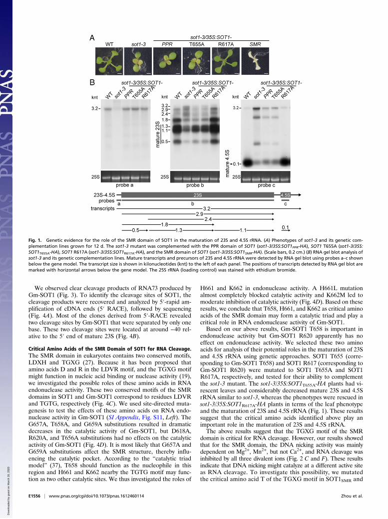
The SMR Domain of SOT1 Is Required for the Maturation of 23S and4.5S rRNA. It has been suggested that SOT1/PPR53 acts in thematuration of 23S and 4.5S rRNA through directly binding to the 5′end of the 23S–4.5S rRNA precursor via the PPR domain and henceblocking the attacks of 5′ exonuclease (33, 34). To address whetherthe SMR domain of SOT1 functions in the maturation of 23S and4.5S rRNA, we attempted to complement sot1-3 plants with the PPRdomain alone (sot1-3/35S:SOT1PPR-HA). The expression of the PPRdomain in sot1-3/35S:SOT1PPR-HA plants was confirmed by RT-PCR and immunoblot assays (SI Appendix, Fig. S8). The sot1-3/35S:SOT1PPR-HA plants displayed a virescent-leaf phenotype and hadconsiderably reduced mature 23S and 4.5S rRNA, similar to sot1-3(Fig. 1 A and B). The lack of phenotype recovery with the PPRdomain alone suggests that the SMR domain of SOT1 plays animportant role in the maturation of 23S and 4.5S rRNA.
The SMR Domain of SOT1 Has Nuclease Activity. Because SMR do-mains typically have DNA nicking activity (20–26), we investi-gated whether the SMR domain of SOT1 retained the ability to
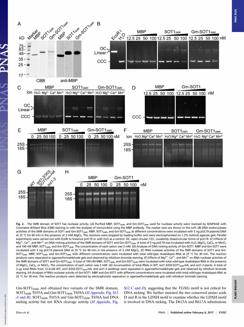
nick supercoiled DNA. We found that recombinant SMR proteinSOT1SMR (amino acids 603–710 of SOT1) cleaved supercoiledpUC19 to open circular and linear conformations and that themetal ions Mn2+ and Mg2+ increased the DNA endonuclease ac-tivity of SOT1SMR (Fig. 2 A–C), indicating that the SMR domain ofSOT1 indeed has DNA endonuclease activity.Given the fact that SOT1 is able to bind the 5′ region of the 23S–
4.5S rRNA precursor (34), we then investigated whether the SMRdomain of SOT1 has RNA nuclease activity. SOT1SMR efficientlydegraded total rRNA isolated from wild-type Arabidopsis, suggestingthat the SMR domain of SOT1 has RNA nuclease activity in vitro(Fig. 2E). However, the metal ions Mn2+, Ca2+, and Mg2+ inhibitedthe RNA nuclease activity of SOT1SMR under these conditions (Fig.2F). To investigate the potential nuclease activity of the SMR do-main in vivo, we complemented sot1-3 with a chloroplast transitpeptide-SMR domain of SOT1 fusion protein (sot1-3/35S:SOT1SMR-HA) (SI Appendix, Fig. S8). This overexpression of the SMR domainin sot1-3 caused a lethal phenotype at the early development ofseedlings (Fig. 1A). Smeared rRNA bands on the RNA gel (Fig. 2G)indicated that the SMR domain of SOT1 has in vivo nuclease ac-tivity. Taken together, these results establish that the SMR domainof SOT1 possesses both DNA and RNA nuclease activities.
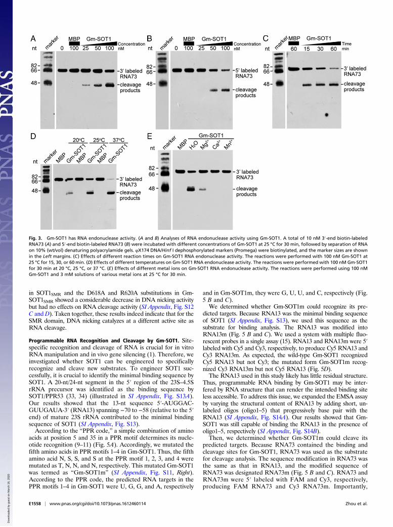
SOT1 Has RNA Endonuclease Activity and Cleaves the 5′ Region of the23S–4.5S rRNA Precursor. SOT1 specifically binds the 5′ region of23S–4.5S rRNA precursor with a 73-nt segment (denoted RNA73hereafter) (34–36); thus, we reasoned that RNA73 should be an invivo substrate for SOT1. To start, we aimed to investigate whetherSOT1 could specifically cleave RNA73 using the recombinant SOT1in vitro. Unfortunately, it was quite difficult to express intact SOT1in Escherichia coli. Therefore, we tried to express the homologousproteins of SOT1 from other species such as Arabidopsis lyrata,Glycine max, Zea mays, and Oryza sativa. After numerous unsuc-cessful attempts, we ultimately obtained Gm-SOT1 from G. max (SIAppendix, Fig. S9). The sequence identity of SOT1 and Gm-SOT1 is∼70%, and they share key residues for RNA recognition (residues 5and 35 in each PPRmotif, marked as red frames in SI Appendix, Fig.S10A). The 5′ region of 23S–4.5S rRNA precursor is highly co-nserved between A. thaliana andG. max (SI Appendix, Fig. S10B). Inaddition, similar to the SMR domain of SOT1, the SMR domain ofGm-SOT1 had both DNA and RNA nuclease activities (Fig. 2 A, C,E, and F). Thus, we used the recombinant protein Gm-SOT1 as anappropriate substitute for SOT1.We used 3′-end and 5′-end biotin-labeled RNA73 to examine
the catalytic activity of Gm-SOT1. With increasing Gm-SOT1concentration, more cleavage products could be detected (Fig. 3A and B). These results suggest that Gm-SOT1 can efficientlycleave the 5′ end of 23S–4.5S rRNA precursor and that thecleavage products are released due to the RNA endonucleaseactivity of Gm-SOT1. In addition, increasing amounts of thecleavage products were detected with increasing incubation time(Fig. 3C). Compared with 20 °C incubation, incubation at highertemperatures (25 °C and 37 °C) led to the production of moreobvious cleavage products (Fig. 3D). The metal ions Mn2+, Ca2+,and Mg2+ inhibited the RNA nuclease activity of Gm-SOT1 (Fig.3E). The cleavage products of RNA73 arose specifically due tothe activity of Gm-SOT1 and not a contaminating ribonuclease,as the incubation of RNA73 with MBP protein that was purifiedfrom E. coli in parallel did not show any RNA cleavage. To-gether, these results demonstrate that Gm-SOT1 has RNAendonuclease activity.Our above results show that the SMR domain of SOT1 pos-
sesses DNA and RNA nuclease activities, cleaving pUC19plasmid and Arabidopsis total RNA, respectively (Fig. 2 B andE). Thus, we investigated whether the full-length Gm-SOT1 cancleave pUC19 plasmid and Arabidopsis total RNA. The full-length Gm-SOT1 did not cleave pUC19 plasmid and total rRNAefficiently (Fig. 2 D and H).
Zhou et al. PNAS | Published online February 6, 2017 | E1555
PLANTBIOLO
GY
PNASPL
US
Dow
nloa
ded
by g
uest
on
Mar
ch 1
6, 2
020
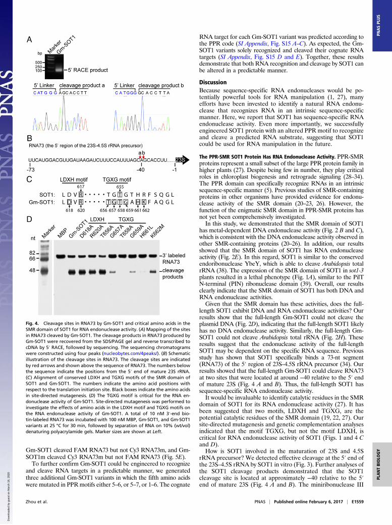
We observed clear cleavage products of RNA73 produced byGm-SOT1 (Fig. 3). To identify the cleavage sites of SOT1, thecleavage products were recovered and analyzed by 5′-rapid am-plification of cDNA ends (5′ RACE), followed by sequencing(Fig. 4A). Most of the clones derived from 5′-RACE revealedtwo cleavage sites by Gm-SOT1 that were separated by only onebase. These two cleavage sites were located at around −40 rel-ative to the 5′ end of mature 23S (Fig. 4B).
Critical Amino Acids of the SMR Domain of SOT1 for RNA Cleavage.The SMR domain in eukaryotes contains two conserved motifs,LDXH and TGXG (27). Because it has been proposed thatamino acids D and R in the LDVR motif, and the TGXG motifmight function in nucleic acid binding or nuclease activity (19),we investigated the possible roles of these amino acids in RNAendonuclease activity. These two conserved motifs of the SMRdomains in SOT1 and Gm-SOT1 correspond to residues LDVRand TGTG, respectively (Fig. 4C). We used site-directed muta-genesis to test the effects of these amino acids on RNA endo-nuclease activity in Gm-SOT1 (SI Appendix, Fig. S11, Left). TheG657A, T658A, and G659A substitutions resulted in dramaticdecreases in the catalytic activity of Gm-SOT1, but D618A,R620A, and T656A substitutions had no effects on the catalyticactivity of Gm-SOT1 (Fig. 4D). It is most likely that G657A andG659A substitutions affect the SMR structure, thereby influ-encing the catalytic pocket. According to the “catalytic triadmodel” (37), T658 should function as the nucleophile in thisregion and H661 and K662 nearby the TGTG motif may func-tion as two other catalytic sites. We thus investigated the roles of
H661 and K662 in endonuclease activity. A H661L mutationalmost completely blocked catalytic activity and K662M led tomoderate inhibition of catalytic activity (Fig. 4D). Based on theseresults, we conclude that T658, H661, and K662 as critical aminoacids of the SMR domain may form a catalytic triad and play acritical role in RNA endonuclease activity of Gm-SOT1.Based on our above results, Gm-SOT1 T658 is important in
endonuclease activity but Gm-SOT1 R620 apparently has noeffect on endonuclease activity. We selected these two aminoacids for analysis of their potential roles in the maturation of 23Sand 4.5S rRNA using genetic approaches. SOT1 T655 (corre-sponding to Gm-SOT1 T658) and SOT1 R617 (corresponding toGm-SOT1 R620) were mutated to SOT1 T655A and SOT1R617A, respectively, and tested for their ability to complementthe sot1-3 mutant. The sot1-3/35S:SOT1T655A-HA plants had vi-rescent leaves and considerably decreased mature 23S and 4.5SrRNA similar to sot1-3, whereas the phenotypes were rescued insot1-3/35S:SOT1R617A-HA plants in terms of the leaf phenotypeand the maturation of 23S and 4.5S rRNA (Fig. 1). These resultssuggest that the critical amino acids identified above play animportant role in the maturation of 23S and 4.5S rRNA.The above results suggest that the TGXG motif of the SMR
domain is critical for RNA cleavage. However, our results showedthat for the SMR domain, the DNA nicking activity was mainlydependent on Mg2+, Mn2+, but not Ca2+, and RNA cleavage wasinhibited by all three divalent ions (Fig. 2 C and F). These resultsindicate that DNA nicking might catalyze at a different active siteas RNA cleavage. To investigate this possibility, we mutatedthe critical amino acid T of the TGXG motif in SOT1SMR and
Fig. 1. Genetic evidence for the role of the SMR domain of SOT1 in the maturation of 23S and 4.5S rRNA. (A) Phenotypes of sot1-3 and its genetic com-plementation lines grown for 12 d. The sot1-3 mutant was complemented with the PPR domain of SOT1 (sot1-3/35S:SOT1PPR-HA), SOT1 T655A (sot1-3/35S:SOT1T655A-HA), SOT1 R617A (sot1-3/35S:SOT1R617A-HA), and the SMR domain of SOT1 (sot1-3/35S:SOT1SMR-HA). (Scale bars, 0.2 cm.) (B) RNA gel blot analysis ofsot1-3 and its genetic complementation lines. Mature transcripts and precursors of 23S and 4.5S rRNA were detected by RNA gel blot using probes a–c shownbelow the gene model. The transcript size is shown in kilonucleotides (knt) to the left of each panel. The positions of transcripts detected by RNA gel blot aremarked with horizontal arrows below the gene model. The 25S rRNA (loading control) was stained with ethidium bromide.
E1556 | www.pnas.org/cgi/doi/10.1073/pnas.1612460114 Zhou et al.
Dow
nloa
ded
by g
uest
on
Mar
ch 1
6, 2
020
Gm-SOT1SMR and obtained two variants of the SMR domain,SOT1SMR T655A and Gm-SOT1SMR T658A (SI Appendix, Fig. S12A and B). SOT1SMR T655A and Gm-SOT1SMR T658A had DNAnicking activity but not RNA cleavage activity (SI Appendix, Fig.
S12 C and D), suggesting that the TGXG motif is not critical forDNA nicking. We further mutated the two conserved amino acidsD and R in the LDXH motif to examine whether the LDXH motifis involved in DNA nicking. The D615A and R617A substitutions
Fig. 2. The SMR domain of SOT1 has nuclease activity. (A) Purified MBP, SOT1SMR, and Gm-SOT1SMR used for nuclease activity were resolved by SDS/PAGE withCoomassie Brilliant Blue (CBB) staining or with the analyses of immunoblot using the MBP antibody. The marker sizes are shown to the Left. (B) DNA endonucleaseactivities of the SMR domains of SOT1 and Gm-SOT1SMR. MBP, SOT1SMR, and Gm-SOT1SMR at different concentrations were incubated with 5 ng pUC19 plasmid DNAat 25 °C for 60 min in the presence of 3 mM MgCl2. The reactions were stopped by loading buffer and were electrophoresed on 1.2% (wt/vol) agarose gels. Parallelexperiments were carried out with EcoRI to linearize pUC19 or with H2O as a control. OC, open circular; CCC, covalently closed-circular forms of pUC19. (C) Effects ofMg2+, Ca2+, andMn2+ on DNA nicking activities of the SMR domains of SOT1 and Gm-SOT1SMR. A total of 5 ng pUC19 was incubated with H2O, MgCl2, CaCl2, or MnCl2and 100 nMMBP, SOT1SMR, and Gm-SOT1SMR. The concentration of each cation was 3 mM. (D) Analyses of DNA nicking activity of Gm-SOT1. MBP and Gm-SOT1 wereincubated with 5 ng pUC19 plasmid DNA at 25 °C for 60 min in the presence of 3 mM MgCl2. (E) RNA nuclease activities of the SMR domains of SOT1 and Gm-SOT1SMR. MBP, SOT1SMR, and Gm-SOT1SMR with different concentrations were incubated with total wild-type Arabidopsis RNA at 25 °C for 30 min. The reactionproducts were separated in agarose/formaldehyde gels and observed by ethidium bromide staining. (F) Effects of Mg2+, Ca2+, and Mn2+ on RNA nuclease activities ofthe SMR domains of SOT1 and Gm-SOT1SMR. A total of 100 nMMBP, SOT1SMR, and Gm-SOT1SMR were incubated with total wild-type Arabidopsis RNA in the presenceof MgCl2, CaCl2, or MnCl2. The concentration of each cation was 3 mM. (G) Accumulation of total RNAs in WT, sot1-3/35S:SOT1SMR-HA, and sot1-3 plants. A total of3 μg total RNAs from 12-d-old WT, sot1-3/35S:SOT1SMR-HA, and sot1-3 seedlings were separated in agarose/formaldehyde gels and observed by ethidium bromidestaining. (H) Analyses of RNA nuclease activity of Gm-SOT1. MBP and Gm-SOT1 with different concentrations were incubated with total wild-type Arabidopsis RNA at25 °C for 30 min. The reaction products were detected by electrophoretic separation in agarose/formaldehyde gels with ethidium bromide staining.
Zhou et al. PNAS | Published online February 6, 2017 | E1557
PLANTBIOLO
GY
PNASPL
US
Dow
nloa
ded
by g
uest
on
Mar
ch 1
6, 2
020
in SOT1SMR and the D618A and R620A substitutions in Gm-SOT1SMR showed a considerable decrease in DNA nicking activitybut had no effects on RNA cleavage activity (SI Appendix, Fig. S12C andD). Taken together, these results indeed indicate that for theSMR domain, DNA nicking catalyzes at a different active site asRNA cleavage.
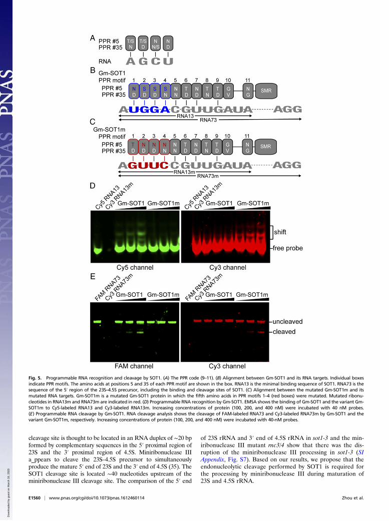
Programmable RNA Recognition and Cleavage by Gm-SOT1. Site-specific recognition and cleavage of RNA is crucial for in vitroRNA manipulation and in vivo gene silencing (1). Therefore, weinvestigated whether SOT1 can be engineered to specificallyrecognize and cleave new substrates. To engineer SOT1 suc-cessfully, it is crucial to identify the minimal binding sequence bySOT1. A 20-nt/24-nt segment in the 5′ region of the 23S–4.5SrRNA precursor was identified as the binding sequence bySOT1/PPR53 (33, 34) (illustrated in SI Appendix, Fig. S13A).Our results showed that the 13-nt sequence 5′-AUGGAC-GUUGAUA-3′ (RNA13) spanning −70 to −58 (relative to the 5′end) of mature 23S rRNA contributed to the minimal bindingsequence of SOT1 (SI Appendix, Fig. S13).According to the “PPR code,” a simple combination of amino
acids at position 5 and 35 in a PPR motif determines its nucle-otide recognition (9–11) (Fig. 5A). Accordingly, we mutated thefifth amino acids in PPR motifs 1–4 in Gm-SOT1. Thus, the fifthamino acid N, S, S, and S at the PPR motif 1, 2, 3, and 4 weremutated as T, N, N, and N, respectively. This mutated Gm-SOT1was termed as “Gm-SOT1m” (SI Appendix, Fig. S11, Right).According to the PPR code, the predicted RNA targets in thePPR motifs 1–4 in Gm-SOT1 were U, G, G, and A, respectively
and in Gm-SOT1m, they were G, U, U, and C, respectively (Fig.5 B and C).We determined whether Gm-SOT1m could recognize its pre-
dicted targets. Because RNA13 was the minimal binding sequenceof SOT1 (SI Appendix, Fig. S13), we used this sequence as thesubstrate for binding analysis. The RNA13 was modified intoRNA13m (Fig. 5 B and C). We used a system with multiple fluo-rescent probes in a single assay (15). RNA13 and RNA13m were 5′labeled with Cy5 and Cy3, respectively, to produce Cy5 RNA13 andCy3 RNA13m. As expected, the wild-type Gm-SOT1 recognizedCy5 RNA13 but not Cy3; the mutated form Gm-SOT1m recog-nized Cy3 RNA13m but not Cy5 RNA13 (Fig. 5D).The RNA13 used in this study likely has little residual structure.
Thus, programmable RNA binding by Gm-SOT1 may be inter-fered by RNA structure that can render the intended binding siteless accessible. To address this issue, we expanded the EMSA assayby varying the structural content of RNA13 by adding short, un-labeled oligos (oligo1–5) that progressively base pair with theRNA13 (SI Appendix, Fig. S14A). Our results showed that Gm-SOT1 was still capable of binding the RNA13 in the presence ofoligo1–5, respectively (SI Appendix, Fig. S14B).Then, we determined whether Gm-SOT1m could cleave its
predicted targets. Because RNA73 contained the binding andcleavage sites for Gm-SOT1, RNA73 was used as the substratefor cleavage analysis. The sequence modification in RNA73 wasthe same as that in RNA13, and the modified sequence ofRNA73 was designated RNA73m (Fig. 5 B and C). RNA73 andRNA73m were 5′ labeled with FAM and Cy3, respectively,producing FAM RNA73 and Cy3 RNA73m. Importantly,
Fig. 3. Gm-SOT1 has RNA endonuclease activity. (A and B) Analyses of RNA endonuclease activity using Gm-SOT1. A total of 10 nM 3′-end biotin-labeledRNA73 (A) and 5′-end biotin-labeled RNA73 (B) were incubated with different concentrations of Gm-SOT1 at 25 °C for 30 min, followed by separation of RNAon 10% (wt/vol) denaturing polyacrylamide gels. φX174 DNA/Hinf I dephosphorylated markers (Promega) were biotinylated, and the marker sizes are shownin the Left margins. (C) Effects of different reaction times on Gm-SOT1 RNA endonuclease activity. The reactions were performed with 100 nM Gm-SOT1 at25 °C for 15, 30, or 60 min. (D) Effects of different temperatures on Gm-SOT1 RNA endonuclease activity. The reactions were performed with 100 nM Gm-SOT1for 30 min at 20 °C, 25 °C, or 37 °C. (E) Effects of different metal ions on Gm-SOT1 RNA endonuclease activity. The reactions were performed using 100 nMGm-SOT1 and 3 mM solutions of various metal ions at 25 °C for 30 min.
E1558 | www.pnas.org/cgi/doi/10.1073/pnas.1612460114 Zhou et al.
Dow
nloa
ded
by g
uest
on
Mar
ch 1
6, 2
020
Gm-SOT1 cleaved FAM RNA73 but not Cy3 RNA73m, and Gm-SOT1m cleaved Cy3 RNA73m but not FAM RNA73 (Fig. 5E).To further confirm Gm-SOT1 could be engineered to recognize
and cleave RNA targets in a predictable manner, we generatedthree additional Gm-SOT1 variants in which the fifth amino acidswere mutated in PPRmotifs either 5–6, or 5–7, or 1–6. The cognate
RNA target for each Gm-SOT1 variant was predicted according tothe PPR code (SI Appendix, Fig. S15 A–C). As expected, the Gm-SOT1 variants solely recognized and cleaved their cognate RNAtargets (SI Appendix, Fig. S15 D and E). Together, these resultsdemonstrate that both RNA recognition and cleavage by SOT1 canbe altered in a predictable manner.
DiscussionBecause sequence-specific RNA endonucleases would be po-tentially powerful tools for RNA manipulation (1, 27), manyefforts have been invested to identify a natural RNA endonu-clease that recognizes RNA in an intrinsic sequence-specificmanner. Here, we report that SOT1 has sequence-specific RNAendonuclease activity. Even more importantly, we successfullyengineered SOT1 protein with an altered PPR motif to recognizeand cleave a predicted RNA substrate, suggesting that SOT1could be used for RNA manipulation in the future.
The PPR-SMR SOT1 Protein Has RNA Endonuclease Activity. PPR-SMRproteins represent a small subset of the large PPR protein family inhigher plants (27). Despite being few in number, they play criticalroles in chloroplast biogenesis and retrograde signaling (28–34).The PPR domain can specifically recognize RNAs in an intrinsicsequence-specific manner (5). Previous studies of SMR-containingproteins in other organisms have provided evidence for endonu-clease activity of the SMR domain (20–23, 26). However, thefunction of the enigmatic SMR domain in PPR-SMR proteins hasnot yet been comprehensively investigated.In this study, we demonstrated that the SMR domain of SOT1
has metal-dependent DNA endonuclease activity (Fig. 2 B and C),which is consistent with the DNA endonuclease activity observed inother SMR-containing proteins (20–26). In addition, our resultsshowed that the SMR domain of SOT1 has RNA endonucleaseactivity (Fig. 2E). In this regard, SOT1 is similar to the conservedendoribonuclease YbeY, which is able to cleave Arabidopsis totalrRNA (38). The expression of the SMR domain of SOT1 in sot1-3plants resulted in a lethal phenotype (Fig. 1A), similar to the PilTN-terminal (PIN) ribonuclease domain (39). Overall, our resultsclearly indicate that the SMR domain of SOT1 has both DNA andRNA endonuclease activities.Given that the SMR domain has these activities, does the full-
length SOT1 exhibit DNA and RNA endonuclease activities? Ourresults show that the full-length Gm-SOT1 could not cleave theplasmid DNA (Fig. 2D), indicating that the full-length SOT1 likelyhas no DNA endonuclease activity. Similarly, the full-length Gm-SOT1 could not cleave Arabidopsis total rRNA (Fig. 2H). Theseresults suggest that the endonuclease activity of the full-lengthSOT1 may be dependent on the specific RNA sequence. Previousstudy has shown that SOT1 specifically binds a 73-nt segment(RNA73) of the 5′ region of 23S–4.5S rRNA precursor (34). Ourresults showed that the full-length Gm-SOT1 could cleave RNA73at two sites that were located at around −40 relative to the 5′ endof mature 23S (Fig. 4 A and B). Thus, the full-length SOT1 hassequence-specific RNA endonuclease activity.It would be invaluable to identify catalytic residues in the SMR
domain of SOT1 for its RNA endonuclease activity (27). It hasbeen suggested that two motifs, LDXH and TGXG, are thepotential catalytic residues of the SMR domain (19, 22, 27). Oursite-directed mutagenesis and genetic complementation analysesindicated that the motif TGXG, but not the motif LDXH, iscritical for RNA endonuclease activity of SOT1 (Figs. 1 and 4 Cand D).How is SOT1 involved in the maturation of 23S and 4.5S
rRNA precursor? We detected effective cleavage at the 5′ end ofthe 23S–4.5S rRNA by SOT1 in vitro (Fig. 3). Further analyses ofthe SOT1 cleavage products demonstrated that the SOT1cleavage site is located at approximately −40 relative to the 5′end of mature 23S (Fig. 4 A and B). The miniribonuclease III
Fig. 4. Cleavage sites in RNA73 by Gm-SOT1 and critical amino acids in theSMR domain of SOT1 for RNA endonuclease activity. (A) Mapping of the sitesin RNA73 cleaved by Gm-SOT1. The cleavage products in RNA73 produced byGm-SOT1 were recovered from the SDS/PAGE gel and reverse transcribed tocDNA by 5′ RACE, followed by sequencing. The sequencing chromatogramswere constructed using four peaks (nucleobytes.com/4peaks/). (B) Schematicillustration of the cleavage sites in RNA73. The cleavage sites are indicatedby red arrows and shown above the sequence of RNA73. The numbers belowthe sequence indicate the positions from the 5′ end of mature 23S rRNA.(C) Alignment of conserved LDXH and TGXG motifs of the SMR domain ofSOT1 and Gm-SOT1. The numbers indicate the amino acid positions withrespect to the translation initiation site. Black boxes indicate the amino acidsin site-directed mutagenesis. (D) The TGXG motif is critical for the RNA en-donuclease activity of Gm-SOT1. Site-directed mutagenesis was performed toinvestigate the effects of amino acids in the LDXH motif and TGXG motifs onthe RNA endonuclease activity of Gm-SOT1. A total of 10 nM 3′-end bio-tin-labeled RNA73 was incubated with 100 nM MBP, Gm-SOT1, and Gm-SOT1variants at 25 °C for 30 min, followed by separation of RNA on 10% (wt/vol)denaturing polyacrylamide gels. Marker sizes are shown at Left.
Zhou et al. PNAS | Published online February 6, 2017 | E1559
PLANTBIOLO
GY
PNASPL
US
Dow
nloa
ded
by g
uest
on
Mar
ch 1
6, 2
020
cleavage site is thought to be located in an RNA duplex of ∼20 bpformed by complementary sequences in the 5′ proximal region of23S and the 3′ proximal region of 4.5S. Miniribonuclease IIIa_ppears to cleave the 23S–4.5S precursor to simultaneouslyproduce the mature 5′ end of 23S and the 3′ end of 4.5S (35). TheSOT1 cleavage site is located ∼40 nucleotides upstream of theminiribonuclease III cleavage site. The comparison of the 5′ end
of 23S rRNA and 3′ end of 4.5S rRNA in sot1-3 and the min-iribonuclease III mutant rnc3/4 show that there was the dis-ruption of the miniribonuclease III processing in sot1-3 (SIAppendix, Fig. S7). Based on our results, we propose that theendonucleolytic cleavage performed by SOT1 is required forthe processing by miniribonuclease III during maturation of23S and 4.5S rRNA.
Fig. 5. Programmable RNA recognition and cleavage by SOT1. (A) The PPR code (9–11). (B) Alignment between Gm-SOT1 and its RNA targets. Individual boxesindicate PPR motifs. The amino acids at positions 5 and 35 of each PPR motif are shown in the box. RNA13 is the minimal binding sequence of SOT1. RNA73 is thesequence of the 5′ region of the 23S–4.5S precursor, including the binding and cleavage sites of SOT1. (C) Alignment between the mutated Gm-SOT1m and itsmutated RNA targets. Gm-SOT1m is a mutated Gm-SOT1 protein in which the fifth amino acids in PPR motifs 1–4 (red boxes) were mutated. Mutated ribonu-cleotides in RNA13m and RNA73m are indicated in red. (D) Programmable RNA recognition by Gm-SOT1. EMSA shows the binding of Gm-SOT1 and the variant Gm-SOT1m to Cy5-labeled RNA13 and Cy3-labeled RNA13m. Increasing concentrations of protein (100, 200, and 400 nM) were incubated with 40 nM probes.(E) Programmable RNA cleavage by Gm-SOT1. RNA cleavage analysis shows the cleavage of FAM-labeled RNA73 and Cy3-labeled RNA73m by Gm-SOT1 and thevariant Gm-SOT1m, respectively. Increasing concentrations of protein (100, 200, and 400 nM) were incubated with 40-nM probes.
E1560 | www.pnas.org/cgi/doi/10.1073/pnas.1612460114 Zhou et al.
Dow
nloa
ded
by g
uest
on
Mar
ch 1
6, 2
020
In bacteria, it is thought that the final maturation of 23S rRNA iscompleted by miniribonuclease III through a one-step cleavage (40,41). However, in chloroplasts, one or two small RNAs adjacently tothe upstream of the miniribonuclease III cleavage site were iden-tified (34, 35), indicating that additional, unknown processingevents exist before miniribonuclease III cleavage in the maturationof 23S rRNA in chloroplasts. Ribonucleases that perform theseadditional unknown processing events remain elusive. Our resultsshowed that SOT1 performed an endonucleolytic cleavage adja-cently to the upstream of the miniribonuclease III cleavage duringmaturation of 23S rRNA in chloroplasts. Such a finding provides usdeep understanding of the molecular mechanism for maturation of23S rRNA in chloroplasts and suggests that new ribonucleases haveemerged as being required for the 23S rRNA processing duringevolution of chloroplasts. Our finding also expands the un-derstanding of the biological functions of PPR proteins, because itis generally believed that PPR proteins stabilize or remodel theirRNA targets (5). However, the exact molecular mechanism ofSOT1 in maturation of 23S rRNA in chloroplasts remains to beinvestigated further.
SOT1 Could Be a Powerful Tool for RNAManipulation.DNA restrictionenzymes were first described ∼40 y ago. However, no analogousRNA endonuclease that recognizes RNA in a sequence-specificmanner was forthcoming. Despite their affinity for many types ofRNA, RNA endonucleases exhibit limited, imprecise recognitionof RNA sequences, which limits their value for RNAmanipulation.Until now, many RNA binding proteins, such as RNA-recognitionmotif (RRM), K homology (KH), zinc finger (ZF), and Pumilio/FBF homology protein (PUF), were found to have modularstructures to recognize RNA sequences and/or structures (42).Among these RNA binding proteins, PUF proteins may representa candidate for engineering highly sequence-specific RNA binding(42). The classical PUF proteins contain eight repeat domains andeach repeat can recognize a single RNA nucleotide (43). The RNAspecificity of the PUF domain has been well decoded (44–46).Based on the properties of the PUF proteins, the programmableRNA binding by the engineered PUF protein has been realized(47–50). Moreover, the PUF domains fusing with a general RNAcleavage domain have been generated to the artificial site-specificRNA endonucleases that specifically recognize and cleave RNAtargets (51, 52).Similar to PUF proteins, several PPR proteins have been
successfully modified to recognize corresponding RNA targetsthrough simply modifying the critical amino acids of each PPRmotif (9, 12–17). Considering the PPR motifs have beendesigned to target a specific transcript, if RNA endonucleaseactivity can be confirmed for the SMR domain of PPR-SMRproteins, the PPR-SMR protein will enable “engineering” ofsequence-specific RNA endonucleases, which will have excitingapplications for RNA manipulation (27). Our current resultsdemonstrated that the PPR-SMR protein SOT1 has RNA en-donuclease activity and can be engineered to recognize andcleave RNA with customizable sequence specificity. We alteredthe critical amino acids for RNA recognition in the SOT1 PPRmotifs, finding that the modified protein could recognize andcleave a new RNA substrate in the expected manner (Fig. 5 andSI Appendix, Fig. S15).Recently, the CRISPR/Cas9 system has been widely used in the
field of genome editing (53, 54). This system can also be used forprogrammable RNA recognition and cleavage (55). In addition, itwas found recently that C2c2 is a programmable RNA-guidedRNA-targeting CRISPR effector (56). These findings suggest thatthe CRISPR/Cas system can be used to develop RNA-targetingtools (55–57). Compared with the CRISPR/Cas system that usesRNA-mediated base pairing to recognize RNA targets, SOT1 re-lies upon PPR–RNA interactions to recognize RNA targets. Theresults in this study reveal that SOT1 can achieve programmable
RNA recognition and cleavage, suggesting that SOT1, similar tothe CRISPR/Cas system, can be used as a tool for RNA manipu-lation. Currently, chloroplast and mitochondrial transformation istechnically very difficult, labor intensive, and time consuming andachieved only in very few organisms (58, 59). It is unclear whetherCRISPR/Cas9 could cleave or edit the mitochondrial genome be-cause it is challenging to import the guide RNA component intomitochondria (60). Thus, developing chloroplast and mitochondrialRNA-targeting tools will be significant for chloroplast and mito-chondrial biology. SOT1 is a chloroplast-localized protein andcould be targeted into mitochondria by fusing a mitochondrialsignaling peptide. Therefore, an advantage for SOT1 is that itcan be used for programmable chloroplast/mitochondrial genes-encoded RNA recognition and cleavage, which enables SOT1 to bea potential RNA-targeting tool in chloroplasts and mitochondria.Just as DNA restriction enzymes have revolutionized the field ofmolecular biology, we look forward to the potential application ofSOT1 to the broad field of RNA manipulation, especially inchloroplasts and mitochondria. Nevertheless, further elucidation ofthe detailed catalytic mechanism used by SOT1 is essential to makeuse of this exciting tool.
Materials and MethodsPlant Materials and Growth Conditions. The A. thaliana wild-type line (eco-type Columbia-0) was obtained from the Arabidopsis Biological ResourceCenter (abrc.osu.edu/). The mutant sot1-3 line was isolated from a pSKI15T-DNA-mutagenized A. thaliana library (ecotype Columbia-0) based on itshigh chlorophyll fluorescence phenotype. The sot1-3 plants were backcrossedthree times to wild-type plants. The mutant rnc3/4 line was kindly provided byD. B. Stern, Boyce Thompson Institute for Plant Research, Ithaca, NY.
The Arabidopsis seeds were surface sterilized and plated on Murashige andSkoog (MS) medium containing 2% (wt/vol) sucrose and 0.8% agar. After in-cubation in darkness for 2 d at 4 °C, the plants were grown under 100 μmolm−2·s−1
14-h light/10-h-dark cycles at 23 °C and 50% relative humidity.
Expression of Recombinant Proteins. The pASK-IBA44 expression system (IBAGmbH) was used to express recombinant proteins in E. coli. The gene encodingthe MBP tag was inserted into pASK-IBA44 to facilitate the protein purification.The coding sequences for the SOT1 SMR domain (amino acids 603–710), theGm-SOT1 SMR domain (amino acids 606–711), and Gm-SOT1 (amino acids 182–706) were subcloned into the pASK-IBA44 vector. Expression of MBP, SOT1SMR,and Gm-SOT1 was induced upon addition of 200 μg anhydrotetracycline perliter E. coli JM83 in a shaking culture (A550 = 0.4). After induction at 17 °C for3 h, E. coli cultures were harvested and homogenized in buffer containing50 mM phosphate, pH 7.5, 400 mM NaCl, 100 mM KCl, and 10% (vol/vol)glycerol. After sonication and centrifugation, the supernatant was applied toNi-NTI (Qiagen) and subsequently loaded onto an amylose resin column (NEB).The purified proteins were further fractionated by size exclusion chromatog-raphy (GE Superdex 200 10/300 GL).
To generate the constructs of Gm-SOT1 variants, SOT1SMR variants, and Gm-SOT1SMR variants, site-directed mutagenesis and multisite-directed mutagenesiswere performed using a Fast Mutagenesis System (Transgen FM111) and a FastMultiSite Mutagenesis System (Transgen FM201), respectively, according to themanufacturer’s instructions (for primers, see SI Appendix, Table S1). Expressionand purification of the Gm-SOT1 variants, SOT1SMR variants and Gm-SOT1SMR
variants were performed as described for Gm-SOT1.
Nuclease Activity Analysis of the SMR Domain of SOT1. DNA nicking nucleaseactivity analysis was performed as described previously (21). The pUC19 (Takara)plasmid DNA (5 ng μL−1) was incubated with MBP, SOT1SMR, SOT1SMR variants,Gm-SOT1SMR, Gm-SOT1SMR variants, or Gm-SOT1 in buffer [20 mM phosphate,pH 7.5, 160 mMNaCl, 40 mM KCl, and 4% (vol/vol) glycerol] at 25 °C for 60 min.The reaction products for pUC19 plasmid DNA were detected by electropho-retic separation in 1% agarose gels with ethidium bromide.
RNA nuclease activity was performed as described previously (38). Thetotal RNA (3 μg) extracted from wild-type plants was incubated with MBP,SOT1SMR, SOT1SMR variants, Gm-SOT1SMR, Gm-SOT1SMR variants, or Gm-SOT1in buffer [20mM phosphate, pH 7.5, 160 mMNaCl, 40mMKCl, and 4% (vol/vol)glycerol] at 25 °C for 30 min. The reaction products for total RNAs were sep-arated in 1.2% (wt/vol) agarose/formaldehyde gels and visualized by ethidiumbromide staining.
Zhou et al. PNAS | Published online February 6, 2017 | E1561
PLANTBIOLO
GY
PNASPL
US
Dow
nloa
ded
by g
uest
on
Mar
ch 1
6, 2
020

RNA Endonuclease Activity Analysis of SOT1. The 5′-end biotin-labeled RNA73,3′-end biotin-labeled RNA73, 5′-end FAM-labeled RNA73, 5′-end Cy3-labeledRNA73m, 3′-end biotin-labeled RNA73PPR5-6, 3′-end biotin-labeled RNA73PPR5-7,and 3′-end biotin-labeled RNA73PPR1-6 were synthesized and labeled byTakara. The reaction buffer contained 20 mM phosphate, pH 7.5, 160 mMNaCl, 40 mM KCl, and 4% (vol/vol) glycerol. The reaction products for the5′-and 3′-end biotin-labeled RNA73, 3′-end biotin-labeled RNA73PPR5-6,3′-end biotin-labeled RNA73PPR5-7, and 3′-end biotin-labeled RNA73PPR1-6were separated in 10% (wt/vol) polyacrylamide gels, transferred into nylonmembranes, and subsequently detected using a chemiluminescent detectionkit (Thermo, 89880). The reaction products for the 5′-end FAM-labeledRNA73, and 5′-end Cy3-labeled RNA73m were resolved on 10% (wt/vol)polyacrylamide gels and detected using a Typhoon Trio imager(GE Healthcare).
Electrophoretic Mobility Shift Assays. Electrophoretic mobility shift assays(EMSA) were carried out with a LightShift Chemiluminescent RNA EMSA Kit(Thermo 20158) following the manufacturer’s instructions (61). The 5′-endCy3-labeled RNA14/RNA13/RNA12a/RNA12b, 5′-end Cy5-labeled RNA13,5′-end Cy3-labeled RNA13m, 5′-end biotin-labeled RNA13PPR5-6, 5′-end biotin-labeled RNA13PPR5-7, 5′-end biotin-labeled RNA13PPR1-6, and oligo1–5 weresynthesized and labeled by Takara. For the RNAs labeled with different fluo-rescent dyes (Cy5 and Cy3), the binding reaction mixture consisted of 10 mM
Hepes (pH 7.3), 20 mM KCl, 2 mM MgCl2, 1 mM DTT, 5% (vol/vol) glycerol,and 40 nM RNA. The protein sample was incubated with the binding re-action mixture at 20 °C for 30 min. The reaction products for the 5′-end Cy3-labeled RNA14/RNA13/RNA12a/RNA12b, 5′-end Cy5-labeled RNA13, and5′-end Cy3-labeled RNA13m were resolved on 6% (wt/vol) native polyacryl-amide gels and were detected using a Typhoon Trio imager (GE Healthcare).For the RNAs labeled with biotin, the binding reaction mixture consisted of10 mM Hepes (pH 7.3), 20 mM KCl, 2 mM MgCl2, 1 mM DTT, 5% (vol/vol)glycerol, and 10 nM RNA. The protein sample was incubated with thebinding reaction mixture at 20 °C for 30 min. The reaction products for the5′-end biotin-labeled RNA13, 5′-end biotin-labeled RNA13PPR5-6, 5′-end bi-otin-labeled RNA13PPR5-7, and 5′-end biotin-labeled RNA13PPR1-6 were re-solved on 6% (wt/vol) native polyacrylamide gels and transferred into nylonmembranes and were subsequently detected according to standard protocolof the chemiluminescent detection kit (Thermo, 89880).
ACKNOWLEDGMENTS. We are grateful to Professor D. B. Stern for providingthe mutant rnc3/4 line and to the Arabidopsis Biological Resource Center forthe seed stocks. This work was supported by the Key Research Plan of Fron-tier Sciences of the Chinese Academy of Sciences (Grant QYZDJ-SSW-SMC003), the State Key Basic Research and Development Plan of China(Grant 2015CB150105), and the Strategic Priority Research Program of theChinese Academy of Sciences (Grant XDB17030100).
1. Choudhury R, Wang Z (2014) Manipulation of RNA using engineered proteins withcustomized specificity. Adv Exp Med Biol 825(6):199–225.
2. Aubourg S, Boudet N, Kreis M, Lecharny A (2000) In Arabidopsis thaliana, 1% of thegenome codes for a novel protein family unique to plants. Plant Mol Biol 42(4):603–613.
3. Small ID, Peeters N (2000) The PPR motif: a TPR-related motif prevalent in plant or-ganellar proteins. Trends Biochem Sci 25(2):46–47.
4. Lurin C, et al. (2004) Genome-wide analysis of Arabidopsis pentatricopeptide repeatproteins reveals their essential role in organelle biogenesis. Plant Cell 16(8):2089–2103.
5. Barkan A, Small I (2014) Pentatricopeptide repeat proteins in plants. Annu Rev PlantBiol 65(1):415–442.
6. Ke J, et al. (2013) Structural basis for RNA recognition by a dimeric PPR-proteincomplex. Nat Struct Mol Biol 20(12):1377–1382.
7. Yin P, et al. (2013) Structural basis for the modular recognition of single-stranded RNAby PPR proteins. Nature 504(7478):168–171.
8. Gully BS, et al. (2015) The solution structure of the pentatricopeptide repeat proteinPPR10 upon binding atpH RNA. Nucleic Acids Res 43(3):1918–1926.
9. Barkan A, et al. (2012) A combinatorial amino acid code for RNA recognition bypentatricopeptide repeat proteins. PLoS Genet 8(8):e1002910.
10. Takenaka M, Zehrmann A, Brennicke A, Graichen K (2013) Improved computationaltarget site prediction for pentatricopeptide repeat RNA editing factors. PLoS One8(6):e65343.
11. Yagi Y, Hayashi S, Kobayashi K, Hirayama T, Nakamura T (2013) Elucidation of theRNA recognition code for pentatricopeptide repeat proteins involved in organelleRNA editing in plants. PLoS One 8(3):e57286.
12. Coquille S, et al. (2014) An artificial PPR scaffold for programmable RNA recognition.Nat Commun 5:5729.
13. Okuda K, et al. (2014) Quantitative analysis of motifs contributing to the interactionbetween PLS-subfamily members and their target RNA sequences in plastid RNAediting. Plant J 80(5):870–882.
14. Gully BS, et al. (2015) The design and structural characterization of a synthetic pen-tatricopeptide repeat protein. Acta Crystallogr D Biol Crystallogr 71(Pt 2):196–208.
15. Kindgren P, Yap A, Bond CS, Small I (2015) Predictable alteration of sequence rec-ognition by RNA editing factors from Arabidopsis. Plant Cell 27(2):403–416.
16. Shen C, et al. (2015) Specific RNA recognition by designer pentatricopeptide repeatprotein. Mol Plant 8(4):667–670.
17. Shen C, et al. (2016) Structural basis for specific single-stranded RNA recognition bydesigner pentatricopeptide repeat proteins. Nat Commun 7:11285.
18. Moreira D, Philippe H (1999) Smr: A bacterial and eukaryotic homologue of theC-terminal region of the MutS2 family. Trends Biochem Sci 24(8):298–300.
19. Fukui K, Kuramitsu S (2011) Structure and function of the small MutS-related domain.Mol Biol Int 2011:691735.
20. Fukui K, Kosaka H, Kuramitsu S, Masui R (2007) Nuclease activity of the MutS ho-mologue MutS2 from Thermus thermophilus is confined to the Smr domain. NucleicAcids Res 35(3):850–860.
21. Fukui K, et al. (2008) Crystal structure of MutS2 endonuclease domain and themechanism of homologous recombination suppression. J Biol Chem 283(48):33417–33427.
22. Diercks T, et al. (2008) Solution structure and characterization of the DNA-bindingactivity of the B3BP-Smr domain. J Mol Biol 383(5):1156–1170.
23. Gui WJ, et al. (2011) Crystal structure of YdaL, a stand-alone small MutS-relatedprotein from Escherichia coli. J Struct Biol 174(2):282–289.
24. Zhang H, et al. (2014) Structural and functional studies of MutS2 from Deinococcusradiodurans. DNA Repair (Amst) 21:111–119.
25. Damke PP, Dhanaraju R, Marsin S, Radicella JP, Rao DN (2015) The nuclease activitiesof both the Smr domain and an additional LDLK motif are required for an efficient
anti-recombination function of Helicobacter pylori MutS2. Mol Microbiol 96(6):1240–1256.
26. Bhandari D, Guha K, Bhaduri N, Saha P (2011) Ubiquitination of mRNA cycling se-quence binding protein from Leishmania donovani (LdCSBP) modulates the RNAendonuclease activity of its Smr domain. FEBS Lett 585(5):809–813.
27. Liu S, Melonek J, Boykin LM, Small I, Howell KA (2013) PPR-SMRs: Ancient proteinswith enigmatic functions. RNA Biol 10(9):1501–1510.
28. Pfalz J, Liere K, Kandlbinder A, Dietz KJ, Oelmüller R (2006) pTAC2, -6, and -12 arecomponents of the transcriptionally active plastid chromosome that are required forplastid gene expression. Plant Cell 18(1):176–197.
29. Koussevitzky S, et al. (2007) Signals from chloroplasts converge to regulate nucleargene expression. Science 316(5825):715–719.
30. Liu X, Yu F, Rodermel S (2010) An Arabidopsis pentatricopeptide repeat protein,SUPPRESSOR OF VARIEGATION7, is required for FtsH-mediated chloroplast bio-genesis. Plant Physiol 154(4):1588–1601.
31. Zoschke R, et al. (2012) The pentatricopeptide repeat-SMR protein ATP4 promotestranslation of the chloroplast atpB/E mRNA. Plant J 72(4):547–558.
32. Zoschke R, Qu Y, Zubo YO, Börner T, Schmitz-Linneweber C (2013) Mutation of thepentatricopeptide repeat-SMR protein SVR7 impairs accumulation and translation ofchloroplast ATP synthase subunits in Arabidopsis thaliana. J Plant Res 126(3):403–414.
33. Zoschke R, Watkins KP, Miranda RG, Barkan A (2016) The PPR-SMR protein PPR53enhances the stability and translation of specific chloroplast RNAs in maize. Plant J85(5):594–606.
34. WuW, et al. (2016) SOT1, a pentatricopeptide repeat protein with a small MutS-relateddomain, is required for correct processing of plastid 23S-4.5S rRNA precursors inArabidopsis thaliana. Plant J 85(5):607–621.
35. Hotto AM, et al. (2015) Arabidopsis chloroplast mini-ribonuclease III participates inrRNA maturation and intron recycling. Plant Cell 27(3):724–740.
36. Bollenbach TJ, et al. (2005) RNR1, a 3′-5′ exoribonuclease belonging to the RNR su-perfamily, catalyzes 3′ maturation of chloroplast ribosomal RNAs in Arabidopsisthaliana. Nucleic Acids Res 33(8):2751–2763.
37. Dodson G, Wlodawer A (1998) Catalytic triads and their relatives. Trends Biochem Sci23(9):347–352.
38. Liu J, et al. (2015) The conserved endoribonuclease YbeY is required for chloroplastribosomal RNA processing in Arabidopsis. Plant Physiol 168(1):205–221.
39. Arcus VL, McKenzie JL, Robson J, Cook GM (2011) The PIN-domain ribonucleases andthe prokaryotic VapBC toxin-antitoxin array. Protein Eng Des Sel 24(1–2):33–40.
40. Redko Y, Bechhofer DH, Condon C (2008) Mini-III, an unusual member of the RNase IIIfamily of enzymes, catalyses 23S ribosomal RNA maturation in B. subtilis. MolMicrobiol 68(5):1096–1106.
41. Redko Y, Condon C (2009) Ribosomal protein L3 bound to 23S precursor rRNA stim-ulates its maturation by Mini-III ribonuclease. Mol Microbiol 71(5):1145–1154.
42. Chen Y, Varani G (2013) Engineering RNA-binding proteins for biology. FEBS J280(16):3734–3754.
43. Wang X, McLachlan J, Zamore PD, Hall TM (2002) Modular recognition of RNA by ahuman pumilio-homology domain. Cell 110(4):501–512.
44. Filipovska A, Razif MFM, Nygård KKA, Rackham O (2011) A universal code for RNArecognition by PUF proteins. Nat Chem Biol 7(7):425–427.
45. Campbell ZT, Valley CT, Wickens M (2014) A protein-RNA specificity code enablestargeted activation of an endogenous human transcript. Nat Struct Mol Biol 21(8):732–738.
46. Hall TMT (2014) Expanding the RNA-recognition code of PUF proteins. Nat Struct MolBiol 21(8):653–655.
47. Wang Y, Cheong C-G, Hall TMT, Wang Z (2009) Engineering splicing factors withdesigned specificities. Nat Methods 6(11):825–830.
48. Cooke A, Prigge A, Opperman L, Wickens M (2011) Targeted translational regulationusing the PUF protein family scaffold. Proc Natl Acad Sci USA 108(38):15870–15875.
E1562 | www.pnas.org/cgi/doi/10.1073/pnas.1612460114 Zhou et al.
Dow
nloa
ded
by g
uest
on
Mar
ch 1
6, 2
020
49. Cao J, Arha M, Sudrik C, Schaffer DV, Kane RS (2014) Bidirectional regulation of
mRNA translation in mammalian cells by using PUF domains. Angew Chem Int Ed Engl
53(19):4900–4904.50. Adamala KP, Martin-Alarcon DA, Boyden ES (2016) Programmable RNA-binding
protein composed of repeats of a single modular unit. Proc Natl Acad Sci USA 113(19):
E2579–E2588.51. Choudhury R, Tsai YS, Dominguez D, Wang Y, Wang Z (2012) Engineering RNA en-
donucleases with customized sequence specificities. Nat Commun 3:1147.52. Zhang W, et al. (2014) Treatment of type 1 myotonic dystrophy by engineering site-
specific RNA endonucleases that target (CUG)(n) repeats. Mol Ther 22(2):312–320.53. Doudna JA, Charpentier E (2014) Genome editing. The new frontier of genome en-
gineering with CRISPR-Cas9. Science 346(6213):1258096.54. Wright AV, Nuñez JK, Doudna JA (2016) Biology and applications of CRISPR systems:
Harnessing nature’s toolbox for genome engineering. Cell 164(1–2):29–44.
55. O’Connell MR, et al. (2014) Programmable RNA recognition and cleavage by CRISPR/Cas9. Nature 516(7530):263–266.
56. Abudayyeh OO, et al. (2016) C2c2 is a single-component programmable RNA-guidedRNA-targeting CRISPR effector. Science 353(6299):aaf5573.
57. East-Seletsky A, et al. (2016) Two distinct RNase activities of CRISPR-C2c2 enableguide-RNA processing and RNA detection. Nature 538(7624):270–273.
58. Larosa V, Remacle C (2013) Transformation of the mitochondrial genome. Int J DevBiol 57(6–8):659–665.
59. Bock R (2015) Engineering plastid genomes: Methods, tools, and applications in basicresearch and biotechnology. Annu Rev Plant Biol 66:211–241.
60. Patananan AN, Wu TH, Chiou PY, Teitell MA (2016) Modifying the mitochondrialgenome. Cell Metab 23(5):785–796.
61. Prikryl J, Rojas M, Schuster G, Barkan A (2011) Mechanism of RNA stabilization andtranslational activation by a pentatricopeptide repeat protein. Proc Natl Acad Sci USA108(1):415–420.
Zhou et al. PNAS | Published online February 6, 2017 | E1563
PLANTBIOLO
GY
PNASPL
US
Dow
nloa
ded
by g
uest
on
Mar
ch 1
6, 2
020