predominance of vagal bradycardia mechanism in...
TRANSCRIPT
J. exp. Biol. 140, 405-420 (1988) 405Printed in Great Britain © The Company of Biologists Limited 1988
PREDOMINANCE OF VAGAL BRADYCARDIA MECHANISMIN THE BRAIN STEM OF TURTLES
BY J. H. HSIEH1, C. M. PAN3, J. S. KUO2 AND C. Y. CHAI3
1Institute of Biomedical Engineering, Chung Yuan Christian University,2Department of Medical Research, Veterans General Hospital and
^Institute of Biomedical Sciences, Academia Sinica, Taipei, Taiwan, ROC
Accepted 19 May 1988
Summary
Cardiovascular parameters of spontaneously breathing pond turtles (Cydemysflavomarginatd) anaesthetized with chloralose (4mgl00g~1) and urethane^OmglOOg"1), were examined during exploratory electrical stimulation of thebrain stem. Turtles exhibited a low mean systemic arterial blood pressure (MSAP,average 25mmHg) and slow heart rate (average 24beatsmin"1). Upon stimu-lation, pressor (sympathetic), depressor (sympatheticinhibition), bradycardia andhypotensive (vagal) responses were elicited from regions of the brain stemextending from the hypothalamus to the medulla, principally in the medial region.The pressor response appeared after a longer latency than did the bradycardia andhypotensive responses. It developed rather slowly, and rarely attained a magni-tude double its resting value. In contrast, stimulation of many points in the brainstem produced marked slowing or even cessation of the heart beat, and thusresulted in an immediate fall of the blood pressure even to zero. This cardio-inhibitory response depended on the integrity of the vagus nerves and wasparticularly marked upon stimulation in the caudal medulla, the areas of theambiguus, solitary and dorsomotor nuclei of the vagus and the midline structures.When such an area was stimulated continuously the heart stopped beatingthroughout the stimulation. The longest period of cardiac arrest before theappearance of escape was 35 min. With continuous stimulation of the peripheralend of the cut vagus, the earliest escape beat occurred even later (65min).Epinephrine given intravenously produced an increase of MSAP and force ofcardiac contraction, although the slope of pressor rise was shallow. Reflexbradycardia, however, was not observed. These experiments show that a veryprominent vagal bradycardia can be evoked from the turtle brain stem, which maycontribute to its well-known capacity for tolerating anoxia.
Introduction
In the mammalian heart, cardiac arrest and resultant anoxia produced by vagalstimulation are transitory: cessation of heart beat lasts for only a short time before
Key words: bradycardia, vagal escape, pressor response, brain stem, hypothalamus, midbrain,medulla, turtle.
406 J. H . HSIEH AND OTHERS
the heart resumes beating in spite of continued stimulation (vagal escapephenomenon). In cats, continuous optimal stimulation of the peripheral end of thecut vagus nerve results in escape in less than 16 s (Chai & Kuo, 1967). Escapeoccurs even earlier (a matter of a few seconds) on stimulation of the vagal nuclei inthe medulla oblongata (Chen & Chai, 1976). In the turtle, the situation has longbeen known to be quite different: the heart may stop beating for more than 1 h inresponse to continuous vagal stimulation (Mills, 1885; Hough, 1895), and thissustained cardiac arrest has been attributed to the low concentration of catechol-amines in the heart (Friedman & Bhagat, 1962).
The turtle is an amphibian reptile characterized by its slow horizontal motion,low systemic arterial pressure (SAP) and heart rate (HR) and extreme tolerance toprolonged anoxia that may last from hours to even months (Robin et al. 1964;Berkson, 1966; Penney, 1974; Felger et al. 1976; Ultsch & Jackson, 1982). Theturtle, therefore, can be expected to have a unique pattern of cardiovascularcontrol as well as special metabolic mechanisms. The low SAP and slow HR,together with the prolonged cardiac arrest on vagal stimulation, suggest that vagalmechanisms in the central nervous system of a turtle might predominate incardiovascular integration. The presence of such mechanisms is investigated in thepresent study.
Materials and methods
Eighty-seven spontaneously breathing turtles (Cyclemys flavomarginata) ofeither sex, weighing between 800 and 1800g, were used. In all cases anaesthesiawas achieved by immersion in iced water for sedation followed by an injection of amixture of chloralose (4mgl00g"1) and urethane (40mgl00g~x) through acannula in the jugular vein, which was also used for drug administration. Thevagus nerves on both sides were carefully isolated. Measurement of the systemicarterial pressure (SAP) from the right carotid artery, monitoring of the meansystemic arterial blood pressure (MSAP), partial representation of cardiaccontractility (dP/dt) and heart rate (HR) have been described previously (Lin etal. 1987; Chai etal. 1988). All recordings were made on a Gould 2800S polygraph.
The head of the animal was fixed in a David-Kopf stereotaxic instrument with amodified mouthpiece. The occipital and the parietal bones were removed.Electrical stimulation was accomplished with a coaxial electrode (NEX-100,Rhodes Medical Instrument; shaft diameter 250 ̂ m, tip diameter 100^m). In theexperiments specifically for chemical activation, stimulation was accomplishedmonopolarly (cathodal) with 30-gauge electrode tubing. The tubing, insulatedexcept for 200 jum at the tip, was connected through a section of PE10polyethylene tube to a Hamilton syringe so that it could be used for identificationof positive reactive sites by electrical stimulation followed by chemical injection atthe same point. The electrode or electrode tubing was positioned perpendicular tothe horizontal plane of the brain. Stimulating current was provided by a constant-current unit connected to a Grass S48 square-wave stimulator. The stimulation

Vagal bradycardia in turtle 407
parameters were 20 Hz, 5 ms and 50-400 [iA, for a 20-s period unless statedotherwise. Various frequencies (10, 20, 40 and 80Hz) and durations (0-1, 0-2, 0-5,1-0, 2-0, 3-0, 4-0, 5-0 and 60ms) were tried. It was found that a pulse train of20 Hz and 5 ms was optimal to produce the most prominent bradycardia. Each siteof stimulation was marked by iron deposited by passing a direct current of 200 fiAfor 15 s through the stimulating point. In the experiments specifically for chemicalstimulation, the stimulation sites were identified by injecting fast green dissolvedin the chemicals.
The following agents were used for chemical stimulation: sodium glutamate(Glu, lmoll"1 , pH8-0 in 0-6% saline), DL-homocysteic acid (DLH,SOOmmoir1), kainic acid (KA, l/zglOOni"1), acetylcholine (ACh, Smmoll"1),norepinephrine (NE, lmmolP 1 ) and epinephrine (Epi, lmmoll"1). Each ofthese agents was mixed with 0-5 % fast green, and administered in a volume of100-200 nl through the electrode tubing by manual pressure.
For testing the reflex bradycardia, Epi (2^gl00g~1) was given intravenously.To determine the nature of the cardiovascular responses, autonomic blockingagents, atropine (O-lmglOOg^1), propranolol (SOjuglOOg"1) or phentolamine(SO^glOOg"1), was administered intravenously.
At the end of each experiment, using the iron marking technique, the animalwas perfused through the jugular vein with 500 ml of 0-6 % saline followed by anequal amount of 10 % formalin (containing 20 % sucrose and 1 % potassiumferrocyanide in saline). The brain was removed and further immersed in the samefixative for 1 week. The brain was then sectioned in 30 /zm slices using a cryostat. Inthose experiments using chemical stimulation, the brain was removed fresh andsectioned in the same way. All the sections were stained with cresyl violet.
Results
Cardiovascular responses to electrical activation
The anaesthetized turtles (TV = 87), at room temperature and breathing spon-taneously, had an average MSAP of 25-2 ± l-2mmHg ( lmmHg= 133-3 Pa) andHR of 23-7 ± 1-3 beats min"1.
Various patterns of cardiovascular responses were obtained on systematicexploration of the whole brain stem with electrical stimulation using rectangularpulses. They included depressor responses alone (Figs IF, 2E), depressor re-sponses with bradycardia varying from slowing to cessation of heart beat(Figs 1D,E, 3A-C, 5A-C), pressor responses alone (Figs 1B,C, 2A) or pressorresponses with tachycardia (Figs 1A, 2B). A pressor response concomitant withbradycardia was rarely seen and occurred only when the stimulation intensity wasso high that it may have involved the neighbouring bradycardia mechanismthrough current spread. In most instances, particularly in the rostral brain stem(Fig. 1A), the pressor response was mild and developed slowly, although amarked hypertension (to a level at least double the resting value) was occasionallyobserved (Figs 1B,C, 2A). An increase of heart rate during the rise of pressure

408
80; ASAP
(mmHg) o t 1 "™"
MSAP 8 0 t(mmHg) OC
dP/dt + 8 0 0
(mmHgs"1) Of-200C
HR 40 [(beats min ') „[
SAP 8Of(mmHg) Q[
80 r
J . H . HSIEH AND OTHERS
B
40 s 40 s 40 s
MSAP(mmHg)
dP/d,(mmHgs-)
HR(beatsmin ')
+ 6 0 0 '_ 2 ( 0
4 0 F
40 s
1 mm
Fig. 1. General types of cardiovascular responses in brain stem stimulation. Pressorresponses on stimulation of (A) the anterior hypothalamus (100 juA); (B) the midbrain(100 ̂ A) and (C) the medulla (100 ̂ A). Note the marked cardioacceleration in A.Depressor responses on stimulation of (D) the posterior hypothalamus (400 /IA),(E) the midbrain (100 fiA) and (F) the medulla (100 ̂ A). Note the early appearance ofan escape beat in the cardiac arrest produced by stimulation of the posteriorhypothalamus (D), and the lack of cardiac change accompanying the depressorresponse (F). Sites of stimulation in this and following figures are indicated as soliddots on each diagram.
was not apparent, and cardiac contractility (dP/dt) varied from a slight increase(Fig. 1C) to a slight decrease (Figs IB, 2A). Under electrical stimulation,therefore, the general picture was that the depressor and bradycardia responsesappeared to predominate over the pressor response. In other words, the site forthe former responses occupied a larger area in the brain stem and its stimulationevoked a more marked response.
Nature of the cardiovascular responses
Bradycardia
The onset of the induced bradycardia was prompt. 1-4 s after the delivery ofelectrical stimulation, bradycardia or cardiac arrest occurred (Figs 1, 3, 5). The

SAP(tnmHg)
MSAP(mmHg)
HR(beats min~')
SAP
50
Vagal bradycardia in turtle
B c
409
D
40
120 s
40r E
(mmHg) I y
MSAP(mmHg)
dP/dt(mmHgs"1) _
A-D
E-H
HR(beats min"1)
30 r
120 s 1 mm
Fig. 2. Effects of sympathetic blocking agents on the pressor and depressor responseselicited from medullary stimulation. (A-D) Pressor response obtained from stimu-lating (50/iA) a point in the medulla of a turtle as shown in the diagram. (A) Control;(B) after bilateral vagotomy; (C) after propranolol (SO^glOOg"1 i.v.); (D) afteradditional phentolamine (SO/zglOOg"1 i.v.). Note the complete abolition of the pressorresponse elicited from stimulation. (E-H) Depressor response elicited from stimu-lation (200/iA) of a point in the medulla of another turtle as shown in the lowerdiagram. (E) Control; (F) after bilateral vagotomy; (G) after propranolol(30/iglOOg"1 i.v.); (H) after additional phentolamine (50/iglOOg"1 i.v.). Note thecomplete elimination of the response.
bradycardia was vagal in origin, brought about mainly through a unilateraldescending pathway, since vagotomy ipsilateral to the site of medullary stimu-lation reduced the bradycardia by 80 ± 2-4 %. Subsequent interruption of thecontralateral vagus nerve then completely eliminated the bradycardia (threeanimals). However, initial vagotomy contralateral to the medullary stimulationdecreased the bradycardia by 22 ±3-4%. Subsequent removal of the remainingvagus then completely eliminated the bradycardia (six animals). This lateralizationwas also noted in the study of vagal escape phenomena resulting from prolongedstimulation of a bradycardia point in the medulla (Fig. 3).
There was little relationship between the bradycardia and the inhibition of thesympathetic system, since intravenous administration of sympathetic blocking
410 J. H . HSIEH AND OTHERS
50 c
050 f
SAP(mmHg)
MSAP(mmHg)
dP/dt +400(mmHgs-')_2 00
40
4-
HR(beats min"1)
SAP(mmHg)
MSAP(mmHg)dP/dt
(mmHgs-')
HR(beats min ')
—i -"
0 ^-
40D
120 s
0c
40 r—D - F
J
50t
0'1 mm
120 s
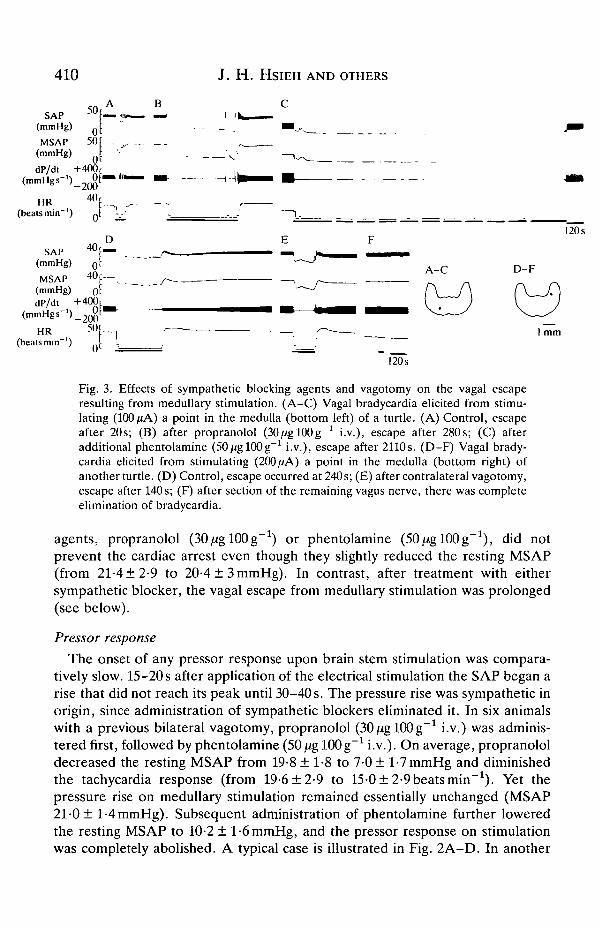
Fig. 3. Effects of sympathetic blocking agents and vagotomy on the vagal escaperesulting from medullary stimulation. (A-C) Vagal bradycardia elicited from stimu-lating (100 fiA) a point in the medulla (bottom left) of a turtle. (A) Control, escapeafter 20s; (B) after propranolol (30^gl00g-1 i.v.), escape after 280s; (C) afteradditional phentolamine (SOjUglOOg"1 i.v.), escape after 2110s. (D-F) Vagal brady-cardia elicited from stimulating (200 ̂ A) a point in the medulla (bottom right) ofanother turtle. (D) Control, escape occurred at 240s; (E) after contralateral vagotomy,escape after 140s; (F) after section of the remaining vagus nerve, there was completeelimination of bradycardia.
agents, propranolol (SOjUglOOg"1) or phentolamine (50//gl00g-1), did notprevent the cardiac arrest even though they slightly reduced the resting MSAP(from 21-4 ±2-9 to 20-4 ± 3 mmHg). In contrast, after treatment with eithersympathetic blocker, the vagal escape from medullary stimulation was prolonged(see below).
Pressor response
The onset of any pressor response upon brain stem stimulation was compara-tively slow. 15-20 s after application of the electrical stimulation the SAP began arise that did not reach its peak until 30-40 s. The pressure rise was sympathetic inorigin, since administration of sympathetic blockers eliminated it. In six animalswith a previous bilateral vagotomy, propranolol (30/^glOOg"1 i.v.) was adminis-tered first, followed by phentolamine (50jUg lOOg"1 i.v.). On average, propranololdecreased the resting MSAP from 19-8 ± 1-8 to 7-0 ± l-7mmHg and diminishedthe tachycardia response (from 19-6±2-9 to 15-0±2-9beatsmin~1). Yet thepressure rise on medullary stimulation remained essentially unchanged (MSAP21-0 ± l-4mmHg). Subsequent administration of phentolamine further loweredthe resting MSAP to 10-2 ± 1-6 mmHg, and the pressor response on stimulationwas completely abolished. A typical case is illustrated in Fig. 2A-D. In another
Vagal bradycardia in turtle All
four animals the blockers were administered in the reverse order. In these animalsphentolamine decreased the resting MSAP from 21-5 ± 1-6 to 18-0 ± l-2mmHgbut both eliminated the pressure rise (21-0 ± 2-6mmHg, no change in BP) and leftthe cardioacceleratory response unchanged (23-0 ± 1-8 vs 23-3 ± l^beatsmin"1).Further intravenous administration of 30/iglOOg"1 propranolol then abolishedthe tachycardia.
The pressure rises noted upon brain stem stimulation appeared to be indepen-dent of inhibition of vagal function. This was demonstrated by the observation thatbilateral vagotomy (nine animals), even with additional administration of atro-pine, lOOjUglOOg"1 i.v., did not reduce the elicited pressor response. Instead, aslight increase of the pressure rise was noted (Fig. 2A,B). Furthermore, explo-ration of the brain stem in these vagotomized animals revealed no significantincrease in the number of the reactive sites for the pressure increase.
Depressor response
Pure depressor responses were occasionally observed, and were due tosympathetic inhibition because bilateral vagotomy did not affect them. Adminis-tration of phentolamine, SO^glOOg"1 i.v., however, completely eliminated thehypotension (three animals, Fig. 2E-H).
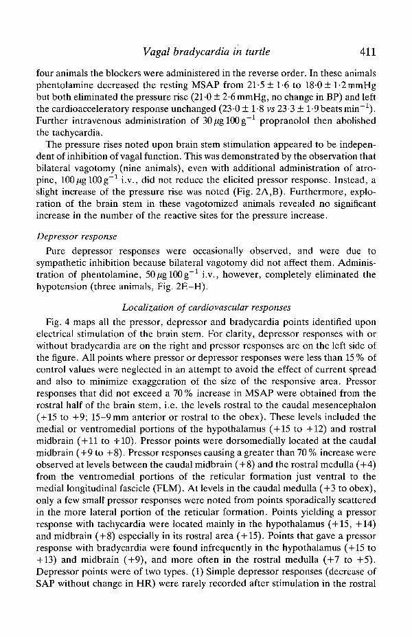
Localization of cardiovascular responses
Fig. 4 maps all the pressor, depressor and bradycardia points identified uponelectrical stimulation of the brain stem. For clarity, depressor responses with orwithout bradycardia are on the right and pressor responses are on the left side ofthe figure. All points where pressor or depressor responses were less than 15 % ofcontrol values were neglected in an attempt to avoid the effect of current spreadand also to minimize exaggeration of the size of the responsive area. Pressorresponses that did not exceed a 70 % increase in MSAP were obtained from therostral half of the brain stem, i.e. the levels rostral to the caudal mesencephalon(+15 to +9; 15-9 mm anterior or rostral to the obex). These levels included themedial or ventromedial portions of the hypothalamus (+15 to +12) and rostralmidbrain (+11 to +10). Pressor points were dorsomedially located at the caudalmidbrain (+9 to +8). Pressor responses causing a greater than 70 % increase wereobserved at levels between the caudal midbrain (+8) and the rostral medulla (+4)from the ventromedial portions of the reticular formation just ventral to themedial longitudinal fascicle (FLM). At levels in the caudal medulla (+3 to obex),only a few small pressor responses were noted from points sporadically scatteredin the more lateral portion of the reticular formation. Points yielding a pressorresponse with tachycardia were located mainly in the hypothalamus (+15, +14)and midbrain (+8) especially in its rostral area (+15). Points that gave a pressorresponse with bradycardia were found infrequently in the hypothalamus (+15 to+ 13) and midbrain (+9), and more often in the rostral medulla (+7 to +5).Depressor points were of two types. (1) Simple depressor responses (decrease ofSAP without change in HR) were rarely recorded after stimulation in the rostral

412 J. H . HSIEH AND OTHERS
+ 13
+ 14 + 12
+ 10 +8
Fig. 4A,B

c+7
SRA
Fig. 4. Distribution of the cardiovascular responsive points in the brain stem of theturtle. For convenience all points producing an increase in arterial pressure (pressorpoints) are placed on the left side of each section and those producing a decrease inarterial pressure (depressor points) on the right side. (A) Pressor response withminimal or no heart rate change; (A) pressor response with tachycardia; (A) pressorresponse with bradycardia. The large, medium and small triangles indicate increases of>70 %, 69-50 % and 49-15 %, respectively. (O) Depressor response with minimal orno change in heart rate. Medium and small circles indicate decreases of 69-50% and49-15 % in the arterial pressure, respectively. ( • ) Depressor response with markedbradycardia. Because very prominent bradycardia or cardiac arrest would inevitablyresult in almost complete suppression of the arterial pressure, the degree of thedepressor response with bradycardia could only be expressed by reference to theduration of the cardiac arrest. Large, medium and small sizes represent the length ofcardiac arrest >20s, 19-10 s and 9-5 s, respectively. The numbers at the left uppercorner of each diagram represent the distance (in mm) rostral (+) to the obex. ACN,anterior commissural nucleus; AN, ambiguus nucleus; DLP, dorsal peduncle of lateralprosencephalic fascicle; DMN, dorsal motor nucleus of vagus nerve; DPC, dorsalnucleus of posterior commissure; DVN, descending vestibular nucleus; FLM, mediallongitudinal fascicle; GC, griseum centrale; HPV, hypothalamic periventricularnucleus; IO, inferior olivary nucleus; IP, interpeduncular nucleus; IRN, inferiorreticular nucleus; LC, locus coeruleus; LHN, lateral habenular nucleus; LM, lentiformmesencephalic nucleus; LVN, lateral vestibular nucleus; M, medial hypothalamicnucleus; MHN,'medial habenular nucleus; MNI, magnocellular part of nucleusisthmus; MRN, medial reticular nucleus; MSN, medial septal nucleus; MSP, medialsuprapeduncular nucleus; NTS, nucleus of tractus solitarius; NR, nucleus reuniens;OC, optic chiasma; PN, parabrachial nucleus; PMN, profound mesencephalic nucleus;R, nucleus rotundus; RN, red nucleus; SM, stria medullaris; SN, substantia nigra; SO,superior olivary nucleus; SON, supraoptic nucleus; SRA, superior raphe nucleus;SRN, superior reticular nucleus; TO, tractus opticum; TT, tectothalamic tract; V,nucleus of descending trigeminal nerve; VI, nucleus of abducent nerve; VII, nucleus offacial nerve; VLP, ventral peduncle of lateral prosencephalic fascicle; XII, nucleus ofhypoglossal nerve.

414 J . H . HSIEH AND OTHERS
SAP(mmHg) Q
MSAP 8 0
(mmHg) Q
dP/dt +600
2%HR
(beats min"1) ol
80r BSAP f(mmHg) 0 ' ^
MSAP 80F(mmHg) ofdP/dt + 4 ° 0 |
(mmHgs-')_2Oo'HR 20r
(beatsmin"1) 0l
SAP 80f(mmHg) Qf
MSAP 80:(mmHg) ot
dP/dt + 6 0 0
1 ^HR
(beats min"1)
1 2 0 5 lS7m
120s
120 s
Fig. 5. Cardiac arrest produced by stimulation of various structures in the medulla.(A) Complete cardiac arrest for 34 min on stimulation of an area of the ambiguusnucleus in the medulla (200 ^A); (B) for 25 min on stimulation of the dorsal motornucleus of the vagus (200 ̂ A), and (C) for 16 min on stimulation of the midlinestructure in the medulla (200 ^A).
levels of the brain stem, scarcely at all after stimulation in the middle levels,but somewhat more frequently after stimulation in the caudal levels (+4 to 0).(2) Depressor responses associated with bradycardia occurred after stimulationthroughout the whole length of the brain stem. The degree of the hypotensiveresponse was proportional to the slowing of the HR, and a cardiac arrest of severalseconds would inevitably result in complete suppression of SAP (Figs 1,5). Thus,the magnitude of this depressor response was determined by the severity of thecardiac arrest rather than by the degree of the peripheral hypotensive effect. Atthe rostral and middle levels of the brain stem, the distribution of the depressorpoints was generally similar to that of the pressor points. In the caudal brain stem,pressor points were sometimes found in the area lateral to the depressor points,whereas moderate depressor responses were attained after stimulation of the areaof the inferior olivary nucleus. The most distinctive feature of these results,however, was the prominent bradycardia found on stimulating the areas of thedorsal motor, solitary and ambiguus nuclei (+2 to 0), as well as the raphe area justventral to the medial longitudinal fascicle (+2, +1).

Vagal bradycardia in turtle 415
The potent vagal bradycardia mechanism in the medulla
A predominance of vagal bradycardia in turtles was evident because stimulationof the caudal medulla immediately stopped the heart beating and thus lowered theSAP to zero with complete arrest of circulation (Figs 3A-C, 5). When stimulationwas deliberately prolonged, beating resumed (escaped) after a considerable time.Six of eleven points of stimulation in the caudal medulla at a level 1 mm rostral tothe obex (+1), when tested for their maximum effect, gave arrests of 25,28, 29, 33,34 and 35 min, whereas the other five caused arrests lasting from 8 to 12 min. Theaverage for the 10 animals was 21 min. Of five points at the obex level tested forthe same purpose, two stopped the heart for 25 and 34min, and the rest for5-17min (average 15min). These values were in contrast to the maximum arrestof 5-10min (average 8min) from 11 points at the +2 level, and the maximumarrest of 5-7 min (average 6 min) from six points at the +3 level. Average lengthsof cardiac arrests caused by stimulation of other areas were: ambiguus, 22 min;dorsal motor of vagus, 15min; and midline structures, 4min. After stimulationrostral to the +3 level, cardiac arrest was not marked, less than lmin, except forone point in the hypothalamus (+13) which caused an arrest of 2 min (Fig. ID).
Vagal escape
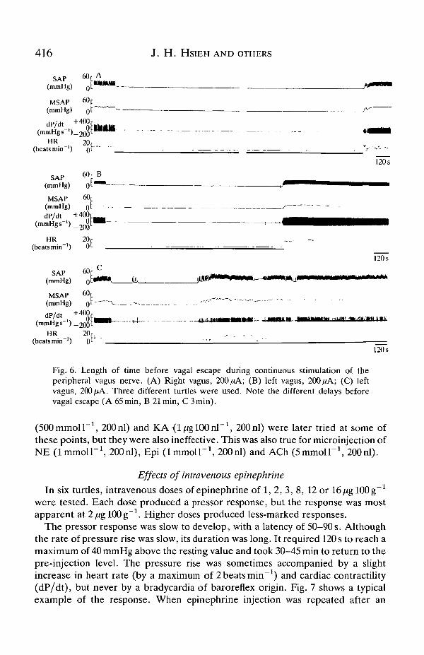
For comparison, in 10 animals the peripheral end of the cut vagus nerve wasstimulated to study the escape phenomenon. The responses differed insignificantlyamong animals. In one turtle the effect of stimulation was so marked that escapedid not occur, and the animal was found dead after stimulation for 63 min. Thevagal escape appeared as late after stimulation as 65 and 21 min (Fig. 6A,B) or asearly as 180 s (Fig. 6C). The cardiac arrest was more prolonged after stimulation ofthe right vagus (30-0 ± 2-1 min) than the left vagus (13-0 ± 1-3min).
The role of sympathetic function in the vagal escape from medullary stimulationwas studied. In six animals, propranolol (30/iglOOg"1 i.v.) was administered first,followed by phentolamine (SOjUglOOg"1 i.v.). Propranolol prolonged the escapeby 230 s (80 ± 26 s vs 310 ± 50 s). Subsequent phentolamine treatment furtherprolonged the escape (84 ± 26 s vs 638 ± 55 s). In one of these six animals theescape prolongation after blockers was very marked, increasing from 20s to 400safter propranolol and to 2110 s after the addition of phentolamine (Fig. 3A-C). Inanother animal, in which the order of administration of blockers was reversed,phentolamine first prolonged the escape by 180s (from 80s to 260s) and thenpropranolol further prolonged the escape by 480s (80s vs 560s).
Chemical stimulations
The points capable of producing significant depressor or pressor responsesunder electrical stimulation were injected with sodium glutamate (10moll"1 ,200 nl) through the same electrode tubing. In general, the response to glutamatewas not marked. Although some points responded in the same way as they did toelectrical stimulation, the response never reached the same magnitude. DLH
416 J. H . HSIEH AND OTHERS
SAP ""I(mmHg) Q[
MSAP(mmHg)
dP/dt 0 .(mmHgs )_200'
HR 20,(beats min ) Q'
120 s
SAP « f B ^
MSAP 60r( m m H g ) o i • - • • - — - /
dP/dt + 4 0 0 F —(mmHgs"1) _ J l * "
HR 20 r(beats min"1) 0l
SAP(mmHg)
120 s
• U.WUtt
120 s
Fig. 6. Length of time before vagal escape during continuous stimulation of theperipheral vagus nerve. (A) Right vagus, 200 ,uA; (B) left vagus, 200fiA; (C) leftvagus, 200,uA. Three different turtles were used. Note the different delays beforevagal escape (A 65 min, B 21 min, C 3min).
(SOOmmoir1, 200 nl) and KA {ljUglOOnP1, 200 nl) were later tried at some ofthese points, but they were also ineffective. This was also true for microinjection ofNE ( l m m o i r 1 , 200nl), Epi ( lmmolP 1 , 200nl) and ACh (Smmoll"1, 200nl).
Effects of intravenous epinephrine
In six turtles, intravenous doses of epinephrine of 1, 2, 3, 8, 12 or 16iugl00g"1
were tested. Each dose produced a pressor response, but the response was mostapparent at 2/ig lOOg"1. Higher doses produced less-marked responses.
The pressor response was slow to develop, with a latency of 50-90 s. Althoughthe rate of pressure rise was slow, its duration was long. It required 120 s to reach amaximum of 40 mmHg above the resting value and took 30-45 min to return to thepre-injection level. The pressure rise was sometimes accompanied by a slightincrease in heart rate (by a maximum of 2 beats min"1) and cardiac contractility(dP/dt), but never by a bradycardia of baroreflex origin. Fig. 7 shows a typicalexample of the response. When epinephrine injection was repeated after an

Vagal bradycardia in turtle All
80 rMSAP [.
(mmHg) £
dp/d, + 4 0 ° r
(beatsmin"1) 20 r_
° A 120 sEpinephrine
Fig. 7. Effect of intravenous injection of epinephrine, 2/ igl00g 1. Note the absenceof reflex bradycardia during pressor responses.
interval of less than 15 min, the magnitudes of the pressure rise, cardiacacceleration and augmentation of contractility decreased (tachyphylaxis), adecrease that was more apparent in the latter two components.
Discussion
The present experiments show that in the brain stem of turtles vagal represen-tation is predominant over sympathetic representation in terms of a larger area ofdistribution in the brain and more intense responses that varied from markedslowing to cessation of heart beat. Furthermore, the latency for producing thisvagal response was shorter, less than 3 s, compared with that of the sympatheticpressor response, 10-20 s. If stimulation was given continuously in the caudalmedulla, the bradycardia could last as long as 35 min before the appearance of anescape beat. Even after stimulation of the hypothalamus the cardiac arrest couldlast for 2 min.
This type of prolonged cardiac arrest resulting from stimulation of the brainstem is not seen in mammals. It is consistent with the finding that the turtle has agreat capacity for tolerating anoxia and undergoing anaerobic metabolism. Forinstance: even in atmospheric air, turtles breathe irregularly and show longperiods of apnoea (Boyer, 1963); turtles (Chrysemys picta) have been found totolerate anoxia (breathing of air without O2) for an average of 1104 min (20 h) andstagnant anoxia (arrest of circulation) for 72 min (Belkin, 1968); at 3°C, turtles cansubmerge for 6 months in an anoxic condition (Ultsch & Jackson, 1982); at16-18°C they can survive an anaerobic dive for up to 2 weeks (Robin et al. 1964);breathing 100 % N2, turtles {Pseudemys scripta) have survived for at least 48 h; andChrysemys can tolerate 93 h in oxygen-free water (Folk, 1974).
The marine green turtle can bury itself in mud on the ocean floor for 1-3 months(Felger etal. 1976). This great tolerance of anoxia, with survival of the brain tissue,

418 J. H . HSIEH AND OTHERS
has been attributed to a remarkable ability to use anaerobic glycolysis as an energysource (Belkin, 1961; Millen etal. 1964; Sick etal. 1982; Caligiuri & Robin, 1985).In contrast, failure of anaerobic glycolysis to maintain adequate levels of ATP andcreatine phosphate limits anaerobic metabolism in freshwater turtles (Pseudemysscripta elegans) (Clark & Miller, 1973). The increased glycolytic capacity is relatedto a high activity of various glycolytic enzymes (Robin et al. 1979). Othermechanisms are also involved: (1) reduced energy requirement during prolongedanoxia in diving (Robin etal. 1981); (2) increased resistance of the brain to acidosisas severe as pH6-20 (Caligiuri & Robin, 1985); (3) cardiovascular adjustmentincluding bradycardia and redistribution of cardiac output such that blood fromthe systemic venous circulation (tissue) bypasses the lung and enters the aortadirectly (Millen et al. 1964; White & Roos, 1966); (4) removal of plasma CO2,probably through the body surface, during apnoea diving (Jackson & Silverblatt,1974). It should be noted that cardiac arrest is a further extension of bradycardia.It entirely stops the circulation and hence curtails the energy expenditure of thewhole body. Thus, in a turtle's brain stem the highly developed vagal mechanismresponsible for cardiac inhibition is an important component of the cardiovascularadjustment that prolongs tolerance to anoxia. It is necessary, however, to find outwhether a turtle heart stops beating completely or continues beating at anextremely slow rate during prolonged diving.
A pressor response effected through activation of the sympathetic system wasalso observed in these experiments. However, because of its long latency, slow riseand low magnitude, this hypertension appeared to be less significant than thebradycardia. Furthermore, during stimulation a concomitant increase of heart rateand cardiac contractility, very common in cats and dogs, was seldom seen in theturtles. Thus, the sympathetic mechanism in the brain stem of turtles seemsprincipally to involve vasomotor action. It is interesting to note, however, thatdespite the low sympathetic function in this species, a mechanism for sympatheticinhibition does exist in the medulla.
Whether turtles are provided with reflex baroreceptor mechanisms like those ofmammals has been a subject of interest for years. Some authors have supportedthe existence of this reflex (Millard & Moalli, 1980; Smits & Kozubowski, 1985)and others have not (Stephens et al. 1983). In experiments utilizing probing andmechanical occlusion of the vessels, including the region of the bulbus cordiproximal to the common pulmonary artery, an increased pressure in these vesselsevoked an immediate but transient increase in synchronized traffic in the vagalefferents to the heart (Faraci etal. 1982). A similar baroreceptor mechanism foundin the pulmonary cutaneous artery of an amphibian (the toad) serves to protect thevasculature of the lung from excessive hydrostatic pressure (West & Van Vliet,1983).
Because the turtle lives in an aquatic habitat, usually maintaining a horizontalposture, its gravitational stress is minimal. Besides, turtles move slowly, they areextremely tolerant of anoxic conditions, and their defence mechanism seems to beescape rather than attack. It is understandable, therefore, that an active

Vagal bradycardia in turtle 419
baroreceptor mechanism for rapid cardiovascular adjustment may not be critical.In the present study, in which systemic injection of epinephrine induced an activeincrease in blood pressure, the heart rate remained unchanged, without the reflexbradycardia commonly seen in mammals after systemic administration of epineph-rine. This observation is consistent with the absence of an active baroreceptorreflex mechanism in turtles. It should be noted, however, that for some reason,despite the use of a large dose of epinephrine (up to 16pig lOOg"1 i.v.), the rate ofpressure rise after administration of this pressor agent was slow in turtles. Whetherthis slow rise is responsible for the failure to induce a reflex bradycardia and thushinders the detection of baroreceptors in turtle remains to be determined.
In the present study, microinjection of transmitter substances, acetylcholine,norepinephrine and epinephrine, and excitatory amino acids into the responsiveregions of the brain stem failed to produce responses comparable to those evokedby electrical stimulation. This apparent difference deserves further study. Re-cently, Reiner (1987) reported a wide distribution of enkephalin peptides in theturtle's brain. It is interesting to note that peptide location in the brain stem showsa certain similarity to the bradycardia areas described in the present study.
In summary, the findings of the present experiments indicate the presence in theturtle brain stem of a very powerful vagal component responsible for bradycardiawhich may override coincident sympathetic activity. Its function may partiallyaccount for the turtle's powerful capacity for tolerating anoxia.
The authors thank R. H. Lin, Y. F. Lin, A. M. Y. Lin and C. L. Na for theirmost valuable assistance in these experiments, and L. L. Chen and G. T. Chen forpreparation of the manuscript and illustrations. Special thanks are due toProfessor John R. Brobeck of University of Pennsylvania for advice and readingthe manuscript. Thanks are due to Drs Paul N. Yu and Shu Chien for their supportand encouragement. This study was supported in part by the Institute ofBiomedical Sciences, Academia Sinica and the National Science Council, ROCnos 755327-070 and 770412-B001.
ReferencesBELKIN, D. A. (1961). Anaerobic mechanisms in the diving of the loggerhead musk turtle,
Sternothaerus minor. Ph.D. dissertation, University of Florida.BELKIN, D. A. (1968). Anaerobic brain function: effects of stagnant and anoxic anoxia on
persistence of breathing in reptiles. Science 162, 1017-1018.BERKSON, H. (1966). Physiological adjustments to prolonged diving in the Pacific green turtle
(Chelonia agassizii). Comp. Biochem. Physiol. 18, 101-119.BOYER, D. R. (1963). Hypoxia: effects on heart rate and respiration in the snapping turtle.
Science 140, 813-814.CALIGIURI, M. A. & ROBIN, E. D. (1985). Prolonged diving and recovery in the freshwater
turtle, Pseudemys scripta. IV. Effects of profound acidosis on O2 consumption in turtle vs. rat(mammalian) brain and heart slices. Comp. Biochem. Physiol. 81A, 603-605.
CHAI, C. Y. & Kuo, J. S. (1967). The optimal frequency and duration of rectangular pulses forefferent vagus stimulation. Chinese J. Physiol. 20, 27-32.
CHAI, C. Y., LIN, Y. F., LIN, A. M. Y., PAN, C. M., LEE, E. H. Y. & Kuo, J. S. (1988).

420 J . H . HSIEH AND OTHERS
Existence of a powerful inhibitory mechanism in the medial region of caudal medulla - withspecial reference to the paramedian reticular nucleus. Brain Res. Bull. 20, 515-528.
CHEN, H. I. & CHAI, C. Y. (1976). Integration of the cardiovagal mechanism in the medullaoblongata of the cat. Am. J. Physiol. 231, 454-461.
CLARK, V. M. & MILLER, A. T., JR (1973). Studies on anaerobic metabolism in the fresh-waterturtle (Pseudemys scripta elegans). Comp. Biochem. Physiol. 44A, 55-62.
FARACI, F. M., SHIRER, H. W., ORR, J. A. & TRANK, J. W. (1982). Circulatory mechanoreceptorsin the pond turtle: Pseudemys scripta. Am. J. Physiol. 242, R216-R219.
FELGER, R. S., CLIFTON, K. & REGAL, P. J. (1976). Water dormancy in sea turtles: independentdiscovery and exploitation in the Gulf of California by two local cultures. Science 191,282-285.
FOLK, G. E. (1974). Textbook of Environmental Physiology, 2nd edn. Philadelphia: Lea &Febiger.
FRIEDMAN, A. H. & BHAGAT, B. (1962). The concentration of catecholamines in the turtle heartand vagal escape. /. Pharm. Pharmac. 14, 764.
HOUGH, T. (1895). On the escape of the heart from vagus inhibition. J. Physiol., Lond. 18,161-200.
JACKSON, D. C. & SILVERBLATT, H. (1974). Respiration and acid-base status of turtles followingexperimental dives. Am. J. Physiol. 226, 903-909.
LIN, A. M. Y., PAN, C. M., LrN, Y. F., Kuo, J. S., CHAN, S. H. H. & CHAI, C. Y. (1987). Acardioinhibitory area in the midbrain central tegmental field of cats. Brain Res. Bull. 18,699-707.
MILLARD, R. W. & MOALLI, R. (1980). Baroreflex sensitivity in an amphibian, Rana catesbeiana,and a reptilian, Pseudemys scripta elegans. J. exp. Zool. 213, 283-288.
MILLEN, J. E., MURDAUGH, H. V., JR, BAUER, C. B. & ROBIN, E. D. (1964). Circulatoryadaptation to diving in the freshwater turtle. Science 145, 591-593.
MILLS, T. W. (1885). The innervation of the heart of the slider terrapin (Pseudemys rugosa).J. Physiol., Lond. 6, 246-286.
PENNEY, D. G. (1974). Effects of prolonged diving anoxia on the turtle, Pseudemys scriptaelegans. Comp. Biochem. Physiol. 47A, 933-941.
REINER, A. (1987). The distribution of proenkephalin-derived peptides in the central nervoussystem of turtles. J. comp. Neurol. 259, 65-91.
ROBIN, E. D., LEWISTON, N., NEWMAN, A., SIMON, L. M. & THEODORE, J. (1979). Bioenergeticpattern of turtle brain and resistance to profound loss of mitochondrial ATP generation. Proc.natn. Acad. Sci. U.S.A. 76, 3922-3926.
ROBIN, E. D., ROBIN, D. A., ACKERMAN, R., LEWISTON, N., HANCE, A. J., CALIGIURI, M. &THEODORE, J. (1981). Prolonged diving and recovery in the fresh water turtle, Pseudemysscripta. Lung and blood gases, pH, lactate concentration and "cation" gap. Comp. Biochem.Physiol. 70, 359-364.
ROBIN, E. D., VESTER, J. W., MURDAUGH, H. V., JR & MILLEN, J. E. (1964). Prolongedanaerobiosis in a vertebrate: anaerobic metabolism in the freshwater turtle. J. cell. comp.Physiol. 63, 287-297.
SICK, T. J., ROSENTHAL, M., LAMANNA, J. C. & LUTZ, P. L. (1982). Brain potassium ionhomeostasis, anoxia, and metabolic inhibition in turtles and rats. Am. J. Physiol. 243,R281-R288.
SMITS, A. W. & KOZUBOWSKI, M. M. (1985). Partitioning of body fluid and cardiovascularresponses to circulatory hypovolaemia in the turtle, Pseudemys scripta elegans. J. exp. Biot.116, 237-250.
STEPHENS, G. A., SHIRER, H. W. & TRANK, J. W. (1983). Arterial baroreceptor reflex control ofheart rate in two species of turtle. Am. J. Physiol. 244, R544-R552.
ULTSCH, G. R. & JACKSON, D. C. (1982). Long-term submergence at 3°C of the turtle,Chrysemyspicta bellii, in normoxic and severely hypoxic water. I. Survival, gas exchange andacid-base status. /. exp. Biol. 96, 11-28.
WEST, N. H. & VAN VLIET, B. N. (1983). Open-loop analysis of the pulmocutaneous baroreflexin the toad Bufo marinus. Am. J. Physiol. 245, R642-R650.
WHITE, F. N. & Ross, G. (1966). Circulatory changes during experimental diving in the turtle.Am. J. Physiol. 211, 15-18.