production and efficacy of a low-cost recombinant ... fileproduction and efficacy of a low-cost...
TRANSCRIPT
Production and efficacy of a low-cost recombinantpneumococcal protein polysaccharide conjugatevaccineHerbert, Jenny; Kay, Emily J; Faustini, Sian; Richter, Alex; Abouelhadid, Sherif; Cuccui, Jon;Wren, Brendan; Mitchell, TimothyDOI:10.1016/j.vaccine.2018.05.036
License:Creative Commons: Attribution (CC BY)
Document VersionPublisher's PDF, also known as Version of record
Citation for published version (Harvard):Herbert, JA, Kay, EJ, Faustini, S, Richter, A, Abouelhadid, S, Cuccui, J, Wren, B & Mitchell, T 2018, 'Productionand efficacy of a low-cost recombinant pneumococcal protein polysaccharide conjugate vaccine', Vaccine, vol.36, no. 26, pp. 3809-3819. https://doi.org/10.1016/j.vaccine.2018.05.036
Link to publication on Research at Birmingham portal
General rightsUnless a licence is specified above, all rights (including copyright and moral rights) in this document are retained by the authors and/or thecopyright holders. The express permission of the copyright holder must be obtained for any use of this material other than for purposespermitted by law.
•Users may freely distribute the URL that is used to identify this publication.•Users may download and/or print one copy of the publication from the University of Birmingham research portal for the purpose of privatestudy or non-commercial research.•User may use extracts from the document in line with the concept of ‘fair dealing’ under the Copyright, Designs and Patents Act 1988 (?)•Users may not further distribute the material nor use it for the purposes of commercial gain.
Where a licence is displayed above, please note the terms and conditions of the licence govern your use of this document.
When citing, please reference the published version.
Take down policyWhile the University of Birmingham exercises care and attention in making items available there are rare occasions when an item has beenuploaded in error or has been deemed to be commercially or otherwise sensitive.
If you believe that this is the case for this document, please contact [email protected] providing details and we will remove access tothe work immediately and investigate.
Download date: 17. Mar. 2019

Vaccine 36 (2018) 3809–3819
Contents lists available at ScienceDirect
Vaccine
journal homepage: www.elsevier .com/locate /vacc ine
Production and efficacy of a low-cost recombinant pneumococcalprotein polysaccharide conjugate vaccine
https://doi.org/10.1016/j.vaccine.2018.05.0360264-410X/� 2018 The Authors. Published by Elsevier Ltd.This is an open access article under the CC BY license (http://creativecommons.org/licenses/by/4.0/).
⇑ Corresponding author.E-mail address: [email protected] (T.J. Mitchell).
1 These authors contributed equally to this work.2 Current address: UCL Great Ormond Street Institute of Child Health, London, UK.
Jenny A. Herbert a,1,2, Emily J. Kay b,1, Sian E. Faustini c,d, Alex Richter c,d,e, Sherif Abouelhadid b, Jon Cuccui b,Brendan Wren b, Timothy J. Mitchell a,⇑a Institute of Microbiology and Infection, College of Medical and Dental Sciences, University of Birmingham, Birmingham, England, UKbDepartment of Pathogen Molecular Biology, London School of Hygiene and Tropical Medicine, London, UKc Institute of Immunology and Immunotherapy, College of Medical and Dental Sciences, University of Birmingham, Birmingham, England, UKdDepartment of Immunology, Queen Elizabeth Hospital, Birmingham, UKeUniversity Hospitals Birmingham NHS Foundation Trust, Birmingham, UK
a r t i c l e i n f o
Article history:Received 30 January 2018Received in revised form 1 May 2018Accepted 5 May 2018Available online 25 May 2018
Keywords:Streptococcus pneumoniaeConjugate vaccineProtein glycan coupling technologyGlycoengineeringPneumonia
a b s t r a c t
Streptococcus pneumoniae is the leading cause of bacterial pneumonia. Although this is a vaccine pre-ventable disease, S. pneumoniae still causes over 1 million deaths per year, mainly in children underthe age of five. The biggest disease burden is in the developing world, which is mainly due to unavailabil-ity of vaccines due to their high costs. Protein polysaccharide conjugate vaccines are given routinely inthe developed world to children to induce a protective antibody response against S. pneumoniae. Oneof these vaccines is Prevnar13, which targets 13 of the 95 known capsular types. Current vaccine produc-tion requires growth of large amounts of the 13 serotypes, and isolation of the capsular polysaccharidethat is then chemically coupled to a protein, such as the diphtheria toxoid CRM197, in a multistep expen-sive procedure. In this study, we design, purify and produce novel recombinant pneumococcal proteinpolysaccharide conjugate vaccines in Escherichia coli, which act as mini factories for the low-cost produc-tion of conjugate vaccines. Recombinant vaccine efficacy was tested in a murine model of pneumococcalpneumonia; ability to protect against invasive disease was compared to that of Prevnar13. This study pro-vides the first proof of principle that protein polysaccharide conjugate vaccines produced in E. coli can beused to prevent pneumococcal infection. Vaccines produced in this manner may provide a low-cost alter-native to the current vaccine production methodology.� 2018 The Authors. Published by Elsevier Ltd. This is an openaccess article under the CCBY license (http://
creativecommons.org/licenses/by/4.0/).
1. Introduction
Streptococcus pneumoniae (the pneumococcus) is the leadingcause of bacterial pneumonia. The highest disease burden isobserved in the developing world due to limited vaccine availabil-ity [1]. In the developed world, since the introduction of pneumo-coccal conjugate vaccines, S. pneumoniae disease burden inchildren has drastically reduced (PCV7/10/13) [2,3]. Prevnar7 wasthe first pneumococcal conjugate vaccine produced and was intro-duced in the UK in 2006. Post introduction, invasive pneumococcaldisease caused by vaccine serotypes dropped by 41% [2]. Afterintroduction of Prevnar13 (PCV13) in 2010 invasive disease causedby the additional 6 serotypes dropped by 75% [4].
PCV13 is a component of the childhood vaccine schedule in theUK and is given to all children in a three-dose schedule at 2, 4 and12 months of age. This vaccine targets the capsular polysaccharidesurrounding the bacteria. PCV13 protects against the 13 (out of 95)pneumococcal serotypes found to be most prevalent in disease [5].Polysaccharide alone is not immunogenic in children under 2 yearsof age, and does not produce a lasting immune response [6]. Con-jugate vaccines work by coupling the polysaccharide component toa protein carrier [7], resulting in a protective T-cell dependentmemory response [8]. This technology was first used for the pro-duction of a Haemophilus influenzae type B vaccine [9], followedby conjugate vaccines to prevent Neisseria meningitidis serogroupC [10], and subsequently pneumococcal infection [11]. Pneumococ-cal conjugate vaccines are the world’s best-selling vaccines, and in2014 PCV13 sales produced revenues of £2.9 billion [12].
Although these glycoconjugate vaccines are very effective, thereare some limitations to their use. Serotype distribution of diseasecausing isolates varies geographically [13]. The PCV7 vaccine

3810 J.A. Herbert et al. / Vaccine 36 (2018) 3809–3819
serotypes are more prevalent in the western world, therefore thisvaccine did not provide adequate protection against serotypesprevalent in developing countries. However, the introduction ofan additional 6 serotypes in PCV13 includes the main disease-causing serotypes in the developing world [5,13]. Serotype replace-ment remains a problem, and introduction of vaccines has resultedin increased incidence of disease from non-vaccine serotypes[2,14,15]. The most recent emerging serotypes (22F and 33F) areincluded in a new 15-valent vaccine preparation [16]. Finally, thehigh cost of conjugate vaccines means they are often not availableto the poorest regions, which have the greatest disease burden.
The complex nature of the production process of the conjugatevaccine is one contributing factor to the high cost. Using standardmethods, growth of large quantities of pathogenic pneumococci isrequired for isolation of the polysaccharide. Post purification thepolysaccharide must then be chemically coupled to the carrier pro-tein, in the case of PCV13 a diphtheria toxoid (CRM197). This pro-cess is time consuming, requires several rounds of purification toremove toxic chemicals and by products, and can often result inbatch to batch variation [17].
An increase in the understanding of bacterial protein glycosyla-tion has led to development of novel ways to couple protein andpolysaccharide (reviewed in [18]). The approach, often referredto as protein glycan coupling technology (PGCT) allows productionof protein polysaccharide conjugate vaccines in Escherichia coli[19]. This technology utilises an oligosaccharyltransferase enzyme,PglB, from the general protein glycosylation locus (Pgl) of Campy-lobacter jejuni [19]. This locus encodes the genes required for theproduction of a C. jejuni heptasaccharide along with PglB, whichis required for coupling of the heptasaccharide to a carrier protein.PglB couples over sixty C. jejuni proteins to this heptasaccharide[20]. These proteins contain an amino acid acceptor sequence,which is recognised by PglB [21]. The consensus, or glycotag,sequence can be engineered into any protein carrier, allowingrecognition by PglB [21]. The glycan specificity of C. jejuni PglBhas been well characterised, using this knowledge researchershave been able to couple a number of different polysaccharidesto chosen carrier proteins using PglB [21–23]. In simple terms,PGCT can be divided into three procedures. In the first stage, genesencoding the target glycan are faithfully cloned and expressed inE. coli on a suitable plasmid. In the second stage, the target carrierprotein containing the appropriate consensus sequon and purifica-tion tag are cloned into a suitable plasmid, and targeted to the peri-plasm. Finally, the coupling enzyme, CjPglB, recognises the initialsugar on the glycan and transfers it to the carrier protein. The plas-mids are introduced into an appropriate E. coli host strain to pro-duce an inexhaustible supply of recombinant glycoprotein thatcan be readily purified [19].
There are a number of vaccines that have been produced usingthis technology that show excellent promise in both animal modelsand in clinical trials [24–27]. Vaccines produced using PGCT willreduce vaccine costs, speed up the production process, and negatethe need for growth of large volumes of pathogenic bacteria. Otherbenefits of using this technology include the ability to readilychange the carrier protein, and to add further compatible polysac-charide types. Further, this technology could allow rapid additionof polysaccharides to vaccine preparations to protect againstemerging serotypes. Vaccines produced in this manner could alsobe tailored to specific geographical regions, by protecting againstthe most prevalent serotypes. To date pneumococcal polysaccha-rides of type 4, 5, 8 and 12F have been expressed in E. coli [28],the first stage of PGCT.
In the current study, we provide the first evidence that recom-binant protein polysaccharide conjugate vaccines can be producedin E. coli and protect against pneumococcal invasive disease in amurine infection model.
2. Materials and methods
2.1. Bacterial strains and plasmids
Escherichia coli strains were grown in modified super optimalbroth, SSOB (Tryptone 2%, Yeast extract 0.5%, NaCl 10 mM, KCl2.5 mM, MgCl2 10 mM, MgSO4 10 mM) at 28 �C, with shaking.Antibiotics were added as necessary for plasmid maintenance:tetracycline 20 mg ml�1; ampicillin 100 mg ml�1; chloramphenicol30 mg ml�1. A table of strains and plasmids used in this study canbe found in the supplementary information (Table S1).
Streptococcus pneumoniae strain (TIGR4) was cultured on BHIagar with 5% horse blood, or statically in BHI broth, in an atmo-sphere containing 5% CO2.
2.2. Vaccine production
Recombinant serotype 4 polysaccharide was produced in E. coli,as previously described [28]. Conjugation to AcrA was carried outusing protein glycan coupling technology [25]. E. coli cultures weregrown for 16 h. These starter cultures were used to inoculate 2 L ofSSOB to an OD600 of 0.03 and incubated with shaking at 28 �C.Once OD600 had reached 0.4–0.6, expression of PglB was inducedwith the addition of 1 mM IPTG. MnCl2 was also added to a finalconcentration of 4 mM. After 20 h growth at 28 �C cells were pel-leted by centrifugation at 5400g for 30 min at 4 �C. Pellets wereresuspended in lysis buffer (50 mM NaH2PO4, 0.3 M NaCl and 10mM imidazole, pH 7.5) with 1 mg/ml lysozyme, and lysed usinga FastPrep instrument (MP Biomedicals) with lysing matrix B.Supernatant was treated with 250 units benzonase for 10 min.Insoluble debris was removed by centrifugation at 7800g for 60min at 4 �C and the supernatant passed through a 0.2 mm filter.The protein/polysaccharide conjugate labeled with a polyhistidineaffinity tag was purified using HisTrap columns (GE Healthcare)using an imidazole gradient of 20–300 mM on an AKTA proteinpurification system (GE Healthcare).
2.3. SDS-PAGE and immuno blot analysis
To verify glycoconjugate production and to select AKTA frac-tions for pooling, samples were subject to SDS-PAGE followed bycoomassie staining or immunoblot. Rabbit anti-serotype 4 capsuleantibody from the Statens Serum Institut, (SSI) Denmark was usedat a dilution of 1:1000 and mouse anti-His monoclonal antibody(Abcam, UK) was used at a dilution of 1:10,000 to detect recombi-nant serotype 4 capsule and His-tagged AcrA respectively. HR6antiserum was used to detect the Campylobacter heptasaccharide(S. Amber and M. Aebi, unpublished data). Secondary goat anti-rabbit IgG IRDye 800 and goat anti-mouse IgG IRDye 680 conju-gates were used at a dilution of 1: 10 000. Fluorescent signal wasdetected using an Odyssey LI-COR detection system (LI-COR Bio-sciences UK Ltd.).
2.4. Protein and polysaccharide (PS) quantification in vaccinepreparations
Selected AKTA fractions were concentrated using Vivaspin pro-tein concentrator spin columns with 10 KDa MWCO (GE Health-care) and protein was quantified using a Qubit protein assay(Thermo Fisher Scientific). Levels of Type 4 polysaccharide in vac-cine preparations was quantified by ELISA using type 4 antiserumand a standard curve generated using purified type 4 polysaccha-ride (SSI, Denmark).
J.A. Herbert et al. / Vaccine 36 (2018) 3809–3819 3811
2.5. Vaccination
All in vivo experiments were carried out in accordance with theUK Animal Scientific Procedures Act (1986). Mice used in this studywere 6–8 week old outbred female MF1 (Harlan, UK). Mice hadfood and water ad libitum, were kept at a constant room temper-ature of 20–22 �C, with a 12 h light/dark cycle. For immunization,each mouse received three subcutaneous injections with aninterval of two weeks between each. All vaccines were made upin phosphate buffered saline (PBS). For the positive controls micewere vaccinated with pneumococcal 13-valent conjugate vaccine(PCV) Prevnar (Pfizer). Two dosing regimes were used for thePrevnar control groups: PCV13 high vaccination contained 0.5 lgof type 4 PS/dose, while PCV13 low contained 0.0001 lg type4 PS/dose. The AcrA-SP4 conjugate vaccine contained 0.0001 lgtype 4 PS/dose. The AcrA alone and AcrA-Pgl vaccines werenormalised to contain the same amount of AcrA to that in theAcrA-SP4 preparation. This was done by western blotting usingan anti-His tag antibody (Abcam, UK). Alhydrogel was added tothe experimental vaccine preparations to the same level as thatfound in the PCV13 high dose (Type 4 PS only controls, AcrA only,AcrA-Pgl, AcrA-SP4). Sham vaccination consisted of PBS withAlhydrogel.
2.6. Intranasal infection model
Four weeks after the last vaccinationmice were challenged withS. pneumoniae serotype 4 strain, TIGR4. Mice were inoculated intra-nasally with 5 � 106 colony forming units (CFU) in 50 ll PBS. Allmice were monitored for symptoms and were sacrificed at a desig-nated clinical endpoint point. Organs were removed (lungs, liver,spleen, brain), blood taken and viable counts performed usingthe Miles and Misra method [29]. Mice that did not reach the clin-ical endpoint were sacrificed at 7 days post infection and processedin the same way. Blood samples from tail veins, body weight andclinical scores were also taken throughout the study. Graphicalrepresentation and statistical analysis was performed in Prism ver-sion 4.0b (GraphPad Software), using a non-parametric Mann-Whitney two sample rank test; significance P < 0.05. Survival ofmice receiving different vaccine preparations was compared usinga Kaplan-Meier survival curve and analysed using a Log-rank Test(P < 0.05).
2.7. Preparation of Luminex beads
Protein (AcrA) and polysaccharide (type 4 polysaccharide) werecoupled to carboxylate microspheres specific for use in the Lumi-nex multiplex machine as described previously [30,31]. Briefly,type 4 pneumococcal polysaccharide (SSI 5 mg/ml) was coupledto poly-L-lysine (PLL) using cyanuric chloride. Type 4 PS coupledto PLL was then purified using a G25 PD-10 Sephadex column(GE healthcare). Type 4 PS-PLL was then coupled to carboxylatedmicrospheres (Bio-Plex COOH bead 11, BIO-RAD, UK). AcrA wascoupled to a different bead set (Bio-Plex COOH bead 47) at a con-centration of 50 lg/ml and did not require prior coupling to PLL.Coupling of beads to antigens was performed using standard meth-ods. Briefly, beads were activated with 5 mg/ml EDC (1-ethyl-3-(3-dimethylaminopropyl)carbodiimide hydrochloride) solution and5 mg/ml NHS (N-hydroxysuccinimide) solution. Bead sets werewashed with PBS and incubated with their individual antigen. Afterincubation, beads were washed in PBS and resuspended in 300 llPBS 0.1% BSA, 0.05% sodium azide and kept at 4 �C in the dark untilused.
2.8. Luminex assay antibody quantification
Assays were adapted from [31]. Briefly, sera taken from individ-ual vaccinated mice prior to bacterial challenge were assessed forantibodies against AcrA and type 4 polysaccharide. Serum wasdiluted 1/100 in sample buffer (PBS 0.05% tween 20, 1% BSA, 5lg/ml CWPS, 5 lg/ml 22F PS). A standard curve was created fromserial 10 fold dilutions of a human standard serum (0 0 7). Thisserum has known anti-pneumococcal PS antibody titres andallowed extrapolation of anti-type 4 PS antibody levels in mousesera from the human serum standard curve. For AcrA, antibodylevels are based on the mean fluorescent intensity values and arerelative quantifications of anti-AcrA antibodies between the differ-ent vaccinated groups.
Assays were run in 96-well filter plates (Millipore, UK) with2500 beads/antigen in each well. To each well 25 ll of diluted serawas added. A type 4 antiserum and a PCV13 mouse control serawere run as internal controls for anti-type 4 PS antibodies for eachassay. An in house anti-AcrA antibody was used as an internal con-trol for AcrA samples. Beads and sera were incubated at room tem-perature for 1 h with shaking at 500 rpm. Beads were washed withPBS 0.05% tween 20 then incubated with a 1/200 dilution of anti-human, anti-rabbit or anti-mouse IgG phycoerythrin (PE) conju-gate antibody (Southern Biotech, UK) for 30 min, shaking at 500rpm. Beads were washed 2� as above and resuspended in 125 llPBS 0.05% tween 20. Data was acquired on a Luminex-100 instru-ment (BIO-RAD, UK). Data analysis was performed on Bio-Plexmanager 4.1.1 software, which created the standard curve fromthe 007 human sera. From this anti-type 4 PS, antibody levels inmouse sera were extrapolated. Graphical representation was per-formed in Prism version 4.0b (GraphPad Software) showing anti-body titres in the different vaccinated groups. Statistical analysiswas performed using a Kruskal-Wallis one-way ANOVA with aDunn’s multiple comparison test (P < 0.05).
2.9. Opsonophagocytic killing assay (OPKA)
HL60 cells were maintained in RPMI medium 1640 GlutaMAX(Thermofisher, UK) supplemented with 20% fetal calf serum, 1�penicillin streptomycin and 2 mM L-glutamine. Differentiationwas performed using a cell density of 4 � 105 cells/ml in 0.8%Dimethylformamide. Cells were differentiated for 5 days and thenused in the OPKA. Cells were washed in HBSS�/+ Ca2+/Mg2+ andresuspended at the desired concentration in opsonisation buffer(OPB buffer) (1 ml 10� HBSS+ Ca2+/Mg2+,1 ml gelatin, 530 ll Fetalbovine serum, 8 ml H2O).
OPKAs were performed on serum samples taken from vacci-nated mice. Serum was heat inactivated for 30 min at 56 �C. Serialtwo-fold dilutions of sera were performed in PBS with a startingdilution of 1:2. TIGR4 was added to each well at a concentrationof 2.5 � 103 cfu and incubated for 30 min at 4 �C. Bacteria werepelleted by centrifugation and 15% baby rabbit complement wasadded, followed by 5 � 105 differentiated HL60 cells. Followingincubation at 37 �C in 5% C02 for 40 min, the contents of each wellwere diluted 1/10 in PBS, and the dilution and neat samples platedonto BAB plates containing 5% horse blood. Plates were incubatedovernight at 37 �C in 5% CO2. Colonies were counted and percent-age killing calculated in comparison to the bacteria, complementand cells only control (no sera added). Type 4 antiserum (SSI)and an in house PCV13 mouse control serum were used as internalcontrols. Graphical representation was performed in Prism version4.0b (GraphPad Software) showing percentage killing of TIGR4with sera from vaccinated mice relative to the bacteria, comple-ment and HL60 cells only control.

Fig. 1. Recombinant glycoconjugate vaccine preparations produced in E. coli. 0.5 mg protein separated on SDS-PAGE gel for batch 1 (A/B) and batch 2 (C/D), A/C – Immunoblotwith anti-His tag antibody (red) and anti- SP4 capsule antibody (green). SP4 antiserum cross reacts with AcrA. B/D – Coomassie stained gel. A/B. Lane 1: AcrA conjugated torecombinant SP4 polysaccharide. C/D Lane 1: AcrA only; lane 2: AcrA conjugated to C. jejuni heptasaccharide; lane 3: AcrA conjugated to recombinant SP4 polysaccharide. M:molecular weight marker PageRuler Plus. i = unglycosylated AcrA, ii = AcrA glycosylated with single glycan unit at one glycosylation site, iii = AcrA glycosylated with singleglycan unit at both glycosylation sites, iv = AcrA glycosylated with polymerized SP4 at one glycosylation site, v = AcrA glycosylated with polymerized SP4 at bothglycosylation sites. Images have been cropped for clarity and ease of labelling. Each sub figure contains lanes cropped from the same gel. Separate, uncropped figures for eachof the fluorescence channels are presented in supplementary data (Fig. S2 and S3). (For interpretation of the references to colour in this figure legend, the reader is referred tothe web version of this article.)
Table 1Comparison of SP4-AcrA P1 and P2.
SP4-AcrA P1 SP4-AcrA P2
Yield per g of cells 201 lg 180 lg% glycosylated AcrA 80% 84%AcrA attached to polymer 3.6% 2.8%AcrA per dose of vaccine 26.8 lg 15.9 lg
3812 J.A. Herbert et al. / Vaccine 36 (2018) 3809–3819
The datasets generated during and/or analysed during the cur-rent study are available from the corresponding author on reason-able request.
3. Results
3.1. Coupling of serotype 4 pneumococcal polysaccharide to AcrA
The oligosaccharyltransferase, CjPglB, was used to transferrecombinantly expressed S. pneumoniae serotype 4 capsularpolysaccharide to AcrA. CjPglB covalently attaches glycans toasparagine residues, within a conserved motif, via an N-glycosidic bond [32,21]. The pglB gene was introduced onto thechromosome of E. coli wild type strain W3110, to form strainW311B (Abouelhadid et al. manuscript in preparation). Previouswork has shown that S. pneumoniae serotype 4 capsular polysac-charide can be recombinantly expressed in E. coli using the plas-mid, pB-4 [28]. AcrA is a protein that forms part of a multidrugefflux pump in C. jejuni [33], and is known to be glycosylated witha heptasaccharide via PglB in vivo [19]. AcrA has also been used as aglycan carrier in a conjugate vaccine produced against brucellosis[34]. In this study, E. coli strain W311B was transformed with plas-mids pB-4 (carrying the serotype 4 capsule locus) and pWA2(carrying acrA) to generate a glycoconjugate vaccine consisting ofAcrA coupled to type 4 polysaccharide. Control strains were alsogenerated by transformation of W311B: with pWA2 only, toexpress AcrA; or in combination with pPgl (pACYC carrying the
whole pgl locus from C. jejuni with pglB mutated) to conjugatethe C. jejuni heptasaccharide to AcrA. Following overnight induc-tion with IPTG, cells were lysed and the AcrA protein was purifiedby affinity chromatography. 0.5 mg of protein purified from eachrecombinant strain was analysed by SDS-PAGE and immunoblot-ting (Fig. 1). When AcrA alone is expressed, a single band of 40KDa is visualized with the anti-His tag antibody (Lane 1 inFig. 1C). AcrA conjugated to the C. jejuni heptasaccharide has threehis-reactive bands, with the most abundant band being the high-est, corresponding to AcrA glycosylated at both sites with the C.jejuni heptasaccharide, which does not form a polymer (Lane 2 inFig. 1C). Yield of conjugate vaccine was low (Table 1: 201 lg and180 lg per g wet-weight of E. coli, for AcrA-SP4 P1 and P2 respec-tively) and therefore two separate batches of vaccine were purifiedfor mouse vaccination and protection studies (Fig. 1: Panel A1 andC3). In the AcrA-SP4 samples, three bands react with the anti-Histag antibody. One of these three bands has the same molecularweight as AcrA and is therefore predicted to be non-glycosylatedAcrA. The two higher molecular weight bands represent AcrA gly-cosylated at one and two glycosylation sites. A further group ofbands at higher molecular weights can be seen above the protein,representing polymer chains attached at one and two sites.
3.2. In vivo vaccine efficacy
To assess the protective efficacy of the recombinant glycoconju-gate vaccine containing type 4 polysaccharide coupled to AcrA(AcrA-SP4), mice were vaccinated with conjugates followed bychallenge with S. pneumoniae strain TIGR4. Type 4 polysaccharidewas chosen to test the conjugation approach because the unconju-gated polysaccharide is not immunogenic in mice [35]. Thisallowed us to test the immunogenicity of the conjugate alone asno protection would be observed from any free polysaccharide pre-sent in the vaccine preparations [35].
The amount of type 4 PS in the vaccine preparation was mea-sured using ELISA against a type 4 PS standard serum from SSI

J.A. Herbert et al. / Vaccine 36 (2018) 3809–3819 3813
(data not shown). The quantity of AcrA in the preparations wasmeasured by western blot using a HIS-tag antibody, and theamount of AcrA normalised between the AcrA containing prepara-tions (data not shown). PCV13 was used as a positive control andthe amount of type 4 polysaccharide in PCV13 was matched to thatin the AcrA-SP4 conjugate (0.0001 lg). This was designated as Pre-vnar low dose (PLD) due to the relatively low amount of type 4polysaccharide in AcrA-SP4. A higher dose of PCV13 (PHD) was also
Fig. 2. Bacterial counts in Blood, Brain and Lungs. Fig. 2A shows Levels of bacteria in the bBacteria in the blood is shown at 24 h post challenge (taken via tail vein bleed) and at timeanalysis was performed on data, n = 3 mice/group. Fig. 2B shows levels of bacteria in the bvein bleed) and at the TOD. Red circular symbol represents data from the mouse thatperformed in GraphPad Prism using a non-parametric Mann-Whitney two sample ranksingle animal.
given as a positive control, as our previous studies showed this wasprotective in our mouse model of infection (containing 0.5 lg type4 polysaccharide/dose).
Two separate batches of the experimental conjugate weretested.
MF1 outbred mice were vaccinated with three doses of: AcrA-SP4, Prevnar13 (PHD/PHL) or one of the control groups (unconju-gated type 4 PS (high/low dose), AcrA alone, AcrA coupled to its
lood, lungs and brain of vaccinated mice (preparation 1) post challenge with TIGR4.of death (TOD, taken via cardiac puncture under terminal anesthesia). No statisticallood of vaccinated mice (preparation 1 and 2) at 24 h post challenge with TIGR4 (tailwas vaccinated with AcrA-SP4 P2 and survived challenge. Statistical analysis wastest, significance *P < 0.05, n = 3–5 mice/group. Each dot represents counts from a
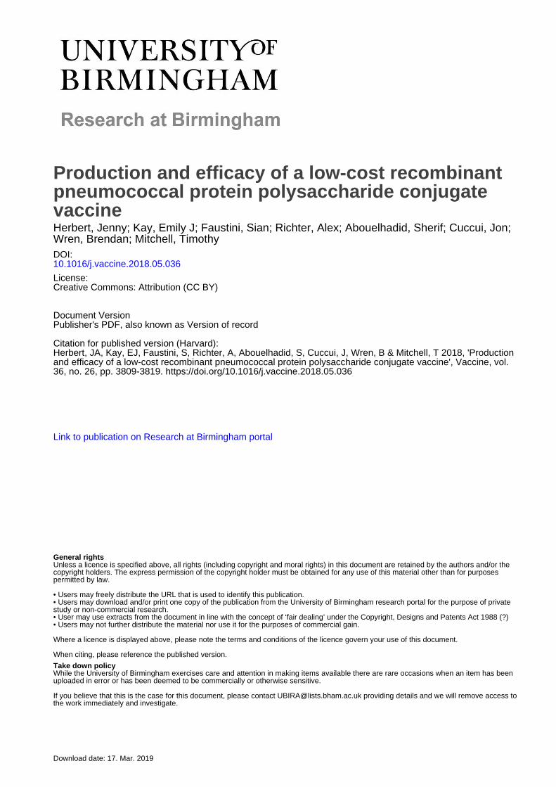
Fig. 3. Survival of vaccinated mice post intranasal challenge with TIGR4. This figureshows survival of vaccinated mice following challenge with TIGR4. Mice werefollowed for disease progression, once clinical symptoms reached a designatedendpoint mice were sacrificed. Experimental endpoint was 7 days post challenge(168 h) and surviving mice were sacrificed at this time point. n = 5 mice/group barAcrA-SP4 P1 where n = 3 and AcrA-SP4 P2 where n = 4. Statistical analysis wasperformed in GraphPad Prism using a Log-rank Test (P < 0.05).
3814 J.A. Herbert et al. / Vaccine 36 (2018) 3809–3819
native C. jejuni heptasaccharide (AcrA-Pgl) or sham vaccinated).Four weeks post the last vaccination mice were challenged viathe intranasal route with a serotype 4 S. pneumoniae strain (TIGR4).Disease progression was assessed in all vaccinated groups.
Initial experiments used three mice to test the first vaccinepreparation, referred to as AcrA-SP4 P1. Small groups of mice wereused due to amounts of conjugate required for the vaccinationschedule. Further experiments were performed using a secondbatch of conjugate vaccine, referred to as AcrA-SP4 P2 (n = 4). Datais presented separately for the two groups due to the differingresults observed for the two preparations.
In the initial experiment, using Preparation 1 of the AcrA-SP4vaccine, three mice were vaccinated for each group. All mice inthe PHD vaccinated group survived the infection, whereas all micein the PLD group succumbed. The experimental AcrA-SP4 conju-gate vaccine conferred 100% protection. Analysis of bacterialcounts in the lungs, brain and blood showed no bacteria presentabove the limit of detection in the organs of mice vaccinated withPHD or AcrA-SP4 P1 (Fig. 2A). Due to the low numbers of mice, sta-tistical analysis was not performed.
Next we attempted to repeat these experiments with a secondbatch of AcrA-SP4 conjugate (AcrA-SP4 P2). However, in this seriesof experiments only one mouse from the group of 4 vaccinated,
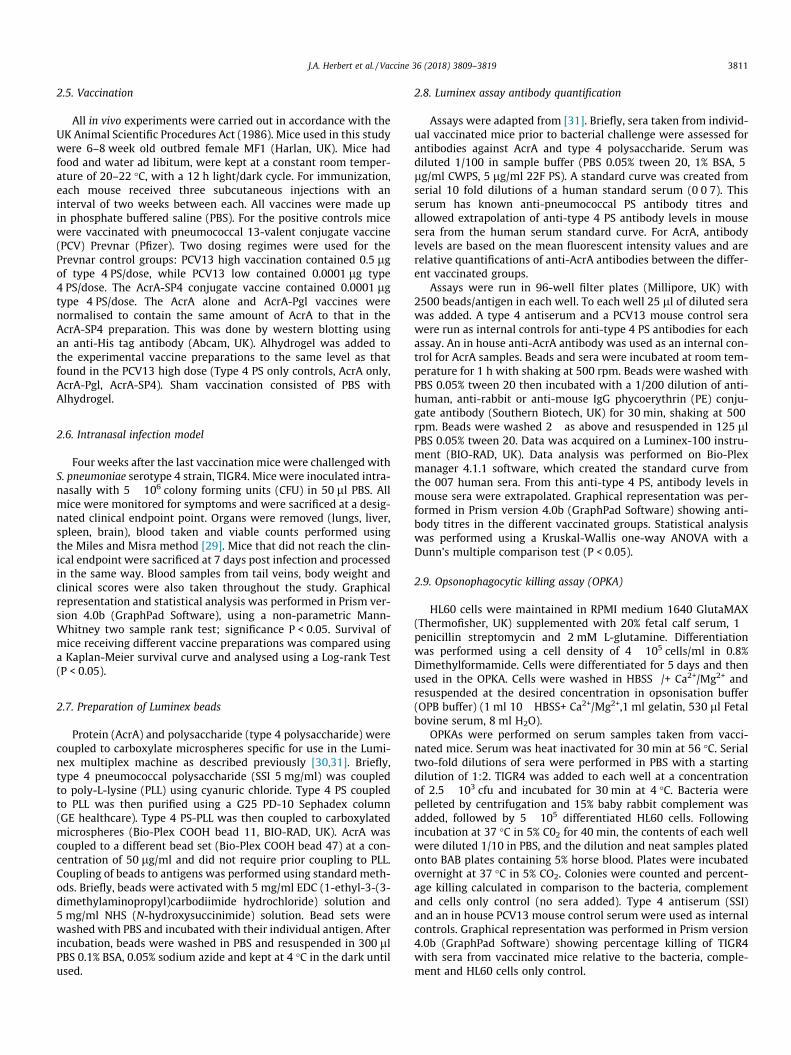
Fig. 4. Antibody responces in vaccinated mice. A shows the IgG antibody responces to ththe IgG antibody responces to type 4 polysaccharide in vaccinated mice, measured usinwith TIGR4. Each dot represent antibody levels in a single animal. Red circular symbol insurvived challenge. Statistical analysis was performed in GraphPad Prism using a Kruskal-***P < 0.001. (For interpretation of the references to colour in this figure legend, the rea
survived bacterial challenge. The surviving mouse showed no bac-terial counts in the blood at 24 h and at the time of death (Fig. 2B).Whereas, the three mice that succumbed to infection all had bac-teria in the blood at 24 h post infection, similar to that observedin the control groups. The PHD positive control group was pro-tected from infection and showed significantly lower (P < 0.05)bacterial counts in the blood compared to all control groups(except AcrA-SP4, analysis was performed on all AcrA-SP4 vacci-nated mice together (n = 7)), data not shown on graph. Mice vacci-nated with AcrA-SP4 P1 had significantly lower bacterial counts inthe blood at both time points compared to the T4PS high/low, AcrA,AcrA-Pgl and sham vaccinated control groups. This was notobserved with mice vaccinated with AcrA-SP4 P2.
Survival curves were used to compare the two preparations(Fig. 3). Mice vaccinated with AcrA-SP4 P1 showed a 100% survivalrate (n = 3), whereas mice vaccinated with AcrA-SP4 P2 only had a25% survival rate (n = 4). Despite a trend towards a difference therewas no significant difference between survival rates with mice vac-cinated with AcrA-SP4 P1 and P2 (P 0.0798). No significant differ-ence was observed between PHD and AcrA-SP4 P1 vaccinated mice.Further, all control groups (sham, T4PS high/low, AcrA and AcrA-Pgl) showed a significant reduction in survival compared to micevaccinated with AcrA-SP4 P1. No significant difference in survivalwas observed between control groups (sham, T4PS high/low, AcrAand AcrA-Pgl) and mice vaccinated with AcrA-SP4 P2.
Using Western blot analysis, the two AcrA-SP4 vaccine prepara-tions were compared using fluorescent intensity measurements(supplementary Fig. 1). As the SSI SP4 antiserum cross-reacts withAcrA all measurements were confined to the anti-his channel. Thisanalysis revealed a higher percentage glycosylation for AcrA-SP4P2 (84% Vs 80%) but a lower percentage of AcrA coupled to poly-mers of SP4 (2.8% Vs 3.6%). In addition, by normalizing to glycancontent of the preparations, the protein content per dose of theP1 was 26.8 lg vs 15.9 lg in P2 (Table 1 and supplementary Fig. 1).
In an attempt to explain the variation in levels of protection ofthe two vaccine preparations, we examined the antibody levels andfunctionality of the antibodies generated by the two vaccines.
3.3. Vaccine induced antibody responses
A Luminex bead based assay was used to evaluate the amountof antibody produced against type 4 PS and the carrier protein
e carrier protein AcrA in vaccinated mice, measured using a luminex assay. B showsg a Luminex assay. Antibody responces were measured in sera taken pre challengedicates the antibody levels in the mouse that was vaccinated with AcrA-SP4 P2 andWallis one way ANOVAwith a Dunn’s multiple comparison test, *P < 0.05, **P < 0.01,der is referred to the web version of this article.)

J.A. Herbert et al. / Vaccine 36 (2018) 3809–3819 3815
AcrA, in serum from vaccinated mice (Fig. 4). Sera used wereobtained from tail vein bleeds taken directly before challenge withTIGR4. Type 4 PS levels are given in lg/ml as samples were com-pared to a standard human serum with known levels of anti-type4 PS antibody (lg/ml) [36]. AcrA levels are presented as relative
Fig. 5. Functional antibody levels in vaccinated mice. (A) shows functional antibodies inline represents sera from a single mouse. Percentage killing of TIGR4 is shown at dilutioseven mice vaccinated with AcrA-SP4 P1 and AcrA-SP4 P2. Each line represents sera fromintranasal infection, and open square those that succumbed to infection. Red filled squaresecond vaccine preparation group. (C) shows functional antibodies in sera from a singldilutions of sera starting at 1:2 dilution. (For interpretation of the references to colour i
amounts of IgG between the different samples, as no serum stan-dard with known levels of AcrA antibody was available.
Anti-AcrA antibodies were detected in all serum samples takenfrom mice that were vaccinated with AcrA conjugates (AcrA-SP4/AcrA-Pgl) or AcrA alone (Fig. 4A). The AcrA-SP4 vaccinated mice
sera from mice vaccinated with AcrA-SP4 P1 (blue), PHD (red) and PLD (black). Eachns of sera starting at 1:2 dilution. (B) shows functional antibodies in sera from alla single mouse with the circular symbol representing the mice that survived TIGR4symbol indicated the functional antibody levels in the mouse that survived from thee mouse from each of the control groups. Percentage killing of TIGR4 is shown atn this figure legend, the reader is referred to the web version of this article.)

3816 J.A. Herbert et al. / Vaccine 36 (2018) 3809–3819
antibody responses have been split into those that were vaccinatedwith the first (AcrA-SP4 P1 n = 3) and second preparation (AcrA-SP4 P2 n = 4). The antibody titer from the mouse that was vacci-nated with the second preparation and survived is shown by ared circular symbol. There was no significant difference in thelevels of anti-AcrA antibodies in AcrA vaccinated groups, suggest-ing this is not the cause of the difference in survival.
Anti-type 4 PS antibody levels in vaccinated groups are shown inFig. 4B. The AcrA-SP4 vaccinated mice antibody responses havebeen split into those that were vaccinated with the first (AcrA-SP4 P1 n = 3) and second preparation (AcrA-SP4 P2 n = 4). Anti-type 4 PS antibody levels were detectable in the PHD vaccinatedmice and AcrA-SP4 vaccinatedmice. Therewas no significant differ-ence in antibody levels between the PHD group and the two AcrA-SP4 groups (analysis was performed on all AcrA-SP4 vaccinatedmice together (n = 7)). There were significantly lower levels ofanti-type 4 PS antibodies in the PLD vaccinated group comparedto PHD high (P = <0.001) and the AcrA-SP4 vaccinated group (n =7, P = <0.01), suggesting the lower PCV13 dose is not sufficient toproduce detectable levels of anti-type 4 PS antibodies in our model.When splitting the AcrA-SP4 vaccinated mice into the two prepara-tions there was no significant difference in the anti-type 4 PS levelsbetween the AcrA-SP4mice vaccinated with the first or second vac-cine preparation. This suggests that the mice that succumbed toinfection did have anti-type 4 PS antibodies, but these were not suf-ficient to clear infection. We know from previous literature [35]that unconjugated type 4 PS is not immunogenic in mice, and thisis confirmed in our model as no anti-type 4 PS antibodies weredetected in the polysaccharide only control groups.
WHO has assigned a protective IgG serotype specific antibodylevel of 0.35 lg/ml as measured by ELISA [37]. However, it hasbeen shown that this does not necessarily correlate with protec-tion; OPKA is seen to be a better measure of true protection[38,39]. There was no significant difference in the total anti-type4 PS IgG antibody levels in mice vaccinated with the first or secondAcrA-SP4 vaccine preparation. Therefore, the ability of the antibod-ies to opsonise TIGR4 was tested using an OPKA. Dilutions of serafrom single vaccinated mice were incubated with TIGR4, differen-tiated HL-60 cells, and a complement source. Percentage killing ofTIGR4, when incubated with sera from vaccinated mice, was com-pared to samples that contained no sera (bacteria, cells and com-plement only control). In our assay, all sera that resulted inbacterial killing above 50% is classed as protective. This assaywas then used to test functional antibodies in the sera from micevaccinated with AcrA-SP4.
Initial experiments were performed on sera frommice given thefirst vaccine preparation. Sera samples taken from vaccinated micewere tested individually. Samples were run alongside a standardtype 4 PS antisera (SSI) to assess any intra-assay variability (datanot shown). This assay confirmed whether antibodies producedfrom vaccination of mice were able to opsonise and kill TIGR4in vitro, which has been shown to correlate with protection [40].Data in Fig. 5A shows the percentage killing of TIGR4 from thethree vaccinated mice with AcrA-SP4 P1, PHD and PLD. All micevaccinated with PHD and AcrA-SP4 P1 showed opsonic antibodiesabove the 50% killing cut off. The three mice vaccinated with thePLD did not show this and all mice in this group succumbed toinfection.
This assay was then used to test the opsonic antibody levels inthe mice vaccinated with AcrA-SP4 P2, and compared to the AcrA-SP4 P1 data (Fig. 5B). One serum sample taken from a mouse ineach of the control groups was also tested (Fig. 5C). From our OPKAdata, we observe functional antibodies (killing above 50%) in allsera samples from mice that survived intranasal challenge withTIGR4. In the seven mice that received AcrA-SP4, functional anti-bodies were observed in the four mice that survived; this was
not the case for the three mice that succumbed to infection. Serafrom mice that were sacrificed due to clinical disease symptomsdid not reach killing above the 50% threshold. Data from the mousethat was vaccinated with the AcrA-SP4 P2 but survived challenge isshown as a red square symbol (Fig. 5B). We observed high levels ofkilling in the sera from one of the PCV13 low vaccinated mice,which survived challenge (data not shown). For the other controlgroups, percentage killing did not reach above 50%; again, this cor-related to the survival status of the mouse. There is therefore astrong correlation between the ability of the antisera to promoteopsonophagocytosis in vitro and the ability to protect from infec-tion in vivo.
4. Discussion
We report the first demonstration of the use of protein glycancoupling technology (PGCT) to produce a recombinant pneumococ-cal conjugate vaccine candidate. We have provided the first proofof principle that a recombinant pneumococcal conjugate vaccinecan be protective in a mouse model of infection, and can producefunctional opsonic antibodies. Production of these opsonic anti-capsular antibodies correlates with the ability to protect fromlethal infection in a mouse model of disease.
Glycoconjugate vaccines are used worldwide to prevent diseasein children caused by encapsulated bacteria. In the UK, conjugatevaccines are given in the routine childhood vaccination scheduleto prevent disease from three important pathogens: H. influenzatype B, N. meningitides, and S. pneumoniae. These vaccines are allcomposed of type specific polysaccharide coupled to a carrier pro-tein, normally diphtheria toxoid or tetanus toxoid. Despite largereductions in disease caused by these pathogens since conjugatevaccine introduction, these pathogens still remain the three big-gest contributors to invasive disease in children worldwide[1,41,42]. Conjugate vaccines that protect against pneumococcalinfection are known for their safety and efficacy. Producing protec-tive type specific opsonic antibodies [5]. However pneumococcalpneumonia remains one of the biggest killers, resulting in393,000 deaths of children under 5 in 2015 [43].
This is mainly due to the limited availability of these vaccines inthe developing world, due to the large cost [12]. Introduction ofconjugate vaccines into a countries vaccine schedules is extremelycostly. Many developing countries are only able to afford these vac-cines with assistance from The Global Vaccine Alliance (GAVI),which is not sustainable. If eligibility for financial help is lost thisleaves a huge financial gap to fill.
PGCT is an alternative approach to making conjugate vaccines.Producing conjugate vaccines in E. coli without the need for com-plex chemical coupling, and growth of large quantities of patho-genic bacteria. Once optimised, this production method has thepotential to significantly reduce the cost of conjugate vaccines,making them more readily available globally. This productionmethod will not help with the problem of serotype replacement,but an easier production method may allow more rapid switchingof vaccine constituents, to include compatible emerging serotypesinto the preparation.
This technology has recently been used for production of anumber of novel vaccines. These include vaccines that protectagainst Francisella tularensis, Burkholderia pseudomellia and Staphy-lococcus aureus [24–26]. These all used the CjPglB enzyme to cou-ple O-antigen (gram negative bacteria) or type specificpolysaccharide (gram positive bacteria) to a carrier protein ofchoice, in this instance to exotoxin A from Pseudomonas aeuriginosa(F. tularensis and S. aureus) or AcrA from C. jejuni (B. pseudomellia).These glycoconjugate vaccines produced using recombinant DNAapproaches all showed at least partial protection in a mouse model

J.A. Herbert et al. / Vaccine 36 (2018) 3809–3819 3817
of infection, and IgG antibody responses against the vaccine com-ponents. This methodology has also been used for production ofa glycoconjugate vaccine to prevent disease caused by Shigella flex-neri 2a [27], coupling the O-antigen to exotoxin A (P. aeuriginosa)using PglB. This conjugate vaccine composition produced throughchemical coupling has already been shown to be protective andsafe in clinical trials [44], however remains expensive to produce.
Using two separately prepared batches of vaccine, we observeda 100% and 25% survival rate of mice vaccinated with AcrA-SP4compared to 25% of mice vaccinated with the equivalent dose ofPrevnar13 (same concentration of type 4 PS – 0.0001 lg). Highersurvival rates of 100% were observed with the higher Prevnar13dose, which is equal to each mouse receiving 100 ml of the neatPCV13 vaccine per dose. Based on the PCV13 vaccine dose, andweight of a 6 week old child, we have estimated a matched dosefor a mouse would contain 0.01 mg of type 4 PS/dose. This dosageof 0.01 mg type 4 PS, when used in our infection model, was 100%protective (data not shown). However, due to small amount ofAcrA-SP4 conjugate available we were unable to match thesedoses, and each mouse received 0.0001 mg of type 4 PS per dose.
We believe the amount of AcrA-SP4 conjugate given to eachmouse was on the cusp of the amount required to protect againstchallenge, and different vaccine efficacies observed to the two vac-cine preparations is likely due to batch-to-batch variation. Optimi-sation therefore is required to ensure a uniform batch, stabilitypost purification and the ability to scale up production.
The variation between batches may be due to reduced amountsof coupled protein and polysaccharide present within the secondpreparation. Total polysaccharide (type 4 PS) was quantified inthe vaccine preparations, and this was used to normalise dosesbetween the two experiments. However, this would not tell us ifthe levels of protein and polysaccharide coupled varied betweenpreparations. To try and evaluate this the two vaccine preparationswere coupled to a Luminex assay bead set. The coupling methodused was for protein, so only the AcrA in the preparations wouldbind to the beads. To measure the amount of AcrA coupled toSP4, a secondary type 4 antisera was used (SSI) to quantify theamount of T4PS coupled to the beads, followed by a PE conjugatedantibody (data not shown). Fluorescence readings were higher forpreparation one, indicating this preparation contained higheramounts of AcrA coupled to T4PS. Type 4 polysaccharide alone isnot protective in a mouse model of infection [35]. Therefore, ifthe level of free polysaccharide was higher in the second prepara-tion this might account for the decrease in protection.
Another possibility for the reduced efficacy in the second prepa-ration could be the composition of repeat unit lengths. It has beenshown previously that for S. pneumoniae serotype 4 glycoconju-gates, a shorter chain lengths (12 repeat units), and a higher ratioof saccharide to protein is required for optimal immunogenicity,than for fully polymerized glycan [35]. Also, for serotype 14 thelength of coupled polysaccharide is important for induction of anti-bodies with high opsonophagocytic activity; higher chain lengthsconfirm higher opsonophagocytic titres [45]. However, this hasnot been shown for other serotypes tested [46].
Relative amounts of IgG against AcrA were measured in all vac-cinated mice. This carrier protein is not a native pneumococcalprotein and therefore provided no protection to mice. This wasclearly shown in the AcrA only and AcrA-Pgl vaccinated mice,which all had anti-AcrA IgG antibodies in sera, yet all mice suc-cumbed to infection. This was further validated using our OPKA,as sera from vaccinated mice, in these two groups, were unableto opsonise and kill TIGR4. A benefit of using PGCT, is that alterna-tive protein carriers, that contribute to vaccine efficacy, could beeasily incorporated into vaccine preparations. One potential candi-date to act as a carrier protein is pneumolysin (Ply), the pneumo-coccal cholesterol dependent cytolysin. This protein is highly
conserved in almost all pneumococcal strains and serotypes [47].Further, a detoxified version of ply (D6ply) has been shown toact as an adjuvant, and upon vaccination, results in antibodies pro-duced against itself and any proteins coupled to it [48,49]. Theseproperties are retained if given mucosally, and would enable thevaccine to be given intranasally [49], a less invasive route than Pre-vnar13 is currently given (intramuscularly).
Total IgG levels against T4PS in mice vaccinated with AcrA-SP4did not seem to correlate to survival in our model. When the AcrA-SP4 vaccinated groups were split into those that survived andthose that died post challenge with TIGR4, there was no significantdifference in antibody levels. For certain serotypes, total IgG levels,as measured by ELISA, have been shown not to correlate with pro-tection, and opsonic antibody activity has been shown to be a morereliable measure of protection [38,39]. This was also the case inthis study, with opsonic antibody activity directly correlating tosurvival.
In future, it may be informative to study antibody isotypes.Pneumococcal conjugate vaccines have been shown to produce apredominant IgG1 followed by IgG3 antibody response in mice[35]. Response to polysaccharide alone manifest in a predomi-nantly IgG2 and IgM response [35,50]. Increased levels of IgM orIgG2 in mice vaccinated with the second preparation would havesupported the theory of increased free polysaccharide within thispreparation.
There are some limitations to overcome before this methodol-ogy can compete with traditional chemical coupling. Currentlythe use of the CjPglB is limited to coupling polysaccharides whichhave an acetamido group at the C2 position of the reducing endsugar [22]. This only includes a small number of the pneumococcalpolysaccharides. Work is underway to overcome these limitations.CjPglB is not only found in campylobacter species, and orthologueshave also been found in a number of other epsilon proteobacteria[51]. This opens up the potential to find glycosyltransferases thathave differing or relaxed glycan specificities to that of CjPglB. Morerecently the crystal structure of PglB from Campylobacter lari hasbeen solved giving an insight into the important structural regionsrequired for PglB function [52]. With this knowledge, it is possibleto modify PglB to increase transfer efficiency and alter the glycanspecificity [53].
5. Conclusions
Here we have provided the first proof of principle that a pneu-mococcal conjugate vaccine, produced by protein glycan couplingtechnology, is protective in a mouse model of infection, and canproduce functional opsonic antibodies. A 57% survival rate of micevaccinated with the novel conjugate was seen, although there wasvariation between vaccine preparations. This is a proof of principlestudy and more work needs to be done to optimise productionmethodology, to improve polymer length, conjugation efficiencyand overall vaccine yield, in order to make a reliable and econom-ical alternative to chemical conjugation methods.
Competing interests
We have no competing interests.
Author contributions
J.A.H carried out all animal work and sample processing, Lumi-nex assays and OPKA assays. Further participated in design of thestudy and drafted the manuscript. E.K produced the vaccine prepa-rations and ran the western blots for vaccine validation. E.K partic-ipated in editing the manuscript. S.F helped with the Luminex

3818 J.A. Herbert et al. / Vaccine 36 (2018) 3809–3819
assays and analysis. S.A. and J.C. developed the genetic tools andgenerated the chromosomally inserted PglB strain. T.M and B.Wconceived of the study and revised the manuscript.
Funding
This work was supported by a Medical Research Council (MRC)Grant MR/K012053/1 to BW and TJM.
Acknowledgements
Wewould like to acknowledge the Medical Research Council forfunding.
Appendix A. Supplementary material
Supplementary data associated with this article can be found, inthe online version, at https://doi.org/10.1016/j.vaccine.2018.05.036.
References
[1] O’Brien KL et al. Burden of disease caused by Streptococcus pneumoniae inchildren younger than 5 years: global estimates. The Lancet2009;374:893–902.
[2] Pilishvili T et al. Sustained reductions in invasive pneumococcal disease in theera of conjugate vaccine. J Infect Dis 2010;31–41. https://doi.org/10.1086/648593.
[3] Hammitt LL et al. Population effect of 10-valent pneumococcal conjugatevaccine on nasopharyngeal carriage of Streptococcus pneumoniae and non-typeable Haemophilus influenzae in Kilifi, Kenya: findings from cross-sectionalcarriage studies. Lancet Glob Health 2014;2:e397–405. https://doi.org/10.1016/s2214-109x(14)70224-4.
[4] Waight PA et al. Effect of the 13-valent pneumococcal conjugate vaccine oninvasive pneumococcal disease in England and Wales 4 years after itsintroduction: an observational cohort study. Lancet Infect Dis2015;15:535–43. https://doi.org/10.1016/s1473-3099(15)70044-7.
[5] Andrews NJ et al. Serotype-specific effectiveness and correlates of protectionfor the 13-valent pneumococcal conjugate vaccine: a postlicensure indirectcohort study. Lancet Infect Dis 2014;14:839–46. https://doi.org/10.1016/s1473-3099(14)70822-9.
[6] Kehrl JH, Fauci AS. Activation of human B lymphocytes after immunizationwith pneumococcal polysaccharides. J Clin Invest 1983;71:1032–40.
[7] Avery OT, Goebel WF. Chemo-Immunological studies on conjugatedcarbohydrate-proteins: II. Immunologoical specificity of synthetic sugar-protein antigens. J Exp Med 1929;50:533–50.
[8] Beuvery EC, Van Rossum F, Nagel J. Comparison of the induction ofimmunoglobulin M and G antibodies in mice with purified pneumococcaltype 3 and meningococcal group C polysaccharides and their proteinconjugates. Infect Immun 1982;37:15–22.
[9] Schneerson R, Barrera O, Sutton A, Robbins JB. Perparation, characterisationand immunogenicity of Haemophilus influenzae type b polysaccharide-proteinconjugates. J Exp Med 1980;152:361–76.
[10] MacDonald NE et al. Induction of immunologic memory by conjugated vs plainmeningococcal C polysaccharide vaccine in toddlers. JAMA 1998;280:1685–9.
[11] Rennels MB et al. Safety and immunogenicity of heptavalent pneumococcalvaccine conjugated to CRM197 in United States Infants. Paediatrics1998;101:604–11.
[12] Gulland A. Charity urges drug firms to reduce price of pneumonia vaccine. BMJ2015;351:h6131. https://doi.org/10.1136/bmj.h6131.
[13] Johnson HL et al. Systematic evaluation of serotypes causing invasivepneumococcal disease among children under five: the pneumococcal globalserotype project. PLoS Med 2010;7. https://doi.org/10.1371/journal.pmed.1000348.
[14] Miller E, Andrews NJ, Waight PA, Slack MPE, George RC. Herd immunity andserotype replacement 4 years after seven-valent pneumococcal conjugatevaccination in England andWales: an observational cohort study. Lancet InfectDis 2011;11:760–8. https://doi.org/10.1016/s1473-3099(11)70090-1.
[15] Hicks LA et al. Incidence of pneumococcal disease due to non-pneumococcalconjugate vaccine (PCV7) serotypes in the United States during the era ofwidespread PCV7 vaccination, 1998–2004. J Infect Dis 2007;196:1346–54.https://doi.org/10.1086/521626.
[16] Skinner JM et al. Pre-clinical evaluation of a 15-valent pneumococcalconjugate vaccine (PCV15-CRM197) in an infant-rhesus monkeyimmunogenicity model. Vaccine 2011;29:8870–6. https://doi.org/10.1016/j.vaccine.2011.09.078.
[17] Frasch CE. Preparation of bacterial polysaccharide-protein conjugates:analytical and manufacturing challenges. Vaccine 2009;27:6468–70. https://doi.org/10.1016/j.vaccine.2009.06.013.
[18] Cuccui J, Wren B. Hijacking bacterial glycosylation for the production ofglycoconjugates, from vaccines to humanised glycoproteins. J PharmPharmacol 2015;67:338–50. https://doi.org/10.1111/jphp.12321.
[19] Wacker M et al. N-linked glycosylation in Campylobacter jejuni and itsfunctional transfer into E. coli. Science (New York, N.Y.) 2002;298:1790–3.https://doi.org/10.1126/science.298.5599.1790.
[20] Scott NE et al., Simultaneous glycan-peptide characterization usinghydrophilic interaction chromatography and parallel fragmentation by CID,higher energy collisional dissociation, and electron transfer dissociation MSapplied to the N-linked glycoproteome of Campylobacter jejuni. Mol CellProteomics 10; 2011: M000031–MCP000201. http://doi.org/10.1074/mcp.M000031-MCP201.
[21] Kowarik M et al. Definition of the bacterial N-glycosylation site consensussequence. EMBO J 2006;25:1957–66. https://doi.org/10.1038/.
[22] Wacker M et al. Substrate specificity of bacterial oligosaccharyltransferasesuggests a common transfer mechanism for the bacterial and eukaryoticsystems. Proc Natl Acad Sci U S A 2006;103:7088–93.
[23] Feldman MF et al. Engineering N-linked protein glycosylation with diverse Oantigen lipopolysaccharide structures in Escherichia coli. Proc Natl Acad Sci U SA 2005;102:3016–21.
[24] Wacker M et al. Prevention of Staphylococcus aureus infections byglycoprotein vaccines synthesized in Escherichia coli. J Infect Dis2014;209:1551–61. https://doi.org/10.1093/infdis/jit800.
[25] Cuccui J et al. Exploitation of bacterial N-linked glycosylation to develop anovel recombinant glycoconjugate vaccine against Francisella tularensis. OpenBiol 2013;3:130002. https://doi.org/10.1098/rsob.130002.
[26] Garcia-Quintanilla F, Iwashkiw JA, Price NL, Stratilo C, Feldman MF. Productionof a recombinant vaccine candidate against Burkholderia pseudomalleiexploiting the bacterial N-glycosylation machinery. Front Microbiol2014;5:381. https://doi.org/10.3389/fmicb.2014.00381.
[27] Kampf MM et al. In vivo production of a novel glycoconjugate vaccine againstShigella flexneri 2a in recombinant Escherichia coli: identification of stimulatingfactors for in vivo glycosylation. Microb Cell Fact 2015;14:12. https://doi.org/10.1186/s12934-015-0195-7.
[28] Kay EJ, Yates LE, Terra VS, Cuccui J, Wren BW. Recombinant expression ofStreptococcus pneumoniae capsular polysaccharides in Escherichia coli. OpenBiol 2016;6:150243. https://doi.org/10.1098/rsob.150243.
[29] Miles AA, Misra SS. The estimation of the bactericidal power of the blood. J Hyg(Lond) 1938;38:732–49.
[30] Lal G et al. Development and validation of a nonaplex assay for thesimultaneous quantitation of antibodies to nine Streptococcus pneumoniaeserotypes. J Immunol Methods 2005;296:135–47. https://doi.org/10.1016/j.jim.2004.11.006.
[31] Whitelegg AM et al. Measurement of antibodies to pneumococcal,meningococcal and haemophilus polysaccharides, and tetanus anddiphtheria toxoids using a 19-plexed assay. J Immunol Methods2012;377:37–46. https://doi.org/10.1016/j.jim.2012.01.007.
[32] Young NM et al. Structure of the N-linked glycan present on multipleglycoproteins in the Gram-negative bacterium, Campylobacter jejuni. J BiolChem 2002;277:42530–9. https://doi.org/10.1074/jbc.M206114200.
[33] Pumbwe L, Piddock LJV. Identifcation and molecular characterisation ofCmeB, a Campylobacter jejuni multidrug efflux pump. FEMS Microbiol Lett2002;185–189.
[34] Iwashkiw JA et al. Exploiting the Campylobacter jejuni protein glycosylationsystem for glycoengineering vaccines and diagnostic tools directed againstbrucellosis. Microb Cell Fact 2012;11.
[35] Peeters CCAM et al. A comparative study of the immunogenicity ofpneumococcal type 4 polysaccharide and oligosaccharide tetanus toxoidconjugates in adult mice. J Immunol 1991;146:4308–14.
[36] Goldblatt D et al. Establishment of a new human pneumococcal standardreference serum, 007sp. Clin Vaccine Immunol 2011;18:1728–36. https://doi.org/10.1128/CVI.05252-11.
[37] World Health Organisation. Recomendations for the production and controlof pneumococcal conjugate vaccines. WHO Tech Rep Ser 2005;Annex2:64–98.
[38] Henckaerts I, Durant N, De Grave D, Schuerman L, Poolman J. Validation of aroutine opsonophagocytosis assay to predict invasive pneumococcal diseaseefficacy of conjugate vaccine in children. Vaccine 2007;25:2518–27. https://doi.org/10.1016/j.vaccine.2006.09.029.
[39] Oishi T et al. Low opsonic activity to the infecting serotype in pediatricpatients with invasive pneumococcal disease. Vaccine 2013;31:845–9. https://doi.org/10.1016/j.vaccine.2012.11.010.
[40] AlonsoDeVelasco E et al. Anti-polysaccharide immunoglobulin isotype levelsand opsonic activity of antisera: relationships with protection againstStreptococcus pneumoniae infection in mice. J Infect Dis 1995;172:562–5.
[41] Watt JP et al. Burden of disease caused by Haemophilus influenzae type b inchildren younger than 5 years: global estimates. The Lancet 2009;374:903–11.
[42] Borrow R et al. The Global Meningococcal Initiative: global epidemiology, theimpact of vaccines on meningococcal disease and the importance of herdprotection. Expert Rev Vaccines 2016;1–16. https://doi.org/10.1080/14760584.2017.1258308.
[43] Murray CJL, Lopez AD, Naghavi M, Wang H. Global, regional, and national lifeexpectancy, all-cause mortality, and cause-specific mortality for 249 causes ofdeath, 1980–2015: a systematic analysis for the Global Burden of DiseaseStudy. The Lancet 2016;2015(388):1459–544. https://doi.org/10.1016/S0140-6736(16)31012-1.

J.A. Herbert et al. / Vaccine 36 (2018) 3809–3819 3819
[44] Cohen D et al. Safety and immunogenicity of investigational shigella conjugatevaccines in israeli volunteers. Infect Immun 1996;64:4074–7.
[45] Laferriere CA, Sood RK, de Muys JM, Michon F, Jennings HJ. Streptococcuspneumoniae type 14 polysaccharide-conjugate vaccines: length stabilization ofopsonophagocytic conformational polysaccharide epitopes. Infect Immun1998;66:2441–6.
[46] Laferriere CA, Sood RK, de Muys JM, Michon F, Jennings HJ. The synthesis ofStreptococcus pneumoniae polysaccharide-tetanus toxoid conjugates and theeffect of chain length on immunogenicity. Vaccine 1997;15:179–86.
[47] Kirkham LA et al. Identification of invasive serotype 1 pneumococcal isolatesthat express nonhemolytic pneumolysin. J Clin Microbiol 2006;44:151–9.https://doi.org/10.1128/JCM.44.1.151-159.2006.
[48] Kirkham LA et al. Construction and immunological characterization of a novelnontoxic protective pneumolysin mutant for use in future pneumococcalvaccines. Infect Immun 2006;74:586–93. https://doi.org/10.1128/IAI.74.1.586-593.2006.
[49] Douce G, Ross K, Cowan G, Ma J, Mitchell TJ. Novel mucosal vaccines generatedby genetic conjugation of heterologous proteins to pneumolysin (PLY) fromStreptococcus pneumoniae. Vaccine 2010;28:3231–7. https://doi.org/10.1016/j.vaccine.2010.02.014.
[50] Barrett DJ, Ayoub EM. IgG2 subclass restriction of antibody to pneumococcalpolysaccharides. Clin Exp Immunol 1986;63:127–34.
[51] Ielmini MV, Feldman MF. Desulfovibrio desulfuricans PglB homolog possessesoligosaccharyltransferase activity with relaxed glycan specificity and distinctprotein acceptor sequence requirements. Glycobiology 2011;21:734–42.https://doi.org/10.1093/glycob/cwq192.
[52] Ihssen J et al. Structural insights from random mutagenesis of Campylobacterjejuni oligosaccharyltransferase PglB. BMC Biotech 2012;12.
[53] Ihssen J et al. Increased efficiency of Campylobacter jejuni N-oligosaccharyltransferase PglB by structure-guided engineering. Open Biol2015;5:140227. https://doi.org/10.1098/rsob.140227.