productive t-cell receptor [}-chain gene rearrangement
TRANSCRIPT

Productive T-cell receptor [}-chain gene rearrangement: coincident regulation of cell cycle and clonality during development in vivo Eric S. Hoffman, 1'2's Lorena Passoni, 1,8 Tessa Crompton, 4 Thomas M.J. Leu, 5 David G. Schatz, 2'3 Andrew Koff, 6 Michael J. Owen, 4 and Adrian C. Hayday 1'2'7
~Department of Biology and ZSection of Immunobiology and ~Howard Hughes Medical Institute, Yale University, New Haven, Connecticut 06510 USA; 4Imperial Cancer Research Fund Laboratories, London WC2 3PX, UK; ~Universit~it Zfirich-Irchel, Veterin~rbiochemie, 8057 Zfirich, Switzerland; 6program in Molecular Biology, Memorial Sloan-Kettering Cancer Center, New York, New York 10021 USA
Productive gene rearrangement at the T-cell receptor (TCR) [}-chain locus facilitates formation of the "pre-TCR," a molecular complex that is important for the subsequent development of ~ T cells. The transition of thymocytes from a population of cells undergoing TCR[} chain genes to a population enriched in cells with productively rearranged TCR~ chain genes is known as "13 selection." This is the first point in ot~ T-cell development at which the products of an activated TCR locus define cell phenotype. Toward an understanding of these events, this study has focused on a set of thymocytes defined by cell surface phenotype as HSA + CD44 l~ CD25 +, in which the bulk of TCR~ gene rearrangement occurs. The analysis of this set, presented here, allows its novel subdivision into two subsets that are respectively strong candidates for cells immediately prior to and immediately following TCR~ selection. Cells that have passed 13 selection differ from the preceding cells by several criteria, including hyperphosphorylation of Rb, increased expression of cyclins A and B, down-regulation of p27, increased CDK2 activity, an induction of cdc2 activity, and progression through DNA synthesis. Consistent with these changes being attributable to productive TCR~ chain gene rearrangement, the identified "~-selected" subset is not detected in mutant mice that cannot assemble a pre-TCR. Interestingly, there is a coincident selective and transient down-regulation of the protein RAG2, on which TCR gene rearrangement obligatorily depends. Together, these findings demonstrate that productive TCR gene rearrangement is associated with events that can ensure thymocyte expansion and monoclonality.
[Key Words: Pre-T-cell receptor; thymocytes; RAG2; cell cycle; immune system development]
Received November 27, 1995; revised version accepted February 28, 1996.
T cells develop from progenitors that enter the thymus expressing very low levels of CD4, which they soon lose, to become CD4-CD8- "double-negative" (DN) cells (Wu et al. 1991) that comprise 1%-2% of thymocytes. The DN cells proceed through a series of differentiation stages (defined by distinct cell surface phenotypes}, until they acquire CD4 and CD8, becoming "double-positive" (DP) cells, essentially all of which have successfully re- arranged genes at the T-cell receptor [3 (TCR[3) chain lo- cus (Mallick et al. 1993}. Contingent upon successful rearrangement of a T-cell receptor o~ (TCRoL) chain gene, DP cells lose either CD4 or CD8, becoming mature "sin-
7Corresponding author. Present address: Kline Biology Tower, 266 Whitney Ave., New Haven, Connecticut 06510 USA. SThe first two authors contributed equally to this study.
gle-positive" (SP} cells that exit to the periphery (Scollay and Shortman 1985; Petrie et al. 1990; Rothenberg et al. 1992).
The productive rearrangement of a TCR[3 chain gene by DN thymocytes is a critical mechanistic stage in T-cell development (Mombaerts et al. 1992b; Mallick et al. 1993; Shinkai et al. 1993; Dudley et al. 1994; Godfrey et al. 1994). The product of the TCR[3 chain gene is com- plexed with a 33-kD surrogate TCRa chain gene (Groet- trup et al. 1993), named pre-To~, to form a "pre-TCR" (Saint-Ruf et al. 1994). Cells that do not express the pre- TCR are, to a first approximation, blocked in their ca- pacity either to proliferate or to differentiate further (Fehling et al. 1995), whereas cells that productively re- arrange the TCR[3 chain genes progress to the DP stage. This is termed "[3 selection." A highly analogous check- point exists in B-cell development, regulated by a pre-B-
948 GENES & DEVELOPMENT 10:948-962 �9 1996 by Cold Spring Harbor Laboratory Press ISSN 0890-9369/96 $5.00
Cold Spring Harbor Laboratory Press on February 26, 2022 - Published by genesdev.cshlp.orgDownloaded from

Thymocyte development, the pre-TCR, and the cell cycle
cell receptor comprising the product of a successful im- munoglobul in heavy-chain gene rearrangement, com- bined wi th a surrogate light-chain, h5/VpreB (Kitamura et al. 1992).
Although the formation and expression of pre-antigen receptors is thus a critical event in lymphocyte develop- ment, events associated with preantigen receptor expres- sion are currently poorly understood. Prior to [3 selec- tion, cells are in a stage defined by cell surface markers as HSA hi C D 4 - C D 8 - CD44 l~ CD25 § (or, for the pur- poses of this paper, CD25 +, for short). 13 Selection facil- itates transit ion to a stage defined as HSA hi CD4 - CD8 - CD44 l~ CD25- (or CD25- , for short). Thus, normal numbers of CD25 - cells do not form in TCR13 - / - mice {Mombaerts et al. 1992b) , p r e T a - / - mice (Fehling et al. 1995), or recombinase activating gene (RAG)I or R A G 2 - / - mice (Mombaerts et al. 1992a; Shinkai et al. 1992) (both RAG proteins being essential for TCR gene rearrangement and hence for expression of the pre-TCR). Most CD25 + cells have been reported to have 2N DNA content, typical of resting cells or cycling cells in G1. A much higher proportion of CD25 cells has been re- ported to be in S phase (Pearse et al. 1989). Thus, it has been hypothesized that 13 selection immedia te ly pro- motes a cell cycle transition, preferentially expanding the pool of precursors deemed useful by virtue of suc- cessful TCR13 chain gene rearrangement (Dudley et al. 1994).
At the same time, a central tenet of immunology is lymphocyte clonality. Over 90% of peripheral T cells express only a single, productively rearranged ~3 chain gene, as a result of "allelic exclusion," the mechan i sm by which expression of an immunoglobul in or TCR chain (in this case, a TCR~ chain) inhibi ts further rear- rangement at the second allele of the corresponding lo-
cus (Mallisen et al. 1992; Robey and Fowlkes 1994). In- terestingly, the capacity of productive TCR13 chain gene to inhibi t further rearrangement may, in part, be related to its above-mentioned putative capacity to promote cell cycle transition. This is because RAG2 protein is ren- dered 20-fold less stable in vitro by the action of cdc2 (Lin and Desiderio 1993, 1995), a kinase associated wi th progression of m a m m a l i a n cells into G2/M.
To investigate the plausibil i ty of this scenario in vivo, we have examined thymocytes actively engaged in TCR13 chain gene rearrangement. The studies have facil- itated redefinition of the stage in thymocyte develop- ment at which ~3 selection occurs, indicating that it is tightly associated wi th the movement of cells from a G~ state to an active cycling state, in which the RAG2 pro- tein is selectively and transiently down-regulated.
Resu l t s
Thymocyte subset purification
Developing thymocytes on either side of the pre-TCR- mediated transit ion were purified by fluorescence-acti- vated cell sorting (FACS) based on surface marker expres- sion (Fig. 1). A typical experiment began wi th 10 C57.BL/ 6J female mice at 3-4 weeks of age. From these, 3x 10⁹ thymocytes were harvested. After complement deple- tion of CD4 + and CD8 + cells, 5 x 10⁷ thymocytes (1.7%) were recovered that were C D 8 - and largely C D 4 - (data not shown). Subsequent sorting e l iminated remaining CD4 dull cells and facilitated recovery of 3x 10⁶ C D 4 - HSA + CD44 l~ CD25 + {CD25 +) cells (0.1%1 and 2.5x10⁶ C D 4 - HSA + CD44 l~ CD25- (CD25-} cells (0.08%) per sort {Wilson et al. 1988; Dudley et al. 1994). Purity was consistently >98%. We have shown previ-
Figure 1. Purification of thymocyte subsets by FACS sorting. Normal mouse thymo- cytes were complement-depleted of CD4 § and CD8 + cells and stained with a panel of fluorescent antibodies. (Top) Presorted cells were separated based on HSA § CD44 l~ CD25 § or CD25- expression; (middle) CD25 + cells after sorting; (bottom) CD25- cells after sorting were reanalyzed for sur- face marker expression. Thymocytes were also sorted only for CD4- cells (see text; data not shown).
GENES & DEVELOPMENT 949
Cold Spring Harbor Laboratory Press on February 26, 2022 - Published by genesdev.cshlp.orgDownloaded from

Hoffman et al.
ously that CD25 + cells purified in this way are largely in the process of rearranging their TCR[3 chain genes, whereas CD25- cells purified in this way have essen- tially all passed [3 selection {Dudley et al. 1994). As a first test of the cell cycle status of the two subsets, their re- spective DNA contents were examined. In this, as in all assays described below, comparison was made between CD25 + and CD25- cells harvested at the same t ime as each other.
DNA content analysis of CD25 + and CD25- ceils
It was reported previously by several investigators that CD25 + cells are largely resting cells, most ly with 2N DNA content, whereas many CD25- cells are prolifer- ating, with > 2 N DNA (Pearse et al. 1989; Penit et al. 1995). This was confirmed using propidium iodide stain- ing of DNA and FACS analysis, with a doublet discrim- ination function to ensure that only single cells were analyzed. CD25 + cells typically included - 8 % - 1 0 % cells in S/G2/M (Fig. 2A), whereas CD25 - cells typically demonstrated - 2 4 % cells in S/G2/M (Fig. 2B). To test whether cells prior to [3 selection were blocked in the cell cycle, analysis was made of CD25 + cells from TCRf~xS-deficient mice, unable to express the products of in-frame TCR[3 rearrangements. These cells typically displayed only a background level of cells in S/G2/M (Fig. 2C). Taken together, the data suggest that most, if not all, CD25 + cells from normal mice that are in S/G2/M were induced to leave Gz because of productive TCR gene rearrangement. This, in turn, suggests that the pre-TCR signal occurs wi th in the CD25 + subset, induc- ing a cell cycle progression before CD25 surface expres- sion is lost. To test this, the CD25 + cells were separated into two distinct subsets for further analysis.
DNA content analysis of subsets of CD25 + cells
Progression through G 1 and into S/G2/M phase often correlates wi th an increase in cell size. By confocal mi- croscopy (see below), most CD25 + cells were sized be- tween 6.5 and 8.5 p.m, an "expected" result based on previous studies (Godfrey et al. 1994; Penit et al. 1995), whereas between 10% and 15% of cells were >8.5 ~,m, commonly - 10.5 l*m {Table 1). In contrast, CD25 + cells from TCR[3xS-deficient mice were homogeneous, with <1% of cells >8.5 ~m (Table 1). The larger 15% of CD25 + cells from normal mice could be separated from other CD25 + cells by forward and side scatter (Fig. 2D), whereas, predicatably, the larger subset was essentially
absent in CD25 + cells from either TCR[3xS-deficient mice (Fig. 2E) or RAG2-deficient mice (Fig. 2F). The cells of expected size, hereafter termed subset E, showed only background levels of cells in S/G2/M (Fig. 2G), indistin- guishable from CD25 § cells from TCR[3xS-deficient mice (Fig. 2C). Conversely, the larger CD25 § cells, here- after termed the "L subset," featured - 6 6 % of cells in S/G2/M (Fig. 2H). This is consistent wi th 8%-10% of bulk CD25 § cells being in S/G2/M (66% of 15%). Thus, subset L describes CD25 + cells that are largely in S/G2/M and that are essentially absent from mutan t mice that for a variety of congenital faults cannot assem- ble a TCR[3{ +) pre-TCR. The prediction would therefore be that L cells should show evidence of [3 selection.
PCR-RFLP analysis of TCR[3 chain joins
To test that the CD25 + L subset had passed through [3 selection, a polymerase chain reaction-restr ict ion frag- ment length polymorphism (PCR-RFLP) analysis of TCR[3 chain gene rearrangements was undertaken as de- scribed previously (Mallick et al. 1993; Dudley et al. 1994). By revealing the status of TCR gene rearrange- ments in a polyclonal population of cells, PCR-RFLP allows rapid elucidation of whether or not cells in a pop- ulation have apparently been selected for on the basis of in-flame and, therefore, productive TCR gene rearrange- ments.
TCR V[34-1132.2 and V[35-1132.2 rearrangements were examined from CD25 + subsets E and L (Fig. 3). V(D)I joining fragments from CD25 + E cells {the majori ty sub- set} showed no enr ichment for productive rearrange- ments [that would have been evident in a repeating pat- tern of bands of periodicity 3 nucleotides [Mallick et al. 1993; Dudley et al. 1994)]. Instead, the sequence lengths of the bands were close to random, wi th some depletion of in-frame rearrangements {E lanes), consistent wi th the analysis of CD25 + cells as a whole {Dudley et al. 1994). In contrast, joins from CD25 + L cells showed a predom- inant ly in-frame triplet periodicity (L lanes), indicating that [3 selection had occurred prior to formation of the CD25 + L cell subset.
This improved definition of the point of [3 selection is illustrated in Figure 4. Although it might be argued that no formal demonstrat ion has been made of a precursor- product relationship between E and L, this is the most likely explanation given that significant TCR[3 chain gene rearrangement does not appear to commence unt i l the CD25 + stage {Godfrey et al. 1994} and that in mice of various genotypes that cannot successfully express the
Figure 2. DNA content analysis of thymocyte subsets by propidium iodide staining. CD25 § cells from normal mice {A), CD25- cells from normal mice (B), and CD25 + cells from TCR[3 x g-deficient mice {C) were stained for DNA content with propidium iodide and analyzed by FACS. Percentages of cells in Go/G~ and S/G2/M are indicated. Doublet discrimination ensured that only single cells were counted. (D) CD25 + cells from normal mice were FACS-sorted by cell size (sorting gates: E = the bulk of the cells of expected size; L = larger cells) based on forward and side scatter. Percentages of cells in the E and L gates are indicated. CD25 + cells from TCR[3 x 8- deficient mice [E) and RAG2-deficient mice (F) were shown to have minimal numbers of L cells based on the same forward and side scatter settings. After sorting, CD25 § E cells {G) and CD25 + L cells (HI from normal mice were analyzed for DNA content by propidium iodide staining.
950 GENES & DEVELOPMENT
Cold Spring Harbor Laboratory Press on February 26, 2022 - Published by genesdev.cshlp.orgDownloaded from

_ . J ,
t
!
r,D
J z
Z
" l - �9 - !
" v
0 . - - I
. . . . ! . . . . ! . . . . ' | . . . . i . . . .
000 I. OOg oog OOt, OOE 0 OSS
C3
. . . . i . . . . i . . . . i . . . . I . . -
ooo L o08 oo9 oo~ oo~ DSS
I L l
! �9 ! ! . . . . T . . . . ! . . . . i . . . . ~ . . . .
O00L O~ 009 ~ 00~ 0
OSS
LJ.
~0
r,,,,3
=l o ,, , ,q
o
,0o
L)_~I, 09~ OL~ O9 t U6 O S lU ,O~ )
Z ,,r
z (',,J
<r,
USC~ Olaf, OUt; 0~ U t t U ~;ILIIIO~)
.~ z o J C M
Igl
o
N
0
GENES & DEVELOPMENT 951
Cold Spring Harbor Laboratory Press on February 26, 2022 - Published by genesdev.cshlp.orgDownloaded from

Hoffman et al.
Tab le 1. CDa4~~ + thymocyte size distribution from normal and TCR-deficient mice measured by confocal microscopy
CD441~ ~- thymocyte size
<~8.5 ~.m >8.5 ,.m Mouse strain (expected} ilargerl
C57.BL/6 {normal) 85.6% 14.4% TCR deficient {[3 and 8) 99.0% 0.97%
pre-TCR, thymocyte development is largely arrested at the CD25 § stage (Mombaerts et al. 1992a; Shinkai et al. 1992; Fehling et al. 1995). Therefore, CD25 § E and CD25 + L subsets are good candidates for cells either side of TCR[3 selection, and their characterization is thus an important step toward understanding events that follow productive TCR[3 chain gene rearrangement.
Con focal microscopy analysis of thyrnocytes for cell cycle regula tors
The progression of mammalian cells through the cell cycle is regulated in large part by the expression of cy- clins, which activate cyclin-dependent kinases {CDKs}. In asynchronously growing cultures of cells, CDK levels remain relatively constant throughout the cell cycle, whereas the levels of most cyclins vary dramatically. Expression of D-type cyclins {e.g., D2, D3), which acti- vate CDK6, generally indicates that cells are in the mi- totic cycle rather than in Go; high expression of cyclin E, which activates CDK2, indicates that cells are progress- ing through mid-late G~; high expression of cyclin A, which also activates CDK2, indicates that cells have pro- gressed into and beyond the late G1; and high expression of cyclin B indicates that cells have progressed into and past the middle of S phase (Koff et al. 1992; Ohtsubo and Roberts 1993). Superimposed upon periodic cyclin-de- pendent activation of CDKs is the capacity of other pro- teins, CDK inhibitors (CDIs), to inhibit CDK activity despite the presence of the requisite cyclin. Thus, CDI p27 can inhibit CDK2-cyclin A/E and, thereby, the pro- gression of cells into S phase (Polyak et al. 1994a,b; Sherr and Roberts 1995).
CD25 + cells were analyzed for the expression of cell cycle-associated proteins by immunofluorescent confo- cal microscopy. In each case, a cell cycle protein anti- body was followed by a Lissamine rhodamine-conjugated secondary antibody, visualized as red staining. CD25 § cells retain the fluorescein isothiocyanate (FITC)-conju- gated anti-CD25 antibody staining used in the sorting, which appears as a green ring around the cells' perime- ters. Compared with the negative control (Fig. 5A), in which cells were treated with the Lissamine rhodamine- conjugated secondary antibody without prior reaction with an antibody against a cell cycle protein, positive (red) staining for cyclins D2 and D3 was detected in all CD25 + cells (Fig. 5B, C). Superposition of cyclin and CD25 staining sometimes resulted in yellow staining, an
example of which is shown in Figure 5B. Cyclin D2 was reproducibly the weaker of the two D cyclins expressed. {Cyclin D I was not detected in CD25 § cells, as is the case in other studies of lymphocytes.} Compared with the negative control of a field of cells (Fig. 5D), CD25 + cells were homogeneously positive for cyclin E (Fig. 5E). In contrast, a heterogeneity in CD25 § cells was apparent for cyclin A: Whereas most cells were weakly positive for cyclin A (Fig. 5F), a few CD25 + cells were strongly stained le.g., arrow in Fig. 5F). Conversely, a few CD25 + cells stained less strongly than most for p27, an inhibitor of GI CDKs (Fig. 5G, arrow). Likewise, whereas most CD25 + cells expressed very little cyclin B, mostly in a punctate pattern underlying the membrane, -10% of CD25 + cells expressed higher levels of cyclin B (data n o t
shown}. Thus, as for DNA content (Fig. 2) and for TCR[3 chain genc rearrangement (Fig. 3}, CD25 § cells fell into t w o subsets according to the expression of cell cycle- associated proteins.
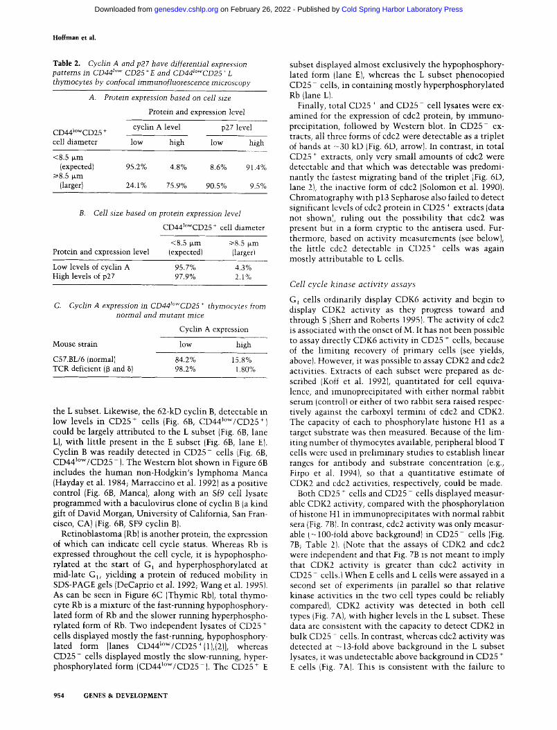
To determine whether cell size was also a good corre- late of differential cyclin-A and p27 expression, cells of diameter <8.5 gm (the E subset) and cells of diameter <~8.5 gna {the L subset) were assayed independently for p27 and cyclin-A staining. This analysis showed that in the E subset >91% of cells were strongly positive for p27 and >95% were only weakly positive for cyclin A (Table 2A}. In contrast, in the L subset, >90% of cells were only weakly positive for p27, whereas >75% were strongly positive for cyclin A. Likewise, when the reciprocal as- say was performed, first assessing cyclin-A and p27 ex- pression and then independently measuring cell size, -95% of cells only weakly positive for cyclin A and -97% of cells strongly positive for p27 were <8.5 p.m (E subsetl ITable 2B1. Together, these data indicate that sub- division of CD25 § cells into E and L subsets according to size operationally defines two cell phenotypes: One sub-
Figure 3. V6 to l~ PCR-RFLP. Analysis of TCR V[3 to I6 joins from CD25- E and CD25 ~ L cells by PCR-RFLP. (Lane 1, E) V64 to I[32.2 joins from CD25- E cells; (lane 2, L} V[34 to 1132.2 joins from CD25" L cells, amplified by PCR and size-deter- mined by comparison to a sequencing gel of known sizes. (Lane 7, E} V~5 to I[32.2 joins from CD25" E cells; (lane 8, L} V[35 to I[32.2 joins from CD25" L cells were likewise analyzed. Aster- isks denote sizes equivalent to in-frame gene rearrangements.
952 GENES & DEVELOPMENT
Cold Spring Harbor Laboratory Press on February 26, 2022 - Published by genesdev.cshlp.orgDownloaded from

Thymocyte development, the pre-TCR, and the cell cycle
Earl ier I Precursors
CD41ow CD44+ CD44+ CD441ow CD2 5- CD25+ CD25+
Small
Double CD4-, CD8-, HSA+ Cells ] Posit ive
I
Cells
Expected Phenotype
CD44~ow CD44~ow CD4+ CD25+ CD2 5- CD8+ Large
Larger and Selected
Pre-TCR Signal
Figure 4. A revised model of postnatal thymoctyte developmental sequence. 13 Se- lection occurs within the CD4- CD8- HSA* CD441~ CD25 * thymocyte popula- tion, signaling the transition to the larger L cells.
set has 2N DNA, expresses high levels of cyclin E and p27, expresses low levels of cyclin A, and displays essen- tially random rearrangements of TCR[3 chain genes; the other subset commonly contains >2N DNA, expresses high levels of cyclin E and cyclin A, expresses low levels of p27, and displays predominant ly in-frame TCR[3 chain gene rearrangements. Consistent with this, only 2 of 110 CD25 + cells from TCR[3x~-deficient (and thus, pre- TCR-deficient) mice were clearly positive for cyclin A (Table 2C). That is, the CD25 § E subset of normal mice appears s imilar to all CD25 + cells in TCR[3 x ~-deficient mice.
Cell cycle protein analysis by Western blot
To confirm and extend the staining data, the expression of several cell cycle proteins was examined by Western blotting of lysates of total CD25 + cells, CD25 + E cells, CD25 § L cells, and CD25- cells. Consistent wi th the confocal data, 60-kD cyclin A was barely detectable in the CD25 + E subset (Fig. 6A, lane E) but was clearly present both in the CD25 + L subset (Fig. 6A, lane L) and in CD25 - cells (Fig. 6A, CD441~ -). The cyclin A detectable in unfractionated CD25 + cells (Fig. 6A, CD441""/CD25 +) can therefore be largely attributed to
Figure 5. Confocal immunofluorescence analysis of cell cycle proteins. CD25" thymocytes from normal mice were stained with one of several antibodies specific for cell cycle proteins followed by a secondary antibody conjugated to Lissamine rhodamine, which is visualized as red staining. CD25 + cells retain the FITC-conlugatcd anti-CD25 antibody staining used in the sorting, which appears as a green ring around the perimeters of the cells. (A,D) Secondary antibody alone shows only the CD25 ring; IB) antibody specific for cyclin D2; (C) antibody specific for cyclin D3; tE) antibody specific for cyclin E; (F] antibody specific for cyclin A; (G) B antibody specific for p27.
GENES & DEVELOPMENT 953
Cold Spring Harbor Laboratory Press on February 26, 2022 - Published by genesdev.cshlp.orgDownloaded from
Hoffman et al.
T a b l e 2. Cyclin A and p27 have differential expression patterns in CD44 l~ CD25 + E and CD441~ + L thymocytes by con focal irnmunofluorescence microscopy
A. Protein expression based on cell size
Protein and expression level
CD441owCD25 + cyclin A level p27 level
cell diameter low high low high
<8.5 txm (expected) 95.2% 4.8% 8.6% 91.4%
i>8.5 txm (larger) 24.1% 75.9% 90.5% 9.5%
B. Cell size based on protein expression level
CD441~ + cell diameter
<8.5 ~xm ~>8.5 txm Protein and expression level (expected) (larger)
Low levels of cyclin A 95.7% 4.3% High levels of p27 97.9% 2.1%
C. Cyclin A expression in CD44t~ + thymocytes from normal and mutant mice
Cyclin A expression
Mouse strain low high
C57.BL/6 (normal) 84.2% 15.8% TCR deficient ([~ and 8) 98.2% 1.80%
the L subset. Likewise, the 62-kD cyclin B, detectable in low levels in CD25 + cells (Fig. 6B, CD441~ +) could be largely attributed to the L subset (Fig. 6B, lane L), with little present in the E subset (Fig. 6B, lane E). Cyclin B was readily detected in CD25- cells (Fig. 6B, CD441~ The Western blot shown in Figure 6B includes the human non-Hodgkin's lymphoma Manca (Hayday et al. 1984; Marraccino et al. 1992) as a positive control (Fig. 6B, Manca), along with an Sf9 cell lysate programmed with a baculovirus clone of cyclin B (a kind gift of David Morgan, University of California, San Fran- cisco, CA)(Fig. 6B, SF9 cyclin B).
Retinoblastoma (Rb) is another protein, the expression of which can indicate cell cycle status. Whereas Rb is expressed throughout the cell cycle, it is hypophospho- rylated at the start of Ga and hyperphosphorylated at mid-late G~, yielding a protein of reduced mobility in SDS-PAGE gels (DeCaprio et al. 1992; Wang et al. 1995). As can be seen in Figure 6C (Thymic Rb), total thymo- cyte Rb is a mixture of the fast-running hypophosphory- lated form of Rb and the slower running hyperphospho- rylated form of Rb. Two independent lysates of CD25 § cells displayed mostly the fast-running, hypophosphory- lated form [lanes CD441~ whereas CD25- cells displayed mostly the slow-running, hyper- phosphorylated form (CD441~ The CD25 § E
subset displayed almost exclusively the hypophosphory- lated form (lane E), whereas the L subset phenocopied CD25- cells, in containing mostly hyperphosphorylated Rb (lane L).
Finally, total CD25 * and CD25- cell lysates were ex- amined for the expression of cdc2 protein, by immuno- precipitation, followed by Western blot. In CD25- ex- tracts, all three forms of cdc2 were detectable as a triplet of bands at - 3 0 kD (Fig. 6D, arrowl. In contrast, in total CD25 + extracts, only very small amounts of cdc2 were detectable and that which was detectable was predomi- nantly the fastest migrating band of the triplet (Fig. 6D, lane 2), the inactive form of cdc2 (Solomon et al. 1990). Chromatography with p 13 Sepharose also failed to detect significant levels of cdc2 protein in CD25 § extracts (data not shown), ruling out the possibility that cdc2 was present but in a form cryptic to the antisera used. Fur- thermore, based on activity measurements (see below), the little cdc2 detectable in CD25 § cells was again mostly attributable to L cells.
Cell cycle k inase ac t iv i ty assays
G~ cells ordinarily display CDK6 activity and begin to display CDK2 activity as they progress toward and through S ISherr and Roberts 1995). The activity of cdc2 is associated with the onset of M. It has not been possible to assay directly CDK6 activity in CD25 ~- cells, because of the limiting recovery of primary cells (see yields, above). However, it was possible to assay CDK2 and cdc2 activities. Extracts of each subset were prepared as de- scribed (Koff et al. 1992), quantitated for cell equiva- lence, and imunoprecipitated with either normal rabbit serum (control) or either of two rabbit sera raised respec- tively against the carboxyl termini of cdc2 and CDK2. The capacity of each to phosphorylate histone H1 as a target substrate was then measured. Because of the lim- iting number of thymocytes available, peripheral blood T cells were used in preliminary studies to establish linear ranges for antibody and substrate concentration (e.g., Firpo et al. 1994), so that a quantitative estimate of CDK2 and cdc2 activities, respectively, could be made.
Both CD25 § cells and CD25 - cells displayed measur- able CDK2 activity, compared with the phosphorylation of histone H 1 in immunoprecipitates with normal rabbit sera (Fig. 7B1. In contrast, cdc2 activity was only measur- able (-100-fold above background) in CD25- cells (Fig. 7B; Table 2). (Note that the assays of CDK2 and edc2 were independent and that Fig. 7B is not meant to imply that CDK2 activity is greater than cdc2 activity in CD25- cells.t When E cells and L cells were assayed in a second set of experiments (in parallel so that relative kinase activities in the two cell types could be reliably compared), CDK2 activity was detected in both cell types iFig. 7A), with higher levels in the L subset. These data are consistent with the capacity to detect CDK2 in bulk CD25 - cells. In contrast, whereas cdc2 activity was detected at -13-fold above background in the L subset lysates, it was undetectable above background in CD25 + E cells (Fig. 7A). This is consistent with the failure to
954 GENES & DEVELOPMENT
Cold Spring Harbor Laboratory Press on February 26, 2022 - Published by genesdev.cshlp.orgDownloaded from

Thymocyte development, the pre-TCR, and the cell cycle
Figure 6. Immunoblots of cyclin A, cy- clin B, Rb, and a cdc2 immunopreeipita- tion. Lysates of total CD25 + cells, CD25 + E cells, CD25 + L cells, and CD25- cells were analyzed by SDS-PAGE and Western immunoblotting as indicated for cyclin A IA1, cyclin BtB), and Rb protein (C}. The cyclin B immunoblot includes the human non-Hodgkin's lymphoma Manca as a pos- itive control (B, lane 1), along with an Sf9 cell lysate programmed with a baculovirus clone of cyclin B, running as a 5-kD larger protein (B, lane 2). The Rb immunoblot includes whole thymus lysate as a positive control for both phosphorylation forms of Rb (C, lane 1}. (D) cdc2 expression was an- alyzed by immunoprecipitation from total CD25 * cells and CD25- cells and similar analysis of the precipitated material. (D, lanes 1,4) Normal rabbit serum was used as the control for the immunoprecipita- tion.
detect s ignif icant cdc2 ac t iv i ty in bulk CD25 + cells (Fig. 7B), because the L subset comprises only - 1 0 % of the total CD25 § subset. Thus, subdivis ion of CD25 § cells in to E and L subsets opera t iona l ly defines a t rans i t ion from low to higher CDK2 ac t iv i ty and negligible to high cdc2 ac t iv i ty in parallel w i th increases in cycl ins A and B (Figs. 5 and 6}, decreases in p27 (Fig. 5), induc t ion of D N A synthes is (Fig. 2), and 13 se lect ion (Fig. 3).
RAG1 and RAG2 expression
Recent work in vi t ro has shown tha t cdc2 k inase can phosphoryla te RAG2 prote in and th rough this phospho- ry la t ion can target RAG2 for rapid degradat ion (Lin and Desiderio 1993, 1995}. Because of the s ignif icant change in cdc2 act ivi ty across the E to L t ransi t ion, the relat ive levels of RAG2 protein in the two subsets (as wel l as in
Figure 7. Immunoprecipitable H1 kinase activ- ity analysis. {A) CDK2 and cdc2 activity from CD25 + E and L cells; (B) CDK2 and cdc2 activity from CD25 * and CD25- cells. The lysates were immunoprecipitated with the antibody indicated above each lane. Shown is a Phosphorlmager rep- resentation of the relative kinase activity using histone H 1 as a substrate. Under each lane in A is the fold increase in H1 kinase activity compared with the amount of activity precipitated with the normal rabbit serum for each type of cell. This was not possible in B, because assays were per- formed at separate times.
GENES & DEVELOPMENT 955
Cold Spring Harbor Laboratory Press on February 26, 2022 - Published by genesdev.cshlp.orgDownloaded from

Hoffman et al.
CD25 + and CD25- cells as a whole) were examined by Western blot using affinity-purified, polyclonal rabbit anti-RAG antibodies, raised against fusions of the Pseudomonas exotoxin with portions of RAG2. The specificity of the antisera has been extensively deter- mined, for example, by the capacity to detect the Pseu- damonas exotoxin fusions, but not Pseudomonas exo- toxin (T.M.J. Leu, K.R. McConnel, E. Corbett, S. Bennet, and D.G. Schatz, in prep.). In Figure 8A, RAG2 is de- tected as a protein of ~58 kD in CD25 + cells ICD441~ CD25 +), that compares well with the detection of a 63- kD RAG2 protein, expressed in a B-cell lymphoma trans- fected with a RAG2 gene tagged with a sequence encoding an additional 5 kD of protein (Fig. 8A, Ind Tr B Ly). RAG2 protein was equally well detected in the pu- rified E subset (Fig. 8A, lane E). In contrast, much lower levels of RAG2 were detectable in CD25 + L cells (Fig. 8A, lane L), in which the prevalent (albeit weak) band migrated - 4 kD more slowly. Likewise, virtually no RAG2 was detected in CD25- cells (Fig. 8A, CD441"w/ CD25 -) [the weak signal from a protein running slighlty faster than RAG2 is a non-RAG2-related protein present in many cell types and detected as a cross-reactive spec- ificity of the polyclonal anti-RAG2 serum (data not shown)]. To assess more accurately the relative levels of RAG2 in CD25 + and CD25- cells, a series of Western blots were performed across a titration of lysate concen- trations of the two subsets, for example, 0.125 x CD25 * versus 2.5x CD25- , 0.25x CD25 + versus 5x CD25 , etc. The data were then subjected to densitometry, which confirmed linear range. Reproducibly, the differ- ence in RAG2 concentration in the two subsets was -22.5-fold (data not shown). Coincidentally, phosphory- lation of RAG2 by cdc2-type activities is estimated to destabilize it by -20-fold (Lin and Desiderio 19951.
The reduction in RAG2 levels in CD25 + L cells and in CD25- cells is transient. By the t ime thymocytes have progressed to the DP stage (Fig. 4), RAG2 protein is re- expressed {Fig. 8B). The reduction in RAG2 levels in CD25 + L cells and in CD25 cells is also selective. Analysis of RAG1 using antisera raised in parallel with those raised against RAG2 revealed a predicted protein of - 125 kD, which was present approximately equally in total CD25 § cells, CD25 + E cells, CD25 + L cells, and CD25- cells (Fig. 8C). (The other bands on the gel are variably detected, again through cross-reactivity of the polyclonal sera, but they also show no distinction in rep- resentation between either E and L cells or CD25 § and CD25- cells).
To determine that the selective changes seen in RAG2 expression were generally representative of cells in the CD25 + and CD25- subsets, RAG2 protein expression was assessed by immunofluorescent confocal micros- copy. RAG2 was clearly detected as a nuclear protein in most CD25 + cells (Fig. 9a). By serial optical section- ing, it could be determined that the larger cells (candi- date L subset cells) showed weaker staining. The residual staining may represent a combinat ion of residual 58- kD RAG2, as detected by Western blot (Fig. 8A) and, possibly, modified forms of RAG2, such as the 62-kD
Figure 8. Immunoblots of RAG1 and RAG2. Lysates of total CD25 cells, CD25 " E cells, CD25" L cells, and CD25 cells were analyzed by SDS-PAGE and Western immunoblotting as indicated for RAG2 IAI and RAG1 ~C), IThe first lane in each gel is a lysate of a B lymphoma transfected and induced to produce RAG1 and RAG2, tagged by a sequence making the protein run 5 kD largcr.1 IB1 Lysates of total thymus, total CD25 ~ cells, CD25 cells, and DP thymocytes were similarly analyzed for RAG2.
form weakly detected in CD25 + L cell lysates (Fig. 8A). Moreover, the confocal representation of RAG2 is not strictly quantitative. This notwithstanding, CD25- cells showed very weak staining larrows in Fig. 9b), con- sistent with the almost total absence of RAG2 protein by Western blot (Fig. 8A}. As controls, CD25 + cells from
956 GENES & DEVELOPMENT
Cold Spring Harbor Laboratory Press on February 26, 2022 - Published by genesdev.cshlp.orgDownloaded from

Thymocyte development, the pre-TCR, and the cell cycle
Figure 9. Confocal immunofluorescence analysis of RAG2. IAI CD25~ thymocytes from normal mice were stained with an antibody specific for RAG2 followed by a secondary antibody conjugated to Lissamine rhodamine, which is visualized as red staining. CD25 + cells retain the FITC-conjugated anti-CD25 antibody staining used in the sorting, which appears as a green ring around the perimeters of the cells. CD25 thymocytes from normal mice (B) and CD25" thymocytes from RAG-defi- cient mice (C) were stained similarly with an antibody specific for RAG2. ID) CD25" thymocytes from normal mice were stained with the secondary antibody alone.
R A G - / - mice and CD25 § cells stained with the con- jugated secondary antibody alone showed only the FITC staining for CD25 {Fig. 9c,d}. In further studies, almost all HSAhiCD441~ + thymocytes from TCRf3x~- deficient mice stained clearly positive for RAG2 protein (data not shown}.
D i s c u s s i o n
An important goal of immunological research is to de- termine the role in development of pre-antigen recep- tors, namely the pre-B-cell receptor, and the pre-TCR. These receptors interact presumably wi th autologous ligands on the stroma and may provide insight into the evolution of receptors that recognize foreign antigens from receptors that originally recognized self ligands. As part of this goal, a definit ion is required of the events surrounding pre-antigen receptor selection. In this paper the definition of events proximal to pre-TCR signaling in
vivo is significantly improved by an improved delinea- tion of where in T-cell development ~ selection is l ikely to occur. This has been accomplished by the novel defi- nit ion of a subset of CD4- C D 8 - HSA + CD44 l~ CD25 § thymocytes that is highly enriched in produc- tively rearranged TCR/3 chain genes. The importance of productive TCR gene rearrangement in the genesis of this cell subset is indicated by its acute depletion in m i c e congenitally unable to synthesize the pre-TCR (Fig. 2C). This subset has been defined as the L subset because although it is as yet indist inguishable by surface markers from the bulk of HSA* CD44 l~ CD25 + cells, the cells are mostly larger than other HSA § CD44 l~ CD25 § cells {the E subset).
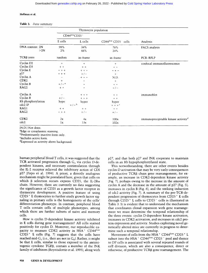
The determination that the L subset clearly differs from the E subset in cell cycle status, as summarized in Table 3, confirms our previous hypothesis that cell cycle activation is a very early event in response to productive TCR[3 chain gene rearrangement (Dudley et al. 1994). Although it has not been formally proven that E cells are precursors to L cells, this is the interpretation that best fits the data, for example, that there is very lit t le TCR~ chain gene rearrangement prior to the HSA + CD44 ~~ CD25 + cell stage IGodfrey et al. 1994} and that thymo- cytes from mutant mice defective in pre-TCR expression lowing to a variety of defects} all show normal thymo- cyte development up to and including the bulk of the HSA ~- CD44 l~ CD25 + cell stage, that is, the E subset (e.g., Fig. 2C).
Because the expansion in thymocyte numbers that oc- curs between the late DN stage and the DP stage appears to be the greatest expansion in thymocyte numbers that occurs, one might conclude that the pre-TCR is the most important growth factor receptor for thymocytes and is thus a primary determinant of the number of T cells an animal makes. This emphasizes the codependent inter- play of the regulation of cell proliferation and cell differ- entiation during T-cell development- - the pre-TCR, a highly T cell-specific differentiation product, promotes a cell cycle transition that is in turn tightly associated with the transient down-regulation of further RAG2-me- diated differentiation.
The findings presented in this paper lay the ground for a more precise analysis of how the pre-TCR functions, in particular, whether it functions by the same mechan i sm as do other growth-stimulatory receptors, such as that for epidermal growth factor. Although the precise inter- relationship of growth-stimulatory factors wi th the cell cycle has not been clarified, it is generally believed that a crucial consequence of their action may be cyclin-D activation and, as a result, phosporylation of Rb. Such a mechanism is plausible for the pre-TCR, based on our findings that Rb in E cells is almost entirely hypophoso phorylated, indicating in turn that cyclin D-dependent kinases in E cells are inactive. Moreover, an analogous situation may hold in B-cell development where Rosen- berg and colleagues showed that in tissue culture, im- munoglobul in light-chain gene rearrangement is re- stricted to a period of the cell cycle in which Rb is hy- pophosphorylated (Wang et al. 1995). In studies of
GENES & DEVELOPMENT 957
Cold Spring Harbor Laboratory Press on February 26, 2022 - Published by genesdev.cshlp.orgDownloaded from
Hoffman et al.
T a b l e 3. Data summary
Thymocyte population
CD441owCD25 +
E cells L cells CD44 z~ CD25 cells Analysis
DNA content: 2N 98% 34% >2N 2% 66%
TCR[3 joins random in-frame
76% 24%
in-frame
FACS analysis
PCR-RFLP
Cyclin D2 + + Cyclin D3 + + + + Cyclin E + + + + + + p27 + + + + / - Cyclin A - + + + CDK2 + + Cyclin B + / _ a + RAG2 + + +
Cyclin A Cyclin B + / - Rb phosphorylation hypo cdc2 IP RAG1 + + RAG2 + + +
+ / _ b
+ + +
+ + +
hyper
+ +
+ / -
+
+ +
+ + +
+
N.D. +
+ +
+ / -
+ +
+ +
hyper + + + c
+ +
+ / - -
CDK2 2x 4x 100x cdc2 lx 13x 102x
confocal immunofluorescence
immunoblot
immunoprecipitable kinase activity d
(N.D.) Not done. aEdge or cytoplasmic staining. bpredominantly inactive form only. r active form. dExpressed as activity above background.
human peripheral blood T cells, it was suggested that the TCR activated progression through G1 via cyclin D-de- pendent kinases, and necessary costimulation through the IL-2 receptor relieved the inhibitory action of CDI p27 {Firpo et al. 1994). A priori, a directly analogous mechanism might be postulated here, given that cells on which ~ selection occurs express CD25, the IL-2R~ chain. However, there are currently no data suggesting the significance of CD25 as a growth factor receptor in thymocyte development. A positive feature of using CD25 + E thymocytes to further study growth factor sig- naling in primary cells is the homogeneity of the cells' differentiation phenotype. In contrast, peripheral blood T cells contain cells of mult iple phenotypes, among which there are further subsets of naive and memory cells.
How is cyclin D-dependent kinase activity inhibited in E cells during gene rearrangement? All cells stained positively for cyclin D. Moreover, our reproducible ca- pacity to measure CDK2 activity in HSA + CD44 ~~ CD25 + E cells (Fig. 7) suggests that the cells have reached mid-G 1 (i.e., they are not in Go). Rather, it might be that E cells, similar to those exposed to the antimi- togenic cytokine TGFI3, contain a member of the INK family of inhibitors (Reynisdottir et al. 1995), along with
p27, and that both p27 and INK cooperate to mainta in cells in an Rb hypophosphorylated state.
This notwithstanding, there are other events besides cyclin-D activation that may be very early consequences of productive TCR[3 chain gene rearrangement; for ex- ample, an increase in CDK2-dependent kinase activity (Fig. 7), perhaps owing to the increase in the amount of cyclin A and the decrease in the amount of p27 (Fig. 5); increases in cyclin B (Fig. 6); and the striking induction of cdc2 activity (Fig. 7). A summary of the pre-TCR-de- pendent progression of thymocytes from CD25 + E cells through CD25 + L cells to C D 2 5 - cells is i l lustrated in Table 3. It is evident that to understand the mechan i sm that coordinates clonal expansion with gene rearrange- ment we must determine the temporal relationship of the three events: cyclin D-dependent kinase activation, increases in CDK2 activation, and increases in cdc2 pro- tein expression and activity. Studies exploiting novel ge- netically altered mice are currently in progress to deter- mine such a temporal relationship.
Movement of cells from the HSA § CD44 l~ CD25 + L subset into the HSA + CD44 l~ C D 2 5 - pool and thence to DP cells is associated with several repeated rounds of cell division, which are also a consequence, direct or otherwise, of productive TCRf~ gene rearrangement. The
958 GENES & DEVELOPMENT
Cold Spring Harbor Laboratory Press on February 26, 2022 - Published by genesdev.cshlp.orgDownloaded from

Thymocyte development, the pre-TCR, and the cell cycle
development of ~/8 T cells does not depend on expression of the TCRI3{ + }-pre-TCR, and most ,/~ T cells lack com- plete V(D)If3 gene rearrangements (Dudley et al. 1994). Thus, most ~/8 T cells would not experience the prolif- eration associated with the CD25 + to CD25- to DP transition. This may in part explain why circulating ~/~ T-cell numbers are commonly 10-50 times lower than oLf3 T-cell numbers (Hayday 1992). At the same time, among ~/~ T cells that contain rearranged V(D)Jf3, pro- ductive rearrangements are disproportionately common (>33%), consistent with those cells benefiting from pre- TCR-stimulated expansion before the completion of their differentiation as ~/~ T cells {Dudley et al. 1994).
Because it was shown recently that many ~8 T cells and a~3 T cells develop along a common lineage (Dudley et al. 1995; Livac et al. 1995), it is intriguing to consider that productive rearrangement of TCR-y and TCR8 genes induces exit from G1, in which gene rearrangement was occurring, but does not induce the repeated proliferation associated with ~ selection. This is perhaps attributable to intrinsic differences in the signaling mechanism em- ployed by the -y8 TCR. Consistent with this, we and our colleagues have recently discovered that in response to antigen stimulation in the spleens of mice, -/8 T cells generally expand less than do o~f3 T cells (W. Pao, L. Wen, A. Smith, A. Judge, I. MacLennan, M.J. Owen, and A.C. Hayday, in prep.). Thus, further comparison of pre-TCR signaling and -/8 TCR signaling in thymocytes may pro- vide important information concerning the signaling pathway that regulates the extent of cell proliferation following growth factor receptor engagement. This path- way may critically involve costimulation through other cell surface receptors that function in concert with the antigen and pre-antigen receptors.
Judging from the phenotype of the pT~ - / - mouse, in which few ~f3 T cells develop (Fehling et al. 1995), thy- mocyte expansion is not the only consequence of pre- TCR action. Rather, essential differentiation events would also seem contingent on pre-TCR signaling. These may include activation of the TCRa locus for sub- sequent expression and rearrangement. Consistent with an effect of productive TCRf3 chain rearrangment (and presumably pre-TCR signaling) on thymocyte differenti- ation is the transient and selective down-regulation of RAG2 protein levels that accompanies the transition to CD25- cells. Changes in RAG2 protein levels during avian thymocyte development were also reported previ- ously (Ferguson et al. 1994), although in that case the study of RAG2 protein in DN cells could not be com- pleted and, thus, no close correlation with ~3 selection was established.
The RAG2 regulation reported here is strikingly con- sistent with the 20-fold decrease in RAG2 stability in vitro reported after its phosphorylation by CDC2 {Lin and Desiderio 1993, 1995). In turn, this finding signifi- cantly strengthens the hypothesis that CDK2/cdc2 is a bona fide regulator of RAG2 in vivo. Because the CDK2/ cdc2 activites are promoted in vivo by productive TCRt3 chain gene rearrangement, this would link the successful rearrangement of a TCRI3 chain with the likely transient
down-regulation of further TCR gene rearrangement. Thus, a contribution to allelic exclusion at the TCRI3 locus may be an important by-product of pre-TCR sig- naling.
This is not to suggest that pre-TCR-associated cell cy- cle transitions are essential for allelic exclusion at the TCR~3 chain locus. Rather, allelic exclusion will be brought about when the TCR~3 locus is rendered "per- manently" inaccessible to further gene rearrangement. Such an event may be analogous to structural alterations of chromosome structure that occur at the yeast mating- type locus, also in response to a signaling cascade pro- moted by engagement of a cell surface receptor (Pringle and Hartwell 1981).
Instead, the pre-TCR-associated cell cycle transition may, through effects on RAG2, create a window of time in which gene rearrangement is reduced while the TCRI3 locus is rendered inaccessible. In support of this idea, allelic exclusion is more efficient after successful rear- rangement at the TCRf3 locus, an event associated with a cell cycle progression, than it is after successful rear- rangement at the TCRc~ locus, an event that is not asso- ciated with cell cycle progression (Padovan et al. 1993). The signal for the recombination machinery to be down- regulated after productive TCRa gene rearrangement seems not to be the completion of gene rearrangement per se, but the successful engagement of a complete TCR by major histocompatibility complex (MHC) products (Turka et al. 19911. Likewise, cell cycle progression and efficient allelic exclusion both appear to be associated with IgH locus rearrangement and the action of the pre- B-cell receptor, whereas productive IgL chain gene rear- rangement is associated with neither efficient allelic ex- clusion nor cell cycle progression (Schlissel and Morrow 19941.
There are numerous precedents for two-step changes in gene expression in response to a change in a cell's status. For example, on differentiation of the human my- elogenous cell line, HL60, c-myc expression is first down-regulated post-transcriptionally, followed some hours later by down-regulation at the promoter {Sieben- list et al. 1988). Such two-step mechanisms are inter- preted as manifesting the progression from a rapid re- sponse to a permanent change. The proliferative conse- quences of productive TCRI3 chain rearrangement and any significant effects on allelic exclusion may them- selves be segregated by mutation. For example, in C D 3 ~ - / - mice, TCRt3 chain allelic exclusion occurs (Liu et al. 1993), whereas the significant expansion in thymocyte numbers that ordinarily accompanies the DN to DP transition does not (Crompton et al. 1994).
Future studies are needed to elucidate several issues concerning RAG2 regulation in vivo. For example, is it most closely correlated with increases in CDK2 activity, perhaps driven by the increases in cyclin A that are ap- parent as cells transit to the CD25 + L subset stage? Or is it most closely correlated with the significant increases in cdc2 activity that similarly accompany the CD25 + to CD25- transition? A contribution of RAG2 RNA regu- lation, suggested previously by MacDonald and col-
GENES & DEVELOPMENT 959
Cold Spring Harbor Laboratory Press on February 26, 2022 - Published by genesdev.cshlp.orgDownloaded from
Hoffman et al.
leagues (Wilson et al. 1994), also needs to be quan t i t a t ed accura te ly now tha t the E and L subsets have been de- fined. Such s tudies promise to improve our unders tand- ing of how t ranscr ip t iona l and pos t - t ranscr ip t ional events in T-cell d i f ferent ia t ion both coordinate and are coordinated by cell cycle regula t ion in vivo.
Materials and methods
Animals and cell sorting
C57.BL/6 mice were obtained from Jackson Labs and the Charles River Company. Normally, female mice at or near 3-4 weeks of age were used. Thymi were teased and ground into single-cell suspensions. Thymocytes were guinea pig comple- ment (GIBCO)depleted of CD4 + and CD8* cells (CD4- and CD8-specific hybridoma supernatant was kindly provided by I.N. Crispe, Yale University, New Haven, CT), stained, and sorted on a FACStar Plus flow cytometer, as described previ- ously (Wilson et al. 1988; Dudley et al. 1994). Antibodies and other staining reagents used for FACS sorting included aCD25- FITC (Pharmingen), aHSA-PE (Pharmingen), ~CD4-Red 670 (Sigma), e~CD44-biotin (Pharmingen), and Avidin D-Texas Red (Vector).
DNA content analysis
Standard staining of cell populations by propidium iodide was done as described previously (Crissman et al. 19761 and analyzed using doublet discrimination on the FACS.
PCR-RFLP analysis
For each experiment, 1 x 10⁶ cells of each subset were used. De- tails of the technique and PCR primers used have been de- scribed previously (Mallick et al. 1993; Dudley et al. 1994).
Immunofluorescent microscopy
Cyclin A, cyclin E, CDK2, and p27 rabbit antisera (Koff et al. 1992), cyclin D2, cyclin D3 rabbit antisera (Santa Cruz), and purified mouse monoclonal specific for cyclin B1 (Pharmingen} have been described and were used to stain cells preincubated with FITC-conjugated anti-CD25 (Pharmingen). Affinity-puri- fied anti-RAG1 and anti-RAG2 rabbit antisera raised against amino acids 56-123 of RAG1 and amino acids 70--516 of RAG2 respectively fused with the Pseudomonas exotoxin protein (T.M.J. Leu, K.R. McConnel, E. Corbett, S. Bennet, and D.G. Schatz, in prep.) were also used to stain similar cells. Reactivity was detected with a Lissamine rhodamine-conjugated goat anti- rabbit immunoglobulin or goat anti-mouse immunoglobulin (Jackson Immuno Labs). A Zeiss Axiovert 10 microscope with a Bio-Rad MRC-600 laser-scanning confocal imaging system and MRC-600 confocal microscope operating software version 6.05.4 was utilized for all studies.
Immunoprecipitation, gel electrophoresis, and immunoblotting
cdc2 immunoprecipitation has been described (Koff et al. 19921. In all other cases, whole-cell lysate was used. Cells were enu- merated and then lysed in an NP-40 lysis buffer containing five protease inhibitors and four phosphatase inhibitors. The protein concentration was determined by BCA assay and equivalent loading in Coommassie blue-stained gels. Defined quantities (normally 2.5x10 s cells/lane) were electrophoresed in 6.5%
(5% for the Rb immunoblot) SDS--polyacrylamide gel electro- phoresis (SDS-PAGE}, transferred to PVDF membranes (Bio- Rad} using standard conditions. Cyclin-A rabbit antiserum was used as described (Koff et al. 1992) at a 1:500 dilution; mono- clonal cyclin-B antibody (Santa Cruz) was used at a 1:1000 di- lution; and monoclonal Rb antibody IPharmingen) was used at a 1:1000 dilution. For RAG1 and RAG2 immunoblots, the membranes were incubated with 1:200 dilutions of either of the two affinity-purified rabbit sera raised against RAG 1 and RAG2, respectively (see above). In all cases, reactivity was determined by HRP-conjugated secondary antibodies (Sigma) and enhanced chemiluminescence (ECL)(DuPont}.
Kinase assays
For each experiment, l xl06 cells were immunoprecipitated with either normal rabbit serum, a carboxy-terminal peptide- specific CDK2 rabbit antiserum, or a carboxy-terminal peptide- specific CDC2 rabbit antiserum as described before (Koff et al. 1992}. Relative histone H1 phosphorylation was measured and quantified using phosphorimaging.
A c k n o w l e d g m e n t s
This work has been supported by National Institutes of Health grants GM37759 (A.H.), GM52597 (A.K.I, and AI32524 (D.G.S.), by a National Science Foundation predoctoral fellowship (E.S.H.), by an Immunology Training Grant (AI07019), by the Imperial Cancer Research Foundation (M.J.O.; T.C.), by the Swiss Bank Corporation (T.M.J.L.), by the Howard Hughes Med- ical Institute (D.G.S.), and by The Society of the Memorial Sloan-Kettering Cancer Center (A.K.). We thank Tom Taylor for cell sorting, Rocco Carbone for DNA analysis, Laurent Caron for confocal microscopy assistance and Sylvia Kim for technical expertise. We also thank William Pao, E.C. Dudley, and those referred to in the text and legends for the kind provision of antibodies and other reagents.
The publication costs of this article were defrayed in part by payment of page charges. This article must therefore be hereby marked "advertisement" in accordance with 18 USC section 1734 solely to indicate this fact.
Note added in proof
Since acceptance of this paper, Xu et al. [Proc. Natl. Acad. Sci 93:2167-2173 (1996)] have refuted the link of thymocyte pro- liferation and allelic exclusion, based on the segregation of the two events in pTc~ - / - mice. As is discussed in this paper, this segregation was already evident in C D 3 ~ - / - mice ILiu et al. 1993; Crompton et al. 19941. Our contention is that cdc2/ CDK2-mediated selective down-regulation of RAG2 protein is likely an important component of a multicomponent process of efficient allelic exclusion, which occurs within the CD25 + sub- set, prior to and potentially independent of subsequent expan- sion at the CD25- stage. This issue is considered in the Dis- cussion.
References
Crissman, H.A., M.S. Oka, and J.A. Steincamp. 1976. Rapid staining method for the analysis of deoxyribonucleic acid and protein in mammalian cells. L Histochem. Cytochem. 24: 64.
Crompton, T., M. Moore, H.R. MacDonald, and B. Malissen. 1994. Double-negative thymocyte subsets in CD3~ chain-
960 GENES & DEVELOPMENT
Cold Spring Harbor Laboratory Press on February 26, 2022 - Published by genesdev.cshlp.orgDownloaded from

Thymocyte development, the pre-TCR, and the cell cycle
deficient mice: Absence of HSA § CD44- CD25 cells. Eur. I. Immunol. 24: 1903-1907.
Davodeau, F., M.-A. Peyrat, I. Houde, M.-M. Mallet, G. De Libero, H. Vie, and M. Bonneville. 1993. Surface expression of two distinct functional antigen receptors on human gamma-delta T cells. Science 260: 1800-1802.
DeCaprio, J.A., J.W. Ludlow, D. Lynch, Y. Furukawa, J. Griffin, H. Piwnica-Worms, C.-M. Huang, and D.M. Livingston. 1992. The retinoblastoma-susceptibility gene product be- comes phosphorylated in multiple stages during cell cycle entry and progression. Proc. Natl. Acad. Sci. 89: 1795-1798.
Dudley, E.C., H.T. Petrie, L.M. Shah, M.J. Owen, and A.C. Hay- day. 1994. T cell receptor ~ chain gene rearrangement and selection during thymocyte development in adult mice. Im- muni ty 1: 83-93.
Dudley, E.C., M. Girardi, M.J. Owen, and A.C. Hayday. 1995. c~ T cells and ~8 T cells can share a late common precursor. Curr. Biol. 5: 659-669.
Fehling, H.J., A. Krotkova, C. Saint-Ruf, and H. yon Boehmer. 1995. Crucial role of the pre-T-cell receptor c~ gene in devel- opment of c~J3 but not ~ T cells. Nature 375: 795-798.
Ferguson, S.E., M.A. Accavitti, D.D. Wang, C.L. Chen, and C.B. Thompson. 1994. Regulation of RAG-2 protein expression in avian thymocytes. Mol. Cell. Biol. 14: 7298-7305.
Firpo, J., A. Koff, M. Solomon, and J. Roberts. 1994. Inactivation of a CDK2 inhibitor during IL2 induced proliferation of hu- man T lymphocytes. Mol. Cell Biol. 14: 4889-4901.
Godfrey, D., J. Kennedy, P. Mombaerts, S. Tonegawa, and A. Zlotnik. 1994. Onset of TCR-~ gene rearrangement and role of TCR-~ expression during CD3 CD4-CD8 thymocyte differentation. ]. Imrnunol. 152: 4783-4792.
Groettrup, M., K. Ungewiss, O. Azogui, R. Palacios, M.J. Owen, A.C. Hayday, and H. von Boehmer. 1993. A novel disulfide- linked heterodimer on pre-T cells consists of the T cell re- ceptor J3 chain and a 33kD glycoprotein. Ceil 75: 283-294.
Hatakeyama, M., J.A. Brill, G.R. Fink, and R.A. Weinberg. 1994. Collaboration of G~ cyclins in the functional inactivation of the retinoblastoma protein. Genes & Dev. 8:1759-1771.
Hayday, A.C. 1992. T cell receptor ~8. In The encylopedia of immunology (ed. I.M. Roitt and P.J. Delves), Vol. 3, pp. 1428-1433. Academic Press, London, UK.
Hayday, A., S.D. Gillies, H. Saito, C. Wood, K. Wiman, W. Hay- ward, and S. Tonegawa. 1984. Activation of a translocated human c-myc gene by an enhancer in the immunoglobulin heavy chain locus. Nature 307: 334-340.
Kitamura, D., A. Kudo, S. Schaal, W. Muller, F. Melchers, and K. Rajewsky. 1992. A critical role of h5 protein in B cell devel- opment. Cell 69: 823-831.
Koff, A., A. Giordano, D. Desai, K. Yamashita, J.W. Harper, S. Elledge, T. Nishimoto, D.O. Morgan, B.R. Franza, and J.M. Roberts. 1992. Formation and activation of a cyclin E-cdk2 complex during the G1 phase of the human cell cycle. Sci- ence 257: 1689-1694.
Lin, W.-C. and S. Desiderio. 1993. Regulation of V(D}J recom- bination activator protein RAG-2 by phosphorylation. Sci- ence 260: 953-959.
�9 1995. V{D)J recombination and the cell cycle. Immunol. Today 16:(6)279-289.
Liu, C.-P., R. Ueda, J. She, J. Sancho, B. Wang, G. Weddell, J. Loring, C. Kurahara, E.C. Dudley, A.C. Hayday, C. Terhorst, and M. Huang. 1993. Abnormal T cell development in CD3-~ -/- mutant mice and identification of a novel T cell popula- tion in the intestine. EMBO J�9 12: 4863-4875.
Livak, F., H.T. Petrie, I.N. Crispe, and D.G. Schatz. 1995. In frame TCR8 gene rearrangements play a critical role in the ~/~/8 T cell lineage decision. Immun i t y 2: 617--627.
Mallick, C., E.C. Dudley, J.L. Viney, M.J. Owen, and A.C. Hay- day. 1993. Rearrangement and diversity of T cell receptor 13 chain genes in thymocytes: A critical role for the J3 chain m development. Cell 73:513-519.
Mallisen, M., J. Trucy, E. Jouvin-Marche, P.A. Cazenave, R. Sol- lay, B. Mallisen. 1992. Regulation of TCR alpha and beta gene allelic exclusion during T-cell development. Immtmol. Today 13:{8)315-322.
Marraccino, R.L., E.J. Firpo, and J.M. Roberts. 1992. Activation of the p34 CDC2 protein kinase at the start of S phase in the human cell cycle. Mol. Biol. Cell 3(4): 389-401.
Mombaerts, P., J. Iacomini, R.S. Johnson, K. Herrup, S. Tone- gawa, and V.E. Papaioannou. 1992a. RAG-l-deficient mice have no mature B and T lymphocytes. Cell 68: 869-877.
Mombaerts, P., A.R. Clark, M.A. Rudnicki, J. Iacomini, S. Ito- hara, J.J. Lafaille, L. Wang, Y. Ichikawa, R. Jaenisch, M.L. Hooper, and S. Tonegawa. 1992b. Mutations in T-cell anti- gen receptor genes ~ and 13 block thymocyte development at different stages. Nature 360: 225-231.
Ohtsubo, M. and J.M. Roberts. 1993. Cyclin dependant regula- tion of G1 in mammalian fibroblasts. Science 259: 1908- 1912.
Padovan, E., G. Casorati, P. Dellabona, S. Meyer, M. Brockhaus, and A. Lanzevecchia. 1993. Expression of two TCR alpha chains: Dual receptor T cells. Science 262: 422-424.
Pearse, M., L. Wu, M. Egerton, A. Wilson, K. Shortman, and R. Scollay. 1989. A murine early thymocyte developmental se- quence is marked by transient expression of the interleukin 2 receptor. Proc. Natl. Acad. Sci. 86: 1614-1618.
Penit, C., B. Lucas, and F. Vasseur. 1995. Cell expansion and growth arrest phases during the transition from precursor (CD4-8-) to immature (CD4+8 +) thymocytes in normal and genetically modified mice. I. Imm unol. 154:5103-5113.
Petrie, H.T., P. Hugo, R. Scollay, and K. Shortman. 1990. Lin- eage relationships and developmental kinetics of immature thymocytes: CD3, CD4, and CD8 acquisition in vivo and in vitro. I. Exp. Med. 172: 1583-1588.
Pines, J. and T. Hunter. 1991. Human cyclins A and B1 are differentially located in the cell and undergo cell cycle-de- pendant nuclear transport. I. Cell Biol. 115: 1-17.
Polyak, K., J. Kato, M.J. Solomon, C.J. Sherr, J. Massague, J.M. Roberts, and A. Koff. 1994a. p27 Kipl, a cyclin-cdk inhibitor, links transforming growth factor-~ and contact inhibition to cell cycle arrest. Genes & Dev. 8: 9-22.
Polyak, K., M. Lee, H. Erdjument-Bromage, A. Koff, J.M. Rob- erts, P. Tempst, and J. Massague. 1994b. Cloning of p27 rapl, a cyclin-dependent kinase inhibitor and a potential mediator of extracellular antimitogenic signals. Cell 78: 59-66.
Pringle, J.R. and L.H. Hartwell. 1981. The Saccharomyces cer- evisiae cell cycle. In Molecular biology of the yeast Saccha- romyces: Life cycle and inheritance, Vol. 1, pp. 97-142, Cold Spring Harbor Laboratory, Cold Spring Harbor, New York.
Reynisdottir, I., K. Polyak, A. Iavarone, and J. Massague. 1995. Kip/Cip and Ink4 Cdk inhibitors cooperate to induce cell cycle arrest in response to TGF-~. Genes & Dev. 9: 1831- 1845.
Robey, E. and B.J. Fowlkes. 1994. Selective events in T cell de- velopment. Annu. Rev. Immunol. 12: 675-705.
Rothenberg, E. 1992. The development of functionally respon- sive T cells. Adv. Immunol. 51: 85-214.
Saint-Ruf, C., K. Ungewiss, M. Groettrup, L. Bruno, H.J. Feh- ling, and H. yon Boehmer. 1994. Analysis and expression of a cloned pre-T cell receptor gene. Science 266: 1208-1212.
Schlissel, M. and T. Morrow. 1994. Ig heavy chain protein con- trols B cell development by regulating germ-line transcrip- tion and retargeting V(D}J recombination. J. Immtmol.
GENES & DEVELOPMENT 961
Cold Spring Harbor Laboratory Press on February 26, 2022 - Published by genesdev.cshlp.orgDownloaded from

Hoffman et al.
153: 1645-1657. Scollay, R. and K. Shortman. 1985. Identification of early stages
of T lymphocyte development in the thymus cortex and me- dulla. J. Immunol . 134: 3632-3642.
Sherr, C.J. and J.M. Roberts. 1995. Inhibitors of mammalian G1 cyclin-dependant kinases. Genes & Dev. 9:1149-1163.
Shinkai, Y., G. Rathbun, K.-P. Lam, E.M. Oltz, V. Stewart, M. Mendelsohn, J. Charron, M. Datta, F. Young, A.M. Stall, and F.W. Alt. 1992. RAG-2-deficient mice lack mature lympho- cytes owing to inability to initiate V(D)J rearrangment. Cell 68: 855-867.
Shinkai, Y., S. Koyasu, K.-I. Nakayama, K.M. Murphy, D.Y. Loh, E.L. Reinherz, and F.W. Alt. 1993. Restoration of T cell de- velopment in RAG-2-deficient mice by functional TCR transgenes. Science 259: 822-825.
Siebenlist, U., P. Bressler, and K. Kelly. 1988. Two distinct mechanisms of transcriptional control operate on c-myc dur- ing differentiation of HL60 cells. Mol. Cell. Biol. 8: 867-874.
Solomon, M.J., M. Glotzer, T.H. Lee, M. Phillipe, and M.W. Kirschner. 1990. Cyclin activation of p34 r162 Cell 63: 1013- 1024.
Turka, L.A., D.G. Schatz, M.A. Oettinger, J.J.M. Chun, C. Gorka, K. Lee, W.T. McCormack, and C.B. Thompson. 1991. Thymocyte expression of RAG-1 and RAG-2: Termination by T cell receptor cross-linking. Science 253: 778-781.
Uematsu, Y., S. Ryser, Z. Dembic, P. Borgulya, P. Krimpenfort, A. Berns, and H. von Boehmer. 1988. In transgenic mice the introduced functional T cell receptor ~ gene prevents expres- sion of endogenous [3 genes. Cell 52: 831-841.
Wang, L.C., Y.Y. Chen, and N. Rosenberg. 1995. Pre-B-cells transformed by ts Abelson virus rearrange kappa and lambda genes in early G1. Curr. Topics Microbiol. Immunol. 194: 355-361.
Wilson, A., A. D'Amico, T. Ewing, R. Scollay, and K. Shortman. 1988. Subpopulations of early thymocytes. A cross-correla- tion flow cytometric analysis of adult mouse Ly-2-L3T4- (CD8-CD4-) thymocytes using eight different surface markers. J. Immunol . 140: 1461-1470.
Wilson, A., W. Held, and H.R. MacDonald. 1994. Two waves of recombinase gene expression in developing thymocytes. L Exp. Med. 179: 1355-1360.
Wiman, K., B. Clarkson, A.C. Hayday, H. Saito, S. Tonegawa, and W.S. Hayward. 1984. Activation of a translocated c-myc gene: role of structural alterations in the upstream region. Proc. Natl. Acad. Sci. 81: 6798-6802.
Wu, L., R. Scollay, M. Egerton, M. Pearse, G. Spangrude, and K. Shortman. 1991. CD4 expressed on earliest T-lineage precur- sor cells in the adult murine thymus. Nature 346: 71-74.
962 GENES & DEVELOPMENT
Cold Spring Harbor Laboratory Press on February 26, 2022 - Published by genesdev.cshlp.orgDownloaded from

10.1101/gad.10.8.948Access the most recent version at doi: 10:1996, Genes Dev.
E S Hoffman, L Passoni, T Crompton, et al. regulation of cell cycle and clonality during development in vivo.Productive T-cell receptor beta-chain gene rearrangement: coincident
References
http://genesdev.cshlp.org/content/10/8/948.full.html#ref-list-1
This article cites 50 articles, 27 of which can be accessed free at:
License
ServiceEmail Alerting
click here.right corner of the article or
Receive free email alerts when new articles cite this article - sign up in the box at the top
Copyright © Cold Spring Harbor Laboratory Press
Cold Spring Harbor Laboratory Press on February 26, 2022 - Published by genesdev.cshlp.orgDownloaded from