pueraria mirifica alleviates cortical bone loss in ...€¦ · research ¤¢¢¯ §¢ ¡¤®¥...
TRANSCRIPT

DOI: 10.1530/JOE-16-0277http://joe.endocrinology-journals.org © 2016 Society for Endocrinology
Printed in Great BritainPublished by Bioscientifica Ltd.
Journ
alofEn
docrinology
121–133D KITTIVANICHKUL and others Pueraria mirifica improvescortical BMD in monkeysResearch
231:2
Abstract
Since the in vitro and in vivo anti-osteoporotic effects of Pueraria mirifica (PM) in
rodents have been verified, its activity in menopausal monkeys was evaluated as
required before it can be applicable for human use. In this study, postmenopausal
osteoporotic monkeys were divided into two groups (five per group), and fed daily
with standard diet alone (PMP0 group) or diet mixed with 1000 mg/kg body weight
(BW) of PM powder (PMP1000 group) for 16 months. Every 2 months, the bone mineral
density (BMD), bone mineral content (BMC) and bone geometry parameters (cortical
area and thickness and periosteal and endosteal circumference) at the distal radius and
proximal tibia were determined using peripheral quantitative computed tomography
together with plasma and urinary bone markers. Compared with the baseline (month 0)
values, the cortical, but not trabecular, BMDs and BMCs and the cortical area and
thickness at the metaphysis and diaphysis of the radius and tibia of the PMP0 group
continuously decreased during the 16-month study period. In contrast, PMP1000
treatment ameliorated the bone loss mainly at the cortical diaphysis by decreasing bone
turnover, as indicated by the lowered plasma bone-specific alkaline phosphatase and
osteocalcin levels. Generally, changes in the cortical bone geometry were in the opposite
direction to the cortical bone mass after PMP1000 treatment. This study indicated that
postmenopausal monkeys continuously lose their cortical bone compartment, and they
have a higher possibility for long bone fractures. Oral PMP treatment could improve
both the bone quantity (BMC and BMD) and quality (bone geometry).
10.1530/JOE-16-0277
Pueraria mirifica alleviates cortical bone loss in naturally menopausal monkeys
Donlaporn Kittivanichkul1, Narattaphol Charoenphandhu2,3, Phisit Khemawoot4 and Suchinda Malaivijitnond1,5
1Department of Biology, Faculty of Science, Chulalongkorn University, Bangkok, Thailand2Center of Calcium and Bone Research (COCAB), Faculty of Science, Mahidol University, Bangkok, Thailand3Department of Physiology, Faculty of Science, Mahidol University, Bangkok, Thailand4Department of Pharmacology and Physiology, Faculty of Pharmaceutical Sciences, Chulalongkorn University, Bangkok, Thailand 5National Primate Research Center of Thailand, Chulalongkorn University, Saraburi, Thailand
Journal of Endocrinology (2016) 231, 121–133
2312
Correspondence should be addressed to S Malaivijitnond Email [email protected]
Key Words
f Pueraria mirifica
f osteoporosis
f cynomolgus monkeys
f estrogen deficiency
Introduction
The increased life expectancy of humans alongside the development of public health has led to a significant increase in the average life expectancy of the population and ageassociated diseases (Sweet et al. 2009).
Osteoporosis is one such disease where the incidence is much higher in the elderly, and is usually associated with bone fracture. It is reported that in the year 2000, there were some 9 million osteoporotic fractures in the world

122Pueraria mirifica improves cortical BMD in monkeys
DOI: 10.1530/JOE-16-0277
Journ
alofEn
docrinology
http://joe.endocrinology-journals.org © 2016 Society for EndocrinologyPrinted in Great Britain
Published by Bioscientifica Ltd.
231:2
population, of which 1.6, 1.7 and 1.4 million were for hip, wrist and vertebral fractures, respectively, which will have a huge impact on the quality of life and socioeconomic consequences (Johnell & Kanis 2006, Woolf 2006). Among the elderly, postmenopausal women are in the highest risk group for osteoporosis, which is attributed to the decreased estrogen level. Estrogen is a key hormonal regulator of bone metabolism, and regulates bone turnover directly via modulating the action of osteocytes, osteoblasts and osteoclasts (Khosla et al. 2012). Consequently, a low bone mass and quality, which account for the low bone composition and structure, occur during the estrogendeficient postmenopausal stage.
In postmenopausal women, the bone mineral density (BMD) and bone mineral content (BMC) decrease by an average of 1.9 and 1.3% per year. Thus, the bone structure changes to a low bone density through endosteal resorption and periosteal deposition (Bouxsein & Karasik 2006). This causes an increased bone diameter, where the medullary and periosteal diameters increase by up to 1.1 and 0.7% per year, respectively, and the cortical thickness increases by 0.4% per year. As a whole, this leads to an annual decrease in the strength index of about 0.7% (Ahlborg et al. 2003). Therefore, women in this postmenopausal period of life have 15% cortical bone loss (Khosla 2013), with cortical bone fractures being the most frequent.
At present, many scientists have tried to search for new chemicals to mitigate bone loss and improve bone quality. Some currently prescribed drugs can increase the bone mass, but at the same time they lower the bone quality and increase the risk of bone fractures afterward (Odvina et al. 2005). Although the positive effect of estrogen on bones is well recognized, estrogen replacement therapy (ERT) is no longer recommended due to the increased incidences of estrogendependent breast cancer, cardiovascular and venous thromboembolic events after the longterm use of ERT (Nelson et al. 2002). Thus, naturebased products, such as plantderived estrogens (or phytoestrogens), which have a similar structure and act in a similar manner with that of 17βestradiol (E2),have attracted attention.
The tuberous roots of Pueraria mirifica (PM) contain at least 17 phytoestrogens (Malaivijitnond 2012), and recently became a focal point of interest for its estrogenic activity. The estrogenic activity of PM powder (PMP) or extract (PME) has been widely tested in vitro and in vivo (Malaivijitnond 2012), where it has also been shown to effectively prevent bone loss by increasing the BMC and BMD in ovariectomized and orchidectomized osteoporotic
rats (Urasopon et al. 2007, 2008, Suthon et al. 2016). Moreover, the underlying mechanism in rat osteoblastlike UMR106 cells has been investigated (Tiyasatkulkovit et al. 2012). Although the effects and mechanism of action of PM have been thoroughly verified in the rodent model, both in vitro and in vivo, based on the safety regulatory guideline of the United States Food and Drug Administration (US FDA) (Thompson et al. 1995), the preclinical test of antiosteoporotic agents needs to be performed in a second animal species, which have similar bone structure and remodeling process to those of humans. Thus, the efficacy of PMP on bone structure modeling should be evaluated in monkeys because of its broadly similarly hormonal patterns (Goodman et al. 1977), reproductive functions (Weinbauer et al. 2008) and bone structures (Schaffler & Burr 1984) to those of humans.
With respect to the previous reports indicating that ovariectomized monkeys, animal models of choice for osteoporosis research, had different patterns of hormonal changes and bone loss from those of postmenopausal monkeys and women (Chen et al. 2013, Kittivanichkul et al. 2016), in this study the effects of PMP on the bone quantity (BMD and BMC) and quality (geometry) were determined in aged, and so naturally menopausal, cynomolgus monkeys.
Materials and methods
Animals
Ten female cynomolgus monkeys were used in this study. They were reared at the Primate Research Unit, Faculty of Science, Chulalongkorn University, Thailand. The selected postmenopausal monkeys met the criteria of (i) being more than 20 years old, (ii) having a complete cessation of menstruation for at least 1 year and (iii) having a total BMD at the radius and tibia of between –0.5 and –1 SD of those of premenopausal cynomolgusmonkeys (Kittivanichkul et al. 2016). The 10 osteoporotic monkeys selected by the above criteria were used in an attempt to mimic the osteoporosis that occurs in naturally postmenopausal aged women. Monkeys were housed in individual cages with a ratio of 12 h light:12 h darkness cycle at ambient temperature. They were fed daily with monkey chow diet (Perfect Companion Group. Co. Ltd. Samut Pakran, Thailand) in the morning (09:00 – 10:00 h) and supplemented with fresh fruits in the afternoon (13:00 – 14:00 h). All animal procedures were approved by the Faculty of Science, Chulalongkorn University Animal Care and Use Committee (Protocol review no. 1123015).
Research D KITTIVANICHKUL and others

123Research D KITTIVANICHKUL and others Pueraria mirifica improves cortical BMD in monkeys
DOI: 10.1530/JOE-16-0277
Journ
alofEn
docrinology
http://joe.endocrinology-journals.org © 2016 Society for EndocrinologyPrinted in Great Britain
Published by Bioscientifica Ltd.
231:2
Clinical records for each animal were performed daily by veterinary staff and animal caretaker.
Phytoestrogen analysis of PMP
Dried PMP (lot no. 141023), as a fine powder, was kindly provided by Dr I Sandford Schwartz, Smith Naturals Co Ltd, Thailand. Liquid chromatography tandem massspectrometry (LC/MS/MS) was used for the analysis of puerarin and miroestrol levels in the PMP. Puerarin is the major phytoestrogen present in PM, while miroestrol shows a high estrogenic activity. Puerarin and miroestrol analytical standards were obtained from SigmaAldrich and Professor Tsutomu Ishikawa (Chiba University, Japan), respectively. Glycyrrhetinic acid, used as an internal standard in the LC/MS/MS, was purchased from Wako. All compounds were tested for quality control and had more than 95% of the active ingredient.
The puerarin and miroestrol standards were prepared by dissolving in DMSO and diluting with methanol. In brief, 100 mg of tested formulation of PMP was dissolved in 1 mL of DMSO, and then diluted 100fold in methanol. The diluted samples were mixed with a 10fold volume
of methanol containing 10 ng of glycyrrhetinic acid as an internal standard for the LC/MS/MS analysis, performed using an Eksigent ekspert UHPLC 100 LC equipped with a QTRAP 6500 MS, controlled by Analyst software version 1.6 (AB Sciex, Framingham, MA, USA). The UHPLC system was equipped with a Synergi FusionRP C18 column as the stationary phase (Phenomenex Inc, Torrance, CA, USA), while the mobile phase was a methanol:water gradient starting at 10% (vol/vol) methanol for 0.5 min, increased linearly to 90% (vol/vol) methanol at 1.5 – 3.5 min, and then decreased linearly to 10% (vol/vol) methanol at 4 – 4.5 min. The retention times of puerarin, miroestrol and glycyrrhetinic acid were 1.56, 1.74 and 2.09 min, respectively (Fig. 1A–C). The MS analysis was performed with negative mode ionization for detection of puerarin (m/z, 415.3/295.0), miroestrol (m/z, 357.2/188.9) and glycyrrhetinic acid (m/z, 469.3/409.2). The fragmentation patterns of puerarin, miroestrol and glycyrrhetinic are shown in Fig. 2A–C. Calibration curves of puerarin and miroestrol showed good correlation coefficients (R2 > 0.99) over the concentration range of 5 – 1000 μg/L. The limitof detection of both compounds was estimated to be 1 μg/L with a signaltonoise ratio of 5. The intraassay
Figure 2Fragmentation pattern of (A) puerarin, (B) miroestrol and (C) glycyrrhetinic acid.
Figure 1LC/MS/MS chromatogram for 100 µg/L of (A) puerarin, (B) miroestrol and (C) glycyrrhetinic acid.

Research 124Pueraria mirifica improves cortical BMD in monkeys
DOI: 10.1530/JOE-16-0277
Journ
alofEn
docrinology
D KITTIVANICHKUL and others
http://joe.endocrinology-journals.org © 2016 Society for EndocrinologyPrinted in Great Britain
Published by Bioscientifica Ltd.
231:2
accuracy and precision for analysis of both compounds were within ±10%. The puerarin and miroestrol contentsin the PMP (lot no. 141023) were 18.6 mg/100 g PMP and 233 µg/100 g PMP, respectively.
Experimental design
Before the onset of the study, the postmenopausal monkeys were randomly assigned into one of the two groups (five monkeys in each group), the control (PMP0) group and that treated with PMP (PMP1000) group. There was no significant difference between the PMP0 and PMP1000 groups in their age, body weight, menopause period and total BMD at both metaphysis and diaphysis at radius and tibia bone (Table 1). The PMP0 group was fed daily with standard diet alone and the PMP1000 group was fed with the same diet mixed with 1000 mg/kg BW of PMP at 08:00 – 09:00 h for 16 months. A PMP dose of 1000 mg/kg BW, equivalent to 0.18 mg/kg BW of puerarin and 2.33 µg/kg BW of miroestrol, was selected for this study because it was shown previously to significantly decrease the serum parathyroid hormone levels in aged female cynomolgus monkeys (Trisomboon et al. 2004). To maintain the consistency of the dose treatment, monkeys were weighed in every 2 months and the amount of PMP fed to the monkeys was adjusted to their BW afterward. To ensure that the same amount of phytoestrogen content was fed to the monkeys, the same batch of PMP was used throughout the experiment. Blood and urine were collected, and the BMD, BMC and bone geometry were measured every 2 months for 16 months (at months 0, 2, 4, 6, 8, 10, 12, 14 and 16, respectively).
Urine and blood collection
For urine collection, monkeys were fasted from 18:00 h on the day before collection and stainless steel trays covered with iron mesh were placed under each monkey cage. On the following day at 06:00 h, the trays were removed and the 12h urine samples were collected by syringe.
Urine samples were clarified by centrifugation at 1700 g, 4˚C for 20 min and then the supernatants were harvested and kept at –20˚C until analyzed for the levels of crosslinked Ntelopeptide of type I collagen (NTX) and creatinine.
After the urine collection was performed, monkeys were anesthetized by i.m. injection with a mixture of tiletamine/zolazepam, as a mixture of 3 mg/kg BW Zoletil 100 (Virbac, Nonthaburi, Thailand) and 40 µg/kg BW medetomidine hydrochloride (Vetcare, Jonesboro, AR, USA). Blood was withdrawn from the femoral vein between 08:00 and 11:00 h and immediately mixed with sodium heparin (Leo Pharmaceutical, Ballerup, Denmark) at 0.1 IU/mL of whole blood, centrifuged (1700 g, 4˚C for 20 min) and the blood plasma was harvested and kept at –80˚C until assayed for E2, folliclestimulating hormone (FSH), luteinizing hormone (LH), calcium and bone turnover markers (see below). After the blood collection, the BMD, BMC and bone geometry were measured at the radius and tibia. Atipamazole (Vetcare) at 40 µg/kg BW was administered by i.m. injection to animals after all the procedures had been completed.
Plasma hormone level assays
Plasma E2, FSH and LH levels were determined only at month 0 to confirm the postmenopausal stage in monkeys. Plasma FSH and LH levels were determined using a heterologous radioimmunoassay as described previously (Trisomboon et al. 2004, Kittivanichkul et al. 2016). Plasma E2 levels were determined by electrochemiluminescence immunoassay at the Department of Pathology, Faculty of Medicine, Ramathibodi Hospital, Mahidol University, Thailand. All samples were processed in a single run to minimize betweenruns experimental variation in the data. The intraassay coefficients of variation were 1.44, 2.4 and 3.2% for the FSH, LH and E2 assays, respectively.
Measurements of plasma calcium levels
At months 0 and 16, the plasma calcium levels were determined by automate clinical chemical analyzer
Table 1 Body weight, age, menopause period and total bone mineral density (BMD) of the monkeys in each treatment group.
Treatment Body weight (kg) Age (years)Menopause period
(years)
Total BMD (mg/cm3)
Metaphysis Diaphysis
Radius Tibia Radius Tibia
PMP0 (n = 5)
5.37 ± 0.23(4.8 – 5.9)
29.6 ± 1.8(24 – 33)
5.8 ± 1.4(2.5 – 10.9)
425.0 ± 38.0(317.7 – 541.7)
384.0 ± 30.8(292.6 – 459.5)
665.3 ± 65.5(439.2 – 837.8)
720.5 ± 39.6(620.1 – 825.5)
PMP1000 (n = 5)
5.39 ± 0.20(5.1 – 6.1)
26.0 ± 2.3(20 – 33)
7.8 ± 1.2(4.3 – 10.6)
541.2 ± 51.7(406.1 – 670.2)
345.8 ± 26.6(304.6 – 449.20)
719.3 ± 58.4(571.4 – 835.3)
721.1 ± 30.6(646.8 – 822.7)

125Research D KITTIVANICHKUL and others Pueraria mirifica improves cortical BMD in monkeys
DOI: 10.1530/JOE-16-0277
Journ
alofEn
docrinology
http://joe.endocrinology-journals.org © 2016 Society for EndocrinologyPrinted in Great Britain
Published by Bioscientifica Ltd.
231:2
(ILAB650, Chema Diagnostica, Monsano, Italy) at the Faculty of Veterinary Science, Chulalongkorn University, Thailand. The intraassay coefficient of variation was 0.75%.
Measurement of the BMC, BMD and bone geometry
The BMC, BMD and bone geometry parameters, including the cortical area and thickness, and periosteal and endosteal circumferences, at the distal radius and proximal tibia, were determined using peripheral quantitative computed tomography (pQCT; Norland Stratec XCT Research SA+ pQCT, Stratec, Pforzheim, Germany) as reported previously (Kittivanichkul et al. 2016). The BMD, BMC and bone geometry parameters of the total, trabecular and cortical bone regions were analyzed using the XCT5.50E software (Stratec).
Measurement of bone turnover markers
Dynamic changes in the bone turnover parameters were determined by measuring the plasma bonespecific alkaline phosphatase (BAP; Quidel, San Diego, CA, USA, catalog no. 8012) and osteocalcin (Quidel, catalog no. 8002) levels and urinary NTX levels (Wampole Labs, Princeton, NJ, USA, catalog no. OST0001) as markers. The BAP and osteocalcin were determined by enzyme immunoassay (EIA), where the intra and interassay coefficients of variation were 4.07 and 8.39% for BAP and 9.33 and 14.17% for osteocalcin, respectively. The urinary NTX levels were also measured by EIA, but normalized to the urinary creatinine levels that were determined by the Jaffe kinetic method (Legrand et al. 2003). The intra and interassay coefficients of variation for NTX were 5.30 and 6.51%, respectively.
Statistical analysis
All data were transformed to percent changes according to the baseline (the month 0). Data are expressed as mean ± s.e.m. Since there was no significant interactionbetween the time and treatment when tested by twoway ANOVA (data not shown), then the changes in all the parameters were analyzed separately using a paired ttest (comparing between the values at month 0 and the other time points in the same group) or an unpaired ttest (comparing between groups at the same time point). A P value of <0.05 was considered to be significant.Statistical analyses were performed using the IBM SPSS Statistics for Windows, version 20.0 software (IBM). Pearson’s correlations were performed using the
R program (R Core Team 2015). Similarity in the slope and intercept of each group regression line was tested using GraphPad Prism version 5.01 (GraphPad Software).
Results
Animal health and appearances
All monkeys remained in good health throughout the 16 months of the study period. The observation revealed that the monkeys showed regular food and water intake, normal behavior and movement. There were no signs of sickness involved in treatment condition.
Plasma E2 levels
Plasma E2 levels at the month 0 did not significantly differ between the PMP0 and PMP1000 groups (73.93 ± 28.10 and 93.95 ± 26.84 pg/mL, respectively). Plasma FSH andLH levels were also not significantly different between the PMP0 and PMP1000 groups (FSH = 2.13 ± 0.14 and 1.82 ± 0.09 ng/mL, respectively, and LH = 2.82 ± 0.52 and 3.01 ± 0.23 ng/mL, respectively).
Plasma calcium levels
Plasma calcium levels at month 0 were comparable between the PMP0 and PMP1000 groups (7.76 ± 0.12 and 7.78 ± 0.09 mg/dL, respectively). However, the level inthe PMP1000 group was lower than the PMP0 group at month 16 (7.76 ± 0.10 mg/dL for PMP0 group and7.20 ± 0.17 mg/dL for PMP1000 group; P = 0.023) and this level was also lower than that of the month 0 (P = 0.009).
Longitudinal changes in the BMC and BMD with advancing age
Since the experiment was performed for 16 months, an increase in the subject age might affect the bone, and so cause changes in the BMC and BMD. Thus, at each time point the respective values of the PMP0 group were also compared with the baseline (month 0) values. Increasing time (subject age) resulted in a gradual and continuous decrease in both the total BMC (Fig. 3A, B, G and H) and BMD (Fig. 4A, B, G and H) at the metaphysis and diaphysis sites of the radius and tibia in the PMP0 group. This decrease was significant from month 4 for the total metaphysis BMC of the radius (Fig. 3A), total diaphysis BMC of the tibia (Fig. 3H) and total metaphysis BMD of the tibia (Fig. 4B).

Research 126Pueraria mirifica improves cortical BMD in monkeys
DOI: 10.1530/JOE-16-0277
Journ
alofEn
docrinology
D KITTIVANICHKUL and others
http://joe.endocrinology-journals.org © 2016 Society for EndocrinologyPrinted in Great Britain
Published by Bioscientifica Ltd.
231:2
A
C
B
D
E F
G
I
H
J
Figure 3Total (A, B, G and H), trabecular (C and D) and cortical (E, F, I and J) bone mineral content (BMC) of the (A, C, E, G, I) radius and (B, D, F, H, J) tibia bone at (A–F) metaphysis and (G–J) diaphysis sites during the 16-month study period. The open square and closed circle indicate the PMP0 and PMP1000 groups, respectively. Data are shown as mean ± s.e.m. derived from the five monkeys in each group. a, b and a′, b′ represent P < 0.05 and P < 0.01 compared between the baseline (month 0) and other time points of the PMP0 and PMP1000 groups, respectively. * represents P < 0.05 compared between the PMP0 and PMP1000 groups.
A B G H
C D
E F
I J
Figure 4The total (A, B, G and H), trabecular (C, D), and cortical (E, F, I and J) bone mineral density (BMD) of the (A, C, E, G, I) radius and (B, F, H, J) tibia bone at (A–F) metaphysis and (G–J) diaphysis sites during the 16-month study period. The open square and closed circle indicate the PMP0 and PMP1000 groups, respectively. Data are shown as mean ± s.e.m., derived from the five monkeys in each group. a, b and a′, b′ represent P < 0.05 and P < 0.01 compared between the baseline (month 0) and other time points of the PMP0 and PMP1000 groups, respectively. * represents P < 0.05 compared between the PMP0 and PMP1000 groups.

127Research D KITTIVANICHKUL and others Pueraria mirifica improves cortical BMD in monkeys
DOI: 10.1530/JOE-16-0277
Journ
alofEn
docrinology
http://joe.endocrinology-journals.org © 2016 Society for EndocrinologyPrinted in Great Britain
Published by Bioscientifica Ltd.
231:2
With respect to the different bone compartments at each bone site, metaphysis comprised the trabecular and cortical bone while diaphysis contained mainly the cortical bone. Thus, the trabecular and cortical BMC and BMD were also analyzed separately. The trabecular BMC (Fig. 3C and D) and BMD (Fig. 4C and D) at the metaphysis site fluctuated highly throughout the 16month period in the PMP0 group. Conversely, changes in the cortical BMC (Fig. 3E and F) and BMD (Fig. 4E and F) at the metaphysis (Fig. 3I and J) and diaphysis (Fig. 4I and J) sites of the radius and tibia showed similar patterns with the total BMC (Fig. 3A, B, G and H) and BMD (Fig. 4A, B, G and H). This was especially the case for the cortical BMC at the diaphysis site of the radius and tibia (Fig. 3I and J), where the significant decrease was greater than that of the total BMC (Fig. 3G and H).
Generally, the changes in the BMC and BMD seen in the PMP1000 group at the metaphysis and diaphysis sites of both the radius and tibia resembled the patterns in the PMP0 group, but to a lesser extent.
Effect of oral PMP administration on the BMC and BMD
Comparing the values of BMC and BMD at the metaphysis site between the PMP0 and PMP1000 groups, significant differences were only detected at month 8 for the cortical BMC (Fig. 3E) and total BMD (Fig. 4A) of the radius and at month 2 for the trabecular BMD (Fig. 4C). At the diaphysis site, as a whole, significantly higher total and cortical BMC and BMD were detected
in the PMP1000 group at the diaphysis sites in both the radius and tibia (Figs 3I and J, and 4G, H, and J), except the total BMC at the radius (Fig. 3G) and tibia (Fig. 3H), diaphysis were not significantly different. Although the cortical BMD at the radius diaphysis in the PMP1000 group (Fig. 4I) was numerically higher in month 16 than in the PMP0 group, this was not significantly different (P = 0.054).
Longitudinal changes in the bone geometry with advancing age
In agreement with the changes in the BMC and BMD, the cortical area and thickness of the PMP0 group gradually and continuously decreased at the metaphysis (Fig. 5A–D) and diaphysis (Fig. 6A–D) sites of the radius and tibia compared with the baseline (month 0) values. However, the values fluctuated less and to a lower extent at the diaphysis site. A significant decrease in the cortical area of the tibia diaphysis was first detected at month 4.
Conversely, as the PMP0 monkeys became older no significant longitudinal changes in the endosteal and periosteal circumferences of the radius and tibia metaphysis (Fig. 5E–H) were observed, but rather the values fluctuated within the baseline range. However, they tended to be higher and lower than the baseline values for the radius and tibia diaphysis (Fig. 6E–H), respectively. It is worth noting that the patterns of change in the endosteal and periosteal circumferences at the same diaphysis site were different between the radius and tibia.
A B
C D
E F
G H
Figure 5Bone geometry, in terms of the cortical area, thickness, endosteal and periosteal circumference, at the metaphysis site of the (A, C, E, G) radius and (B, D, F, H) tibia bone during the 16-month study period. The open square and closed circle indicate the PMP0 and PMP1000 groups, respectively. Data are shown as mean ± s.e.m, derived from the five monkeys in each group. a, b and a′, b′ represent P < 0.05 and P < 0.01 compared between the baseline (month 0) and other time points of the PMP0 and PMP1000 groups, respectively. * represents P < 0.05 compared between the PMP0 and PMP1000 groups.

Research 128Pueraria mirifica improves cortical BMD in monkeys
DOI: 10.1530/JOE-16-0277
Journ
alofEn
docrinology
D KITTIVANICHKUL and others
http://joe.endocrinology-journals.org © 2016 Society for EndocrinologyPrinted in Great Britain
Published by Bioscientifica Ltd.
231:2
Generally, the patterns of change in the bone geometry of the PMP1000 group at the metaphysis and diaphysis sites in both the radius and tibia resembled those of the PMP0 group, except at the tibia diaphysis where the endosteal and periosteal circumferences (Fig. 6F and H) decreased to a greater extent.
Effect of PMP on bone geometry
In the same line with the changes in the BMC and BMD, PMP treatment improved the bone quality by alleviating the decreased cortical thickness and area that occurred during estrogen deprivation and increasing age (Figs 5 and 6A–D). However, significant differences between the PMP0 and PMP1000 groups could only be detected for the cortical thickness at the diaphysis site of both the radius (only at month 16; Fig. 6C) and tibia (Fig. 6D), especially at the tibia diaphysis where PMP1000 treatment significantly increased the cortical thickness to a higher level than the baseline value. In agreement with the increased cortical area and thickness, only the endosteal circumference at the diaphysis site of the tibia of the PMP1000 group was significantly smaller than the PMP0 group (Fig. 6F).
The patterns of change in the endosteal and periosteal circumferences were also different in the radius and tibia diaphysis in response to PMP treatment. While the values in the PMP0 group fluctuated near the baseline values in both the bone types, the endosteal and periosteal
circumferences at the diaphysis site were significantly lower than the baseline values for the tibia bone of the PMP1000 group, but not significantly different from the PMP0 group for the radius bone (Fig. 6E–H).
Plasma and urinary bone turnover markers
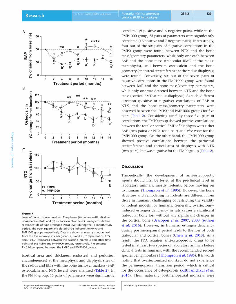
The increasing age of the postmenopausal monkeys during the 16month study period in the PMP0 group resulted in a significant decrease in plasma BAP (at months 2 and 8) and osteocalcin levels (starting from month 10), while no significant changes were detected in the NTX levels (Fig. 7A–C).
Treatment of the PMP1000 decreased the plasma BAP and osteocalcin levels to a greater degree than those observed in the PMP0 group (Fig. 7A and B), and this was significant from month 4 (P < 0.05 or 0.01) comparedwith the baseline values. Moreover, the NTX level was also significantly lower than the baseline value at month 16 (P < 0.01) in the PMP1000 group (Fig. 7C). Thus, thelevels of BAP and osteocalcin in the PMP1000 group were significantly lower than those in the PMP0 group starting from months 4 and 6, respectively.
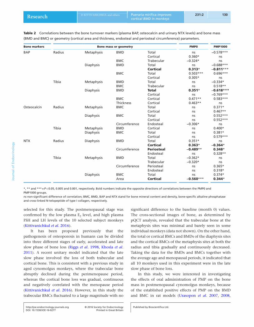
Correlation between bone mass/geometry and bone turnover markers
Correlations between the bone mass (total, cortical and trabecular BMC and BMD) and the geometry
A B
C D
E F
G H
Figure 6Bone geometry, in terms of the cortical area, thickness, endosteal and periosteal circumference, at the diaphysis site of the (A, C, E, G) radius and (B, D, F, H) tibia bone during the 16-month study period. The open square and closed circle indicate the PMP0 and PMP1000 groups, respectively. Data are shown as mean ± s.e.m, derived from the five monkeys in each group. a, b and a′, b′ represent P < 0.05 and P < 0.01 compared between the baseline (month 0) and other time points of the PMP0 and PMP1000 groups, respectively. * represents P < 0.05 compared between the PMP0 and PMP1000 groups.
129Research D KITTIVANICHKUL and others Pueraria mirifica improves cortical BMD in monkeys
DOI: 10.1530/JOE-16-0277
Journ
alofEn
docrinology
http://joe.endocrinology-journals.org © 2016 Society for EndocrinologyPrinted in Great Britain
Published by Bioscientifica Ltd.
231:2
(cortical area and thickness, endosteal and periosteal circumferences) at the metaphysis and diaphysis sites of the radius and tibia with the bone turnover markers (BAP, osteocalcin and NTX levels) were analyzed (Table 2). In the PMP0 group, 15 pairs of parameters were significantly
correlated (9 positive and 6 negative pairs), while in the PMP1000 group, 23 pairs of parameters were significantly correlated (16 positive and 7 negative pairs). Interestingly, four out of the six pairs of negative correlations in the PMP0 group were found between NTX and the bone mass/geometry parameters, while only one each between BAP and the bone mass (trabecular BMC at the radius metaphysis), and between osteocalcin and the bone geometry (endosteal circumference at the radius diaphysis) were found. Conversely, six out of the seven pairs of negative correlations in the PMP1000 group were found between BAP and the bone mass/geometry parameters, while only one was detected between NTX and the bone mass (cortical BMD at radius diaphysis). As such, different direction (positive or negative) correlations of BAP or NTX and the bone mass/geometry parameters were observed between the PMP0 and PMP1000 groups for five pairs (Table 2). Considering carefully those five pairs of correlations, the PMP0 group showed positive correlations between the total or cortical BMD of diaphysis with either BAP (two pairs) or NTX (one pair) and vice versa for the PMP1000 group. On the other hand, the PMP1000 group showed positive correlations between the periosteal circumference and cortical area of diaphysis with NTX (two pairs), but was negative for the PMP0 group (Table 2).
Discussion
Theoretically, the development of antiosteoporotic agents should first be tested at the preclinical level in laboratory animals, mostly rodents, before moving on to humans (Thompson et al. 1995). However, the bone structure and remodeling in rodents are different from those in humans, challenging or restricting the validity of rodent models for humans. Generally, ovariectomyinduced estrogen deficiency in rats causes a significant trabecular bone loss without any significant changes in the cortical bone (Urasopon et al. 2007, 2008, Suthon et al. 2016). However, in humans, estrogen deficiency during postmenopausal period leads to the loss of both trabecular and cortical bones (Chen et al. 2013). As a result, the FDA requires antiosteoporotic drugs to be tested in at least two species of laboratory animals before clinical tests in humans, with the recommended second species being monkeys (Thompson et al. 1995). It is worth noting that ovariectomized monkeys do not experience the perimenopausal transition period, which is critical for the occurrence of osteoporosis (Kittivanichkul et al. 2016). Thus, naturally postmenopausal monkeys were
B
C
A
Figure 7Level of bone turnover markers. The plasma (A) bone-specific alkaline phosphatase (BAP) and (B) osteocalcin plus the (C) urinary cross-linked N-telopeptide of type I collagen (NTX) levels during the 16-month study period. The open square and closed circle indicate the PMP0 and PMP1000 groups, respectively. Data are shown as mean ± s.e.m, derived from the five monkeys in each group. a, b and a′, b′ represent P < 0.05 and P < 0.01 compared between the baseline (month 0) and other time points of the PMP0 and PMP1000 groups, respectively. * represents P < 0.05 compared between the PMP0 and PMP1000 groups.
Research 130Pueraria mirifica improves cortical BMD in monkeys
DOI: 10.1530/JOE-16-0277
Journ
alofEn
docrinology
D KITTIVANICHKUL and others
http://joe.endocrinology-journals.org © 2016 Society for EndocrinologyPrinted in Great Britain
Published by Bioscientifica Ltd.
231:2
selected for this study. The postmenopausal stage was confirmed by the low plasma E2 level, and high plasma FSH and LH levels of the 10 selected subject monkeys (Kittivanichkul et al. 2016).
It has been proposed previously that the pathogenesis of osteoporosis in humans can be divided into three different stages of early, accelerated and late slow phase of bone loss (Riggs et al. 1998, Khosla et al. 2011). A recent unitary model indicated that the late slow phase involved the loss of both trabecular and cortical bone. This is consistent with a previous study in aged cynomolgus monkeys, where the trabecular bone abruptly declined during the perimenopause period, whereas the cortical bone loss was gradual, continuous and negatively correlated with the menopause period (Kittivanichkul et al. 2016). However, in this study the trabecular BMCs fluctuated to a large magnitude with no
significant difference to the baseline (month 0) values. The crosssectional images of bone, as determined by pQCT analysis, revealed that the trabecular bone at the metaphysis sites was minimal and barely seen in some individual monkeys (data not shown). On the other hand, the total or cortical BMCs and BMDs of the diaphysis sites and the cortical BMCs of the metaphysis sites at both the radius and tibia gradually and continuously decreased. Taking the data for the BMDs and BMCs together with the average age and menopausal periods, it indicated that all 10 monkeys used in this experiment were in the late slow phase of bone loss.
In this study, we were interested in investigating the effects of oral administration of PMP on the bone mass in postmenopausal cynomolgus monkeys, because of the established positive effects of PMP on the BMD and BMC in rat models (Urasopon et al. 2007, 2008,
Table 2 Correlations between the bone turnover markers (plasma BAP, osteocalcin and urinary NTX levels) and bone mass
(BMD and BMC) or geometry (cortical area and thickness, endosteal and periosteal circumference) parameters.
Bone markers Bone mass or geometry PMP0 PMP1000
BAP Radius Metaphysis BMD Total ns −0.578***Cortical 0.360* ns
BMC Trabecular −0.324* nsDiaphysis BMD Total ns −0.688***
Cortical 0.313* −0.811***BMC Total 0.503*** 0.696***
Cortical 0.305* nsTibia Metaphysis BMD Total ns −0.334*
BMC Trabecular ns 0.518**Diaphysis BMD Total 0.351* −0.618***
Cortical ns −0.769***BMC Cortical 0.471** 0.583***Thickness Cortical 0.463** ns
Osteocalcin Radius Metaphysis BMC Total ns 0.371*Cortical ns 0.467**
Diaphysis BMC Total ns 0.552***Cortical ns 0.552***
Circumference Endosteal −0.306* nsTibia Metaphysis BMD Cortical ns 0.400*
Diaphysis BMC Total ns 0.381*Cortical ns 0.579***
NTX Radius Diaphysis BMD Total 0.351* nsCortical 0.363* −0.364*
Circumference Periosteal −0.489** 0.348*Endosteal ns 0.328**
Tibia Metaphysis BMD Total −0.362* nsTrabecular −0.320* ns
Circumference Periosteal ns 0.365*Endosteal ns 0.318*
Diaphysis BMC Total ns 0.374*Area Cortical −0.560*** 0.344*
*, ** and *** = P < 0.05, 0.005 and 0.001, respectively. Bold numbers indicate the opposite directions of correlations between the PMP0 and PMP1000 groups.ns non-significant difference of correlation; BMC, BMD, BAP and NTX stand for bone mineral content and density, bone-specific alkaline phosphatase and cross-linked N-telopeptide of type I collagen, respectively.
131Research D KITTIVANICHKUL and others Pueraria mirifica improves cortical BMD in monkeys
DOI: 10.1530/JOE-16-0277
Journ
alofEn
docrinology
http://joe.endocrinology-journals.org © 2016 Society for EndocrinologyPrinted in Great Britain
Published by Bioscientifica Ltd.
231:2
Tiyasatkulkovit et al. 2012, Suthon et al. 2016). With regard to the in vitro study in rat osteoblastlike UMR106 cells, the PME could enhance expression of gene associated with osteoblast differentiation (i.e. alkaline phosphatase) and suppress expression of gene associated with osteoclast differentiation (i.e. receptor activator of nuclear factor kappaB ligand (Rankl)). Besides, PME also increased the expression of osteoprotegerin (Opg), a decoy protein receptor of RANKL, mRNA level (Tiyasatkulkovit et al. 2012). Although it was reported that PME could enhance the Opg/Rankl ratio in baboon primary osteoblast (Tiyasatkulkovit et al. 2014), and could also decrease the serum parathyroid hormone levels in postmenopausal monkeys (Trisomboon et al. 2004), it was still required to test if PMP or PME can ameliorate bone loss in naturally postmenopausal monkeys.
With respect to this relatively longterm study (16 months) in postmenopausal monkeys, the two parameters age and estrogen deficiency, although not related, were evaluated for changes in the bone mass. Interestingly, either the age or PMP treatment induced or extenuated the loss of BMD and BMC, principally at the diaphysis sites and especially in the cortical bone. The cortical, but not the trabecular, region is the predominant site of bone fractures in agerelated osteoporotic patients (Zebaze et al. 2010). Several studies have reported that the cortical bone becomes more brittle and weaker with advancing age (McCalden et al. 1993). Generally, most bone fractures occur in the long bones that mainly consist of cortical bone tissue (Li et al. 2013), and so drug targeting cortical bone should be prescribed for agerelated bone loss late in life (Zebaze et al. 2010). Indeed, bone cells comprise both estrogen receptor (ER)α and ERβpositive cells (Bordet al. 2001), and the phytoestrogens in PMP can bind with either ERβ or ERα (Setchell 1998). Thus, PMP shouldhave the potential to increase the trabecular as well as the cortical bone. However, changes in the trabecular bone of the PMPtreated monkeys were minimal and so PMP might affect the cortical bone to a greater extent in these postmenopausal aged monkeys. Thus, PMP could be a candidate for therapeutic intervention to reduce agerelated bone fractures in osteoporotic menopausal women.
To prevent bone fractures, we also need to pay attention to the skeletal structure or bone geometry in addition to the bone mass, as both the bone density and its size/shape are required to predict the risk of osteoporotic fracture. These results in postmenopausal monkeys are consistent with a previous report in menopausal women,
where expansions in the medullary cavity and periosteal diameter were observed in the radius (diaphysis) bone after menopause (Ahlborg et al. 2003). It is worth noting that the response in the tibia bone geometry to estrogen deprivation and increasing age (PMP0 group) was different from that in the radius. Both the endosteal and periosteal circumferences of the tibia bone fluctuated within the baseline values throughout the 16month period, but they tended to increase in the radius bone. This significant difference might be because of the effect of the weightbearing load on the tibia bone. The monkeys were kept in captivity in individual cages, and therefore rarely walked in a quadrupedal pattern, which resulted in a lower weightbearing load on the radius than on the tibia. Loading is known to directly regulate osteogenesis by increasing bone formation via mechanoresponsive cells (Ehrlich & Lanyon 2002). Moreover, estrogen deficiency has been proposed to reduce the sensitivity of bone cells to mechanical force resulting in a reduced bone mass (Frost 1992). Indeed, a synergistic relationship between estrogen and mechanical loading on bone metabolism has been described (Kohrt et al. 1995). Therefore, any compound that can stimulate ERs, like PMP, would be expected to increase the mechanostat and improve the mechanical properties of the tibia bone. We also found that the radius and tibia responded to PMP treatment differently, where in the radius PMP predominantly increased the bone mass, while in the tibia it mainly affected the bone structure. After 16 months of PMP treatment, the increased cortical BMD and BMC were more evident at the diaphysis site of the radius bone. However, PMP treatment decreased endosteal resorption (significantly decreased endosteal circumferences in Fig. 6F) and periosteal deposition (marginally decreased periosteal circumferences in Fig. 6H) of the tibia bone. This correlated with the effect of estrogen that promoted endosteal formation and inhibited periosteal apposition in humans (Vanderschueren et al. 2006). It has been reported previously that ERβ is responsible for the effect ofestrogen on the periosteal and endosteal surface (Bellido & Gallant 2014). Thus, the cooperative actions of PMP treatment and weight bearing support the different results between the radius and tibia observed in this study.
Together with the reduction in cortical bone mass, area and thickness during the 16month study period in the PMP0 monkeys, the plasma BAP and osteocalcin levels decreased (although they transiently fluctuated to a high degree) with time. This is consistent with a previous crosssectional study in baboon monkeys, which revealed

Research 132Pueraria mirifica improves cortical BMD in monkeys
DOI: 10.1530/JOE-16-0277
Journ
alofEn
docrinology
D KITTIVANICHKUL and others
http://joe.endocrinology-journals.org © 2016 Society for EndocrinologyPrinted in Great Britain
Published by Bioscientifica Ltd.
231:2
that the plasma BAP level was lowest among the oldest individuals (Havill et al. 2006). Moreover, it has been reported previously that serum osteocalcin levels were negatively correlated with the cortical diaphysis BMDs in postmenopausal monkeys (Kittivanichkul et al. 2016). Generally, osteocalcin is regarded as a bone turnover marker, where decreased plasma osteocalcin levels lead to a decreased bone turnover rate. Together with the lower plasma osteocalcin level in the PMP1000 group compared with the PMP0 group, our data also indicated the reduction of plasma calcium level in the PMP1000 group. Previously, it was reported that PMP could reduce serum parathyroid hormone and calcium levels in aged menopausal monkeys (Trisomboon et al. 2004). Thus, it might be hypothesized that PMP can play a part in reducing bone turnover rate through the parathyroid hormone and osteocalcin action, and might indirectly increase the cortical diaphysis BMD. Based on the fact that the rate of bone resorption (~3 weeks) and bone formation (~3 – 4 months) are not equal (Sims & Martin 2014), the reduced bone turnover might alleviate the cortical bone loss by uncoupling the osteoblast and osteoclast activity.
Although PMP treatment reduced in the plasma osteocalcin levels, no correlation between the osteocalcin levels and bone mass/geometry parameters was found. In contrast, positive correlations between the urinary NTX levels and bone geometry parameters (diaphysis periosteal circumference and cortical area) and negative correlations between the urinary NTX and plasma BAP levels with the bone mass parameters (diaphysis cortical and total BMDs) were observed following PMP1000 treatment. Taking these results of bone markers and bone mass/geometry parameters together, we can conclude that if the bone turnover (accounting for the BAP and NTX levels) increased in the PMP1000 group, the changes in bone geometry were in the opposite direction to the changes in the bone mass.
Although naturally menopausal cynomolgus monkeys are a more valuable animal model than ovariectomized animals to mimic the changes in the slow phase of bone loss in postmenopausal aged women, it is very difficult to obtain a large enough sample size of old animals in most primate facilities, which is a limitation for many researchers.
Declaration of interestThe authors declare that there is no conflict of interest that could be perceived as prejudicing the impartiality of the research reported.
FundingThis work was supported by the Grant for International Research Integration: Chula Research Scholar, Ratchadaphiseksomphot Endowment Fund (GCURS_58_06_23_02 to S Malaivijitnond) and the Thailand Research Fund through the Royal Golden Jubilee PhD Program (PHD/0097/2557 to D Kittivanichkul).
AcknowledgementsWe thank all members of the Veterinary Physiology Laboratory, Tokyo University of Agriculture and Technology, Japan for radioimmunoassay measurement and the veterinarians and animal caretakers at the National Primate Research Center of Thailand for animal procedure.
ReferencesAhlborg HG, Johnell O, Turner CH, Rannevik G & Karlsson MK 2003
Bone loss and bone size after menopause. New England Journal of Medicine 349 327–334. (doi:10.1056/NEJMoa022464)
Bellido T & Gallant KH 2014 Hormonal effect on bone cells. In Basic and Applied Bone Biology, pp 300–313. Eds DB Burr & MR Allen. San Diego, CA, USA: Academic Press. (doi:10.1016/B9780124160156.000150)
Bord S, Horner A, Beavan S & Compston J 2001 Estrogen receptors alpha and beta are differentially expressed in developing human bone. Journal of Clinical Endocrinology and Metabolism 86 2309–2314. (doi:10.1210/jcem.86.5.7513)
Bouxsein ML & Karasik D 2006 Bone geometry and skeletal fragility. Current Osteoporosis Reports 4 49–56. (doi:10.1007/s1191400600029)
Chen H, Zhou X, Fujita H, Onozuka M & Kubo KY 2013 Agerelated changes in trabecular and cortical bone microstructure. International Journal of Endocrinology 2013 213234. (doi:10.1155/2013/213234)
Ehrlich PJ & Lanyon LE 2002 Mechanical strain and bone cell function: a review. Osteoporosis International 13 688–700. (doi:10.1007/s001980200095)
Frost HM 1992 Perspectives: bone’s mechanical usage windows. Bone and Mineral 19 257–271. (doi:10.1016/01696009(92)90875E)
Goodman AL, Descalzi CD, Johnson DK & Hodgen GD 1977 Composite pattern of circulating LH, FSH, estradiol, and progesterone during the menstrual cycle in cynomolgus monkeys. Proceedings of the Society for Experimental Biology and Medicine 155 479–481. (doi:10.3181/0037972715539834)
Havill LM, Hale LG, Newman DE, Witte SM & Mahaney MC 2006 Bone ALP and OC reference standards in adult baboons (Papio hamadryas) by sex and age. Journal of Medical Primatology 35 97–105. (doi:10.1111/j.16000684.2006.00150.x)
Johnell O & Kanis JA 2006 An estimate of the worldwide prevalence and disability associated with osteoporotic fractures. Osteoporosis International 17 1726–1733. (doi:10.1007/s0019800601724)
Khosla S 2013 Pathogenesis of agerelated bone loss in humans. Journals of Gerontology: Series A, Biological Sciences and Medical Sciences 68 1226–1235. (doi:10.1093/gerona/gls163)
Khosla S, Melton LJ 3rd & Riggs BL 2011 The unitary model for estrogen deficiency and the pathogenesis of osteoporosis: is a revision needed? Journal of Bone and Mineral Research 26 441–451. (doi:10.1002/jbmr.262)
Khosla S, Oursler MJ & Monroe DG 2012 Estrogen and the skeleton. Trends in Endocrinology and Metabolism 23 576–581. (doi:10.1016/j.tem.2012.03.008)
Kittivanichkul D, Watanabe G, Nagaoka K & Malaivijitnond S 2016 Changes in bone mass during the perimenopausal transition in naturally menopausal cynomolgus monkeys. Menopause 23 87–99. (doi:10.1097/GME.0000000000000556)
Kohrt WM, Snead DB, Slatopolsky E & Birge SJ Jr 1995 Additive effects of weightbearing exercise and estrogen on bone mineral density in

133Research D KITTIVANICHKUL and others Pueraria mirifica improves cortical BMD in monkeys
DOI: 10.1530/JOE-16-0277
Journ
alofEn
docrinology
http://joe.endocrinology-journals.org © 2016 Society for EndocrinologyPrinted in Great Britain
Published by Bioscientifica Ltd.
231:2
older women. Journal of Bone and Mineral Research 10 1303–1311. (doi:10.1002/jbmr.5650100906)
Legrand JJ, Fisch C, Guillaumat PO, Pavard JM, Attia M, De Jouffrey S & Claude JR 2003 Use of biochemical markers to monitor changes in bone turnover in cynomolgus monkeys. Biomarkers 8 63–77. (doi:10.1080/1354750021000042448)
Li S, AbdelWahab A & Silberschmidt VV 2013 Analysis of fracture processes in cortical bone tissue. Engineering Fracture Mechanics 110 448–458. (doi:10.1016/j.engfracmech.2012.11.020)
Malaivijitnond S 2012 Medical applications of phytoestrogens from the Thai herb Pueraria mirifica. Frontiers in Medicine 6 8–21. (doi:10.1007/s1168401201848)
McCalden RW, McGeough JA, Barker MB & CourtBrown CM 1993 Agerelated changes in the tensile properties of cortical bone. The relative importance of changes in porosity, mineralization, and microstructure. Journal of Bone and Joint Surgery. American Volume 75 1193–1205.
Nelson HD, Humphrey LL, Nygren P, Teutsch SM & Allan JD 2002 Postmenopausal hormone replacement therapy: scientific review. JAMA 288 872–881. (doi:10.1001/jama.288.7.872)
Odvina CV, Zerwekh JE, Rao DS, Maalouf N, Gottschalk FA & Pak CY 2005 Severely suppressed bone turnover: a potential complication of alendronate therapy. Journal of Clinical Endocrinology and Metabolism 90 1294–1301. (doi:10.1210/jc.20040952)
R Core Team 2015 R: a language and environment for statistical computing. R Foundation for Statistical Computing, Vienna, Austria. http://www.Rproject.org/.
Riggs BL, Khosla S & Melton LJ 3rd, 1998 A unitary model for involutional osteoporosis: estrogen deficiency causes both type I and type II osteoporosis in postmenopausal women and contributes to bone loss in aging men. Journal of Bone and Mineral Research 13 763–773. (doi:10.1359/jbmr.1998.13.5.763)
Schaffler MB & Burr DB 1984 Primate cortical bone microstructure: relationship to locomotion. American Journal of Physical Anthropology 65 191–197. (doi:10.1002/ajpa.1330650211)
Setchell KD 1998 Phytoestrogens: the biochemistry, physiology, and implications for human health of soy isoflavones. American Journal of Clinical Nutrition 68 1333S1346S.
Sims NA & Martin TJ 2014 Coupling the activities of bone formation and resorption: a multitude of signals within the basic multicellular unit. BoneKEy Reports 3 481. (doi:10.1038/bonekey.2013.215)
Suthon S, Jaroenporn S, Charoenphandhu N, Suntornsaratoon P & Malaivijitnond S 2016 Antiosteoporotic effects of Pueraria candollei var. mirifica on bone mineral density and histomorphometry in estrogendeficient rats. Journal of Natural Medicines 70 225–233. (doi:10.1007/s1141801609655)
Sweet MG, Sweet JM, Jeremiah MP & Galazka SS 2009 Diagnosis and treatment of osteoporosis. American Family Physician 79 193–200.
Thompson DD, Simmons HA, Pirie CM & Ke HZ 1995 FDA Guidelines and animal models for osteoporosis. Bone 17 125S–133S. (doi:10.1016/87563282(95)00285l)
Tiyasatkulkovit W, Charoenphandhu N, Wongdee K, Thongbunchoo J, Krishnamra N & Malaivijitnond S 2012 Upregulation of osteoblastic differentiation marker mRNA expression in osteoblastlike UMR106 cells by puerarin and phytoestrogens from Pueraria mirifica. Phytomedicine 19 1147–1155. (doi:10.1016/j.phymed.2012.07.010)
Tiyasatkulkovit W, Malaivijitnond S, Charoenphandhu N, Havill LM, Ford AL & VandeBerg JL 2014 Pueraria mirifica extract and puerarin enhance proliferation and expression of alkaline phosphatase and type I collagen in primary baboon osteoblasts. Phytomedicine 21 1498–1503. (doi:10.1016/j.phymed.2014.06.019)
Trisomboon H, Malaivijitnond S, Suzuki J, Hamada Y, Watanabe G & Taya K 2004 Longterm treatment effects of Pueraria mirifica phytoestrogens on parathyroid hormone and calcium levels in aged menopausal cynomolgus monkeys. Journal of Reproduction and Development 50 639–645. (doi:10.1262/jrd.50.639)
Urasopon N, Hamada Y, Asaoka K, Cherdshewasart W & Malaivijitnond S 2007 Pueraria mirifica, a phytoestrogenrich herb, prevents bone loss in orchidectomized rats. Maturitas 56 322–331. (doi:10.1016/j.maturitas.2006.09.007)
Urasopon N, Hamada Y, Cherdshewasart W & Malaivijitnond S 2008 Preventive effects of Pueraria mirifica on bone loss in ovariectomized rats. Maturitas 59 137–148. (doi:10.1016/j.maturitas.2008.01.001)
Vanderschueren D, Venken K, Ophoff J, Bouillon R & Boonen S 2006 Clinical review: sex steroids and the periosteum—reconsidering the roles of androgens and estrogens in periosteal expansion. Journal of Clinical Endocrinology and Metabolism 91 378–382. (doi:10.1210/jc.20051766)
Weinbauer GF, Niehoff M, Niehaus M, Srivastav S, Fuchs A, Van Esch E & Cline JM 2008 Physiology and Endocrinology of the Ovarian Cycle in Macaques. Toxicologic Pathology 36 7S23S. (doi:10.1177/0192623308327412)
Woolf AD 2006 The global perspective of osteoporosis: the ILAR lecture, June 2004, Berlin. Clinical Rheumatology 25 613–618. (doi:10.1007/s1006700602657)
Zebaze RM, GhasemZadeh A, Bohte A, IulianoBurns S, Mirams M, Price RI, Mackie EJ & Seeman E 2010 Intracortical remodelling and porosity in the distal radius and postmortem femurs of women: a crosssectional study. Lancet 375 1729–1736. (doi:10.1016/S01406736(10)603200)
Received in final form 31 August 2016Accepted 6 September 2016Accepted Preprint published online 6 September 2016