pulpotomies for primary teeth july 2010
DESCRIPTION
pediatric dentistryTRANSCRIPT

PULPOTOMIES FOR PRIMARY TEETH
I. Pulpal Anatomy and Physiology in Primary Teeth

Dental and Pulpal Anatomy
The enamel and dentine of primary teeth are thinner than in permanent teeth
The pulps of primary teeth are relatively larger than those of permanent teeth
The pulp chambers of primary mandibular molars are relatively larger than those of maxillary molars
There are pulp horns under each primary tooth cusp, and these can be quite prominent


Dental and Pulpal Anatomy cont.
• The mesial pulp horns in primary molars are usually closer to the occlusal surface than the distal pulp horns, and therefore easier to expose
• The outline of the pulp chamber roof follows that of the occlusal surface
• Primary molars have a thin pulpal floor which may have accessory canals that communicate with the inter-radicular area
•

Dental and Pulpal Anatomy cont.
The root canals of primary molars can be narrow, ribbon-like and tortuous, and with deposition of secondary dentine, multiple canals may form with many ramifications
Maxillary molars usually have 4 canals (2 in the mesio-buccal root)
Mandibular molars usually have 3 canals (2 in the mesial root)
Dentine
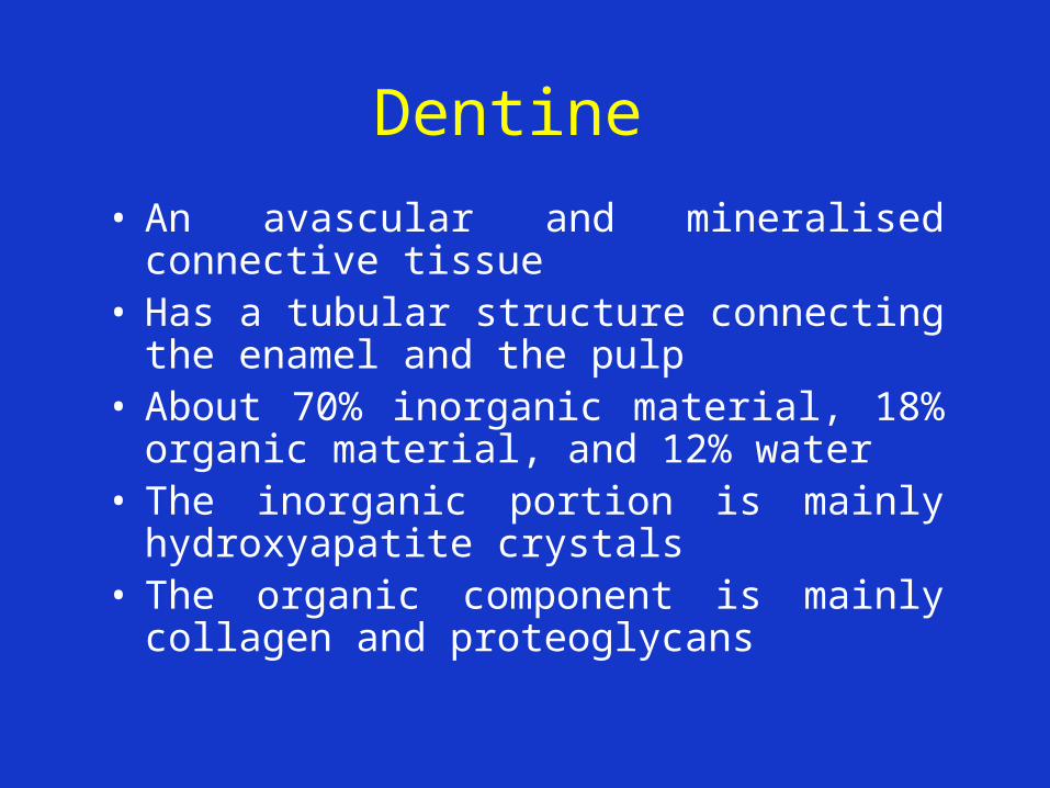
• An avascular and mineralised connective tissue
• Has a tubular structure connecting the enamel and the pulp
• About 70% inorganic material, 18% organic material, and 12% water
• The inorganic portion is mainly hydroxyapatite crystals
• The organic component is mainly collagen and proteoglycans
Dentine cont.
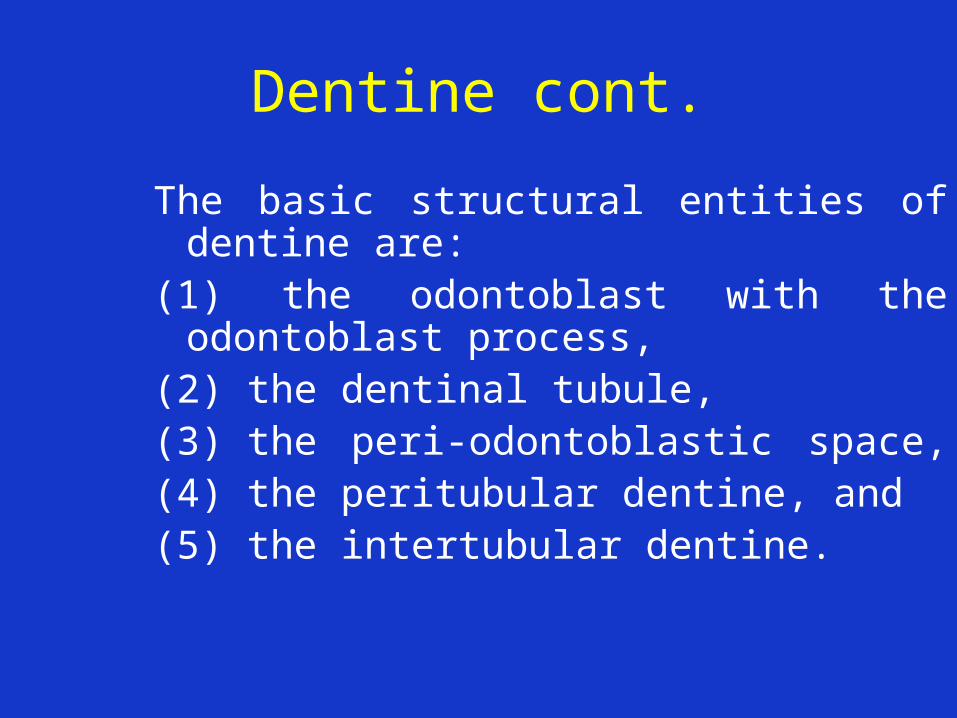
The basic structural entities of dentine are:
(1) the odontoblast with the odontoblast process,
(2) the dentinal tubule, (3) the peri-odontoblastic space, (4) the peritubular dentine, and (5) the intertubular dentine.


Odontoblasts
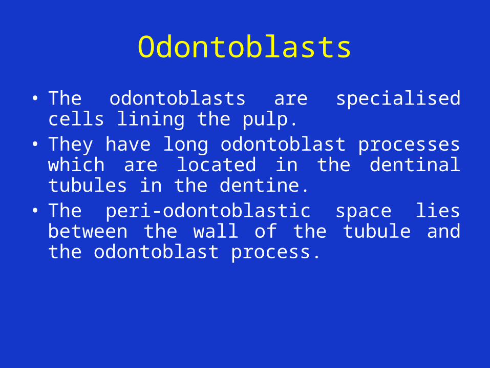
• The odontoblasts are specialised cells lining the pulp.
• They have long odontoblast processes which are located in the dentinal tubules in the dentine.
• The peri-odontoblastic space lies between the wall of the tubule and the odontoblast process.


Odontoblasts cont.
• The peri-odontoblastic space contains tissue fluid and a few organic constituents.
• The odontoblast processes and tubules may branch, particularly peripherally.
• Intercellular junctions are found between odontoblasts, and between odontoblasts and the nerve-like fibroblasts in the pulp, and may be important in the transmission of nerve impulses.

Primary and Secondary Dentine
• Primary dentine is formed during tooth development.
• Secondary dentine is formed after the crown of the tooth is fully formed. This slowly reduces the size of the pulp chamber with age.
• The number and course of the tubules in the secondary dentine become more irregular due to progressive crowding as the odontoblasts move towards the pulp.


Secondary Dentine cont.
• Also, peritubular dentine is laid down with increasing age and may eventually completely obliterate the tubules.
• Under areas of external irritation such as attrition, caries and restorations increased depositions of secondary dentine with a particularly irregular structure are found.


The Pulp
• The dentine and the pulp constitute the major
part of the tooth. • The pulp is surrounded by dentine except at the
apical foramen where it communicates with the periodontal tissues.
• There is an intimate relationship between the dentine and pulp both developmentally and functionally.

The Pulp cont.
• The pulp is a loose connective tissue made up of nerves, blood vessels and connective tissue.
• It is about 25% organic material and 75% water by weight.
• The most common cells in the pulp are fibroblasts - flattened cells with an oval nucleus.
• Other cells are also found, for example, the predentine is lined by a layer of specialised pulp cells, the odontoblasts.

The Pulp cont.
• Small blood vessels (arterioles and venules) enter and leave the pulp through the apical foramen, and through any accessory root canals.
• The arterioles end in a dense capillary network especially in the odontoblastic and sub-odontoblastic region.
• Because the pulp is rich in blood vessels local trauma or irritation can result in a hyperaemic reaction.

The Pulp cont.
• Pulp stones are islands of mineralised material in the pulp that are more common in pathologically altered teeth.
• These are sometimes seen on radiographs.• With increasing age the pulp changes from a
cell-rich and fibre-poor young tissue, to a more cell-poor and fibre-rich old tissue.
• These changes are accentuated if the teeth are subjected to external irritation eg attrition, caries, or restorative procedures.
• The functional processes within the pulp also decrease with age.

CLINICAL PULP TREATMENT FOR PRIMARY TEETH

Why pulp therapy for primary teeth?
• Important to maintain primary teeth for the development of dentition (maintain arch length)
• Prevent infection and pain• Can help to nurture a positive attitude about oral
health in children• Can preserve primary tooth where permanent
successor is missing• Can helps maintain masticatory function• Preserves aesthetics• Sends message that “primary teeth are
important!”

Space Loss Due to Premature Extraction of Primary Molar


What is a pulpotomy?
• Vital amputation of the (inflamed or infected) coronal pulp
• Wound surfaces at orifaces of root canals are then treated in order to preserve vitality of radicular pulp, and retain the tooth
• Success depends on type of wound dressing, pulpal diagnosis at time of treatment, amputation technique and treatment of wound surface

Large carious pulp exposure

PULPOTOMY

Properties of an Ideal Pulpotomy Material
• Bactericidal
• Harmless to the pulp, surrounding tissues and underlying tooth germ
• Promote healing
• Not interfere with the physiological process of root resorption

Types of Medicament
Two main groups:– Calcium hydroxide preparations – stimulate
a hard tissue barrier; promote healing; eg Vitapex, plain Ca(OH)2
– Devitalizing medicaments – aim to maintain primary tooth irrespective of pulpal condition eg formocresol, Ledermix, glutaraldehyde, ferric sulphate
– Also - MTA

(1) Calcium Hydroxide• Strongly alkaline (pH about 12)• Irritation causes initial necrosis adjacent to vital
pulp tissue• Pulp responds with an inflammatory reaction• Later repair process starts with proliferation of
cells and formation of new collagen• Odontoblasts differentiate and form a hard
dentine-like tissue (but this is NOT a tight seal)• Only successful for healthy pulps and those with
partial chronic reversible pulpitis • Success rates only about 60% in some studies

Complications of use of Ca(OH)2
• Internal dentine resorption
• Often due to chronic inflammation of the residual pulp, or the presence of a blood clot over the pulp wound surface
• If placed in the right situation directly onto the pulp wound, can be very successful
(2) Formocresol
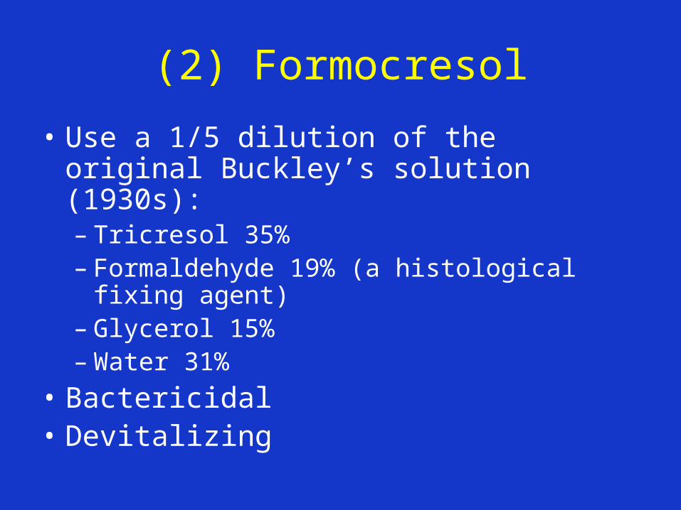
• Use a 1/5 dilution of the original Buckley’s solution (1930s):– Tricresol 35% – Formaldehyde 19% (a histological fixing
agent)– Glycerol 15%– Water 31%
• Bactericidal• Devitalizing

Formocresol (cont)
• Aim to create a chemically altered zone at the pulp-medicament interface – leaving the deeper untreated pulp tissue vital and un-inflamed
• Diffuses into the pulp tissue - degree of penetration is time and dose-dependent
• May end up with chronic inflammation or even partial necrosis of residual pulp

Formocresol cont.
• Higher success rate than calcium hydroxide
• Systemic absorption of FC shown in animal studies where large nos of teeth had pulpotomies with full strength FC
• Suggested FC may have immunogenic, toxic, mutagenic potential – but no data to support toxic effects from pulpotomies in humans

Complications of Use of Formocresol
• Can get chronic inflammation or necrosis of residual pulp – but clinical and radiological problems may not occur for several years
• One early study reported a few cases of hypoplasia of underlying permanent tooth
• Success rates vary – clinical success rates over 90%; radiographic success >60% in most studies

(3) Ferric Sulphate (15.5%)
• Used in dentistry to control gingival bleeding eg before crown impression taking (called Astringident)
• Forms a ferric ion-protein complex on contact with blood seals vessels
• Clinical studies show similar clinical success to formocresol
• Little chance of success if pulp in roots inflamed

(4) Glutaraldehyde
• Better fixative cf FC
• Larger molecules less penetration
• Toxic properties eg allergies, eye irritation
• Seldom used

Other Methods
• Electrosurgery (may be in combination with FC)
• Lasers
• MTA

Radiographs
• Must have preoperative radiograph which shows the furcation area (or preferably the whole tooth) – PA is best
• Should also have a post-op radiograph
• Follow up radiograph at 6 months and then annually

PULPOTOMY TECHNIQUE
Steps
• Pre-op radiograph• Isolate tooth• Remove all caries• Remove roof of pulp chamber with high speed bur• Remove pulp with no. 6 or 8 round low speed burs
and/or large sharp excavator (do NOT cut through furcation!)
• Arrest bleeding from root orifaces with cotton pellets• Place damp cotton pellet with FC or FS in pulp chamber
for 1-5 min• Mix ZOE or IRM (thick mix) and fill pulp chamber• Restore tooth eg with ss crown


Note:
• Studies show that if the marginal ridge is broken down, there is often inflammation of the pulp horn
• Direct pulp capping has a poor success rate in primary teeth – pulpotomy better
• In Cambodia there are many abscessed teeth following placement of deep restorations because pulp already inflamed
• Teeth with pulpotomies tend to exfoliate early

INDICATIONS FOR PULPOTOMY
• Large carious lesion with substantial loss of marginal ridge
• Restorable tooth• No radicular pulpitis:
– No spontaneous / persistent / severe pain– No persistent bleeding (or pus) from radicular amputation site
• No abscess or fistula or swelling or mobility• >1/2 of root remaining• No periapical/furcal bone loss• No internal resorption of crown or root• Bleeding disorders


CONTRA-INDICATIONS FOR PULPOTOMY
• Unrestorable tooth• Abscess/fistula/swelling/mobility• Periapical / furcal radiolucency• <2/3 of root remaining• Internal resorption• Continued bleeding after application of FC• Medical conditions:
– Heart problems requiring Ab cover– immunocompromised


Suitable Case (if pulp healthy)

Unsuitable Case – Why?

ABSCESS

Unsuitable case – Why?


Pulpotomized Teeth are Best Restored with SS Crowns

FOLLOW UP
• Review clinically and radiographically at 6 mo and then annually
• PA or PBWs which show furcation area
• Appearance of a radiolucency, internal resorption, or abscess indicate failure extraction or pulpectomy

Complications/Failures

Notes
• If health of pulp doubtful, some dentists add a drop of FC the ZOE/IRM mix