purkinje cells of rat and chicken cerebellum contain ... proteins from rat and chicken cerebel- lum...
TRANSCRIPT

Volume 294, number 1,2. 47 50 FEBS 10445 c 1991 Federation of European Biochemical Societies 0014S793/91/$3.50
December 1991
Purkinje cells of rat and chicken cerebellum contain calreticulin (CaBP3)
Dominique Perk, Birte Sbnnichsen, Hans-Dieter Siiling and
A htrilwq Kliuis& Biochernic~. Zentrum Itmere Medizin, Robert Koch Strasse 40,
Received 14 October 1991
Phuc Nguyen-Van
D-3400 Giittingen, Germany
The presence of the calcium-binding protein calreticulin (CaBP3) was assessed in rat and chicken cerebellum by immunoblotting, and its localization in Purkinje cells was established by immunocytochemistry and in situ hybridization.
Calreticulin; Purkinje cell
1. INTRODUCTION 2.2. Methods
Calrcticulin (CaBP3) is a low affinity, high capacity calcium-binding protein which has been found in rat liver and brain [I,21 and subsequently in adrenal me- dulla and cortex, and in endothelial cells (Nguyen-Van and Perrin, unpublished results). This protein has an apparent molecular mass of 60 kDa on SDS-PAGE, and a molecular mass of 46 kDa has been established by molecular cloning [3,4]. Calreticulin is found mainly in the ER [3] and in vesicles called calciosomes ([2], see [5] as a review). Its function remains unclear although a role in calcium sequestration has been proposed. It seemed therefore surprising that calreticulin could not be found in the rat cerebellum and particularly not in Purkinje cells from rat [6] and chicken cerebellum [5,6]. On the other hand, the presence of calsequestrin, a cal- cium-binding protein presenting an apparent molecular mass of 5 1 kDa observed otherwise only in muscle cells has been described for Purkinje cells from chicken, but not from other species [7].
Our results demonstrate clearly the presence of calre- ticulin in Purkinje cells of both rat and chicken by im- munochemical and by in situ hybridization methods.
2. MATERIALS AND METHODS
2.1. Materials Chicken cerebellum came from the Institut fiir Tierhygiene. Rat
cerebellum was prepared from male Wistar rats. The random primed DNA labeling kit and the in situ hybridization
kit (DIG luminescent detection kit) as well as proteinase K were purchased from Boehringer Mannheim (Germany). The goat anti- rabbit IgG antibody coupled to peroxidase was purchased from Dia- nova Hamburg (Germany).
Ahhrevia~ions: DIG, digoxigenin; ER, endoplasmic reticulum; PBS, phosphate-buffered saline; dUTP, deoxyuridine trisphosphate.
Correspondence address; D. Perrin, Abeilung Klinische Biochemie, Zentrum Innere Medizin, Robert Koch Strasse 40, D-3400 Gdttingen, Germany.
Published by Elsevier Science Publishers B. V.
2.2. I. Preparation of cerebellum sections Rat and chicken cerebella were fixed in Bouin’s fixative, demyeli-
nated, embedded in paraffin and cut in 4 PM sections. Sections were subsequently dewaxed in xylene before use.
2.2.2. Immunocytochemistry Anti-calreticulin antibodies were raised in rabbit as described in [8].
Dewaxed cerebellum sections were incubated for 2 h in the presence of either preimmune serum or anti-calreticulin serum diluted 1:250, followed by an incubation for 2 h in the presence of a goat anti-rabbit IgG antibody coupled to peroxidase at a dilution of 1:250. AlI incuba- tions were performed at room temperature in PBS containing 10% fetal calf serum. Diaminobenzidine and H202 were used as substrates, and the sections were mounted in glycerol and examined in a Zeiss Axioskop microscope.
2.2.3. In situ hybridization A 750 bases PCR fragment from a rat liver cDNA clone coding for
the C-terminal region of calreticulin was used as a probe. This frag- ment missed the sequence coding for the ER retention signal KDEL to avoid crosshybridization with the mRNA of proteins displaying the same motive. Our probe was non-radioactively labeled with digoxige- nin-II-dUTP by the random priming method [9] according to the Boehringer random primed DNA labeling kit protocol. The control probe was prepared under the same conditions, but missed the cDNA fragment coding for calreticulin. In situ hybridization was performed under the conditions described in the protocol of the Boehringer DIG luminescent detection kit. Shortly, the dewaxed sections were treated for 2 min at 37°C with 50 pg/rnl proteinase K, and subsequently incubated in presence of the DIG-labeled probe for 10 h at 42°C. Afterwards the probes were labeled with anti-DIG antibodies coupled to alkaline phosphatase diluted 1:300 for 10 h at room temperature. Nitroblue tetrazolium chloride and 5-bromo-4-chloro-3-indolyl- phosphate were used as substrates. Controls were performed under the same conditions, the DIG-labeled cDNA probe being replaced the control probe.
2.2.4. Miscellaneous SDS-PAGE was performed according to [IO], and blotting onto
nitrocellulose according to [l 11. After blotting, nitrocellulose sheets were labeled with rabbit anti-rat calreticulin antibodies diluted to I:1000 and further incubated in presence of a goat anti-rabbit IgG antibody coupled to peroxidase at a dilution of 1:lOOO. All incubations were performed in 3% (w/v) milk powder in PBS.
Microsomes from rat and chicken cerebella were prepared as de- scribed in [8,12].
47
Volume 294, number 1,2 FEBS LETTERS
3. RESULTS AND DISCUSSION
December 1991
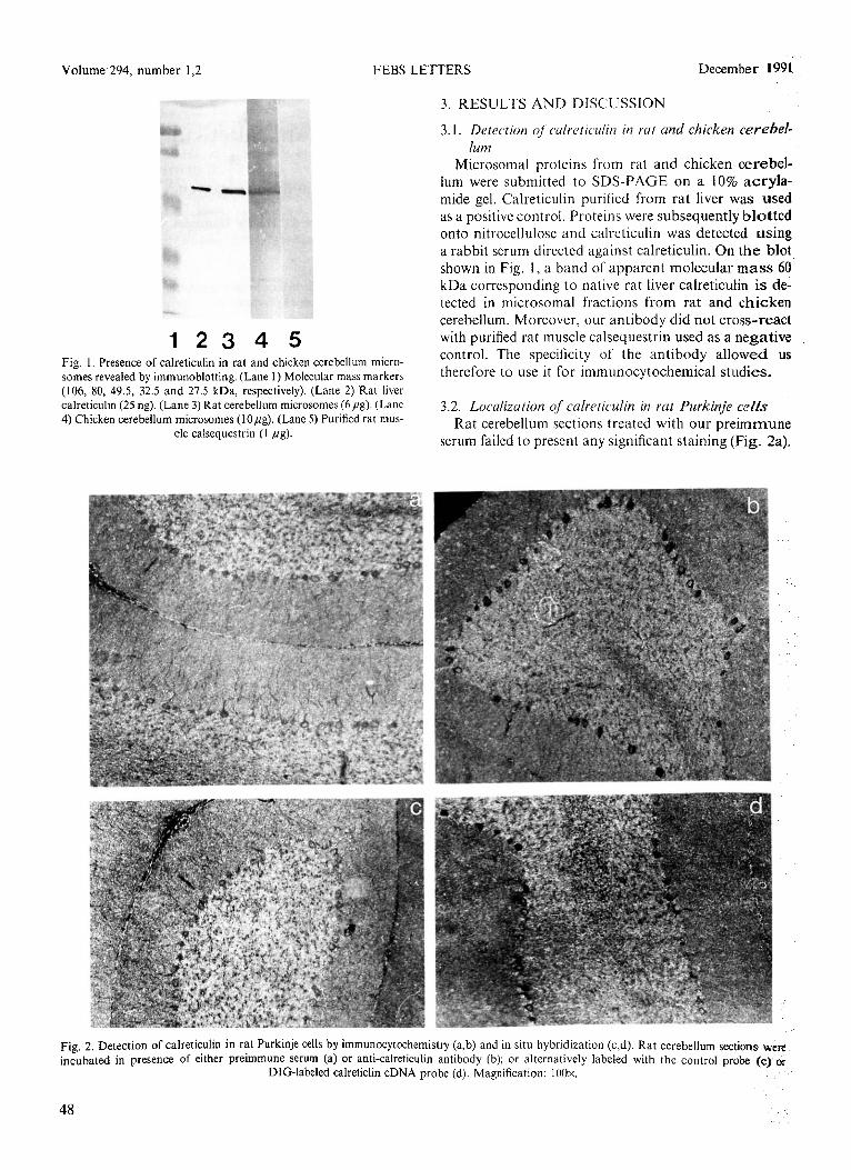
123 4 5 Fig. I. Presence of calreticulin in rat and chicken cerebellum micro- somes revealed by immunoblotting. (Lane 1) Molecular mass markers (106, 80, 49.5, 32.5 and 27.5 kDa, respectively). (Lane 2) Rat liver calreticulin (25 ng). (Lane 3) Rat cerebellum microsomes (6pg). (Lane 4) Chicken cerebellum microsomes (IOpg). (Lane 5) Purified rat mus-
cle calsequestrin (I fig).
3.1. Detection of cwlreticulin in rut and chicken cerebef- lum
Microsomal proteins from rat and chicken cerebel- lum were submitted to SDS-PAGE on a 10% acryla- mide gel. Calreticulin purified from rat liver was used as a positive control. Proteins were subsequently blotted onto nitroccllulose and calreticuhn was detected using a rabbit serum directed against calreticulin. On the blot shown in Fig. 1, a band of apparent molecular mass 60 kDa corresponding to native rat liver calreticulin is de- tected in microsomal fractions from rat and chicken cerebellum. Moreover, our antibody did not cross-react with purified rat muscle calsequestrin used as a negative control. The specificity of the antibody allowed us therefore to use it for immunocytochemical studies.
3.2. Localization qf calretidin in rut Purkinje cells Rat cerebellum sections treated with our preimrnune
serum failed to present any significant staining (Fig. 2a).
Fig, 2. Detection of calreticulin in rat Purkinje Cells by immunocytochemistry (a,b) and in situ hybridization (c,d). Rat cerebellum se&-ma were incubated in presence of either preimmune serum (a) or anti-calreticulin antibody (b); or alternatively labeled with the control probe [c) or
DIG-labeled calreticlin cDNA probe (d). Magnification: 100x.
48

Volume 294, number I 7 ,.- FEBS LETTERS December 199 1
Fig. 3. Localization of calreticulin in chicken cerebellum. Calreticulin was localized in chicken cerebellum sections by immunocytochemistry (a,b) using anti-calreticulin antibody (b). Preimmune serum was used as a control (a). Calreticulin mRNA was detected by in situ hybridization (c,d);
(c) control, (d), DIG-labeled calreticulin cDNA probe. Magnification: 100x.
In contrast, the sections incubated in presence of the antibody directed against calreticulin revealed an in- tense cytoplasmic labeling predominantly in Purkinje cells (Fig. 2b).
The DIG-labeled calreticulin cDNA probe was tested versus the control probe in rat liver sections, the liver being an organ rich in calreticulin. No staining was observed in the liver sections treated with the control probe. However, a strong signal was detected in the sections treated with the DIG-labeled calreticulin cDNA probe (data not shown). The same probes were used to label rat and chicken cerebellum sections.
One could hardly detect any signal in the rat cerebel- lum sections labeled with the control probe (Fig. 2~). In contrast, an intense staining was observed with the DIG-labeled calreticulin cDNA probe in the Purkinje cells of rat cerebellum (Fig. 2d).
3.3. Loculizcrtion of calreticulin in chicken Purkinje cells
In contrast with control sections (Fig. 3a), a specific staining of Purkinje cells was detected when chicken cerebellum sections were incubated in presence of our anti-rat calreticulin antibody (Fig. 3b). However, the intensity of calreticulin staining in Purkinje cells was apparently lower in chicken than in rat cerebellum sec-
tions. This result could reflect either a difference in the quantity of calreticulin present, or the fact that our anti-calsequestrin antibody recognizes better the rat than the avian protein. Moreover, the general back- ground level was higher in chicken than in rat cerebel- lum sections with either preimmune or immune serum.
Nevertheless, when in situ hybridization techniques were used, the contrast between control (Fig. 3c) and probe (Fig. 3d) was striking, demonstrating the massive presence of calreticulin mRNA in chicken Purkinje cells. Therefore, the difference between rat and chicken in terms of intensity of the calreticulin staining in Pur- kinje cells could be due to immunological rather than quantitative reasons.
Both immunological and in situ hybridization tech- niques clearly demonstrate the presence of the microso- ma1 calcium-binding protein calreticulin (CaBP3) in rat and chicken cerebellum, particularly in Purkinje cells. Those results are in line with the idea that calreticulin is an ubiquitous protein in vertebrate serving most likely an important although yet unknown function. Moreo- ver, the concomitant presence of calreticulin and calse- questrin in chicken Purkinje cells - as well as in skeletal muscle cells - raises the question of the relative localiza- tion and function of these two proteins.
49
Volume 294, number 1,2 FEBS LETTERS December 1991
Acknowledgements: We are grateful to Dr. Inge Brandt for the gen- erous gift of rat cerebellum and liver sections, to Antje Ape1 for skilled tissue preparation work, to Annette Germeshausen for technical sup- port, and to Anne-Helene Perrin-Voltz for critical reading of the manuscript.
REFERENCES
[I] Waisman, D.M., Smallwood, J., Lafreniere, E. and Rasmussen, H. (1983) Biochem. Biophys. Res. Commun. 119,440446.
[2] Treves, S., De Mattei, M., Lanfredi, M., Villa, A., Green, N.M., MacLennan, D.H., Meldolesi, J. and Pozzan, T. (1990), Biochem. J. 271,473-480.
[3] Smith, M.J. and Koch, G.L.E. (1989) EMBO J. 8, 3581-3586. [4] Fliegel, L., Burns, K., MacLennan, D.H., Reithmeier, R.A.F.
and Michalak, M. (1989) J. Biol. Chem. 269, 21522-21528.
[S] Meldolesi, J., Madeddu, L. and Po7m1, T. (1990) Biochim. Bio- phys. Acta 1055, 130.-140.
[6] Satoh, T., Ross, C.A., Villa, A., Supattapone, S., Pozzan, T., Snyder, S.H. and Mcldolesi, J. (1990) J. Cell Biol. 11, 615-624.
[7) Volpe, P., Alderson-Lang, B.H.. Madeddu, L., Damiani, E., Cal- lins, J.H. and Margreth, A. (1990) Neuron 5, 713-721.
[8] Nguyen-Van, P. and Soling, H.-D. (1989) J. Biol. Chem. 29, 17494-1501.
[9] Feinberg, A.P. and Vogelstein, B. (1983) Anal. Biochem. 132, 613.
[lo] Laemmli, U.K. (1970) Nature 227, 680.-688. [II] Towbin, H., Staehelin, T. and Gordon, J. (1979) Proc. Nat].
Acad. Sci. USA 76, 4350 4354. [l2] Dawson, PA. and Irvine, R.F. (1984) Biochem. Biophys. Res.
Commun. 120, 858 864.
50