rapid fire ecg challenge: putting your interpretation...
TRANSCRIPT

Evert Torrejon , MDSociedad Peruana Cardiologia
Presenter
Rapid Fire ECG Challenge: Putting Your
Interpretation Skills to the Test

Male 36 years old, athlete, with prior to start swimming, gastric discomfort
Life in the Fasttlane Dr Ed Burns

Point to the Diagnostics:
• Sinus bradycardia• Sinus Arrhythmia• ECG athlete• Ventricular Tachycardia• Accelerated idioventricular Rythm• Junctional Escape Rhythm.

Accelerated idioventricularRythm: 60 b.p.m
Sinus Arrhythmia
sinus capture

isorhythmic A-V dissociation

isorhythmic A-V dissociation:
• An ectopic ventricular rhythm that consists of three or more ventricular complexes that occur at a speed of 50-110 bpm.
• The frequency differentiates the AIVR from the ventricular escape rhythms (frequency <50 bpm) and TV (> 110 bpm).

isorhythmic A-V dissociation
• AV dissociation with complexes of sinus and ventricular origin that occur at a similar frequency, unlike BAV III, where atrial fr is usually greater than ventricular.
• DAVI: due to functional block in the AV node due to retrograde ventricular conduction, leaving it refractory to anterograde impulses

isorhythmic A-V dissociation:Mechanism
• Increased automaticity of ventricular ectopic pacemaker, associated with sinus bradycardia and sinus arrhythmia due to training in athletes (increased vagal tone and sympathetic decrease: hypervagotonicstate)

isorhythmic A-V dissociation : other causes
• During the reperfusion phase of an acute myocardial infarction• Toxicity of medications, especially digoxin, cocaine and volatile anesthetics such as
desflurane• Electrolyte abnormalities• Cardiomyopathy• Congenital heart disease• Myocarditis• After the return of spontaneous circulation in the post-cardiac arrest period

ECG athlete: anomalies due to autonomic conditioning: • Sinus arrhythmia• Sinus bradycardia• Rhythms of articular or ventricular escape.• AV Blocking I degree• Wenckebach phenomenon• Hypertrophy of the left ventricle on ECG• Axis deviation• Incomplete RBBB

A 70-year-old man with palpitations, dyspnea:
Fr 150QRS 160QRS EJE 130°

Point correct diagnosis:
• Ventricular tachycardia• SV tachycardia: intranodal reentry with aberrance• AV reentrant tachycardia antidromic• Tachycardia with pacemaker


68 years old woman with ischemic cardiomyopathy with CDI comes by fatigue and presincope
Matt Needleman :ISHNE ECG University 2018

What is your diagnosis?
• 1.SVT with Right Bundle Branch Block• 2. Atrioventricular reentrant antidromic tachycardia• 3. Atrial Flutter with 2:1 AV Block and Bundle Branch
Block • 4. Ventricular tachycardia


Wide QRS tachycardia
Regular Irregulars
SVT+ BBB SVT+AcVia
TV Pacemak FA T des P
Fixed
Intermit
Antidromic
Atrial tachy
Flutter
BBB
VA


Vereckei Criteria for the Diagnosis of Ventricular Tachycardia
• If there is an initial R wave in aVR: It is a Ventricular Tachycardia. If not, move on to the next
• If the width of the Wave Q or of the initial R wave is greater than 40 msec (a small square): It is a Ventricular Tachycardia. If not, move on to the next
• If there are notches or notches in the initial descending portion of a predominantly negative QRS complex: It is a Ventricular Tachycardia. If not, move on to the next
• If Vi / Vt is less than 1: It is a Ventricular Tachycardia. If not, move on to the next• If none of the previous statements are met: It is a Supraventricular Tachycardia.
Vereckei A, Duray G, Szenasi G, Altemose GT, Miller JM. New algorithm using only lead aVR for differential diagnosis of wide QRS complex tachycardia. Heart Rhythm. 2008


Other Ventricular Tachycardia Data
• Presence of beats with fusion and with capture• Ventriculo / auricular ratio> 1 (more QRS than P waves)• When they present Morphology of Right Branch Block, if QRS> 140
ms• When they present Morphology of Left Branch Block, if QRS> 160 ms• Cardiac axis between -90º and -180º (extreme deviation)

Capture

Fusion

Positive Concordance

Negative Concordance

Brugada sign (red tweezers) and Josephson's sign (blue arrow)

TV criteria if it presents a picture of Right Branch Block in V1
Rabbit ear higher left (RSr) on TV Right rabbit ear higher (SNR) in RBB

TV criteria if it presents a picture of Right Branch Block in V1
•In V1: Wide R wave only or with recesses, or Wave R greater than R '•In V6: Wave R less than Wave S

Criteria TV if you have left bundle branch block pattern in V1
•In V1: initial and wide r wave (> 30 msec), S wave in its descending portion and duration from the beginning of the QRS to the deepest point of the S wave greater than 60 ms.•In V6: Presence of initial Q wave (qR) or QS morphology.
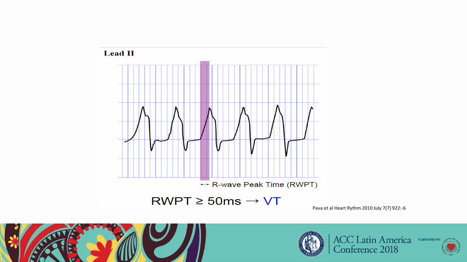
Pava et al Heart Rythm 2010 July 7(7) 922:-6


Woman 68 years old with ischemic cardiomyopathy with CDI comes by fatigue and presincope
Matt Needleman :ISHNE ECG University 2018
EjeQRS -90º,-QRS 160 msec,-Max peak QRS in second 50% of RR, -concordance only up to V5

Woman 68 years old with ischemic cardiomyopathy with CDI comes by fatigue and presincope
Matt Needleman :ISHNE ECG University 2018
EjeQRS -90º,-QRS 160 msec,-Max peak QRS in second 50% of RR, -concordance only up to V5
Fusion Beat
Fusion Beat

Male, 49 years of age. myocardial infarction history, goes by shortness of breath and palpitations.
Pdrid,s Real World ECGs Books 2016

What is your diagnosis?
• 1.- Synusal tachycardia with left bundle branch block• 2.- AV antidromic reentrant tachycardia• 3.- Atrial flutter with AV block 2/1, LBBB• 4.- Ventricular tachycardia

Male, 49 years of age. myocardial infarction history, goes by shortness of breath and palpitations.
Pdrid,s Real World ECGs Books 2016

Electrocardiogram after treatment
Pdrid,s Real World ECGs Books 2016

Female 71 years old: Sudden shortness of breath and dizzy: first ECG
Podrid,s Real World ECGs

What are possible diagnoses?
• 1.) Ventricular Tachycardia: Flutter V• 2.) AV reentry tachycardia antidromic• 3.) Sinus Tachycardia with Left Branch Block• 4.) Atrial tachycardia with aberrance• 6.) Tachycardia Atrial Flutter 1: 1 conduction


Female 71 years old: Sudden shortness of breath and dizzy: first ECG- Regular rhythm at 250 x min- QRS: 0.16 sec Typical LBBB.- For frequency atrial Flutter with 1: 1
conduction vs ventricular flutter
Podrid,s Real World ECGs
- Wave R is narrower (<0.08 msec) than the S wave in V2 to V4: consistent with SV rhythm with aberration of the Left Branch because the initial forces are normal, while the terminal portions, which is the aberratedpart responsible for the spreading of the QRS, for example a branch block.
Dg: atrial Flutter with LBBB conduction 1:1

ECG , A few minutes in Emergency:Regular rhythm 125 lpmwithout LBBQRS 0.08 sec
Atrial activity: 250 bpm with continuous flutter undulation
V4 to V6 transient conduction one to one with LBB

Evert Torrejon , MDSociedad Peruana Cardiologia
Presenter
Rapid Fire ECG Challenge: Putting Your
Interpretation Skills to the Test

48 year old woman with palpitations

What is your diagnosis ?:
• 1.-Sinus tachycardia with branch block• 2.-Atrial tachycardia with aberrance• 3.-Ventricular tachycardia left posterior fascicle• 4.-SV tachycardia with bifascicular block RBB + LAFB

48 year old woman with palpitationsQRS 120Morfología BRD:RSR’ en V1Eje -90ºCaptura Ondas P disociadas en II

Male 27 years: palpitations, pre-syncope, normal ECG, negative troponins and during stress test is observed:

After endovenous BB:

What is your diagnosis?
• Sinus tachycardia with aberrance• Atrial Tachycardia with Left Branch Block• Antidromic AV reentrant tachycardia• Ventricular tachycardia• Atrial fibrillation with blocking Left Branch

Male 27 years: palpitations, pre-syncope, normal ECG, negative troponins and during stress test is observed:

No pre-excitation, conclusion: TV tract output VD, "malignant"
Hossein Shenasa MD

Male, 22 years old, consulted for presyncopes preceded by rapid palpitations

What is your diagnosis?
• Sinus tachycardia with aberrance• Atrial Tachycardia with Left Branch Block• Antidromic AV reentrant tachycardia• Ventricular tachycardia origin tract output Right Ventricle• Atrial fibrillation with LBB

Male, 22 years old, consulted for presyncopes preceded by rapid palpitations

Woman, 18 years old, attends by palpitations, precordialgia, syncope

Woman, 18 years old, attends by palpitations, precordialgia, syncope
Wide QRS, similar to LBBQRS Front axle: Inferior
Predominant R II III aVF
Transition R / S> 1 beyond V2

Male 17 years old, with palpitations: passes to sinus rhythm with adenosine
Fr : 198QRS 180QT 288QTc 523Eje QRS -20

ECG Criteria for Distinguishing Left from Right Ventricular Outflow Tract Tachycardia• Outflow tract (OT) ventricular arrhythmia rep-re sents the most
common subgroup of idiopathic premature ventricular premature contractions (PVCs)/ventricular tachycardia (VT). It typically occurs in healthy young to middle-aged patients without structural heart disease and can be provoked by emotional stress, exercise, or dietary stimulants.1)Prognosis is generally excellent; OTVT can be effectively treated by drugs or radiofrequency (RF) catheter ablation.

ECG Criteria for Distinguishing Left from Right Ventricular Outflow Tract Tachycardia• Detailed intracardiac electrical mapping has shown that the vast majority of OTPVCs/OTVTs
originate from the right ventricular (RV) OT.3)However, in approximately 10% to 15% of cases, the arrhythmia originates from the left ventricular (LV) OT and can be mapped to the region of the aortic cusps.4) Typically, OTPVCs/VT originating in the RV manifests as an inferior axis in the frontal ECG plane and a left bundle branch block (LBBB) configurationwith precordial R/S transition at or after V3.5) In contrast, LVOT PVCs/VT usually manifests either as a right bundle branch block (RBBB)/inferior axis or LBBB/inferior axis with a precordial R/S-wave transition at or before lead V3.6) Criteria to distinguish an RVOT from an LVOT origin for patients with precordial transition occurring at lead V3 are lacking


Idiopathic TV• No Heart disease is demonstrated.• It represents 10% of all TV.• Subgroups: 1) TV of the outflow tract (TS), (88-90%):• RVOT (80-90%) (septal region posterosuperior most)• LVOT (10-20%): basal endocardium 60% cases in mitral-• aortic continuity - epicardial 40%
• 2) Fascicular TV, (10-12%)•• The TV of the aortic cusps represents 0.7% of all TV.•

Idiopathic TV• Focal electrophysiological mechanism: Activity triggered by late postpotentials
dependent on cAMP-Calcium and less frequently increased automatism.• Sensitive to adenosine and verapamil, appears with exercise or adrenergic
influence, and are not inducible or present ??? entrainment ?? transient during the programmed electrical stimulation.
• The most frequent symptoms are palpitations with or without dyspnea and dizziness; Syncope is rare.
• From the electrocardiographic point of view, they can present as frequent ventricular extrasystoles (VS), repetitive non-sustained TV and sustained TV induced by exercise

TIPS :• 1.- wide QRS rhythmic tachycardia: first choice :TV (the most frequent), mainly if the patient
has a structurally ill heart (ischemic heart disease, cardiomyopathy, etc.)• 2- Tachycardias with previous branch blocks (BRI or BRD) will maintain their QRS width.
Important to compare with previous ECG.• 3- In case of doubt the administration of ADENOSINE, in bolus, can help us by its transitory effect
on the AV node:• a) It interrupts intranodal reentry and restores sinus rhythm.• b) It slows down the ventricular response, transiently, and allows us to see the F waves of the
flutter or the f of atrial fibrillation.• c) Adenosine has no effect on the ventricle so it does not modify the TV (with some
exception: idiopathic TV), nor does it affect the conduction of the accessory pathways, only to the AV NODE.
• d) Caution: Adenosine can sometimes cause degeneration to ventricular fibrillation, particularly in patients with coronary artery disease

Evert Torrejon , MDSociedad Peruana Cardiologia
Presenter
Rapid Fire ECG Challenge: Putting Your
Interpretation Skills to the Test

42-year-old woman with rapid heart beat for 2 hours ago
Adrián Baranchuk ISHNE ECG University

Choose option that is correct:
• 1. Sinus Tachycardia• 2. Atrial Tachycardia• 3. Atrial flutter with conduction 2/1• 4. Fascicular tachycardia• 5. Typical AVNRT• 6. Atypical AVNRT • 7. AVRT Orthodromic AV reentrant tachycardia• 8. Junctional tachycardia


42-year-old woman with rapid heart beat for 2 hours ago
Taquicardia QRS estrecho P retrógradas
Adrián Baranchuk ISHNE ECG University

42-year-old woman with rapid heart beat for 2 hours ago
Adrián Baranchuk ISHNE ECG University
R a P intervalo corto menos del 50% de RR

42-year-old woman with rapid heart beat for 2 hours ago
Adrián Baranchuk ISHNE ECG University
Seudo “s” II III aVF

42-year-old woman with rapid heart beat for 2 hours ago
Adrián Baranchuk ISHNE ECG University
Seudo “r”


27 year old woman with palpitations last months, calm with cough:
Podrid ‘s Real World ECGs Books 2018

Choose option that is correct:
• 1. Sinus Tachycardia• 2. Atrial Tachycardia• 3. Atrial flutter with conduction 2/1• 4. Fascicular tachycardia• 5. Typical AVNRT• 6. Atypical AVNRT • 7. AVRT Orthodromic AV reentrant tachycardia• 8. Junctional tachycardia
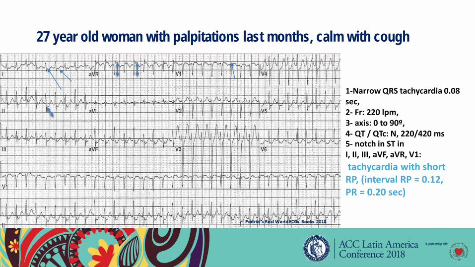
27 year old woman with palpitations last months, calm with cough
1-Narrow QRS tachycardia 0.08 sec,2- Fr: 220 lpm,3- axis: 0 to 90º,4- QT / QTc: N, 220/420 ms5- notch in ST inI, II, III, aVF, aVR, V1:tachycardia with short RP, (interval RP = 0.12, PR = 0.20 sec)

ECG without tachycardia1. Sinus rhythm 75 lpm,2. Normal P-waves3. short PR interval 0.12,4. QRS wide 0.12, by delta waves,5. QT / QTc 360/140 and 360/410 when QRS isconsidered prolonged.
Podrid ‘s Real World ECGs Books 2018
6. Negative Delta I and aVL, positive V1: left lateral tract.
7. intermittent preexcitation: The last two beats, without preexcitationof similar morphology during tachycardia

Woman 88 years old, with severe CMD, with dyspnea

Choose option that is correct:
• 1. Sinus Tachycardia with BAV I and LBB• 2. Atrial Tachycardia With BAV I and LBB• 3. Atrial flutter with conduction 2/1• 4. Ventricular tachycardia• 5. Typical AVNRT• 6. Atypical AVNRT • 7. AVRT tachycardia

Woman 88 years old, with severe CMD, with dyspnea
Fr 112 lpm QRS :0.16seg BCRI
SinusTachycardia vs Atrial T.
PR o RP :0.24 seg

Woman 88 years old, with severe CMD, with dyspnea : ECG taken months before

80-year-old man with precordial discomfort

Choose option that is correct:
• 1. Sinus Tachycardia• 2. Atrial Tachycardia• 3. Atrial flutter with conduction 2/1• 4. Fascicular tachycardia• 5. Typical AVNRT• 6. Atypical AVNRT • 7. AVRT Orthodromic AV reentrant tachycardia• 8. Junctional tachycardia

80-year-old man with precordial discomfortQRS estrecho139 por min
P retrogradas II III aVF

Despues de 6 mg en bolo de Adenosina EV :

Paroxysmal supraventricular tachycardia: TYPES of reentry:
• I) INTRANODAL reentry (the most frequent).• II) Reentry accessory pathway with two forms of
presentation:• a) Orthodromic: narrow QRS and• b) antidromic: wide QRS (more rare).

P´ waves after QRS :
• 1) P‘ waves not visible or attached at the end of the QRS, giving the false appearance of S in inferior derivations or of R 'in derivation V1 -> COMMON INTRANODAL REENTRANT (slow-fast). Most common (80-90%)
• 2) P‘ waves separated from the QRS -> REENTRANT by ACCESSORY ROUTE or ATYPICAL AV NODE REENTRY (fast-slow or slow-slow). -> only by means of an EEF can we differentiate them.