recommendations on angiographic revascularization...
TRANSCRIPT

2650
See related article, p 2509
Intra-arterial therapy (IAT) for acute ischemic stroke (AIS) has dramatically evolved during the past decade to include
aspiration and stent-retriever devices. Recent randomized con-trolled trials have demonstrated the superior revascularization efficacy of stent-retrievers compared with the first-generation Merci device.1,2 Additionally, the Diffusion and Perfusion Imaging Evaluation for Understanding Stroke Evolution (DEFUSE) 2, the Mechanical Retrieval and Recanalization of Stroke Clots Using Embolectomy (MR RESCUE), and the
Interventional Management of Stroke (IMS) III trials have con-firmed the importance of early revascularization for achieving better clinical outcome.3–5 Despite these data, the current het-erogeneity in cerebral angiographic revascularization grading (CARG) poses a major obstacle to further advances in stroke therapy. To date, several CARG scales have been used to mea-sure the success of IAT.6–14 Even when the same scale is used in different studies, it is applied using varying operational criteria, which further confounds the interpretation of this key metric.10 The lack of a uniform grading approach limits comparison of revascularization rates across clinical trials and hinders the
Recommendations on Angiographic Revascularization Grading Standards for Acute Ischemic Stroke
A Consensus Statement
Osama O. Zaidat, MD; Albert J. Yoo, MD; Pooja Khatri, MD; Thomas A. Tomsick, MD; Rüdiger von Kummer, MD; Jeffrey L. Saver, MD; Michael P. Marks, MD;
Shyam Prabhakaran, MD; David F. Kallmes, MD; Brian-Fred M. Fitzsimmons, MD; J. Mocco, MD; Joanna M. Wardlaw, MD; Stanley L. Barnwell, MD; Tudor G. Jovin, MD;
Italo Linfante, MD; Adnan H. Siddiqui, MD; Michael J. Alexander, MD; Joshua A. Hirsch, MD; Max Wintermark, MD; Gregory Albers, MD; Henry H. Woo, MD; Donald V. Heck, MD;
Michael Lev, MD; Richard Aviv, MD; Werner Hacke, MD; Steven Warach, MD; Joseph Broderick, MD; Colin P. Derdeyn, MD; Anthony Furlan, MD; Raul G. Nogueira, MD;
Dileep R. Yavagal, MD; Mayank Goyal, MD; Andrew M. Demchuk, MD; Martin Bendszus, MD; David S. Liebeskind, MD; for the Cerebral Angiographic Revascularization Grading (CARG) Collaborators, STIR Revascularization working group, and STIR Thrombolysis in
Cerebral Infarction (TICI) Task Force
Received May 1, 2013; accepted June 21, 2013.Endorsed by the American Academy of Neurology/Stroke System Work Group, American Association of Neurological Surgeon/Congress of Neurological
Surgeon Cerebrovascular Section, American Society of Neuroradiology, Society of Neurointerventional Surgery, and Society of Vascular and Interventional Neurology.
From the Department of Neurology, Medical College of Wisconsin, Milwaukee, WI (O.O.Z., B.-F.M.F.); Department of Radiology, Harvard Medical Center, Boston, MA (A.J.Y., J.A.H., M.L.); Department of Neurology (P.K., J.B.), Department of Radiology (T.A.T.), University of Cincinnati, Cincinnati, OH; Department of Radiology, Technical University Dresden, Dresden, Germany (R.v.K.); Department of Neurology, University of Los Angeles, Los Angeles, CA (J.L.S., D.S.L.); Department of Radiology (M.P.M.), Department of Neurology (G.A.), Stanford University, San Francisco, CA; Department of Neurology, Northwestern University, Chicago, IL (S.P.); Department of Radiology, Mayo Clinic, Rochester, MN (D.F.K.); Department of Neurosurgery, Vanderbilt University, Nashville, TN (J.M.); Department of Clinical Neurosciences, University of Edinburgh, Edinburgh, United Kingdom (J.M.W.); Department of Neurosurgery, Oregon State University, Portland, OR (S.L.B.); Department of Neurology, University of Pittsburgh, Pittsburgh, PA (T.G.J.); Baptist Vascular Center, Miami, FL (I.L.); Department of Neurosurgery, University of Buffalo, Buffalo, NY (A.H.S); Department of Neurosurgery, Mount Sinai Medical Center, Los Angeles, CA (M.J.A.); Department of Radiology, University of Virginia, Charlottesville, VA (M.W.); Department Neurosurgery, Stony Brook University, East Setauket, NY (H.H.W.); Department of Radiology, Forsyth Medical Center, Kernersville, NC (D.V.H.); Department of Medical Imaging, University of Toronto, Toronto, ON, Canada (R.A.); Department of Neurology (W.H.), Department of Neuroradiology (M.B.), University of Heidelberg, Heidelberg, Germany; Department of Neurology, University Medical Center, Brackenbridge, Bethesda, MD (S.W.); Department of Radiology, Washington University, St Louis, MO (C.P.D.); Department of Neurology, Case Western University, Cleveland, OH (A.F.); Department of Neurology, Emory University, Atlanta, GA (R.G.N.); Department of Neurology, Miami University, Miami, FL (D.R.Y.); and Department of Radiology (M.G.), Department of Neurology (A.M.D.), University of Calgary, Calgary, AB, Canada.
This statement is also endorsed by Cerebrovascular Coalition (CVC), Stroke Imaging Repository (STIR) Consortium and Stroke Treatment Academic Industry Roundtable (STAIR) group.
A list of all STAIR Participant Endorsees is given in the Appendix.Guest Editor for this article was Bruce Ovbiagele, MD, MSc, MAS.Correspondence to Osama O. Zaidat, MD, MS, Department of Neurology, Neurosurgery, and Radiology, Froedtert Hospital and Medical College of
Wisconsin, 9200 W Wisconsin Ave, Milwaukee, WI. E-mail [email protected](Stroke. 2013;44:2650-2663.)© 2013 American Heart Association, Inc.
Stroke is available at http://stroke.ahajournals.org DOI: 10.1161/STROKEAHA.113.001972
by guest on July 10, 2018http://stroke.ahajournals.org/
Dow
nloaded from
Zaidat et al Angiographic Revasc Grading Consensus 2651
translation of promising, early phase angiographic results into proven, clinically effective treatments.6–14 For these reasons, it is critical that CARG scales be standardized and end points for successful revascularization be refined.6 This will lead to a greater understanding of the aspects of revascularization that are strongly predictive of clinical response.
The optimal grading scale must demonstrate (1) a strong cor-relation with clinical outcome, (2) simplicity and feasibility of scale interpretation while ensuring characterization of relevant angiographic findings, and (3) high inter-rater reproducibility.
To address these issues, a multidisciplinary panel of neuro-interventionalists, neuroradiologists, and stroke neurologists with extensive experience in neuroimaging and IAT, convened at the “Consensus Meeting on Revascularization Grading Following Endovascular Therapy” with the goal of establish-ing consensus recommendations for the standardization of revascularization grading in AIS clinical studies. Presented here are the recommendations of the panel.
MethodsA hybrid approach based on established processes described by Glaser15 and by the National Institutes of Health (http://prevention.nih.gov/cdp/about.aspx)16 was used to structure the conference and consensus statement. Before the meeting, a comprehensive literature review was performed, and relevant documents were distributed to each participant. Twelve expert panelists attended the full-day round-table meeting held in Chicago, IL, on January 7, 2012. The panel comprised a multidisciplinary group of noninvasive stroke neurolo-gists and neurointerventionalists with neuroradiology, neurosurgery, and neurology backgrounds.
Panelists presented their views on CARG after IAT using case il-lustrations, followed by an open discussion. A preselected set of angio-grams from IAT-treated patients with AIS was reviewed at the beginning of the meeting and again at the end of the day to identify key points for discussion. After the meeting, draft proposals were presented for voting via an online survey to arrive at consensus recommendations. The final consensus document was based on a systematic literature review, the roundtable discussion, and the postmeeting survey results.
The consensus recommendations were presented to the revascular-ization grading task force assembled at the Stroke Imaging Repository (STIR) Consortium/Stroke Treatment Academic Industry Roundtable (STAIR) meetings in March 2013, and their feedback was incorporated into the final draft. The article was distributed to 68 national and inter-national experts for further comments or suggestions. The final copy was endorsed by stakeholder Cerebrovascular Coalition societies.
ResultsThe consensus recommendations are classified into the fol-lowing 4 key areas: (1) CARG, (2) anatomic and collateral considerations, (3) additional grading considerations, and (4) technical considerations and documentation.
Cerebral Angiographic Revascularization Grading
Effective Revascularization: Recanalization Versus ReperfusionIssue: Recanalization and reperfusion are often interchanged in CARG scales. Recanalization and reperfusion scales lack clear definitions.
Revascularization encompasses all treatment-related improvements in blood flow, including local recanalization of the arterial occlusion and reperfusion of the downstream territory. Although these end points are closely related,
recanalization and reperfusion are different. Recanalization is required for antegrade tissue reperfusion, but recanalization may not necessarily lead to reperfusion in regions where distal emboli or established infarctions are present. Because of their intimate relationship, both measures have been shown to pre-dict improved clinical outcomes after IAT.17–28 Of the 2 param-eters, reperfusion is more strongly associated with tissue and clinical outcomes because of its measurement of tissue-level effects.18,28 Therefore, reperfusion is the best marker of effec-tive revascularization. However, recanalization is an important metric of the treatment effect on the target lesion. Future stud-ies are needed to assess whether recanalization imparts addi-tional prognostic information in combination with reperfusion.
Reperfusion ScalesOn digital subtraction angiography (DSA) in normal patients, tissue-level perfusion is evidenced by a capillary blush. After IAT, reperfusion is operationally defined as the antegrade (ie, not by pial collaterals) restoration of a capillary blush. As such, reperfusion scales account for distal emboli and would identify treatment modalities that achieve revascularization without clot fragmentation and distal embolization (DE).Thrombolysis in Myocardial Infarction Scale. Originally con-structed to measure myocardial reperfusion, the Thrombolysis in Myocardial Infarction (TIMI) scale has been widely adopted for use in the cerebral circulation (Table 1).29–36 However, vari-able permutations of TIMI have been used in previous IAT studies, most of which measure recanalization.1,32–36 In the Mechanical Embolus Removal in Cerebral Ischemia (MERCI) and Multi MERCI trials,33,34 treatment success required recan-alization of all treatable vessels. For internal carotid artery (ICA) terminus occlusions, this translated to recanalization of the ICA, M1, and all M2 segments. The investigators stated that “the status of arterial branches distal to the treatable ves-sel was not considered when ascribing the TIMI scale.”33 The general uncertainty on how to grade TIMI was demonstrated in a recent study of 48 patients with AIS treated with IAT, where the inter-rater reliability was poor using the modified TIMI grading scale (κ=0.4).37 Similarly, there was wide discordance between the site and core laboratory TIMI scale readings in the Solitaire with Intention for Thrombectomy (SWIFT) trial (14.8% and 19.1% difference in the Solitaire FR and Merci arms, respectively).1 Nevertheless, TIMI has been predictive of clinical outcome across studies; reflecting the strong correla-tion of recanalization and reperfusion.12,19–21,24,27
Table 1. Thrombolysis in Myocardial Ischemia Scale
TIMI Grades Definitions
Grade 0 Absence of any antegrade flow beyond the target occlusion (no perfusion)
Grade 1 Any faint antegrade flow beyond the target occlusion, with incomplete filling of the distal branches (penetration without perfusion)
Grade 2 Delayed or sluggish antegrade flow with complete filling of the distal M2 branches flow (partial perfusion)
Grade 3 Normal flow that fills all distal branches, including M3 and M4 (complete perfusion)
TIMI indicates thrombolysis in myocardial ischemia.
by guest on July 10, 2018http://stroke.ahajournals.org/
Dow
nloaded from
2652 Stroke September 2013
The original TIMI scheme has 2 issues that limit its use in the cerebral vasculature. First, the major criterion for grading myocardial reperfusion is the rate of contrast clearance from the distal tissue bed. However, in the carotid circulation, Mori et al38 found that if there is tissue-level reperfusion, flow delay is an infrequent occurrence; the return of a parenchymal con-trast blush is a sign of sufficient flow. The second issue is that the extent of reperfusion is not considered in the TIMI criteria. This is critical in cerebral ischemia because final infarct vol-ume is the single best predictor of outcome after IAT,39–41 and greater degrees of reperfusion are associated with higher rates of good outcome.3,12,18,25,26
Thrombolysis in Cerebral Infarction Scale. The Thrombolysis in Cerebral Infarction (TICI) scale was originally proposed in a position statement that attempted to standardize clinical trial design and reporting for IAT.14,32 The TICI scale specifi-cally addresses the extent of tissue reperfusion, as represented by the capillary blush on DSA.14 TICI is graded by visually estimating how much of the initial antegrade capillary blush defect (or target downstream territory [TDT]; see Target Downstream Territory section; denominator) is reperfused (numerator). The TICI scale distinguishes no perfusion (TICI grade 0; Figure 1), minimal flow past the occlusion but no per-fusion (grade 1; Figure 2), minor partial reperfusion (grades 2a; Figures 3 and 4), major partial reperfusion (2b; Figure 1), and complete reperfusion without any flow defects (grade 3; Figures 2 and 5; Table 2). Two variations of the TICI scale exist on the threshold for major partial reperfusion. The origi-nal TICI system defined TICI 2b as restoration of more than two thirds of the TDT.14 This is in contrast to the subsequent
modification (modified treatment in cerebral ischemia [mTICI]) introduced by the IMS investigators, which uses a threshold of more than half of the TDT.18,42,43 There is indirect evidence of better inter-rater variability using the original TICI scale than TIMI. The discordance was higher between site and core laboratory readers in the SWIFT (using TIMI) versus TREVO2 (using original TICI) trials (≈15% versus 1% in the stent-retriever arms, respectively).1,2 The advantage of mTICI is its simplicity (ease of visually estimating 1/2 versus 2/3 reperfusion), and previous work has demonstrated excellent inter-rater agreement for distinguishing <50% versus ≥50% reperfusion of the downstream territory.21 Moreover, mTICI has proven value for predicting clinical outcome.18,23,25,26,44 In a comparative study of TIMI and mTICI, the c-statistic for predicting 90-day good outcome (mRS 0–2) was significantly higher for mTICI versus TIMI (0.74 versus 0.68; P<0.0001).25
Recanalization ScalesRecanalization measures the direct action of the therapeutic intervention on the target arterial lesion (TAL). Although it neglects downstream effects within the ischemic bed, it may provide additional prognostic information, particularly in the setting of incomplete vessel patency where there may be an increased risk of reocclusion or DE.Arterial Occlusive Lesion Scale. The only grading scale explicitly intended to measure the degree of recanalization at the TAL is the Arterial Occlusive Lesion (AOL) scale, which was proposed by the IMS investigators (Table 3).5,12,18,42,43 AOL describes arterial patency at the site of occlusion based on the degree of luminal opening (none, partial, or complete) with
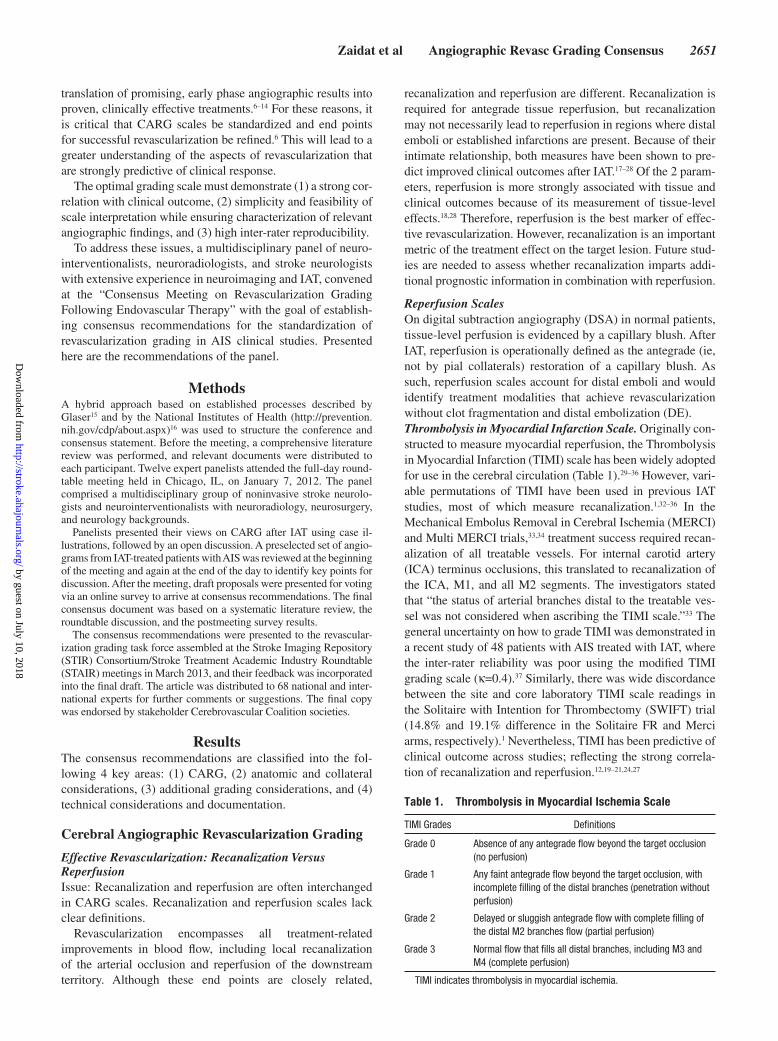
Figure 1. Example of thrombolysis in cerebral infarction (TICI) 0 and TICI 2b. Top, Anteroposterior (first 2 boxes) and lateral (last 2 boxes) in an early arterial and late capillary phases depicting TICI 0 at baseline. Bottom, Same phases depicting TICI 2b after intra-arterial therapy. Black arrow indicating the tar-get arterial lesion (TAL): middle cerebral artery/M1 horizontal segment occlusion (TAL) distal to the lenticulostriate (LS). Black half circles approximate the target downstream territory (TDT; the presumed area supplied by the TAL).
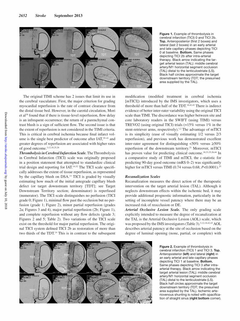
Figure 2. Example of thrombolysis in cerebral infarction (TICI) 1 and TICI 3. Top, Anteroposterior (left) and lateral (right) in an early arterial and late capillary phases depicting TICI 1 at baseline. Bottom, Same phases depicting TICI 3 after intra-arterial therapy. Black arrow indicating the target arterial lesion (TAL): middle cerebral artery/M1 horizontal segment occlusion (TAL) distal to the lenticulostriate (LS). Black half circles approximate the target downstream territory (TDT; the presumed area supplied by the TAL). Ischemic arte-riovenous shunting is noted with opacifica-tion of straight sinus (right bottom corner).
by guest on July 10, 2018http://stroke.ahajournals.org/
Dow
nloaded from

Zaidat et al Angiographic Revasc Grading Consensus 2653
further qualification based simply on the presence (grades 2 or 3) or absence (grades 0 or 1) of any downstream flow.11,18
Reflecting the strong relationship between recanaliza-tion and reperfusion, AOL and mTICI showed good agree-ment (κ=0.66), with no difference in their ability to predict clinical outcome (61% versus 71% for AOL 2–3 and mTICI 2a-3, respectively; P=0.9) in 96 angiograms from IAT cases.23 Similarly, AOL and TIMI scales showed modest agreement (κ=0.30) with no difference in their ability to predict clinical outcome (49% versus 54% in AOL 2–3 and TIMI 2–3, respec-tively; P=0.13) in 61 angiograms from IMS I trial.12 However, as previously noted, both mTICI 2a-3 and 2b-3 were dem-onstrated to be superior to AOL 2 to 3 for predicting 90-day functional independence in the combined IMS II data set, sup-porting the preeminent role of tissue reperfusion in shaping clinical outcome.18 Nevertheless, recanalization grading is important and is likely to contribute further prognostic infor-mation to reperfusion alone.
Although easy to use, there are 3 main issues with the AOL grading system. First, in IMS II and III, AOL scoring was applied only to the TAL.5,18 For example, recanalization of a carotid terminus occlusion yielded AOL 3, but mTICI 0, if the middle cerebral artery (MCA) remained occluded. The alternative approach is to use the entire clot span for grading recanalization, which raises a second issue; the distal extent of clot is frequently unknown, particularly when it extends into several branches. For example, if a clot within the MCA M1 segment extends into both proximal M2 segments, is the TAL within the M1 only, or the M1 and both M2 segments? In the former case, opening the M1 and one of the M2 branches
only would yield an AOL 3; in the latter case, the same result would yield an AOL 2. The third issue relates to the difficulty in distinguishing between AOL 2 (partial recanalization) ver-sus possible underlying stenosis, or device-related vasospasm. Despite these shortcomings, AOL is the only scale that specifi-cally addresses device efficiency at the TAL.
Other CARG ScalesIn addition to the commonly used revascularization grading schemes, other scales have been proposed. The Qureshi scale rates both occlusion site and collaterals; however, its major disadvantages are scale complexity and incorporation of dis-parate elements of stroke physiology. These issues make it difficult to distinguish treatment-related gains from baseline anatomic and collateral status, and also complicate statistical analysis.45 Similarly, an additional attempt to develop specific comprehensive scale, recanalization in brain ischemic (RBI), did not yield wide adaptation.46 Another reperfusion grading system, the Mori scale, considers the proximal arterial occlu-sive lesion and distal perfusion, but has been infrequently used in AIS studies.38
Consensus Revascularization ScalesIssue: What CARG scale should be recommended?
There was consensus agreement that proposing a novel CARG system with completely new scale definitions would add further confusion to the field, given the multiple grading schemes already established. The panelists strongly supported the recommendation of adopting a single CARG system for perfusion and one for recanalization with consistent opera-tional definitions across AIS trials.
Figure 4. Example of thrombolysis in cerebral infarction (TICI) 0 and TICI 2a (compare with TICI 3 and 2b above). Top, Anteroposterior (first 2 boxes) and lateral (last 2 boxes) in an early arterial and late capillary phases depicting TICI 0 at base-line. Bottom, Same phases depicting TICI 2a after intra-arterial therapy. Black arrow indicating the target arterial lesion (TAL): middle cerebral artery/M1 horizontal seg-ment occlusion (TAL) distal to the lenticulo-striate (LS). Black half circles approximate the target downstream territory (TDT; the presumed area supplied by the TAL).
Figure 3. Example of thrombolysis in cerebral infarction (TICI) 0 and TICI 2a (compared with TICI 2b and 3 above). Top, Anteroposterior (first 2 boxes) and lateral (last 2 boxes) in an early arterial and late capillary phases depicting TICI 0 at baseline. Bottom, Same phases depicting TICI 2a after intra-arterial therapy. Black arrow indicating the tar-get arterial lesion (TAL): middle cerebral artery/M1 horizontal segment occlusion (TAL) distal to the lenticulostriate (LS). Black half circles approximate the target downstream territory (TDT; the presumed area supplied by the TAL).
by guest on July 10, 2018http://stroke.ahajournals.org/
Dow
nloaded from

2654 Stroke September 2013
From the most commonly used scales, the panel recom-mended mTICI as the primary reperfusion scale because it was specifically designed for the cerebral circulation, has good inter-rater reliability, and strongly predicts clinical out-come.18,23,25,26 Furthermore, the consensus was to maintain the acronym mTICI, which now stands for modified treat-ment in cerebral ischemia (previously modified thrombolysis in cerebral ischemia), to more accurately reflect current IAT practice.
For assessing recanalization, the AOL scale is preferred because of its ease of use and precise assessment of device efficacy at the occlusion. The AOL scale can be used at dif-ferent locations.
Because mTICI and AOL assess different dimensions of revascularization, (reperfusion and recanalization, respec-tively), it is optimal to obtain and report both measures.
Recommendations:
• The angiographic criterion for tissue reperfusion is resto-ration of capillary-level opacification, which is achieved in an antegrade fashion (ie, across the occlusive lesion).
• For primary measures of procedural success, reperfusion scales should be used.
• The mTICI scale should be used as the standard reperfu-sion grading scale.
• mTICI should stand for modified treatment in cerebral ischemia.
• Recanalization should be measured using the AOL scale.
Defining TICI Grades and the Target Angiographic End PointIssue: Consistent thresholds for TICI grades are lacking. The target angiographic end point is variable and not well defined in stroke studies.
Clear operational definitions for TICI grades 3 and 2 have not been standardized. These variations may have dif-ferent clinical implications and limit comparisons between device studies. For mTICI 3, there is no clear consensus on whether it should represent complete versus near complete antegrade reperfusion. Although complete reperfusion is associated with the best outcomes,5,25 the benefits and risks of attempting to achieve full reperfusion are not yet fully understood. However, the requirement for complete reperfu-sion may potentially increase the reliability of mTICI 3 grad-ing between readers and help to distinguish mTICI 2b from mTICI 3. Hence, the consensus was to define mTICI 3 as complete antegrade reperfusion, without any distal branch occlusion or stagnation.
As previously noted, TICI 2b has been dichotomized into 2 main variations: (1) more than half (mTICI) and (2) more than two thirds (original TICI) reperfusion. In available studies, good interobserver agreement was demonstrated using either threshold.25,26 Given the current reliability data, and potential simplicity of the one half threshold, the panel recommended that TICI 2b should be defined as more than half but less than full antegrade reperfusion of the TDT.
In IAT studies, procedural success had been variably defined using different target angiographic end points. The most frequent definition of IAT angiographic success has been TIMI 2 to 3 or TICI 2a-3 reperfusion grades.1–5,32–36 Several studies have shown that mTICI 2b yields significantly better
Figure 5. Example of downstream terri-tory (TDT) for an ICA occlusion with pre–intra-arterial therapy (IAT) data from CT angiogram showing cross-filling from the contralateral anterior cerebral artery. Top, Anteroposterior (first 2 boxes) and lateral (last 2 boxes) in an early arterial and late capillary phases depicting thrombolysis in cerebral infarction (TICI) 0 at baseline. Bottom, Same phases depicting TICI 3 after IAT. Black arrow indicating the target arterial lesion (TAL): Distal ICA proximal to the ophthalmic artery. Black half circles approximate the target downstream ter-ritory (TDT; the presumed area supplied by the TAL). Early ischemic arteriovenous shunting is noted in the right lower corner.
Table 2. Modified Treatment in Cerebral Ischemia Scale
mTICI Grades Definitions
Grade 0 No perfusion
Grade 1 Antegrade reperfusion past the initial occlusion, but limited distal branch filling with little or slow distal reperfusion
Grade 2a Antegrade reperfusion of less than half of the occluded target artery previously ischemic territory (eg, in 1 major division of the MCA and its territory)
Grade 2b Antegrade reperfusion of more than half of the previously occluded target artery ischemic territory (eg, in 2 major divisions of the MCA and their territories)
Grade 3 Complete antegrade reperfusion of the previously occluded target artery ischemic territory, with absence of visualized occlusion in all distal branches
MCA indicates middle cerebral artery; and mTICI, Modified Treatment in Cerebral Ischemia Scale.
Table 3. Arterial Occlusive Lesion Scale
AOL Grades Definitions
Grade 0 Complete occlusion of the target artery
Grade 1 Incomplete occlusion or partial local recanalization at the target artery with no distal flow
Grade 2 Incomplete occlusion or partial local recanalization at the target artery with any distal flow
Grade 3 Complete recanalization and restoration of the target artery with any distal flow
AOL indicates arterial occlusive lesion.
by guest on July 10, 2018http://stroke.ahajournals.org/
Dow
nloaded from

Zaidat et al Angiographic Revasc Grading Consensus 2655
outcomes than minor reperfusion.18,25,26,44 Data from IMS II revealed a trend favoring mTICI 2b for predicting outcome versus mTICI 2a (P=0.08).18 Furthermore, mTICI 2b-3 was demonstrated as the optimal threshold for predicting good outcome in 313 AIS undergoing IAT (sensitivity 78%, speci-ficity 65%) versus mTICI 2a-3.25 In core laboratory adjudi-cated results from the IMS III trial, mTICI 3 and 2b grades were associated with 80% and 46.3% good clinical outcome (90-day mRS ≤2), respectively, versus 19.4% for mTICI 2a.5 Given that mTICI 2b-3 reperfusion seems to have a higher accuracy for discriminating a good functional outcome than mTICI 2a-3,18,25,26 its use as the target angiographic end points is recommended until further data are available.
Recommendations:
• mTICI 3 should be defined as complete antegrade reper-fusion of the TDT, with absence of visualized occlusion in all distal branches.
• mTICI 2b should be defined as more than half, but less than complete antegrade reperfusion of the TDT.
• The target angiographic end point for technical success should be defined as mTICI 2b or higher.
Adjusting the mTICI Grading ScaleIssue: Arterial occlusion locations and new embolization may be associated with different clinical outcomes that may not be accounted for in the mTICI score.
Different vessel occlusions are associated with different clinical outcomes; for example, distal ICA and basilar artery occlusions are associated with worse clinical outcome when compared with other locations.47–51 Given how the occlusion location influences outcome, should an adjustment to the mTICI scale be implemented based on the occlusion site? One scale attempted to incorporate the site of occlusion into the revascularization grade; however, this modification made the scale more complex and prohibitive.45,46
The panel decided against adjusting or imposing a negative mTICI score based on location. Instead, the recommendation was to define the location of the vessel occlusion clearly to allow pooling of similar location data within and across trials. To improve mTICI correlation with clinical outcome, studies should report subgroup analyses by the level of occlusion, or alternatively studies should enroll patients with homoge-neous clot location, such as the Prolyse in Acute Cerebral Thromboembolism (PROACT) in MCA, MCA Embolism Local Fibrinolytic Intervention (MELT), and the Basilar Artery International Cooperation Study (BASICS) trials.32,50,52
Similarly, embolization into new territory (ENT) should be captured separately without adjustment of the mTICI scale. It is recognized that the mTICI scale will incorporate informa-tion on distal emboli. New embolization is discussed in more detail below (see ENT and DE in the Target Downstream Territory section).
Recommendations:
• mTICI grading should not be modified based on the level of vessel occlusion or new embolization.
• mTICI grading should be reported by level of occlusion for comparison of treatment arms within studies and to allow comparison across studies.
Anatomic and Collateral ConsiderationsOnce all arterial stenoses and occlusions (eg, single or mul-tiple emboli, tandem lesions) and baseline anatomic variants (eg, hypoplastic A1 segment or posterior communicating artery) have been identified and documented, the TAL can be designated.
Target Arterial LesionIssue: The terminology used to describe the TAL is not well defined.
Anatomic definitions of TAL, or the primary focus of intra-arterial treatment, at baseline angiography have varied across the literature. The consensus recommendation was to desig-nate the proximal-most intracranial occlusive lesion as the TAL. Therefore, in cases of multiple intracranial emboli (eg, distal anterior cerebral artery [ACA] and MCA emboli), the most proximal one will be considered the TAL. Alternatively, in the setting of tandem extracranial-intracranial occlusions (eg, cervical ICA plus proximal MCA occlusions), the intra-cranial lesion will be considered the TAL. The only exception to this definition would be in cases where there is no intracra-nial occlusion; in this case, the extracranial lesion is the TAL. In addition to specifying the location of the most proximal intracranial lesion, a more granular descriptive terminology should be used, whenever possible, to allow for subsequent detailed and supraordinate analyses.
Recommendation:
• The TAL is the most proximal treated intracranial occlu-sive lesion.
Intracranial ICA Occlusion Nomenclature. Issue: Intracranial ICA clot topography has been largely neglected in stroke studies.
The intracranial carotid occlusion may have variable out-comes depending on the extent of the clot.47–49 Such lesions are often grouped under the term carotid T, but this designation implies a particular shape of the occlusion, namely contiguous clot extending from the distal ICA into both A1 and M1 arter-ies. However, a distinction should be made from other con-figurations, such as L when it extends only to the M1 segment, or even as I where there is flow across the circle of Willis into both the A1 and M1 segments (ie, clot is isolated to the distal ICA only). This distinction may be made from injecting the contralateral ICA or from pretreatment computed tomography (CT) angiography (CTA) or MR angiography when available.
Recommendation:
• Intracranial carotid occlusions should be documented based on the best available data: carotid T if terminal ICA, origin of M1, and origin of A1 are all occluded; carotid L if only both terminal ICA and the M1 origin are occluded; isolated intracranial ICA if only the intra-cranial ICA is occluded.
MCA Occlusion Nomenclature. Issue: Description of MCA clot location varies across studies.
Anatomic variations have made MCA clot location nomen-clature challenging. The panel supports the definition of the M1 segment as the first portion of the MCA up to the major bifurcation. Occlusion of the proximal M1 segment should be assigned when it involves the proximal half of the M1 segment
by guest on July 10, 2018http://stroke.ahajournals.org/
Dow
nloaded from
2656 Stroke September 2013
or when no lenticulostriate vessels are visualized. Distal M1 occlusions involve the distal half of the M1 segment or spare the lenticulostriate perforators.
Detailed description of any anatomic variant must be reported, including duplicated M1 segments, dominant ante-rior temporal artery (ATA) branches, and A1 segment varia-tions (ie, aplastic, hypoplastic, normal caliber).
Recommendations:
• MCA clot location should be clearly and precisely described.
• The M1 segment should be defined as the first portion of the MCA up to the major bifurcation.
• M1 clot location should be further localized as proximal (ie, involving the proximal half of the M1 or occluding the lenticulostriate vessels) or distal (ie, involving the distal half of the M1 segment or sparing the lenticulo-striate vessels).
• Anatomic variations should be described (eg, duplicate M1 segment, dominant anterior temporal artery, A1 anatomy).
Target Downstream TerritoryIssue: Criteria for determining the TDT are not well defined.
The TDT is the region of the brain that was previously sup-plied in an antegrade fashion, but attributable to the target lesion, now has absent capillary-level opacification or capillary opacification, which is achieved via pial collaterals. Territory receiving retrograde collateral perfusion should be considered as part of the TDT. The full extent of this territory serves as the denominator in calculating the percentage of tissue reper-fusion. This definition is particularly important for ICA or vertebrobasilar occlusion. For instance, in an intracranial ICA occlusion, the TDT comprises both ipsilateral ACA and MCA territories in cases with no evidence of ipsilateral ACA filling from contralateral carotid injection (or equivalent data from CTA or MR angiography). In many cases of intracranial ICA occlusion, there is antegrade filling of the ipsilateral ACA ter-ritory over the anterior communicating artery such that the TDT is the MCA territory only. In cases of vertebrobasilar occlusion, the TDT is the territory of the basilar artery, supe-rior cerebellar arteries, and bilateral posterior cerebral arteries territories, unless imaging data from CTA, MR angiography, or DSA demonstrate the distal extent of the clot or anterior cir-culation supplying the posterior cerebral arteries (fetal origin).
Recommendations:
• The TDT is the brain region that lacks antegrade capil-lary staining secondary to the target occlusion. Capillary opacification that is achieved via pial collaterals should be considered part of the TDT.
• In intracranial ICA occlusion, the TDT should include both ACA and MCA territories.
• In vertebrobasilar occlusion, the TDT should be basilar artery branches above the level of occlusion and both posterior cerebral arteries.
• If evidence from other baseline imaging data shows a different pattern than expected above, adjustment to the TDT must be made (eg, in most ICA occlusions, the TDT will be the MCA territory attributable to anterior communicating artery supply to the ipsilateral ACA territory).
Cerebral Collateral Flow GradingIssue: No consensus or consistency exists in obtaining and grading collateral circulation data during IAT.
The pial collateral circulation predicts the response to IAT revascularization and clinical outcomes in AIS cases.53–58 Collateral maintenance may prolong the time window for revascularization, and failure may hasten ischemia and lead to futile revascularization.53 Collateral grading has been reported using several scales.59 The American Society of Interventional and Therapeutic Neuroradiology (ASITN) collateral grading (ACG) system is the most commonly used scale, and assigns scores based on the extent and rate of retrograde collateral flow to the TDT. Score ranges between 0 (no collaterals) and 4 (complete and rapid collateral perfusion to the ischemic bed; Table 4). Higher ACG score has been associated with a higher rate of successful revascularization after IAT.54 The same group reported the highest rates of infarct growth on day 3 to 5 brain MRI in those with no recanalization and poor ACG score at baseline.55
In addition to the ACG scale, the pial collateral scale has been described by Christoforidis et al.56 The pial collateral scale grade measures the proximal-most extent of retrograde vessel filling from pial collaterals, with the best grade defined as retrograde filling of the occluded segment up to the dis-tal extent of the clot. The authors demonstrated smaller final infarct volumes and better discharge mRS scores in those with a better collateral grade.
Recently, a third collateral scale has been described in a study of 26 IAT-treated patients with ICA or MCA occlusion. In the Capillary Index Score,57 the TDT is divided into 3 equal parts, and each part is given a score of 1 if normal capillary blush is present, or 0 if absent. The individual scores are sum-mated to yield a total Capillary Index Score between 0 and 3. A high Capillary Index Score scale was associated with better outcome and recanalization in that study.
Collateral scales have been shown to predict revascu-larization and clinical and tissue outcome after IAT. The panel recommends that the ASITN collateral grading scale should be used for cerebral collateral flow grading when angiographic data are available and adequate. However, performing a complete diagnostic angiogram for collateral assessment poses a delay to treatment. In cases where this information cannot be obtained quickly (eg, because of dif-ficult vessel catheterization), collaterals may be imputed from noninvasive imaging modalities, such as CTA and CT/MR perfusion.58
Recommendations:
• Cerebral collateral flow grading on DSA should be per-formed using the ASITN grading scale.
• When DSA data are not available, noninvasive imag-ing should be used to evaluate the collateral circulation before IAT.
Additional Considerations
ENT and DE Into the Target Downstream TerritoryIssue: Criteria for new embolization (into the target or new territories) are not well defined, and these data are not rou-tinely recorded.
by guest on July 10, 2018http://stroke.ahajournals.org/
Dow
nloaded from

Zaidat et al Angiographic Revasc Grading Consensus 2657
The panel supports the definition of ENT as any treatment-related embolus outside of the TDT. The consensus defini-tion of DE is any treatment-related embolus within the TDT. Therefore, in a terminal ICA occlusion, a treatment-related ACA embolus would represent an ENT if the ipsilateral ACA was patent and received antegrade flow from the contralateral ICA at baseline. The same embolus would represent a DE if no such ACA flow was seen at baseline (eg, isolated hemisphere).
ENT during IAT was reported in 3.8% of the MERCI trial cohort, 1.5% in a retrospective study of the Solitaire device, and in 1.7% of treated MCA occlusions in the pooled IMS I and II data.60–62 ENT after treatment of ICA terminus occlu-sions has been shown to be associated with poor clinical out-come in the pooled IMS I and II analyses.62 Similarly, in IMS III, ENT was found in 14% (28/200) of M1 and ICA occlu-sions after IAT, and was associated with poor clinical outcome (mRS≤2 in 17.9% versus 30.2% in those without ENT).63
Downstream DE has been reported in ≈7% to 16% of IAT cases, and has not been independently associated with worse outcome in these studies.61,63–65 However, this may be attribut-able to the lack of a clear definition of DE, or may be more likely because of the fact that DE is accounted for by the mTICI scale. The panel recognizes that accurate documenta-tion of DE is difficult because it requires knowledge of whether distal emboli were present in the TDT before treatment. To obtain this information would require microcatheter selection of branches distal to the clot with subsequent contrast injec-tion, which would add a time delay to treatment, as well as the potential increased risk of hemorrhage from microcatheter injections. Alternatively, close inspection of pretreatment CTA would be necessary for identifying such occlusions, although this may not be reliable. Given these challenges and the fact that treatment-related DE is incorporated into the final mTICI grade, separate documentation of DE is of uncertain value.
Given the potential deleterious effect of ENT, the panel rec-ommends that any occurrence of ENT be documented. In ICA terminus occlusions, any new post-treatment ACA occlusion is considered DE unless there was normal ACA flow at base-line, then it would be considered ENT. Similar definitions can be applied to the vertebrobasilar occlusion, where any down-stream occlusion distal to the TAL is considered DE unless
complete angiogram or pre-IAT noninvasive imaging demon-strated patency of these distal branches.
Recommendations:
• ENT denotes treatment-related embolization to territo-ries outside of the TDT (eg, ACA embolization during MCA occlusion treatment).
• DE is defined as any treatment-related embolization into the TDT.
• Occurrences of ENT after IAT should be reported.
Venous ShuntingIssue: Few studies have documented venous shunting or cor-related this finding with outcome.
The presence of unexpected findings such as early venous shunting (Figures 2 and 5) or a prominent capillary phase blush should be noted, as they may provide information about not just reperfusion, but tissue infarction, impaired autoregu-lation, and risk for hemorrhagic transformation.66,67
Recommendation:
• Early venous shunting during IAT should be reported.
Technical Considerations and Documentation
Procedural Time MetricsIssue: Recording of important time milestones and procedural events is not standardized; consensus on terminology is cur-rently lacking.
Given the negative impact of time delays on clinical out-come after IAT, documentation of time variables is important.68 The key management milestone’s times must be documented in all studies, including the time of patient arrival in the suite, groin puncture, first access of the target clot by the micro-catheter, and procedure end.69–71 Time to first achievement of revascularization should be carefully defined (see below) and documented. If functional flow is not established during the case, time to the end of the procedure, defined as the time to the final angiogram, should be recorded (Table 5 showing sug-gested time intervals).
When performing clinical studies, additional procedural steps should be documented in a manner that matches the pub-lished protocols. In PROACT-II and the IMS trials, drug deliv-ery or device use was followed by intermittent DSA to assess interval revascularization results (eg, hourly in PROACT-II and every 15 minutes in the IMS III trial or after each device pass).5,32 It is imperative to define the main study treatment and subsequent rescue phases to allow investigators and treat-ing physicians to scale revascularization separately for the investigational intervention alone and for the entire procedure.
Recommendation:
• Suggested Stroke Intervention Times Intervals are onset to door, door to imaging, imaging (noncontrast head CT scan) to room entry, room entry to puncture, puncture to clot, clot to substantial reperfusion, and clot to end of procedure.
Therapeutic Intervention DetailsIssue: Device or thrombolytic delivery documentation is not consistent.
Table 4. American Society of Interventional and Therapeutic Neuroradiology Collateral Grading System
ACG Grades Definitions
Grade 0 No collaterals visible to the ischemic site
Grade 1 Slow collaterals to the periphery of the ischemic site, with persistence of some of the defect
Grade 2 Rapid collaterals to the periphery of the ischemic site, with persistence of the defect, and only to a portion of the ischemic territory
Grade 3 Collaterals with slow but complete angiographic blood flow of the ischemic bed by the late venous phase
Grade 4 Complete and rapid collateral blood flow to the vascular bed in the entire ischemic territory by retrograde perfusion
N/A Not applicable based on the territory or injections available
ACG indicates the American Society of Interventional and Therapeutic Neuroradiology collateral grading.
by guest on July 10, 2018http://stroke.ahajournals.org/
Dow
nloaded from

2658 Stroke September 2013
It is important to describe exactly what was done from a therapeutic standpoint. For intra-arterial drug delivery, it is important to define the site of arterial injection, including the vessel injected and the position of the catheter tip relative to the target thrombus (proximal to the clot, intraclot, or distal to the clot). The amount and duration of drug delivery must be denoted. When devices are placed, the deployment loca-tion and time, and adjuvant approaches, should be detailed. Individual device pulls should be reported as discrete steps. Adjunctive medications, such as vasodilator infusion, are important to document because they may affect ultimate revas-cularization results. Such treatments may be used to relieve or prevent vasospasm, or to enhance the retrieval process.72,73
Recommendation:
• Therapeutic intervention steps need to be reported in a standardized format and documented with proper imag-ing acquisition.
Angiography Imaging TechniquesIssue: Inconsistent image acquisition may hinder assessment of revascularization.
Obtaining adequate images during acquisition of cere-bral angiography for endovascular AIS therapy and the subsequent filming and annotation of such data are criti-cal (Table 6). Such information is imperative for compre-hensive revascularization grading. Angiography should be
Table 5. Suggested Stroke Intervention Time Intervals
Time Interval Variables Definitions
Onset to door (OtD)* Time from last known well to Emergency Department registration
Door to imaging (DtI)* Time from ED arrival to start of noncontrast CT head imaging used for treatment selection
Imaging to endovascular team activation Time from start of noncontrast CT head to activation (contacting) the endovascular team
Endovascular team activation to arrival to angiosuite (ATAtAA)
Imaging to angiosuite (ItA) Time from start of noncontrast CT head imaging to angiosuite room entry
Angiosuite entry to puncture (AtP)* Time from room entry to arterial puncture
Puncture to baseline angiogram (PtBA) Puncture to placing the main catheter/sheath in the base vessel (carotid or vertebral)
Puncture to clot (PtC) Time from arterial puncture to first access of the clot by microcatheter
Clot to TICI 2a perfusion (CtP) Time from first access of the clot by microcatheter to first achievement of persistent TICI 2a or better reperfusion
Clot to substantial perfusion (CtSP)* Time from first access of the clot by microcatheter to first achievement of persistent TICI 2b or better reperfusion
Clot to end of procedure (CtE)* Time from first access of the clot by microcatheter to final angiogram
SITI indicates stroke interventional time intervals; and TICI, thrombolysis in cerebral infarction.*Strong consideration.
Table 6. Imaging Acquisitions
Images Instructions Comments
Baseline angiogram Acquired in biplane frontal and lateral projections and filmed throughout the arterial and venous phases
Intermittent biplane angiograms Every 15 min or after each device passAcquired in biplane frontal and lateral projections and filmed
Some studies have suggested that additional intraprocedural contrast injections may increase hemorrhage risk
Superselective microcatheter angiograms Acquired in biplane frontal and lateral projections, with injections beyond the target occlusion
Some studies have suggested that additional microcatheter contrast injections may increase hemorrhage risk
Final angiogram Acquired final biplane angiogram at the same angles and magnification of the baseline angiogram inclusive of the target ischemic territory
Angiographic image annotation Annotation of each angiogram critical for each imaging obtained.
For example: Side and vessel name, baseline angiogram, baseline microcatheter angiogram before first pass, after first pass angiogram, angiogram after XX mg of thrombolytic…etc.
Filming rate Angiographic acquisition at filming rate adequate for assessment of all angiographic phases
Still images Still images should be obtained when any device is deployed with annotation indicating the name of the device and the order of the deployment
by guest on July 10, 2018http://stroke.ahajournals.org/
Dow
nloaded from

Zaidat et al Angiographic Revasc Grading Consensus 2659
Table 7. Summary of Panel Recommendations
Consensus Issues Recommendations Considerations
Cerebral angiographic revascularization grading (CARG)
• Revascularization scales should measure recanalization and reperfusion separately
• mTICI should be used as the standard reperfusion grading scale• mTICI should stand for modified treatment in cerebral ischemia (mTICI)
instead of thrombolysis• Arterial occlusive lesion (AOL) should be used to measure recanalization
• The panelists strongly recommended adopting a single CARG scale for reperfusion and another for recanalization with consistent definitions across studies
• Because mTICI and AOL assess different revascularization aspects, it is optimal to obtain and report both measures
Definition of mTICI grades • mTICI 2b should be defined as more than half, but less than complete, antegrade reperfusion of the target downstream territory (TDT)
• mTICI 3 should be defined as complete antegrade reperfusion of the TDT, with absence of visualized occlusion in all distal branches
• The recommended definition of mTICI 2b seems to have good inter-rater reliability and is highly predictive of clinical outcome
Target angiographic end point • Target angiographic end point for assigning technical success should be mTICI 2b or higher
• mTICI 2b reperfusion seems to have higher specificity and sensitivity in discriminating between poor and good clinical outcome than mTICI 2a
Target arterial lesion (TAL) • TAL is the most proximal treated intracranial occlusive lesion • Identification of the TAL is important for describing and systematically evaluating procedural technical efficacy
Intracranial ICA occlusion evaluation • Carotid terminus occlusion extent must be documented based on the best available data:
• Carotid T if terminal ICA, M1, and A1 are occluded• Carotid L if both terminal ICA and M1 are occluded (sparing of A1)• Carotid I if only terminal ICA occluded (sparing of M1 and A1)
• Both functional and anatomic considerations should be taken into account when reporting ICA occlusions
MCA segment clot location • The M1 segment should be taken as the first portion of the MCA until the major bifurcation
• Clot location in the M1 should be further localized as proximal or distal based on which half of the M1 segment is involved or whether the lenticulostriates are involved (proximal) or spared (distal)
• Normal anatomic variations should be described (eg, duplicated M1, dominant anterior temporal branch, or aplastic, hypoplastic or normal caliber A1)
• The prognosis of M1 occlusion in the setting of a duplicated horizontal segment, dominant temporal artery, or aplastic A1 may be different
Target downstream territory (TDT) • TDT is the brain region that lacks antegrade capillary staining secondary to the target lesion
• In ICA occlusion, the TDT should be both ACA and MCA• In VB occlusion, the TDT should be BA, both superior and posterior
cerebral arteries• If evidence from other baseline imaging data shows a different pattern
than expected above, adjustment to the TDT must be made
• Territory receiving retrograde collateral perfusion should be considered as part of the TDT
• The full extent of this territory serves as the denominator in calculating the percentage of tissue reperfusion
Cerebral collateral flow grading (CCFG) • For DSA data, CCFG must be assessed using the ASITN grading scale• In the absence of DSA collateral information, noninvasive imaging data
should be evaluated
Embolization into new territory (ENT) and distal embolization (DE) into the TDT
• ENT denotes treatment-related embolization into areas outside the TDT• DE is defined as any treatment-related embolization into the TDT• ENT after IAT should be reported• DE is difficult to accurately characterize (ie, requires knowledge of
baseline emboli in the TDT), and is accounted for in the mTICI grade
• Given the potential deleterious effect of ENT and unknown prognosis of DE, it is important to define and document their occurrence
Venous shunting • Early venous shunting during IAT should be recorded
Procedural time metrics • The following Stroke Interventional Time Intervals should be documented: onset to door, door to imaging, door to needle (IV thrombolysis), picture to puncture and puncture to revascularization/end of the procedure
• When performing clinical studies, additional procedural steps should be documented in a manner that matches the published protocols
Therapeutic intervention details • Therapeutic intervention steps need to be reported in a standardized format and documented with proper imaging acquisition
Angiography imaging techniques • Baseline and final biplane images must be obtained• Oblique images should be obtained to identify the site and extent of
the clot• Filming at adequate rate for assessment of all angiographic phases• Filming throughout the angiographic phases into the venous phase• Capturing still images of each device used during the intervention
• Digital subtraction angiography should be performed at baseline, at key intervals, and at the end of the procedure in biplane projection and performed in the venous phase
• Archiving of at least every other frame is necessary for adequate documentation
ACA indicates distal anterior cerebral artery; ASITN, American Society of Interventional and Therapeutic Neuroradiology; BA, basilar artery; DSA, digital subtraction angiography; IAT, intra-arterial therapy; ICA, internal carotid artery; MCA, middle cerebral artery; and VB, vertebrobasilar.
by guest on July 10, 2018http://stroke.ahajournals.org/
Dow
nloaded from

2660 Stroke September 2013
performed as DSA and according to the following specifi-cations at baseline, at key intervals, and at the termination of the procedure. Both true anteroposterior (slightly cranial because transfacial more likely not to be true anteroposte-rior and lead to superior orbital edge bony contamination of M1 segment) and lateral projections centered on the target cerebral artery distribution including the distal branches (unmagnified) should be acquired. It should span the vertex superiorly and the inferior orbit inferiorly on anteroposte-rior projection and the frontal bone anteriorly and occipital bone posteriorly on lateral projection aligning the ear canals. Subsequent oblique views should be acquired as needed to allow clear documentation of the location, extent, and ves-sel caliber at the occlusion site, as well as to demonstrate the extent of collaterals. In addition to selective injections proximal to the target lesion, suggested additional runs to evaluate collateral circulation depending on the target vessel lesion location may be obtained.
At a minimum of the initial and final angiographic runs, image acquisition should be performed to the terminal venous phase, until all contrast is cleared through.
When a device is used, a still image of the aspiration device, stent-retriever, balloon, stent, or other device should be captured each time the device is deployed with proper annotation. All microcatheter injections should also be cap-tured, and all procedural steps should be imaged (pre- and postintervention), including all passes, aspiration devices change, intra-arterial thrombolytic or antiplatelet infusion, balloon inflations, stent placement, and other mechanical device intervention.
Once images are acquired, the full set of angiography sequences from the start of the procedure to images of the femoral artery should be stored together under 1 file. These time sequences on each of the angiographic images provide important data used to verify integrity of the angi-ography data set. The entire data set from each procedure should ideally be archived in DICOM file type format, reflecting the nature of original image acquisition. Proper documentation of the angiographic procedure, including both diagnostic and therapeutic aspects, should always be maintained.
Recommendations:
• Baseline and final biplane images must be inclusive of all TDT, obtained throughout the venous phase, and used for interpretation of reperfusion scale.
• Oblique images should be obtained to identify the site and extent of the clot as needed, capturing still images of each device used during the intervention.
Future Directions and Conclusion
Future Collaborative ResearchThe pace of research and development for IAT has acceler-ated in recent years. There has been a remarkable evolution of technologies, procedural refinements, and systematic studies and trials that have yielded a wealth of angiographic informa-tion. The pathophysiology of ischemic stroke and the impact of endovascular interventions can be readily ascertained from these data sets, in both routine clinical practice and the
research realm. Ongoing collaborations should address the following key areas for further study:
1. Future revascularization consensus meetings should be convened on a regular basis for refinement of these grad-ing scales because endovascular therapies evolve and more data become available.
2. The reliability and clinical predictive value of CARG scales need to be further established in future prospec-tive randomized, blinded trials.
3. Different TICI thresholds (eg, TICI 2c defined as >90% but less than TICI 3 or near complete reperfusion) should be tested against the standard mTICI thresholds for im-proving outcome prediction.
4. Future studies must clarify the optimal definition of the TDT for each level of vessel occlusion. This will have important consequences for revascularization grading and for documentation of DE and ENT.
5. Prospective blinded collateral grading data are needed concerning the reliability and clinical predictive values.
6. Incorporation of a time metric (eg, time from groin puncture to reperfusion) into the mTICI scale may fur-ther improve outcome prediction.
7. Online training modules for revascularization grading should be developed, in a similar fashion to training modules for stroke severity grading.
ConclusionsThese definitions for the pathophysiology, procedural details, therapeutic interventions, and revascularization results are not perfect at present, yet use of standard methodology will allow for common terminology to describe what takes place in the setting of ischemic stroke studies and multicenter tri-als. Interpretation of any subsequent publication is critically dependent on the use of such definitions. A summary of the key consensus recommendations is provided in Table 7.
AcknowledgmentsThe writing committee consisted of Drs Zaidat, Yoo, Khatri, and Liebeskind. We thank Dr Castonguay, for her hard work and dedica-tion in editing the article and fusing several authors’ comments, and Dr Issa for his hard work and logistical help in organizing the con-sensus meeting, providing literature search, and collating the data that was provided during the meeting.
Sources of FundingAn investigator-initiated, independent, unrestricted research grant was submitted to Codman Neurovascular (Boston, MA) to support the cerebral angiographic revascularization grading consensus meet-ing. The meeting was academically, administratively, and financially managed by the statement group, with no control, oversight, or condi-tions by the sponsors.
DisclosuresNone.
References 1. Saver JL, Jahan R, Levy EI, Jovin TG, Baxter B, Nogueira RG, et al;
SWIFT Trialists. Solitaire flow restoration device versus the Merci Retriever in patients with acute ischaemic stroke (SWIFT): a randomised, parallel-group, non-inferiority trial. Lancet. 2012;380:1241–1249.
by guest on July 10, 2018http://stroke.ahajournals.org/
Dow
nloaded from

Zaidat et al Angiographic Revasc Grading Consensus 2661
2. Nogueira RG, Lutsep HL, Gupta R, Jovin TG, Albers GW, Walker GA, et al; TREVO 2 Trialists. Trevo versus Merci retrievers for thrombec-tomy revascularisation of large vessel occlusions in acute ischaemic stroke (TREVO 2): a randomised trial. Lancet. 2012;380:1231–1240.
3. Lansberg MG, Straka M, Kemp S, Mlynash M, Wechsler LR, Jovin TG, et al; DEFUSE 2 study investigators. MRI profile and response to endovascular reperfusion after stroke (DEFUSE 2): a prospective cohort study. Lancet Neurol. 2012;11:860–867.
4. Kidwell CS, Jahan R, Gornbein J, Alger JR, Nenov V, Ajani Z, et al; the MR RESCUE Investigators. A trial of imaging selection and endovascular treatment for ischemic stroke. N Engl J Med. 2013;368: 914–923.
5. Broderick JP, Palesch YY, Demchuk AM, Yeatts SD, Khatri P, Hill MD, et al; the Interventional Management of Stroke (IMS) III Investigators. Endovascular therapy after intravenous t-PA versus t-PA alone for stroke. N Engl J Med. 2013;368:893–903.
6. Goyal M, Fargen KM, Turk AS, Mocco J, Liebeskind DS, Frei D, et al. 2C or not 2C: defining an improved revascularization grading score and the need for standardization of angiography outcomes in stroke trials. J Neurointerv Surg. 2013. http://www.ncbi.nlm.nih.gov/pubmed/23390038. Accessed January 2013.
7. Zaidat OO, Liebeskind DS, Edgell RC, Amlie-Lefond CM, Kalia JS, Alexandrov AV. Clinical trial design for endovascular ischemic stroke intervention. Neurology. 2012;79(13 suppl 1):S221–S233.
8. Kallmes DF. TICI: if you are not confused, then you are not paying atten-tion. AJNR Am J Neuroradiol. 2012;33:975–976.
9. Zaidat OO, Lazzaro MA, Liebeskind DS, Janjua N, Wechsler L, Nogueira RG, et al. Revascularization grading in endovascu-lar acute ischemic stroke therapy. Neurology. 2012;79(13 suppl 1): S110–S116.
10. Saver JL, Liebeskind DS, Nogueira RG, Jahan R. Need to clarify Thrombolysis In Myocardial Ischemia (TIMI) scale scoring method in the Penumbra Pivotal Stroke Trial. Stroke. 2010;41:e115–e116.
11. Tomsick T. TIMI, TIBI, TICI: I came, I saw, I got confused. AJNR Am J Neuroradiol. 2007;28:382–384. http://www.ncbi.nlm.nih.gov/pubmed/17297017. Accessed January 2013.
12. Khatri P, Neff J, Broderick JP, Khoury JC, Carrozzella J, Tomsick T; IMS-I Investigators. Revascularization end points in stroke inter-ventional trials: recanalization versus reperfusion in IMS-I. Stroke. 2005;36:2400–2403.
13. Albers GW, Goldstein LB, Hess DC, Wechsler LR, Furie KL, Gorelick PB, et al; STAIR VII Consortium. Stroke Treatment Academic Industry Roundtable (STAIR) recommendations for maximizing the use of intra-venous thrombolytics and expanding treatment options with intra-arterial and neuroprotective therapies. Stroke. 2011;42:2645–2650.
14. Higashida RT, Furlan AJ, Roberts H, Tomsick T, Connors B, Barr J, et al; Technology Assessment Committee of the American Society of Interventional and Therapeutic Neuroradiology; Technology Assessment Committee of the Society of Interventional Radiology. Trial design and reporting standards for intra-arterial cerebral thrombolysis for acute ischemic stroke. Stroke. 2003;34:e109–e137.
15. Glaser EM. Using behavioral science strategies for defining the state-of-the-art. J Applied Behav Sci. 1980;16:79–92.
16. National Institutes of Health. NIH Consensus Development Program. http://prevention.nih.gov/cdp/about.aspx. Accessed February 2013.
17. Zaidat OO, Suarez JI, Sunshine JL, Tarr RW, Alexander MJ, Smith TP, et al. Thrombolytic therapy of acute ischemic stroke: correlation of angi-ographic recanalization with clinical outcome. AJNR Am J Neuroradiol. 2005;26:880–884.
18. Tomsick T, Broderick J, Carrozella J, Khatri P, Hill M, Palesch Y, et al; Interventional Management of Stroke II Investigators. Revascularization results in the Interventional Management of Stroke II trial. AJNR Am J Neuroradiol. 2008;29:582–587.
19. Fields JD, Lutsep HL, Smith WS; MERCI Multi MERCI Investigators. Higher degrees of recanalization after mechanical thrombectomy for acute stroke are associated with improved outcome and decreased mor-tality: pooled analysis of the MERCI and Multi MERCI trials. AJNR Am J Neuroradiol. 2011;32:2170–2174.
20. von Kummer R, Holle R, Rosin L, Forsting M, Hacke W. Does arte-rial recanalization improve clinical outcome in carotid territory stroke? Stroke. 1995;26:581–587.
21. Arnold M, Nedeltchev K, Remonda L, Fischer U, Brekenfeld C, Keserue B, et al. Recanalisation of middle cerebral artery occlu-sion after intra-arterial thrombolysis: different recanalisation grading
systems and clinical functional outcome. J Neurol Neurosurg Psychiatry. 2005;76:1373–1376.
22. Nam HS, Lee KY, Kim YD, Choi HY, Cho HJ, Cha MJ, et al. Failure of complete recanalization is associated with poor outcome after cardioem-bolic stroke. Eur J Neurol. 2011;18:1171–1178.
23. Sugg R, Holloway W, Martin C, Akhtar N, M Rymer M. Recanalization vs reperfusion as vascular end points in acute ischemic stroke endovas-cular intervention [abstract]. J NeuroIntervent Surg. 2011;3:A1–A2.
24. Nogueira RG, Liebeskind DS, Sung G, Duckwiler G, Smith WS; MERCI; Multi MERCI Writing Committee. Predictors of good clinical outcomes, mortality, and successful revascularization in patients with acute ischemic stroke undergoing thrombectomy: pooled analysis of the Mechanical Embolus Removal in Cerebral Ischemia (MERCI) and Multi MERCI Trials. Stroke. 2009;40:3777–3783.
25. Yoo AJ; Simonsen CZ; Prabhakaran S; Chaudhry ZA; Issa M; Fugate JE; et al. Refining angiographic biomarkers of reperfusion: modified TICI is superior to TIMI for predicting clinical outcomes after intra-arterial therapy. Stroke. 2013;44:A62.
26. Suh HS, Cloft HJ, Fugate JE, Rabinstein AA, Liebeskind DS, Kallmes DF. Clarifying differences among thrombolysis in cerebral infarction scale variants is the artery half open or half closed? Stroke. 2013. In press.
27. Rha JH, Saver JL. The impact of recanalization on ischemic stroke out-come: a meta-analysis. Stroke. 2007;38:967–973.
28. Soares BP, Tong E, Hom J, Cheng SC, Bredno J, Boussel L, et al. Reperfusion is a more accurate predictor of follow-up infarct volume than recanalization: a proof of concept using CT in acute ischemic stroke patients. Stroke. 2010;41:e34–e40.
29. TIMI Study Group. The Thrombolysis in Myocardial Infarction (TIMI) trial. Phase I findings. The N Engl J Med.. 1985;312:932–936.
30. Chesebro JH, Knatterud G, Roberts R, Borer J, Cohen LS, Dalen J, et al. Thrombolysis in myocardial infarction (TIMI) Trial, Phase I: A comparison between IV tPA and IV streptokinase. Circulation. 1987;76:142–154.
31. Cannon CP, Braunwald E, McCabe CH, Antman EM. The Thrombolysis in Myocardial Infarction (TIMI) trials: the first decade. J Interv Cardiol. 1995;8:117–135.
32. Furlan A, Higashida R, Wechsler L, Gent M, Rowley H, Kase C, et al. Intra-arterial prourokinase for acute ischemic stroke. The PROACT II study: a randomized controlled trial. J. Am Med Assoc. 1999;282:2003–2011.
33. Gobin YP, Starkman S, Duckwiler GR, Grobelny T, Kidwell CS, Jahan R, et al. MERCI 1: a phase 1 study of Mechanical Embolus Removal in Cerebral Ischemia. Stroke. 2004;35:2848–2854.
34. Smith WS. Safety of mechanical thrombectomy and intravenous tissue plasminogen activator in acute ischemic stroke. Results of the multi Mechanical Embolus Removal in Cerebral Ischemia (MERCI) trial, part I. AJNR Am J Neuroradiol. 2006;27:1177–1182.
35. Bose A, Henkes H, Alfke K, Reith W, Mayer TE, Berlis A, et al; Penumbra Phase 1 Stroke Trial Investigators. The Penumbra system: a mechanical device for the treatment of acute stroke due to thromboem-bolism. AJNR Am J Neuroradiol. 2008;29:1409–1413.
36. The penumbra pivotal stroke trial Investigators. The penumbra pivotal stroke trial: safety and effectiveness of a new generation of mechanical devices for clot removal in intracranial large vessel occlusive disease. Stroke. 2009;40:2761–2768.
37. Bar M, Mikulik R, Jonszta T, Krajina A, Roubec M, Skoloudik D, et al. Diagnosis of recanalization of the intracranial artery has poor inter-rater reliability. AJNR Am J Neuroradiol. 2012;33:972–974.
38. Mori E, Yoneda Y, Tabuchi M, Yoshida T, Ohkawa S, Ohsumi Y, et al. Intravenous recombinant tissue plasminogen activator in acute carotid artery territory stroke. Neurology. 1992;42:976–982.
39. Yoo AJ, Verduzco LA, Schaefer PW, Hirsch JA, Rabinov JD, González RG. MRI-based selection for intra-arterial stroke therapy: value of pre-treatment diffusion-weighted imaging lesion volume in selecting patients with acute stroke who will benefit from early recanalization. Stroke. 2009;40:2046–2054.
40. Yoo AJ, Chaudhry ZA, Nogueira RG, Lev MH, Schaefer PW, Schwamm LH, et al. Infarct volume is a pivotal biomarker after intra-arterial stroke therapy. Stroke. 2012;43:1323–1330.
41. Wheeler HM, Mlynash M, Inoue M, Tipirneni A, Liggins J, Zaharchuk G, et al; DEFUSE 2 Investigators. Early diffusion-weighted imaging and perfusion-weighted imaging lesion volumes forecast final infarct size in DEFUSE 2. Stroke. 2013;44:681–685.
by guest on July 10, 2018http://stroke.ahajournals.org/
Dow
nloaded from

2662 Stroke September 2013
42. The IMS investigators. The Interventional Management of Stroke (IMS) II Study. Stroke. 2007;38:2127–2135.
43. Khatri P, Hill MD, Palesch YY, Spilker J, Jauch EC, Carrozzella JA, et al; Interventional Management of Stroke III Investigators. Methodology of the Interventional Management of Stroke III Trial. Int J Stroke. 2008;3:130–137.
44. Marks MP, Lansberg MG, Mlynash M, Straka M, Kemp S, Inoue M, et al; DEFUSE 2 Investigators. Correlation of AOL recanalization, TIMI reperfusion and TICI reperfusion with infarct growth and clinical out-come in the DEFUSE 2 Trial [abstract]. Stroke. 2013;44:A63.
45. Qureshi AI. New grading system for angiographic evaluation of arterial occlusions and recanalization response to intra-arterial thrombolysis in acute ischemic stroke. Neurosurgery. 2002;50:1405–1414; discussion 1414.
46. Fredieu A, Duckwiler G, Kidwell CS, Vinuela F, Jahan R, Gobin YP, et al. The Recanalization in Brain Ischemia (RBI) scale: design and vali-dation. Stroke. 2002;33:352.
47. Lin R, Vora N, Zaidi S, Aleu A, Jankowitz B, Thomas A, et al. Mechanical approaches combined with intra-arterial pharmacological therapy are associated with higher recanalization rates than either intervention alone in revascularization of acute carotid terminus occlusion. Stroke. 2009;40:2092–2097.
48. Flint AC, Duckwiler GR, Budzik RF, Liebeskind DS, Smith WS; MERCI and Multi MERCI Writing Committee. Mechanical thrombectomy of intracranial internal carotid occlusion: pooled results of the MERCI and Multi MERCI Part I trials. Stroke. 2007;38:1274–1280.
49. Zaidat OO, Suarez JI, Santillan C, Sunshine JL, Tarr RW, Paras VH, et al. Response to intra-arterial and combined intravenous and intra-arterial thrombolytic therapy in patients with distal internal carotid artery occlu-sion. Stroke. 2002;33:1821–1826.
50. Schonewille WJ, Wijman CA, Michel P, Rueckert CM, Weimar C, Mattle HP, et al; BASICS study group. Treatment and outcomes of acute basilar artery occlusion in the Basilar Artery International Cooperation Study (BASICS): a prospective registry study. Lancet Neurol. 2009;8:724–730.
51. Arnold M, Nedeltchev K, Schroth G, Baumgartner RW, Remonda L, Loher TJ, et al. Clinical and radiological predictors of recanalisa-tion and outcome of 40 patients with acute basilar artery occlusion treated with intra-arterial thrombolysis. J Neurol Neurosurg Psychiatry. 2004;75:857–862.
52. Ogawa A, Mori E, Minematsu K, Taki W, Takahashi A, Nemoto S, et al; MELT Japan Study Group. Randomized trial of intraarterial infusion of urokinase within 6 hours of middle cerebral artery stroke: the middle cerebral artery embolism local fibrinolytic intervention trial (MELT) Japan. Stroke. 2007;38:2633–2639.
53. Liebeskind DS, Kim D, Starkman S, Changizi K, Ohanian AG, Jahan R, et al. Collateral failure? Late mechanical thrombectomy after failed intravenous thrombolysis. J Neuroimaging. 2010;20:78–82.
54. Bang OY, Saver JL, Kim SJ, Kim GM, Chung CS, Ovbiagele B, et al. Collateral flow predicts response to endovascular therapy for acute isch-emic stroke. Stroke. 2011;42:693–699.
55. Bang OY, Saver JL, Buck BH, Alger JR, Starkman S, Ovbiagele B, et al; UCLA Collateral Investigators. Impact of collateral flow on tis-sue fate in acute ischaemic stroke. J Neurol Neurosurg Psychiatry. 2008;79:625–629.
56. Christoforidis GA, Mohammad Y, Kehagias D, Avutu B, Slivka AP. Angiographic assessment of pial collaterals as a prognostic indicator fol-lowing intra-arterial thrombolysis for acute ischemic stroke. AJNR Am J Neuroradiol. 2005;26:1789–1797.
57. Al-Ali F, Jefferson A, Barrow T, Cree T, Louis S, Luke K, et al. The cap-illary index score: rethinking the acute ischemic stroke treatment algo-rithm. Results from the Borgess Medical Center Acute Ischemic Stroke Registry. J Neurointerv Surg. 2013;5:139–143.
58. Miteff F, Levi CR, Bateman GA, Spratt N, McElduff P, Parsons MW. The independent predictive utility of computed tomography angio-graphic collateral status in acute ischaemic stroke. Brain. 2009;132 (pt 8):2231–2238.
59. McVerry F, Liebeskind DS, Muir KW. Systematic review of methods for assessing leptomeningeal collateral flow. AJNR Am J Neuroradiol. 2012;33:576–582.
60. Smith WS, Sung G, Starkman S, Saver JL, Kidwell CS, Gobin YP, et al; MERCI Trial Investigators. Safety and efficacy of mechanical embo-lectomy in acute ischemic stroke: results of the MERCI trial. Stroke. 2005;36:1432–1438.
61. Dávalos A, Pereira VM, Chapot R, Bonafé A, Andersson T, Gralla J; Solitaire Group. Retrospective multicenter study of Solitaire FR for revascularization in the treatment of acute ischemic stroke. Stroke. 2012;43:2699–2705.
62. King S, Khatri P, Carrozella J, Spilker J, Broderick J, Hill M, et al; IMS & IIMS II Investigators. Anterior cerebral artery emboli in com-bined intravenous and intra-arterial rtPA treatment of acute ischemic stroke in the IMS I and II trials. AJNR Am J Neuroradiol. 2007;28: 1890–1894.
63. Tomsick T, Broderick J, For IMS III investigators. ISC. Feb 7, 2013; Honolulu, HA.
64. Janjua N, Alkawi A, Suri MF, Qureshi AI. Impact of arterial reocclusion and distal fragmentation during thrombolysis among patients with acute ischemic stroke. AJNR Am J Neuroradiol. 2008;29:253–258.
65. Castaño C, Dorado L, Guerrero C, Millán M, Gomis M, Perez de la Ossa N, et al. Mechanical thrombectomy with the Solitaire AB device in large artery occlusions of the anterior circulation: a pilot study. Stroke. 2010;41:1836–1840.
66. Dorn F, Kuntze-Soderqvist A, Popp S, Lockau H, Haller B, Zimmer C, et al. Early venous drainage after successful endovascular recanalization in ischemic stroke—a predictor for final infarct volume? Neuroradiology. 2012;54:745–751.
67. Ohta H, Nakano S, Yokogami K, Iseda T, Yoneyama T, Wakisaka S. Appearance of early venous filling during intra-arterial reperfusion therapy for acute middle cerebral artery occlusion: a predictive sign for hemorrhagic complications. Stroke. 2004;35:893–898.
68. Khatri P, Abruzzo T, Yeatts SD, Nichols C, Broderick JP, Tomsick TA; IMS I and II Investigators. Good clinical outcome after ischemic stroke with successful revascularization is time-dependent. Neurology. 2009;73:1066–1072.
69. Sun CH, Nogueira RG, Glenn BA, Connelly K, Zimmermann S, Anda K, et al. “Picture to puncture”: a novel time metric to enhance outcomes in patients transferred for endovascular reperfusion in acute ischemic stroke. Circulation. 2013;127:1139–1148.
70. Gupta R, Horev A, Nguyen T, Gandhi D, Wisco D, Glenn BA, et al. Higher volume endovascular stroke centers have faster times to treat-ment, higher reperfusion rates and higher rates of good clinical out-comes. J Neurointerv Surg. 2013;5:294–297.
71. Eesa M, Menon BK, Hill MD, Demchuk A, Goyal M. Achieving faster recanalization times by IA thrombolysis in acute ischemic stroke: where should we direct our efforts? Interv Neuroradiol. 2011;17:228–234.
72. Shah QA, Georgiadis A, Suri MF, Rodriguez G, Qureshi AI. Preliminary experience with intra-arterial nicardipine in patients with acute ischemic stroke. Neurocrit Care. 2007;7:53–57.
73. Gupta R. Arterial vasospasm during mechanical thrombectomy for acute stroke. J Neuroimaging. 2009;19:61–64.
KEY WORDS: AOL ◼ embolectomy ◼ endovascular therapy ◼ grading ◼ IA therapy ◼ recanalization ◼ revascularization ◼ score ◼ scoring ◼ stent-retrievers ◼ stroke ◼ thrombectomy ◼ TICI ◼ TIMI
by guest on July 10, 2018http://stroke.ahajournals.org/
Dow
nloaded from

Zaidat et al Angiographic Revasc Grading Consensus 2663
AppendixListing of STAIR Participant Endorsees
1. Erkan Akyuz2. Andrei V. Alexandrov3. Jean-Claude Baron4. Johannes Boltze5. Arani Bose6. Robert Croop7. Christopher D. d’Esterre8. Jens Fiehler9. Brian Frake
10. Philip B. Gorelick11. David C. Hess12. Michael D. Hill13. William Holt14. Reza Jahan15. Kennedy R. Lees16. Richard Leigh
17. Weili Lin18. Marie Luby19. Patrick Lyden20. Jose Merino21. Keith W. Muir22. Mark W. Parsons23. Howard Rowley24. Sean I. Savitz25. Richard D. Scheyer26. Larry Wechsler27. Ona Wu28. Hui Jing Yu29. Vallabh Janardhan30. Edgard Pereira31. Rishi Gupta32. Syed I Hussain33. Thanh Nguyen34. Alicia Castonguay35. Mohammad Issa
by guest on July 10, 2018http://stroke.ahajournals.org/
Dow
nloaded from

ForceRevascularization working group, and STIR Thrombolysis in Cerebral Infarction (TICI) Task
for the Cerebral Angiographic Revascularization Grading (CARG) Collaborators, STIRR. Yavagal, Mayank Goyal, Andrew M. Demchuk, Martin Bendszus and David S. Liebeskind
DileepSteven Warach, Joseph Broderick, Colin P. Derdeyn, Anthony Furlan, Raul G. Nogueira, Gregory Albers, Henry H. Woo, Donald V. Heck, Michael Lev, Richard Aviv, Werner Hacke,
Linfante, Adnan H. Siddiqui, Michael J. Alexander, Joshua A. Hirsch, Max Wintermark, Fitzsimmons, J. Mocco, Joanna M. Wardlaw, Stanley L. Barnwell, Tudor G. Jovin, Italo
Jeffrey L. Saver, Michael P. Marks, Shyam Prabhakaran, David F. Kallmes, Brian-Fred M. Osama O. Zaidat, Albert J. Yoo, Pooja Khatri, Thomas A. Tomsick, Rüdiger von Kummer,
Ischemic Stroke: A Consensus StatementRecommendations on Angiographic Revascularization Grading Standards for Acute
Print ISSN: 0039-2499. Online ISSN: 1524-4628 Copyright © 2013 American Heart Association, Inc. All rights reserved.
is published by the American Heart Association, 7272 Greenville Avenue, Dallas, TX 75231Stroke doi: 10.1161/STROKEAHA.113.001972
2013;44:2650-2663; originally published online August 6, 2013;Stroke.
http://stroke.ahajournals.org/content/44/9/2650World Wide Web at:
The online version of this article, along with updated information and services, is located on the
http://stroke.ahajournals.org//subscriptions/
is online at: Stroke Information about subscribing to Subscriptions:
http://www.lww.com/reprints Information about reprints can be found online at: Reprints:
document. Permissions and Rights Question and Answer process is available in the
Request Permissions in the middle column of the Web page under Services. Further information about thisOnce the online version of the published article for which permission is being requested is located, click
can be obtained via RightsLink, a service of the Copyright Clearance Center, not the Editorial Office.Strokein Requests for permissions to reproduce figures, tables, or portions of articles originally publishedPermissions:
by guest on July 10, 2018http://stroke.ahajournals.org/
Dow
nloaded from