redig kappan final · 4 clinical aspects of olfaction annika brämerson md, department of...
TRANSCRIPT
Clinical aspects of olfaction
Annika Brämerson
Department of Otorhinolaryngology Institute of Clinical Sciences at Sahlgrenska Academy
University of Gothenburg, Sweden
Skövde and Göteborg 2008
2
Correspondence to: [email protected] Annika Brämerson ÖNH-mottagningen Kärnsjukhuset, Skaraborg Hospital SE 541 85 Skövde, Sweden © Annika Brämerson, 2008 ISBN 978-91-628-7569-5 Printed by Intellecta DocuSys AB, V. Frölunda, Sweden 2008

3
To Peter, Mattias, Amanda, Johanna

4
Clinical aspects of olfaction Annika Brämerson MD, Department of Otorhinolaryngology, Institute of Clinical Sciences at
Sahlgrenska Academy, University of Gothenburg, Gothenburg.
Abstract The most important aims of this thesis were: (1) to investigate the prevalence of olfactory dysfunction in an adult Swedish population and to relate dysfunction to factors of possible impact on olfaction, (2) to present our experience with patients seeking medical attention for olfactory disorders, (3) to describe the relationship between detection and identification tests for olfactory disorders, (4) to assess olfactory event-related potentials (OERPs) in patients with olfactory impairment and in healthy controls, thereby exploring possibilities and limitations regarding to the clinical utility of the method, and (5) to evaluate the effect on the sense of smell of radiation in patients treated for head and neck cancer. Materials and methods: In the population-based study, 1900 adult subjects were drawn from the municipal register (I). Of these, 1387 were tested for olfactory impairment and questioned with regard to their sense of smell and relevant medical conditions. Three hundred and three patients seeking medical care for olfactory disturbances were clinically examined and tested with detection and identification tests: 102 of these patients also answered a questionnaire with regard to quality of life (II). OERPs were obtained after stimulation with an olfactometer in 23 patients and 24 healthy controls (III). The effect of radiation on the olfactory organ was investigated in 71 patients with head and neck tumors, before and after radiotherapy (IV). Results: The overall prevalence of olfactory dysfunction in adults was 19.1%, with 13.3% suffering hyposmia and 5.8% anosmia (I). Olfactory dysfunction (hyposmia and anosmia), was related to high age, male gender, and nasal polyps. Prevalence of anosmia in particular was found to be related to high age, nasal polyps, and diabetes mellitus. In neither case was there a relation to smoking (I). Upper respiratory tract infection was the most likely cause of olfactory disorder in close to 30% of the cases, followed by nasal polyposis, head trauma, and aging. However, the suggested etiology was not possible to determine in one third of the cases. Clinically, a strong relationship was found between the detection and identification tests (II). Patients with olfactory disorders have reduced quality of life relating to paid employment, household work, and social and family life (II). The OERPs in patients showed reduced amplitude and longer latencies compared to controls. Increasing concentration of the stimulant odor increased the OERPs (III). Patients who were exposed to a high dose of radiation against the olfactory organ showed reduced olfactory capability compared to those exposed to a low dose (IV). Conclusions: Olfactory dysfunction is common in society. Patients seeking medical attention for olfactory disorders should be investigated for both quantitative and qualitative disorders with reliable tests because impaired olfactory function is associated with a reduced quality of life. Patients who will be treated with radiation for tumors near the olfactory region should be informed of the risk of olfactory deterioration. Key words: olfactory disorders, epidemiology, quality of life, diagnosis, event-related potentials, radiation therapy. ISBN 978-91-628-7569-5
5
Table of contents Abstract 4
List of original papers 6
Abbreviations 7
Background 8
Classification of olfactory disturbances 9
Diagnostics of olfactory disturbances 9
General aims 12
Subjects 12
Methods 13
Results 14
Discussion 16
Conclusions 19
Perspectives for the future 20
Populärvetenskaplig sammanfattning på svenska 21
Acknowledgements 23
References 25

6
This thesis is based on studies reported in the following papers, which will be referred to in the text by their Roman numerals: I Brämerson A, Johansson L, Ek L, Nordin S, Bende M. Prevalence of
olfactory dysfunction: the Skövde population-based study. Laryngoscope 2004; 114:733-7.
II Brämerson A, Nordin S, Bende M. Clinical experience with patients with olfactory complaints and their quality of life. Acta Otolaryngol 2007; 127:167-74.
III Brämerson A, Millqvist E, Ydse B, Larsson C, Olofsson JK, Bende M. Event-related potentials in patients with olfactory loss. Acta Otolaryngol, E-publ 2008.
IV Brämerson A, Mercke C, Nordin S, Bende M. Olfactory loss after head and neck radiation therapy. In manuscript.

7
Abbreviations CCCRC Connecticut Chemosensory Clinical Research Center EEG Electroencephalogram ENT Ear, nose, and throat NHP Nottingham Health Profile OERP Olfactory event-related potential OR Odds ratio SOIT Scandinavian Odor-Identification Test

8
Background All living beings have the ability to detect and identify chemical and natural substances in the environment. In animals, the sense of smell is of decisive importance. A newborn mammal depends on its sense of smell to find its mother’s nipple and suck; without the sense of smell, it would hardly survive. Also for many adult animals, the sense of smell is essential to survival and procreation. They use their sense of smell to find food, to avoid danger, and to find a mate to reproduce with. Special chemical compounds associated with chemosensory communication in all kinds of behavior are called pheromones. Many animals have a sense of smell far superior to that of humans. A dog has an area of olfactory epithelium 40 times as large as that of a human, and it can perceive more and fainter odorants. However, from an evolutionary perspective, the basic role of the sense of smell is the same in humans as in animals. It directs our attention to chemical environments that might be important for survival and reproduction. We do not eat food that smells rotten, we run when we notice a smell of fire, and we may even rely on our sense of smell when we select our partner [1]. Mothers and newborn babies identify each other by their body odors [2]. Nothing is as rich in memories as a scent. It can trigger memories from episodes that happened long ago and give rise to associations and emotions, positive and negative. Who has not heard of Marcel Proust’s flood of memories, set off by a madeleine biscuit? When something we eat tastes good it is mainly because of its smell. Taste actually distinguishes only sweet, salty, sour, bitter, and umami: all subtle differences in flavor are expressions of the sense of smell. Also, the trigeminal sensations of some chemicals are sometimes misinterpreted as odors. For example, ammonia not only smells, it also produces a pungent, burning sensation in the nose. The sense of smell interacts with taste and trigeminal perception during eating and drinking, producing complex sensory impressions [3]. When our sense of smell is normal, we usually pay it little attention but when this sense is lost, our quality of life deteriorates. Without the sense of smell we cannot enjoy the scent of a lilac, the taste of food is at best salty or sweet, and we worry about how our own body smells. Visual and hearing impressions are easily defined and can be described in simple physical terms. The sense of smell, however, and the odorants that stimulate it are not so easy to define. There are no definite rules to identify an odor. We often describe odors in very general terms, such as “floral” or “woodsy”. Hedonic scales grade odors according to how foul or pleasant they are. Carl von Linné, for example, classified odors into seven hedonic groups: herbal, balsamic, musky, garlicky, goaty, nasty, and disgusting.
9
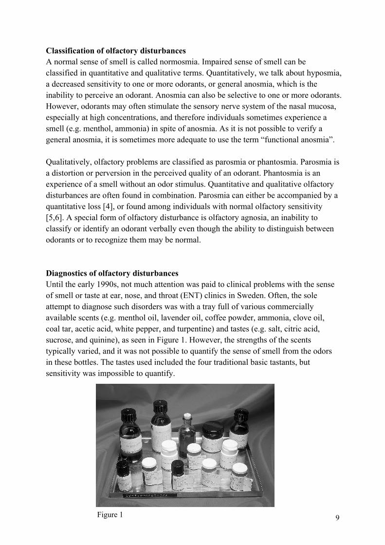
Classification of olfactory disturbances A normal sense of smell is called normosmia. Impaired sense of smell can be classified in quantitative and qualitative terms. Quantitatively, we talk about hyposmia, a decreased sensitivity to one or more odorants, or general anosmia, which is the inability to perceive an odorant. Anosmia can also be selective to one or more odorants. However, odorants may often stimulate the sensory nerve system of the nasal mucosa, especially at high concentrations, and therefore individuals sometimes experience a smell (e.g. menthol, ammonia) in spite of anosmia. As it is not possible to verify a general anosmia, it is sometimes more adequate to use the term “functional anosmia”. Qualitatively, olfactory problems are classified as parosmia or phantosmia. Parosmia is a distortion or perversion in the perceived quality of an odorant. Phantosmia is an experience of a smell without an odor stimulus. Quantitative and qualitative olfactory disturbances are often found in combination. Parosmia can either be accompanied by a quantitative loss [4], or found among individuals with normal olfactory sensitivity [5,6]. A special form of olfactory disturbance is olfactory agnosia, an inability to classify or identify an odorant verbally even though the ability to distinguish between odorants or to recognize them may be normal. Diagnostics of olfactory disturbances Until the early 1990s, not much attention was paid to clinical problems with the sense of smell or taste at ear, nose, and throat (ENT) clinics in Sweden. Often, the sole attempt to diagnose such disorders was with a tray full of various commercially available scents (e.g. menthol oil, lavender oil, coffee powder, ammonia, clove oil, coal tar, acetic acid, white pepper, and turpentine) and tastes (e.g. salt, citric acid, sucrose, and quinine), as seen in Figure 1. However, the strengths of the scents typically varied, and it was not possible to quantify the sense of smell from the odors in these bottles. The tastes used included the four traditional basic tastants, but sensitivity was impossible to quantify.
Figure 1

10
Without methods to diagnose smell and taste disorders, not many patients were referred to ENT clinics with such problems, and the lack of patients contributed to the lack of clinical research in this area in Sweden. Because adequate tests are prerequisite for prospective studies of the cause of impaired sense of smell and the prevalence of olfactory disturbances in society had not previously been investigated in population-based studies, either in Sweden or elsewhere, we were motivated to develop test equipment, evaluate the extent of smell and taste disorders in society, and structure a consulting program for patients. Since the 1990s, smell and taste problems in Sweden have received attention, and new tests have been introduced. The highlight for olfactory research was in 2004 when the Nobel Prize in Physiology or Medicine was awarded to Linda Buck and Richard Axel for their work on olfactory receptors. The olfactory epithelium is found predominately on the superior turbinate and on the upper part of the nasal septum. Axel and Buck demonstrated that the odor receptor cells in the olfactory mucosa are highly specialized. Each cell has only one type of receptor and each of these can recognize only a few odorous substances. Olfactory receptor neurons extend their axons through the thin cribriform plate to well-defined microregions, glomeruli, in the olfactory bulb. Receptor cells carrying the same type of receptor send their neurons to the same glomerulus. In the glomeruli the neurons excite mitral cells. Each mitral cell is activated only by one glomerulus, and the specificity of the information flow is thereby maintained. The nerve signals then reach defined microregions in the brain cortex and combine into a characteristic pattern for each odor. The most commonly used psychophysical methods for diagnosing olfactory disturbances are detection and identification tests, which are inexpensive and simple to perform. Discrimination tests for both quantity and quality are also available. Psychophysical tests, although different, do to some extent overlap each other and data from such tests should be combined with information from the individual’s history, especially in diagnosing parosmia and phantosmia [7]. For detection tests, the odorant is diluted in several concentrations and the individual tries to detect the lowest possible concentration, the threshold. This test often uses a force-choice technique, where the subject is asked to choose between two bottles, of which one contains the odorant and the other is a blank. At wrong answers, a more concentrated odorant is presented until a specified number of consecutive correct answers is reached. This test can be performed with some variations and with different odorants; in the present studies the Connecticut Chemosensory Clinical Research Center (CCCRC) threshold test with butanol was used [8]. The threshold criterion was the level at which the individual made four consecutive correct responses, but later, to

11
better avoid false positive results, it was suggested the level of correct answers be raised to five (Cain, personal communication). Butanol is hedonically neutral and is supposed not to affect the trigeminal nerve at the concentrations used [9]. A threshold test is a very adequate method to measure impaired sense of smell, but only one odorant can be tested for at a time. The most commonly used identification (naming) tests are the University of Pennsylvania Smell Identification Test (UPSIT) and Sniffin´Sticks [10,11]. However, the nature of odor identification being closely related to familiarity with aromatic items [12] limits the valid use of each test to the region of its development. For that reason we developed the Scandinavian Odor-Identification Test (SOIT) with test stimuli and response alternatives selected to be appropriate for clinical odor identification for application to a Scandinavian population [13]. The 16 test stimuli represent a wide range of aromatic qualities, which provide a good basis to generalize test performance to olfactory status. The SOIT was demonstrated to have good reliability and validity, with separate assessment of olfactory and trigeminal function. Although the intention in developing the SOIT was to be able to test all Scandinavian inhabitants, there are people with different cultural backgrounds who are not familiar with the presented odorants; therefore, there is a risk for false negative test results in these subjects. Besides using psychophysical tests, it is also possible to study olfactory capability with neurophysiological methods. These methods are, in contrast to psychophysical tests, objective in the respect that they do not always demand active participation of the person. By using an olfactometer, an exact dose, concentration, and stimulus duration of an odorant can be delivered. By repeated odor provocations in standardized circumstances, during continuous registration with an electroencephalograph, it is possible to register olfactory event-related potentials (OERPs). This technique has been widely used during the last decades and has proven fruitful for investigating olfaction in various conditions [14]. Recordings of event-related potentials involving the auditory, visual, and somatosensory systems have provided useful, noninvasive information on neurophysiological function of sensory and cognitive character over the past four decades [15,16]. The electroencephalographs are analyzed by averaging the electrical brain responses to a series of identical events, e.g., sensory stimulations. The resulting waveform represents brain activity related to the processing of the event. The signals are quantified by means of their amplitudes (size) and latencies (timing). The amplitude has traditionally been regarded as reflecting the magnitude and orientation of neuroelectrical dipoles engaged by cortical processing of the stimulus, and the latency reflecting the speed of such processing [17-20]. A general finding in the literature is that sensory and cognitive impairment results in decreased amplitudes
12
and prolonged latencies, reflecting diminished and prolonged cortical responses [21,22]. The OERPs have a typical shape and a complex of peaks (named N1, P1, N2, P2, etc) [14,23,24]. It is very important that a neurophysiological method is able to distinguish not only between odors and groups of individual odors, but also, between different concentrations of an odor. There are divergent reports of the ability of OERPs to record a dose-response to an olfactory stimulus. Increasing doses of odors typically lead to larger amplitudes and shorter latencies; however, a dose-dependent effect on both amplitudes and latencies is not always found [24-30]. Dose-response effects have mostly been studied in healthy individuals, and there are few studies on patients with impaired olfactory function [31-34]. General aims The aims of this thesis were:
• to investigate the prevalence of olfactory dysfunction in an adult Swedish population and to relate dysfunction to factors of possible impact on olfaction;
• to present our experience with patients seeking medical attention for olfactory disorders and to diagnose types of olfactory disturbance, evaluate possible causes, and relate the patients´ olfactory complaints to performance on psychophysical tests;
• to evaluate the impact of impaired sense of smell on quality of life in patients with olfactory disorders;
• to describe the relationship between detection and identification tests for olfactory disorders;
• to assess OERPs in patients with olfactory impairment and in healthy controls and to investigate possible dose-response effects of odor concentration on OERP parameters in these groups, thereby exploring possibilities and limitations regarding the clinical utility of the OERP method; and
• to evaluate the effect on the sense of smell of radiation in patients treated for head and neck cancer.
Subjects The sense of smell was investigated in a population-based study in Skövde (I). To obtain a representative sample, 1900 individuals, aged 20 years or older, were randomly recruited and stratified by gender and age. The community of Skövde has approximately 50 000 inhabitants, of these, 67% live in the city, 19% in villages, and 14% in the countryside. The demographic profile of Skövde is similar to that of Sweden as a whole, and the study was therefore assumed to be representative for the Swedish population.

13
Patients seeking medical care for olfactory and taste disturbances were consecutively recruited for the study and investigated in a standardized way in order to classify type and degree of the disorder (II). In total, 303 patients were included, 156 women and 147 men, between 10 and 88 years of age. Twenty-three of these patients (10 women and 13 men, aged 18 to 74 years), together with 24 healthy controls (19 women and 5 men, aged 28 to 63 years), were investigated with respect to OERPs (III).
The effect of radiation on the olfactory organ was investigated in patients with head and neck tumors, before and after radiotherapy (IV). During a 10-year period, all patients with such tumors were included after informed consent. In total, 141 patients (49 women and 92 men) participated in the first olfactory test, but only 71 patients, 20 women and 51 men, between 35 and 86 years of age were investigated after the radiation treatment. Of these, 29 received a high dose of radiation against the olfactory organ and the other 42 received a low or no dose and served as controls.
Methods The nasal cavity in all individuals was examined with rigid or flexible endoscopy. Each individual’s self-reported function of the sense of smell was assessed with a structured interview. The psychophysical tests CCCRC butanol threshold test (II-IV) and SOIT (I-IV) were used. As they provide information about somewhat different aspects of olfactory function, there may be a discrepancy between results from the two tests, which for diagnostic purposes made it necessary to define a key for the evaluation of olfactory status. Regarding the diagnoses of anosmia, hyposmia, and normosmia, the following criteria were used in evaluation of the outcome on the butanol threshold test and the SOIT:
1. If agreement between the two tests, the diagnosis was established. 2. If the threshold test showed anosmia, and the SOIT showed normosmia or
hyposmia, the patient was given the diagnosis of hyposmia because the patient was able to experience smells, which precluded anosmia. The threshold test is better than the SOIT for odor sensitivity.
3. If the threshold test showed hyposmia, and the SOIT showed normosmia or anosmia, the patient was given the diagnosis of hyposmia because it is not always possible to prove reduced sensitivity with the SOIT since the odorants are above the smell threshold. The threshold test proves that the patient could not be anosmic.
14
4. If the threshold test showed normosmia, and the SOIT showed hyposmia or anosmia, the patient was given the diagnosis of hyposmia because the patient was able to identify some odorants and could therefore not be anosmic. A normal threshold does not exclude quantitative disorders for other odorants.
The neurophysiological method of OERP recordings was used in study III. The session consisted of OERP recordings during presentations of two concentrations of butanol. A constantly flowing air stream was led into one nasal cavity and the stimuli were presented with controlled temperature and humidity, with a 30 s interstimulus interval, generated by a dynamic olfactometer. The two concentrations were presented 40 times each to the participant in two separate series. Electroencephalogram (EEG) readings were obtained from electrodes placed on the scalp. The records were amplified and filtered. An independent trained observer examined each registration in order to find the peaks of the curves. The peaks were averaged separately for each butanol concentration and electrode site and analyzed for latency and amplitude of the signal. To evaluate quality of life, we used the Nottingham Health Profile (NHP) [35], which is a well-known and well-evaluated instrument that has been used for a number of different conditions to measure the impact on wellbeing, i.e., how disease and symptoms affect a patient’s health and ability to function in daily life (II). Results In the population-based study of 1900 individuals, 1387 (73%) participated in the clinical investigation. The overall prevalence of olfactory dysfunction in adults was 19.1%, with hyposmia in 185 subjects (81 women and 104 men) and functional anosmia in 80 subjects (33 women and 47 men). This represents a prevalence of hyposmia of 13.3% and anosmia of 5.8% (I). Regarding risk for olfactory dysfunction (both hyposmia and anosmia), the prevalence increased with age, and dysfunction was more common in men than in women (odds ratio, OR, for men = 1.7), and more common in individuals with nasal polyps (OR = 2.1). There was no statistically significant increased risk for olfactory dysfunction in people with diabetes mellitus or in smokers. In an additional analysis, current heavy smokers were compared with those who never had smoked, and after adjustments for gender and age, no statistical difference in performance on the SOIT was observed (I). Prevalence of anosmia per se was found to be related to high age. The risk increased with the presence of nasal polyps (OR = 3.8) and diabetes mellitus (OR = 2.6), but there was no statistically significant relation to either gender or smoking (I).

15
For enhanced interpretation of SOIT-based prevalence of olfactory dysfunction, sensitivity and specificity of these diagnoses (hyposmia or anosmia vs. normosmia) were calculated by comparison with self-reported olfactory sensitivity (worse than normal vs. normal or better than normal). These comparisons provided a sensitivity of 43.9%, a specificity of 85.4%, and a correct classification rate of 79.0%. However, 12% were not aware of their olfactory dysfunction and 9% experienced olfactory dysfunction that was not confirmed. In total, 17% reported better olfactory acuity than normal. The SOIT scores were lower in those who reported normal acuity than in those who reported better-than-normal acuity, and even lower in those who reported worse-than-normal acuity (I). Of the 303 patients with olfactory complaints attending the ENT clinic, 288 complained of reduced or total loss of olfactory sensitivity. Of these, 52% were diagnosed as hyposmic, 44% as anosmic, and 4% as normosmic. Out of the 145 patients complaining of total loss of sensitivity, 19% were diagnosed as hyposmic and 3% as normosmic. Out of the 143 patients complaining of reduced loss in sensitivity, 10% were classified as anosmic and 5% as normosmic. Based on the diagnoses, the correct classification rate for the patients’ evaluation of their smell sensitivity was 81.2%. Independent of these test results, 24% of the patients experienced parosmia, and 11% experienced phantosmia. Clinically, a strong relationship between the outcome on the butanol threshold test and the SOIT was found. The suggested etiology of the olfactory disorders was not possible to determine in one third of the cases. Upper respiratory tract infection was the most likely cause in close to 30% of the cases, followed by nasal polyposis, head trauma, and aging (II). The NHP was completed by 102 patients (50 women and 52 men, mean age 52 years). Of these patients, 44% were diagnosed with hyposmia, 44% with anosmia, 21% with parosmia, and 16% with phantosmia. There was a significant difference between the patient group and the reference values for four of the seven questions, namely, relating to paid employment, household work, social life, and family life (II). The OERP grand average, the average of all recordings, shows a distinct response in healthy subjects, while in patients, the effect of the olfactory stimuli (0.3% and 7.5% butanol) were not so evident. Statistical analyses of the individual registrations show significant differences between groups. Patients had lower amplitudes and longer latencies than controls. Within each group there was a significant increase in amplitude and decrease in latency with increased concentration (III). The patients with head and neck tumors were divided into two groups according to the proposed dose of radiation against the olfactory organ. A high radiation dose was

16
given to 29 patients with tumors of the sino-nasal cavity and nasopharynx, the parotid gland, the tongue, the gingival-maxillary region, the bucca, the skin of the temple, the lachrymal duct, and the ear. A low radiation dose against the olfactory epithelium was given to 42 patients with tumors of the tonsils and tongue base, the larynx, the submandibular gland, the hypopharynx, the bottom of the oral cavity, and the lower lip and mandible. In total, 32 patients also received 2 to 3 courses of chemotherapy, usually before radiation therapy. Of these, 25% received a high radiation dose and 75% a low dose. Among those 39 patients who were not treated with chemotherapy 13% received a high radiation dose and 87% a low dose (IV). The butanol threshold test (olfactory detection) before treatment showed a mean threshold of 5.5 in dilution step in the high-dose group and 6.1 in the low-dose group. The corresponding values after treatment were 3.8 and 6.6, respectively. After treatment, there was a significant difference between the groups. The difference between the first and the second test also differed statistically between the groups (IV). The identification test showed before treatment a mean correct identification score of 12.2 in the high-dose group and 12.5 in the low-dose group. The corresponding scores after treatment were 9.3 and 12.5, respectively. After treatment, there was a significant difference between the groups. The difference between the first and the second test also differed statistically between the groups. A multiple regression analysis showed that no variable but radiation dose was important for the difference in olfactory function (IV). Discussion Population-based studies are an important supplement to patient-based studies in examining medical disorders in society. The knowledge of the epidemiology of a disease is fundamental in planning investigations, as well as in treatment and research concerning the disease. To achieve a representative population, the study sample should be randomized and stratified for age and gender and not weakened by too many drop-outs. There are few studies regarding olfactory function that fulfill these epidemiologic criteria. Three non population-based studies, measuring odor identification are of interest. Doty et al reported a study of 1955 individuals from 5 to 99 years old and Ship and Weiffenbach studied 387 healthy individuals from 19 to 95 years [36,37]. Landis et al reported frequency of olfactory dysfunction in 1240 patients from 5 to 86 years old visiting an outpatient ENT clinic [38]. In a population-based cross-sectional study, odor identification was measured in 2491 elderly individuals between 53 and 97 years of age [39]. These studies, in agreement with our study (I), show that impaired olfactory function increases with age. Odor identification
17
impairment in the elderly can be caused by many factors, both peripheral and central. The olfactory epithelium and the olfactory bulb undergo atrophy with time [40-42], and age-related volume loss in temporal lobe areas important to olfactory processing has been found by structural magnetic resonance imaging [43]. OERPs show longer latencies and smaller amplitudes in elderly individuals [44,45]. Odor identification ability is decreased in early stages of common dementia disorders such as Alzheimer’s disease [46,47]. The prevalence of parosmia in the present study of adults (I) was 4.0%, as has been presented elsewhere together with a similar randomized population-based study of 326 teenagers who reported a prevalence of parosmia of 3.4% [48]. In study II, the qualitative disorder was found to be accompanied with quantitative loss. Because good olfactory capability is important for flavor perception, impaired sense of smell may cause decreased appetite that could in turn lead to poor nutrition [49]. However, in additional evaluation of the data from study I, no relationship was found between the degree of olfactory dysfunction and the body mass index in elderly individuals (either ≥60 years or ≥70 years old). Additionally, there was no difference in weight reduction between those who received high or low dose radiation against the olfactory epithelium in spite of differences in olfactory deterioration (IV). Gender-related differences in odor perception have previously been reported. Usually, females perform better in identification tests [36,37,39], as confirmed by our study (I). However, in others studies, differences were not statistically significant [50,51]. The clinical importance of gender-related differences in odor perception is doubtful. There is a discrepancy between the great number of elderly men and women with olfactory dysfunction in the population-based study (I) and the age profile in the clinical study (II). It is also remarkable that in ten years, no more than 303 patients of the catchment area of 270 000 inhabitants have attended the ENT clinic for olfactory disorders compared to the 19% prevalence of olfactory dysfunction in the population-based study. Why do not individuals with impaired olfactory function contact health care for their problem? Of 1175 individuals who reported “normal” or “better than normal” sense of smell, 15% had impaired sense of smell measured by the SOIT in study I. Similar results were obtained in the study by Murphy et al [39]. There may be several reasons people do not seek help for olfactory problems, but one important factor may be that society in general is unaware that olfactory tests exist and that help may be possible. Other reasons could be that individuals are not aware of a dysfunction or that it is of secondary importance, i.e. they do not experience substantial negative consequences in everyday life. Thus, selection bias has to be
18
considered when studying quality of life. Several studies reported consequences of quality of life in patients with olfactory disorders. One revealed that patients with loss of the sense of smell showed impairment in areas of eating, safety and personal hygiene [52]. In a study of 135 patients with olfactory impairment quality of life was investigated with both the General Well-Being Schedule [53] and the disease-specific Multi-Clinic Smell and Taste Questionnaire [54] and reported negative effects mainly concerned personal hygiene, eating, and drinking [55]. These results are in accordance with those seen in study II where patients with olfactory disorders experienced a significantly reduced quality of life regarding not only household work and social and family life, but also paid employment. However, it is interesting to note that subjects with congenital anosmia do not indicate impairment of their quality of life [56]. Any measure of olfactory impairment with an identification test uses a limited number of stimulants. That may leave some relatively rare specific anosmias undetected. More important is that by using only an identification test, such as the SOIT, in the population-based study (I) the prevalence of olfactory dysfunction will likely be underestimated. Extracted data from the patient-based study, where both the SOIT and butanol threshold tests were performed (II), showed that even if the tests were in accordance in 76%, they differed between normal and abnormal sense of smell; in 21% of the patients the SOIT indicated normosmia, whereas the threshold test indicated hyposmia or anosmia. One explanation may be that it is possible to identify the odorants in the SOIT with even a very slightly elevated olfactory threshold. Given an identification test’s high test-retest performance, low cost, acceptability to participants, and short test time required, such tests are useful for population-based studies [39]. However, in clinical settings, an identification test alone is not sufficient. Both an identification test and a threshold test are necessary, and it is important that these tests be accompanied by both an adequate interview (especially to find those with qualitative olfactory disorders) and a clinical examination of the nasal cavity to find treatable conditions (e.g. foreign bodies, nasal polyps, tumors, infections) [57]. What is the use of OERP recordings in the clinical setting? According to Lötsch and Hummel, the OERP recording is regarded as a useful addition to psychophysical tests of olfactory function since they are direct correlates of neuronal activation, have an extremely high temporal resolution, allow the investigation of the sequential processing of information, can be obtained independently of the subject’s response bias, are largely independent of the subject’s cooperation, and produce results which are independent of the person performing the measurement. Weaknesses of this technique are related to its vulnerability to artifacts, the necessity of repeated stimulation at relatively long interstimulus intervals, and the fact that responses have to be extracted from a potentially noisy EEG background [58]. OERPs have in many

19
studies been able to show differences between groups of patients and healthy controls, and they have thus illustrated olfactory deterioration in diseases, e.g. head trauma and dementia [59,60]. However, problems arise when investigating a single patient with heavily impaired olfactory capability. In such a patient it may be difficult to identify any signals from the background activity and therefore impossible to differentiate hyposmia from total anosmia. Furthermore, if OERPs are absent, in spite of normal psychophysical test performance, it is of low diagnostic value [58]. On the other hand, when OERP signals are obtained from a patient with abnormal psychophysical test performance, it could be caused by high trigeminal sensitivity, even if odorants with very low trigeminal stimulation are used. Conclusions • Almost 20% of the adult Swedish population has an olfactory dysfunction, with
males at higher risk than females. Risk factors for olfactory dysfunction are age, nasal polyps, and diabetes, but not smoking.
• Patients seek medical attention for olfactory disorders for both quantitative and qualitative disorders. Impaired olfactory function can reduce quality of life on many levels regarding ability to function in daily life, but seems to have no impact on general health.
• Reliable diagnostic tools for olfactory disorders include patient’s history, a clinical examination, and psychophysical tests. Detection and identification tests complement each other. Possible causes of impaired olfactory function are often upper respiratory tract infection, nasal polyposis, head trauma, and aging, but in most cases no clinical findings or plausible anamnesis data explains the deterioration.
• With OERP recordings it is possible to register a dose-response effect and to differentiate between healthy subjects and individuals with impaired olfactory function. Further validation is needed before OERPs can be routinely applied in clinical assessments of olfactory dysfunction.
• Radiation against the olfactory organ impairs the olfactory capability regarding both detection sensitivity and identification. The pathophysiology underlying this effect is not known.

20
Perspectives for the future Ideas about new items in the clinical field of olfactory research arose with time. A follow-up of the olfactory function of the Skövde population-based study would illustrate the impact of normal aging on olfactory capability and the incidence of olfactory disorders. It might even be possible to add psychological tests to study whether impaired olfactory function really is an early sign of dementia. From this population it would also be possible to investigate selective anosmia. The test result of each odorant of the SOIT is registered at the first investigation. Selective anosmia could be found among individuals with normal test performance who at a second test missed the same odorant as previously. To identify selective anosmia for an odorant, the olfactometer and OERP recordings would be most useful. OERP assessment is used around the world to measure olfactory function. Although the measurement of the OERPs is quite objective, the interpretation of the registrations are subjective. It is not very difficult to identify the peaks (N1, P1, N2, and P2) when stimulating healthy subjects with a strong odorant. However, with weaker odorants, and in individuals with impaired olfactory function, the task becomes more difficult. As identifying the peaks is the subjective part in the evaluation of OERPs, it is important to validate this performance from different aspects:
1. Do different independent researchers provide the same results when analyzing the peaks in one and the same recording?
2. What is the agreement when one person analyzes the same recording at different occasions?
3. To what extent do trained researchers and clinicians do better than untrained?

21
Populärvetenskaplig sammanfattning på svenska Alla levande varelser har förmågan att känna dofter. För djuren är luktsinnet många gånger livsavgörande. De är beroende av ett fungerande luktsinne för att söka föda och de uppmärksammas på faror i omgivningen. Artens överlevnad är beroende av de vuxna djurens luktsinne, med hjälp av lukten hittar de en lämplig partner att para sig med. Många djur har ett luktsinne överlägset människans men även för oss spelar det en väsentlig roll. Vi äter inte skämd mat, vi flyr när vi känner brandrök och kanske väljer även vi partner med inverkan av luktsinnet. Dofter kan väcka minnen som ligger långt tillbaka, ge upphov till associationer och känslor, såväl positiva som negativa. Lukt och smak har nära samband. I själva verket är det vi kallar smak till största delen en effekt av luktsinnet. Vår tungas smaklökar kan endast känna fem smaker: sött, salt, surt, beskt och umami. 2004 tilldelades Linda Buck och Richard Axel Nobelpriset i fysiologi och medicin efter att ha visat hur högspecialiserat vårt luktsinne är. När vårt luktsinne fungerar normalt, är vi ofta inte så uppmärksamma på det, men den som förlorat sitt luktsinne beskriver ofta en försämring i livskvalitet. En luktstörning kan innebära avsaknad av eller nedsättning av luktförmågan. Det kan också visa sig som en förvrängning i uppfattningen av dofter eller att vi upplever en doft utan att det finns ett stimuli. Den metod man tidigare hade att testa luktsinnet var den s.k. luktbrickan, vilken ses i figur 1. Numera finns ett flertal metoder att tillgå för att testa luktsinnet. Vi kan bestämma tröskeln för förmågan att känna dofter och vi kan testa förmågan att känna igen och att särskilja dofter. Allt genom att lukta på olika doftämnen och beskriva vad vi känner. Vi kan även mäta hjärnans elektriska svar på passivt tillförda doftimpulser genom elektroencefalografi, EEG. Metoden kallas Olfactory Event-Related Potentials, OERP. Det huvudsakliga syftet med denna avhandling var
• att undersöka förekomsten av luktstörning hos en vuxen, svensk befolkning och relatera störningen till möjliga bidragande orsaker
• att redogöra för vår erfarenhet av patienter som söker vård för luktsinnets sjukdomar
• att beskriva sambandet mellan olika lukttester • att mäta OERP hos patienter med luktstörning och hos friska och att beskriva
möjligheter och begränsningar med mätmetoden • att utvärdera effekten på luktsinnet av strålbehandling hos patienter med cancer
inom huvud- och halsområdet I det första arbetet studerades förekomsten av luktstörning i befolkningen. Av ett slumpmässigt urval av 1900 vuxna invånare i Skövde lukttestades 1387 personer. 13.3% hade en nedsättning av luktsinnet vilket kunde relateras till hög ålder, manligt

22
kön och näspolyper. Avsaknad av luktsinnet sågs hos 5.8% och kunde relateras till hög ålder, näspolyper och diabetes. Inte i något fall fanns samband med rökning. I studie II undersöktes 303 patienter som sökte för luktstörning. Den mest troliga orsaken till luktstörning var luftvägsinfektion, följt av näspolyper, skallskada och ålder. Hos en tredjedel var det inte möjligt att bestämma trolig orsak. Patienter med störning av luktsinnet har nedsatt livskvalitet relaterat till yrkesliv, hushållsarbete, socialt liv och familjeliv. I studie III användes OERP för att undersöka 23 patienter med luktstörning och 24 friska individer. Patienterna fick ett svagare elektriskt svar i hjärnans aktivitet av doftimpulser än de friska. I studie IV påvisades att 71 patienter med cancer inom huvud- halsområdet som får hög stråldos mot luktorganet får försämrat luktsinne jämfört med dem som får låg stråldos. Slutsatsen blir att luktstörning är vanligt förekommande i samhället och att patienter som söker vård för luktsinnets sjukdomar ska testas och undersökas noggrant eftersom luktstörning ger påverkan på livskvalitet. Patienter som skall strålbehandlas för cancer i närheten av luktorganet skall informeras om risken för luktsinnesnedsättning.

23
Acknowledgements I would like to express my sincere gratitude to all the people who have contributed to this work. In particular I express my acknowledgement to: All the patients, and other study subjects who participated in the studies. Professor Mats Bende, my tutor, for his wealth of ideas, his knowledge in the field of research, his enormous enthusiasm, and his never-ending patience with me. Professor Steven Nordin, also my tutor for sharing with me his enormous knowledge of the sense of smell (in all dimensions), for encouraging me, and for always being willing to talk and offer good advice. Professor Gösta Granström at the department of Otorhinolaryngology, Sahlgrenska Academy, University of Gothenburg, for his support. Associate professor Eva Millqvist, for all her valuable help in the OERP study and technical support with the olfactometer. Professor Claes Mercke for his assistance as a co-author in the radiation therapy study. Research assistant Christel Larsson for her help with study administration, her excellent monitoring, her always positive attitude to my projects and her willingness to listen. Assistant nurse Ulla Thunberg for her encouragement and skilful technical assistance. MD Karin Kullberg for help with the statistical analyses. Christina Bende at the medical library, for all her help. My friends: colleagues and staff at the ENT department at Skaraborg Hospital for their support in my everyday work at the clinic. Special thanks to Bodil Paulsson, my colleague and roommate, sharing my ups and downs, both private and professional. My dear husband Peter, and our wonderful children Mattias, Amanda, and Johanna. In addition to being my life, they help in the work with my studies: Peter always supports and helps me through my (frequent) computer problems and the children are a patient audience, listening as I go through my lectures again and again.

24
During this work I have been supported by grants from Draco AB, the Department of Research and Development at Skaraborg Hospital, the Foundations of Acta Oto-Laryngologica and Rosa & Emanuel Nachmansson.

25
References 1. Jacob S, McClintock MK, Zelano B, Ober C. Paternally inherited HLA alleles
are associated with women’s choice of male odor. Nat Gen 2002; 30:175-9. 2. Doucet S, Soussignan R, Sagot P, Schaal B. The “smellscape” of mother’s
breast: Effects of odor masking and selective unmasking on neonatal arousal, oral, and visual responses. Dev Psychobiol 2007; 49:129-38.
3. Verhagen JV, Engelen L. The neurocognitive bases of human multimodal food perception: Sensory integration. Neurosci Biobehav Rev 2006; 30:613-50.
4. Bonfils P, Avan P, Faulcon P, Malinvaud D. Distorted odorant perception: analysis of a series of 56 patients with parosmia. Arch Otolaryngol Head Neck Surg 2005; 131:107-12.
5. Douek E. Some abnormalities of smell. J Laryngol Otol 1970; 84:1185-91. 6. Goodspeed RB, Catalanotto FA, Gent JF, et al. Clinical characteristics of
patients with taste and smell disorders. In: Meiselman HL, Rivlin RS (eds), Clinical Measurement of Taste and Smell. Macmillan, New York, 1986, pp 451-66.
7. Lötsch J, Reichmann H, Hummel T. Different odor tests contribute differently to the evaluation of olfactory loss. Chem Senses 2008; 33:17-21.
8. Cain WS. Testing olfaction in a clinical setting. Ear Nose Throat J 1989; 68:78-86.
9. Lorig T, Sapp A, Campbell A. Event-related potentials to odor stimuli. Bull Psychonomic Soc 1993; 31:131-4.
10. Doty RL, Shaman P, Dann M. Development of the University of Pennsylvania Smell Identification Test: A standardized microencapsulated test of olfactory function. Physiol Behav 1984; 32:489-502.
11. Hummel T, Sekinger B, Wolf S, Pauli E, Kobal G. ‘Sniffin´sticks’: olfactory performance assessed by the combination testing of odor identification, odor discrimination and olfactory threshold. Chem Senses 1997; 22:39-52.
12. Goldman WP, Seamon JG. Very long-term memory for odors: retention of odor-name associations. Am J Psychol 1992; 105:549-63.
13. Nordin S, Brämerson A, Liden E, Bende M. The Scandinavian Odor-Identification Test: development, reliability, validity and normative data. Acta Otolaryngol 1998; 118:226-34.
14. Hummel T, Kobal G. Olfactory event-related potentials. In: Simon SA, Nicolelis MAL (eds) Methods and frontiers in chemosensory research. CRC Press, Boca Raton, FL, USA, 2001, pp 429-64.
15. Chiappa KH, Ropper AH. Evoked potentials in clinical medicine (first of two parts). N Engl J Med 1982; 306:1140-50.
16. Sutton S, Braren M, Zubin J, John ER. Evoked-potential correlates of stimulus uncertainty. Science 1965; 150:1187-8.

26
17. Donchin E, Coles MGH. Is the P300 component a manifestation of context updating? Behav Brain Sci 1988; 11:357-74.
18. Kutas M, McCarthy G, Donchin E. Augmenting mental chronometry: the P300 as a measure of stimulus evaluation time. Science 1977; 197:792-5.
19. Polich J. Normal variation of P300 from auditory stimuli. Electroencephalogr Clin Neurophysiol 1986; 65:236-40.
20. Jing H, Pivik RT, Dykman RA. A new scaling method for topographical comparisons of event-related potentials. J Neurosci Meth 2006; 151:239-49.
21. Kugler CF, Taghavy A, Platt D. The event-related P300 potential analysis of cognitive human brain aging: a review. Gerontology 1993; 39:280-303.
22. Polich J, Herbst KL. P300 as a clinical assay: rationale, evaluation, and findings. Int J Psychophysiol 2000; 38:3-19.
23. Evans WJ, Cui L, Starr A. Olfactory event-related potentials in normal human subjects: effects of age and gender. Electroencephalogr Clin Neurophysiol 1995; 95:293-301.
24. Frasnelli J, Lotsch J, Hummel T. Event-related potentials to intranasal trigeminal stimuli change in relation to stimulus concentration and stimulus duration. J Clin Neurophysiol 2003; 20:80-6.
25. Olofsson JK, Nordin S. Gender differences in chemosensory perception and event-related potentials. Chem Senses 2004; 29:629-37.
26. Kobal G. Electrophysiologische Untersuchungen des menschlichen Geruchsinns. Stuttgart: Thieme. 1981.
27. Frasnelli J, Wohlgemuth C, Hummel T. The influence of stimulus duration on odor perception. Int J Psychophysiol 2006; 62:24-9.
28. Pause BM, Sojka B, Ferstl R. Central processing of odor concentration is a temporal phenomenon as revealed by chemosensory event-related potentials (CSERP). Chem Senses 1997; 22:9-26.
29. Prah J, Benignus V. Olfactory evoked responses to odorous stimuli of different intensities. Chem Senses 1992; 17:417-25.
30. Tateyama T, Hummel T, Roscher S, Post H, Kobal G. Relation of olfactory event-related potentials to changes in stimulus concentration. Electroencephalogr Clin Neurophysiol 1998; 108:449-55.
31. Hummel T, Pauli E, Schuler P, Kettenmann B, Stefan H, Kobal G. Chemosensory event-related potentials in patients with temporal lobe epilepsy. Epilepsia 1995; 36:79-85.
32. Geisler MW, Schlotfeldt CR, Middleton CB, Dulay MF, Murphy C. Traumatic brain injury assessed with olfactory event-related brain potentials. J Clin Neurophysiol 1999; 16:77-86.
33. Lorig TS. The application of electroencephalographic techniques to the study of human olfaction: a review and tutorial. Int J Psychophysiol 2000; 36:91-104.

27
34. Morgan CD, Murphy C. Olfactory event-related potentials in Alzheimer’s disease. J Int Neuropsychol Soc 2002; 8:753-63.
35. Hunt SM, McKenna SP, McEwen J, Williams J, Papp E. A quantitative approach to perceived health status. J Epidemiol Comm Health 1980; 34:281-6.
36. Doty RL, Shaman P, Applebaum SL, Giberson R, Siksorski L, Rosenberg L. Smell identification ability: changes with age. Science 1984; 226:1441-3.
37. Ship JA, Weiffenbach JM. Age, gender, medical treatment and medication effects on smell identification. J Gerontol 1993; 48:M26-32.
38. Landis B N Konnerth CG, Hummel T. A study on the frequency of olfactory dysfunction. Laryngoscope 2004; 114(10):1764-9.
39. Murphy C, Schubert CR, Cruickshanks KJ, Klein BEK, Klein R, Nondahl DM. Prevalence of olfactory impairment in older adults. JAMA 2002; 288:2307-12.
40. Smith CG. Incidence of atrophy of the olfactory nerves in man Arch Otolaryngol 1941; 34:533-9.
41. Liss L, Gomez F. The nature of senile changes of the human olfactory bulb and tract. Arch Otolaryngol 1958; 67:167-71.
42. Nakishima T, Kimmelman CP, Snow JB. Immunohistopathology of human olfactory epithelium, nerve, and bulb. Laryngoscope 1985; 95:391-6.
43. Jernigan TL, Archibald SL, Fennema-Notestine C, et al. Effect of age on tissues and regions of the cerebrum and cerebellum. Neurobiol Aging 2001; 22:581-94.
44. Murphy C, Nordin S, de Wijk RA, Cain Ws, Polich J. Olfactory evoked potentials: assessment of young and elderly, and comparison to psychophysical threshold. Chem Senses 1994; 19:47-56.
45. Murphy C, Morgan CD, Geisler MW, et al. Olfactory event-related potentials and aging: normative data. Int J Psychophysiol 2000; 36:133-45.
46. Nordin S, Murphy C. Odor memory in Alzheimer’s disease. In: C Rouby, B Schaal, D Dubois, R Gervais, A Holley (eds), Olfaction, Taste, and Cognition. Cambridge University Press, New York, 2002, pp 261-77.
47. Doty RL. Odor perception in neurodegenerative diseases. In: RL Doty (ed), Handbook of olfaction and gestation (pp. 479-501), 2003. Marcel Dekker, New York.
48. Nordin S, Brämerson A, Millqvist E, Bende M. Prevalence of parosmia: the Skövde population-based studies. Rhinology 2007; 45:50-3.
49. Schiffman SS, Graham BG. Taste and smell perception affect appetite and immunity in the elderly. Eur J Clin Nutr 2000; 54, Suppl 3:54-63.
50. Larsson M, Finkel D, Pedersen NL. Odor identification: influence of age, gender, cognition, and personality. J Gerontol B Psychol Sci Soc Sci 2000; 55:304-10.
51. Hummel T, Konnerth CG, Rosenheim K, Kobal G. Screening of olfactory function with a four-minute odor identification test: reliability, normative data,

28
and investigations in patients with olfactory loss. Ann Otol Rhinol Laryngol 2001; 110:976-81.
52. Miwa T, Furukawa M, Tsukatani T, Costanzo RM, DiNardo LJ, Reiter ER. Impact of olfactory impairment on quality of life and disability. Arch Otolaryngol Head Neck Surg 2001; 127:497-503
53. Fazio A.F. A concurrent validational study of the NCHC General Well-Being Schedule. Vital Health Stat 1977; 2(73):1-53
54. Nordin S, Brämerson A, Murphy C, Bende M. A Scandinavian adaptation of the Multi-Clinic Smell and Taste Questionnaire: evaluation of questions about olfaction. Acta Otolaryngol 2003; 123:536-42
55. Blomqvist EH, Brämerson A, Stjärne P, Nordin S. Consequences of olfactory loss and adopted coping strategies. Rhinology 2004; 42(4):189-94
56. Temmel AF, Quint C, Schickinger-Fischer B, Klimek L, Stoller E, Hummel T. Characteristics of olfactory disorders in relation to major causes of olfactory loss. Arch Otolaryngol Head Neck Surg 2002; 128:635-41
57. Nordin S, Brämerson A, Bende M. Prevalence of self-reported poor odor detection sensitivity: The Skövde Population-based Study. Acta Otolaryngol 2004; 124:1171-3.
58. Lötsch J, Hummel T. The clinical significance of electrophysiological measures of olfactory function. Behav Brain Res 2006; 170:78-83.
59. Geisler MW, Schlotfeldt CR, Middleton CB, Dulay MF, Murphy C. Traumatic brain injury assessed with olfactory event-related brain potentials. J Clin Neurophysiol 1999; 16:77-86.
60. Morgan CD, Murphy C. Olfactory event-related potentials in Alzheimer’s disease. J Int Neuropsychol Soc 2002; 8:753-63.