research open access copy number gain of mycn gene is …
TRANSCRIPT
Wang et al. Diagnostic Pathology 2013, 8:5http://www.diagnosticpathology.org/content/8/1/5
RESEARCH Open Access
Copy number gain of MYCN gene is a recurrentgenetic aberration and favorable prognosticfactor in Chinese pediatric neuroblastomapatientsMiao Wang1, Chunju Zhou2, Rongqin Cai1, Yong Li1 and Liping Gong1*
Abstract
Background: Amplification of MYCN oncogene is an established marker indicating aggressive tumor progression ofneuroblastoma (NBL). But copy number analyses of MYCN gene in ganglioneuroblastoma (GNBL) andganglioneuroma(GN) is poorly described in the literature. In the study, we evaluated the copy number aberrationsof MYCN gene in clinical samples of NBLs, GNBLs and GNs and analyzed their association with clinical outcome ofthe patients.
Methods: In this study, we analyzed MYCN gene and chromosome 2 aneusomy by using fluorescence in situhybridization (FISH) method in a total of 220 patients with NBL, GNBL and GN cases. Kaplan-Meier curves weregenerated by using SPSS 12.0 software.
Results: Of 220 patients, 178 (81.0%) were NBLs, 32 (14.5%) were GNBLs and 10 (4.5%) were GNs. MYCN gain is arecurrent genetic aberration of neuroblastic tumors (71.8%, 158/220), which was found in 129 NBLs (58.6%,129/220), 25 GNBLs (11.4%, 25/220) and 4 GN cases (1.8%, 4/220). However, MYCN amplification was only present in24 NBL tumors (13.5%, 24/178) and 1 GNBL case (3.1%, 1/32). Kaplan-Meier survival analysis indicated that MYCNamplification is significantly correlated with decreased overall survival in NBLs (P=0.017). Furthermore, a betterprognosis trend was observed in patients with MYCN gain tumors compared with those with MYCN gene normalcopy number tumors and MYCN amplification tumors (P=0.012).
Conclusions: In summary, the frequency of MYCN amplification in NBLs is high and is rarely observed in GNBLs andGNs, which suggest MYCN plays an important role in neuroblastic tumors differentiation. MYCN gain appeared todefine a subgroup of NBLs with much better outcome and classification of MYCN gene copy number alteration asthree groups (amplification, gain and normal) can provide a powerful prognostic indicator in NBLs.
Virtual Slides: The virtual slide(s) for this article can be found here: http://www.diagnosticpathology.diagnomx.eu/vs/6417541528559124
* Correspondence: [email protected] of Pathology, Basic Medical College, Capital Medical University,Beijing, ChinaFull list of author information is available at the end of the article
© 2013 Wang et al.; licensee BioMed Central Ltd. This is an Open Access article distributed under the terms of the CreativeCommons Attribution License (http://creativecommons.org/licenses/by/2.0), which permits unrestricted use, distribution, andreproduction in any medium, provided the original work is properly cited.
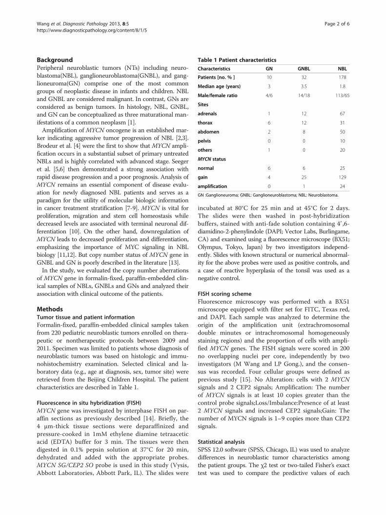
Table 1 Patient characteristics
Characteristics GN GNBL NBL
Patients [no. % ] 10 32 178
Median age (years) 3 3.5 1.8
Male/female ratio 4/6 14/18 113/65
Sites
adrenals 1 12 67
thorax 6 12 31
abdomen 2 8 50
pelvis 0 0 10
others 1 0 20
MYCN status
normal 6 6 25
gain 4 25 129
amplification 0 1 24
GN :Ganglioneuroma; GNBL: Ganglioneuroblastoma; NBL: Neuroblastoma.
Wang et al. Diagnostic Pathology 2013, 8:5 Page 2 of 6http://www.diagnosticpathology.org/content/8/1/5
BackgroundPeripheral neuroblastic tumors (NTs) including neuro-blastoma(NBL), ganglioneuroblastoma(GNBL), and gang-lioneuroma(GN) comprise one of the most commongroups of neoplastic disease in infants and children. NBLand GNBL are considered malignant. In contrast, GNs areconsidered as benign tumors. In histology, NBL, GNBL,and GN can be conceptualized as three maturational man-ifestations of a common neoplasm [1].Amplification of MYCN oncogene is an established mar-
ker indicating aggressive tumor progression of NBL [2,3].Brodeur et al. [4] were the first to show that MYCN ampli-fication occurs in a substantial subset of primary untreatedNBLs and is highly correlated with advanced stage. Seegeret al. [5,6] then demonstrated a strong association withrapid disease progression and a poor prognosis. Analysis ofMYCN remains an essential component of disease evalu-ation for newly diagnosed NBL patients and serves as aparadigm for the utility of molecular biologic informationin cancer treatment stratification [7-9]. MYCN is vital forproliferation, migration and stem cell homeostasis whiledecreased levels are associated with terminal neuronal dif-ferentiation [10]. On the other hand, downregulation ofMYCN leads to decreased proliferation and differentiation,emphasizing the importance of MYC signaling in NBLbiology [11,12]. But copy number status of MYCN gene inGNBL and GN is poorly described in the literature [13].In the study, we evaluated the copy number aberrations
of MYCN gene in formalin-fixed, paraffin-embedded clin-ical samples of NBLs, GNBLs and GNs and analyzed theirassociation with clinical outcome of the patients.
MethodsTumor tissue and patient informationFormalin-fixed, paraffin-embedded clinical samples takenfrom 220 pediatric neuroblastic tumors enrolled on thera-peutic or nontherapeutic protocols between 2009 and2011. Specimen was limited to patients whose diagnosis ofneuroblastic tumors was based on histologic and immu-nohistochemistry examination. Selected clinical and la-boratory data (e.g., age at diagnosis, sex, tumor site) wereretrieved from the Beijing Children Hospital. The patientcharacteristics are described in Table 1.
Fluorescence in situ hybridization (FISH)MYCN gene was investigated by interphase FISH on par-affin sections as previously described [14]. Briefly, the4 μm-thick tissue sections were deparaffinized andpressure-cooked in 1mM ethylene diamine tetraaceticacid (EDTA) buffer for 3 min. The tissues were thendigested in 0.1% pepsin solution at 37°C for 20 min,dehydrated and added with the appropriate probes.MYCN SG/CEP2 SO probe is used in this study (Vysis,Abbott Laboratories, Abbott Park, IL). The slides were
incubated at 80°C for 25 min and at 45°C for 2 days.The slides were then washed in post-hybridizationbuffers, stained with anti-fade solution containing 4’ ,6-diamidino-2-phenylindole (DAPI; Vector Labs, Burlingame,CA) and examined using a fluorescence microscope (BX51;Olympus, Tokyo, Japan) by two investigators independ-ently. Slides with known structural or numerical abnormal-ity for the above probes were used as positive controls, anda case of reactive hyperplasia of the tonsil was used as anegative control.
FISH scoring schemeFluorescence microscopy was performed with a BX51microscope equipped with filter set for FITC, Texas red,and DAPI. Each sample was analyzed to determine theorigin of the amplification unit (extrachromosomaldouble minutes or intrachromosomal homogeneouslystaining regions) and the proportion of cells with ampli-fied MYCN genes. The FISH signals were scored in 200no overlapping nuclei per core, independently by twoinvestigators (M Wang and LP Gong.), and the consen-sus was recorded. Four cellular groups were defined asprevious study [15]. No Alteration: cells with 2 MYCNsignals and 2 CEP2 signals; Amplification: The numberof MYCN signals is at least 10 copies greater than thecontrol probe signals;Loss/Imbalance:Presence of at least2 MYCN signals and increased CEP2 signals;Gain: Thenumber of MYCN signals is 1–9 copies more than CEP2signals.
Statistical analysisSPSS 12.0 software (SPSS, Chicago, IL) was used to analyzedifferences in neuroblastic tumor characteristics amongthe patient groups. The χ2 test or two-tailed Fisher’s exacttest was used to compare the predictive values of each
Wang et al. Diagnostic Pathology 2013, 8:5 Page 3 of 6http://www.diagnosticpathology.org/content/8/1/5
marker analyzed. A p value of less than 0.05 was consid-ered statistically significant. Kaplan-Meier curves weregenerated by using SPSS 12.0 software.
ResultsClinical featuresWe analyzed a panel of 220 pediatric patient samplescomprising 178 NBLs, 32 GNBLs, and 10 GNs. Patientcharacteristics are summarized in Table 1. There was amale predominance in NBL disease group, with the maleto female ratio being 1.7:1. The median age at diagnosiswas 3 years for GNs, 3.5 years for GNBLs and 1.8 yearsfor NBLs. Patients with NBL were significantly youngerthan patients with GN and GNBL (P < 0.001). The dis-tribution of these three diseases generally follows thedistribution of the sympathetic ganglia. In our study,about 36.4% (80/220) tumors arise in adrenal gland. Inaddition, adrenal involvement at diagnosis was 1 case forGN, 12 cases for GNBL and 67 cases for NBL.
Morphology featuresNeuroblastic tumors were divided into three histologicalsubtypes on morphologic criteria of neuroblastic tumorswhich was recommendations by the International Neuro-blastoma Pathology Committee [16]. GN, a fully differen-tiated tumor, is characterized by a mixture of matureschwann cells and ganglion cells (Figure 1A). GNBL hasprimitive neuroblasts along with maturing ganglion cells(Figure 1B); the number and arrangement of the cells varyso the tumor assumes a wide range of appearances. NBL,the least differentiated, resembles the fetal adrenal medullaand is composed of primitive neuroblasts (Figure 1C).
Figure 1 Representative HE image of neuroblastic tumors. A, GanglionGanglioneuroblastoma: Increased schwannian stroma. C, Neuroblastoma: Acytoplasm.
GNBLs and GNs are usually of favorable histology. In fact,GN is considered a benign neuroblastic tumor.
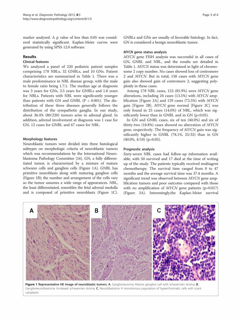
MYCN gene status analysisMYCN gene FISH analysis was successful in all cases ofGN, GNBL and NBL, and the results are detailed inTable 1. MYCN status was determined in light of chromo-some 2 copy number. No cases showed loss of centromere2 and MYCN. But in total, 158 cases with MYCN genegain also showed gain of centromere 2, suggesting poly-ploidy in these cases.Among 178 NBL cases, 153 (85.9%) were MYCN gene
alterations, including 24 cases (13.5%) with MYCN amp-lification [Figure 2A] and 129 cases (72.5%) with MYCNgain [Figure 2B]. MYCN gene normal [Figure 2C] wasonly found in 25 cases (14.0%) of NBL, which was sig-nificantly lower than in GNBL and in GN (p<0.05).In GN and GNBL cases, six of ten (40.0%) and six of
thirty-two (18.8%) cases showed no aberration of MYCNgene, respectively. The frequency of MYCN gain was sig-nificantly higher in GNBL (78.1%, 25/32) than in GN(40.0%, 4/10) (p<0.05).
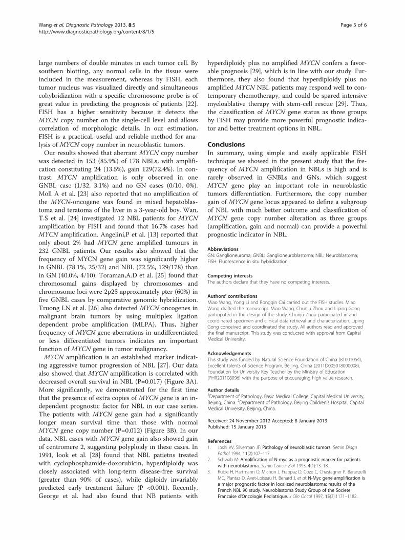
Prognostic analysisSixty-seven NBL cases had follow-up information avail-able, with 50 survived and 17 died at the time of writingup of the study. The patients typically received multiagentchemotherapy. The survival time ranged from 0 to 47months and the average survival time was 37.4 months. Asignificant trend was observed between MYCN gene amp-lification tumors and poor outcome compared with thosewith no amplification of MYCN gene patients (p=0.017)(Figure 3A). Interestingly,the Kaplan-Meier survival
euroma: Mature ganglion cell with schwannian stroma. B,monotonous population of hyperchromatic cells with scant

Figure 2 Representative FISH image of neuroblastic tumor cells displaying MYCN gene status. A: Amplification:The number of MYCNsignals (green) is more than 10 copies of the CEP2 probe signals (red). B: Gain:The number of MYCN signals (green) is 1 copy greater than theCEP2 probe signals; C: No Alteration: Cells with MYCN signals (green) showing the same numbers of the CEP2 probe signals. (DAPI counterstain,original magnification ×1000).
Wang et al. Diagnostic Pathology 2013, 8:5 Page 4 of 6http://www.diagnosticpathology.org/content/8/1/5
analysis also indicated a significant better prognosis inpatients with MYCN gene gain tumors compared withthose with MYCN gene normal tumors (p=0.012)(Figure 3B).
DiscussionThe peripheral neuroblastic tumour group includesNBL, GNBL and GN. NBL is the most common extra-cranial solid tumour of childhood and the incidence ofpediatric neuroblastoma are increasing [17,18]. MYCNgene amplification is a known molecular marker for ag-gressive progression of NBL [4]. In the study, we evalu-ated the histological presentation and MYCN gene copynumber in 220 pediatric neuroblastic tumors, which in-clude 178 NBLs, 32 GNBLs and 10 GNs and analyzed
Figure 3 Overall survival analysis. A. MYCN amplification is correlated wcorrelated with decreased overall survival and MYCN gene gain is correlate
their association with clinical outcome of the patients.To our knowledge, this is the first article for MYCNgene and chromosome 2 aneusomy analyses by usingfluorescence in situ hybridization (FISH) method inchinese pediatric patients.Our study reaffirmed the need for MYCN copy num-
ber to be determined in light of chromosome 2 copynumber. MYCN copy number had been determined bysouthern blot analysis [19]. After 1993, fluorescence insitu hybridization (FISH) was used to determine thepresence of MYCN amplification [20,21]. In these stud-ies, the results of southern blotting and FISH analysiswere prospectively compared and a MYCN copy numberof ≥ 10 was determined to be the optimal cutoff by FISH[20], as the vast majority of amplified tumors have very
ith decreased overall survival in NBL. P=0.017. B. MYCN amplification isd with good outcome in NBL. P=0.012.
Wang et al. Diagnostic Pathology 2013, 8:5 Page 5 of 6http://www.diagnosticpathology.org/content/8/1/5
large numbers of double minutes in each tumor cell. Bysouthern blotting, any normal cells in the tissue wereincluded in the measurement, whereas by FISH, eachtumor nucleus was visualized directly and simultaneouscohybridization with a specific chromosome probe is ofgreat value in predicting the prognosis of patients [22].FISH has a higher sensitivity because it detects theMYCN copy number on the single-cell level and allowscorrelation of morphologic details. In our estimation,FISH is a practical, useful and reliable method for ana-lysis of MYCN copy number in neuroblastic tumors.Our results showed that aberrant MYCN copy number
was detected in 153 (85.9%) of 178 NBLs, with amplifi-cation constituting 24 (13.5%), gain 129(72.4%). In con-trast, MYCN amplification is only observed in oneGNBL case (1/32, 3.1%) and no GN cases (0/10, 0%).Moll A et al. [23] also reported that no amplification ofthe MYCN-oncogene was found in mixed hepatoblas-toma and teratoma of the liver in a 3-year-old boy. Wan,T.S et al. [24] investigated 12 NBL patients for MYCNamplification by FISH and found that 16.7% cases hadMYCN amplification. Angelini,P et al. [13] reported thatonly about 2% had MYCN gene amplified tumours in232 GNBL patients. Our results also showed that thefrequency of MYCN gene gain was significantly higherin GNBL (78.1%, 25/32) and NBL (72.5%, 129/178) thanin GN (40.0%, 4/10). Toraman,A.D et al. [25] found thatchromosomal gains displayed by chromosomes andchromosome loci were 2p25 approximately pter (60%) infive GNBL cases by comparative genomic hybridization.Truong LN et al. [26] also detected MYCN oncogenes inmalignant brain tumors by using multiplex ligationdependent probe amplification (MLPA). Thus, higherfrequency of MYCN gene aberrations in undifferentiatedor less differentiated tumors indicates an importantfunction of MYCN gene in tumor malignancy.MYCN amplification is an established marker indicat-
ing aggressive tumor progression of NBL [27]. Our dataalso showed that MYCN amplification is correlated withdecreased overall survival in NBL (P=0.017) (Figure 3A).More significantly, we demonstrated for the first timethat the presence of extra copies of MYCN gene is an in-dependent prognostic factor for NBL in our case series.The patients with MYCN gene gain had a significantlylonger mean survival time than those with normalMYCN gene copy number (P=0.012) (Figure 3B). In ourdata, NBL cases with MYCN gene gain also showed gainof centromere 2, suggesting polyploidy in these cases. In1991, look et al. [28] found that NBL patietns treatedwith cyclophosphamide-doxorubicin, hyperdiploidy wasclosely associated with long-term disease-free survival(greater than 90% of cases), while diploidy invariablypredicted early treatment failure (P <0.001). Recently,George et al. had also found that NB patients with
hyperdiploidy plus no amplified MYCN confers a favor-able prognosis [29], which is in line with our study. Fur-thermore, they also found that hyperdiploidy plus noamplified MYCN NBL patients may respond well to con-temporary chemotherapy, and could be spared intensivemyeloablative therapy with stem-cell rescue [29]. Thus,the classification of MYCN gene status as three groupsby FISH may provide more powerful prognostic indica-tor and better treatment options in NBL.
ConclusionsIn summary, using simple and easily applicable FISHtechnique we showed in the present study that the fre-quency of MYCN amplification in NBLs is high and israrely observed in GNBLs and GNs, which suggestMYCN gene play an important role in neuroblastictumors differentiation. Furthermore, the copy numbergain of MYCN gene locus appeared to define a subgroupof NBL with much better outcome and classification ofMYCN gene copy number alteration as three groups(amplification, gain and normal) can provide a powerfulprognostic indicator in NBL.
AbbreviationsGN: Ganglioneuroma; GNBL: Ganglioneuroblastoma; NBL: Neuroblastoma;FISH: Fluorescence in situ hybridization.
Competing interestsThe authors declare that they have no competing interests.
Authors’ contributionsMiao Wang, Yong Li and Rongqin Cai carried out the FISH studies. MiaoWang drafted the manuscript. Miao Wang, Chunju Zhou and Liping Gongparticipated in the design of the study. Chunju Zhou participated in andcoordinated specimen and clinical data retrieval and characterization. LipingGong conceived and coordinated the study. All authors read and approvedthe final manuscript. This study was conducted with approval from CapitalMedical University.
AcknowledgementsThis study was funded by Natural Science Foundation of China (81001054),Excellent talents of Science Program, Beijing, China (2011D005018000008),Foundation for University Key Teacher by the Ministry of Education(PHR201108096) with the purpose of encouraging high-value research.
Author details1Department of Pathology, Basic Medical College, Capital Medical University,Beijing, China. 2Department of Pathology, Beijing Children’s Hospital, CapitalMedical University, Beijing, China.
Received: 24 November 2012 Accepted: 8 January 2013Published: 15 January 2013
References1. Joshi VV, Silverman JF: Pathology of neuroblastic tumors. Semin Diagn
Pathol 1994, 11(2):107–117.2. Schwab M: Amplification of N-myc as a prognostic marker for patients
with neuroblastoma. Semin Cancer Biol 1993, 4(1):13–18.3. Rubie H, Hartmann O, Michon J, Frappaz D, Coze C, Chastagner P, Baranzelli
MC, Plantaz D, Avet-Loiseau H, Benard J, et al: N-Myc gene amplification isa major prognostic factor in localized neuroblastoma: results of theFrench NBL 90 study. Neuroblastoma Study Group of the SocieteFrancaise d'Oncologie Pediatrique. J Clin Oncol 1997, 15(3):1171–1182.
Wang et al. Diagnostic Pathology 2013, 8:5 Page 6 of 6http://www.diagnosticpathology.org/content/8/1/5
4. Brodeur GM, Seeger RC, Schwab M, Varmus HE, Bishop JM: Amplification ofN-myc in untreated human neuroblastomas correlates with advanceddisease stage. Science 1984, 224(4653):1121–1124.
5. Brodeur GM, Seeger RC, Schwab M, Varmus HE, Bishop JM: Amplification ofN-myc sequences in primary human neuroblastomas: correlation withadvanced disease stage. Prog Clin Biol Res 1985, 175:105–113.
6. Seeger RC, Brodeur GM, Sather H, Dalton A, Siegel SE, Wong KY, HammondD: Association of multiple copies of the N-myc oncogene with rapidprogression of neuroblastomas. N Engl J Med 1985, 313(18):1111–1116.
7. Tonini GP, Verdona G, Garaventa A, Cornaglia-Ferraris P: Antiblastictreatment does not affect N-myc gene amplification in neuroblastoma.Anticancer Res 1987, 7(4B):729–732.
8. Cetinkaya C, Hultquist A, Su Y, Wu S, Bahram F, Pahlman S, Guzhova I,Larsson LG: Combined IFN-gamma and retinoic acid treatment targetsthe N-Myc/Max/Mad1 network resulting in repression of N-Myc targetgenes in MYCN-amplified neuroblastoma cells. Mol Cancer Ther 2007,6(10):2634–2641.
9. Pession A, Tonelli R: The MYCN oncogene as a specific and selective drugtarget for peripheral and central nervous system tumors. Curr CancerDrug Targets 2005, 5(4):273–283.
10. Rouah E, Wilson DR, Armstrong DL, Darlington GJ: N-myc amplificationand neuronal differentiation in human primitive neuroectodermaltumors of the central nervous system. Cancer Res 1989, 49(7):1797–1801.
11. Kang JH, Rychahou PG, Ishola TA, Qiao J, Evers BM, Chung DH: MYCNsilencing induces differentiation and apoptosis in human neuroblastomacells. Biochem Biophys Res Commun 2006, 351(1):192–197.
12. Janardhanan R, Banik NL, Ray SK: N-Myc down regulation induceddifferentiation, early cell cycle exit, and apoptosis in human malignantneuroblastoma cells having wild type or mutant p53. Biochem Pharmacol2009, 78(9):1105–1114.
13. Angelini P, London WB, Cohn SL, Pearson AD, Matthay KK, Monclair T,Ambros PF, Shimada H, Leuschner I, Peuchmaur M, et al: Characteristicsand outcome of patients with ganglioneuroblastoma, nodular subtype: areport from the INRG project. Eur J Cancer 2012, 48(8):1185–1191.
14. Yu R, Chen G, Zhou C, Gao Z, Shi Y, Shi Y, Zhou X, Xie J, Liu H, Gong L:Extra copies of ALK gene locus is a recurrent genetic aberration andfavorable prognostic factor in both ALK-positive and ALK-negativeanaplastic large cell lymphomas. Leuk Res 2012, 36(9):1141–1146.
15. Ambros PF, Ambros IM: Pathology and biology guidelines for resectableand unresectable neuroblastic tumors and bone marrow examinationguidelines. Med Pediatr Oncol 2001, 37(6):492–504.
16. Shimada H, Ambros IM, Dehner LP, Hata J, Joshi VV, Roald B: Terminologyand morphologic criteria of neuroblastic tumors: recommendations bythe International Neuroblastoma Pathology Committee. Cancer 1999,86(2):349–363.
17. Hsieh MH, Meng MV, Walsh TJ, Matthay KK, Baskin LS: Increasing incidenceof neuroblastoma and potentially higher associated mortality of childrenfrom nonmetropolitan areas: analysis of the surveillance, epidemiology,and end results database. J Pediatr Hematol Oncol 2009, 31(12):942–946.
18. Heck JE, Ritz B, Hung RJ, Hashibe M, Boffetta P: The epidemiology ofneuroblastoma: a review. Paediatr Perinat Epidemiol 2009, 23(2):125–143.
19. Schwab M, Alitalo K, Klempnauer KH, Varmus HE, Bishop JM, Gilbert F,Brodeur G, Goldstein M, Trent J: Amplified DNA with limited homology tomyc cellular oncogene is shared by human neuroblastoma cell lines anda neuroblastoma tumour. Nature 1983, 305(5931):245–248.
20. Mathew P, Valentine MB, Bowman LC, Rowe ST, Nash MB, Valentine VA,Cohn SL, Castleberry RP, Brodeur GM, Look AT: Detection of MYCN geneamplification in neuroblastoma by fluorescence in situ hybridization: apediatric oncology group study. Neoplasia 2001, 3(2):105–109.
21. Shapiro DN, Valentine MB, Rowe ST, Sinclair AE, Sublett JE, Roberts WM,Look AT: Detection of N-myc gene amplification by fluorescence in situhybridization. Diagnostic utility for neuroblastoma. Am J Pathol 1993,142(5):1339–1346.
22. Hachitanda Y, Saito M, Mori T, Hamazaki M: Application of fluorescence insitu hybridization to detect N-myc (MYCN) gene amplification onparaffin-embedded tissue sections of neuroblastomas. Med Pediatr Oncol1997, 29(2):135–138.
23. Moll A, Krenauer A, Bierbach U, Till H, Hirsch W, Leuschner I, Schmitz N,Wittekind C, Aigner T: Mixed hepatoblastoma and teratoma of the liver ina 3-year-old child: a unique combination and clinical challenge. DiagnPathol 2009, 4:37.
24. Wan TS, Ma ES, Chan GC, Chan LC: Investigation of MYCN status inneuroblastoma by fluorescence in situ hybridization. Int J Mol Med 2004,14(6):981–987.
25. Toraman AD, Keser I, Luleci G, Tunali N, Gelen T: Comparative genomichybridization in ganglioneuroblastomas. Cancer Genet Cytogenet 2002,132(1):36–40.
26. Truong LN, Patil S, Martin SS, LeBlanc JF, Nanda A, Nordberg ML, BecknerME: Rapid Detection of high-level oncogene amplifications in ultrasonicsurgical aspirations of brain tumors. Diagn Pathol 2012, 7:66.
27. Suita S, Zaizen Y, Kaneko M, Uchino J, Takeda T, Iwafuchi M, Utsumi J,Takahashi H, Yokoyama J, Nishihira H, et al: What is the benefit ofaggressive chemotherapy for advanced neuroblastoma with N-mycamplification? A report from the Japanese Study Group for theTreatment of Advanced Neuroblastoma. J Pediatr Surg 1994,29(6):746–750.
28. Look AT, Hayes FA, Shuster JJ, Douglass EC, Castleberry RP, Bowman LC,Smith EI, Brodeur GM: Clinical relevance of tumor cell ploidy and N-mycgene amplification in childhood neuroblastoma: a Pediatric OncologyGroup study. J Clin Oncol 1991, 9(4):581–591.
29. George RE, London WB, Cohn SL, Maris JM, Kretschmar C, Diller L, BrodeurGM, Castleberry RP, Look AT: Hyperdiploidy plus nonamplified MYCNconfers a favorable prognosis in children 12 to 18 months old withdisseminated neuroblastoma: a Pediatric Oncology Group study. J ClinOncol 2005, 23(27):6466–6473.
doi:10.1186/1746-1596-8-5Cite this article as: Wang et al.: Copy number gain of MYCN gene is arecurrent genetic aberration and favorable prognostic factor in Chinesepediatric neuroblastoma patients. Diagnostic Pathology 2013 8:5.
Submit your next manuscript to BioMed Centraland take full advantage of:
• Convenient online submission
• Thorough peer review
• No space constraints or color figure charges
• Immediate publication on acceptance
• Inclusion in PubMed, CAS, Scopus and Google Scholar
• Research which is freely available for redistribution
Submit your manuscript at www.biomedcentral.com/submit