targeting of the mycn protein with small molecule c-myc inhibitors
TRANSCRIPT
Targeting of the MYCN Protein with Small Moleculec-MYC InhibitorsInga Muller1, Karin Larsson1, Anna Frenzel1, Ganna Oliynyk1, Hanna Zirath1, Edward V. Prochownik2,
Nicholas J. Westwood3, Marie Arsenian Henriksson1*
1 Department of Microbiology, Tumor and Cell Biology, Karolinska Institutet, Stockholm, Sweden, 2 Section of Hematology/Oncology, Children’s Hospital of Pittsburgh of
UMPC, Pittsburgh, Pennsylvania, United States of America, 3 School of Chemistry and Biomedical Sciences Research Complex, University of St. Andrews and EaStCHEM, St.
Andrews, Fife, Scotland, United Kingdom
Abstract
Members of the MYC family are the most frequently deregulated oncogenes in human cancer and are often correlated withaggressive disease and/or poorly differentiated tumors. Since patients with MYCN-amplified neuroblastoma have a poorprognosis, targeting MYCN using small molecule inhibitors could represent a promising therapeutic approach. We havepreviously demonstrated that the small molecule 10058-F4, known to bind to the c-MYC bHLHZip dimerization domain andinhibiting the c-MYC/MAX interaction, also interferes with the MYCN/MAX dimerization in vitro and imparts anti-tumorigeniceffects in neuroblastoma tumor models with MYCN overexpression. Our previous work also revealed that MYCN-inhibitionleads to mitochondrial dysfunction resulting in accumulation of lipid droplets in neuroblastoma cells. To expand ourunderstanding of how small molecules interfere with MYCN, we have now analyzed the direct binding of 10058-F4, as wellas three of its analogs; #474, #764 and 10058-F4(7RH), one metabolite C-m/z 232, and a structurally unrelated c-MYCinhibitor 10074-G5, to the bHLHZip domain of MYCN. We also assessed their ability to induce apoptosis, neurite outgrowthand lipid accumulation in neuroblastoma cells. Interestingly, all c-MYC binding molecules tested also bind MYCN as assayedby surface plasmon resonance. Using a proximity ligation assay, we found reduced interaction between MYCN and MAXafter treatment with all molecules except for the 10058-F4 metabolite C-m/z 232 and the non-binder 10058-F4(7RH).Importantly, 10074-G5 and 10058-F4 were the most efficient in inducing neuronal differentiation and lipid accumulation inMYCN-amplified neuroblastoma cells. Together our data demonstrate MYCN-binding properties for a selection of smallmolecules, and provide functional information that could be of importance for future development of targeted therapiesagainst MYCN-amplified neuroblastoma.
Citation: Muller I, Larsson K, Frenzel A, Oliynyk G, Zirath H, et al. (2014) Targeting of the MYCN Protein with Small Molecule c-MYC Inhibitors. PLoS ONE 9(5):e97285. doi:10.1371/journal.pone.0097285
Editor: Alfred Nordheim, University of Tuebingen, Germany
Received September 20, 2013; Accepted April 17, 2014; Published May 23, 2014
Copyright: � 2014 Muller et al. This is an open-access article distributed under the terms of the Creative Commons Attribution License, which permitsunrestricted use, distribution, and reproduction in any medium, provided the original author and source are credited.
Funding: This study was funded by grants from the Swedish Research Council and the Swedish Cancer Society. IM and HZ were recipients of graduate studentgrants from KI (KID), MAH was recipient of a Senior Investigator Award from the Swedish Cancer Society, and NJW was a Royal Society University Research Fellowwhen this work began. The funders had no role in study design, data collection and analysis, decision to publish, or preparation of the manuscript.
Competing Interests: The authors have declared that no competing interests exist.
* E-mail: [email protected]
Introduction
The MYC family members c-MYC, MYCN and L-MYC are
transcription factors crucial for the regulation of normal cellular
functions including proliferation, cell growth, differentiation,
metabolism and apoptosis. However, the genes encoding these
proteins are also the most frequently deregulated oncogenes in
several types of human cancers [1,2]. c-MYC and MYCN
(hereafter MYC), exert their functions mainly through transcrip-
tional modulation of their target genes. The C-terminal domain of
MYC comprises a basic helix-loop-helix leucine zipper domain
(bHLHZip), necessary for the dimerization with its partner MAX
and for sequence-specific binding to DNA [3], while the N-
terminal transactivation domain interacts with co-factors to
regulate transcription [2]. There is a large overlap between the
downstream targets of c-MYC and MYCN and insertion of the
mycn gene into the c-myc locus can fully rescue the embryonic lethal
phenotype of a c-myc knockout mouse [4]. However, in normal
tissue the expression pattern of these two proteins differ
significantly [5,6]. In the developing embryo, MYCN is expressed
in certain tissues including the central and peripheral nervous
systems, lung and spleen, whereas in adults its expression is very
low or absent. In contrast, c-MYC is expressed in all proliferating
cells in adults [6–9].
In human tumors, oncogenic alterations in MYC are common
and include point mutations that increase protein stability, gene
amplification, gene translocation, and enhanced translation [1,2].
MYCN is amplified in cancers such as neuroblastoma (NB),
medulloblastoma, lung cancer and glioma [1,10–12]. In NB, a
pediatric cancer of the sympathetic nervous system, MYCN-
amplification is strongly correlated with poor prognosis and
advanced tumor stage, and these tumors are often resistant to
multimodal therapy [11,12]. MYC is therefore an attractive target
for cancer therapy [13,14]. It has been shown that downregulation
of MYC leads to cancer cell growth arrest, senescence, enhanced
apoptosis, differentiation and/or tumor regression in mouse
models of human cancer [15]. Importantly, even transient
downregulation of MYC has been reported sufficient to diminish
the tumor burden in animal models [15], and the effects of MYC
inhibition on normal tissue has been shown to be well tolerated
PLOS ONE | www.plosone.org 1 May 2014 | Volume 9 | Issue 5 | e97285
and reversible in adult mice [16–23]. Several groups have made
efforts to target MYC at different levels, including its transcription,
translation, hetero-dimerization with MAX, as well as targeting its
direct or indirect downstream targets [10,14,24–26]. A number of
small molecular compounds inhibiting c-MYC-MAX dimerization
have been identified [27,28] and among them 10058-F4 is by far
the most studied. Biophysical experiments have shown that it
interacts with the C-terminal bHLHZip region of c-MYC [28–32].
A fluorescence polarization assay was used to determine the
affinity as well as to identify the binding site of 10058-F4 on c-
MYC using different deletions, point mutations and synthetic
peptides [30]. NMR measurements confirmed that 10058-F4
binds to amino acid residues 402–412 in the bHLHZip domain of
c-MYC [30]. Furthermore, metadynamic molecular simulations
and an ion mobility mass spectrometry using a peptide
corresponding to the identified binding site, indicated that the
compound binds to an inactive, disordered conformation of c-
MYC [31,32]. Together these studies suggest that 10058-F4
inhibits the function of c-MYC in a direct manner by preventing c-
MYC/MAX hetero-dimerization. Importantly, several reports
have shown that 10058-F4 affects c-MYC expression and induces
cell cycle arrest, inhibits cell growth, promotes apoptosis and
confers chemo-sensitivity in a c-MYC specific manner in various
cancer cell types [28,33–35]. In addition, treatment of acute
myeloid leukemia (AML) cells with 10058-F4 leads to myeloid
differentiation [33]. The effect of 10058-F4 treatment in vivo has
been investigated in xenograft models of prostate cancer but no
significant antitumor activity could be observed, probably due to
its rapid clearance and low potency [36]. In contrast, we have
recently demonstrated anti-tumorigenic effects of 10058-F4 in two
tumor models of MYCN-amplified neuroblastoma, suggesting that
direct MYC inhibition using a small molecule is achievable in vivo
[36].
The structurally unrelated small molecule 10074-G5 was
identified simultaneously as 10058-F4 as another substance that
inhibits the c-MYC/MAX interaction. This molecule also
decreased c-MYC protein levels and inhibited cell growth [28],
but failed to show any antitumor activity in a xenograft model
using a Burkitt’s lymphoma cell line [37]. The cognate binding site
for 10074-G5 on c-MYC was found to be distinct from that of
10058-F4, spanning amino acid residues 363–381 [30,38] (see
Figure 1). Both molecules were found to bind independently of
each other, and probably induce only local conformation changes
in the bHLHZip domain of c-MYC preventing its interaction with
MAX [30,38]. In order to identify more potent compounds,
several analogs of 10058-F4 have been synthesized, some of which,
including #015, #474 and #764, exhibited improved growth
inhibition of c-MYC expressing cells [39].
Since c-MYC and MYCN share structural similarity in the
bHLHZip domain we hypothesized that 10058-F4 also targets
MYCN. We have previously shown that this compound interferes
with the MYCN/MAX interaction leading to cell cycle arrest,
apoptosis, and neuronal differentiation in MYCN-overexpressing
NB cell lines [40]. In addition, using 10058-F4 as a tool, we found
that inhibition of MYCN results in mitochondrial dysfunction
leading to lipid accumulation. Importantly, 10058-F4 treatment
furthermore increased the survival of TH-MYCN transgenic mice
and showed anti-tumor effects in established aggressive NB
xenografts [40].
Here, we determined the direct binding of 10058-F4 and
additional selected c-MYC-targeting compounds to MYCN by
surface plasmon resonance (SPR) (see Figure S1 for the structures
of the compounds used). We found that all molecules previously
reported to bind to c-MYC also bound to MYCN. Treatment with
the small molecules furthermore interfered with the MYCN/
MAX interaction and caused protein degradation, apoptosis,
differentiation and lipid formation to different extents in MYCN-
amplified NB cells.
Results
Folding of the human bHLHZip domain of MYCN andc-MYC
The bHLHZip domains of the MYC proteins are intrinsically
disordered with little secondary structure without binding to MAX
[41]. In order to verify the folding of the protein domains following
purification under denaturing conditions and refolding, circular
dichroism (CD) was performed. Comparison of the CD spectra of
the purified and refolded bHLHZip domains with a model
spectrum of a a-helical protein, indicated the presence of some a-
helical structure in both c-MYC and MYCN (Figure S2). Analyses
of the obtained spectra using the CONTIN, SELCON and the
K2D algorithms [41–47] predicted the unstructured content to be
very similar in both proteins, approximately 58% for c-MYC and
55% for MYCN. The a-helical structure was predicted to be 26%
for c-MYC whereas MYCN had a slightly higher a-helical
content, 33%. These values are in agreement with the 27% a-
helical content documented for the v-MYC bHLHZip [48]. We
found approximately 16% b-sheets in c-MYC and 13% in
MYCN, respectively (Table S1). These results are indicative of
two incompletely folded protein domains with some a-helical
structure, as previously reported for the purified bHLHZip of c-
MYC [30,31]. Since the secondary structure content of the
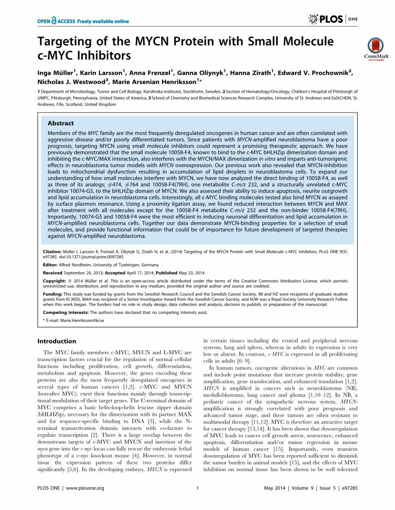
Figure 1. Alignment of the bHLHZip domains of c-MYC and MYCN. The amino acids in the bHLHZip domains of human c-MYC and humanMYCN were aligned, while the secondary structure is indicated above. Identical amino acid are denoted (*), conserved substitution (:) and semi-conserved substitution (.). The two binding sites for the specified compounds as reported by Hammoudeh et al is indicated by a colored square [38].Each small molecule is positioned under their reported or assumed binding site [38,39]. For the 10058-F4 analogs #474 and #764 as well as itspotential metabolite C-m/z 232 the binding sites have not been determined experimentally [39]. Through the similarity of their chemical structure to10058-F4, it has been assumed that these compounds bind to the same site as indicated.doi:10.1371/journal.pone.0097285.g001
Targeting of MYCN with c-MYC Inhibitors
PLOS ONE | www.plosone.org 2 May 2014 | Volume 9 | Issue 5 | e97285
bHLHZip domains confirmed results from previous reports, we
next performed surface plasmon resonance (SPR) experiments in
order to examine the binding of the selected compounds to the
purified bHLHZip domains of both proteins.
Binding of small molecules to the bHLHZip of MYCN andc-MYC
The protein sequences of the human c-MYC and MYCN
bHLHZip domains share 56% similarity (Figure 1). Given the
highly structural and functional similarity of the C-terminal
regions of c-MYC and MYCN it is very likely that molecules
binding to c-MYC also bind to MYCN. In addition to 10058-F4
we included three of its structural analogs (Figure 1; Figure S1) in
our study. The analogs #474 and #764 have previously been
shown to have greater potency compared to the parent compound
as judged by enhanced growth inhibition of c-MYC expressing
leukemic cells [39]. In contrast, the analog 10058-F47RH (referred
to as 7RH throughout) was found to be inactive in a c-MYC
binding assay [39], and was thus included as a negative control.
We also tested the binding to MYCN of a predicted in vivo
metabolite of 10058-F4, C-m/z 232, in order to examine if the
modified molecule still possesses some of the capacities of 10058-
F4 [36]. Furthermore we included the structurally unrelated
compound 10074-G5, previously shown to bind to c-MYC, in
order to test the conservation of binding to a second site in the
bHLHZip domain of MYC [28,30,38] (Figure 1).
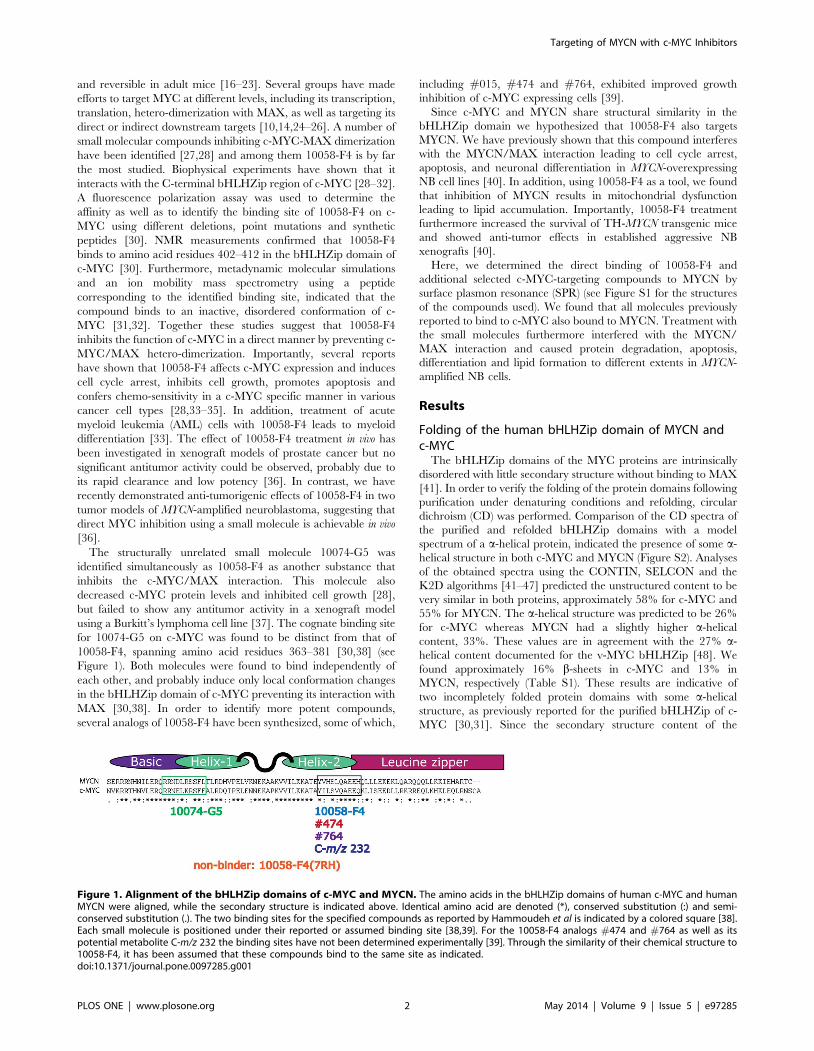
For all SPR binding measurements the compounds were
injected at increasing concentrations. After protein immobilization
on the CM5 chip surface most of the c-MYC protein appeared to
be active, since the expected maximal response (Rmax, the binding
signal at saturation) was reached after injection of increasing
concentrations of 10058-F4 (Figure S3). However for MYCN, only
one fourth of the theoretical Rmax was reached, indicating that not
all protein molecules were able to bind to the analytes after
immobilization (Figure S3). However, despite some of the MYCN
protein being inactive, increased binding of the molecules was still
detected in a dose-dependent manner and KD values could be
calculated for most of the compounds (Table 1, Figure 2 and
Figure S3). Surprisingly, the obtained Rmax values for C-m/z 232
to both c-MYC and MYCN were twice as high as those for 10058-
F4, and double those of the theoretical Rmax value for a single site
binding to c-MYC, thus suggesting a possible second binding site.
The analog #764 as well as 10074-G5 showed especially poor
solubility in aqueous buffers and could not be analyzed at
concentrations above 50 mM. Hence the Rmax for c-MYC and
MYCN could not be obtained for these molecules. Some
unspecific binding, which was visible in the sensorgrams by a
continuous, slightly upward trend of the curves, especially at
higher concentrations, was detected for 10058-F4, C-m/z-232 and
#474 (Figure S3), probably reflecting weak non-specific binding to
other sites in c-MYC [30,32] and MYCN, respectively. As
expected, the 10058-F4 analog 7RH previously reported not to
bind to c-MYC [39], was also a non-binder in our assays and was
thus regarded as a negative control (Figure S3).
In order to determine the affinity of the compounds to c-MYC
and MYCN, the response for each small molecule was plotted
against the analyte (compound) concentration. The KD was
defined as the concentration of analyte at 50% of the experimental
maximal Response (Rmax). Approximate KD-values could be
determined for all compounds except for #764. Similar affinities
were determined for binding of 10058-F4 to both c-MYC and
MYCN, with a mean KD value of 39.768.1 mM and
41.9610.6 mM, respectively (Table 1 and Figure 2). The affinities
determined for the potential metabolite, C-m/z 232, were the
lowest of all the molecules tested with a mean KD of 82.365.5 mM
for c-MYC and a mean KD of 87.164.9 mM for MYCN.
Compared to 10058-F4 the analog #474 analog showed an
enhanced affinity both to c-MYC (mean KD value of
16.661.4 mM) and to MYCN (mean KD = 26.968.1 mM).
Importantly, we demonstrated binding of the structurally unrelat-
ed compound 10074-G5 to both MYC proteins, and determined
the affinity to both MYCN (KD = 19.2611.5 mM) and to c-MYC
(KD = 31.7624.9 mM). Although the sensorgrams for #764
indicated binding to both proteins (Figure S3), we were not able
to acquire data reliable enough to calculate its KD. As expected,
binding of 7RH could not be detected to either of the two MYC
proteins, even at the highest concentrations used (Figure S3). In
summary, all active c-MYC inhibitors tested were also found to
bind directly to MYCN.
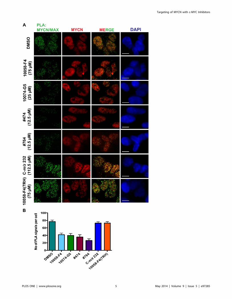
Targeting the MYCN/MAX interaction in NB cellsIn our previous study we observed a reduction in MYCN/MAX
interaction after treatment with 10058-F4 [40]. To investigate if
the other small molecules also have an effect on MYCN/MAX
dimerization we performed a proximity ligation assay (PLA) using
MYCN-amplified SMS-KCN69n NB cells. Compared to DMSO
treated cells, 10058-F4, its analogs #474 and #764 as well as the
structurally unrelated compound 10074-G5 reduced the MYCN/
Figure 2. Equilibrium binding response for compoundsbinding to the bHLHZip of c-MYC and MYCN. Equilibrium bindingresponse determined by Surface Plasmon Resonance of small moleculesto c-MYC (A) or MYCN (B) as a function of the concentration asindicated. The concentrations were plotted versus the response unitsafter solvent correction and subtraction from the reference surface. Theplots are from one representative experiment. For each molecule theexperiments were performed at least with two different immobilizationsfollowed by at least three independent experiments.doi:10.1371/journal.pone.0097285.g002
Targeting of MYCN with c-MYC Inhibitors
PLOS ONE | www.plosone.org 3 May 2014 | Volume 9 | Issue 5 | e97285
MAX interaction following 6 hours treatment (Figure 3). In
contrast, the metabolite C-m/z 232 and the non-binder 7RH did
not affect MYCN/MAX dimer formation (Figure 3). No signal
was detected in any of the experimental negative controls (Figure
S4). In summary, all molecules that compared to 10058-F4
showed approximately similar or higher affinity to MYCN were
also able to reduce the interactions between MYCN and MAX in
NB cells.
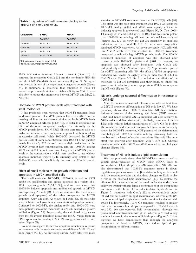
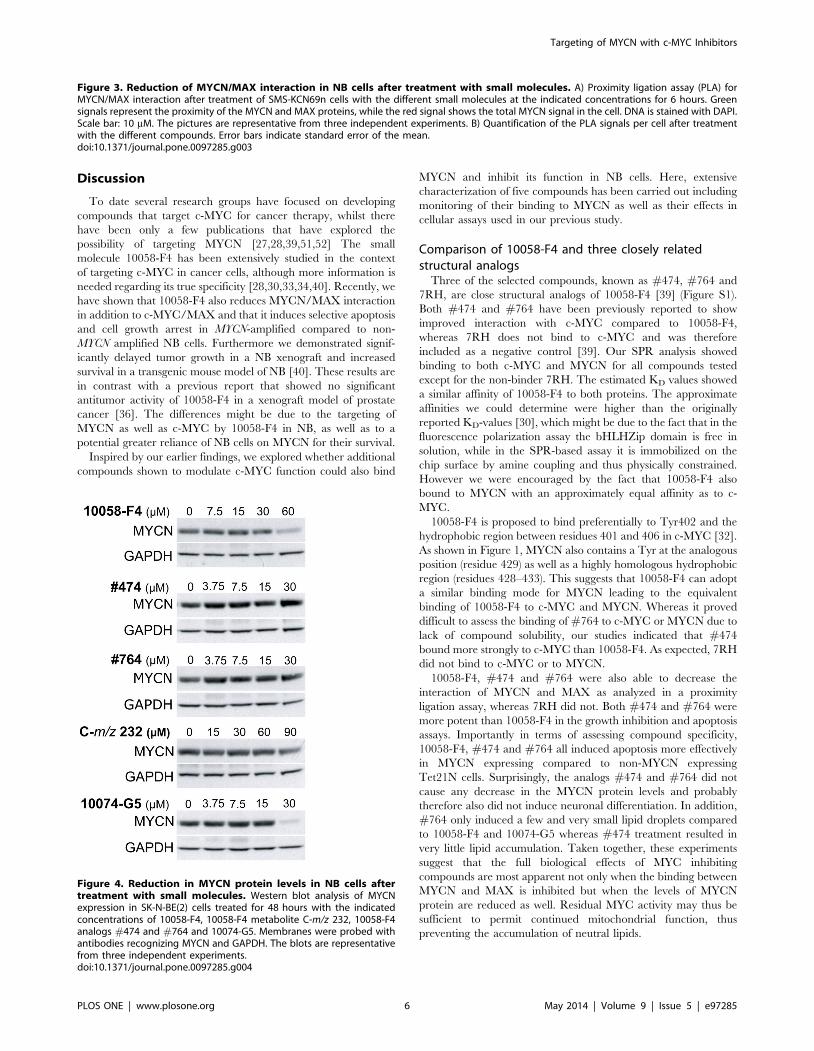
Decrease of MYCN protein levels after treatment withsmall molecules
It has previously been reported that 10058-F4 treatment leads
to down-regulation of c-MYC protein levels in c-MYC overex-
pressing cell lines and we observed similar results for MYCN levels
in MYCN-amplified NB cells [33,34,40]. In order to determine the
effect of the other compounds analyzed in this study on the
MYCN protein levels, SK-N-BE(2) NB cells were treated with as a
high concentration of each compound as possible without resulting
in excessive cell death. While 10074-G5 decreased the MYCN
protein levels at lower concentrations than 10058-F4, the putative
metabolite C-m/z 232 showed only a slight reduction in the
MYCN levels at high concentrations, and the 10058-F4 analogs
#474 and #764 did not cause any changes in the MYCN protein
levels at the concentrations which were possible to use without
apoptosis induction (Figure 4). In summary, only 10058-F4 and
10074-G5 were able to efficiently decrease the MYCN protein
levels.
Effect of small-molecules on growth inhibition andapoptosis in MYCN-amplified cells
The small molecules 10058-F4, 10074-G5, as well as #474
inhibit cell proliferation and induce apoptosis in a variety of c-
MYC expressing cells [28,33,34,39], and we have shown that
10058-F4 induces apoptosis and inhibits cell growth in MYCN
overexpressing NB cells [40]. Here we examined the effect on cell
growth (and apoptosis) of the other compounds in MYCN-
amplified Kelly NB cells. As shown in Figure 5A, all molecules
tested inhibited cell growth in a concentration dependent manner.
Compared to 10058-F4, the analog #474 and 10074-G5 were
more potent whereas the metabolite C-m/z 232 caused less growth
inhibition (Figure 5A). Importantly, the calculated IC50 values
from the cell growth inhibition assays and the KD-values from the
SPR experiment for binding to MYCN strongly correlated to each
other (Figure 5B).
We next investigated the level of apoptosis induction in response
to treatment with the molecules using two different MNA NB cell
lines (Figure 5C, D). As previously shown, Kelly cells were more
sensitive to 10058-F4 treatment than the SK-N-BE(2) cells [40].
This effect was also seen after treatment with 10074-G5, while the
10058-F4 analogs #474 and #764 were equally efficient in
inducing apoptosis in the two cell lines (Figure 5C, D). The 10058-
F4 analogs #474 and #764 as well as 10074-G5 were more potent
than 10058-F4 in inducing cell death in both cell lines analyzed
(Figures 5C, D). To verify the MYCN specificity in apoptosis
induction, we next used Tet21N NB cells with doxycycline
regulated MYCN expression. As shown previously [40], cells with
low MYCN-levels were less sensitive to 10058-F4 treatment
compared to cells with high MYCN protein levels. The MYCN
dependent induction of apoptosis was also observed after
treatment with 10074-G5, #474 and #764. In contrast, no
apoptosis was observed after incubation with C-m/z 232
independently of MYCN status (Figure S5). As previously reported
for c-MYC expressing cells [39], the effect of #764 on apoptosis
induction was similar or slightly stronger than that of #474 in
Tet21N cells (Figure 5C, D). In conclusion, the affinity of the
molecules to MYCN correlated with their ability to inhibit cell
growth and to selectively induce apoptosis in MYCN overexpress-
ing NB cells (Figure S5).
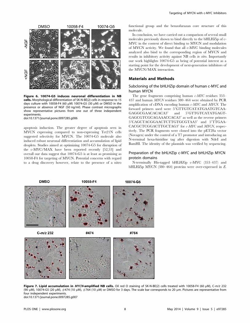
NB cells undergo neuronal differentiation in response to10074-G5
MYCN counteracts neuronal differentiation whereas inhibition
of MYCN promotes differentiation of NB cells [49,50]. We have
previously shown that 10058-F4 stimulates NB differentiation,
induces expression of the nerve growth factor (NGF) receptor
TrkA and hence renders MYCN-amplified NB cells sensitive to
NGF-mediated differentiation [40]. Similarly, treatment of SK-N-
BE(2) cells with sub-lethal concentrations of 10074-G5 for 14 days
also resulted in evident neurite outgrowth (Figure 6). As previously
shown for 10058-F4 treatment, NGF potentiated the differentiated
morphology of 10074-G5 treated cells by increasing both the
number and the length of neurite protrusions (Figure 6). The same
effect was observed after treatment with C-m/z 232, whereas
incubation with neither #474 nor #764 resulted in morphological
changes (Figure S6).
Treatment of NB cells induces lipid accumulationWe have previously shown that 10058-F4 treatment as well as
genetic down-regulation of MYCN using shRNA, leads to
accumulation of lipid droplets in MYCN-amplified NB cells. We
also demonstrated that 10058-F4 treatment results in down-
regulation of proteins involved in b-oxidation of fatty acids as well
as in the respiratory chain, and that these changes are likely to play
a role in the observed lipid accumulation [40]. To explore the
effect on lipid accumulation of the small molecules studied here
cells were treated with sub-lethal concentrations of the compounds
and stained with Oil Red O in order to detect lipids. As seen in
Figure 7, treatment with C-m/z 232 at concentrations under
80 mM did not resulted in lipid accumulation, whereas at 90 mM
the amount of lipid droplets was similar to after incubation with
10058-F4. Interestingly, 10074-G5 treatment resulted in smaller
but more numerous lipid droplets compared to 10058-F4 and C-
m/z 232. We also observed lipid accumulation, although less
pronounced, after treatment with #474, whereas #764 led to only
a minor increase in the amount of lipid droplets (Figure 7). Taken
together, we have demonstrated that although the analyzed
compounds all bind to MYCN, they induce lipid droplet
accumulation to different extents.
Table 1. KD values of small molecules binding to thebHLHZip of c-MYC and MYCN.
Compound c-MYC MYCN
KD (mM)a KD (mM)a
10058-F4 39.7 (68.1) 41.9 (610.6)
C-m/z 232 82.3 (65.5) 87.1 (64.9)
#474 16.6 (61.4) 26.9 (64.9)
10074-G5 31.7 (624.9) 19.2 (611.5)
aKD values are shown as mean 6 SDdoi:10.1371/journal.pone.0097285.t001
Targeting of MYCN with c-MYC Inhibitors
PLOS ONE | www.plosone.org 4 May 2014 | Volume 9 | Issue 5 | e97285
Targeting of MYCN with c-MYC Inhibitors
PLOS ONE | www.plosone.org 5 May 2014 | Volume 9 | Issue 5 | e97285
Discussion
To date several research groups have focused on developing
compounds that target c-MYC for cancer therapy, whilst there
have been only a few publications that have explored the
possibility of targeting MYCN [27,28,39,51,52] The small
molecule 10058-F4 has been extensively studied in the context
of targeting c-MYC in cancer cells, although more information is
needed regarding its true specificity [28,30,33,34,40]. Recently, we
have shown that 10058-F4 also reduces MYCN/MAX interaction
in addition to c-MYC/MAX and that it induces selective apoptosis
and cell growth arrest in MYCN-amplified compared to non-
MYCN amplified NB cells. Furthermore we demonstrated signif-
icantly delayed tumor growth in a NB xenograft and increased
survival in a transgenic mouse model of NB [40]. These results are
in contrast with a previous report that showed no significant
antitumor activity of 10058-F4 in a xenograft model of prostate
cancer [36]. The differences might be due to the targeting of
MYCN as well as c-MYC by 10058-F4 in NB, as well as to a
potential greater reliance of NB cells on MYCN for their survival.
Inspired by our earlier findings, we explored whether additional
compounds shown to modulate c-MYC function could also bind
MYCN and inhibit its function in NB cells. Here, extensive
characterization of five compounds has been carried out including
monitoring of their binding to MYCN as well as their effects in
cellular assays used in our previous study.
Comparison of 10058-F4 and three closely relatedstructural analogs
Three of the selected compounds, known as #474, #764 and
7RH, are close structural analogs of 10058-F4 [39] (Figure S1).
Both #474 and #764 have been previously reported to show
improved interaction with c-MYC compared to 10058-F4,
whereas 7RH does not bind to c-MYC and was therefore
included as a negative control [39]. Our SPR analysis showed
binding to both c-MYC and MYCN for all compounds tested
except for the non-binder 7RH. The estimated KD values showed
a similar affinity of 10058-F4 to both proteins. The approximate
affinities we could determine were higher than the originally
reported KD-values [30], which might be due to the fact that in the
fluorescence polarization assay the bHLHZip domain is free in
solution, while in the SPR-based assay it is immobilized on the
chip surface by amine coupling and thus physically constrained.
However we were encouraged by the fact that 10058-F4 also
bound to MYCN with an approximately equal affinity as to c-
MYC.
10058-F4 is proposed to bind preferentially to Tyr402 and the
hydrophobic region between residues 401 and 406 in c-MYC [32].
As shown in Figure 1, MYCN also contains a Tyr at the analogous
position (residue 429) as well as a highly homologous hydrophobic
region (residues 428–433). This suggests that 10058-F4 can adopt
a similar binding mode for MYCN leading to the equivalent
binding of 10058-F4 to c-MYC and MYCN. Whereas it proved
difficult to assess the binding of #764 to c-MYC or MYCN due to
lack of compound solubility, our studies indicated that #474
bound more strongly to c-MYC than 10058-F4. As expected, 7RH
did not bind to c-MYC or to MYCN.
10058-F4, #474 and #764 were also able to decrease the
interaction of MYCN and MAX as analyzed in a proximity
ligation assay, whereas 7RH did not. Both #474 and #764 were
more potent than 10058-F4 in the growth inhibition and apoptosis
assays. Importantly in terms of assessing compound specificity,
10058-F4, #474 and #764 all induced apoptosis more effectively
in MYCN expressing compared to non-MYCN expressing
Tet21N cells. Surprisingly, the analogs #474 and #764 did not
cause any decrease in the MYCN protein levels and probably
therefore also did not induce neuronal differentiation. In addition,
#764 only induced a few and very small lipid droplets compared
to 10058-F4 and 10074-G5 whereas #474 treatment resulted in
very little lipid accumulation. Taken together, these experiments
suggest that the full biological effects of MYC inhibiting
compounds are most apparent not only when the binding between
MYCN and MAX is inhibited but when the levels of MYCN
protein are reduced as well. Residual MYC activity may thus be
sufficient to permit continued mitochondrial function, thus
preventing the accumulation of neutral lipids.
Figure 3. Reduction of MYCN/MAX interaction in NB cells after treatment with small molecules. A) Proximity ligation assay (PLA) forMYCN/MAX interaction after treatment of SMS-KCN69n cells with the different small molecules at the indicated concentrations for 6 hours. Greensignals represent the proximity of the MYCN and MAX proteins, while the red signal shows the total MYCN signal in the cell. DNA is stained with DAPI.Scale bar: 10 mM. The pictures are representative from three independent experiments. B) Quantification of the PLA signals per cell after treatmentwith the different compounds. Error bars indicate standard error of the mean.doi:10.1371/journal.pone.0097285.g003
Figure 4. Reduction in MYCN protein levels in NB cells aftertreatment with small molecules. Western blot analysis of MYCNexpression in SK-N-BE(2) cells treated for 48 hours with the indicatedconcentrations of 10058-F4, 10058-F4 metabolite C-m/z 232, 10058-F4analogs #474 and #764 and 10074-G5. Membranes were probed withantibodies recognizing MYCN and GAPDH. The blots are representativefrom three independent experiments.doi:10.1371/journal.pone.0097285.g004
Targeting of MYCN with c-MYC Inhibitors
PLOS ONE | www.plosone.org 6 May 2014 | Volume 9 | Issue 5 | e97285
Analysis of the 10058-F4 metabolite C-m/z 232In a previous study it was shown that C-m/z 232 is a major
metabolite of 10058-F4 resulting from conversion of the thio-
carbonyl group in 10058-F4 to the corresponding carbonyl group
[36]. We compared 10058-F4 and C-m/z 232 across our series of
assays to assess whether the activity observed for 10058-F4 in cells
and in our animal model of NB could in fact be due to the
presence of C-m/z 232. In general, C-m/z 232 retained some of
the activities displayed by 10058-F4 but to a significantly reduced
extent. More specifically, C-m/z 232, was able to bind to c-MYC
and MYCN but with much lower affinity compared to 10058-F4,
and whilst neuronal differentiation and the formation of lipid
droplets were seen for cells treated with C-m/z 232, the phenotype
was considerably less strong than that observed after 10058-F4
treatment.
Importantly, C-m/z 232 was not able to effect the MYCN/
MAX interaction in the proximity ligation assay even at a
concentration of 112.5 mM. In addition, treatment of cells with C-
m/z 232 only slightly reduced the MYCN protein levels at a high
concentration, and it only caused low levels of growth inhibition
and apoptosis. Furthermore, we did not observe any induction of
apoptosis in MYCN or non-MYCN expressing Tet21N cells.
Taken together these results suggest that when 10058-F4 is used in
cells or animal models, the contribution of the metabolite C-m/z
232 to the outcome of the experiment is only minor.
Comparison of the 10058-F4 and 10074-G5 moleculesThe 10074-G5 compound which is structurally unrelated to
10058-F4 has previously been reported to modulate c-MYC
activity by preventing the c-MYC/MAX interaction. Importantly
the regions of the bHLHZip domain of c-MYC that interact with
these two molecules have been shown to be distinct [29,36]
(Figure 1). Here, we demonstrate that 10074-G5 also binds to
MYCN. However due to the relatively large standard deviation
associated with the data no more conclusions can be drawn other
than that its affinity is approximate the same as that for 10058-F4.
In addition, a good correlation between the affinities of both
10058-F4 and 10074-G5 for MYCN and their inhibitory effects on
MYCN-amplified NB cells was observed. Importantly, 10074-G5
reduced the MYCN/MAX interaction in NB cells as demonstrat-
ed by PLA. In general the observed activity of 10074-G5 across
the wide range of cell-based assays was very robust, and consistent
with its previously reported rapid intracellular accumulation
during cell culture [53]. In fact, 10074-G5 mirrored the effects
of 10058-F4 but was more effective at reducing MYCN protein
levels and was more potent for both growth inhibition and
Figure 5. Growth inhibition and apoptosis induction in MYCN amplified cells. (A) The amount of viable of cells was determined using crystalviolet assay following 48 hours treatment of Kelly cells with increasing concentration of the indicated compound. Data are shown as percent ofcontrol (DMSO) treated cells and represent the mean of three independent experiments. Error bars indicate standard deviation. (B) Pearson’scorrelation between the IC50 values in the growth inhibition assay (A, Table 2) and the KD values for binding to MYCN (Table 1). (C–D) Quantificationof apoptosis by propidium iodide staining for sub G1 DNA content of SK-N-BE(2) (C) and Kelly (D) cells. Data represent the means of at least threeindependent experiments. Error bars indicate standard deviation.doi:10.1371/journal.pone.0097285.g005
Table 2. IC50 values from the crystal violet assay in Kelly cells.
Compound IC50 (mM) 95% Confidence Interval (mM)
10058-F4 41.1 33.9–49.7
C-m/z 232 63.6 55.2–73.3
#474 16.3 14.9–17.8
10074-G5 22.5 20.1–25.3
doi:10.1371/journal.pone.0097285.t002
Targeting of MYCN with c-MYC Inhibitors
PLOS ONE | www.plosone.org 7 May 2014 | Volume 9 | Issue 5 | e97285
apoptosis induction. The greater degree of apoptosis seen in
MYCN expressing compared to non-expressing Tet21N cells
suggested selectivity for MYCN. The 10074-G5 molecule also
induced robust neuronal differentiation and accumulation of lipid
droplets. Studies aimed at optimizing 10074-G5 for disruption of
the c-MYC/MAX have been reported recently [52,53] and
overall our data suggest that 10074-G5 is at least as promising as
10058-F4 for targeting of MYCN. Potential concerns with regard
to a drug discovery however, relate to the presence of a nitro
functional group and the benzofurazan core structure of this
molecule.
In conclusion, we have carried out a comparison of several small
molecules previously shown to bind directly to the bHLHZip of c-
MYC in the context of direct binding to MYCN and modulation
of MYCN activity. We found that all c-MYC binding molecules
analyzed also bind to the corresponding region of MYCN and
results in inhibitory activity against NB cells in vitro. Importantly
our work highlights 10074-G5 as being of potential interest as a
starting point for the development of next-generation inhibitors of
the MYCN/MAX interaction.
Materials and Methods
Subcloning of the bHLHZip domain of human c-MYC andhuman MYCN
The gene fragments comprising human c-MYC residues 353–
437 and human MYCN residues 380–464 were obtained by PCR
amplification of cDNA encoding human c-MYC and MYCN. The
forward primers used were 59GTTGTCATATGAATGTCAA-
GAGGCGAACACACA39 and 59GTTGTCATATGAGT-
GAGCGTCGCAGAAACCACA39 as well as the reverse primers
59CAGCTACGGAACTCTTGTGCGTAA39 and 59TTGAA-
CACGCTCGGACTTGCTAG39 for c-MYC and MYCN, respec-
tively. The PCR fragments were cloned into the pET28a vector
(Novagen) under the control of a T7 promotor and introducing an
N-terminal hexa-histidine tag after digestion with NdeI and
BamHI. The identity of the plasmids was verified by sequencing.
Preparation of the bHLHZip c-MYC and bHLHZip MYCNprotein domains
N-terminally His-tagged bHLHZip c-MYC (353–437) and
bHLHZip MYCN (380–464) proteins were over-expressed in E.
Figure 6. 10074-G5 induces neuronal differentiation in NBcells. Morphological differentiation of SK-N-BE(2) cells in response to 15days culture with 10058-F4 (60 mM) 10074-G5 (30 mM) or DMSO in thepresence or absence of NGF (50 ng/ml). Phase contrast micrographsshow representative pictures from one out of three independentexperiments.doi:10.1371/journal.pone.0097285.g006
Figure 7. Lipid accumulation in MYCN-amplified NB cells. Oil red O staining of SK-N-BE(2) cells treated with 10058-F4 (60 mM), C-m/z 232(90 mM), 10074-G5 (20 mM), #474 (10 mM), #764 (10 mM) or DMSO for 3 days. The scale bar corresponds to 20 mm. Pictures are representative fromfour independent experiments.doi:10.1371/journal.pone.0097285.g007
Targeting of MYCN with c-MYC Inhibitors
PLOS ONE | www.plosone.org 8 May 2014 | Volume 9 | Issue 5 | e97285
coli BL21 (DE3) bacteria (Stratagene) at 37uC in LB media with
kanamycin. Cultures were induced with Isopropyl b-D-1-thioga-
lactopyranoside (0,1 mg/ml) at an OD of 0.6 and harvested after
4 hours. After sonication of the proteins in 50 mM NaH2PO4,
100 mM NaCl and 10 mM Tris-HCl (pH = 8,0) for 6630
seconds, the pellet was resuspended in 100 mM NaH2PO4,
10 mM Tris-HCl, 10 mM imidazole and 8 M urea (pH = 8,0)
prior to additional sonication for 6630 seconds. The proteins were
then loaded onto a Ni-Sepahrose High Performance His-Trap
column (GE Healthcare), washed with ten column volumes of
100 mM NaH2PO4, 10 mM Tris-HCl, 40 mM imidazole and
8 M urea (pH = 8,0) and finally eluted with two column volumes
100 mM NaH2PO4, 10 mM Tris-HCl, 500 mM imidazole and
8 M urea (pH = 8,0). Fractions without visible higher molecular
mass contaminations, as judged by SDS-page, were pooled and
refolded by dialysis in 50 mM NaH2PO4, 100 mM NaCl, 10 mM
Tris-HCl (pH = 8,0), 5 mM EDTA, 2 mM DTT and decreasing
concentrations of urea (6 M, 4 M, 2 M and 0 M). Finally the
buffer was exchanged to either PBS (Hyclone) for use in circular
dichroism measurements or 20 mM Na2HPO4*2H2O, 300 mM
NaCl, 4 mM KH2PO4, 0,05% Surfactant P20 (GE Healthcare)
and 5% DMSO (pH = 7,5) for use in Surface Plasmon Resonance
analysis. Afterwards the proteins were concentrated up to a
concentration of 5 mg/ml. Protein concentrations were deter-
mined by SDS-page comparing purified proteins to a standard
curve of BSA loaded on the gel.
Circular DichroismThe wavelength scan were performed with a protein concen-
tration of 1 mg/ml in PBS at 20uC in a 1 mm path-length cell
with a JASCO J-810 spectrometer equipped with a thermoelectric
temperature controller. The residual secondary structure of the
bHLHZip of c-MYC and MYCN was estimated by means of
spectral deconvolution with CONTIN, SELCON and K2D on the
Dichroweb server using reference sets 4 and 7. Reported values
represent the average result of CONTIN, SELCON and K2D
algorithms in percent as well as the relative standard deviation.
Surface Plasmon ResonanceSurface Plasmon Resonance (SPR) experiments were performed
using a Biacore T200 (GE Healthcare) instrument at 25uC. The
proteins were immobilized on a CM5 (GE Healthcare) chip
through amine coupling using the amine coupling kit (GE
Healthcare) resulting in immobilization levels between 1900–
2100 RU. For each immobilization a new purified batch of
protein was used. The samples were flowed over the surface with
30 ml/min for 60 seconds with a regeneration time of 600 seconds.
The experiments were performed in the same batch of running
buffer as used for dialysis. After each injection the flow delivery
system was washed with 50% DMSO to avoid possible binding of
molecules. To remove all remaining analyte the surface was
regenerated with 10 mM NaOH. The obtained data was analyzed
by the Biacore T200 Evaluation Software 2.0 (GE Healthcare).
Rmax is the binding signal at saturation. The KD-values were
obtained from steady state fitting of equilibrium curves by plotting
the response against the concentration of the analyte (compound).
The KD value is calculated as 50% of the experimental Rmax. The
theoretical Rmax = (MW analyte/MW ligand) x immobilized
ligand level on the chip (RU).
Cell cultureHuman MYCN-amplified neuroblastoma SK-N-BE(2) and
SMS-KCN69n cells were grown in MEME:F12-Ham medium
(1:1, v/v) supplemented with 10% FBS, 1% glutamine, 1% non-
essential amino acids, and 1% penicillin/streptomycin. Human
MYCN-amplified neuroblastoma Kelly cells were grown in RPMI
1640 medium supplemented with 10% FBS, 1% glutamine, and
1% penicillin/streptomycin. Tet21N cells derived from the human
non-MYCN amplified NB cell line SH-EP, contain a repressible
(Tet-Off) MYCN gene and were cultured as previously described
[51]. Doxycycline was added as indicated to repress MYCN
expression at a final concentration of 1 mg/ml.
Reagents10058-F4 (1RH) and 10074-G5 were purchased from Sigma.
10058-F4 (7RH) (# 5321418), #474 (# 6123474), #764 (#6863764) and C-m/z 232 (# 5955535) were purchased from
Chembridge. Propidium iodide and doxycycline were from Sigma.
Proximity Ligation Assay (PLA)406103 SMS-KCN69n cells were seeded on collagen coated
coverslips and grown for 24 hours and treated for 6 hours the next
day. After 6 hours the cells were washed in PBS and treated as
previously described [40]. The PLA assay was performed after
manufacturer’s instructions with the Duolink kit (Olink, Uppsala,
Sweden). The cells were incubated with primary antibody against
MYCN (NCMII100, St Cruz Bio) 1:500 and Max (C-17, St Cruz
Bio) 1:500 for 1 hour in a humidity chamber at 37uC, which were
diluted in antibody diluent provided by the manufacturer. MYCN
(1:500) and FAS (A-20, St Cruz Bio) 1:500 as well as MAX (1:500)
and GFP (C-2, St Cruz Bio) 1:500 antibodies were used in
combination in order to test for any unspecific binding of the
secondary antibodies or unspecific signals. All of the following
procedures were performed according to manufacturer’s instruc-
tions. During the amplification step an additional secondary
antibody (Alexa fluor 568, goat anti-mouse; Invitrogen) was added
1:1000 to the amplification solution to counterstain for MYCN
protein.
Microscopy platformThe confocal images of the cells were acquired at 636
magnification using a motorized axiovert fluorescence microscope
200 M LCI (Zeiss GmBH, Gottingen, Germany) including a
CSU10 Yokogawa head; a multilens/multipinhole array aperture
(Yokogawa, Japan), an ORCA ER cold CCD camera; detector
array 134461024px (Hammamatsu, Japan) and two continuous
wave diode-pumped solid-state (DPSS) lasers for fluorescence
excitation (Cobolt, Sweden); Cobolt Calypso 491 nm DPSS
(excitation of green PLA fluorophore) and Cobolt Jive 561 nm
DPSS (excitation of Alexa Fluor 568). Emission filters 525/50 and
607/45 were used respectively. Open lab software (Perkin Elmer,
USA) was used to navigate the microscope. Image capture
parameters were set to pixel binning 1, the space between each
z-layer to 0.6 mm and the z-depth of each capture was on average
25 layers per picture. A total number of 5 to 10 confocal image
stacks were captured per compound and per experiment. A total
amount of at least 120 cells were analyzed for all compounds. The
PLA signals were analyzed using the open source image analysis
program Image J (NIH, Bethseda, USA) and the amount of PLA
signal per cell was calculated. Two persons analyzed the images
independently in a blind fashion.
Image analysisIn order to quantify the PLA signal from the total z-depth of
each cell confocal z-stacks was projected in z and identically
processed regarding background subtraction. The PLA signal
was then quantified blindly and automatically using a find
Targeting of MYCN with c-MYC Inhibitors
PLOS ONE | www.plosone.org 9 May 2014 | Volume 9 | Issue 5 | e97285
maxima-based algorithm in ImageJ (plugin routine developed by
Dr. E. Flaberg and Professor L. Szekely, KI and available upon
request). The images for visualization of the results were identically
processed regarding background subtraction.
ImmunoblottingWhole cell extracts were prepared and lysed using RIPA-
buffer. Western blot analysis was performed as previously
described [54]. In brief, membranes were probed with a mouse
anti-MYCN antibody (B8.4.B, St Cruz Bio), MAX antibody
(C-2, St Cruz Bio) and an HRP-conjugated anti-mouse
antibody (DAKO) was used as secondary antibody. The
membranes were developed using enhanced chemilumines-
cence substrate (ECL, Amersham). The membranes were
subsequently re-probed with antibodies specific for GAPDH
(6C5, St Cruz Bio) in order to confirm equal loading.
Apoptosis detectionSmall molecule-induced apoptosis was scored by quantifying the
sub-G0/G1 fraction of the cell cycle. Treated neuroblastoma cells,
were fixed in 70% ice-cold methanol and stored at 220uC. Cells
were stained with propidium iodide solution (5 mg/ml propidium
iodide and 25 mg/ml RNase A in PBS) for 30 min at 37uC and
analyzed in a FACScan flow cytometer (Becton Dickinson).
Apoptotic cells were identified within the PI-stained population
by virtue of exhibiting an apparent sub-diploid DNA content.
Tet21N cells were pre-treated with doxycycline for 4 days to turn
MYCN off or grown without doxycycline. Both cell sets were
incubated with the respective compounds followed by FACS
analysis.
Crystal violet stainingCell viability was assayed by crystal violet staining. Kelly cells
treated for 48 h with the indicated compound or DMSO were
fixed in 1% trichloroacetic acid (TCA) (Merck-Schuchardt) for
1 h in 4uC and stained with 0.04% crystal violet (Sigma) for
30 min at room temperature. Following washing of unbound
stain in water, the bound crystal violet was dissolved in 1% SDS.
The absorbance was measured at 570 nm using 650 nm as
reference wavelength. The IC50 was determined after scaling the
percentage of viable cells in log using a nonlinear regression with
graph pad prism 6.
Neuronal differentiation of NB cellsSK-N-BE(2) cells were grown for 15 days in 6 well-plates
and treated twice per week with DMSO or compound, with
and without NGF. Differentiation was assessed based on
number and length of neuronal outgrowths using a phase
contrast microscope (Axiovert 40 CFL, Zeiss) and the
Axiovision Rel. 4.8 software.
Oil Red O staining of lipidsSK-N-BE(2) cells grown on cover slips were fixed in 4% PFA,
washed in PBS and then in 60% isopropanol before staining with
filtered Oil Red O solution (2 mg/ml) in 60% isopropanol. Cells
were washed briefly in distilled water and mounted onto glass
slides using water-soluble mounting medium (Aqua Pertex,
Histolab products AB). For visualization, bright field images
were captured at 40X magnification using an Axiovert 40 CFL
inverted fluorescence microscope (Zeiss) and Axiovision Rel. 4.8
software.
Supporting Information
Figure S1 Chemical structures of the compounds usedin this study.
(TIF)
Figure S2 Circular Dichroism spectra for the bHLHZipdomains of c-MYC and MYCN. The spectra of both proteins
are similar to a model spectrum of an a-helical protein with
minima at 208 and 222 nm. Shown spectra are averaged from
three individual measurements.
(TIF)
Figure S3 BIAcore sensorgrams for compound bindingto the bHLHZip of c-MYC and MYCN. The solvent
corrected and background subtracted sensorgrams of c-MYC are
displayed in the left panel, while the corresponding sensorgrams
for MYCN are displayed in the right panel. The concentrations
used are indicated to the right. All data was plotted with the
injection point at 0 s. The reported KD-values are an average of at
least three independent measurements and at least two different
immobilizations.
(TIF)
Figure S4 Proximity ligation assay control experiments.SMS-KCN69n cells were treated with DMSO for 6 hours. Cover
slips were incubated as described in the PLA procedure except for
incubation with primary antibody where the following controls
were used as indicated: no primary antibody, MYCN antibody,
MAX antibody, MYCN and FAS antibodies or MAX and GFP
antibodies. DNA was stained with DAPI. Scale bar: 10 mM.
Photographs are representative from three independent experi-
ments.
(TIF)
Figure S5 Cell death induction in cells with an inducibleMYCN expression. Tet21N cells were treated with 1 ug/ml of
doxycycline for downregulation of MYCN expression for 96 hours
followed by a 48 hour treatment with the respective small
molecules. In cells with and without MYCN expression. A)
Western blot analysis of MYCN and MAX expression with and
without doxycycline treatment. GAPDH was used as loading
control. One representative blot from three independent experi-
ments is shown. B) Quantification of cell death by propidium
iodide staining for sub G1 DNA content of Tet21N (Tet-OFF)
cells with high or low MYCN protein levels after treatment with
the respective small molecules at the indicated concentrations.
Data represent the means of at least three independent
experiments. Error bars indicate standard error.
(TIF)
Figure S6 Effects of C-m/z 232, #474 and #764 onneuronal differentiation of NB cells. Morphological differ-
entiation of SK-N-BE(2) cells in response to 15 days culture with
10058-F4 (60 mM) C-m/z 232 (70 mM), #474 (20 mM), #764
(20 mM) or DMSO. Phase contrast micrographs show represen-
tative pictures from one out of 3 to 5 independent experiments.
(TIF)
Table S1 Comparison of secondary structure predic-tions for c-MYC353-437 and MYCN380-464 based on the CDspectra.
(DOCX)
Acknowledgments
We are indebted to Dr. A. McCarthy for expertise in optimizing the SPR
analyses, to Dr. N. Zinin for subcloning of the gene fragments encoding the
Targeting of MYCN with c-MYC Inhibitors
PLOS ONE | www.plosone.org 10 May 2014 | Volume 9 | Issue 5 | e97285
bHLHZip domains of c-MYC and of MYCN into the pET28a expression
vector, to Dr. E. Flaberg for expert analysis of the PLA slides, to Professor
M. Schwab (Cancer Research Center, Heidelberg, Germany) for the
Tet21N cells and all present and past members of the Arsenian Henriksson
laboratory for helpful discussions. We acknowledge GE Healthcare for a
fruitful collaboration and access to the Biacore T200.
We dedicate this article in the memory of our friend and colleague Dr.
Anna McCarthy, a dedicated chemist who generously shared expert
advice. She tragically passed away on January 31st 2014 after battling with
acute illness.
Author Contributions
Conceived and designed the experiments: IM KL AF MAH. Performed
the experiments: IM KL AF GO HZ. Analyzed the data: IM KL AF NJW.
Contributed reagents/materials/analysis tools: EVP. Wrote the paper: IM
MAH AF NJW KL.
References
1. Vita M, Henriksson M (2006) The Myc oncoprotein as a therapeutic target forhuman cancer. Semin Cancer Biol 16: 318–330.
2. Meyer N, Penn LZ (2008) Reflecting on 25 years with MYC. Nat Rev Cancer 8:
976–990.
3. Luscher B, Larsson LG (1999) The basic region/helix-loop-helix/leucine zipper
domain of Myc proto-oncoproteins: function and regulation. Oncogene 18:2955–2966.
4. Malynn BA, de Alboran IM, O’Hagan RC, Bronson R, Davidson L, et al.(2000)N-myc can functionally replace c-myc in murine development, cellular growth
and differentiation. Genes and Dev 14: 1390–1399.
5. Downs KM, Martin GR, Bishop JM (1989) Contrasting patterns of myc and N-myc expression during gastrulation of the mouse embryo. Genes Dev 3: 860–
869.
6. Zimmerman KA, Yancopoulos GD, Collum RG, Smith RK, Kohl NE, et al.
(1986) Differential expression of myc family genes during murine development.Nature 319: 780–783.
7. Hirvonen H, Makela TP, Sandberg M, Kalimo H, Vuorio E, et al. (1990)
Expression of the myc proto-oncogenes in developing human fetal brain.Oncogene 5: 1787–1797.
8. Mugrauer G, Alt FW, Ekblom P (1988) N-myc proto-oncogene expressionduring organogenesis in the developing mouse as revealed by in situ
hybridization. J Cell Biol 107: 1325–1335.
9. Stanton BR, Parada LF (1992) The N-myc proto-oncogene: developmental
expression and in vivo site-directed mutagenesis. Brain Pathol 2: 71–83.
10. Albihn A, Johnsen JI, Henriksson MA (2010) MYC in oncogenesis and as atarget for cancer therapies. Adv Cancer Res 107: 163–224.
11. Brodeur GM (2003) Neuroblastoma: biological insights into a clinical enigma.Nat Rev Cancer 3: 203–216.
12. Maris JM, Hogarty MD, Bagatell R, Cohn SL (2007) Neuroblastoma. Lancet369: 2106–2120.
13. Larsson LG, Henriksson MA (2010) The Yin and Yang functions of the Myc
oncoprotein in cancer development and as targets for therapy. Exp Cell Res 316:1429–1437.
14. Prochownik EV, Vogt PK (2010) Therapeutic Targeting of Myc. Genes Cancer1: 650–659.
15. Felsher DW (2010) MYC Inactivation Elicits Oncogene Addiction through BothTumor Cell-Intrinsic and Host-Dependent Mechanisms. Genes Cancer 1: 597–
604.
16. Felsher DW, Bishop JM (1999) Reversible tumorigenesis by MYC inhematopoietic lineages. Mol Cell 4: 199–207.
17. Jain M, Arvanitis C, Chu K, Dewey W, Leonhardt E, et al. (2002) Sustained lossof a neoplastic phenotype by brief inactivation of MYC. Science 297: 102–104.
18. Pelengaris S, Khan M, Evan GI (2002) Suppression of Myc-induced apoptosis inbeta cells exposes multiple oncogenic properties of Myc and triggers
carcinogenic progression. Cell 109: 321–334.
19. Pelengaris S, Littlewood T, Khan M, Elia G, Evan G (1999) Reversibleactivation of c-Myc in skin: induction of a complex neoplastic phenotype by a
single oncogenic lesion. Mol Cell 3: 565–577.
20. Shachaf CM, Felsher DW (2005) Tumor dormancy and MYC inactivation:
pushing cancer to the brink of normalcy. Cancer Res 65: 4471–4474.
21. Shachaf CM, Kopelman AM, Arvanitis C, Karlsson A, Beer S, et al. (2004)
MYC inactivation uncovers pluripotent differentiation and tumour dormancy in
hepatocellular cancer. Nature 431: 1112–1117.
22. Soucek L, Evan GI (2010) The ups and downs of Myc biology. Curr Opin Genet
Dev 20: 91–95.
23. Soucek L, Whitfield J, Martins CP, Finch AJ, Murphy DJ, et al. (2008)
Modelling Myc inhibition as a cancer therapy. Nature 455: 679–683.
24. Dang CV (2012) MYC on the path to cancer. Cell 149: 22–35.
25. Loke SL, Stein C, Zhang X, Avigan M, Cohen J, et al. (1988) Delivery of c-myc
antisense phosphorothioate oligodeoxynucleotides to hematopoietic cells inculture by liposome fusion: specific reduction in c-myc protein expression
correlates with inhibition of cell growth and DNA synthesis. Curr Top MicrobiolImmunol 141: 282–289.
26. Siddiqui-Jain A, Grand CL, Bearss DJ, Hurley LH (2002) Direct evidence for a
G-quadruplex in a promoter region and its targeting with a small molecule torepress c-MYC transcription. Proc Natl Acad Sci U S A 99: 11593–11598.
27. Berg T, Cohen SB, Desharnais J, Sonderegger C, Maslyar DJ, et al. (2002)Small-molecule antagonists of Myc/Max dimerization inhibit Myc-induced
transformation of chicken embryo fibroblasts. Proc Natl Acad Sci U S A 99:3830–3835.
28. Yin X, Giap C, Lazo JS, Prochownik EV (2003) Low molecular weight
inhibitors of Myc-Max interaction and function. Oncogene 22: 6151–6159.
29. Follis AV, Hammoudeh DI, Daab AT, Metallo SJ (2009) Small-moleculeperturbation of competing interactions between c-Myc and Max. Bioorg Med
Chem Lett 19: 807–810.
30. Follis AV, Hammoudeh DI, Wang H, Prochownik EV, Metallo SJ (2008)
Structural rationale for the coupled binding and unfolding of the c-Myconcoprotein by small molecules. Chem Biol 15: 1149–1155.
31. Harvey SR, Porrini M, Stachl C, MacMillan D, Zinzalla G, et al. (2012) Small-
molecule inhibition of c-MYC:MAX leucine zipper formation is revealed by ionmobility mass spectrometry. J Am Chem Soc 134: 19384–19392.
32. Michel J, Cuchillo R (2012) The impact of small molecule binding on the energy
landscape of the intrinsically disordered protein C-myc. PLoS One 7: e41070.
33. Huang MJ, Cheng YC, Liu CR, Lin S, Liu HE (2006) A small-molecule c-Myc
inhibitor, 10058-F4, induces cell-cycle arrest, apoptosis, and myeloid differen-tiation of human acute myeloid leukemia. Exp Hematol 34: 1480–1489.
34. Lin CP, Liu JD, Chow JM, Liu CR, Liu HE (2007) Small-molecule c-Myc
inhibitor, 10058-F4, inhibits proliferation, downregulates human telomerasereverse transcriptase and enhances chemosensitivity in human hepatocellular
carcinoma cells. Anticancer Drugs 18: 161–170.
35. Rahl PB, Lin CY, Seila AC, Flynn RA, McCuine S, et al. (2010) c-Myc
Regulates Transcriptional Pause Release. Cell 141: 432–445.
36. Guo J, Parise RA, Joseph E, Egorin MJ, Lazo JS, et al. (2009) Efficacy,pharmacokinetics, tisssue distribution, and metabolism of the Myc-Max
disruptor, 10058-F4 [Z,E]-5-[4-ethylbenzylidine]-2-thioxothiazolidin-4-one, in
mice. Cancer Chemother Pharmacol 63: 615–625.
37. Clausen DM, Guo J, Parise RA, Beumer JH, Egorin MJ, et al. (2010) In vitrocytotoxicity and in vivo efficacy, pharmacokinetics, and metabolism of 10074-
G5, a novel small-molecule inhibitor of c-Myc/Max dimerization. J PharmacolExp Ther 335: 715–727.
38. Hammoudeh DI, Follis AV, Prochownik EV, Metallo SJ (2009) Multiple
independent binding sites for small-molecule inhibitors on the oncoprotein c-
Myc. J Am Chem Soc 131: 7390–7401.
39. Wang H, Hammoudeh DI, Follis AV, Reese BE, Lazo JS, et al. (2007) Improvedlow molecular weight Myc-Max inhibitors. Mol Cancer Ther 6: 2399–2408.
40. Zirath H, Frenzel A, Oliynyk G, Segerstrom L, Westermark UK, et al. (2013)
MYC inhibition induces metabolic changes leading to accumulation of lipiddroplets in tumor cells. Proc Natl Acad Sci U S A 110: 10258–10263.
41. Dyson HJ, Wright PE (2005) Intrinsically unstructured proteins and theirfunctions. Nat Rev Mol Cell Biol 6: 197–208.
42. Andrade MA, Chacon P, Merelo JJ, Moran F (1993) Evaluation of secondary
structure of proteins from UV circular dichroism spectra using an unsupervisedlearning neural network. Protein Eng 6: 383–390.
43. Sreerama N, Venyaminov SY, Woody RW (2000) Estimation of protein
secondary structure from circular dichroism spectra: inclusion of denatured
proteins with native proteins in the analysis. Anal Biochem 287: 243–251.
44. Sreerama N, Woody RW (1994) Protein secondary structure from circulardichroism spectroscopy. Combining variable selection principle and cluster
analysis with neural network, ridge regression and self-consistent methods. J MolBiol 242: 497–507.
45. van Stokkum IH, Spoelder HJ, Bloemendal M, van Grondelle R, Groen FC
(1990) Estimation of protein secondary structure and error analysis from circular
dichroism spectra. Anal Biochem 191: 110–118.
46. Whitmore L, Wallace BA (2004) DICHROWEB, an online server for proteinsecondary structure analyses from circular dichroism spectroscopic data. Nucleic
Acids Res 32: W668–673.
47. Whitmore L, Wallace BA (2008) Protein secondary structure analyses from
circular dichroism spectroscopy: methods and reference databases. Biopolymers89: 392–400.
48. Fieber W, Schneider ML, Matt T, Krautler B, Konrat R, et al. (2001) Structure,
Function, and Dynamics of the Dimerization and DNA-binding Domain ofOncogenic Transcription Factor v-Myc. J Mol Biol 307: 1395–1410.
49. Edsjo A, Holmquist L, Pahlman S (2007) Neuroblastoma as an experimental
model for neuronal differentiation and hypoxia-induced tumor cell dedifferen-
tiation. Semin Cancer Biol 17: 248–256.
50. Knoepfler PS, Cheng PF, Eisenman RN (2002) N-myc is essential duringneurogenesis for the rapid expansion of progenitor cell populations and the
inhibition of neuronal differentiation. Genes Dev 16: 2699–2712.
51. Loven JZ, Zinin N, Wahlstrom T, Muller I, Brodin P, et al. (2010) MYCN-regulated microRNAs repress estrogen receptor-a (ESR 1) expression and
Targeting of MYCN with c-MYC Inhibitors
PLOS ONE | www.plosone.org 11 May 2014 | Volume 9 | Issue 5 | e97285
neuronal differentiation in human neuroblastoma. Proc Natl Acad Sci U S A
107: 1553–1558.
52. Yap JL, Wang H, Hu A, Chauhan J, Jung KY, et al. (2013) Pharmacophore
identification of c-Myc inhibitor 10074-G5. Bioorg Med Chem Lett 23: 370–
374.
53. Wang HC, Hu J, Pendleton A, Yap K, Sabato JL, et al. (2013) Disruption of
Myc-Max heterodimerization with improved cell-penetrating analogs of thesmall molecule 10074-G5. Oncotarget 4: 936–947.
54. Albihn A, Loven J, Ohlsson J, Osorio LM, Henriksson M (2006) c-Myc-
dependent etoposide-induced apoptosis involves activation of Bax and caspases,and PKCdelta signaling. J Cell Biochem 98: 1597–1614.
Targeting of MYCN with c-MYC Inhibitors
PLOS ONE | www.plosone.org 12 May 2014 | Volume 9 | Issue 5 | e97285