research open access developing aptamer probes … · research open access developing aptamer...
TRANSCRIPT

JOURNAL OF HEMATOLOGY& ONCOLOGY
Yang et al. Journal of Hematology & Oncology 2014, 7:5http://www.jhoonline.org/content/7/1/5
RESEARCH Open Access
Developing aptamer probes for acutemyelogenous leukemia detection and surfaceprotein biomarker discoveryMingli Yang†, Guohua Jiang†, Wenjing Li, Kai Qiu, Min Zhang, Christopher M Carter, Samer Z Al-Quran and Ying Li*
Abstract
Background: The majority of patients with acute myelogenous leukemia (AML) still die of their disease. In order toimprove survival rates in AML patients, new strategies are necessary to discover biomarkers for the detection andtargeted therapy of AML. One of the advantages of the aptamer-based technology is the unique cell-basedselection process, which allows us to efficiently select for cell-specific aptamers without knowing which targetmolecules are present on the cell surface.
Methods: The NB4 AML cell line was used as the target cell population for selecting single stranded DNAaptamers. After determining the affinity of selected aptamers to leukocytes, the aptamers were used to phenotypehuman bone marrow leukocytes and AML cells in clinical specimens. Then a biotin-labelled aptamer was used toenrich and identify its target surface protein.
Results: Three new aptamers were characterized from the selected aptamer pools (JH6, JH19, and K19). All of themcan selectively recognize myeloid cells with Kd in the low nanomole range (2.77 to 12.37 nM). The target of thebiotin-labelled K19 aptamer probe was identified as Siglec-5, a surface membrane protein in low abundance whoseexpression can serve as a biomarker of granulocytic maturation and be used to phenotype AML. More importantly,Siglec-5 expression can be used to detect low concentrations of AML cells in human bone marrow specimens, andfunctions as a potential target for leukemic therapy.
Conclusions: We have demonstrated a pipeline approach for developing single stranded DNA aptamer probes,phenotyping AML cells in clinical specimens, and then identifying the aptamer-recognized target protein. Thedeveloped aptamer probes and identified Siglec-5 protein may potentially be used for leukemic cell detection andtherapy in our future clinical practice.
Keywords: Acute myeloid leukemia, Aptamer, Biomarker, Cell-SELEX, Siglec-5
IntroductionAcute myelogenous leukemia (AML) is a heterogeneousgroup of malignant hematopoietic neoplasms derivedfrom hematopoietic stem cells postulated to arise due tomutations of genes that regulated the orderly prolifera-tion, differentiation, and maturation of hematopoieticcells. In the past two decades, scientific advances utiliz-ing molecular techniques and cytogenetic detection haveyielded new insights into the genetic and biologic
* Correspondence: [email protected]†Equal contributorsUF/Shands Medical Laboratory at Rocky Point, 4800 35th Drive, Gainesville,FL 32608, USA
© 2014 Yang et al.; licensee BioMed Central LtCommons Attribution License (http://creativecreproduction in any medium, provided the orwaiver (http://creativecommons.org/publicdomstated.
features of acute leukemia. Despite these advances, themajority of patients who suffered from AML still died oftheir disease [1-3]. With the exception of a subtype ofAML, AML M3 (i.e. acute promyelocytic leukemia,APL), we have not yet succeeded in translating ourscientific discoveries into more effective treatments forthe majority of AML patients. While therapeutic in-tensification, improved supportive care, and bone mar-row transplantation have led to gradual improvementsof outcome in children and younger adults with AML,the overall survival rate approaches 50%. In older indi-viduals (>55-60 years) and in secondary AML patients,the outlook is more dismal with overall survival rates of
d. This is an Open Access article distributed under the terms of the Creativeommons.org/licenses/by/2.0), which permits unrestricted use, distribution, andiginal work is properly cited. The Creative Commons Public Domain Dedicationain/zero/1.0/) applies to the data made available in this article, unless otherwise

Yang et al. Journal of Hematology & Oncology 2014, 7:5 Page 2 of 14http://www.jhoonline.org/content/7/1/5
10-15% [4], typically attributed to an increase in un-favorable cytogenetic features.Currently, immunophenotyping via immunohistochem-
istry and flow cytometric analysis plays a pivotal role inthe detection and diagnosis of AML. However, the surfacebiomarkers currently used for immunophenotyping AMLare adapted from the advancement of immunology re-search in the last several decades instead of being spe-cifically developed for leukemic cell detection. While weuse these biomarkers in our daily practice to classifyleukemia into myeloid or lymphoid lineages, they nei-ther identify the molecular events underlying the neo-plastic processes nor provide adequate insight into theaggressiveness or prognosis of these diseases. As a con-sequence, many different leukemic variants becomegrouped together under the same name due to the lackof adequate biomarkers for effective stratification, des-pite not representing the same exact disease by natureor behavior [5,6]. In addition, when only a small numberof leukemic cells are present it is often difficult, if notimpossible, to determine the disease status due to theirimmunophenotypic similarity to normal cells. This isoften the case following chemotherapy when minimalresidual leukemia is present. Therefore, a new strategyusing molecular aptamers is envisioned to discover bio-markers and apply them in clinical practice to improvetherapeutic efficiency and the survival rate of AMLpatients.Molecular aptamers consist of single stranded DNA or
RNA that can recognize target proteins, peptides, andother small molecules. Through a process called SELEX(Systematic Evolution of Ligands by Exponential enrich-ment) [7-11], DNA or RNA aptamers specific for a knownprotein of interest can be selected from a random pool ofoligonucleotide sequences, and then used as diagnosticand therapeutic reagents [12]. Traditionally, the targets(most often proteins), in most instances, have to be identi-fied first before specific molecular probes, includingmonoclonal antibodies, can be developed. However, usinglive cells from leukemic cell lines, we established a uniquecell-based selection process (Cell-SELEX) that allows forthe selection of aptamers that can recognize live leukemiccells from patients [13]. Most importantly, the Cell-SELEX method allows us to select a group of cell-specificaptamers in a relatively short time period and selectedaptamers can readily be tested and verified in clinicalspecimens, without knowing which target molecules arepresent on the cell surface. Thus far, while many DNA orRNA aptamers have been selected against various types ofcells, a few surface proteins targeted by individual apta-mers of interest were identified [14], which is probablydue to the technical challenge in purification and identi-fication of low-abundance membrane proteins [14]. Thefew reported proteins identified through individual
aptamer probes include pigpen from the rat endothelialcell line YPEN-1 [15], Tenascin-C of U251 glioblastomacells [16], immunoglobulin heavy mu chain in Burkitt’slymphoma cells [17] and protein tyrosine kinase-7 (PTK7)on CCRF-CEM T-cell acute lymphoblastic leukemiccells [18].In order to develop biomarkers for AML, we intended
to design a pipeline approach for biomarker discovery:1) To employ the Cell-SELEX technique to select forDNA aptamer probes against live leukemic cells; 2) Totest selected aptamers by phenotyping normal humanbone marrow cells or leukemic cells in clinical speci-mens; 3) To identify target proteins on leukemic cellsurfaces with meaningful molecular signatures as dem-onstrated with the aptamers. In this study, we selectedaptamers against NB4 AML cells. More importantly, withbiotin-labelled aptamers we were able to demonstrate thatthe target protein for one of the new aptamers was amember of the sialic-acid-binding immunoglobulin-likelectins (Siglecs). Then the aptamer recognizing Siglec-5was used to detect small numbers of AML cells in humanbone marrow specimens.
Material and methodsCell culture and ReagentsNB4 and HL60 human leukemic cell lines were obtainedfrom ATCC (American Type Culture Collection, Manassas,Virginia) and were cultured in RPMI 1640 medium(Thermo Scientific HyClone, South Logan, Utah) supple-mented with 10% fetal bovine serum (FBS) (heat inacti-vated, Thermo Scientific HyClone, South Logan, Utah),and antibiotics (100 units/ml penicillin-Streptomycin fromFisher BioReagents, Fairlawn, NJ). Before binding to apta-mers, cells were washed with phosphate-buffered saline(PBS). The buffer used for aptamer binding and selectionwas prepared by adding 4.5 g/L glucose, 5 mM MgCl2,0.1 mg/ml yeast tRNA (Fisher BioReagents, Fairlawn, NJ,USA) and 1 mg/ml Bovine Serum Albumin (BSA) (FisherBioReagents) into Phosphate buffered saline (PBS) [13].The used fluorochromes include allophycocyanin (APC),fluorescein isothiocyanate (FITC), phycoerythrin (PE), andperidinin chlorophyll protein (PerCP).
Cell-SELEX procedures for aptamer selectionHPLC purified libraries (Sigma-Aldrich, St. Louis,MO) contain a segment of randomized sequence of50 nucleotides (nt) flanked by PCR primer hybridi-zation sites (5′-GACGCTTACTCAGGTGTGACTCG-50 nt-CGAAGGACGCAGATGAAGTCTC-3′). BiotinylatedPCR-primers were used in the PCR reactions for thesynthesis of biotin-labelled DNA molecules. After heatdenaturation at 95°C for 5 min, the denatured DNAswere placed on ice immediately and the biotinylatedstrands were separated from the complement strands

Yang et al. Journal of Hematology & Oncology 2014, 7:5 Page 3 of 14http://www.jhoonline.org/content/7/1/5
by streptavidin-coated magnetic beads (Thermo ScientificPierce, Pittsburgh, PA).The selection processes were performed similarly as
described before [13,19]. 20 nmoles of synthesized singlestranded DNA pool were dissolved in 1 ml of bindingbuffer and used for the first round selection. 100–200pmoles of pool dissolved in 400 μL binding buffer wereused for the remaining rounds of selection. The DNApools were denatured by heating at 95°C for 5 min andplaced on ice for 10 min before binding. The singlestranded DNA pool was incubated with 1–2 × 10⁶ target(NB4) cells on ice for 1 hr. After washing, the boundDNAs were eluted by heating at 95°C for 5 min in400 μL of 2 mM Tris–HCl buffer (pH 8.0). The elutedDNAs were amplified by PCR (Taq-polymerase anddNTP’s were obtained from Fisher (Fairlawn, NJ)) for25–30 cycles of 0.5 min at 94°C, 0.5 min at 60°C, and0.5 min at 72°C, followed by 5 min at 72°C. Theamplified sense DNA pool used for the next roundselection was separated from antisense PCR products bystreptavidin-coated magnetic beads. After 8–20 roundsof selection, biotinylated selected DNA pools or singlestranded DNA control were bound to NB4 cells on icefor 30 min, washed twice with cold PBS and then incu-bated with 5 μL PE-streptavidin (0.5 mg/ml, BectonDickinson, NJ) on ice for 30 min. After washed twicewith PBS, the NB4 cells were subject to flow cytometryanalysis. After the selected pool showed significantlyhigher fluorescence signals than the unselected one (seeAdditional file 1: Figure S1), selected pool was PCR-amplified using unlabeled primers, cloned into pPCR-Script Amp SK(+) vector with PCR-Script Amp CloningKit (Agilent Technologies, San Diego, CA) and trans-formed into Escherichia coli (DH5α), as described inprevious studies [13,19]. 100 white colonies were pickedand grew for minipreprations of plasmid DNA withQIAprep Spin Miniprep Kit (Hilden, Germany). TheDNA sequences were determined by the DNA sequen-cing facility at the Interdisciplinary Center for Biotech-nology Research, University of Florida. DNA sequencesthat were present in more than two clones were consid-ered as aptamer candidates.
Flow cytometric analysis of aptamer binding to targetcellsBiotin-labelled, selected single stranded DNA pools orindividual aptamers of interest were incubated with 5 ×10⁵ cells in 200 μL of binding buffer with 0.1% NaN3 onice for 30 min. Cells were washed twice with 4 ml ofPBS buffer and incubated with 5 μL PE-streptavidin(0.5 mg/ml, Becton Dickinson, NJ) for 30 min. Biotin-labelled unselected library was used as a negative control.The cells were washed once and cell-bound fluorescencewas determined with a FACScan or FACSCalibur flow
cytometer (Becton Dickinson, NJ) by counting 20,000-50,000 events. The FITC, PE and PERCP were activatedby blue laser (488 nm) and APC by red laser (635 nm).Fluorescence-labelled monoclonal antibodies were usedwith aptamers to define lineages of bone marrow leuko-cytes and leukemic cells in clinical specimens. To deter-mine the binding affinity of selected aptamers, allexperiments for the aptamer binding assay were repeated2–4 times. The GraphPad Software (San Diego, CA, USA)was used to analyze the data for obtaining the equilibriumdissociation constants (Kd) of the fluorescent aptamersand the 95% confidence interval.
Clinical sample preparation and testingAll clinical samples were submitted for pathologicalevaluation to the Shands Hospital HematopathologyLaboratory, University of Florida. The studies were ap-proved by the University of Florida Institutional ReviewBoard. The presented data include thirty-six cases ofAML. Ten cases of non-malignant human bone marrowwere also used for the studies.Erythrocytes in all bone marrow samples specimens
were lysed as described before [20]. Human bone mar-row or leukemic cells were immunophenotyped withthirty fluorochrome-conjugated monoclonal antibodiesin our clinical flow cytometry panels [20], for immuno-phenotyping mature or immature granulocytes, mono-cytes, blasts and lymphocytes so that we can determinehow to selectively gate the cell population of interest.The data analysis was performed using FCS Express soft-ware (De Novo Software, Los Angeles, CA, USA, http://www.denovosoftware.com). Initial cell subpopulations wereestablished using the levels of CD45 expression and side-scatter (SSC) properties [21,22]. After defining immu-nophenotypes of leukemic cells, antibodies for CD45,CD34, CD117, CD33, HLA-DR, CD64 or CD14 (BectonDickinson, NJ) were used to select cells of interest todetermine fluorescence levels of bound aptamers forindividually gated subpopulations.
Statistical analysesGraphPad Software was used for statistical analyses. TheOne-way Analysis of Variance (ANOVA) or T test wasused to compare fluorescence levels of aptamers boundon the different cell populations. Unless stated other-wise, results were given as mean ± standard deviation(SD) and the P values were also given for comparison asnecessary.
Protease treatment for cellsNB4 cells (5 × 10⁶) were washed with PBS and thenincubated with 1 ml of 0.25% trypsin/0.1% EDTA inHank’s buffered salt solution (HBSS) (Thermo ScientificHyClone, Pittsburgh, PA) at 37°C for 10 min. FBS was

Yang et al. Journal of Hematology & Oncology 2014, 7:5 Page 4 of 14http://www.jhoonline.org/content/7/1/5
then added to quench the protease. After washing withPBS, the treated cells were used for aptamer-bindingassays as described earlier.
Enrichment and identification of the aptamer-boundtarget proteinA total of, 8 × 10⁸ NB4 cells in the active growing phasewere harvested, and used as target cells for aptamer K19binding followed by enrichment of the aptamer-boundtarget protein. The NB4 cells were pre-incubated with8 ml of RPMI media containing 1 mg of heat-denaturedHerring Sperm DNA (Promega) at 4°C for 15 min toblock potential nonspecific binding of the aptamer tothe cells. The cells were then incubated in the bindingbuffer with or without biotin-labelled aptamer K19 (atthe final concentration of 300 nM) and the binding wasperformed without any aptamers was used as a negativecontrol. To determine the specificity of aptamer binding,an additional negative control was made by pre-incubatingthe cells with 300 nM of the unlabeled K19 aptamer for1 hr prior to the binding of the biotin-labelled aptamer.After binding, the cells were washed three times withPBS to remove the unbound aptamer. A small aliquot ofeach cell sample (5 × 10⁵ cells) was taken, and analysedby flow cytometry with PE-streptavidin to monitor theaptamer binding.The aptamer-bound or control cells were then lysed in
10 ml of lysis buffer containing 10 mM HEPES pH 7.4,150 mM NaCl, 1% Triton X-100 and 1 mM EDTA plusHaltTM protease inhibitor cocktail (Thermo ScientificPierce, Pittsburgh, PA) on ice for 15 min. After centrifu-gation at 14000 g for 15 min, the supernatant was incu-bated with 1 mg (100 μl) of magnetic streptavidin beadsat 4°C for 30 min to capture the protein-aptamer com-plexes. The beads with bound aptamer-protein com-plexes were then collected on an EasySep magnet stand(Stemcell Technologies, Vancouver, BC, Canada) andwashed five times with 15 ml of the lysis buffer. Theenriched proteins were heated for elution and separatedby 11% SDS-polyacrylamide gel electrophoresis (SDS-PAGE). The gels were then silver-stained with the PierceSilver Stain Kit (Thermo Scientific Pierce, Rockford, IL).The aptamer-specific protein bands were excised andtrypsin-digested in situ [23] and analysed by QSTARLC-MS/MS and a MASCOT database search at theInterdisciplinary Center for Biotechnology ResearchMass Spectrometry Core Facility, University of Florida.
Studies of aptamer-antibody competitionFluorescein-conjugated mouse monoclonal anti-humanSiglec-5 (Clone 194128, R&D Systems, Minneapolis,MN, USA) and biotin-labelled or unlabeled K19 apta-mers were used in the competition studies. Competitionexperiments were carried out in two ways: 1) NB4 cells
(2 × 10⁵) were incubated with 300 nM of the unlabeledK19 aptamer or a control aptamer in 100 μL of bindingbuffer at 4°C for 45 min. After washing with PBS to re-move the unbound aptamers, cells were incubated with5 μg/ml fluorescein-conjugated anti-Siglec-5 antibody orcontrol IgG1 antibody in 50 μL of PBS with 0.5% BSA at4°C for 45 min. After washing off of the unbound anti-bodies, the cells were analysed by flow cytometry. 2) TheNB4 cells were incubated with the anti-Siglec-5 or thecontrol antibody and then with the biotin-labelledaptamer K19 or control aptamers. After PBS washing,PE-streptavidin was added followed by flow cytometricanalysis as described earlier.
Non-Radioactive Cell Proliferation AssayCellTiter 96® Non-Radioactive Cell Proliferation AssayKit (Promega, Wisconsin) was used to determine viablecell numbers after NB4 cells were incubated with variousamounts of aptamer-streptavidin-saporin complexes ormixtures of aptamer and unlabeled saporin. After incu-bation for 72 hours, the assay is performed by adding apremixed, optimized Dye Solution to culture wells of a96-well plate. In 4-hour incubation, living cells convertthe tetrazolium component of the Dye Solution into aformazan product. The Solubilization/Stop Solution thenwas added to the culture wells to solubilise the formazanproduct, and the absorbance at 570 nm is recordedusing a 96-well plate reader.
ResultsUsing Cell-SELEX for selection of aptamers bound to NB4cellsCultured AML NB4 and HL60 cell lines have been usedfor aptamer selection, and aptamers selected againstHL60 cells can recognize monocytic cells [19]. Becauseof previous unsuccessful attempts to select aptamersagainst NB4 cells, we focused on the viability of the cul-tured cells used for aptamer selection. Through carefuloptimization, we critically improved the cell cultureconditions necessary to maintain NB4 cells in the activeproliferation phase. The use of cells in the active proli-feration phase improved the cells viability during theaptamer selection procedures, which in turn reducednonspecific aptamer binding caused by dead cell frag-ments or debris. As a result, we were able to select apanel of aptamers for NB4 cells. In addition, we signifi-cantly reduced the time period needed for Cell-SELEX,and were able to obtain the aptamers with approxi-mately eight rounds of selection. 10 aptamer candidateswere obtained through sequencing 100 individual clonesand we chose three representative aptamers (JH6, JH19and K19) (Table 1) for further studies because the threenew aptamers showed much better recognition to NB4cells than to HL60 cells, and the bound aptamers

Table 1 Sequences and binding affinity of selected aptamers
Aptamers Kd (nM) Confidence interval (nM) (95%) Aptamer sequences*
JH6 2.77 0.69-4.85 5′-GTACGCCGCAAGACGAGTTGTGTATAAGCCGGC-3′
JH19 7.57 4.13-11.02 5′-AGGTGTGACTCGATCTGTGGGGGTT GGGGGGTGGTTTTTCGGAA-3′
K19 12.37 7.79-16.94 5′-AAGGGGTT GGGTGGGTTT ATACAAATTA ATTAATATTGTATGGTATATTT-3′
*All aptamers have two short sequences (5′GACGCTTACTCAGGTGTGACTCG3′ and 5′CGAAGGACGCAGATGAAGTCTC3′) attached to the 5′ and 3′ ends, respectively,of the aptamer sequences shown above. The short sequences at 5′ and 3′ ends were used for PCR amplification.
Yang et al. Journal of Hematology & Oncology 2014, 7:5 Page 5 of 14http://www.jhoonline.org/content/7/1/5
exhibited up to 8 to 22 fold increases in fluore-scence intensity compared to the DNA library control(Figure 1a). We then determined the affinity of thethree aptamers to NB4 cells. All the three aptamershave high affinity for NB4 cells with calculated Kd of2.77 nM for JH6, 7.57 nM for JH19 and 12.37 nM forK19 (Figure 1b and Table 1).
Figure 1 Aptamer recognition of cultured NB4 and HL60 leukemic celeukemic cells. Individually synthesized biotin-labelled aptamers and PE-streability to recognize NB4 and HL60 cells. Single-stranded library DNA was usis illustrated as the following: negative control (black); JH6 (green); JH19 (blbuffer was 150 nM. (b). Determination of the aptamer binding affinities toused for the binding assays. The background binding was measured by usgeometric means of bound aptamers was determined by flow cytometry. Twere obtained by fitting the dependence of specific binding fluorescenceY = Bmax*X/(Kd + X) using the GraphPad Software (San Diego, CA, USA) as
The selected aptamers can differentially recognizemyeloid cells in normal human bone marrow specimensBecause all three aptamers were selected against theAML NB4 cell line, we tested whether the selected apta-mers (JH6, JH19 and K19) have an ability to recognizedifferent types of leukocytes in human bone marrowspecimens. While no binding on lymphocytes was seen,
lls. (a). Comparison of aptamer recognition of cultured NB4 and HL60ptavidin were analyzed with flow cytometry in order to compare theired as a negative control. The binding of selected aptamers with cellsue); K19 (red). The final concentration of these aptamers in bindingNB4 cells. The biotin-labeled aptamers and PE-labeled streptavidin wereing unselected single-stranded library DNA. The fluorescence intensityhe equilibrium dissociation constants (Kd) of the fluorescent ligandsintensity on the concentration of the ligands to the Equationdescribed in previous studies [13].

Yang et al. Journal of Hematology & Oncology 2014, 7:5 Page 6 of 14http://www.jhoonline.org/content/7/1/5
all the three aptamers showed high levels of binding(6 to 17 folds of fluorescence intensity over background)on mature and immature granulocytes and monocytes(Figure 2). The results suggest that the three aptamersmay recognize myeloid-specific surface markers. Thebound aptamer K19 had higher fluorescence intensity ongranulocytes, monocytes, and NB4 cells than boundaptamers JH6 and JH19 (Figures 1 and 2). In addition, allthree aptamers had low, but statistically significant, levelsof binding on CD34(+) early hematopoietic precursors(Figure 2).
The selected aptamers can differentially recognizeleukemic cells from AML non-M3 and AML M3 casesBecause the three aptamers recognized maturing granulo-cytes and monocytes better than CD34(+) early progeni-tors, we separated AML clinical specimens into threegroups: 1) AML non-M3 CD34(+); 2) AML non-M3CD34(−); and 3) AML M3. We then determined if apta-mers JH6, JH19, and K19 could differentially recognizeany groups of AML cases. While these aptamers showedlow levels of reactivity on normal CD34(+) progenitors, allthree aptamers can recognize both CD34(+) and CD34(−)cells of AML non-M3 cases with the median values offluorescence intensity being ~8 to 30 fold higher thanthose of background binding (Figure 3). However, thelevels of the three aptamers bound on AML non-M3 casesvaried significantly, and there was no statistical signifi-cance in aptamer binding levels between the normalCD34(+) cells and leukemic cells from AML non-M3cases. Critically, all three aptamers had much lower levelsof binding on leukemic cells of AML M3 cases than nor-mal CD34(+) early progenitors or leukemic cells of AMLnon-M3 cases. These differences were statistically signifi-cant (Figure 3).
Figure 2 Recognition of normal bone marrow leukocytes by aptameror single-stranded negative control DNA on normal human bone marrow cwas determined by flow cytometry. Fluorescence intensity is shown as meof < 0.05, < 0.01, and < 0.001, respectively.
Using biotin-labelled K19 aptamers to enrich and identifyits target proteinIn order to determine if the targets of the aptamers mayrepresent surface proteins or moieties associated withsurface membrane proteins, we treated NB4 cells withtrypsin before binding the aptamers on cells. As shownin Figure 4a, the binding sites of aptamers JH6, JH19and K19, as indicated by the fluorescence intensity ofbound aptamers, were partially or almost completelyabolished by 10 min of trypsin digestion. These resultssuggest that the target molecules recognized by theseaptamers may be directly or indirectly related to surfaceproteins anchored on the cell membrane.Since aptamer K19 bound NB4 cells demonstrate
relatively higher fluorescent intensity, suggesting moreabundant aptamer K19 binding sites as compared to thecells bound with aptamers JH6 and JH19, and threeaptamers showed similar binding patterns when appliedto bone marrow CD34(+) cells, granulocytes and mono-cytes, we focused on identification of the protein targetassociated with the binding site of aptamer K19. Flowcytometric analysis is a very sensitive technology, and weestimated that there were only a few hundred aptamerK19 binding sites on individual NB4 cells when wecompared the fluorescence intensity of K19 to those ofPE-beads (QuantiBRITE PE, Becton Dickinson), whichare designed to estimate the number of bound antibodymolecules per cell.To verify the specific binding of aptamer K19 during
target protein enrichment, we used a negative control, inwhich unlabeled aptamer K19 was used to block thebinding of biotin-labelled aptamer K19 to NB4 cells.Flow cytometric analysis of small aliquots of the aptamer-bound cell samples, which were made to enrich targetproteins, demonstrated that the unlabeled aptamer can
s JH6, JH19, and K19. The fluorescence intensity of bound aptamersells, including lymphocytes, granulocytes, monocytes, and CD34+ cellsan ± standard deviation, and “*”, “**”, and “***” represent the P values
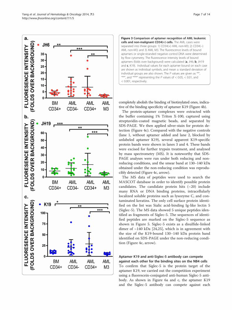
Figure 3 Comparison of aptamer recognition of AML leukemiccells and non-malignant CD34(+) cells. The AML cases wereseparated into three groups: 1) CD34(+) AML non-M3; 2) CD34(−)AML non-M3; and 3) AML M3. The fluorescence levels of boundaptamers or single-stranded negative control DNA were determinedby flow cytometry. The fluorescence intensity levels of boundaptamers (folds over background) were calculated (a, JH6; b, JH19and c, K19). Individual values for each aptamer bound on each caseare shown as individual symbols, and mean ± standard deviation ofindividual groups are also shown. The P values are given as “*”,“**”, and “***” representing the P values of < 0.05, < 0.01, and< 0.001, respectively.
Yang et al. Journal of Hematology & Oncology 2014, 7:5 Page 7 of 14http://www.jhoonline.org/content/7/1/5
completely abolish the binding of biotinylated ones, indica-tive of the binding specificity of aptamer K19 (Figure 4b).The protein-aptamer complexes were extracted with
the buffer containing 1% Triton X-100, captured usingstreptavidin-coated magnetic beads, and separated bySDS-PAGE. We then applied silver-stain for protein de-tection (Figure 4c). Compared with the negative controls(lane 1, without aptamer added and lane 2, blocked byunlabeled aptamer K19), several apparent K19-specificprotein bands were shown in lanes 3 and 4. These bandswere excised for further trypsin treatment, and analysedby mass spectrometry (MS). It is noteworthy that SDS-PAGE analyses were run under both reducing and non-reducing conditions, and the smear band at 130–140 kDaobtained under the non-reducing condition was reprodu-cibly detected (Figure 4c, arrow).The MS data of peptides were used to search the
MASCOT database in order to identify possible proteincandidates. The candidate protein hits (~20) includemany RNA or DNA binding proteins, intracellularlylocalized soluble proteins such as lysozyme C, and con-taminated keratins. The only cell surface protein identi-fied on the list was Sialic acid-binding Ig-like lectin 5(Siglec-5). The MS data showed 5 unique peptides iden-tified as fragments of Siglec-5. The sequences of identi-fied peptides are marked on the Siglec-5 sequence asshown in Figure 5. Siglec-5 exists as a disulfide-linkeddimer of ~140 kDa [24,25], which is in agreement withthe size of the K19-bound 130–140 kDa protein bandidentified on SDS-PAGE under the non-reducing condi-tion (Figure 4c, arrow).
Aptamer K19 and anti-Siglec-5 antibody can competeagainst each other for the binding sites on the NB4 cellsTo confirm that Siglec-5 is the protein target of theaptamer K19, we carried out the competition experimentusing a fluorescein-conjugated anti-human Siglec-5 anti-body. As shown in Figure 6a and c, the aptamer K19and the Siglec-5 antibody can compete against each
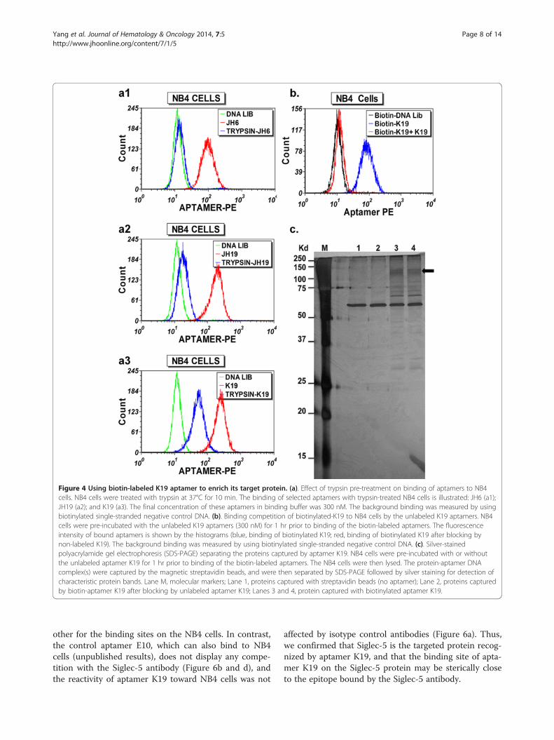
Figure 4 Using biotin-labeled K19 aptamer to enrich its target protein. (a). Effect of trypsin pre-treatment on binding of aptamers to NB4cells. NB4 cells were treated with trypsin at 37°C for 10 min. The binding of selected aptamers with trypsin-treated NB4 cells is illustrated: JH6 (a1);JH19 (a2); and K19 (a3). The final concentration of these aptamers in binding buffer was 300 nM. The background binding was measured by usingbiotinylated single-stranded negative control DNA. (b). Binding competition of biotinylated-K19 to NB4 cells by the unlabeled K19 aptamers. NB4cells were pre-incubated with the unlabeled K19 aptamers (300 nM) for 1 hr prior to binding of the biotin-labeled aptamers. The fluorescenceintensity of bound aptamers is shown by the histograms (blue, binding of biotinylated K19; red, binding of biotinylated K19 after blocking bynon-labeled K19). The background binding was measured by using biotinylated single-stranded negative control DNA. (c). Silver-stainedpolyacrylamide gel electrophoresis (SDS-PAGE) separating the proteins captured by aptamer K19. NB4 cells were pre-incubated with or withoutthe unlabeled aptamer K19 for 1 hr prior to binding of the biotin-labeled aptamers. The NB4 cells were then lysed. The protein-aptamer DNAcomplex(s) were captured by the magnetic streptavidin beads, and were then separated by SDS-PAGE followed by silver staining for detection ofcharacteristic protein bands. Lane M, molecular markers; Lane 1, proteins captured with streptavidin beads (no aptamer); Lane 2, proteins capturedby biotin-aptamer K19 after blocking by unlabeled aptamer K19; Lanes 3 and 4, protein captured with biotinylated aptamer K19.
Yang et al. Journal of Hematology & Oncology 2014, 7:5 Page 8 of 14http://www.jhoonline.org/content/7/1/5
other for the binding sites on the NB4 cells. In contrast,the control aptamer E10, which can also bind to NB4cells (unpublished results), does not display any compe-tition with the Siglec-5 antibody (Figure 6b and d), andthe reactivity of aptamer K19 toward NB4 cells was not
affected by isotype control antibodies (Figure 6a). Thus,we confirmed that Siglec-5 is the targeted protein recog-nized by aptamer K19, and that the binding site of apta-mer K19 on the Siglec-5 protein may be sterically closeto the epitope bound by the Siglec-5 antibody.

Figure 5 The sequences of Siglec-5 protein and peptides recovered by mass spectrometric analysis of aptamer K19-enriched proteins.The Ig-like V-type domain and two Ig-like C2-type domains of Siglec-5 are underlined while the signal peptide, the transmembrane region, andthe immunoreceptor tyrosine-based inhibitory (ITIM) motif are marked with dotted underlines. The peptides identified by mass spectrometry arehighlighted by bordering.
Yang et al. Journal of Hematology & Oncology 2014, 7:5 Page 9 of 14http://www.jhoonline.org/content/7/1/5
Siglec-5 can be used as a biomarker for granulocyticmaturation and AML cell detection as well as be used asa potential target for leukemic cell growth inhibitionSiglec-5 was reported to be expressed on granulocytes[26], but its expression during granulocytic or monocyticmaturation has not been well characterized. Since apta-mer K19 recognized maturing granulocytes much betterthan CD34(+) early progenitors in normal human bonemarrow (Figure 2), we further determined whether itsbinding sites (i.e. Siglec-5 protein levels) on granulocytesvary during granulocytic maturation. By flow cytometricanalysis, we separated maturing granulocytes or mono-cytes into three subsets: early stage, immediate stage,and matured stage, according to the expression levels ofCD13 and CD11b for granulocytes and CD64 and CD14for monocytes (Figure 7, left panel) [27-29]. We thendetermined the fluorescence levels of aptamer K19bound on each subset. Compared with the negative con-trol, the fluorescence intensity of bound aptamer K19on granulocytes gradually increased during granulocyticmaturation (Figure 7, right panel), indicating progres-sive up-regulation of Siglec-5 levels during granulocyticmaturation. However, persistently high levels of Siglec-5expression were observed on both CD64(+)/CD14(−)immature and CD64(+)/CD14(+) mature monocytes.Because Siglec-5 is overexpressed in a subset of AML
cells, we selected an AML case with relatively highlevels of Siglec-5 expression, and spiked small numbersof the AML cells into a normal human bone marrowspecimen. Then, based on the differential expression levels
of Siglec-5 on normal CD34(+) cells and CD34 (+)leukemic cells, we used aptamer K19 to aid in the de-tection of AML cells mixed into a normal bone mar-row specimen (Figure 8). Additionally, to demonstrateSiglec-5 can be a potential biomarker for targetedtherapy, we tested biotinylated Siglec-5 aptamer K19and saporin-cross-linked to streptavidin (SA-SAP) forinhibiting NB4 cell proliferation in vitro. Comparedwith unlabeled saporin (SAP) (Figure 9a) or the biotiny-lated single stranded DNA control (Figure 9b), theSiglec-5 aptamer K19 can enhance the toxicity of SA-SAP to NB4 cells with an estimated IC50 of 25 to 50nM. The enhanced toxic effect of biotinylated K19 apta-mer can be blocked by non-labelled aptamer K19,indicating that the enhanced cell toxicity is mediatedthrough the specific binding to surface Siglec-5 proteins(Figure 9c).
DiscussionThe molecular characteristics of leukemic cells, especiallyat the proteomic level, are critical for understandingleukemia pathogenesis and designing targeted therapy. Inthe last several decades, proteomic analysis has been per-formed to advance the discovery of diseased cell-specificprotein biomarkers, but so far only a few AML biomarkershave been introduced into clinical practice for AMLdetection and therapy. Currently, we still lack effectivebiomarkers for AML diagnosis and targeted therapy.Thus, our intent in this study is to develop new
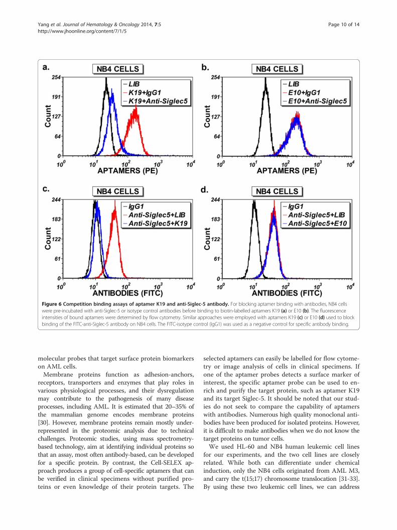
Figure 6 Competition binding assays of aptamer K19 and anti-Siglec-5 antibody. For blocking aptamer binding with antibodies, NB4 cellswere pre-incubated with anti-Siglec-5 or isotype control antibodies before binding to biotin-labelled aptamers K19 (a) or E10 (b). The fluorescenceintensities of bound aptamers were determined by flow cytometry. Similar approaches were employed with aptamers K19 (c) or E10 (d) used to blockbinding of the FITC-anti-Siglec-5 antibody on NB4 cells. The FITC-isotype control (IgG1) was used as a negative control for specific antibody binding.
Yang et al. Journal of Hematology & Oncology 2014, 7:5 Page 10 of 14http://www.jhoonline.org/content/7/1/5
molecular probes that target surface protein biomarkerson AML cells.Membrane proteins function as adhesion-anchors,
receptors, transporters and enzymes that play roles invarious physiological processes, and their dysregulationmay contribute to the pathogenesis of many diseaseprocesses, including AML. It is estimated that 20–35% ofthe mammalian genome encodes membrane proteins[30]. However, membrane proteins remain mostly under-represented in the proteomic analysis due to technicalchallenges. Proteomic studies, using mass spectrometry-based technology, aim at identifying individual proteins sothat an assay, most often antibody-based, can be developedfor a specific protein. By contrast, the Cell-SELEX ap-proach produces a group of cell-specific aptamers that canbe verified in clinical specimens without purified pro-teins or even knowledge of their protein targets. The
selected aptamers can easily be labelled for flow cytome-try or image analysis of cells in clinical specimens. Ifone of the aptamer probes detects a surface marker ofinterest, the specific aptamer probe can be used to en-rich and purify the target protein, such as aptamer K19and its target Siglec-5. It should be noted that our stud-ies do not seek to compare the capability of aptamerswith antibodies. Numerous high quality monoclonal anti-bodies have been produced for isolated proteins. However,it is difficult to make antibodies when we do not know thetarget proteins on tumor cells.We used HL-60 and NB4 human leukemic cell lines
for our experiments, and the two cell lines are closelyrelated. While both can differentiate under chemicalinduction, only the NB4 cells originated from AML M3,and carry the t(15;17) chromosome translocation [31-33].By using these two leukemic cell lines, we can address

Figure 7 Using Siglec-5 as a biomarker for granulocytic different maturation in human bone marrow. Leukocytes from normal bonemarrow aspirates were incubated with aptamer K19 or the single-stranded negative control DNA. (a). Siglec-5 expression of maturing granulocytes.The granulocyte population was first identified using levels of side-scattered light (SSC) in combination with the fluorescence intensity of CD45. Then,maturing granulocytes were separated into three subsets according to the expression levels of CD13 and CD11b (left panel): early stage (yellow),immediate stage (purple), and matured stage (green). Fluorescence intensity (PE) of the granulocyte subsets bound with single stranded DNA control(middle panel) or aptamer K19/Siglec-5 (right panel) is shown in relation to fluorescence levels of CD11b (APC). (b). Siglec-5 expression of maturingmonocytes. The mature and immature monocytes were first identified using levels of side-scattered light (SSC) in combination with the fluorescenceintensity of CD64. Then, maturing monocytes were separated into three subsets according to the expression levels of CD64 and CD14 (left panel): earlystage (yellow), immediate stage (teal) and matured stage (fuchsia). Fluorescence intensity (PE) of the monocyte subsets bound with single-strandednegative control DNA (middle panel) or aptamer K19/Siglec-5 (right panel) is shown in relation to fluorescence levels of CD14 (APC).
Yang et al. Journal of Hematology & Oncology 2014, 7:5 Page 11 of 14http://www.jhoonline.org/content/7/1/5
three questions: A). Is it possible to select single strandedDNA aptamers that are capable of detecting differencesin surface protein expression between two closely rela-ted leukemic cell lines (HL60 and NB4)? B). Can theseselected aptamers be further used on clinical specimensfor phenotyping AML and identifying new biomarkers?C). Can the newly identified biomarker be used to aidthe detection of AML cells in human bone marrowspecimens?As a result, we used NB4 leukemic cells to select and
characterize three new DNA aptamers (JH6, JH19, and
K19), which have more binding sites on NB4 cells thanon HL60 cells. This is in contrast to the aptamer KH1C12previously selected from HL60 cells, which selectivelyrecognized HL60 cells [19]. Gene expression profilingstudies showed that NB4 and HL60 cell lines had the mostclosely related profiles of mRNA expression [33]. Thus,our results with aptamers selected against NB4 and thosepreviously selected against HL60 cells indicate that it ispossible to select aptamers capable of detecting differencesin surface protein expression between two closely relatedleukemic cell lines.
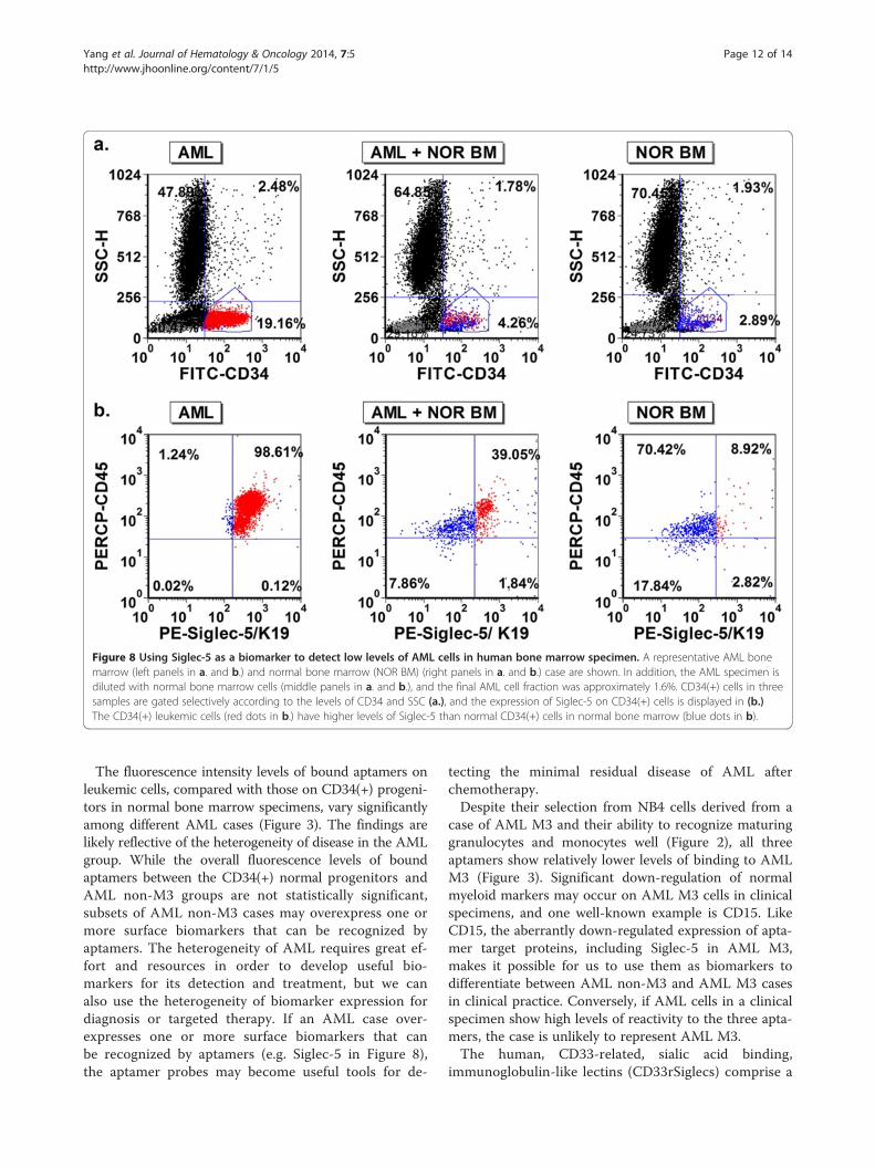
Figure 8 Using Siglec-5 as a biomarker to detect low levels of AML cells in human bone marrow specimen. A representative AML bonemarrow (left panels in a. and b.) and normal bone marrow (NOR BM) (right panels in a. and b.) case are shown. In addition, the AML specimen isdiluted with normal bone marrow cells (middle panels in a. and b.), and the final AML cell fraction was approximately 1.6%. CD34(+) cells in threesamples are gated selectively according to the levels of CD34 and SSC (a.), and the expression of Siglec-5 on CD34(+) cells is displayed in (b.)The CD34(+) leukemic cells (red dots in b.) have higher levels of Siglec-5 than normal CD34(+) cells in normal bone marrow (blue dots in b).
Yang et al. Journal of Hematology & Oncology 2014, 7:5 Page 12 of 14http://www.jhoonline.org/content/7/1/5
The fluorescence intensity levels of bound aptamers onleukemic cells, compared with those on CD34(+) progeni-tors in normal bone marrow specimens, vary significantlyamong different AML cases (Figure 3). The findings arelikely reflective of the heterogeneity of disease in the AMLgroup. While the overall fluorescence levels of boundaptamers between the CD34(+) normal progenitors andAML non-M3 groups are not statistically significant,subsets of AML non-M3 cases may overexpress one ormore surface biomarkers that can be recognized byaptamers. The heterogeneity of AML requires great ef-fort and resources in order to develop useful bio-markers for its detection and treatment, but we canalso use the heterogeneity of biomarker expression fordiagnosis or targeted therapy. If an AML case over-expresses one or more surface biomarkers that canbe recognized by aptamers (e.g. Siglec-5 in Figure 8),the aptamer probes may become useful tools for de-
tecting the minimal residual disease of AML afterchemotherapy.Despite their selection from NB4 cells derived from a
case of AML M3 and their ability to recognize maturinggranulocytes and monocytes well (Figure 2), all threeaptamers show relatively lower levels of binding to AMLM3 (Figure 3). Significant down-regulation of normalmyeloid markers may occur on AML M3 cells in clinicalspecimens, and one well-known example is CD15. LikeCD15, the aberrantly down-regulated expression of apta-mer target proteins, including Siglec-5 in AML M3,makes it possible for us to use them as biomarkers todifferentiate between AML non-M3 and AML M3 casesin clinical practice. Conversely, if AML cells in a clinicalspecimen show high levels of reactivity to the three apta-mers, the case is unlikely to represent AML M3.The human, CD33-related, sialic acid binding,
immunoglobulin-like lectins (CD33rSiglecs) comprise a
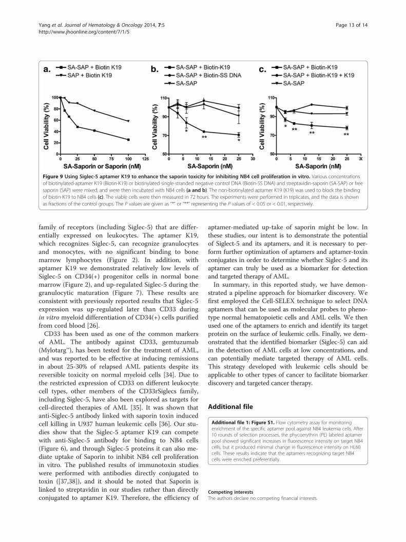
Figure 9 Using Siglec-5 aptamer K19 to enhance the saporin toxicity for inhibiting NB4 cell proliferation in vitro. Various concentrationsof biotinylated-aptamer K19 (Biotin-K19) or biotinylated single-stranded negative control DNA (Biotin-SS DNA) and streptavidin-saporin (SA-SAP) or freesaporin (SAP) were mixed, and were then incubated with NB4 cells (a and b). The non-biotinylated aptamer K19 (K19) was used to block the bindingof biotin-K19 to NB4 cells (c). The viable cells were then measured in 72 hours. The experiments were performed in triplicates, and the data is shownas fractions of the control groups. The P values are given as “*” or “**” representing the P values of < 0.05 or < 0.01, respectively.
Yang et al. Journal of Hematology & Oncology 2014, 7:5 Page 13 of 14http://www.jhoonline.org/content/7/1/5
family of receptors (including Siglec-5) that are differ-entially expressed on leukocytes. The aptamer K19,which recognizes Siglec-5, can recognize granulocytesand monocytes, with no significant binding to bonemarrow lymphocytes (Figure 2). In addition, withaptamer K19 we demonstrated relatively low levels ofSiglec-5 on CD34(+) progenitor cells in normal bonemarrow (Figure 2), and up-regulated Siglec-5 during thegranulocytic maturation (Figure 7). These results areconsistent with previously reported results that Siglec-5expression was up-regulated later than CD33 duringin vitro myeloid differentiation of CD34(+) cells purifiedfrom cord blood [26].CD33 has been used as one of the common markers
of AML. The antibody against CD33, gemtuzumab(Mylotarg™), has been tested for the treatment of AML,and was reported to be effective at inducing remissionsin about 25-30% of relapsed AML patients despite itsreversible toxicity on normal myeloid cells [34]. Due tothe restricted expression of CD33 on different leukocytecell types, other members of the CD33rSiglecs family,including Siglec-5, have also been explored as targets forcell-directed therapies of AML [35]. It was shown thatanti-Siglec-5 antibody linked with saporin toxin inducedcell killing in U937 human leukemic cells [36]. Our stu-dies show that the Siglec-5 aptamer K19 can competewith anti-Siglec-5 antibody for binding to NB4 cells(Figure 6), and through Siglec-5 proteins it can also me-diate uptake of Saporin to inhibit NB4 cell proliferationin vitro. The published results of immunotoxin studieswere performed with antibodies directly conjugated totoxin ([37,38]), and it should be noted that Saporin islinked to streptavidin in our studies rather than directlyconjugated to aptamer K19. Therefore, the efficiency of
aptamer-mediated up-take of saporin might be low. Inthese studies, our intent is to demonstrate the potentialof Siglect-5 and its aptamers, and it is necessary to per-form further optimization of aptamers and aptamer-toxinconjugates in order to determine whether Siglec-5 and itsaptamer can truly be used as a biomarker for detectionand targeted therapy of AML.In summary, in this reported study, we have demon-
strated a pipeline approach for biomarker discovery. Wefirst employed the Cell-SELEX technique to select DNAaptamers that can be used as molecular probes to pheno-type normal hematopoietic cells and AML cells. We thenused one of the aptamers to enrich and identify its targetprotein on the surface of leukemic cells. Finally, we dem-onstrated that the identified biomarker (Siglec-5) can aidin the detection of AML cells at low concentrations, andcan potentially mediate targeted therapy of AML cells.This strategy developed with leukemic cells should beapplicable to other types of cancer to facilitate biomarkerdiscovery and targeted cancer therapy.
Additional file
Additional file 1: Figure S1. Flow cytometry assay for monitoringenrichment of the specific aptamer pool against NB4 leukemia cells. After10 rounds of selection processes, the phycoerythrin (PE) labeled aptamerpool showed significant increases in fluorescence intensity on target NB4cells, but it produced minimal change in fluorescence intensity on HL60cells. These results indicate that the aptamers recognizing target NB4cells were enriched preferentially.
Competing interestsThe authors declare no competing financial interests.

Yang et al. Journal of Hematology & Oncology 2014, 7:5 Page 14 of 14http://www.jhoonline.org/content/7/1/5
Authors’ contributionsMY and GJ, performed research and analyzed the data and wrote the paper;WL, KQ and MZ, performed research work; SA and CC, performed leukemiadata analysis and wrote the paper; YL, designed research, analyzed the data,and wrote the paper. All authors read and approved the final manuscript.
FundingThis work was supported by the National Institutes of Health [CA129311 to Y.L.].
Received: 2 November 2013 Accepted: 24 December 2013Published: 9 January 2014
References1. Appelbaum FR, Gundacker H, Head DR, Slovak ML, Willman CL, Godwin JE,
Anderson JE, Petersdorf SH: Age and acute myeloid leukemia. Blood 2006,107:3481–3485.
2. Dohner H, Estey EH, Amadori S, Appelbaum FR, Buchner T, Burnett AK,Dombret H, Fenaux P, et al: Diagnosis and management of acute myeloidleukemia in adults: recommendations from an international expertpanel, on behalf of the European LeukemiaNet. Blood 2010, 115:453–474.
3. O’Donnell MR, Abboud CN, Altman J, Appelbaum FR, Arber DA, Attar E,Borate U, Coutre SE, et al: Acute myeloid leukemia. J Natl Compr CancNetw 2012, 10:984–1021.
4. Sekeres MA: Treatment of older adults with acute myeloid leukemia: stateof the art and current perspectives. Haematologica 2008, 93:1769–1772.
5. Zaidi SZ, Owaidah T, Al SF, Ahmed SY, Chaudhri N, Aljurf M: The challengeof risk stratification in acute myeloid leukemia with normal karyotype.Hematol Oncol Stem Cell Ther 2008, 1:141–158.
6. Gregory TK, Wald D, Chen Y, Vermaat JM, Xiong Y, Tse W: Molecularprognostic markers for adult acute myeloid leukemia with normalcytogenetics. J Hematol Oncol 2009, 2:23.
7. Gold L, Janjic N, Jarvis T, Schneider D, Walker JJ, Wilcox SK, Zichi D:Aptamers and the RNA world, past and present. Cold Spring Harb PerspectBiol 2012, 4:1–9.
8. Barbas AS, Mi J, Clary BM, White RR: Aptamer applications for targetedcancer therapy. Future Oncol 2010, 6:1117–1126.
9. Cerchia L, Giangrande PH, McNamara JO, de F,V: Cell-specific aptamers fortargeted therapies. Methods Mol Biol 2009, 535:59–78.
10. Ellington AD, Conrad R: Aptamers as potential nucleic acidpharmaceuticals. Biotechnol Annu Rev 1995, 1:185–214.
11. Brody EN, Willis MC, Smith JD, Jayasena S, Zichi D, Gold L: The use ofaptamers in large arrays for molecular diagnostics. Mol Diagn 1999,4:381–388.
12. Bunka DH, Platonova O, Stockley PG: Development of aptamertherapeutics. Curr Opin Pharmacol 2010, 10:557–562.
13. Shangguan D, Li Y, Tang Z, Cao ZC, Chen HW, Mallikaratchy P, Sefah K,Yang CJ, et al: Aptamers evolved from live cells as effective molecularprobes for cancer study. Proc Natl Acad Sci USA 2006, 103:11838–11843.
14. Dua P, Kim S, Lee DK: Nucleic acid aptamers targeting cell-surfaceproteins. Methods 2011, 54:215–225.
15. Blank M, Weinschenk T, Priemer M, Schluesener H: Systematic evolution ofa DNA aptamer binding to rat brain tumor microvessels. selectivetargeting of endothelial regulatory protein pigpen. J Biol Chem 2001,276:16464–16468.
16. Daniels DA, Chen H, Hicke BJ, Swiderek KM, Gold L: A tenascin-C aptameridentified by tumor cell SELEX: systematic evolution of ligands byexponential enrichment. Proc Natl Acad Sci USA 2003, 100:15416–15421.
17. Mallikaratchy P, Tang Z, Kwame S, Meng L, Shangguan D, Tan W: Aptamerdirectly evolved from live cells recognizes membrane boundimmunoglobin heavy mu chain in Burkitt’s lymphoma cells. Mol CellProteomics 2007, 6:2230–2238.
18. Shangguan D, Cao Z, Meng L, Mallikaratchy P, Sefah K, Wang H, Li Y, Tan W:Cell-specific aptamer probes for membrane protein elucidation in cancercells. J Proteome Res 2008, 7:2133–2139.
19. Sefah K, Tang ZW, Shangguan DH, Chen H, Lopez-Colon D, Li Y, Parekh P,Martin J, et al: Molecular recognition of acute myeloid leukemia usingaptamers. Leukemia 2009, 23:235–244.
20. Jiang G, Zhang M, Yue B, Yang M, Carter C, Al-Quran SZ, Li B, Li Y: PTK7: A newbiomarker for immunophenotypic characterization of maturing T cells andT cell acute lymphoblastic leukemia. Leuk Res 2012, 36:1347–1353.
21. Borowitz MJ, Guenther KL, Shults KE, Stelzer GT: Immunophenotyping ofacute leukemia by flow cytometric analysis. Use of CD45 and right-anglelight scatter to gate on leukemic blasts in three-color analysis. Am J ClinPathol 1993, 100:534–540.
22. Wood BL: Ten-color immunophenotyping of hematopoietic cells.Curr Protoc Cytom 2005, Chapter 6:6.21.1–6.21.11.
23. Granvogl B, Ploscher M, Eichacker LA: Sample preparation by in-geldigestion for mass spectrometry-based proteomics. Anal Bioanal Chem2007, 389:991–1002.
24. Cornish AL, Freeman S, Forbes G, Ni J, Zhang M, Cepeda M, Gentz R,Augustus M, et al: Characterization of siglec-5, a novel glycoproteinexpressed on myeloid cells related to CD33. Blood 1998, 92:2123–2132.
25. Crocker PR, McMillan SJ, Richards HE: CD33-related siglecs as potentialmodulators of inflammatory responses. Ann N Y Acad Sci 2012,1253:102–111.
26. Virgo P, Denning-Kendall PA, Erickson-Miller CL, Singha S, Evely R, Hows JM,Freeman SD: Identification of the CD33-related Siglec receptor, Siglec-5(CD170), as a useful marker in both normal myelopoiesis and acutemyeloid leukaemias. Br J Haematol 2003, 123:420–430.
27. Loken MR, Wells DA: The role of flow cytometry in myelodysplasticsyndromes. J Natl Compr Canc Netw 2008, 6:935–941.
28. Stetler-Stevenson M, Arthur DC, Jabbour N, Xie XY, Molldrem J, Barrett AJ,Venzon D, Rick ME: Diagnostic utility of flow cytometricimmunophenotyping in myelodysplastic syndrome. Blood 2001, 98:979–987.
29. Wood BL: Flow cytometric diagnosis of myelodysplasia andmyeloproliferative disorders. J Biol Regul Homeost Agents 2004, 18:141–145.
30. Savas JN, Stein BD, Wu CC, Yates JR III: Mass spectrometry acceleratesmembrane protein analysis. Trends Biochem Sci 2011, 36:388–396.
31. Pass MB, Borregaard N, Cowland JB: Derangement of transcription factorprofiles during in vitro differentiation of HL60 and NB4 cells. Leuk Res2007, 31:827–837.
32. Ballerini P, Besancon F, Cayre YE: [Effect of translocation t(15;17) on thegene expression regulation of myeloblastin during all trans retinoic acidinduced myeloid differentiation in human leukemic cells]. C R SeancesSoc Biol Fil 1995, 189:521–530.
33. Leupin N, Kuhn A, Hugli B, Grob TJ, Jaggi R, Tobler A, Delorenzi M, Fey MF:Gene expression profiling reveals consistent differences between clinicalsamples of human leukaemias and their model cell lines. Br J Haematol2006, 135:520–523.
34. Walter RB, Appelbaum FR, Estey EH, Bernstein ID: Acute myeloid leukemiastem cells and CD33-targeted immunotherapy. Blood 2012, 119:6198–6208.
35. O’Reilly MK, Paulson JC: Siglecs as targets for therapy in immune-cell-mediated disease. Trends Pharmacol Sci 2009, 30:240–248.
36. Nguyen DH, Ball ED, Varki A: Myeloid precursors and acute myeloidleukemia cells express multiple CD33-related Siglecs. Exp Hematol 2006,34:728–735.
37. Chu TC, Marks JW III, Lavery LA, Faulkner S, Rosenblum MG, Ellington AD,Levy M: Aptamer:toxin conjugates that specifically target prostate tumorcells. Cancer Res 2006, 66:5989–5992.
38. Zhang Y, Hong H, Cai W: Tumor-targeted drug delivery with aptamers.Curr Med Chem 2011, 18:4185–4194.
doi:10.1186/1756-8722-7-5Cite this article as: Yang et al.: Developing aptamer probes for acutemyelogenous leukemia detection and surface protein biomarkerdiscovery. Journal of Hematology & Oncology 2014 7:5.