resistanceindexofpenicillin...
TRANSCRIPT
International Scholarly Research NetworkISRN MicrobiologyVolume 2012, Article ID 789474, 6 pagesdoi:10.5402/2012/789474
Research Article
Resistance Index of Penicillin-Resistant Bacteria to VariousPhysicochemical Agents
M. Kazemi,1 R. Kasra Kermanshahi,2 E. Heshmat Dehkordi,2 F. Payami,2 and M. Behjati1
1 Department of Genetics and Molecular Biology, Medical School, Isfahan University of Medical Sciences, Isfahan 81746-73461, Iran2 Department of Microbiology, Faculty of Science, The University of Isfahan, Isfahan, Iran
Correspondence should be addressed to M. Kazemi, m [email protected]
Received 10 September 2011; Accepted 23 October 2011
Academic Editors: C. M. Manaia and D. Rodriguez-Lazaro
Copyright © 2012 M. Kazemi et al. This is an open access article distributed under the Creative Commons Attribution License,which permits unrestricted use, distribution, and reproduction in any medium, provided the original work is properly cited.
Widespread use of various antimicrobial agents resulted in the emergence of bacterial resistance. Mechanisms like direct efflux,formation, and sequestration of metals and drugs in complexes and antiporter pumps are some examples. This investigation aimsto investigate the resistance pattern of penicillin-resistant bacterial strains to some physicochemical agents. Sensitivity/resistancepattern of common bacterial strains to antimicrobial agents were evaluated by disk diffusion assay. Broth and agar dilution methodwere used for determination of minimum inhibitory concentration and minimal bactericidal concentration. The impact of UV rayon the bacterial growth under laminar flow hood was measured using photonmeter. Our data demonstrates that the most prevalentmetal resistance was against arsenate (95.92%), followed by cadmium (52.04%) and mercury (36.73%). There was significantdifference between cetrimide resistances among studied microbial strains especially for P. aeruginosa (P < 0.05). High rate ofpathogen resistance to various antibacterial agents in our study supports previously published data. This great rate of bacterialresistance is attributed to the emergence of defense mechanisms developed in pathogens. The higher general bacterial resistancerate among Staphylococcus strains rather than E. coli and P. aeruginosa strains draws attention towards focusing on designing newertherapeutic compounds for Staphylococcus strains.
1. Introduction
Various resistance mechanisms have been developed by bac-teria to counteract heavy metal stress. Some of these mech-anisms include metal efflux out of the cell, sequestrationof heavy metals in complexes, and reduction of a metal toless toxic species [1, 2]. Metal efflux is seen in the case ofCd which is detoxified in gram-negative bacteria by RND-driven system like Czc and Ncc as zinc and Nickel exporters,respectively [3]. In gram-positive bacteria this metal efflux isdone via Cadmium-exporting P-type ATPase as seen in CadApump in S. aureus. Hg2+ is transported inside the cells byspecific uptake system which is rapidly reduced to abolish thetoxic effects of Hg2+ on periplasmic proteins by reoxidation.In this way, Mercury leaves the cell by passive diffusion anddoes not remain inside the cell [4, 5]. The first detoxificationstep in gram-negative bacteria is attachment of periplasmicHg2+-binding protein MerP to cations. It probably deliverstoxic cations to the mercury transporter system which is
subsequently transported into the cytoplasm. An alternativeuptake route is also present which involves MerC protein.Inside the cell, Hg2+ is reduced into Hg0 by MerA, relatedto the glutathione reductase system. MerA protein is alsoinvolved in the reduction process of Hg2+ after cleavage byMerA. MerB detoxifies organomercurial agents which aremuch more toxic than Hg2+ [5].
Multidrug resistance pumps (MDR), responsible for theextrusion of chemically unrelated antimicrobial agents com-poses alternative resistance pathway. This process is mediatedby active export of the toxic compounds by means of theproton motive forces [6]. Multidrug resistance to variousorganic cationic antiseptics and disinfectant compounds ascetyltrimethylammonium bromide (cetrimide) has been re-ported in various organisms including S. aureus, E. coli, andB. subtilis [7, 8]. Mutations leading to overexpression of thepumps have been identified in clinical isolates of multidrug-resistant strains [9]. Among these strains, P. aeruginosa isan opportunistic human pathogen with innate resistance

2 ISRN Microbiology
to multiple antimicrobial agents. This intrinsic multidrugresistance is attributed to the lowpermeability of outer mem-brane and expression of a number of broadlyspecific mul-tidrug efflux (Mex) pumps as MexAB-OprM and MexXY-OprM [2, 9]. These pumps are composed of three dif-ferent peptides: MexA, a fusion membrane protein whichapparently docks MexB to OprM, MexB translocase whichbelongs to the resistance nodulation division (RND) familyof solute/proton antiporters and OprM which is an outermembrane porin [10]. The broad specificity of MDRsseems to be matched with the broad resistance of biofilmsto antimicrobial agents. MDRs are mostly regulated byenvironmental factors exemplified by induced expression ofE. coli EmrAB and RND pump by drug substrate and stress,respectively [10]. The presence of three additional effluxsystems as MexCD-OprJ, MexEF-OprN, and MexJK-OprMdue to overexpression of efflux genes by mutational eventsis addressed by other studies. However these pumps playdifferent roles rather than drug efflux including export ofthe biocides, dyes, detergents, metabolic inhibitors, organicsolvents, and molecules participated in bacterial cell-to-cell communication [11]. In P. aeruginosa, the MexAB-OprM multidrug efflux system exports a number of antimi-crobial compounds such as β-Lactams and is responsiblefor its “intrinsic resistance” to antibiotics. The substratesfor MexAB-OprM include quinolones, tetracycline, and β-lactams [2, 9, 12].
UV light is recognized as an effective antiorganism byinactivation of pathogens [13]. The effectiveness of UVlight in biological inactivation aroused from observation ofdouble stranded DNA breaks occurred by UV ray [14]. Thebreak point is particularly between pyrimidine bases whichalters base pairing and induces formation of new linkagebetween adjacent nucleotides on the same DNA strand.Unrepaired damage blocks DNA replication which ultimatelylead to cell cycle arrest and death [14]. Many organismsdeveloped reparative mechanisms to compensate destructiveeffects of UV radiation as nucleotide excision repair andphoto reactivation [11]. Since UV radiation is widely usedto sterilize operation rooms, utensils, and drinking water,the emergence of bacterial resistance will cause great hygieneproblems.
As mentioned above, various drug resistance mecha-nisms are involved in the emergence of pathogen-drug re-sistance. It is not unusual for pathogens to use combinationof these resistance strategies against antimicrobial agents.Understanding the synergistic effects of these resistance strat-egies helps us choose appropriate therapeutic agents againstdeveloped resistance. Therefore, the aim of this study is toevaluate the prevalence of simultaneous resistance to theabove-mentioned factors in some common resistant patho-gens.
2. Materials and Methods
2.1. Bacterial Strains. Standard bacterial strains were cul-tured in commercial culture medias as Nutrient agar (NA),Nutrient broth (NB), Trypticase-soy agar (TSA), Trypticase-soy broth (TSB), Muller Hinton agar (MHA), Eosin
Table 1: Standard bacterial strains applied in this study.
Group Strain Application of strains
S. aureus
ATCC 6538PMeasurement of antimicrobialmaterial
ATCC 9144Measurement of materials suchas Cetrimide erythromycin,Penicillin and tetracycline
ATCC 25923Study of resistance against UVray
E. coliATCC 8739
Measurement of antimicrobialmaterials
ATCC 25922Study of resistance against UVray
P. aeruginosaATCC 9027
Measurement of antimicrobialmaterials
ATCC 27853Study of resistance against UVray
methylene blue (EMB), and MacConkey agar. PHG-II andbiochemical culture medias were also used for heavy metalresistance and identification of isolated bacteria, respectively.Cadmium nitrate, sodium arsenate, and mercury nitratewere used as heavy metals in this study (Table 1).
2.2. Determination of Bacterial Sensitivity to AntimicrobialAgents. In order to determine the sensitivity/resistance pat-tern of bacterial strains to antimicrobial agents, disk diffu-sion assay (Kirby-Bauer method) was used. Broth and agardilution method was used for determination of minimuminhibitory concentration (MIC) and minimal bactericidalconcentration (MBC). Determination of the optimal con-centration of antimicrobial compounds with the capacityto annihilate 99.9% of the microorganisms was performedusing agar on plate method. Bacterial resistance to cetrimidewas assessed by replica-plating method using steer’s repli-cator [15, 16]. Sterile PHG-II medium supplemented withthe Peptone (4 g/L), yeast extract (1 g/L), Glucose (2 g/L),and Agar (15 g/L) was used to determine MIC against heavymetals. Various concentrations of heavy metals added to thismedium in plates (adjusted pH and 55◦C).Then, 0.1 mL ofmicrobial suspension in Log-phase was spread on plates.Subsequently, plates were incubated at 35◦C for 24–72 h[17, 18]. Different concentrations of heavy metals were usedas follow:
(i) cadmium nitrate: 0.037–0.075–0.15–0.31–0.62–1.23–2.46–4.93 (μg/mL),
(ii) mercury nitrate: 0.12–0.25–0.50–1.00–2.00–4.00 (μg/mL),
(iii) sodium arsenate: 2–4–8–16–32–64–128 (mg/mL).
Resistance strains to chemical agents were based on thefollowing relations:
(i) cetrimide [19],
(ii) in S. aureus and E. coli, respectively, growth at theconcentrations of 4.16 and 20.83 μg/mL in screeningmethod and P. aeruginosa, MIC ≥ 400 μg/mL,

ISRN Microbiology 3
0
10
20
30
40
50
60
70
80
90
100
1.25 4.16 5.21 6.25 9.37 10.41 14.58 20.83 26.04 208.33 260.41 312.5
Bac
teri
al g
row
th (
%)
Cetrimide concentration (mg/mL)
E. coliP. aeruginosa
S.aureusS.COA (−)
Figure 1: Comparison of bacterial growth rate in the presence ofvarious concentration of cetrimide.
(iii) metals [17, 19],
(iv) cadmium, MIC ≥ 0.62 μg/mL,
(v) mercury, MIC ≥ 1 μg/mL,
(vi) arsenic, MIC ≥ 4 mg/mL.
2.3. Determination of Bacterial Sensitivity to UV Radiation.The impact of UV ray (UVC) on the bacterial growth underlaminar flow hood (Slee Mains VLFS 636) was measuredusing photonmeter (Hausatech Quantum sensor Q SPAR).Grew bacteria in TBS (24 h, 30 rpm) were diluted in order toachieve approximately 1.5 × 10⁸/mL Bacteria. Then, 0.1 mLof the solution was spread evenly on TSA plates. Thereafter,bacterial cultures were exposed to UV radiation in differenttime intervals (0, 30, 60, 120, and 240 Sec) at the intensity of0.25 j/m2s. All samples were incubated at 35◦C for 4 h beforescoring. The coefficiencies of sensitivity to UV radiationwere determined using the following formula: SUV = Ln[(CFU) d/(CFU) 0]/d where (CFU) 0 indicates the numberof bacteria in the certain volume of control samples beforeUV radiation, (CFU) d indicates the number of bacteria inthe same volume of sample after UV radiation, and d standsfor applied dose in terms of j/m2 [15, 20].
2.4. Statistical Analysis. Statistical analysis between differentexperimental assays was performed by SAS software usinggeneral linear models and correlation analysis procedures.All data are expressed as Mean ± SD. P values less than 0.05were accepted as statistically significant difference.
3. Results and Discussion
The percentage of bacterial growth in the presence of differ-ent concentrations of cetrimide is demonstrated in Figure 1.
SUV
Number of strains
−0.56
−0.54
−0.52
−0.48
−0.46
−0.44
−0.42
−0.4
−0.5
1 2 3 4 6 7 9 10 11 12 13 15 16 17 18 22 24 25 26 27 28 30 31 32 33 34 36
AT
CC
6538
PA
TC
C91
44A
TC
C25
923
Figure 2: Comparison of coefficient of sensitivity to UV radiation(SUV) in S. aureus strains.
Number of strains
SUV
−0.55
−0.53
−0.51
−0.49
−0.47
−0.45
−0.43
−0.41
1 2 3 4 5 6 7 8 9 10 12 13 14 15 17 18 19 2220 28 32 34 36 3938
AT
CC
8739
AT
CC
2592
2
Figure 3: Comparison of coefficient of sensitivity to UV radiation(SUV) in E. coli strains.
A considerable difference in resistance to cetrimide has beenobserved between microbial strains. The greatest resistancerate to cetrimide was imparted by P. aeruginosa. The greatestMIC and MBC values were 6.25 μg/mL and 25 μg/mL,50 μg/mL and 200 μg/mL, and 400 μg/mL and 800 μg/mLfor Staphylococcus, E. coli, and P. aeruginosa, respectively. Asignificant difference (P < 0.05) was observed between MICand MBC of Staphylococcus and E. coli with P. aeruginosastrains.
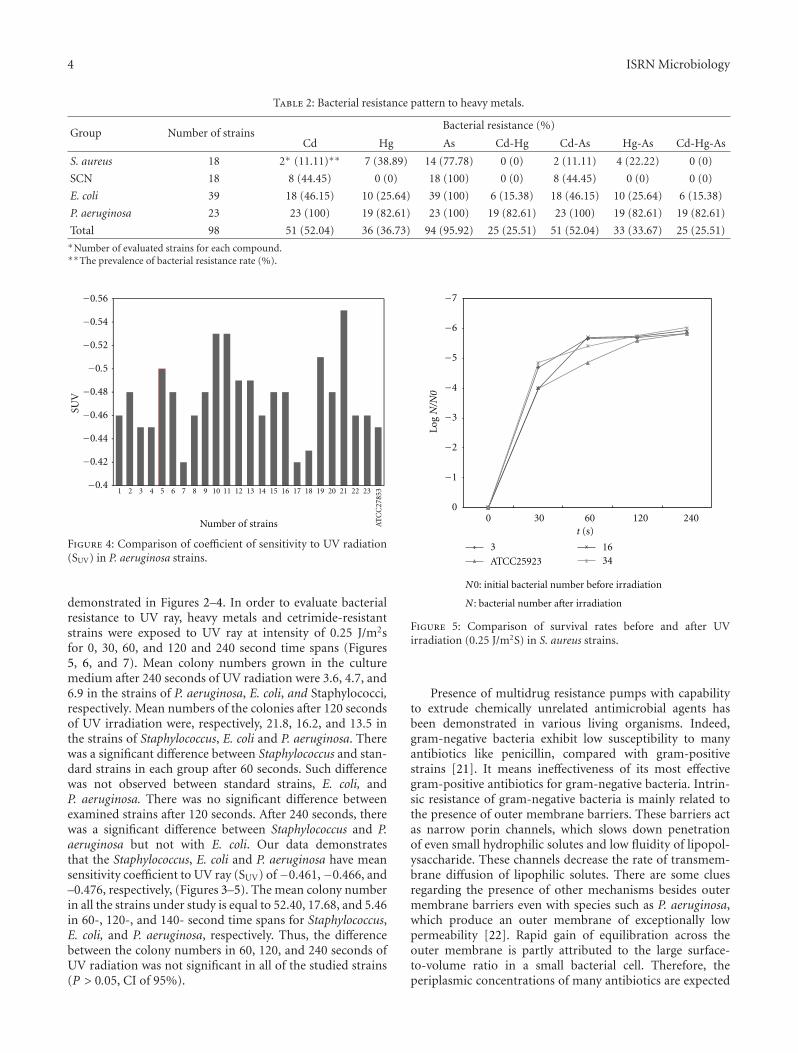
In general, the most prevalent metal resistance wasagainst Arsenate (95.92%), followed by Cadmium (52.04%)and Mercury (36.73%) (Table 2). The rate of double metalresistance was 25.51%, 52.04%, and 36.67% for Cd-Hg, Cd-As and Hg-As, respectively. The rate of triple metal resistance(Cd-Hg-As) was 25.51.
Comparison of coefficient of sensitivity to UV rays forthe strains of Staphylococcus, E. coli, and P. aeruginosa, are
4 ISRN Microbiology
Table 2: Bacterial resistance pattern to heavy metals.
Group Number of strainsBacterial resistance (%)
Cd Hg As Cd-Hg Cd-As Hg-As Cd-Hg-As
S. aureus 18 2∗ (11.11)∗∗ 7 (38.89) 14 (77.78) 0 (0) 2 (11.11) 4 (22.22) 0 (0)
SCN 18 8 (44.45) 0 (0) 18 (100) 0 (0) 8 (44.45) 0 (0) 0 (0)
E. coli 39 18 (46.15) 10 (25.64) 39 (100) 6 (15.38) 18 (46.15) 10 (25.64) 6 (15.38)
P. aeruginosa 23 23 (100) 19 (82.61) 23 (100) 19 (82.61) 23 (100) 19 (82.61) 19 (82.61)
Total 98 51 (52.04) 36 (36.73) 94 (95.92) 25 (25.51) 51 (52.04) 33 (33.67) 25 (25.51)∗Number of evaluated strains for each compound.∗∗The prevalence of bacterial resistance rate (%).
SUV
Number of strains
−0.56
−0.54
−0.52
−0.48
−0.46
−0.44
−0.42
−0.4
−0.5A
TC
C27
8531 2 3 4 5 6 7 8 9 10 11 12 13 14 15 16 17 18 19 21 22 2320
Figure 4: Comparison of coefficient of sensitivity to UV radiation(SUV) in P. aeruginosa strains.
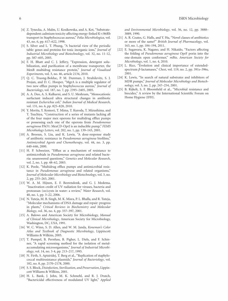
demonstrated in Figures 2–4. In order to evaluate bacterialresistance to UV ray, heavy metals and cetrimide-resistantstrains were exposed to UV ray at intensity of 0.25 J/m2sfor 0, 30, 60, and 120 and 240 second time spans (Figures5, 6, and 7). Mean colony numbers grown in the culturemedium after 240 seconds of UV radiation were 3.6, 4.7, and6.9 in the strains of P. aeruginosa, E. coli, and Staphylococci,respectively. Mean numbers of the colonies after 120 secondsof UV irradiation were, respectively, 21.8, 16.2, and 13.5 inthe strains of Staphylococcus, E. coli and P. aeruginosa. Therewas a significant difference between Staphylococcus and stan-dard strains in each group after 60 seconds. Such differencewas not observed between standard strains, E. coli, andP. aeruginosa. There was no significant difference betweenexamined strains after 120 seconds. After 240 seconds, therewas a significant difference between Staphylococcus and P.aeruginosa but not with E. coli. Our data demonstratesthat the Staphylococcus, E. coli and P. aeruginosa have meansensitivity coefficient to UV ray (SUV) of−0.461,−0.466, and–0.476, respectively, (Figures 3–5). The mean colony numberin all the strains under study is equal to 52.40, 17.68, and 5.46in 60-, 120-, and 140- second time spans for Staphylococcus,E. coli, and P. aeruginosa, respectively. Thus, the differencebetween the colony numbers in 60, 120, and 240 seconds ofUV radiation was not significant in all of the studied strains(P > 0.05, CI of 95%).
00 30 60 120 240
Log
N/N
0
t (s)
3
ATCC25923
1634
−7
−6
−5
−4
−3
−2
−1
N0: initial bacterial number before irradiation
N : bacterial number after irradiation
Figure 5: Comparison of survival rates before and after UVirradiation (0.25 J/m2S) in S. aureus strains.
Presence of multidrug resistance pumps with capabilityto extrude chemically unrelated antimicrobial agents hasbeen demonstrated in various living organisms. Indeed,gram-negative bacteria exhibit low susceptibility to manyantibiotics like penicillin, compared with gram-positivestrains [21]. It means ineffectiveness of its most effectivegram-positive antibiotics for gram-negative bacteria. Intrin-sic resistance of gram-negative bacteria is mainly related tothe presence of outer membrane barriers. These barriers actas narrow porin channels, which slows down penetrationof even small hydrophilic solutes and low fluidity of lipopol-ysaccharide. These channels decrease the rate of transmem-brane diffusion of lipophilic solutes. There are some cluesregarding the presence of other mechanisms besides outermembrane barriers even with species such as P. aeruginosa,which produce an outer membrane of exceptionally lowpermeability [22]. Rapid gain of equilibration across theouter membrane is partly attributed to the large surface-to-volume ratio in a small bacterial cell. Therefore, theperiplasmic concentrations of many antibiotics are expected

ISRN Microbiology 5
00 30 60 120 240
t (s)
−7
−8
−6
−5
−4
−3
−2
−1
9 ATCC25922
38
Log
N/N
0
N0: initial bacterial number before irradiation
N : bacterial number after irradiation
Figure 6: Comparison of survival rates before and after UV irradi-ation (0.25 J/m2S) in E. coli strains.
to reach 50% of their external concentrations in 10 to 30seconds in P. aeruginosa and in a much shorter time periodthan E. coli. Additional mechanisms are therefore needed toexplain intrinsic resistance like the hydrolysis of the earlier β-lactam compounds by the periplasmic β-lactamases encodedby chromosomal genes in many gram-negative bacteria [23].However, recent studies showed that multiple drug effluxpumps with broad specificities play a major role in intrinsicresistance of gram-negative bacteria [24].
Cetrimide, used widely as biocide and disinfectants,entered in the pathogen cell wall via outer membrane per-meability porins. Cetrimide resistance phenotype encodedby plasmid genes in E. coli was shown to be associated withaltered composition of outer membrane lipopolysaccharideand diminished porin numbers. Cetrimide resistance hasbeen shown with Staphylococcus, E. coli, and P. aeruginosa,similar to our findings. The greatest cetrimide resistance by P.aeruginosa seen in our study supports the finding regardinggreat gram-negative bacteria, mainly Pseudomonas, resis-tance to cetrimide and multiple antibiotics, especially duringnosocomial infections. Generally, gram-negative bacteria aremore biocide resistant than gram-positive bacteria, but it isnot always the case [25]. Pattern of heavy metal resistancein our studied isolates remark the relative effectiveness ofmercury rather than cadmium and specially arsenate forthese isolates. Great bacterial resistance against arsenate andcadmium makes them poor antimicrobial agents for theseorganisms, but new agents composed of mercury can beapplied more effectively. Interestingly, our data demonstratesthat double (mainly with mercury and cadmium) or triplemetal usage might be more effective because of the lowerbacterial resistance to these heavy metals. Application of
0
517
21
22
0 30 60 120 240
t (s)
−7
−8
−6
−5
−4
−3
−2
−1
ATCC27853
Log
N/N
0N : bacterial number after irradiation
N0: initial bacterial number before irradiation
Figure 7: Comparison of survival rates before and after UV irradi-ation (0.25 J/m2S) in P. aeruginosa strains.
ultraviolet spectrums, germicidal (ultraviolet C) and solar(ultraviolet A and B), as adjunctive therapy for bioburdenshave been previously documented [20]. In our study, UV-radiation resistance in Staphylococcus strains was higher thanthat of the other groups and the strains of E. coli have a higherresistance than P. aeruginosa.
Overall, our data demonstrates that the general bacterialresistance rate was higher among Staphylococcus strains thanE. coli strains. The same ranking was seen with E. colistrains comparing with P. aeruginosa strains. Despite of thepreviously published data, regarding the greatest rate ofmultiple antimicrobial resistances in gram-negative bacteria,our results demonstrated greater general bacterial resistancein Staphylococcus strains, a gram-positive bacterium. Know-ing the great antimicrobial resistance rate in gram-negativebacteria, it will be easy to understand the meaning of thegreater rate of resistance among Staphylococcus strains. Thesefindings imply the very limited remained therapeutic optionsand consequently the need for finding new powerful antimi-crobial agents.
References
[1] B. E. Burke, K. Wing Tsang, and R. M. Pfister, “Cadmiumsorption by bacteria and freshwater sediment,” Journal ofIndustrial Microbiology, vol. 8, no. 3, pp. 201–207, 1991.
[2] G. M. Teitzel and M. R. Parsek, “Heavy metal resistance ofbiofilm and planktonic Pseudomonas aeruginosa,” Applied andEnvironmental Microbiology, vol. 69, no. 4, pp. 2313–2320,2003.
[3] Z. Tynecka and A. Malm, “Energetic basis of cadmium toxicityin Staphylococcus aureus,” BioMetals, vol. 8, no. 3, pp. 197–204, 1995.
6 ISRN Microbiology
[4] Z. Tynecka, A. Malm, U. Kosikowska, and A. Kot, “Substrate-dependent cadmium toxicity affecting energy-linked K+/86Rbtransport in Staphylococcus aureus,” Folia Microbiologica, vol.43, no. 6, pp. 617–622, 1998.
[5] S. Silver and L. T. Phung, “A bacterial view of the periodictable: genes and proteins for toxic inorganic ions,” Journal ofIndustrial Microbiology and Biotechnology, vol. 32, no. 11-12,pp. 587–605, 2005.
[6] F. H. Bhatt and C. J. Jeffery, “Expression, detergent solu-bilization, and purification of a membrane transporter, theMexB multidrug resistance protein,” Journal of VisualizedExperiments, vol. 3, no. 46, article 2134, 2010.
[7] Q. C. Truong-Bolduc, P. M. Dunman, J. Strahilevitz, S. J.Projan, and D. C. Hooper, “MgrA is a multiple regulator oftwo new efflux pumps in Staphylococcus aureus,” Journal ofBacteriology, vol. 187, no. 7, pp. 2395–2405, 2005.
[8] A. A. Deo, A. S. Kulkarni, and S. U. Meshram, “Monocationicsurfactant induced ultra structural changes in antibioticresistant Escherichia coli,” Indian Journal of Medical Research,vol. 131, no. 6, pp. 825–828, 2010.
[9] Y. Morita, Y. Komori, T. Mima, T. Kuroda, T. Mizushima, andT. Tsuchiya, “Construction of a series of mutants lacking allof the four major mex operons for multidrug efflux pumpsor possessing each one of the operons from Pseudomonasaeruginosa PAO1: MexCD-OprJ is an inducible pump,” FEMSMicrobiology Letters, vol. 202, no. 1, pp. 139–143, 2001.
[10] A. Brooun, S. Liu, and K. Lewis, “A dose-response studyof antibiotic resistance in Pseudomonas aeruginosa biofilms,”Antimicrobial Agents and Chemotherapy, vol. 44, no. 3, pp.640–646, 2000.
[11] H. P. Schweizer, “Efflux as a mechanism of resistance toantimicrobials in Pseudomonas aeruginosa and related bacte-ria: unanswered questions,” Genetics and Molecular Research,vol. 2, no. 1, pp. 48–62, 2003.
[12] K. Poole, “Multidrug efflux pumps and antimicrobial resis-tance in Pseudomonas aeruginosa and related organisms,”Journal of Molecular Microbiology and Biotechnology, vol. 3, no.2, pp. 255–263, 2001.
[13] W. A. M. Hijnen, E. F. Beerendonk, and G. J. Medema,“Inactivation credit of UV radiation for viruses, bacteria andprotozoan (oo)cysts in water: a review,” Water Research, vol.40, no. 1, pp. 3–22, 2006.
[14] N. Tuteja, M. B. Singh, M. K. Misra, P. L. Bhalla, and R. Tuteja,“Molecular mechanisms of DNA damage and repair: progressin plants,” Critical Reviews in Biochemistry and MolecularBiology, vol. 36, no. 4, pp. 337–397, 2001.
[15] A. Balows and American Society for Microbiology, Manualof Clinical Microbiology, American Society for Microbiology,Washington, DC, USA, 1991.
[16] W. C. Winn, S. D. Allen, and W. M. Janda, Koneman’s ColorAtlas and Textbook of Diagnostic Microbiology, LippincottWilliams & Wilkins, 2005.
[17] T. Pumpel, B. Pernfuss, B. Pigher, L. Diels, and F. Schin-ner, “A rapid screening method for the isolation of metal-accumulating microorganisms,” Journal of Industrial Microbi-ology, vol. 14, no. 3-4, pp. 213–217, 1995.
[18] N. Firth, S. Apisiridej, T. Berg et al., “Replication of staphylo-coccal multiresistance plasmids,” Journal of Bacteriology, vol.182, no. 8, pp. 2170–2178, 2000.
[19] S. S. Block, Disinfection, Sterilization, and Preservation, Lippin-cott Williams & Wilkins, 2001.
[20] H. L. Bank, J. John, M. K. Schmehl, and R. J. Dratch,“Bactericidal effectiveness of modulated UV light,” Applied
and Environmental Microbiology, vol. 56, no. 12, pp. 3888–3889, 1990.
[21] A. R. Coates, G. Halls, and Y. Hu, “Novel classes of antibioticsor more of the same?” British Journal of Pharmacology, vol.163, no. 1, pp. 184–194, 2011.
[22] E. Sugawara, K. Nagano, and H. Nikaido, “Factors affectingthe folding of Pseudomonas aeruginosa OprF porin into theone-domain open conformer,” mBio, American Society forMicrobiology, vol. 1, no. 4, 2010.
[23] L. Rice, “Evolution and clinical importance of extended-spectrum β-lactamases,” Chest, vol. 119, no. 2, pp. 391s–396s,2001.
[24] K. Lewis, “In search of natural substrates and inhibitors ofMDR pumps,” Journal of Molecular Microbiology and Biotech-nology, vol. 3, no. 2, pp. 247–254, 2001.
[25] B. Rijkelt, S. F. Bloomfield et al., “Microbial resistance andbiocides,” A review by the International Scientific Forum onHome Hygiene (IFH).

Submit your manuscripts athttp://www.hindawi.com
Hindawi Publishing Corporationhttp://www.hindawi.com Volume 2014
Anatomy Research International
PeptidesInternational Journal of
Hindawi Publishing Corporationhttp://www.hindawi.com Volume 2014
Hindawi Publishing Corporation http://www.hindawi.com
International Journal of
Volume 2014
Zoology
Hindawi Publishing Corporationhttp://www.hindawi.com Volume 2014
Molecular Biology International
Hindawi Publishing Corporationhttp://www.hindawi.com
GenomicsInternational Journal of
Volume 2014
The Scientific World JournalHindawi Publishing Corporation http://www.hindawi.com Volume 2014
Hindawi Publishing Corporationhttp://www.hindawi.com Volume 2014
BioinformaticsAdvances in
Marine BiologyJournal of
Hindawi Publishing Corporationhttp://www.hindawi.com Volume 2014
Hindawi Publishing Corporationhttp://www.hindawi.com Volume 2014
Signal TransductionJournal of
Hindawi Publishing Corporationhttp://www.hindawi.com Volume 2014
BioMed Research International
Evolutionary BiologyInternational Journal of
Hindawi Publishing Corporationhttp://www.hindawi.com Volume 2014
Hindawi Publishing Corporationhttp://www.hindawi.com Volume 2014
Biochemistry Research International
ArchaeaHindawi Publishing Corporationhttp://www.hindawi.com Volume 2014
Hindawi Publishing Corporationhttp://www.hindawi.com Volume 2014
Genetics Research International
Hindawi Publishing Corporationhttp://www.hindawi.com Volume 2014
Advances in
Virolog y
Hindawi Publishing Corporationhttp://www.hindawi.com
Nucleic AcidsJournal of
Volume 2014
Stem CellsInternational
Hindawi Publishing Corporationhttp://www.hindawi.com Volume 2014
Hindawi Publishing Corporationhttp://www.hindawi.com Volume 2014
Enzyme Research
Hindawi Publishing Corporationhttp://www.hindawi.com Volume 2014
International Journal of
Microbiology