respiratory system. lungs and air passages nose, pharynx, larynx, trachea, bronchi, alveoli and...
TRANSCRIPT

Respiratory System
Lungs and Air Passages
Nose, pharynx, larynx, trachea, bronchi, alveoli and lungs • Responsible for – 1. Taking in O2 (needed by all body cells) – 2. Removing CO2 (metabolic waste product)
• Body has 4-6 minute supply of O2 • Must work continuously or death will occur
Nose
Has 2 nostrils or nares – Openings through which air enters
• Nasal septum – 1. Partition or wall of cartilage – 2. Divides the nose into 2 hollow spaces called
nasal cavities
Nasal Cavities
• A. Lined with mucous membrane • B. Rich blood supply • C. As air enters it is warmed, filtered and moistened• D. Mucous also helps trap pathogens and dirt • E. Cilia: tiny hair-like structures which also trap dirt and
pathogens, pushing them toward the esophagus to be swallowed
• F. Olfactory receptors for the sense of smell • G. Nasolacrimal ducts drain tears from the eye into the
nose to provide additional moisture for the air

Paranasal Sinuses
• A. Hollow air-containing spaces within the skull
• B. Cavities in the skull around the nasal area • C. Connected to the nasal cavity by short ducts • D. Lined with mucous membrane that warms
and moistens air • E. Provide resonance for the voice
Pharynx
• A. The throat • B. Lies directly behind the nasal cavities • C. As air leaves the nose it enters the pharynx • D. Has three sections
Pharynx
• Nasopharynx • a. Upper portion behind the nasal cavities • b. Contain the pharyngeal tonsils or adenoids (lymphatic tissue) and
the auditory (eustachian) tube openings • 2. Oropharynx • a. Middle section located behind the oral cavity • b. Contains the palatine tonsils (two rounded masses of lymphatic
tissue) • c. Received both air from the nasopharynx and food and air from the
mouth • 3. Laryngopharynx a. Bottom section of the pharynx • b. Branches into the trachea, which carries air to and from the lungs,
and the esophagus – the tube that carries food to the stomach
Epiglottis
• A. A flap of cartilage attached to the root of the tongue
• B. Prevents choking or aspiration of food • C. Acts as a lid over the opening of the larynx • D. During swallowing when food and liquid
move through the throat, the epiglottis closes over the larynx
Larynx
• A. Voice box • B. Lies between the pharynx and trachea • C. Has a framework of cartilage commonly called the
“Adam’s apple” • D. Contains two folds called vocal cords
1. Opening between the vocal cords is the glottis 2. As air leaves the lungs, the vocal cords vibrate
and produce sound 3. Tongue and lips act on the sound to produce
speech
Trachea
• A. Windpipe • B. Tube extending from the larynx to the center of
the chest (about 4.5” long) • C. Carries air between the pharynx and bronchi • D. Series of c-shaped cartilages, which are open
on the dorsal or back surface, and help keep the trachea open
•
Alveoli
• A. Air sacs that resemble a bunch of grapes • B. Adult lung contains approximately 300 million
alveoli • C. Made of one layer of squamous epithelium tissue • D. Contains a rich network of blood capillaries • E. Capillaries allow O2 and CO2 to be exchanged
between the blood and the lungs • F. Inner surface of alveoli are covered with surfactant
– 1. Lipid or fatty substance – 2. Helps prevent alveoli from collapsing
Bronchi
• A. Two divisions of the trachea near the center of the chest – 1. Right and left bronchus (singular) – 2. Right bronchus is shorter, wider and extends more vertically
than the left bronchus • B. Each bronchus enters a lung and carries air from the
trachea to the lungs • C. In the lungs, the bronchi continue to divide into smaller
and smaller bronchi • D. Smaller branches are called bronchioles • E. Smallest bronchioles, called terminal bronchioles; end in
the air sacs called alveoli
Erythrocytes
• A. Carry oxygen to all parts of the body • B. Carries carbon dioxide to the lungs for
exhalation
Lungs
• A. Organs that contain divisions of the bronchi and alveoli • B. Right lung has 3 sections or lobes: superior, middle and
inferior • C. Left lung has only two lobes, superior and inferior • D. Left lung is smaller because the heart lies more to the left
side of the chest • E. Both the lungs are located in the thoracic cavity • F. Apex: uppermost part of the lung • G. Base: lower part of the lung• H. Hilum: the midline region in which blood vessels, nerves,
lymphatic tissue, and bronchial tubes enter and exit the lung • I. The lungs extend from the collarbone to the diaphragm
Diaphragm
• A. A muscular partition • B. Separates the thoracic from the abdominal cavity • C. Aids in the process of breathing • 1. Contracts
a. Moves downward, enlarging the area in the thoracic cavity b. Decreasing internal air pressure, so that air flows into the
lungs to equalize the pressure • 2. Relaxes
– a. When the lungs are full, the diaphragm relaxes and elevates – b. Makes the area in the thoracic cavity smaller, thus
increasing air pressure in the chest – c. Air is expelled out of the lungs to equalize pressure
Pleura
• A. Consists of two layers • 1. Visceral pleura attached to surface of lung; inner layer
closer to the lungs • 2. Parietal pleura attached to the chest wall; outer layer,
closer to the ribs • B. Pleural space • 1. Located between the two layers • 2. Filled with a thin layer of pleural fluid that lubricates the
membranes and prevents friction as the lungs expand during breathing
Pathway of Air
• A. Nose • B. Nasal cavities and paranasal sinuses • C. Pharynx (adenoids and tonsils) • D. Larynx (epiglottis) • E. Trachea • F. Bronchi • G. Bronchioles • H. Alveoli • I. Lung capillaries
Ventilation
• A. Process of breathing • B. Respiration • C. Two phases • 1. Inspiration or inhalation
– a. Process of breathing in air– b. Diaphragm contracts and enlarges the thoracic cavity
• 2. Expiration or exhalation – a. Process where air leaves the lungs – b. Diaphragm and intercostal muscles relax and air is forced out
of the lungs
Respiration
• Inspiration + Expiration = Respiration • 1. The mechanical process of breathing • 2. The exchange of air between the lungs and
the external environment • 3. Process is controlled by the respiratory
center in the medulla oblongata of the brain
Respiration
• Respiration is a Vital Sign • 1. Normal adult respiration rate is 12-20
breaths/minute • 2. How do we know if a person is breathing?
We can see the chest rise and fall
Abnormal Breathing
• A. Dyspnea: difficult or labored breathing • B. Apnea: absence of respiration • C. Tachypnea: breathing rate above 25/breaths/minute • D. Bradypnea: slow respirations, below 10
breaths/minute • E. Orthopnea: dyspnea in any position other than sitting
erect or standing • F. Cyanosis: bluish discoloration of the skin, lips or nail
beds as a result of decreased O2
Form a group of four studentsResearch the selected Respiratory DisorderPrepare a presentation using a trifold poster describe: disease symptoms, etiology of disease, the signs and symptoms, treatments, and ways to prevent it
include: pictures, pamphlets, local treatment centers, USE YOUR CREATIVITY AND KNOWLEDGE
Select one Respiratory Disorder• Asthma• Cystic Fibrosis• Chronic Obstruction Pulmonary Disease• Emphysema• ARDS (acute respiratory distress syndrome)• Pleurisy• Pneumonia• Tuberculosis• SIDS (sudden infant death syndrome)• Sleep Apnea
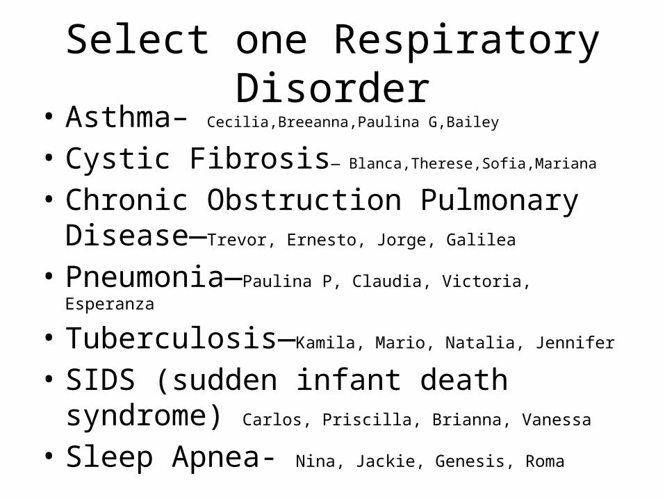
Select one Respiratory Disorder• Asthma– Cecilia,Breeanna,Paulina G,Bailey
• Cystic Fibrosis— Blanca,Therese,Sofia,Mariana
• Chronic Obstruction Pulmonary Disease—Trevor, Ernesto, Jorge, Galilea
• Pneumonia—Paulina P, Claudia, Victoria, Esperanza
• Tuberculosis—Kamila, Mario, Natalia, Jennifer
• SIDS (sudden infant death syndrome) Carlos, Priscilla, Brianna, Vanessa
• Sleep Apnea- Nina, Jackie, Genesis, Roma
