reverse genetics with tilling in phytophthora sojae
TRANSCRIPT
University of Tennessee, Knoxville University of Tennessee, Knoxville
TRACE: Tennessee Research and Creative TRACE: Tennessee Research and Creative
Exchange Exchange
Masters Theses Graduate School
12-2005
Reverse Genetics with TILLING in Reverse Genetics with TILLING in Phytophthora sojae
Melinda Beth Tierney University of Tennessee - Knoxville
Follow this and additional works at: https://trace.tennessee.edu/utk_gradthes
Part of the Life Sciences Commons
Recommended Citation Recommended Citation Tierney, Melinda Beth, "Reverse Genetics with TILLING in Phytophthora sojae. " Master's Thesis, University of Tennessee, 2005. https://trace.tennessee.edu/utk_gradthes/2552
This Thesis is brought to you for free and open access by the Graduate School at TRACE: Tennessee Research and Creative Exchange. It has been accepted for inclusion in Masters Theses by an authorized administrator of TRACE: Tennessee Research and Creative Exchange. For more information, please contact [email protected].

To the Graduate Council:
I am submitting herewith a thesis written by Melinda Beth Tierney entitled "Reverse Genetics
with TILLING in Phytophthora sojae." I have examined the final electronic copy of this thesis for
form and content and recommend that it be accepted in partial fulfillment of the requirements
for the degree of Master of Science, with a major in Life Sciences.
Kurt Lamour, Major Professor
We have read this thesis and recommend its acceptance:
Beth Mullin, Carrie Fritz
Accepted for the Council:
Carolyn R. Hodges
Vice Provost and Dean of the Graduate School
(Original signatures are on file with official student records.)

To the Graduate Council: I am submitting herewith a thesis written by Melinda Beth Tierney entitled “Reverse Genetics with TILLING in Phytophthora sojae.” I have examined the final electronic copy of this thesis for form and content and recommend that it be accepted in partial fulfillment of the requirements for the degree of Master of Science, with a major in Life Sciences.
Kurt Lamour Major Professor
We have read this thesis and recommend its acceptance: Beth Mullin Carrie Fritz
Accepted for the Council: Ann Mayhew Vice Chancellor and
Dean of Graduate Studies
(Original signatures are on file with official student records)

Reverse Genetics with TILLING in
Phytophthora sojae
A Thesis presented for the Master of Science Degree
The University of Tennessee, Knoxville
Melinda Beth Tierney
December 2005

ii
DEDICATION
This thesis is dedicated to John Tierney for always believing in me, for his invaluable
support and encouragement, and most of all his love
.

iii
ACKNOWLEDGEMENTS
To my major professor, Dr. Kurt Lamour for his guidance and enthusiasm.
To Ledare Finley for all her help and for her expertise figuring out the nuances of
TILLING.
To the Lamour lab for being fun to work with.
To the Genome Science and Technology Program.
To the Department of Entomology and Plant Pathology for the use of office space.
To my committee: Dr. Beth Mullin and Dr. Carrie Fritz.
To Jessie for always being there to relieve stress.
To my parents, Greg and DeAnna Lawrence, and my parents-in-law, Bill and Pati
Tierney, for their love, support, and encouragement.

iv
ABSTRACT
The reverse genetic method TILLING (Targeting Induced Local Lesions in
Genomes) is being applied to the plant pathogen Phytophthora sojae. The objective is to
recover gene-specific mutants carrying allelic series and/or knockout induced mutations.
A library of 3000 mutant individuals was generated using the chemical mutagens
ethylmethanesulfonate (EMS) or ethylnitrosourea (ENU). Gene-specific induced
mutations are detected by screening a mirror library of genomic DNA. PCR is used to
amplify and fluorescently label 1kb portions of specific genes from the mutant library
and the PCR products are then heated and cooled slowly to form hetero- and homo-
duplexes. The PCR fragments are treated with the CEL 1 enzyme, a single strand
specific endonuclease, and the treated fragments resolved on a LI-COR gel analyzer
system. Amplicons harboring induced SNPs are cleaved at the site of the mutation thus
producing novel fragments. To date we have identified nine SNP’s in the Necrosis
Inducing Protein (NIP) gene in P. sojae using TILLING. Although beyond the scope of
this work, the generation of recombinant sexual progeny to recover mutants homozygous
for these mutations is underway.

v
TABLE OF CONTENTS
Section Page
1. INTRODUCTION.............................................................................. 1
A. Genetics ....................................................................................... 1
B. Forward Genetics vs. Reverse Genetics ....................................... 1
C. Specific Reverse Genetic Approaches........................................... 2
C.1. Gene Silencing ............................................................ 4
C.2. Targeted Gene Disruption by Homologous
Recombination ........................................................... 5
C.3. Insertional Mutagenesis/Transposon Mediated
Mutagenesis ................................................................ 6
D. Chemical Mutagenesis and TILLING ........................................... 7
E. Examples of TILLING ................................................................. 8
E.1. Arabidopsis ................................................................. 8
E.2. Zebrafish ..................................................................... 9
F. Limitations and Roadblocks to Completing Reverse Genetics..... 10
G. Phytophthora.............................................................................. 10
H. TILLING Phytophthora.............................................................. 12
2. MATERIALS AND METHODS...................................................... 16
A. Mutant Library Construction ...................................................... 16
B. DNA Extraction.......................................................................... 16
C. Target Selection.......................................................................... 16
D. PCR Amplification ..................................................................... 17

vi
E. CEL Treatment ........................................................................... 17
F. Sephadex Cleanup ...................................................................... 22
G. Polyacrylamide Gel Separation of PCR Products........................ 22
H. Sequencing for Single Nucleotide Polymorphism Confirmation.. 24
I. Germination of Oospores............................................................ 24
J. DNA Extraction and Sequencing ................................................ 26
3. RESULTS, DISCUSSION, AND CONCLUSIONS ......................... 27
A. Mutant Library ........................................................................... 27
B. Recovery of Gene Specific Mutants............................................ 27
C. Oospore Progeny ........................................................................ 32
D. Conclusions................................................................................ 32
Literature Cited ................................................................................................. 33
Appendices ................................................................................................... 42
Vita ................................................................................................... 48

vii
LIST OF TABLES
Table Page
1. Primers targeting PsojNIP....................................................................... 18
2. PCR amplification reaction for TILLING............................................... 19
3. CEL reaction........................................................................................... 21
4. CODDLE mutation predictions ............................................................... 31

viii
LIST OF FIGURES
Figure Page
1. Illustration of the major differences between forward and reverse genetics ..... 3
2. Basic Local Alignment Search Tool results aligning PsojNIP with
other similar proteins in the National Center for Biotechnology
Information database.................................................................................... 13
3. Identification of Phytophthora isolates with gene specific point mutations
using the TILLING strategy......................................................................... 14
4. Gene sequence for PsojNIP .......................................................................... 18
5. PCR amplification of mutant genomic DNA containing PsojNIP.................. 20
6. Overview of mutation detection method........................................................ 25
7. Genomic DNA quantification gel.................................................................. 28
8. TILLING gel with observed single nucleotide polymorphism (snp) .............. 29
9. Induced polymorphism confirmed by sequencing.......................................... 30

1
1. INTRODUCTION
This chapter is a lightly revised version of a paper by the name “An Introduction to Reverse Genetic Tools for Investigating Gene Function” published on the American Phytopathological Society educational website: Tierney, M.B. and Lamour, K.H. 2005. An Introduction to Reverse Genetic Tools for Investigating Gene Function. The Plant Health Instructor. DOI: 10.1094/PHI-I-2005-1025-01. My primary contributions to this paper include literature review and research of topic information and most of the writing. A. Genetics
Genetics began in the 1860’s with Gregor Mendel, an Augustinian monk who
performed experiments that suggested the existence of genes (Griffiths et al. 2000). The
discovery of the structure of DNA by James Watson and Francis Crick in the 1950’s
(Watson and Crick 1953), followed by the development of di-deoxy terminator
sequencing by Sanger in the late 1970’s (Sanger, Nicklen, and Coulson 1977) and then of
PCR (polymerase chain reaction) technology by Kary Mullis in the 1980’s (Mullis and
Faloona 1987; Saiki et al. 1985) set off a genetic revolution. Technological advances in
sequencing have greatly accelerated our accumulation of genetic sequence data to the
point now where whole genome sequences are publicly available for a large number of
organisms including plant pathogens. The toolbox for genetic research is expanding
rapidly and this overview presents a suite of tools generally referred to as reverse genetics
that can be used to investigate gene function.
B. Forward Genetics vs. Reverse Genetics
Variants help us understand the ‘normal’. Variation can be measured at many
scales – from macro (body size, morphology) to different levels of micro variation (crude

2
protein profiles to DNA sequence variation). Forward genetics refers to a process where
studies are initiated to determine the genetic underpinnings of observable phenotypic
variation. In many cases the observable variation has been induced using a DNA
damaging agent (mutagen) but also may be naturally occurring. The investigator
eventually ends up sequencing the gene or genes thought to be involved (Figure 1).
With the advent of whole genome sequencing many researchers are now in a very
different position. They have access to all of the gene sequences within a given organism
and would like to know their function. So, instead of going from phenotype to sequence
as in forward genetics, reverse genetics works in the opposite direction – a gene sequence
is known but its exact function is uncertain. In reverse genetics a specific gene or gene
product is disrupted or modified and then the phenotype is measured (Figure 1). Here we
will overview some of the techniques for reverse genetics with a special emphasis on the
TILLING (Targeting Induced Local Lesions IN Genomes) technique which is being
utilized on plant pathogens in the genus Phytophthora.
C. Specific Reverse Genetic Approaches
The goal in reverse genetics is to investigate the impact of induced variation
within a specific gene and to infer gene function. The process of disruption or alteration
can either be targeted specifically as in the case of gene silencing or homologous
recombination or can rely on non-targeted random disruptions (e.g. chemical
mutagenesis, transposon mediated mutagenesis) followed by screening a library of
individuals for lesions at a specific location. Following is a brief overview of some
commonly used targeted and non-targeted approaches.

3
Figure 1: Illustration of the major differences between forward and reverse genetics.
The blue oospore symbolizes an observable phenotype that may have been produced via
any one of a number of pathways (e.g. mutation, recombination, etc.). This is the starting
point for A, forward genetics where the investigator proceeds from the phenotype to
characterize the underlying genetic difference. B, illustrates reverse genetics where the
sequence for a gene is known but the gene's function is unknown and the investigator
disrupts the gene and investigates the resulting phenotype.

4
C.1. Gene Silencing
RNA interference (RNAi) is the process by which expression of a target gene is
inhibited by antisense and sense RNAs. It works based on the ability of double-stranded
sequences to recognize and degrade sequences that are complementary to them (Lewin
2004). RNAi was first discovered in Caenorhabditis elegans when the introduction of
double-stranded RNA was observed to be an efficient method for silencing gene
expression (Fire et al. 1998; Kuttenkeuler and Boutros 2004). RNAi- based silencing is
an exciting strategy for reverse genetics (Waterhouse, Graham, and Wang 1998). RNA
interference has recently become a powerful tool to silence the expression of genes and
analyze their loss-of-function phenotype, allowing analysis of gene function when mutant
alleles are not available. Having been shown to work in a similar manner in all
metazoans, RNAi has proven to be applicable to many organisms and has been used to
generate a wide variety of loss-of-function phenotypes (Kuttenkeuler and Boutros 2004).
The phenomenon of post-transcriptional gene silencing observed in plants may also be
due to a related RNAi mechanism (Waterhouse, Graham, and Wang 1998). RNAi based
silencing utilizes the endonuclease Dicer to cleave single stranded RNAs, abbreviated
siRNAs, from double stranded RNA; the RISC complex then destroys specific target
mRNAs based on sequence complementarity with the siRNA (Pattanayak et al. 2005).
RNAi has been used for a systematic analysis of gene function in C. elegans by
generating loss of function phenotypes, creating a library of worms expressing dsDNA
corresponding to different genes (Lewin 2004). Genome-wide RNAi screens against
these libraries of predicted genes have allowed study of a variety of biological processes
in C. elegans. A genome-wide library of double-stranded RNAs that target every gene in

5
the Drosophila genome has also been published that is suitable for high throughput cell-
based assays (Kuttenkeuler and Boutros 2004). One difficulty in using RNAi as a reverse
genetic technique is that throughput is limited by the ability to deliver siRNAs to target
loci (Henikoff, Till, and Comai 2004). It is also labor intensive, can give ambiguous
results, and can be unsuitable for isolating mutants that have lethal or sterile phenotypes
(Gilchrist and Haughn 2005).
C.2. Targeted Gene Disruption by Homologous Recombination
Homologous recombination is a reciprocal exchange of DNA sequences, as in
between two chromosomes that carry the same genetic loci (Lewin 2004). Just as
homologous recombination has been found to be mainly initiated with a double-strand
break, gene targeting by homologous recombination is associated with the repair of
double strand breaks. The double-strand break repair and synthesis-dependent strand-
annealing models are the most generally accepted models to explain gene targeting (Iida
and Terada 2004).
Homologous recombination has been widely used in embryonic stem (ES) cells in
mice, and has allowed construction of precise mutations in nearly every gene. A reverse-
genetic system using homologous recombination has recently been developed for
Drosophila. It is promising, but is a lengthy procedure and requires generation of
specific transgenic flies (Stemple 2004). Reproducible gene targeting by homologous
recombination is now also feasible in rice. With the combination of site-specific
recombination systems (such as Cre-lox), the future of gene targeting by homologous
recombination as a routine procedure for engineering the genome of rice and presumably
other plants is bright (Iida and Terada 2004).

6
C.3. Insertional Mutagenesis/Transposon Mediated Mutagenesis
Transposons are mobile genetic elements that can relocate from one genomic
location to another (Hayes 2003). They are DNA sequences that can insert themselves at
a new location in the genome without having any sequence relationship with the target
locus (Lewin 2004). Transposon-based signature-tagged mutagenesis has been
successful in identifying essential genes as well as genes involved in infectivity of a
variety of pathogens. Strategies for insertional mutagenesis using transposons have been
developed for a number of animal and plant models (Hayes 2003). Reverse-genetics is
currently being done in Drosophila and C. elegans by utilizing libraries of individuals
who carry transposable element insertions, many of which have been mapped, and some
of which will disrupt the expression of nearby genes. In Drosophila P-elements,
imprecise excision can be driven to generate a mutation in the nearest gene. Transposon-
based methods are also being used in Arabidopsis, maize and other plants (Stemple
2004). One drawback of insertional mutagenesis is the low frequency of mutations,
necessitating the screening of large numbers of individuals to find mutations in any given
gene (Gilchrist and Haughn 2005). Also, insertions in essential genes will usually cause
lethality, and less severe mutations must be generated in these genes in order to
understand gene function (Till et al. 2003).
The segment of the Ti plasmid of Agrobacterium tumefaciens known as T-DNA
that carries genes to transform the plant cell has also been utilized for insertional
mutagenesis. T-DNA insertional mutagenesis has been used to obtain gene knockouts for
greater than 70% of Arabidopsis genes (Alonso et al. 2003), but no comparable resources
exist for rice or maize even as high-coverage genomic sequence is becoming increasingly

7
available (Henikoff, Till, and Comai 2004). Unlike other successful gene targeting
systems (namely mouse, Physcomitrella, and Drosophila), the precise mechanism of T-
DNA integration into the plant genome remains largely unknown (Iida and Terada 2004).
Like RNA suppression techniques, insertional mutagenesis is limited by its host range
and by its limited range of allele types (McCallum et al. 2000).
Gene replacement via transformation is a commonly used tool for many
filamentous fungi (Fang, Hanau, and Vaillancourt 2002; Lalucque and Silar 2004;
Takano et al. 2000). The transformation can be mediated in many ways, including
Agrobacterium (Zhang et al. 2003) and various other transformation vectors (Scott-Craig
et al. 1998; Takano et al. 2000).
D. Chemical Mutagenesis and TILLING
Two of the most widely used mutagens for chemical mutagenesis experiments are
EMS (ethylmethanesulfonate) and ENU (ethylnitrosourea). EMS (Sciences 2002; Sega
1984)is a chemical mutagen that alkylates guanine bases. The alkylated guanine will
then pair with thymine instead of the preferred cytosine base, ultimately resulting in a
G/C to A/T transition. EMS is the most commonly used mutagen in plants. In
Arabidopsis, five percent of EMS-induced mutations in targeted coding regions result in
premature termination of the gene product, while fifty percent result in missense
mutations that alter the amino-acid sequence of the encoded protein (Gilchrist and
Haughn 2005). This high level of missense mutations relative to terminated gene
products is very useful in analyzing gene function. ENU (Justice et al. 1999) also
induces point mutations, and is a more potent mutagen than EMS. It is also an alkylating
agent, mutagenizing by transferring an ethyl group to oxygen or nitrogen radicals in the

8
DNA molecule, which leads to mispairing and ultimately results in base pair
substitutions, and sometimes base pair losses if not repaired (Guénet 2004).
Chemical mutagenesis is attractive for reverse genetics because it results in
induced point mutations, which create a diverse range of alleles for genetic analysis. It
induces a large number of recessive mutations per genome that are randomly distributed
(Gilchrist and Haughn 2005). Because chemical mutagenesis is already widely used in
many organisms for forward genetic screens (Smits et al. 2004), it promises to be
generally applicable for reverse genetics (Coghill et al. 2002; Henikoff, Till, and Comai
2004; Jansen et al. 1997; Wienholds et al. 2002). Until recently, chemical mutagenesis
has not been widely used as a tool for reverse genetics because of the lack of high-
throughput techniques for detecting point mutations (Gilchrist and Haughn 2005). The
TILLING strategy provides a high throughput strategy to detect single base changes
within genetic targets (Colbert et al. 2001; Henikoff and Comai 2003) and can be applied
to a wide variety of organisms (McCallum et al. 2000; Perry et al. 2003; Smits et al.
2004; Till, Reynolds et al. 2004).
E. Examples of TILLING
TILLING has been an instrumental tool in the study of several organisms.
Following is the description of two of the most widely recognized TILLING projects.
E.1. Arabidopsis
A public TILLING resource was set up in 2001 for the Arabidopsis community.
This effort was called the Arabidopsis TILLING Project (ATP) and is now called the
Seattle Tilling Project (STP) and is a joint effort between the Comai Laboratory at the
University of Washington and the Henikoff Laboratory at the Fred Hutchinson Cancer

9
Research Center in Seattle, Washington. Users are charged a fee that covers partial costs
of the services provided to request mutations in genes of interest (Gilchrist and Haughn
2005; Till et al. 2003). Training sessions, workshops, and on-going support to
researchers interested in developing TILLING in other organisms has been made
available through the STP and has served to make the TILLING technique more wide-
spread. This group has also developed and made publicly available web-based software
programs for PCR primer design and visualization of polymorphisms (Gilchrist and
Haughn 2005). Further information about the STP can be found at the following url:
http://tilling.fhcrc.org:9366/.
E.2. Zebrafish
Target selected mutagenesis using TILLING is also being done in zebrafish. The
Hubrecht Laboratory at The Netherlands Institute for Developmental Biology has
generated a library of 4608 ENU-mutagenized F1 animals and has kept a living stock.
The DNA from these animals has been screened for mutations in 16 genes using
TILLING followed by re-sequencing. This resulted in 255 mutations being identified, 14
of which resulted in a premature stop codon, 7 in a splice donor/acceptor site mutation,
and 119 in an amino acid change. They were able to knock out 13 different genes in only
a few months time through reverse genetics (Wienholds et al. 2003). Thus far, mutant
phenotypes have been characterized for two of the zebrafish genes modified via
TILLING - the gene for Dicer1 (Stemple 2004; Wienholds et al. 2003) and the gene for
adenomatous polyposis, a tumor suppressor, which through this research was found to
have a previously unknown function for signaling in cardiac-valve formation (Hurlstone
et al. 2003; Stemple 2004).

10
F. Limitations and Roadblocks to Completing Reverse Genetics
Completing reverse genetics is not without its pitfalls and not all techniques can
be applied to all organisms. In order to be successful, there are several aspects that must
be checked. For organisms that do not have efficient transformation systems available
techniques such as TILLING that can be applied without transformation may be the only
practical choice. In these cases, the rate of mutagenesis is an important factor that can be
difficult to determine. The load of mutations must be balanced with the recovery of
mutants (Till et al. 2003) – in other words, the genome can’t be so riddled with mutations
that it is impossible to see a mutant phenotype. Also to be considered is the fertility of
the mutagenized organism, especially in the first generation but also in subsequent
generations (Perry et al. 2003), both before and after mutagenesis. This is especially true
for diploid organisms, because if the sexual machinery is not intact and working properly,
then it is impossible to obtain a homozygous mutant. The mutagenized organisms must
also be kept alive long enough to screen a mutant population for a specific target. For
some organisms, like Arabidopsis or Phytophthora, this is not a problem as the seeds or
cultures are relatively easy to store. It presents a challenge for other organisms, such as
zebrafish or rats, because they must be stored and kept alive through the mutant screening
stage.
G. Phytophthora
Phytophthora is an oomycete pathogen that is important for many reasons ranging
from scientific to economic. The genus has been responsible for a host of diseases and
outbreaks with dire consequences, including the Irish potato famine (May and Ristaino
2004; Ristaino 2002) and the more recent Sudden Oak Death (Martin and Tooley 2003;

11
Rizzo, Garbelotto, and Hansen 2005) epidemic. The economic consequences of damage
from Phytophthora species is billions of dollars a year in the United States, and much
more than that worldwide (Erwin and Ribeiro 1996). Most Phytophthora species
resemble the true fungi, but are actually more closely related to brown algae and diatoms
(Baldauf et al. 2000; Huitema et al. 2004).
Phytophthora diseases can be difficult to diagnose due the similarity of symptoms
to other pathogens, causing late recognition and misdiagnoses that often result in heavy
losses. Because they are not true fungi they are not affected by most fungicides (Erwin
and Ribeiro 1996), so even once identified Phytophthora infections are difficult to
control. The current effort toward understanding this group of pathogens lies in studying
Phytophtora at the molecular level, using genome sequence resources to determine their
underlying biology (Huitema et al. 2004; Qutob et al. 2000).
P. sojae is a species of Phytophthora that is responsible for root and stem rot in
soybean, which is considered to be its sole host (Bhat and Schmitthenner 1993; Jackson,
Kirpatrick, and Rupe 2004; Leitz et al. 2000; MacGregor et al. 2002; Moy et al. 2004;
Tyler 2002; Tyler, Forster, and Coffey 1995; Whissen et al. 1994). P. sojae is
homothallic, allowing it to self fertilize (Erwin and Ribeiro 1996). This makes it largely
inbred and a good background for mutation studies.
The gene target chosen for this study was PsojNIP (Phytophthora sojae Necrosis
Inducing Protein). PsojNIP is expressed during the transition from biotrophy to
necrotrophy in P. sojae infection. The mechanism of necrosis induction is unknown, but
studies have indicated that it may occur through manipulation of intrinsic host cell death
programs. Evidence also supports that PsojNIP is a secreted protein, an elicitor-toxin

12
that aids in colonization and accelerates host cell death in the necrotrophic phase of the
disease (Qutob, Kamoun, and Gijzen 2002).
PsojNIP is similar in sequence to proteins from other oomycetes, bacteria, and
fungi. This can be visualized by performing a BLAST (Basic Local Alignment Search
Tool) search at the National Center for Biotechnology Information (NCBI) website
(http://www.ncbi.nlm.nih.gov/). At the BLAST window of the NCBI website, the
position-specific iterated and pattern-hit initiated BLAST (PSI- and PHI-BLAST) was
chosen under the protein section. The protein sequence for PsojNIP was entered as input,
and the BLAST program then searched through the NCBI protein databases and looked
for other proteins containing conserved regions similar to PsojNIP (Figure 2).
H. TILLING Phytophthora
Whole genome sequences for the soybean pathogen Phytophthora sojae and the
Sudden Oak Death pathogen P. ramorum were made public near the end of 2004. Large-
scale genomic sequencing projects are underway for the potato late blight pathogen P.
infestans and the vegetable pathogen P. capsici. The compilation of these genomic
sequences allows the application of many sophisticated research tools and promises a
better understanding of this devastating group of pathogens (Huang et al. 2005; Torto et
al. 2003). In an effort to better understand gene function within Phytophthora a project
to develop a reverse genetic TILLING resource for Phytophthora has been initiated.
Figure 3 provides an overview of the process as it is being applied to Phytophthora.
Zoospores present an ideal life stage for mutagenesis as they are uni-nucleate and single
mutant individuals can readily be isolated following mutagenesis. The mutation rate for
EMS or ENU is not known for Phytophthora and lethality is being used as an indicator of

13
1. necrosis-inducing peptide [Phytophthora sojae] 2. necrosis and ethylene-inducing protein 4-2
[Phytophthora megakarya] 3. necrosis and ethylene-inducing protein 6 [Phytophthora
megakarya] 4. necrosis-inducing protein NPP1 [Phytophthora
parasitica] 5. necrosis-inducing-like protein [Phytophthora sojae] 6. necrosis-inducing protein NPP1 [Phytophthora
infestans] 7. necrosis and ethylene-inducing protein 4 [Phytophthora
megakarya] 8. necrosis-inducing-like protein [Phytophthora sojae] 9. necrosis and ethylene-inducing protein 2 [Phytophthora
megakarya] 10. necrosis and ethylene-inducing protein 1 [Phytophthora
megakarya] 11. 25 kDa protein elicitor [Pythium aphanidermatum] 12. 25 kDa protein elicitor-like protein [Pythium aff.
vanterpoolii] 13. necrosis and ethylene-inducing protein 6-2
[Phytophthora megakarya] 14. hypothetical protein BL03562 [Bacillus licheniformis
ATCC 14580] 15. necrosis and ethylene inducing protein [Bacillus
halodurans C-125] 16. hypothetical protein BLi01595 [Bacillus licheniformis
ATCC 14580] 17. hypothetical protein MG08454.4 [Magnaporthe grisea
70-15] 18. hypothetical protein FG06017.1 [Gibberella zeae PH-1] 19. hypothetical protein AN3211.2 [Aspergillus nidulans
FGSC A4] 20. NPP1 domain protein, putative [Aspergillus fumigatus
Af293] 21. necrosis and ethylene-inducing protein 5 [Phytophthora
megakarya]
22. hypothetical protein MG10532.4 [Magnaporthe grisea 70-15]
23. necrosis and ethylene inducing peptide [Fusarium oxysporum f. sp. erythroxyli]
24. NEP-like [Fusarium oxysporum] 25. necrosis and ethylene-inducing protein 3 [Phytophthora
megakarya] 26. necrosis and ethylene inducing peptide [Verticillium
dahliae] 27. necrosis and ethylene-inducing protein 7 [Phytophthora
megakarya] 28. Necrosis inducing [Frankia sp. EAN1pec] 29. putative secreted protein [Streptomyces coelicolor
A3(2)] 30. MOSQUITOCIDAL TOXIN PROTEIN [Bacillus
thuringiensis serovar israelensis ATCC 35646] 31. hypothetical protein MG00401.4 [Magnaporthe grisea
70-15] 32. putative exported protein [Erwinia carotovora subsp.
atroseptica SCRI1043] 33. hypothetical protein FG03394.1 [Gibberella zeae PH-1] 34. hypothetical protein [Vibrio pommerensis] 35. conserved hypothetical protein [Neurospora crassa] 36. hypothetical protein MG02332.4 [Magnaporthe grisea
70-15] 37. hypothetical protein FG07787.1 [Gibberella zeae PH-1] 38. conserved hypothetical protein [Aspergillus fumigatus
Af293] 39. hypothetical protein FG11493.1 [Gibberella zeae PH-1] 40. Endoglucanase 2 precursor (Endo-1,4-beta-glucanase 2)
(Cellulase 2) 41. endo-beta-1,4-glucanase precursor [Clostridium
cellulolyticum] 42. predicted protein [Magnaporthe grisea 70-15] 43. mitochondrial processing peptidase, putative
[Cryptococcus neoformans var. neoformans JEC21] 44. hypothetical protein CNBE4620 [Cryptococcus
neoformans var. neoformans B-3501A] Figure 2: Basic Local Alignment Search Tool results aligning PsojNIP with other similar
proteins in the National Center for Biotechnology Information database. The colors
divide the range of scores into five groups based on similarity.

14
Figure 3: Identification of Phytophthora isolates with gene specific point mutations
using the TILLING strategy. A, zoospores are treated with EMS or ENU. Single
colonies are arrayed in 384-well microtiter plates and grown for long-term storage and
DNA extraction. B, genomic DNA is extracted, pooled, and a 1000-bp target amplified
with PCR. C, PCR products are heated and re-annealed to form hetero- and
homoduplexes, treated with the mismatch endonuclease CEL I, and the resulting
fragments resolved on a denaturing polyacrylamide gel. The CEL I enzyme cuts 3’ of
mismatched DNA and novel fragments are present in pools containing mutant isolates.
Positive pools are then analyzed to identify the isolate carrying the mutation.
15
dose response. Genomic DNA is extracted from the mutant individuals and pooled 2 to
4-fold. The genomic DNA library can then be repeatedly screened. Specific genes are
amplified from the pools of genomic DNA and the PCR products are heated up and
allowed to cool slowly to form heteroduplexes between wild type and mutant strands
ofDNA. The heteroduplexes are treated with the single strand specific endonuclease
CEL1 which cuts 3’ of single base mismatches producing novel fragments of DNA
(Oleykowski et al. 1998; Till, Burtner et al. 2004). CEL1 treated PCR products are then
resolved on a polyacrylamide gel and screened for the presence of novel fragments.
Pools containing novel fragments are then analyzed to determine exactly which mutant is
carrying an induced point mutation and the PCR product from this mutant is sequenced to
determine if the induced change is predicted to be silent, missense, or a knockout
mutation.
Phytophthora is diploid through most of its life cycle (including the zoospore
stage) and all of the induced point mutations exist in the heterozygous state. Once a non-
silent point mutation has been identified the mutant isolate is taken through the sexual
stage and the sexual progeny are screened to identify individuals homozygous for the
mutation under investigation. Homozygous mutants are then tested to determine if there
is an altered phenotype.

16
2. MATERIALS AND METHODS
A. Mutant Library Construction
P. sojae isolate 6497 was used for all experiments. Zoospores were mutagenized
at differing rates producing between 15 and 90% lethality. Following mutagenesis the
zoospores were diluted and arrayed into 96 or 384-well plates to achieve approximately 1
zoospore per 5 wells. Once the mutant isolates filled the well with mycelium the colonies
were transferred to PARP-V8 agar plates and allowed to grow for 5 to 7 days. The
isolates were then transferred to both long term storage (2ml tubes with 1ml sterile water
and 2 hemp seeds) and to 24-well deepwell plates containing PARP-V8 broth to produce
mycelium for DNA extraction.
B. DNA Extraction
Mutagenized P. sojae isolates were grown in broth for 7 days and the mycelium
harvested into 2 ml deepwell plates containing glass balls (two 3 mm balls per well) and
lyophilized for 48 hours. The plates were then sealed and disrupted for a total of 2
minutes to bash the dried mycelium into a fine powder. Genomic DNA was isolated
from the samples using the solutions given in Appendix 1 using the protocol outlined in
Appendix 2. Concentration of the DNA was estimated by running 3-5µL of each sample
of DNA on a 1.5% agarose gel along with lambda DNA of known concentrations. Based
on the average concentration of the DNA in each plate, the DNA was then diluted to
approximately 0.07 ng/ul for TILLING.
C. Target Selection
Upon consulting the Phytophthora research community for genes of interest,
PsojNIP (Phytophthora sojae Necrosis Inducing Protein) was chosen as a gene target for

17
this study. PsojNIP is expressed during the transition from biotrophy to necrotrophy in
P. sojae infection, and is proposed to accelerate host cell death (Qutob, Kamoun, and
Gijzen 2002). Primers (Table 1) were optimized using the Codons Optimized to Detect
Deleterious Lesions (CODDLE) website (http://www.proweb.org/input/) to amplify
~1,000 nt portions of the gene for PsojNIP (Figure 4) from the mutant genomic DNA.
D. PCR Amplification
A master mix of all PCR reagents except the DNA was made and thoroughly
mixed. The primer cocktail (see Appendix 3) is a mixture of both forward and reverse,
labeled and unlabeled primers and was pre-made and aliquoted (as fluorescent primers
should not be repeatedly thawed and refrozen) and kept in the -80oC freezer. 5µL of
master mix was then aliquoted into each well of a 96-well PCR plate and 5µL of mutant
genomic DNA was then added to each well (Table 2). The plate was then sealed and the
contents quickly mixed and centrifuged, and run on the PCR program described in Figure
5. During this and all subsequent steps, care was taken to shield the reactions from light
as the fluorescent primers are light sensitive.
E. CEL Treatment
CEL reaction reagents were mixed together in a master mix (Table 3) and 10µL of
the mix was added to each well of the plate containing the PCR reactions. Upon addition
of the CEL reagents, the plate was covered with foil tape, vortexed, and centrifuged
briefly before being placed into a thermocycler to incubate at 45o C for 15 minutes. After
15 minutes, the CEL reaction was stopped by adding 5µL 75mM EDTA to each well and
mixing.

18
Table 1: Primers targeting PsojNIP
Oligo Start Length Tm %GC Sequence
Left Primer 39 24 69.791 50.000 GATTGCCCCGCCTTTTCTTGCTTA
Right Primer 1032 24 69.841 54.167 GCGCGATTAGCGAACGAGATTCAC
Amplified Region
994
ATCATGACATGCCTACCCACCGAACGGCTCAACGAGGTGATTGCCCCGCCTTTTCTTGCTTACCTCCTTTGACTCACAGATTCAACAAGCCTTTCCCGCCCAAGAGGCTGCACGACCTACGTGAATGCTATGCTGCCTCAAAGCTTTGTGCTCTAGATCGTGGAGCTCTATCATTTCCCAACTCGCTCCGTCCTGCACACAACAAAACAA GCTCCTCATCTGACCATGAACCTCCGCCCTGCACTCCTCGCTACGCTGGCTTCATTCGCGTACGTGAGCGCCAGCGTTATCAACCACGACCAGGTCGTGCCATTCACCCAGCCGACGCCTACGACCGCTCTCCAACAAGCGGCCGTCAAGTACAAGCCTCAAATCCACATCAGCAACGGCTGCCACCCGTACCCTGCCGTGGACAATAACGGCAACACGAGCGGCGGGTTGAATCCTACCGGCAGCGAGAGCGCCGGGTGCAAGGGCTCCGGCTACGGCACTCAAATCTACGGTCGCGCCGTCAAGTACCAAGGTGTCTACGCCTTCATGTACTCGTGGTACATGCCCAAAGACGAAACCTTGACCGGGCTGGGGCACCGCCACGACTGGGAGGCGTGCGTTGTCTGGGTCGACGACATCGCTGCGTCCAGTCCGAAGATCGTCGCGCTGTCCGCTTCAGCGCACAGCGGATACAACAAGTACTACCCGCCGAGCTCCTCCTACTTCAGTGGCAACAGCGCCAAGATCGACTACTCGTCCAGCTACGTGGTCATCAACCACGCGCTGTCGGCCACGTCAACTGCGGGCGAGACGCAGCCTCTGATCATGTGGGACCAGCTCACGGACGCGGCCCGCAGGGCACTGGAGGACACGGACTTTGGCGACGCCAACGTGCCGTTTAAGGATGCCAACTTCCAGACCAAGCTCGGCAACGCCTACTACGCTTAACGTTGACTCAGGCTTTTAACCTGTCTCGCATACTTGACGGACGGTGCAGCATTTGAATGTGACGACTTTGTGAATCTCGTTCGCTAATCGCGCCCTGGTCTTGGCAGTTTGTAATGCGCCAGGATCCATGGCGTTGAACCTTCCGTTAGCCTCGCCGTCTCCACGTTTTTGGCGCTTCCGTGACGTCGACTTTGTCGTTC
Figure 4: Gene sequence for PsojNIP. Coding region is in red. Primer sequences are
underlined.

19
Table 2: PCR amplification reaction for TILLING
Reagent Volume for 1 reaction
Sterile Water 3.46
10x PCR buffer 1
5mM dNTPs 0.4
Primer Cocktail 0.04
Taq 0.1
Genomic DNA 5Μl
Total Volume 10Μl

20
1. 95oC 2 minutes
2. 94oC 20 seconds
3. 73oC 30 seconds Decrease by 1oC every cycle
4. 72oC 1 minute Ramp to 72oC at 0.5oC/second
5. Cycle to step 2 for 7 more times
6. Incubate at 94oC for 20 seconds
7. Incubate at 65oC for 30 seconds
8. Incubate at 72oC for 1 minute Ramp to 72oC at 0.5oC/second
9. Cycle to step 6 for 44 more times
10. Incubate at 72oC for 5 minutes
11. Incubate at 99oC for 10 minutes
12. Incubate at 70oC for 20 seconds
13. Cycle to step 12 for 69 more times
14. Incubate at 4oC forever
Figure 5: PCR amplification of mutant genomic DNA containing PsojNIP. Steps of
the PCR program as set on the thermocycler are listed.

21
Table 3: CEL reaction
Reagent Volume for 1 reaction
Water 7.925 µL
10x CEL buffer 2 µL
CEL I enzyme 0.075 µL
Total 10 µL

22
F. Sephadex Cleanup
A 96-well deepwell filter plate loaded with Sephadex G-50 Medium (Amersham
Biosciences, Uppsala, Sweden) was used to clean up the fluorescently labeled PCR
reactions prior to loading them on the polyacrylamide gel. Prior to loading the sample
onto the Sephadex, 500µL sterile water was pipetted into each well of a 96-well
Whatman Unifilter 800 plate (Whatman Inc., Florham, New Jersey), and the plate was
placed on a 1mL deepwell plate and centrifuged at 850xG for 2-3 minutes to clean the
column. The water in the deepwell plate was discarded. Sephadex (previously prepared
according to the protocol in Appendix 4) was pipetted into each well of the filter plate
using a P1000 multichannel with cut tips to load 750µL into each well. The plate was
then placed on a 1mL deepwell plate and spun at 850xG for 5 minutes to spin the water
through. After spinning, the water from the deepwell plate was discarded and the plate
was placed on a PCR plate pre-loaded with 5µL per well of formamide/dye solution. The
CEL reactions were then loaded onto the Sephadex columns. Care was taken not to touch
the sides of the wells or insert the tips into the sephadex as the sample was slowly applied
to the middle of the column. The plate was allowed to sit covered with foil for 5 minutes
before centrifuging to bring the sample through the column into the dye plate. The
cleaned product in the dye mixture plate was then covered, and incubated at 85 degrees
for 45 minutes in a thermocycler.
G. Polyacrylamide Gel Separation of PCR Products
A LI-COR 4300 DNA Analyzer (LI-COR Inc., Lincoln, NE) was used to separate
the CEL treated PCR products and is an important tool for detecting the novel fragments
produced by single base mismatches. The gel was prepared by the directions according

23
to the bottle of KBPLUS 6.5% Gel Matrix (LI-COR Biosciences, Lincoln, NE) by adding
150µL of 10%APS (ammonium persulfate) and 15µL TEMED
(tetramethylethylenediamine) to 20 mL gel matrix and pouring the solution into the gel
apparatus. After being allowed to set (approximately one and one half hours), the gel
apparatus is placed in the LICOR and allowed to prerun for 20 minutes. Once the prerun
is done, the well is flushed out with buffer and filled with Ficoll dye solution to allow the
well to be easily visualized and aid in clearing acrylamide chunks from the well. A 96-
well comb loader tray, designed for samples to be loaded with the multichannel pipettor,
was loaded with 1ul of sample from each reaction and size ladders labeled with both the
700 and 800 infrared dye. 96-tooth paper combs were used to wick the samples from the
comb loader and the paper comb was placed into the well of the gel. After the comb was
inserted, the gel was allowed to run for 4 minutes to move the sample into the gel matrix.
The comb was removed and the well rinsed before the run was allowed to continue to
completion.
Gel images were analyzed using the photo editing program Photoshop (Adobe
Systems Inc., USA). Polymorphisms were revealed by the presence of novel fragments
in the gel. The LICOR produces two different gel images; one for the spectrum produced
by the forward primer which is labeled with a dye that emits signal at 700nm and one for
the reverse primer which is labeled with a dye that emits light at 800nm. PCR products
harboring a SNP will be cut into two fragments – one labeled with the 700 dye and one
labeled 800 dye that together add up to the full length wild type product. This provides a
robust means to confirm that novel PCR products shorter than the expected wildtype are
the product of CEL cleavage and not spurious products. For example, since the PCR

24
product is 1000 nt, or 1kb, a novel fragment 600 bases long on the 700 gel image can be
confirmed as a SNP with the presence of a 400 base fragment in the 800 gel image in the
same lane. This also helps to locate the heterozygous base in the sequencing
electropherogram because we know that the SNP is located 600 bases in from one end
and 400 from the other. This is illustrated in Figure 6 (see also Figure 3).
H. Sequencing for Single Nucleotide Polymorphism Confirmation
Full length PCR products are amplified and sequenced in both directions for
mutants that were found to contain SNP’s based on the LICOR gel data. For sequencing,
the mutant genomic DNA was PCR amplified with non-fluorescent PsojNIP primers, and
then cleaned up using the Qiagen PCR clean-up kit (Qiagen, Valencia, CA). PCR
products were submitted to the sequencing facility at the University of Tennessee for
sequencing and the trace electropherograms visualized to determine if the SNP
previously seen on the gel was in fact present in the genomic DNA.
I. Germination of Oospores
Mutant isolates carrying amino acid changing SNPs were transferred to PARP-V8
agar plates and incubated at room temperature in the dark for 2 to 3 months. Mycelium
and oospores were scraped from the top of the plates and thoroughly homogenized using
a Tissue Terror (Biocold Scientific, Fenton, MO) and the resulting slurry filtered through
a single layer of sterile KimWipe (Kimberly-Clark, Neenah, WI). The filtrate was treated
with 0.5mg/ml crude lysing enzymes from Trichoderma harzianum (Sigma Aldrich, St.
Louis, MO) overnight and the germinated oospores visualized under a light microscope.
Germinated oospores were recovered using a suction device constructed from a Pasteur
pipette as previously described (Lamour and Hausbeck 2000). Following isolation the

25
Figure 6: Overview of the mutation detection method.

26
oospore progeny were analyzed as described for the original mutant isolates.
J. DNA Extraction and Sequencing
DNA was extracted from the mutant progeny in the same manner as from the
original mutants (see Appendices 1 and 2). The DNA extracted from the mutant progeny
were sequenced to find progeny that are homothallic for the snp. For sequencing, the
mutant genomic DNA was PCR amplified with non-fluorescent PsojNIP primers, and
then cleaned up using the Qiagen kit as in the earlier step. The sequence data was then
screened to determine if the snp previously seen was in fact present in the genomic DNA
of the progeny.

27
3. RESULTS, DISCUSSION, AND CONCLUSIONS
A. Mutant Library
In order to process the large number of mutant isolates needed to build a robust
mutant library it was crucial to develop a cost-effective and labor-saving way to extract
DNA from P. sojae. Over the course of this project the strategies to isolate mutants,
grow sufficient mycelium for DNA production, and separate the genomic DNA from
other cellular constituents evolved considerably. As of 2005 the mutant DNA production
protocol presented here has remained relatively stable and consistently produces high
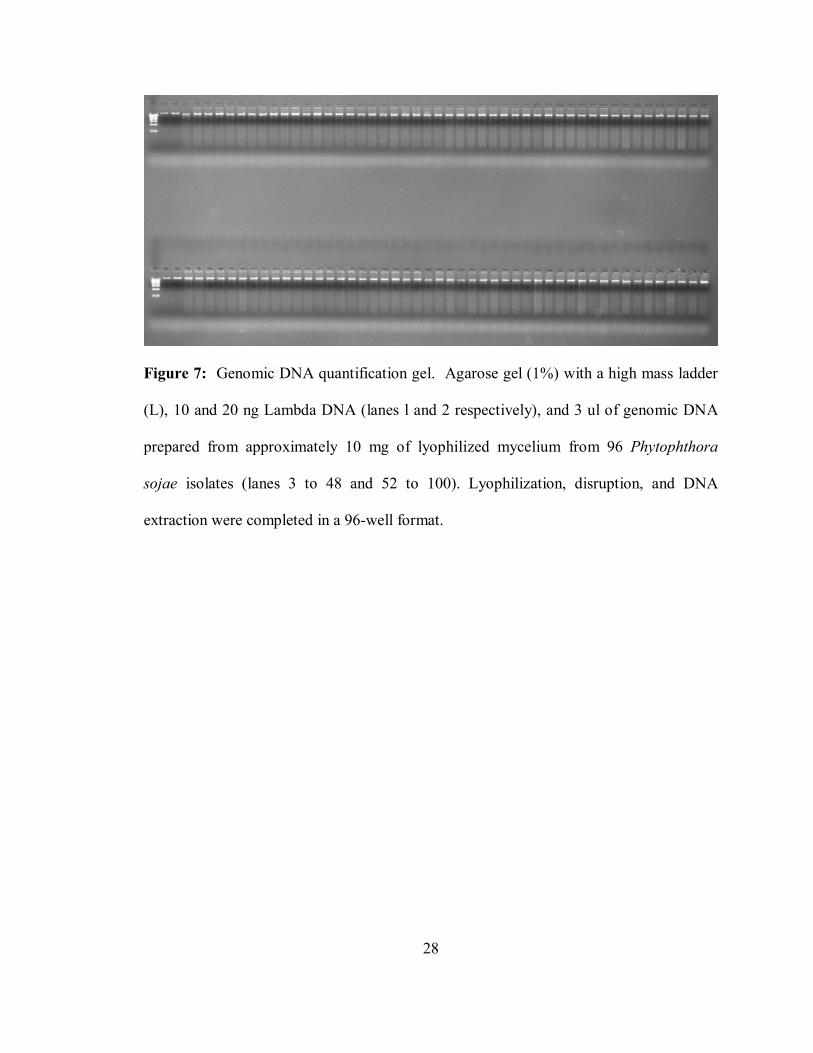
quality, high molecular weight DNA (Figure 7). Currently, there are approximately 3000
mutants and their respective genomic DNA’s available for screening.
B. Recovery of Gene Specific Mutants
Primers were optimized to amplify 1,000 nt portions of the gene for PsojNIP
(Phytophthora sojae Necrosis Inducing Protein) from the mutant genomic DNA. A total
of ten SNP’s have been observed (Figure 8) and sequenced (Figure 9). Two of these
SNP’s, in isolates NR108 and NR146, were found to be false positives. Three of the
SNP’s were found to cause silent mutations – NR562 (Tyrosine to Tyrosine) at position
658, E113 (Tyrosine to Tyrosine) at position 706, and K2302 (Glutamine to Glutamine)
at position 265. One SNP, in mutant K2141, was confirmed by sequencing but is 10bp
upstream of the coding region. The remaining four SNP’s were found to cause an amino
acid change. Mutant E16 had an induced change from the wildtype Valine (V) to
Glutamic Acid (E) at position 441. This is a drastic change, as V is small, hydrophobic
and aliphatic while E is larger, polar, and charged. Mutant E158 has Phenylalanine (F) in
place of the wildtype Serine (S) at position 720. The change from F to S is also likely to
28
Figure 7: Genomic DNA quantification gel. Agarose gel (1%) with a high mass ladder
(L), 10 and 20 ng Lambda DNA (lanes l and 2 respectively), and 3 ul of genomic DNA
prepared from approximately 10 mg of lyophilized mycelium from 96 Phytophthora
sojae isolates (lanes 3 to 48 and 52 to 100). Lyophilization, disruption, and DNA
extraction were completed in a 96-well format.

29
Figure 8: TILLING gel with observed single nucleotide polymorphism (snp). Presence
of novel fragments in both gel images in lane 32 adding up to the amplified fragment size
(~1000 bp) indicates snp. Top gel is the 700 gel image (fragment size ~260 bp) and
bottom gel is the 800 gel image (fragment size ~750 bp).

30
Figure 9: Induced polymorphism confirmed by sequencing. Arrow shows the clear
double peak at position 169 which indicates a single nucleotide polymorphism (SNP). In
this instance the SNP falls just upstream of the coding sequence and will not impact the
gene.

31
be significant – F is hydrophobic and has an aromatic side chain which, due to its
bulkiness, is likely to greatly disrupt protein structure when substituted for the tiny polar
sidechain of S. Mutant N37 has an induced change from the wildtype Valine (V) to
Isoleucine (I) at position 427. Again, V is small hydrophobic and aliphatic. I is similar
to V except that is larger. Although this change is more subtle than the previous two, the
size difference could still potentially affect protein folding and structure. Mutant K1857
was a change from wildtype Aspartic Acid (D) to Asparagine (N) at position 557. D is
negatively charged, small and polar. N is also small and polar, but it is uncharged. Due
to electrostatic forces, this change also has the potential to alter protein folding and
function.
From the CODDLE website (http://www.proweb.org/input/), predictions can be
made based on the mutation method as to what types of mutations will result in your
sequence of interest. When the PsojNIP sequence was entered, the following mutations
were predicted (Table 4). CODDLE also calculated that 57.3% of the changes would be
Table 4: CODDLE mutation predictions
Mutation Type Percentage of total mutations
A ↔ T Transversions 44.0%
A:T → G:C Transitions 38.0%
G:C → A:T Transitions 8.0%
A:T → C:G Transversions 5.0%
G ↔ C Transversions 3.0%
G:C → T:A Transversions 2.0%

32
nonsilent, with 1.8% of these polymorphisms being truncation (nonsense) changes and
55.5% being missense changes. These numbers correlate with the experimental data
described above
C. Oospore Progeny
The next step was to recover sexual progeny from the mutants that are
homothallic for allelic and knockout mutations. Phytophthora sojae is self-fertile and
makes sexual oospores in single culture. Unfortunately, the wildtype copy of isolate
P6497 that we obtained from another Phytophthora research group at the start of this
project does not produce normal oospores. This has made it very difficult to recover
sexual progeny from the mutants harboring amino acid changing mutations. We have
since procured another copy of P6497 that produces normal oospores from the long term
Phytophthora culture collection at the University of California, Riverside. Attempts are
being made to salvage the mutants isolated from the defective P. sojae isolates (E16 and
E158) isolated during the course of this work. Due to the very low number of normal
oospores produced by the mutant isolates described here and the need for extended
periods of dormancy (2 to 3 months) for oospore germination, we have only been able to
analyze six sexual progeny from the E16 mutant parent. Sequencing of the PsojNIP
gene for these six isolates did not reveal any carrying the homozygous mutant allele.
D. Conclusions
TILLING was successfully adapted to P. sojae and PsojNIP specific mutants
were recovered. Further research using a P. sojae strain that is more efficient at
producing oospores will be required to ascertain the heritability of the mutations and the
effects on pathogenicity of the mutants.

33
LITERATURE CITED

34
1. Alonso, J.M., Stepanova, A.N., Leisse, T.J., Kim, C.J., Chen, H., Shinn, P.,
Stevenson, D.K., Zimmerman, J., Barajas, P., Cheuk, R., Gadrinab, C., Heller, C.,
Jeske, A., Koesema, E., Meyers, C. C., Parker, H., Prednis, L., Ansari, Y., Choy,
N., Deen, H., Geralt, M., Hazari, N., Hom, E., Karnes, M., Mulholland, C.,
Ndubaku, R., Schmidt, I., Guzman, P., Aguilar-Henonin, L., Schmid, M., Weigel,
D., Carter, D.E., Marchand, T., Risseeuw, E., Brodgden, D., Zeko, A., Crosby,
W.L., Berry, C.C., and Ecker, J.R. 2003. Genome-Wide Insertional Mutagenesis
of Arabidopsis thaliana. Science 301:653-657.
2. Baldauf, S.L., Roger, A.J., Wenk-Siefert, I., and Doolittle, W.F. 2000. A
Kingdom-Level Phylogeny of Eukaryotes Based on Combined Protein Data.
Science 290:972-.
3. Bhat, R.G., and Schmitthenner, A.F. 1993. Selection and characterization of
inhibitor-resistant mutants of Phytophthora sojae. Experimental Mycology
17:109-121.
4. Coghill, E.L., Hugill, A., Parkinson, N., Davison, C., Glenister, P., Clements, S.,
Hunter, J., Cox, R.D., and Brown, S.D.M. 2002. A gene-driven approach to the
identification of ENU mutants in the mouse. Nature Genetics 30:255-256.
5. Colbert, T., Till, B.J., Tompa, R., Reynolds, S., Steine, M.N., Yeung, A.T.,
McCallum, C.M., Comai, L., and Henikoff, S. 2001. High-throughput screening
for induced point mutations. Plant Physiology 126:480-484.
6. Erwin, D.C., and Ribeiro, O.K. 1996. Phytophthora Diseases Worldwide: The
American Phytopathological Society.

35
7. Fang, G.-C., Hanau, R.M., and Vaillancourt, L.J. 2002. The SOD2 gene, encoding
a manganese-type superoxide dismutase, is up-regulated during conidiogenesis in
the plant-pathogenic fungus Colletotrichum graminicola. Fungal Genetics and
Biology 36:155-165.
8. Fire, A., XU, Siqun, Montgomery, M.K., Kostas, S.A., Driver, S.E., and Mello,
C.C. 1998. Potent and specific genetic interference by double-stranded RNA in
Caenorhabditis elegans. Nature 391:806-811.
9. Gilchrist, E.J., and Haughn, G.W. 2005. TILLING without a plough: a new
method with applications for reverse genetics. Current Opinion in Plant Biology
8:211-215.
10. Griffiths, A.J.F., Miller, J.H., Suzuki, D.T., Lewontin, R.C., and Gelbart, W.M.
2000. An Introduction to Genetic Analysis. 7 ed. New York: W.H. Freeman and
Company.
11. Guénet, J. 2004. Chemical Mutagenesis of the mouse genome: an overview.
Genetica 122:9-24.
12. Hayes, F. 2003. Transposon-Based Strategies for Microbial Functional Genomics
and Proteomics. Annual Review of Genetics 37:3-29.
13. Henikoff, S., Till, B.J., and Comai, L. 2004. TILLING. Traditional Mutagenesis
Meets Functional Genomics. Plant Physiology 135:1-7.
14. Henikoff, S. , and Comai, L. 2003. Single-Nucleotide Mutations for Plant
Functional Genomics. Annual Review of Plant Biology 54:375-401.
15. Huang, S., van der Vossen, E.A., Kuang, H., Vleeshouwers, V.G., Zhang, N.,
Borm, T.J., van Eck, H.J., Baker, B., Jacobson, E., and Visser, R.G. 2005.

36
Comparative genomics enabled the isolation of the R3a late blight resistance gene
in potato. Plant Journal 42 (2):251-261.
16. Huitema, E., Bos, J.I.B., Tian, M., Win, J., Waugh, M.E., and Kamoun, S. 2004.
Linking sequence to phenotype in Phytophthora-plant interactions. TRENDS in
Microbiology 12 (4):193-200.
17. Hurlstone, A.F.L., Haramis, A.G., Wienholds, E., Begthel, H., Korving, J., Eeden,
F. van, Cuppen, E., Zivkovic, D., Plasterk, R.H.A., and Clevers, H. 2003. The
Wnt/Beta-catenin pathway regulates cardiac valve formation. Nature 425:633-
637.
18. Iida, S. , and Terada, R. 2004. A tale of two integrations, transgene and T-DNA:
gene targeting by homologous recombination in rice. Current opinion in
Biotechnology 15:132-138.
19. Jackson, T.A., Kirpatrick, T.L., and Rupe, J.C. 2004. Races of Phytophthora
sojae in Arkansas Soybean Fields and Their Effects on Commonly Grown
Soybean Cultivars. Plant Disease:345-351.
20. Jansen, G., Hazendonk, E., Thijssen, K.L., and Plasterk, R.H. 1997. Reverse
genetics by chemical mutagenesis in Caenorhabditis elegans. Nature Genetics
17:119-121.
21. Justice, M.J., Noveroske, J.K., Weber, J.S., Zheng, B., and Bradley, A. 1999.
Mouse ENU Mutagenesis. Human Molecular Genetics 8 (10):1955-1963.
22. Kuttenkeuler, D., and Boutros, M. 2004. Genome-wide RNAi as a route to gene
function in Drosophila. Briefings in Functional Genomics and Proteomics 3
(2):168-176.

37
23. Lalucque, H., and Silar, P. 2004. Incomplete Penetrance and Variable
Expressivity of a Growth Defect as a Consequence of Knocking Out Two K+
Transporters in the Euascomycete Fungus Podospora anserina. Genetics
166:125-133.
24. Lamour, K.H., and Hausbeck, M.K. 2000. Mefenoxam Insensitivity and the
Sexual Stage of Phytophthora capsici in Michigan Cucurbit Fields.
Phytopathology 90:396-400.
25. Leitz, R.A., Hartman, G.L., Pedersen, W.L., and Nickell, C.D. 2000. Races of
Phytophthora sojae on Soybean in Illinois. Plant Disease (84):487.
26. Lewin, B. 2004. Genes VII. Upper Saddle River, NJ: Pearson Prentice Hall.
27. MacGregor, T., Bhattacharyya, M., Tyler, B.M., Bhat, R.G., Schmitthenner, A.F.,
and Gijzen, M. 2002. Genetic and physical mapping of Avr1a in Phytophthora
sojae. Genetics 160:949-959.
28. Martin, F.N., and Tooley, P.W. 2003. Phylogenetic relationships of Phytophthora
ramorum, P. nemorosa, and P. pseudosyringae, three species recovered from
areas in California with sudden oak death. Mycological Research 107 (12):1379-
1391.
29. May, K.J., and Ristaino, J.B. 2004. Identity of the mtDNA haplotype(s) of
Phytophthora infestans in historical specimens from the Irish potato famine.
Mycological Research 108:1-9.
30. McCallum, C.M., Comai, L., Greene, E.A., and Henikoff, S. 2000. Targeting
Induced Local Lesions IN Genomes (TILLING) for Plant Functional Genomics.
Plant Physiology 123:439-442.

38
31. McCallum, C.M., Comai, L., Greene, E.A., and Henikoff, S. 2000. Targeted
Screening for Induced Mutations. Nature Biotechnology 18:455-457.
32. Moy, P., Qutob, D., Chapman, B.P., Atkinson, I., and Gijzen, M. 2004. Patterns
of Gene Expression Upon Infection of Soybean Plants by Phytophthora sojae.
Molecular Plant-Microbe Interactions 17 (10):1051-1062.
33. Mullis, K.B., and Faloona, F.A. 1987. Specific synthesis of DNA in vitro via a
polymerase-catalyzed chain reaction. Methods in Enzymology 155:335-350.
34. Oleykowski, C.A., Mullins, C.R.B., Godwin, A.K., and Yeung, A.T. 1998.
Mutation detection using a novel plant endonuclease. Nucleic Acids Research
26:4597-4602.
35. Pattanayak, D., Agarwal, S., Sumathi, S., Chakrabarti, S.K., Naik, P.S., and
Khurana, S.M. 2005. Small but mighty RNA-mediated interference in plants.
Indian Journal of Experimental Biology 43 (1):7-24.
36. Perry, J.A., Wang, T.L., Welham, T.J., Gardner, S., Pike, J.M., Yoshida, S., and
Parniske, M. 2003. A TILLILNG Reverse Genetics Tool and a Web-Accessible
Collection of Mutants of the Legume Lotus japonicus. Plant Physiology 131:866-
871.
37. Qutob, D., Kamoun, S., and Gijzen, M. 2002. Expression of a Phytophthora sojae
necrosis-inducing protein occurs during transition from biotrophy to necrotrophy.
The Plant Journal 32:361-373.
38. Qutob, D., Hraber, P.T., Sobral, B.W.S., and Gijzen, M. 2000. Comparative
Analysis of Expressed Sequences in Phytophthora sojae. Plant Physiology
123:243-253.

39
39. Ristaino, J.B. 2002. Tracking historic migrations of the Irish potato famine
pathogen, Phytophthora infestans. Microbes and Infection 4 (13):1369-1377.
40. Rizzo, D.M., Garbelotto, M., and Hansen, E.M. 2005. PHYTOPHTHORA
RAMORUM: Integrative Research and Management of an Emerging Pathogen in
California and Oregon Forests. Annual Review of Phytopathology 43 (1):309-
335.
41. Saiki, R.K., Scharf, S., Faloona, F., Mullis, K.B., Horn, G.T., Erlich, H.A., and
Arnheim, N. 1985. Enzymatic Amplification of Beta-Globin Genomic Sequences
and Restriction Site Analysis for Diagnosis of Sickle Cell Anemia. Science
230:1350-1354.
42. Sanger, F., Nicklen, S., and Coulson, A.R. 1977. DNA Sequencing with chain-
terminating inhibitors. Proceedings of the National Academy of Science 74
(12):5463-5467.
43. Sciences, National Institute of Environmental Health. 2002. Ethyl
methanesulfonate. In Report on carcinogens.
44. Scott-Craig, J.S., Cheng, Y., Cervone, F., Lorenzo, G. De, Pitkin, J.W., and
Walton, J.D. 1998. Targeted Mutants of Cochliobolus carbonum Lacking the Two
Major Extracellular Polygalacturonases. Applied and Environmental
Microbiology:1497-1503.
45. Sega, G.A. 1984. A review of the genetic effects of ethyl methanesulfonate.
Mutation Research 134:113-142.
46. Smits, B.M.G. , Mudde, J., Plasterk, R.H.A., and Cuppen, E. 2004. Target-
selected mutagenesis of the rat. Genomics 83:332-334.

40
47. Stemple, D.L. 2004. TILLING - a high-throughput harvest for functional
genomics. Nature Reviews Genetics 5:145-150.
48. Takano, Y., Kikuchi, T., Kubo, Y., Hamer, J.E., Mise, K., and Furusawa, I. 2000.
The Colletotrichum lagenarium MAP Kinase Gene CMK1 Regulates Diverse
Aspects of Fungal Pathogenesis. Molecular Plant-Microbe Interactions 13
(4):374-383.
49. Till, B.J., Burtner, C., Comai, L., and Henikoff, S. 2004. Mismatch cleavage by
single-strand specific nucleases. Nucleic Acids Research 32 (8):2632-2641.
50. Till, B.J., Reynolds, S.H., Greene, E.A., Codomo, C.A., Enns, L.C., Johnson, J.E.,
Burtner, C., Odden, A.R., Young, K., Taylor, N.E., Henikoff, J.G., Comai, L., and
Henikoff, S. 2003. Large-Scale Discovery of Induced Point Mutations With High-
Througput TILLING. Genome Research 13:524-530.
51. Till, B.J., Reynolds, S.H., Weil, C., Springer, N., Burtner, C., Young, K., Bowers,
E., Codomo, C.A., Enns, L.C., Odden, A.R., Greene, E.A., Comai, L., and
Henikoff, S. 2004. Discovery of induced point mutations in maize genes by
TILLING. BMC Plant Biology 4 (12).
52. Torto, T.A., Shuang, L., Styer, A., Huitema, E., Testa, A., Gow, N.A.R., van
West, P., and Kamoun, S. 2003. EST Mining and Functional Expression Assays
Identify Extracellular Effector Proteins From the Plant Pathogen Phytophthora.
Genome Research 13:1675-1685.
53. Tyler, B.M. 2002. Molecular Basis of Recognition Between Phytophthora
Pathogens and Their Hosts. Annual Review of Phytopathology 40:137-167.

41
54. Tyler, B.M., Forster, H., and Coffey, M.D. 1995. Inheritance of avirulence factors
and restriction fragment length polymorphism markers in outcrosses of the
oomycete Phytophthora sojae. Molecular Plant-Microbe Interactions 8:515-523.
55. Waterhouse, P.M., Graham, M.W., and Wang, M. 1998. Virus resistance and gene
silencing in plants can be induced by simultaneous expression of sense and
antisense RNA. Proceedings of the National Academy of Science 95:13959-
13964.
56. Watson, J.D., and Crick, F.H.C. 1953. A Structure for Deoxyribose Nucleic Acid.
Nature 171:737-738.
57. Whissen, S.C., Drenth, A., Maclean, D.J., and Irwin, J.A.G. 1994. Phytophthora
sojae avirulence genes, RAPD, and RFLP markers used to construct a detailed
genetic linkage map. Molecular Plant-Microbe Interactions 8:988-995.
58. Wienholds, E., Schulte-Merker, S., Walderich, B., and Plasterk, R.H. 2002.
Target-Selected Inactivation of the Zebrafish rag1 Gene. Science 297:99-102.
59. Wienholds, E., Eeden, F. van, Kosters, M., Mudde, J., Plasterk, R.H.A., and
Cuppen, E. 2003. Efficient Target-Selected Mutagenesis in Zebrafish. Genome
Research 13:2700-2707.
60. Zhang, A., Lu, P., Dahl-Roshak, A.M., Paress, P.S., Kennedy, S., Tkacz, J.S., and
An, Z. 2003. Efficient disruption of a polyketide synthase gene (pks1) required for
melanin synthesis through Agrobacterium-mediated transformation of Glarea
lozoyensis. Molecular Genetics and Genomics 268:645-655.

42
APPENDICES

43
APPENDIX I: SOLUTIONS FOR DNA EXTRACTION
AP1 = Lysis Buffer 100 mM Tris (pH 8.0) 100 ml 1M Tris stock 50 mM EDTA 100 ml 0.5M EDTA stock 500 mM NaCl 29.2 g NaCl 1.33% SDS 13.5 g SDS Start with about 500 ml MilliQ water in a 1 liter beaker with a stir bar. Add the liquid stock solutions first. Add the NaCl and SDS (wear a face mask when weighing SDS). Stir until in solution and bring final volume to 1 liter. Store in a 1 liter glass bottle and do not autoclave. AP2 = Potassium Acetate (pH 8.0) Make a 5M solution of KOAc. Weigh out 245.4 g potassium acetate and add to 200 ml of MilliQ water stirring in a beaker gradually. Add water to bring volume to 500 ml. Using the pH meter, add glacial acetic acid to bring pH to 8.0. Filter sterilize. AP3 = Guanidine Hydrochloride and Alcohol Make a 5 M stock of Guanidine Hydrochloride (GH). Dissolve 238.8 g of GH in MilliQ Water (start with small amount, 200 ml). Bring final volume to 500 ml. Filter sterilize. Use 125 ml of 5M GH to add to 250 ml 95% EtOH. Keep stock solution of 5M GH for use later. AW = Tris, EDTA, NaCl stock (AW salt stock). 33 mM Tris (pH 8.0) 16.5 ml 1 M Tris 3.3 mM EDTA 3.3 ml EDTA 165 mM NaCl 4.82 g NaCl Dissolve in 200 ml MilliQ water and bring final volume to 500 ml. Filter sterilize and keep as salt stock for AW. Mix 300 ml of the salt stock with 700 ml EtOH to make home-made AW. AE = 10 mM Tris (pH 8.0) To make 1 liter, add 10 ml 1 M Tris to 990 ml MilliQ water. Autoclave and aliquot into sterile 15 ml or 50 ml tubes in the sterile hood.

44
APPENDIX 2: PLATE DNA EXTRACTION PROTOCOL
1. □ There are pre-made bottles of AP1. Use one bottle per 2 plate extraction. Heat
the bottle in the 65º oven until the chunks are gone. Heat tube of Fighter F at same time.
2. □ While heating - bash the samples for 30 seconds on each side on a setting of 30. Make sure to rotate the plates and do each side at this setting. After the first bash – check to make sure there is powder in each well. If no powder – then there is probably no beads so you’ll need to add 2-4 beads to the well. Spin the plate for five minutes and carefully remove cap and put it in the sink.
3. □ Add 800µl of Fighter F and 200µl of RNase to the preheated AP1. Don’t fret if the fighter F is finicky – just get as much as you can in there. Shake the tubes vigorously.
4. □ Add 500µl of the AP1 mix. Put a cap on, and gently shake the plates until all the chunks of mycelium are in solution.
5. □ Incubate the plate in the 65º oven for 30 minutes. Make sure the cap is sealed (it gets loose during the incubation). Spin for 1 minute at max speed and take the cap off (never reuse caps!).
6. □ Add 200ul AP2 Buffer using the 8-channel pipette (there are pre-made tubes with enough for 2 plates) to the plates, add a new cap, and shake the plate vigorously to mix the solutions for 15 seconds. Put in the -20C freezer for 20 minutes or leave in the -20C overnight.
AFTER FREEZING:
7. □ Spin plates for 10 minutes at max speed. If you left the plates in the -20°
freezer overnight, let them stand at RT for a few minutes before spinning the plates. If you spin them frozen, nothing will happen!
8. □ While the plates are spinning add 800µl of AP3 Solution (there are pre-made tubes of AP3 for 2 plates) to properly labeled 2ml plates that will hold the AP3+AP2 mixture. Use the 8 channel pipette.
9. □ Transfer 500µl of the supernatant from the spun plate to the correctly oriented/labeled deepwell plate from step 8. The goal is to get only supernatant. There is a mark on the Apricot in rm 403 – use this. If you look very closely you can see where the tips of the multichannel are in the plate. Try to get them just above the precipitated sludge. Have Kurt help you do this the first time.
10. □ Put a new cap on the plate, shake the plate vigorously for 15 seconds, and spin the plate for 1 minute at 5220 RPM.
11. □ Put a properly labeled/oriented glass filter plate on a clean 2 ml deepwell plate.
Be sure to label the 2ml plate properly. 12. □ Transfer 1300µl (two 650 µl aliquots using the 8 channel) to the glass filter
plate. 13. □ Spin at 5220 RPMs for 5 minutes. Discard flow through.

45
14. □ Add 800µl of Buffer AW. There is premade AW in tubes for 2 plates. Cover with a rayon breathable tape. Spin for 5 minutes. Discard flow through.
15. □ Add 400µl 95% ethanol. Cover with a rayon breathable tape. Spin for 5 minutes. Discard flow through.
16. □ Put AE buffer (to preheat) into the 65º oven and ALSO put the spin plate (alone, without the 2 ml deepwell plate and without the rayon tape) into the 65º oven to dry for 15 minutes. There is prepared AE in tubes for 2 plates.
17. □ Place the dried glass fiber plate onto a new (never used) properly labeled and oriented 1ml deepwell plate.
18. □ Put 200µl of preheated AE buffer into the plate. Let it sit 30 minutes at room temperature.
19. □ Spin for 2 minutes at 5220 RPM. 20. □ Run 3µl DNA from Rows B and G on a 1% agarose gel. (3 ul DNA + 3 ul Tris
+ 2 ul dye). 21. □ Seal the plate with the thick tinfoil tape (expensive tape). Store the labeled
DNA plate in the refrigerator and record on the log that the DNA has been extracted.

46
APPENDIX 3: PRIMER COCTAIL RECIPE
To make primer cocktail, mix following in a tube and aliquot into small volumes (enough for 2 gels; about 10µl per tube works well) All primers at 100 µm concentration 24 µl L700 blue primer 16 µl L unlabeled primer 32 µl R800 green primer 8 µl R unlabeled primer

47
APPENDIX 4: SEPHADEX PREPARATION
Weigh out 11 grams of Sephadex and mix in 160 mL MilliQ water in glass bottle
Autoclave 15 minutes.
This makes approximately enough for 2 plates.

48
VITA
Melinda Tierney was born Melinda Beth Lawrence on December 29, 1980 in
Rolla, MO. She was raised in Joplin, MO and attended Royal Heights Elementary
School, North Middle School, and Joplin Junior High. She graduated from Joplin High
School in 1999. From there, she went to the University of Missouri-Rolla and met and
married John Tierney. She graduated from UMR in December of 2003 with a B.S. in
Biological Sciences and minors in Chemistry and Psychology. Melinda was then
accepted to the Genome Science and Technology program with the University of
Tennessee and Oak Ridge National Lab and moved to Knoxville, TN to pursue graduate
work. In December 2005, Melinda earned a Master of Science degree in Life Sciences.