review article biologic complexity in sickle cell...
TRANSCRIPT

Hindawi Publishing CorporationThe Scientific World JournalVolume 2013, Article ID 694146, 12 pageshttp://dx.doi.org/10.1155/2013/694146
Review ArticleBiologic Complexity in Sickle Cell Disease: Implications forDeveloping Targeted Therapeutics
Beatrice E. Gee
Department of Pediatrics, Cardiovascular Research Institute, Morehouse School of Medicine, 720 Westview Drive SW,Atlanta, GA 30310-1495, USA
Correspondence should be addressed to Beatrice E. Gee; [email protected]
Received 29 December 2012; Accepted 29 January 2013
Academic Editors: Y. Al-Tonbary, M. A. Badr, A. Mansour, and F. Tricta
Copyright © 2013 Beatrice E. Gee. This is an open access article distributed under the Creative Commons Attribution License,which permits unrestricted use, distribution, and reproduction in any medium, provided the original work is properly cited.
Current therapy for sickle cell disease (SCD) is limited to supportive treatment of complications, red blood cell transfusions,hydroxyurea, and stem cell transplantation. Difficulty in the translation of mechanistically based therapies may be the result ofa reductionist approach focused on individual pathways, without having demonstrated their relative contribution to SCD com-plications. Many pathophysiologic processes in SCD are likely to interact simultaneously to contribute to acute vaso-occlusion orchronic vasculopathy. Applying concepts of systems biology and network medicine, models were developed to show relationshipsbetween the primary defect of sickle hemoglobin (Hb S) polymerization and the outcomes of acute pain and chronic vasculopathy.Pathophysiologic processes such as inflammation and oxidative stress are downstream by-products of Hb S polymerization,transduced through secondary pathways of hemolysis and vaso-occlusion. Pain, a common clinical trials endpoint, is also complexand may be influenced by factors outside of sickle cell polymerization and vascular occlusion. Future sickle cell research needs tobetter address the biologic complexity of both sickle cell disease and pain.The relevance of individual pathways to important sicklecell outcomes needs to be demonstrated in vivo before investing in expensive and labor-intensive clinical trials.
1. Introduction
Sickle cell disease (SCD) is a group of disorders caused bya mutation in the sequence of beta globin, leading to poly-merized hemoglobin (sickle hemoglobin, hemoglobin S),hemolytic anemia, painful vaso-occlusive events, vascularremodeling, acute and chronic organ injury, and shortenedlifespan. Sickle cell disease affects over 70,000 individuals intheUnited States, and there are at least 75,000 hospitalizationscosting over $500 million annually for treatment of SCDcomplications [1]. While survival has greatly improved, theaverage lifespan for people with hemoglobin SS was esti-mated in 1994 to be in the midforties [2], significantly lessthan the average American. Despite well-described geneticand biochemical properties of sickle hemoglobin and manybasic science discoveries about sickle cell pathophysiology,modern-day therapy continues to be limited to symptomatictreatment of pain, oxygen supplementation, antibiotics, redblood cell transfusions, and hydroxyurea. Hydroxyurea is anagent that induces fetal hemoglobin production and is the
only drug approved for adults by the United States Foodand Drug Administration that directly affects sickle cell out-comes. Stem cell transplantation from a histocompatibledonor has a high cure rate, but many patients do not have asuitable donor.
Since the passing of the National Sickle Cell Control Actin 1972, over one billion dollars have been allocated fromthe National Heart, Lung and Blood Institute of the NationalInstitutes of Health (NIH) for SCD research [3].This fundinghas resulted in a significant body of research on SCD. TheUnited States National Library of Medicine website lists over7000 articles since 1950 meeting the search terms of “sicklecell research;” 482 are human clinical trials. As of December1, 2012, the website http://www.clinicaltrials.gov/ showed 96open intervention trials in sickle cell disease. Table 1 showsthe most common types of studies. Some of these emergingtherapies have been recently reviewed [4].
Director of the Division of Blood Diseases and Resourcesat the NIH, W. Keith Hoots, recently wrote, “Research overthe decades indicates that the primary defect in hemoglobin

2 The Scientific World Journal
that results in polymerization of the protein under lowoxygen conditions and resultant cellular deformity of thered blood cell initiates a complex downstream pathogenesisassociated with vascular injury, and organ ischemia. Deci-phering this in a manner that informs successful therapiesthat improve all target organs continues to challenge hema-tologists” [5]. It is likely that this complexity is a barrier tosuccessful translation of basic science discoveries in SCDto effective therapeutics. Sickle hemoglobin polymerizationis associated with many abnormal downstream processes,but no single pathway has been shown to play a primary orcritical role in complications occurring in people with SCD.Most mechanistically based clinical interventions have beendesigned to target individual pathways, but there is likelyto be ongoing interaction within the body between differentprocesses, so that even if one pathway is successfully blocked,others may still be active and continue to promote vaso-occlusion or other complications. To address biologic com-plexity in SCD, this paper will analyze examples of promisingclinical trials that did not yield expected benefits, contrastreductionism with systems biology, present models thatfacilitate the visualization of interactions of mechanisms inSCD complications, and then discuss implications for futureresearch.
2. Unexpected Outcomes of PromisingClinical Trials
Preclinical studies and clinical trials targeting three differentsickle cell pathways will be reviewed, including inhibition ofadhesion by poloxamer 188, inhibition of Gardos channel-induced erythrocyte dehydration by senicapoc (ICA-17043),and treatment of acute pain episodes with inhaled nitricoxide. Comprehensive reviews of approaches to sickle celltreatment have been published elsewhere [4, 6, 7].
2.1. AntiadhesionTherapy with Poloxamer 188. Fluorocarbonemulsions, including identical but variously named com-pounds Pluronic F-68, Flocor, RheoThRx, and poloxamer188, have been studied in SCD since 1975 [8]. Pluronic F-68 was demonstrated to reduce in vitro sickle red cell staticrigidity (stiffness), filterability through a 5 micron filter, andabolish adherence to endothelialmonolayers [9]. It is believedto bind nonspecifically to the red cell membrane, “lubricat-ing” the cells and “providing a hydrated, poorly compressiblebarrier that appears to block hydrophobic adhesive inter-actions (cell-cell, cell-protein, and protein-protein) in theblood [10].” Preclinical studies demonstrated “a reduction inblood viscosity, erythrocyte aggregation, adhesion to vascularendothelium, and an improvement in microvascular bloodflow.”
A phase II randomized, double-blinded placebo-control-led trial (RCT) testing poloxamer 188 was conducted withfifty subjects with SCD who presented within 4–18 hours ofonset of acute pain. They were treated with either placeboor poloxamer 188 infusion of 300mg/kg for 60 minutes,followed by 47-hour maintenance infusion of 30mg/kg/hr.In the 31 subjects who completed the 48 hour infusion, there
Table 1: Major types of sickle cell intervention studies registered onthe website http://www.clinicaltrials.gov/, as of December 1, 2012, ofa total of 96 trials.
Pathway/mechanism Number of studiesBone marrow transplantation 21Hemoglobin F induction 9Nitric oxide related 8Analgesic regimens 6Nutritional supplements 4Adhesion inhibition 3Transfusion therapy 3Red cell hydration 3Noninvasive ventilation 3Statins 2Renin-angiotensin pathway in nephropathy 2Iron chelation 2Educational tools 2Antiinflammation 2Gene transfer 1Carbon monoxide donation 1Anti-coagulation 1
was a statistically significant reduction in pain duration by 36hours and a 3–5-fold reduction in analgesic use.
This was followed by a phase III RCT using poloxamer188, with 255 individuals enrolled at 40 study sites [11]. Inthe poloxamer 188-treated group, there was a “modest” 9-hour reduction in pain duration, with more pronouncedeffects in children under 15 years old (21-hour reduction) andsubjects receiving hydroxyurea (16-hour reduction). Therewere no significant differences between the poloxamer 188-versus placebo-treated groups in time to discharge, painseverity ratings, or total analgesic use. Pharmacokineticsshowed mean steady state drug concentration “within thetherapeutic range of rheological and antiadhesive effects.”The differences in responses between the phase II and IIItrials were attributed to the more stringent definition of painduration used in the Phase III trial, and the authors concludedthat poloxamer 188 may most benefit children with SCDunder 15 years of age or those being treatedwith hydroxyurea.
Close comparison shows differences between the PhaseII and III trials. There was a shorter duration of pain priorto beginning study drug in the Phase II trial. The inclusioncriteria for the Phase II trial included 4–18 hours of moderatepain on presentation, and there was a median of 17 hoursbetween onset of pain and start of study infusion. In contrast,subjects in the phase III trial had 1.87–2.25 days of pain fromonset of crisis to randomization, with an additional 2.3 hoursbetween randomization to start of study infusion, or almost3 times longer pain duration before study drug infusion wasbegun. In addition, the loading dose of poloxamer 188 was 3-fold higher in the phase II trial compared to phase III. Lastly,the phase II trial allowed nonsteroidal anti-inflammatorydrugs (NSAIDs) for analgesia, but they were not allowedduring the study drug infusion and for 12 hours following
The Scientific World Journal 3
discontinuation in the phase III trial. The absence of opioid-sparing effect of NSAIDs may have contributed to the lack ofdifference in opioid usage.
2.2. Gardos Channel Inhibition. Erythrocyte water content isan important determinant of sickle hemoglobin concentra-tion and polymer formation. Erythrocyte hydration statusis controlled by KCl and water loss through two transportsystems, K-Cl cotransport and the calcium-dependent potas-sium, or Gardos channel. Gardos channel inhibition in sicklecells was found to improve erythrocyte hydration status [12].The orally available antifungal agent, clotrimazole, couldcompletely inhibit deoxygenation-induced Gardos channel-mediated K+ loss. Oral clotrimazole was found to reduceK+ loss and improve erythrocyte volume in transgenic miceexpressing hemoglobin S, Antilles, andD Punjab (SADmice)[13] and in a phase I-II trial in five adults with hemoglobin SS[14]. However, clotrimazole as a potential red cell hydratingagent was felt to be limited by its poor oral absorption,short half-life, and the development of elevated hepatictransaminases at high doses.
Based on the structure of clotrimazole, alternative agentswere developed to more potently and specifically inhibitthe erythrocyte Gardos channel. Senicapoc (ICA-17043) wasfound to inhibit SADmouse red cell dehydration in vitro andin vivo and an increase in hematocrit of 7% [15]. A phaseII RCT in humans showed favorable hematologic responsesto senicapoc 10mg daily compared to placebo, with 0.68gm/dL increase in hemoglobin concentration and reductionin dense erythrocytes, reticulocytes, lactose dehydrogenase(LDH), and indirect bilirubin [16].
This was followed by a phase III RCT with 297 adultsubjects randomized at 75 study centers to receive senicapocfor 52 weeks [17]. The dose was 20mg twice daily for fourdays, followed by 10mg daily. The primary endpoint wasthe frequency of sickle cell-related painful crises requiringmedical facility treatment. The study was discontinued earlydue to lack of efficacy. Despite improvement in hematologicparameters, there was no difference in painful crisis ratesbetween subjects receiving senicapoc compared to placebo.The authors proposed that a possible reason for the discrep-ancy between hematologic responses and painful crisis rateswas that reduction in hemolysis rate increases bioavailabilityof NO, which in turn enhances nocioceptive pain signaling.
Of note, the Gardos channel, also known as the inter-mediate conductance Ca2+-activated K+ channel, KCa3.1,KCNN4, or SK4, is expressed in other cell types, suchas T- and B-lymphocytes, macrophages, endothelial cells,fibroblasts, vascular smooth muscle cells, and neurons. Inblood vessels, the KCa3.1 channel plays a role in endothelium-derived hyperpolarizing factor- (EDHF-) induced vasodila-tion, whichmay play amajor role in themicrocirculation [18].Chemical inhibition of the channel or KCNN4−/− knockoutin mice resulted in hyperresponsiveness to stress due toenhanced adrenocorticotropic hormone (ACTH) secretionby the anterior pituitary [19].The KCa3.1 channel has approx-imately 50% homology with KCa2 family channels, which aresmall conductance channels located in neurons and involvedin afterhyperpolarization. These channels are expressed in
high concentrations in the brain and are critical for learningandmemory [20].Therefore, it is possible that the unexpectedresults of the senicapoc trial may be related to the drug’s effecton cell types other than sickle erythrocytes, particularly onEDHF-dependent vasodilation or in the nervous system.
2.3. Inhaled Nitric Oxide for Acute Painful Episodes. Nitricoxide (NO) is an important vasoactive molecule, witheffects on vascular smooth muscle dilation and modulatingleukocyte and platelet activation. The use of nitric oxide intreatment sickle cell disease acute chest syndrome (ACS) wasfirst reported in 1997 [21], followed by additional criticalcare medicine reports of clinical benefit in ACS, stroke, andmultiorgan failure syndrome [22–24]. Cell-free hemoglobinhas been found to reduce NO concentrations [25]. Individ-uals with hemoglobin SS had 20-fold higher plasma hemeconcentrations (4.2𝜇M), and NO consumption was greaterin sickle cell plasma and linearly correlatedwith plasma hemeconcentration. Forearm blood flowwas lower in patients withhemoglobin SS, which improved after infusion of sodiumnitroprusside (SNP), a NO donor. Giving inhaled NO at80 ppm reduced NO consumption of sickle cell plasmaby oxidizing and nitrosylating hemoglobin. Inhaled NO at80 ppm was found to increase skin oxygenation in peoplewith SCD but had no effect on forearm blood flow [26].
In SAD mice, inhaled NO at 20 ppm improved survivalrates during exposure to hypoxia when it was given 30minutes prior to hypoxic exposure and continued during theentire exposure [27]. However, there was no benefit if a lowerconcentration of NO was used, or if it was given only beforeor only during hypoxia. The benefits of NO preinhalationand treatment during hypoxia-induced vaso-occlusion wereproposed to include reduction of platelet and erythrocyteadhesion, but these mechanisms were not tested.
There have been three published studies of inhaled NOtherapy for acute pain episodes in SCD. In the first study,20 pediatric subjects with SCD were enrolled, 10 who weretreated with inhaled NO 80 ppm for 4 hours and 10 withplacebo [28]. The group who received inhaled NO had lowerpain ratings and less morphine used at 6 hours, but nosignificant difference in duration of hospitalization. In thesecond study, nine adults with SCDwere treated with inhaledNO80 ppm for 4 hours and ninewith placebo [29].The groupwho received inhaled NO had lower pain ratings at six hours,but no difference inmorphine utilization. Amulticenter RCTof inhaled NO was conducted with 150 children and adultsage 10 years and older with SCD presenting with acute paincrisis [30]. Inhaled NO was given for up to 72 hours, withan initial concentration of 80 ppm for the first four hours,40 ppm for the next four hours, and then pulsed delivery at5 ppm for the remainder of the 72 hour period. There was nodifference between groups for the primary outcomemeasure,time to resolution of pain, or duration of hospitalization, painscores at 24 hours, or total opioid use.
3. Lost in Translation?
With the strength of the preclinical findings, why were thesetherapeutic strategies not effective in reducing the impact
4 The Scientific World Journal
of acute pain, the most common complication of sickle celldisease? Sickle cell clinical research probably suffers from thesame challenges as other clinical trials in the United States.In 2012, the Institute of Medicine published a report thatdiscusses the major challenges with current clinical trials,including high costs due to elaborate administrative proce-dures, failure to enroll sufficient numbers of participants,regulatory issues, and failure to publish negative results [31].For sickle cell disease research, it has been recommendedthat clinical trials design be improved, especially to ensuresufficient enrollment, redefine study endpoints, and accountfor different clinical subphenotypes [4].
In the studies reviewed in this paper, the outcomemeasures for acute pain were different in many of the studiesconducted. The need to use multiple study sites to accom-modate the number of subjects necessary for large clinicaltrials makes consistency of study treatments and outcomemeasures extremely important. For example, in the phase IIIinhaledNORCT, therewere institutional differences betweenstudy sites, such that two sites had significantly differentoutcomes than the others. In the poloxamer 188 studies, therewere several methodologic differences between the phase IIand III trials which may account for the disparity in efficacyresults.
3.1. Lack of Models for Acute Pain in Sickle Cell Disease.Beyond challenges inherent to clinical trials, there are somerecurring themes in these studies. First, preclinical studieswere surrogates, but not sufficiently good model systems,for actual pain episodes. In general, preclinical in vivo evi-dence of the relevance of individual pathways on importantsickle cell outcomes has been lacking. For antiadhesiontherapy with poloxamer 188, the general “masking” effectwas a good approach to address the redundancy of multiplered cell, leukocyte, platelet, and endothelial cell adhesionmolecules, but the nearly complete endothelial adhesionblockade demonstrated in vitro had not been reported inpublished animal studies prior to human clinical trials. In thecase of Gardos channel inihibition, there was strong evidencefor improvement in red cell volume, reduced hemolysis, andanemia, but no testing in a model mimicking painful crisisprior to the phase III clinical trial. Preclinical data forinhaled NO showed beneficial effects on survival in hypoxicconditions in transgenic sickle cell mice, but it was onlyhelpful when given before hypoxia was initiated. InhaledNO had no effect on forearm blood flow, but somewhatimproved skin hemoglobin oxygen saturation. In healthyadults, it has been shown that inhaled NO 40 ppm correctshypoxia-induced pulmonary hypertension without an effecton systemic vasodilation [32]. While it would be predictedthat inhaled NOwould promote vasodilation, reduce inflam-mation, and improve red cell hydration, none of theseoutcomes were reported in the three inhaled NO clinicaltrials, making it difficult to ascertain whether there was anyimprovement to these proposed mechanisms resulting fromtreatment.
At the heart of the matter, it remains an article of faiththat sickle cell polymerization and microvascular occlusionis the actual cause of acute pain in SCD. There is currently
no way to visualize vaso-occlusion to corroborate that sitesof sickle erythrocyte microvascular occlusion correspond tolocations where patients feel pain or to show that therapiesthat restore or maintain normal blood flow will relieve vaso-occlusion and pain. Intravital microscopy of various vascularbeds in rodents has been used to study agents that blockadhesion of sickle erythrocytes and leukocytes, includingex vivo rat mesenteric vessels, transgenic mouse cremaster,and brain window [33–35]. While vascular occlusions can bevisualized, animals are anesthetized during the proceduresand cannot be evaluated for pain. In humans, conjunctivaland skin blood flow have been shown to be abnormal inpeople with SCD [26, 36], but these are not typically sites ofpain, and blood flow has not been specifically measured atthese sites during acute pain episodes. Bulbar conjunctivalblood flow was found to improve after erythrocytapheresis[37] and in a small number of subjects treatedwith poloxamer188 [38].
3.2. DrugDelivery and Timing. Even if a therapy is effective inthe person with SCD, there may be difficulty in maintainingeffective pharmacokinetics, timing of therapy, and/or drugdelivery to the site of vaso-occlusion. A treatment may workwell at certain concentrations in vitro or in transgenic mice,but these same concentrations may not be achievable in thesetting of a person with a larger volume of distribution forthe necessary duration of the acute painful crisis. In earlynitric oxide inhalation studies, acute treatment with inhaledNO 80 ppm was for 4 hours, and followup was for 4–24hours. It is possible that these time courses were too shortto be effective, since vaso-occlusion commonly does notresolve in 6 hours. In the case of established tissue injuryfrom ischemia, pain may not necessarily resolve within aday. In the larger randomized controlled trial of inhaled NO,the equivalent of NO 5 ppm was given for up to 72 hours,an overall lower concentration, not previously reported tohave an effect on vasodilation or inflammatory markers, andthe authors state that this lower rate of administration mayhave been insufficient to generate systemic nitrite, an NOmetabolite which may mediate its tissue effects.
As in stroke therapy with neuroprotectants and throm-bolytics [39], timing of therapy in acute painful crisis maybe an important factor in effectiveness. Sickle cell pain maybe similar to myocardial and cerebral infarction, in that thebenefits of therapies to reverse vessel occlusion and restoreperfusion need to be initiated very early in the ischemicprocess to be effective, and otherwise the ischemic injuryis established and cannot be readily reversed. We currentlyhave no way of knowing whether there is still ongoing vaso-occlusion when a person with SCD presents for treatmentof pain, if the pain is instead associated with reperfusion ordue to persistent transmission from afferent sensory neuronsthat have been activated in ischemic tissues. If pain is apostocclusive event, then treatments that reduce sickle cellpolymerization, adhesion, thrombosis, or vaso-constrictionmay not have much effect on the acute pain experience,though they may prevent worsening or progressive vaso-occlusion.
The Scientific World Journal 5
In the hemoglobin SAD mouse hypoxia studies, inhaledNO was only effective in preventing mortality when theanimals were pretreated for 30 minutes prior to hypoxicexposure, which would not be feasible in people. Nitric oxidemay be effective if therapy is begun at the very outset ofvaso-occlusion and if vaso-constriction and inflammation areimportant inciting factors. Individuals in the clinical trialsmay present for treatment to the clinical research site severaldays after the initiation of the vaso-occlusive event, afterunsuccessful home therapy. A similar effect of timing wasseen in poloxamer 188 studies, where the drug was moreeffective in the Phase II trial when it was given earlier in theonset of the painful episode.
3.3. Complexity of Sickle Cell Disease. Lastly, it is likely thattherapies which target specific components of sickle cellpathophysiology do not sufficiently inhibit the entire processthat makes up acute pain episodes. Poloxamer 188 has thepotential to inhibit multiple cell-cell interactions, but adhe-sion has not yet been demonstrated to be a major mech-anism contributing to acute painful episodes. While theeffects of senicapoc on sickle erythrocyte hydration and totalhemoglobin concentration has been consistent between invitro experiments, transgenic mice, and humans, it may alsohave effects on the KCa3.1 channels of vascular cells and/orneurons that could adversely affect acute painful episodes.Inhaled NO could potentially improve blood flow by vasodi-lating vessels and reduce inflammation and platelet adhesion,but it may also promote cyclic GMP-mediated nociceptivepain transmission. Even if each of these therapies had fullybeneficial effects on vaso-occlusion, alone they may not beadequate to reverse a painful crisis in progress if vessels arecompletely occluded with irreversibly sickled erythrocytes orif the pain is due to reperfusion response rather than vaso-occlusion.
Early reports discussing complexity in SCD date back to1974 and 1983 [40, 41]. Frenette in 2002 described sickle cellvaso-occlusion is as a “multistep andmulticellular paradigm,”involving sickle erythrocyte and leukocyte adherence inaddition to sickle hemoglobin polymerization [42]. In 2009,HebbeI provided a detailed review of themany “subbiologies”that are involved in sickle cell vaso-occlusion and recom-mended multimodality chemoprophylaxis to target themsimultaneously [43], a recommendation reiterated by others[4, 44]. Complexity in SCD has been recognized for severaldecades, but for practical considerations and the mandatefor feasible hypothesis-driven research, most SCD researchhas focused on understanding and intervening in individualpathways.
To date, systems biology high-throughput and large data-set methodologies, or “omics” studies, of SCD have includedtranscriptome analysis of blood outgrowth endothelial cells,monocytes and reticulocytes [45] in humans, and kid-neys in transgenic sickle cell mice [46]. Blood outgrowthendothelial cell transcriptome analysis showed that indi-viduals with SCD and arterial occlusion in the Circle ofWillis had higher expression of genes regulated by NFK-B and RelA, regulators of inflammation [47]. Sickle cell
monocytes demonstrated differential expression of genesinvolved in heme metabolism, cell-cycle regulation, antiox-idant and stress responses, inflammation, and angiogene-sis [48]. Proteome analysis of sickle cell erythrocytes andplasma have shown upregulation of antioxidant proteins,an increase in cytoskeletal defects, an increase in proteinrepair and turnover components, a decrease in lipid raftproteins, and apolipoprotein dysregulation [49]. Genome-wide association studies (GWAS)were performed usingDNAfrom over 1000 subjects in the Comprehensive Study of SickleCell Disease cohort [50]. Several genes that were identifiedwere associated with severity of SCD symptoms and certaincomplications. Epigenetic analysis of reticulocytes beforeand after hydroxyurea demonstrated increased expression ofmicroRNAs (miR)-26b and miR-151-3p to be associated withfetal hemoglobin response [51].
4. A Systems and Network Approach toSickle Cell Disease
Reductionism in science dates back to Rene Descartes inthe 1600s [52]. His approach was to “divide all the diffi-culties under examination into as many parts as possible. . .beginning with simplest and most easily understood objects,and gradually ascending. . . to the knowledge of the mostcomplex.”
Systems theory was first coined by Ludwig von Berta-lanffy in the 1940s, defining it as the transdisciplinary studyof the abstract organization of phenomena, independent oftheir substance, type, or spatial or temporal scale of existence.It investigates both the principles common to all complexentities and the (usually mathematical) models which can beused to describe them [53].
Donella Meadows defines a system as “a set of thingsinterconnected in such a way that they produce their ownpattern of behavior over time. The system may be buffeted,constricted, triggered, or driven by outside forces. But thesystem’s response to these forces is characteristic of itself,and that response is seldom simple in the real world. . . Ourown bodies are magnificent examples of integrated, inter-connected, self-maintaining complexity [54].” Key definitionsused in systems models include stocks, which are measurablematerials or information; flows, or the movement of stocks,including inflow and outflow; and feedback loops, closedchains of causal connections from a stock that regulate thebehavior of stocks. The concept of stabilizing or balancingfeedback loops is analogous to its physiologic definition,where balancing feedback is required for homeostasis. Insickle cell disease, an example would be the interaction bet-ween free heme released by hemolysis and natural hemescavengers, such as haptoglobin and hemopexin. Healthreflects a state of balanced equilibrium, whereas illness resultsfrom disequilibrium or loss of homeostasis.
Systems biology is the application of systems thinking tothe study of biologic processes. Kitano wrote that systemsbiology is a ““holistic” approach to interconnect differentcellular processes, such asmetabolism and genetic regulation,instead of traditional reductionist methods [55].” Breitling

6 The Scientific World Journal
states that it is “based on the comprehensive study of themolecular diversity of living systems, both natural and syn-thetic, the identification of simplifying general principles andpatterns that are recurring features in living and engineeredsystems, and the integration of our biological knowledge incomplex models of the regulatory networks that characterizelife [56].” Machado defined that “systems biology is usedto model complex biological processes using computationaltools and high throughput experimental data [57].” However,Joyner warns that systems biology methods need to be usedin the context of physiologic principles, such as homeostasis,feedback, redundancy, and adaptation, rather than appliedgenerically and without guiding hypotheses [58].
Network thinkingwas defined byMitchell as “focusing onrelationships between entities rather than the entities them-selves [52].” A network is a collection of nodes connectedby links. Nodes to connected to many others are hubs. Hubsare highly vulnerable to failure or can be targeted for attack.Examples of nodes are cells or molecules, and links canbe relationships or physical connections, such as synapses,fibers, or routes. Alon has applied engineering principles tomodel biologic functions [59]. He wrote that, “Simplicityoccurs in biologic networks. There are only a few types ofrecurring interactions, or network motifs. Each motif canperform defined information processing functions. . . Mostbiologic functions are carried out by specific groups of genesand proteins which form functional modules. For example,proteins work in coregulated groups such as pathways andcomplexes. This is analogous to modules in engineering,subroutines in software, or replaceable parts inmachines.Theworking definition of a module is a set of nodes that havestrong interactions and a common function.”
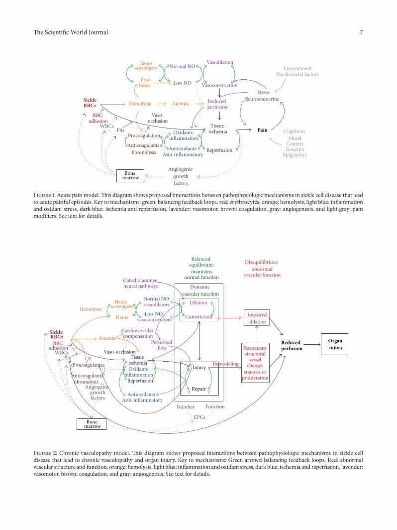
These concepts of systems thinking and network analysiswere used to develop diagrammatic models that incorpo-rate and which show potential interactions between multi-ple mechanisms putatively involved in acute pain episodes(Figure 1) and chronic vasculopathy (Figure 2) in SCD.Theseare not classic biologic network models with specific proteinsor genes as nodes but include as modules major mechanisticpathways in SCD for which there is existing evidence.In these models, the primary defect of sickle hemoglobinpolymerization causes secondary processes of hemolysis andvaso-occlusion, which further transduce their effects to bloodvessels and organs through oxidative, inflammatory, vasomo-tor, coagulation, and angiogenic intermediaries. The roles ofbalancing feedback loops in homeostasis and the effects onthe vascular bed are central features of the models.
4.1. Acute Pain Model. Figure 1 shows potential relationshipsbetween mechanistic pathways in SCD that lead to acutepain episodes. The most commonly considered pathway inthis process begins with sickle hemoglobin polymeriza-tion, leading to formation of rigid sickle erythrocytes andmicrovascular occlusion when these cells become trapped insmall vessels. In the presence of inflammation (infection orischemia and reperfusion), vascular endothelial cells expressa number of adhesion molecules that facilitate adherenceof mature erythrocytes, reticulocytes, activated leukocytes,
and/or platelets. Endothelial tissue factor expression canactivate coagulation factors and thrombosis.
Hemolysis can also contribute to vaso-occlusion, throughthe release of free heme, reactive iron species, andmembranemicroparticles. Free heme can bind NO and reduce itsbioavailability, which promotes vaso-constriction, inflam-mation, and platelet aggregation. Heme and reactive ironspecies can directly cause injury and oxidative damage toendothelium. Erythrocyte membrane microparticles withexposed phosphatidylserinemay active platelets and promotecoagulation. Hemolytic anemia reduces oxygen delivery totissues and contributes to reduced tissue and organ perfusionchronically, likely leaving them vulnerable to the effects ofacute vaso-occlusion.
After vaso-occlusion has occurred, there is local tissueischemia and reperfusion, which includes inflammatory andoxidant responses. Our group has demonstrated elevatedlevels of hypoxia-inducible factor- (HIF-) associated angio-genic growth factors in children with hemoglobin SS duringsteady state [60], suggesting a chronic baseline state of tissueischemia even in the absence of symptomatic pain or acutecomplications. In particular, levels of stromal-derived factor-(SDF-) 1𝛼, produced by ischemic endotheliumandother cells,were associated with the number of bone marrow-derivedcirculating progenitors with angiogenic potential (CD34+/VEGFR2+) and total white blood cell (WBC) count. TotalWBC has been found to be associated with SCD severityin some series, and the relationship between WBC andSDF-1𝛼 suggests that those people with the most ongoingtissue ischemia may have the most vaso-occlusive organinjury. Tissue ischemia has a reinforcing effect on inflamma-tion, through HIF-mediated angiogenic stimulation of WBCrelease from the bone marrow.
Pain is itself a complex process and is not diagrammedin detail in this model. The pain experience is the prod-uct of the nociceptive input from injured tissue but alsoinvolves cognitive, contextual, mood, and individual differ-ences, such as sex, age, and genetics. Stress related to acutepain can induce neuro-endocrine responses, such as stresssteroids, catecholamines, and pain peptides (substance P,neurokinins). Catecholamines promote vaso-constriction,and the pain peptides can be proinflammatory. The currentmodel also does not attempt to delineate all of the externalfactors that may also influence a pain episode, such as envi-ronment (ambient temperature, second-hand smoke) or psy-chosocial factors. Neuropathic pain resulting from ischemicinjury directly to nerves may account for some of the painexperienced in SCD and would not necessarily respond toantisickling therapies or those targeting inhibition of vaso-occlusion. Chronic pain may involve “imprinting” of thenervous system by epigenetic modifications (DNA methy-lation, histone modification) that regulate gene expression[61]. Chronic pain and opioids can alter the structure of thebrain [62, 63]. To summarize, there are many pathways thatcan interact in acute pain episodes in SCD, some positivelyreinforce others, and some factors are outside of the body.
4.2. Chronic Vasculopathy Model. A model with similar fea-tures describes the development of chronic vasculopathy in
The Scientific World Journal 7
WBCsPlts
Anti-inflammatory
Freeheme Low NO
Normal NO
Procoagulation
Anemia
Tissueischemia
Reperfusion
Oxidantsinflammation
Angiogenicgrowthfactors
Reducedperfusion
Bonemarrow
RBCadhesion
Environment
Stress
Hemescavengers
Anticoagulantsfibronolysis
Vaso-occlusion
Hemolysis
Vasodilation
Vasoconstriction
Antioxidants
Neuroendocrine
CognitionMood
ContextGenetics
Epigenetics
Psychosocial factors
Pain
SickleRBCs
Figure 1: Acute pain model.This diagram shows proposed interactions between pathophysiologic mechanisms in sickle cell disease that leadto acute painful episodes. Key tomechanisms: green: balancing feedback loops, red: erythrocytes, orange: hemolysis, light blue: inflammationand oxidant stress, dark blue: ischemia and reperfusion, lavender: vasomotor, brown: coagulation, gray: angiogenesis, and light gray: painmodifiers. See text for details.
RBCadhesion Permanent
structuralvessel
change:stenosis or
proliferation
WBCsPlts
Anti-inflammatory
Dilation
Constriction
Dynamicvascular function
Injury
Repair
Heme Low NOvasoconstrictors
Normal NOvasodilators
HemolysisImpaireddilation
Procoagulants Remodeling
Vaso-occlusion
AnemiaCardiovascularcompensation
EPCs
Number Function
Disequilibrium:abnormal
vascular function
Perturbedflow
Tissueischemia
Angiogenicgrowthfactors
Catecholaminesneural pathways
Hemescavengers
fibronolysis
Balancedequilbrium:maintains
normal function
Reperfusion
Bonemarrow
Oxidantsinflammation
Antioxidants
Anticoagulants
SickleRBCs
Organinjury
Reducedperfusion
Figure 2: Chronic vasculopathy model. This diagram shows proposed interactions between pathophysiologic mechanisms in sickle celldisease that lead to chronic vasculopathy and organ injury. Key to mechanisms: Green arrows: balancing feedback loops, Red: abnormalvascular structure and function, orange: hemolysis, light blue: inflammation and oxidant stress, dark blue: ischemia and reperfusion, lavender:vasomotor, brown: coagulation, and gray: angiogenesis. See text for details.

8 The Scientific World Journal
SCD (Figure 2). In the vasculopathymodel, dynamic vascularfunction is the central process affected by SCDderangements.In the healthy state, there is balanced equilibrium betweenvasodilation and constriction, and between endothelial injuryand repair.
Hemolysis will cause disequilibrium in favor of vaso-constriction, and longterm exposure can eventually causesustained impairment in vasodilation and reduced vesselcompliance (stiffness). Early stage remodeling may causearterial wall stiffness and reduced compliance before arterialstenosis is apparent in imaging studies. This may be thecase in those children with elevated transcranial doppler(TCD) velocities in the cerebral arteries that is associatedwith high stroke risk, but whose brain magnetic resonancearteriography (MRA) shows no arterial stenoses.
Anemia reduces oxygen delivery and organ perfusion.Ischemia and reperfusion due to the combination of ane-mia and repetitive vaso-occlusion stimulate HIF-associatedangiogenic growth factors. Anemia-associated compensatorycirculatory changes are likely to cause disturbed bloodflow, which can promote endothelial injury, and potentiallycontribute to vessel wall remodeling (intimal proliferationand arterial stenosis). Our group has demonstrated ele-vated serumconcentrations of platelet-derived growth factor-(PDGF-) AA, a mediator of vascular remodeling, and brainderived neurotrophic factor (BDNF), a biomarker of cerebralischemia, in children with hemoglobin SS and high TCDvelocities [64], consistent with the model. High frequencyof red cell transfusion therapy reduced PDGF-AA, solubleVCAM-1, and RANTES in children with hemoglobin SS andhigh TCD velocities, suggesting that correction of anemiaand reduction in sickle cells by transfusion reduces vascularinjury, inflammation, and vascular remodeling responses[65].
Considering sickle cell disease through the lenses of thesemodels helps explain the potential limitations of therapiesthat are targeted to single pathways. While the therapiesdiscussed earlier may have some multimodal effects, thereare many additional contributors to acute pain that were notmodulated by individual therapy.Thesemodels contrast con-ceptually to the bimodal model proposed by Kato, in whichcertain SCD complications are primarily related to eitherhemolysis or vaso-occlusion, and that individuals exhibit oneor the other subphenotype [66]. In these models, hemolysisand vaso-occlusion occur simultaneously in both acute painand chronic vasculopathy, although not necessarily equally inmagnitude at any given time.
Complexity in SCD does not necessarily mean that thedisease is hopelessly complicated and cannot be successfullytreated. In these models, sickle hemoglobin polymerizationis the network hub, and therefore most vulnerable to attack(correction). This is consistent with the clinical observationthat red cell transfusions and hydroxyurea, which correcthemoglobin S polymerization and/or sickled erythrocytes,are often effective SCD treatments and also reduce down-stream mediators. However, while hydroxyurea is an alter-native to transfusions for certain SCD indications, it is notan equivalent therapy. In the setting of established structuralvascular disease, such as significant cerebral vasculopathy in
individuals with stroke, hydroxyurea therapy in combinationwith phlebotomy to relieve iron overload was not as effectiveas transfusions in preventing stroke [67]. Even chronicred blood cell transfusions are not effective in preventingrecurrent stroke in up to 20% of individuals [68], suggestingadditional unidentified stroke mediators in a subset of indi-viduals with the most severe disease.
Viewed from this perspective, replacement of sickleerythrocytes by stem cell transplantation, gene therapy cor-rection of the hemoglobin S mutation, or very effective fetalhemoglobin induction are likely to be the most effective SCDtreatments in the long run. However, until these are widelyavailable, should severe sickle cell disease be treated likethalassemia, with lifelong chronic red cell transfusions? Howdo we identify those at highest risk who would benefit fromlife-long transfusions begun early in life?
5. Future Directions
This paper of selected clinical trials and discussion of com-plexity in SCD has identified some challenges in the searchfor alternative effective therapies. There are logistical andmethodological issues related to clinical trials, such as con-sistent study endpoint definition and effective timing anddelivery of therapeutics; lack of goodmodel systems in whichto test the effect of therapeutics; and the larger problem posedby complexity—it is difficult to shut down all aspects of thesystem at once without using transfusions or stem cell trans-plantation. Acknowledgment of complexity does not implythat therapies targeting individual intermediary mechanismsin SCD should be abandoned but necessitates that theireffectiveness needs to be tested in highly predictive modelsystems prior to embarking on large-scale clinical trials. Inthis section, some possible approaches to these challenges aresuggested.
5.1. Strategic Mechanistic Testing of Therapeutics in the Trans-genic Sickle Cell Mouse Model. There is a need to developpreclinical model systems for complications such as acutepain or chronic vasculopathy that are highly predictive ofthose processes in people with SCD. There are currentlytransgenic sickle cell mouse models of acute chest syn-drome and pulmonary hypertension, but none yet for SCD-associated stroke or acute painful crisis. The most extrememodel used in transgenic sickle cell mice is hypoxia-induceddeath, which presumably occurs from sickle cell-inducedvaso-occlusion in the entire animal. While not similar tohuman acute pain episodes in severity, drugs that are potentenough to prevent this degree of sickling or vaso-occlusionshould be effective in less extreme situations. For example, thelack of beneficial effect of inhaled NO in the phase III RCTwas accurately predicted by the lack of rescue in the hypoxia-exposed SAD mice who were treated with only posthypoxiainhaled NO. If there was a way to image sites of pain whilesimultaneously measuring pain behaviors in the sickle cellmouse, it might be useful to titrate the severity of the hypoxicexposure to a sublethal dose thatmight simulate acute painfulevents.
The Scientific World Journal 9
Transplantation of sickle cell mouse bone marrow orinjection of sicklemouse erythrocytes into a transgenic strainwith a desired gene knockoutmay be used to prove the essen-tial role of an individual molecule in sickle cell pathogenesis.These approaches have been used to demonstrate the roleof P-selectin in sickle cell microvascular occlusion [69] andsuperoxide produced by NADPH oxidase in cerebral arterymicrovascular occlusions [35]. Tissue-specific conditionalknockouts in a transgenic sickle cell mouse would be anelegant but technically challenging experimental approach totest the role of individual molecules in the “native” environ-ment of the transgenic sickle cell mouse. However, such anapproach should only be used in pathways in which thereis no physiologic redundancy. At least ten different sickleerythrocyte membrane proteins or components have beenidentified as potentially involved in adhesion or interactionwith endothelial cells [70], so that knocking down only oneis unlikely to have major impact on vaso-occlusion, unless itis a critically important molecule that has greater impact thanall of the others in combination.
5.2. Develop Imaging Systems for Vaso-Occlusion. One of themost basic tenets of the field is that acute pain episodesare caused by the occlusion of microvessels by sickled ery-throcytes. While this makes theoretic sense on the basisof hemoglobin polymerization, rheologic and microvascularstudies, this has never been proven in peoplewith SCD.Whendoes acute pain begin relative to vaso-occlusion? Do individ-ualswith full body pain really have sickling everywhere?Doesvaso-occlusion in one part of the body induce pain at distantsites through crosstalk between neurons in the sensorypathway or central nervous system? The field needs ways tovisualize both sites of vaso-occlusion and pain pathways todemonstrate that they are actually related and to have anoutcome measure for testing pain treatments. Vaso-occlusivesites could potentially be visualized by radionuclide-taggedparticles that home to ischemic tissue markers, such as SDF-1𝛼.
5.3. Interdisciplinary Pain Research. The pain research fieldapparently has similar challenges in developing chronic paintreatments as has been described here for SCD. Borsookwrote “Drug development for pain often fails, parallelingmany other CNS areas, because preclinical and experimentalclinical proof-of-concept (POC) studies do not translate wellto clinical conditions and patient populations [71].” Proposedbiomarkers for pain include functional brain imaging withanalysis of focal brain regions and chemical biomarkers,such as CNS neurotransmitters (e.g., glutamate, GABA, andglycine) and brain metabolites (e.g., NAA, choline), whichcan be measured in vivo using magnetic resonance spec-troscopy (MRS) [72]. Functional MRI (fMRI) with supportvector machine learning analysis of the whole brain is abledistinguish pain without the need for verbal communication[73]. Since pain is such a large part of SCD symptomatology,future research collaborations with established pain neuro-biologists equipped to use these state-of-art approaches arewarranted as suggested by the NIH Blueprint for Neuro-science “Grand Challenge on Pain [74].”
5.4. Develop a Consensus Set of Meaningful Study Endpointsand Test for Mechanistic Markers during Clinical Trials. Foragents that prevent pain, the number and duration of acutepainful episodes, including both facility- and home-treatedevents, and quality of life measures related to pain may bea more accurate assessment of the true effectiveness of apain prevention therapy. In the treatment of acute pain, thereneeds to be agreement on the definition of who should behospitalized for therapy to assure comparability of groupsand on the definition of what constitutes “resolution of pain.”It has also been recommended that study endpoints bettermatch the mechanism of action of the therapeutic [4, 44].For example, pain outcomes may not be applicable to agentsthat reduce hemolysis (Gardos channel inhibitors) or targetNO signaling, since they may be more likely to be beneficialin the vasculopathy subphenotype (pulmonary hypertension,priapism). Analogously, there needs to be consensus onvasculopathy endpoints. For example, is echocardiographicmeasurement of tricuspid regurgitant velocity acceptable asa noninvasive surrogate of pulmonary hypertension? In thecase of any endpoint, does it respond rapidly enough totherapy to be feasibly measured as a study outcome?
To better understand the role of therapeutic mechanisms,biomarkers or other functional outcome measures should beincluded as part of early phase clinical trials. Asmentioned inthe earlier discussion of inhaled NO, none of the publishedclinical trials reported on vasodilation, platelet aggregation,or inflammatory biomarkers, such as sVCAM. Senicapoc’sred cell effects were consistently measured in each clinicaltrial, so that lack of efficacy in reducing pain frequencyappears to be unrelated to improvement in cell hydrationstatus. In this situation, the data helps guide investigatorsin the analysis of therapeutic and unwanted side effects.Measurement of biomarkers makes clinical trials more laborintensive and is unlikely to be feasible at every study site.However, it provides important evidence to explain drugefficacy or lack thereof.
5.5. Directed Delivery of Therapeutics to Ischemic Sites. Incombination with biomarker and functional assays, earlyphase clinical trials should include well-described pharma-cokinetics to establish that the administered doses result inplasma concentrations that are comparable to those usedin preclinical studies. However, systemic concentrations ofa drug may not be the same as the amount delivered tothe affected tissues, especially in areas of reduced perfusion.There is an opportunity to apply nanotechnology to selec-tively deliver analgesics, antisickling, other vaso-occlusiondisrupting agents, or drugs that improve tissue oxygenationto ischemic regions (presumably corresponding to vaso-occlusion) by targeting ischemia markers, such as SDF-1𝛼.
5.6. Systems Biology, “Omics,” and Computational Modeling.Systems biology and “omics” technologies may be useful inunderstanding complex biologic systems. This approach is inits infancy in SCD and could be applied to dissecting specificproblems, such as identifying gene variants or microRNAsthat predispose to higher risk of well-defined complications
10 The Scientific World Journal
such as stroke, chronic pain, changes in gene expression, orepigenetic modifications that occur with therapies. Anothergoal for the application of “omics” to SCDwould be to identifymaster regulators that control multiple pathophysiologicmechanisms, so that these could be targeted for inhibition.
Predictive computational models can potentially be usedto integrate multiple types of patient data, such as laboratoryvalues, radiographic findings, circulating biomarkers, andgenetic and epigenetic data, with disease phenotype to definerisk categories. The ability to identify individuals at highestrisk for severe complications would allow the option ofearly treatment with high-risk therapies, currently red bloodcell transfusion or stem cell transplantation, well beforethe onset of complications. The predictive strength of suchmodeling approaches would be enhanced by including asmany individuals with SCD as possible, perhaps through acollaborative national data registry and biorepository system.
5.7. Improve Understanding of the Effects of Psychosocial Deter-minants and the Environment on SCD Complications. It hasbeenwell described that stresses related to poverty and racismaffect cardiovascular risk and disease and disproportionatelyaffect African Americans. It is very likely that such gene-environment interactions are additional factors influencingSCD complexity and outcomes and are currently not ade-quately understood. For example, how do chronic undernu-trition, lack of utilities, poverty, or personal or familialmentalillness affect the frequency and severity of acute illness or theresponse to therapy in a person with SCD? Epidemiologicmethodologies including geocoding could be applied tostudying some of these factors in SCD. Such variables can beadded to computational predictive models to help us beginto understand the relative contributions of factors in SCDcomplications.
6. Conclusions
SCD is caused by a single mutation in beta globin but triggersseveral pathophysiologic pathways and results in a highlycomplex disease. This complexity is likely to be one of themajor barriers to the development of successful new treat-ments which, to date, has largely concentrated on individualmechanistic pathways. Future development of therapeuticsneeds to continue to focus on correcting the underlyingproblem of sickle hemoglobin polymerization but shouldalso include development of better model systems for acuteand chronic SCD complications, methods for visualizingand measuring vaso-occlusion and associated pain, directeddelivery of therapies to sites of vaso-occlusion, systemsbiology approaches to identify master regulators of the mul-tiple downstream effectors of hemolysis and vaso-occlusion,and better understanding of the contribution of gene-environment interactions on sickle cell disease complica-tions. Considering the number of pathophysiologic processescaused by SCD, it is astonishing how well the body maintainshomeostasis sufficient for growth, development, and generalhealth for periods between acute illnesses. The approachto this disease should also include an effort to identify
mechanisms that are crucial to maintaining homeostasis andwellness. While there have been many life-saving advancesin the treatment of SCD, much work remains to achieve thegoal of curing the disease and developing safe and effectivetherapies to improve health and well-being.
Conflict of Interests
Theauthor has no financial interests in any of the commercialproducts mentioned.
Acknowledgments
The author receives research funding from the Atlanta Clin-ical Translational Science Institute (UL1 TR000454), More-house School of Medicine RCENTER (U54 RR026137), andGrants 5R01HL095647 and P20 MD006881. The authorwould like to thank her family, teachers, mentors, colleagues,and patients for teaching her much about the art and scienceof medicine.
References
[1] “Sickle Cell Disease—Data and Statistics,” 2010, http://www.cdc.gov/ncbddd/sicklecell/data.html.
[2] O. S. Platt, D. J. Brambilla, W. F. Rosse et al., “Mortality in sicklecell disease—life expectancy and risk factors for early death,”TheNew England Journal of Medicine, vol. 330, no. 23, pp. 1639–1644, 1994.
[3] Sickle Cell Research for Treatment and Cure, National Institutesof Health—National Heart Lung, and Blood Institute, 2002.
[4] E. Vichinsky, “Emerging “A” therapies in hemoglobinopathies:agonists, antagonists, antioxidants, and arginine,” HematologyAmerican Society of Hematology Education Program, vol. 2012,pp. 271–275, 2012.
[5] W. K. Hoots and S. B. Shurin, “Future directions of sickle celldisease research: the NIH perspective,” Pediatric Blood andCancer, vol. 59, no. 2, pp. 353–357, 2012.
[6] J. Hankins and B. Aygun, “Pharmacotherapy in sickle cell dis-ease—state of the art and future prospects,”The British Journalof Haematology, vol. 145, no. 3, pp. 296–308, 2009.
[7] R. I. Raphael, “Pathophysiology and treatment of sickle celldisease,” Clinical Advances in Hematology and Oncology, vol. 3,no. 6, pp. 492–505, 2005.
[8] F. Padilla, J. O.Wear, andW.H. vanWagner, “Effect of fluorocar-bon emulsions on the mechanical fragility of normal and sicklecells: in vitro studies,” Federation Proceedings, vol. 34, no. 6, pp.1510–1512, 1975.
[9] C. M. Smith II, R. P. Hebbel, D. P. Tukey, C. C. Clawson, J.G. White, and G. M. Vercellotti, “Pluronic F-68 reduces theendothelial adherence and improves the rheology of ligandedsickle erythrocytes,” Blood, vol. 69, no. 6, pp. 1631–1636, 1987.
[10] P. Adams-Graves, A. Kedar, M. Koshy et al., “RheothRx (Polox-amer 188) injection for the acute painful episode of sickle celldisease: a pilot study,” Blood, vol. 90, no. 5, pp. 2041–2046, 1997.
[11] E. P. Orringer, J. F. Casella, K. I. Ataga et al., “Purified poloxamer188 for treatment of acute vaso-occlusive crisis of sickle celldisease: a randomized controlled trial,” The Journal of theAmerican Medical Association, vol. 286, no. 17, pp. 2099–2106,2001.
The Scientific World Journal 11
[12] C. Brugnara, L. de Franceschi, and S. L. Alper, “Inhibition ofCa2+-dependent K+ transport and cell dehydration in sickleerythrocytes by clotrimazole and other imidazole derivatives,”Journal of Clinical Investigation, vol. 92, no. 1, pp. 520–526, 1993.
[13] L. de Franceschi, N. Saadane, M. Trudel, S. L. Alper, C.Brugnara, and Y. Beuzard, “Treatment with oral clotrimazoleblocks Ca2+-activated K+ transport and reverses erythrocytedehydration in transgenic SAD mice. A model for therapy ofsickle cell disease,” Journal of Clinical Investigation, vol. 93, no.4, pp. 1670–1676, 1994.
[14] C. Brugnara, B. Gee, C. C. Armsby et al., “Therapy with oralclotrimazole induces inhibition of the Gardos channel andreduction of erythrocyte dehydration in patients with sickle celldisease,” Journal of Clinical Investigation, vol. 97, no. 5, pp. 1227–1234, 1996.
[15] J. W. Stocker, L. de Franceschi, G. A. McNaughton-Smith, R.Corrocher, Y. Beuzard, and C. Brugnara, “ICA-17043, a novelGardos channel blocker, prevents sickled red blood cell dehy-dration in vitro and in vivo in SAD mice,” Blood, vol. 101, no. 6,pp. 2412–2418, 2003.
[16] K. I. Ataga, W. R. Smith, L. M. de Castro et al., “Efficacy andsafety of the Gardos channel blocker, senicapoc (ICA-17043), inpatients with sickle cell anemia,” Blood, vol. 111, no. 8, pp. 3991–3997, 2008.
[17] K. I. Ataga, M. Reid, S. K. Ballas et al., “Improvements inhaemolysis and indicators of erythrocyte survival do not corre-late with acute vaso-occlusive crises in patients with sickle celldisease: a phase III randomized, placebo-controlled, double-blind study of the Gardos channel blocker senicapoc (ICA-17043),” The British Journal of Haematology, vol. 153, no. 1, pp.92–104, 2011.
[18] D. L. Tharp and D. K. Bowles, “The intermediate-conductanceCa2+-activated K+ channel (KCa3.1) in vascular disease,” Car-diovascular and Hematological Agents in Medicinal Chemistry,vol. 7, no. 1, pp. 1–11, 2009.
[19] Z. Liang, L. Chen, H. McClafferty et al., “Control of hypo-thalamic-pituitary-adrenal stress axis activity by the interme-diate conductance calcium-activated potassium channel, SK4,”Journal of Physiology, vol. 589, part 24, pp. 5965–5986, 2011.
[20] E. F. Kuiper, A. Nelemans, P. Luiten, I. Nijholt, A. Dolga, andU. Eisel, “K(Ca)2 and k(ca)3 channels in learning and memoryprocesses, and neurodegeneration,” Frontiers in Pharmacology,vol. 3, article 107, 2012.
[21] A. M. Atz and D. L. Wessel, “Inhaled nitric oxide in sickle celldisease with acute chest syndrome,” Anesthesiology, vol. 87, no.4, pp. 988–990, 1997.
[22] K. J. Sullivan, S. R. Goodwin, J. Evangelist, R. D. Moore, andP. Mehta, “Nitric oxide successfully used to treat acute chestsyndrome of sickle cell disease in a young adolescent,” CriticalCare Medicine, vol. 27, no. 11, pp. 2563–2568, 1999.
[23] P. Montero-Huerta, D. R. Hess, and C. A. Head, “Inhaled nitricoxide for treatment of sickle cell stroke,”Anesthesiology, vol. 105,no. 3, pp. 619–621, 2006.
[24] W. L. Chang, L. M. Corate, J. M. Sinclair, and H. C. van derHeyde, “Continuous inhaled nitric oxide therapy in a case ofsickle cell disease with multiorgan involvement,” Journal ofInvestigative Medicine, vol. 56, no. 8, pp. 1023–1027, 2008.
[25] C. D. Reiter, X.Wang, J. E. Tanus-Santos et al., “Cell-free hemo-globin limits nitric oxide bioavailability in sickle-cell disease,”Nature Medicine, vol. 8, no. 12, pp. 1383–1389, 2002.
[26] K. J. Zuzak, M. T. Gladwin, R. O. Cannon III, and I. W. Levin,“Imaging hemoglobin oxygen saturation in sickle cell disease
patients using noninvasive visible reflectance hyperspectraltechniques: effects of nitric oxide,” The American Journal ofPhysiology, vol. 285, no. 3, pp. H1183–H1189, 2003.
[27] R.Martinez-Ruiz, P.Montero-Huerta, J. Hromi, andC.A.Head,“Inhaled nitric oxide improves survival rates during hypoxia ina sickle cell (SAD) mouse model,” Anesthesiology, vol. 94, no. 6,pp. 1113–1118, 2001.
[28] D. L. Weiner, P. L. Hibberd, P. Betit, A. B. Cooper, C. A.Botelho, and C. Brugnara, “Preliminary assessment of inhalednitric oxide for acute vaso-occlusive crisis in pediatric patientswith sickle cell disease,” The Journal of the American MedicalAssociation, vol. 289, no. 9, pp. 1136–1142, 2003.
[29] C. A. Head, P. Swerdlow,W. A.McDade et al., “Beneficial effectsof nitric oxide breathing in adult patients with sickle cell crisis,”The American Journal of Hematology, vol. 85, no. 10, pp. 800–802, 2010.
[30] M. T. Gladwin, G. J. Kato, D. Weiner et al., “Nitric oxide forinhalation in the acute treatment of sickle cell pain crisis:a randomized controlled trial,” The Journal of the AmericanMedical Association, vol. 305, no. 9, pp. 893–902, 2011.
[31] Institute of Medicine, Envisioning a Transformed Clinical TrialsEnterprise in the United States: Establishing an Agenda for 2020,Workshop Summary, National Academies Press, 2012.
[32] C. G. Frostell, H. Blomqvist, G. Hedenstierna, J. Lundberg, andW. M. Zapol, “Inhaled nitric oxide selectively reverses humanhypoxic pulmonary vasoconstriction without causing systemicvasodilation,” Anesthesiology, vol. 78, no. 3, pp. 427–435, 1993.
[33] J. Chang, J. T. Patton, A. Sarkar, B. Ernst, J. L. Magnani, and P. S.Frenette, “GMI-1070, a novel pan-selectin antagonist, reversesacute vascular occlusions in sickle cell mice,” Blood, vol. 116, no.10, pp. 1779–1786, 2010.
[34] D. K. Kaul, X. D. Liu, X. Zhang et al., “Peptides based on𝛼V-binding domains of erythrocyte ICAM-4 inhibit sicklered cell-endothelial interactions and vaso-occlusion in themicrocirculation,”The American Journal of Physiology, vol. 291,no. 5, pp. C922–C930, 2006.
[35] K. C. Wood, R. P. Hebbel, and D. N. Granger, “Endothelial cellNADPH oxidase mediates the cerebral microvascular dysfunc-tion in sickle cell transgenic mice,” FASEB Journal, vol. 19, no. 8,pp. 989–991, 2005.
[36] A. T. Cheung, J. W. Miller, S. M. Craig et al., “Comparison ofreal-time microvascular abnormalities in pediatric and adultsickle cell anemia patients,” The American Journal of Hematol-ogy, vol. 85, no. 11, pp. 899–901, 2010.
[37] A. T. Cheung, J. W. Miller, M. G. Miguelino et al., “Exchangetransfusion therapy and its effects on real-timemicrocirculationin pediatric sickle cell anemia patients: an intravital microscopystudy,” Journal of Pediatric Hematology/Oncology, vol. 34, no. 3,pp. 169–174, 2012.
[38] A. T. W. Cheung, M. S. Chan, S. Ramanujam et al., “Effects ofpoloxamer 188 treatment on sickle cell vaso-occlusive crisis:computer-assisted intravitalmicroscopy study,” Journal of Inves-tigative Medicine, vol. 52, no. 6, pp. 402–406, 2004.
[39] J. Grotta, “Timing of thrombolysis for acute ischemic stroke:“timing is everything” or ‘everyone is different’,” Annals of theNew York Academy of Sciences, vol. 1268, pp. 141–144, 2012.
[40] I. M. Rutkow and J. M. Lipton, “The sickle cell complexity,”TheJournal of the American Medical Association, vol. 228, no. 5, pp.608–609, 1974.
[41] J. A. Warth and D. L. Rucknagel, “The increasing complexity ofsickle cell anemia,” Progress in Hematology, vol. 13, pp. 25–47,1983.

12 The Scientific World Journal
[42] P. S. Frenette, “Sickle cell vaso-occlusion: multistep and multi-cellular paradigm,” Current Opinion in Hematology, vol. 9, no.2, pp. 101–106, 2002.
[43] R. P. Hebbel, G. M. Vercellotti, and K. A. Nath, “A systemsbiology consideration of the vasculopathy of sickle cell anemia:the need for multi-modality chemo-prophylaxis,” Cardiovascu-lar and Hematological Disorders, vol. 9, no. 4, pp. 271–292, 2009.
[44] O. L. Castro,V. R.Gordeuk,M.T.Gladwin, andM.H. Steinberg,“Senicapoc trial results support the existence of different sub-phenotypes of sickle cell disease with possible drug-inducedphenotypic shifts,”The British Journal of Haematology, vol. 155,no. 5, pp. 636–638, 2011.
[45] L. S. Moreira, T. G. de Andrade, D. M. Albuquerque et al.,“Identification of differentially expressed genes induced byhydroxyurea in reticulocytes from sickle cell anaemia patients,”Clinical and Experimental Pharmacology and Physiology, vol. 35,no. 5-6, pp. 651–655, 2008.
[46] A. C. Rybicki, M. E. Fabry, M. D. Does, D. K. Kaul, and R. L.Nagel, “Differential gene expression in the kidney of sickle celltransgenic mice: upregulated genes,” Blood Cells, Molecules, andDiseases, vol. 31, no. 3, pp. 370–380, 2003.
[47] L. C. Milbauer, P. Wei, J. Enenstein et al., “Genetic endothelialsystems biology of sickle stroke risk,” Blood, vol. 111, no. 7, pp.3872–3879, 2008.
[48] M. L. Jison, P. J. Munson, J. J. Barb et al., “Blood mononu-clear cell gene expression profiles characterize the oxidant,hemolytic, and inflammatory stress of sickle cell disease,” Blood,vol. 104, no. 1, pp. 270–280, 2004.
[49] S. Yuditskaya, A. F. Suffredini, and G. J. Kato, “The proteomeof sickle cell disease: insights from exploratory proteomicprofiling,” Expert Review of Proteomics, vol. 7, no. 6, pp. 833–848, 2010.
[50] P. Sebastiani, N. Solovieff, S. W. Hartley et al., “Genetic mod-ifiers of the severity of sickle cell anemia identified througha genome-wide association study,” The American Journal ofHematology, vol. 85, no. 1, pp. 29–35, 2010.
[51] A. L. Walker, S. Steward, T. A. Howard et al., “Epigenetic andmolecular profiles of erythroid cells after hydroxyurea treat-ment in sickle cell anemia,” Blood, vol. 118, no. 20, pp. 5664–5670, 2011.
[52] M. Mitchell, Complexity: A Guided Tour, Oxford UniversityPress, New York, NY, USA, 2009.
[53] L. Von Bertalanffy, “The theory of open systems in physics andbiology,” Science, vol. 111, no. 2872, pp. 23–29, 1950.
[54] D. Wright, Ed., Thinking in Systems: A Primer/ Donella H.Meadows, Chelsea Green Publishing Company, White RiverJunction, Vt, USA, 2008.
[55] H. Kitano, “Systems biology: a brief overview,” Science, vol. 295,no. 5560, pp. 1662–1664, 2002.
[56] R. Breitling, “What is systems biology?” Frontiers in Physiology,vol. 1, article 9, 2010.
[57] D. Machado, R. S. Costa, M. Rocha, E. C. Ferreira, B. Tidor,and I. Rocha, “Modeling formalisms in systems biology,” AMBExpress, vol. 1, article 45, 2011.
[58] M. J. Joyner, “Giant sucking sound: can physiology fill the intel-lectual void left by the reductionists?” Journal of Applied Physi-ology, vol. 111, no. 2, pp. 335–342, 2011.
[59] U. Alon, An Introduction to Systems Biology: Design Principlesof Biologic Circuits, Chapman & Hall/CRC, Taylor and FrancisGroup, Boca Raton, Fla, USA, 2007.
[60] S. F. Ofori-Acquah, I. D. Buchanan, I. Osunkwo et al., “Elevatedcirculating angiogenic progenitors and white blood cells areassociated with hypoxia-inducible angiogenic growth factors inchildren with sickle cell disease,” Anemia, vol. 2012, Article ID156598, 9 pages, 2012.
[61] T. Buchheit, T. van de Ven, and A. Shaw, “Epigenetics and thetransition from acute to chronic pain,” Pain, vol. 13, no. 11, pp.1474–1490, 2012.
[62] J. W. Younger, L. F. Chu, N. T. D’Arcy, K. E. Trott, L. E. Jastrzab,and S. C. MacKey, “Prescription opioid analgesics rapidlychange the human brain,” Pain, vol. 152, no. 8, pp. 1803–1810,2011.
[63] A. V. Apkarian, Y. Sosa, S. Sonty et al., “Chronic back pain isassociated with decreased prefrontal and thalamic gray matterdensity,” Journal ofNeuroscience, vol. 24, no. 46, pp. 10410–10415,2004.
[64] H. I. Hyacinth, B. E. Gee, T. V. Adamkiewicz et al., “PlasmaBDNF and PDGF-AA levels are associated with high TCDvelocity and stroke in children with sickle cell anemia,”Cytokine, vol. 60, no. 1, pp. 302–308, 2012.
[65] H. I. Hyacinth, B. E. Gee, J. H. Voeks, R. J. Adams, and J.M. Hibbert, “High frequency RBC transfusion is associatedwith decreased serum markers of neurodegeneration, vascularremodeling and inflammation,” Blood, vol. 120, no. 21, p. 244,2012.
[66] G. J. Kato,M. T. Gladwin, andM.H. Steinberg, “Deconstructingsickle cell disease: reappraisal of the role of hemolysis in thedevelopment of clinical subphenotypes,” Blood Reviews, vol. 21,no. 1, pp. 37–47, 2007.
[67] R. E.Ware and R.W.Helms, “Stroke with transfusions changingto hydroxyurea (SWiTCH),” Blood, vol. 119, no. 17, pp. 3925–3932, 2012.
[68] D. J. Scothorn, C. Price, D. Schwartz et al., “Risk of recurrentstroke in children with sickle cell disease receiving blood trans-fusion therapy for at least five years after initial stroke,” Journalof Pediatrics, vol. 140, no. 3, pp. 348–354, 2002.
[69] S. H. Embury, N. M. Matsui, S. Ramanujam et al., “The contri-bution of endothelial cell P-selectin to the microvascular flowof mouse sickle erythrocytes in vivo,” Blood, vol. 104, no. 10, pp.3378–3385, 2004.
[70] R. P.Hebbel, “Adhesion of sickle red cells to endothelium:mythsand future directions,” Transfusion Clinique et Biologique, vol.15, no. 1-2, pp. 14–18, 2008.
[71] D. Borsook, L. Becerra, and R. Hargreaves, “Biomarkers forchronic pain and analgesia—part 1: the need, reality, challenges,and solutions,” Discovery Medicine, vol. 11, no. 58, pp. 197–207,2011.
[72] D. Borsook, L. Becerra, and R. Hargreaves, “Biomarkers forchronic pain and analgesia—part 2: how, where, and what tolook for using functional imaging,” Discovery Medicine, vol. 11,no. 58, pp. 209–219, 2011.
[73] J. E. Brown, N. Chatterjee, J. Younger, and S. Mackey, “Towardsa physiology-based measure of pain: patterns of human brainactivity distinguish painful from non-painful thermal stimula-tion,” PLoS ONE, vol. 6, no. 9, Article ID e24124, 2011.
[74] National Institutes of Health Blueprint for Neurosciences FactSheet, U.S.Department ofHealth andHumanServices,NationalInstitutes of Health, 2011.

Submit your manuscripts athttp://www.hindawi.com
Stem CellsInternational
Hindawi Publishing Corporationhttp://www.hindawi.com Volume 2014
Hindawi Publishing Corporationhttp://www.hindawi.com Volume 2014
MEDIATORSINFLAMMATION
of
Hindawi Publishing Corporationhttp://www.hindawi.com Volume 2014
Behavioural Neurology
EndocrinologyInternational Journal of
Hindawi Publishing Corporationhttp://www.hindawi.com Volume 2014
Hindawi Publishing Corporationhttp://www.hindawi.com Volume 2014
Disease Markers
Hindawi Publishing Corporationhttp://www.hindawi.com Volume 2014
BioMed Research International
OncologyJournal of
Hindawi Publishing Corporationhttp://www.hindawi.com Volume 2014
Hindawi Publishing Corporationhttp://www.hindawi.com Volume 2014
Oxidative Medicine and Cellular Longevity
Hindawi Publishing Corporationhttp://www.hindawi.com Volume 2014
PPAR Research
The Scientific World JournalHindawi Publishing Corporation http://www.hindawi.com Volume 2014
Immunology ResearchHindawi Publishing Corporationhttp://www.hindawi.com Volume 2014
Journal of
ObesityJournal of
Hindawi Publishing Corporationhttp://www.hindawi.com Volume 2014
Hindawi Publishing Corporationhttp://www.hindawi.com Volume 2014
Computational and Mathematical Methods in Medicine
OphthalmologyJournal of
Hindawi Publishing Corporationhttp://www.hindawi.com Volume 2014
Diabetes ResearchJournal of
Hindawi Publishing Corporationhttp://www.hindawi.com Volume 2014
Hindawi Publishing Corporationhttp://www.hindawi.com Volume 2014
Research and TreatmentAIDS
Hindawi Publishing Corporationhttp://www.hindawi.com Volume 2014
Gastroenterology Research and Practice
Hindawi Publishing Corporationhttp://www.hindawi.com Volume 2014
Parkinson’s Disease
Evidence-Based Complementary and Alternative Medicine
Volume 2014Hindawi Publishing Corporationhttp://www.hindawi.com